prevalence of candidiasis, uremic stomatitis, uremic...
TRANSCRIPT

PREVALENCE OF CANDIDIASIS, UREMIC
STOMATITIS, UREMIC FROST AND
COLONIZATION OF CANDIDAL
SUBSPECIES IN HEMODIALYSIS
PATIENTS
DISSERTATION
Submitted to The Tamil Nadu Dr. M.G.R Medical University in
partial fulfillment of the requirement for the degree of
MASTER OF DENTAL SURGERY
BRANCH - IX
ORAL MEDICINE AND RADIOLOGY
2015 - 2018

CERTIFICATE
This is to certify that this dissertation titled “Prevalence of
Candidiasis, Uremic Stomatitis, Uremic Frost and Colonization of
Candidal Subspecies in Hemodialysis Patients” is a bonafide research
work done by Dr. DIVYA N.L. under our guidance during her post
graduate study during the period of 2015-2018 under The Tamil Nadu
Dr. M.G.R Medical University, Chennai, in partial fulfilment of the
degree of Master Of Dental Surgery in Oral Medicine and Radiology,
Branch IX. It has not been submitted (partial or full) for the award of any
other degree or diploma.
Dr. J. EUGENIA SHERUBIN, MDS
Guide
Reader
Department of Oral Medicine and Radiology
Sree Mookambika Institute of Dental
Sciences
Kulasekharam.
Dr. R.V. MOOKAMBIKA MD, DM
Co-guide
Associate Professor
Department of Nephrology
Sree Mookambika Institute of Medical
Sciences
Kulasekharam.
Dr. BEENA UNNIKRISHNAN MD, DM
Co-guide
Associate Professor
Department of Nephrology
Sree Mookambika Institute of Medical
Sciences
Kulasekharam


CERTIFICATE II
This is to certify that this dissertation work titled “Prevalence of
Candidiasis, Uremic Stomatitis, Uremic Frost and Colonization of
Candidal Subspecies in Hemodialysis Patients” of the candidate Dr.
Divya N.L. with registration Number for the award of MASTER OF
DENTAL SURGERY in the branch of Oral Medicine and Radiology,
[Branch- IX]. I personally verified the urkund.com website for the purpose of
plagiarism Check. I found that the uploaded thesis file contains from
introduction to conclusion pages and result shows 1 percentage of plagiarism
in the dissertation.
Guide & Supervisor sign with Seal.
Date:
Place:

SREE MOOKAMBIKA INSTITUTE OF DENTAL
SCIENCES, KULASEKHARAM.
ENDORSEMENT BY HEAD OF THE DEPARTMENT
This is to certify that this dissertation titled “Prevalence of Candidiasis,
Uremic Stomatitis, Uremic Frost and Colonization of Candidal
Subspecies in Hemodialysis Patients” is a bonafide research work done by
Dr. Divya N.L under the guidance of Dr. Eugenia Sherubin J MDS,
Reader, Department of Oral Medicine and Radiology, Sree Mookambika
Institute of Dental Sciences, Kulasekharam.
Dr TATU JOY E, MDS
Professor and Head
Department of Oral Medicine and
Radiology
Sree Mookambika Institute of Dental
Sciences
Kulasekharam.

SREE MOOKAMBIKA INSTITUTE OF DENTAL
SCIENCES, KULASEKHARAM.
ENDORSEMENT BY THE PRINCIPAL/HEAD OF THE
INSTITUTION
This is to certify that this dissertation titled “Prevalence of Candidiasis,
Uremic Stomatitis, Uremic Frost and Colonization of Candidal
Subspecies in Hemodialysis Patients” is a bonafide research work done by
Dr. Divya N.L under the guidance of Dr. Eugenia Sherubin J MDS,
Reader, Department of Oral Medicine and Radiology, Sree Mookambika
Institute of Dental Sciences, Kulasekharam.
Dr. Elizabeth Koshi MDS,
Principal,
Sree Mookambika Institute of
Dental Sciences.
V.P.M Hospital Complex,
Padanilam, Kulasekharam,
KanyaKumari District,
Tamil Nadu - 629 161

ACKNOWLEDGEMENT
I extend my profound gratitude to my guide Dr. Eugenia Sherubin J MDS,
Reader of the Department of Oral Medicine and Radiology for her invaluable
guidance, co-operation, constant encouragement and immense patience with me in
every step of this endeavor. I thank her for the trust she had in me that made me
complete this work and also I consider it as a great opportunity to do my
postgraduate programme under her guidance.
I humbly thank my co-guides Dr. Beena Unnikrishnan MD, DM and
Dr. R.V. Mookambika MD,DM Assistant Professors , Department of Nephrology
for their constant enthusiasm, strive for perfection, and patience they showed me
for writing this study.
I am thankful to Dr. Tatu Joy E., Professor & HOD, Department of Oral
Medicine & Radiology, for his valuable help, suggestions and supervision
throughout the study.
I take this opportunity to express my sincere gratitude to my teachers
Dr. Shashi Kiran M, Reader, Dr. Redwin Dhas, and Dr.Farakath Khan Senior
Lecturers for the constant encouragement throughout the course of this study.
I would like to acknowledge the help & support given by Dr. Velayuthan Nair,
Chairman and Dr. Rema V Nair Director Sree Mookambika Institute of Medical
Sciences, for academic support and facilities to carry out my dissertation work.
I thank my batch mates Dr. Lakshmi and Dr. Kartheesan and my seniors
Dr. Indhu Krishnan, Dr. Melbia Shiny, Dr. Hema and Dr. Aravind for their
constant support and encouragement.

I would like to thank my fellow Post Graduates Dr. Sajitha, Dr. Dhanya,
Dr. Thanuja, Dr. Godwi, Dr.Monisha and Dr. Janetha for helping me to get
through difficult times and for all emotional support and caring.
I would like to thank Mr. Sarath Babu and Dr. Ravi Chandran for helping
me with my statistical and lab work.
Words are less to express my deep gratitude and love to my husband
Sankaralingam, my lovely son Siddharth, my mother Mrs. Latha, and my
grandmother Mrs. Ambika and for their constant tireless support and encouragement
that they give me throughout my life.
Last but not least I thank God Almighty for His profound blessings,
wisdom, health and strength he has showered on me.

CONTENTS
Sl. No Index Page No
1 List of Abbreviations i
2 List of Tables ii
3 List of Graphs iii
4 List of color plates iv
5 List of Annexure v
6 Abstract vi-viii
7 Introduction 1-4
8 Aims and objectives 5
9 Review of literature 6-52
10 Materials and Methods 53-55
11 Results and Observations 56-63
12 Discussion 64-72
13 Summary and Conclusion 73-74
14 Bibliography ix-xvi
15 Annexures

i
LIST OF ABBREVEATIONS
AKI Acute Kidney Injury
CKD Chronic Kidney Disease
ESRD End Stage Renal Disease
HD Haemodialysis
PD Peritoneal Dialysis
TX Renal Transplantation
HT Hemodialysis Treatment
GFR Glomerular Filtration Rate
IC Invasive Candidiasis
OC Oral Candidiasis
ATN Acute Tubular Necrosis
BUN Blood Urea Nitrogen
CADIS Candida Associated Denture Induced Stomatitis
HR Hazard Ratio
RAS Renin Angiotensin System
GUT Genito Urinary Tract
CFU Colony Forming Unit
GVHD Graft Versus Host Disease
CSA Cyclosporine
AZA Azathioprine
EBV Epstein Barr Virus
OLP Oral Lichen Planus
BMS Burning Mouth Syndrome
DDAVP 1-Deamino 8 D Arginine Vasopressin
OL Oral Lesion
BMI Body Mass Index
DM Diabetes Mellitus

ii
LIST OF TABLES
TABLE NO TITLE OF THE TABLE
Table -1 Distribution of patients based on age
Table - 2 Distribution of patients based on gender
Table – 3 Distribution of patients based on oral examination
Table – 4 Distribution of patient based on duration of ailment
Table – 5 Distribution of patients based on duration of dialysis
Table – 6 Distribution of patients based on dental history
Table – 7 Distribution of patients based on family history
Table – 8 Distribution of patients based on the status of oral hygiene
Table – 9 Distribution of patients based on oral lesions
Table -10 Distribution of patients based on Candida species
Table – 11 Distribution of patients based on provisional diagnosis/final
diagnosis

iii
LIST OF GRAPHS
Graph No TITLE OF THE GRAPH
Graph - 1 Distribution of patients based on age
Graph - 2 Distribution of patients based on gender
Graph - 3 Distribution of patients based on oral examination
Graph - 4 Distribution of patient based on duration of ailment
Graph - 5 Distribution of patients based on duration of dialysis
Graph - 6 Distribution of patients based on dental history
Graph-7 Distribution of patients based on family history
Graph-8 Distribution of patients based on the status of oral hygiene
Graph-9 Distribution of patients based on oral lesions
Graph-10 Distribution of patients based on Candida species

iv
LIST OF COLOR PLATES
COLOR PLATE NO TITLE OF COLOR PLATE
CP - 1 Armamentarium 1
CP - 2 Armamentarium 2
CP - 3 Acute pseudomembranous candidiasis
CP - 4 Denture stomatitis
CP- 5 Geographic tongue
CP6 Median Rhomboid glossitis
CP7 Angular cheilitis
CP8 Uremic Stomatitis
CP9 Collecting Sample
CP10 Saliva sample collection
CP11 Laminar air flow
CP12 Autoclave
CP13 Incubator
CP14 SDA plate with Candida growth
CP15 KB006 HiCandida TM
Identification Kit
CP16 Color indicators

v
LIST OF ANNEXURE
NO TITLE
Appendix -1 Certificate from Institutional Research Committee
Certificate from Institutional Human Ethics Committee
Appendix - 2 Patient information sheet
English
Malayalam
Tamil
Appendix - 3 Patient consent form
English
Malayalam
Tamil
Appendix - 4 Case Sheet

ABSTRACT

Abstract
vi
Background of the study:
With impaired renal function, decreased glomerular filtration rate and
accumulation and retention of various products of renal failure the oral cavity may
shows a variety of changes as the body progresses through an azotemic to uremic
state. In 90% of hemodialysis patient oral manifestation are evident which may be due
to disease process or as a result of treatment. Studies have been conducted regarding
colonization of yeast in hemodialysis patients. However there is a gap in the
knowledge regarding assessment of Candida subspecies and its correlation with oral
lesions in hemodialysis patient which aid in prophylactic treatment. Prevalence of
Candidiasis, Uremic Stomatitis, Uremic frost and colonization of Candida Subspecies
will open eyes in this new research.
Aim:
To assess the presence of Candidiasis, Uremic Stomatitis, and Uremic
frost in patients undergoing hemodialysis.
To assess the presence of various subspecies of Candida in patients
with chronic renal disease undergoing hemodialysis
Materials & Methods:
A comparative cross-sectional study to determine the presence of Candidiasis,
Uremic Stomatitis, and Uremic frost and various subspecies of Candida in patients
undergoing hemodialysis was carried out in the outpatient department and the study
sample consisted of 50 chronic renal failure patients. The oral health status was
assessed clinically for each patient and recorded. The data was analysed by Statistical

Abstract
vii
Package for Social Sciences (SPSS 16.0). Chi square test was applied to find the
statistical significance between the groups.
Results:
The analysis demonstrated significant association between colonization by
yeasts and patient’s age (P = 0.04), chief complaint (P = 0.004), duration of the
diseases (P = 0.03), duration of dialysis treatment (P = 0.03) and oral hygiene status
(P = 0.03). However, no significant association was observed with gender.
The percentage of patients affected by the disease was found to be higher
between the age group of 40 -60 years of age and the analysis demonstrated that the
results in relation to the presence of oral lesion and age changed and were no longer
significant for the presence of yeasts. Common dental problems associated with these
patients are periodontitis and dental caries. Despite optimal dialysis, oral disease may
be increased in these patients treated due to their lower uptake of public dental
services, as well as increased malnutrition and inflammation.
Nevertheless, chronic kidney disease patients undergoing hemodialysis
treatment were more likely to carry yeasts and its prevalence is higher when lesions
were present in the oral cavity. But our results demonstrated only 18 (36%) patients
with CKD undergoing HT were colonized by yeasts, from which a total of 2
subspecies were only isolated and identified, all belonging to the genus Candida with
the predominance of C. albicans (22%).
Clinical assessment detected more than one type of lesion in one of the 18
patients with CKD undergoing HT with oral lesions. Candidiasis (acute

Abstract
viii
pseudomembranous candidiasis, chronic atrophic candidiasis or angular cheilitis) was
confirmed in 17 (34%) of the 18 lesions suggestive of the disease. Among the
remaining lesion found, the diagnosis suggested uremic stomatits. Uremic frost was
not reported in any patients.
The only type of lesion to show positive association with the presence of
yeasts was chronic erythematosus candidiasis, which is the kind of lesion typically
resulting from the use of dental prosthesis.
Conclusion:
It is concluded that the oral cavity can reveal signs of undiagnosed renal
failure, hence oral health care providers must be well aware of signs and symptoms to
refer such patients to a physician for further investigation as well as manage and treat
the oral health complications.

INTRODUCTION

Introduction
1
The kidneys have a vital role in maintaining a stable internal environment. The
function of kidneys include regulating the acid–base and fluid–electrolyte balance by
filtering blood, selectively reabsorbing water and electrolytes, and excreting urine. In
addition, the kidneys also excrete metabolic waste products, including urea,
creatinine, and uric acid. Apart from these functions, the kidneys also have a vital
endocrine function, secreting renin, the active form of vitamin D, and erythropoietin.
These hormones are important in maintaining blood pressure, calcium metabolism,
and the synthesis of erythrocytes.1
Renal failure expresses the loss of functional capacity of the nephrons
independently of its etiology. Disorders of the kidneys can be classified into disorders
of hydrogen ion concentration (pH) and electrolytes, acute kidney injury (AKI),
chronic kidney disease (CKD), and end-stage renal disease (ESRD) or uremic
syndrome. It is classified into acute and chronic renal failure, based on its form of
onset and on the possibilities for recovery of the structural lesion.1 Although acute
renal failure is reversible in the majority of cases, chronic renal failure presents a
progressive course towards terminal renal failure, even if the cause of the initial
nephropathy disappears.
ESRD occurs when the kidney function is impaired towards 5-10% of the
original capacity requiring either hemodialysis (HD) or peritoneal dialysis treatment
(PD) or renal transplantation (TX). An extra-corporal device is used in HD, whereas
the peritoneal membrane acts as a filter in PD .2 TX patients receive their allograft
from living or cadaveric donors. To prevent allograft rejection, immunosuppressant

Introduction
2
therapy is required including the use of prednisolone, cyclosporine or tacrolimus,
which could affect the oral health.
End stage renal disease (ESRD) encompasses a wide range of metabolic
disorders affecting every system which leads to a very immunocompromised
situation. More than a million people worldwide have their lives extended through
the development of renal replacement therapy. In India, a conservative estimate of
ESRD burden, based on a population of 1.1 billion is that 1,650,000-2,200,000 people
develop ESRD annually and the number of patients with chronic kidney disease, stage
5 (CKD), undergoing hemodialysis treatment (HT) has been increasing at a rate of
6.3% a year.1
CKD is defined by the National Kidney Foundation Kidney Disease Outcomes
Quality Initiative as either of the following: (1) the presence of markers of kidney
damage for >3 months, as defined by structural or functional abnormalities of the
kidney with or without a decrease in GFR manifest either by pathologic abnormalities
or other markers of kidney damage, including abnormalities in the composition of
blood or urine, or abnormalities in imaging tests; (2) the presence of GFR < 60
mL/min/1.73 m2 for >3 months, with or without other signs of kidney damage.
Chronic renal failure creates a predisposition to opportunistic infections,
which include mainly fungal origin. In this stage of the renal disease, patients are
often colonized by yeasts of the genus Candida spp., and consequently, the chance of
these microorganisms evolving to fungal infections is high. Yeast colonization is seen
as a predisposing factor for invasive fungal infection. 2In the majority of cases, these
infections are cutaneous affecting moist mucosal membranes.
However, the

Introduction
3
dissemination of yeasts into the bloodstream is common, which can lead to
candidemia. Candidemia or invasive candidiasis (IC) is a serious public health
problem due to the high rates of morbidity and mortality.1 In addition hemodialysis
has been considered among the main risk factor for candidemia.3
Oral candidiasis (OC) is an intermediary form between yeast colonization and
IC. Although OC is a superficial oral infectious lesion which is clinically detectable,
serious consideration should be taken due to its high potential to develop into a
systemic infection in immune-compromised patients. 3
Oral presentations of
candidiasis vary from the large white plaques of pseudomembraneous candidiasis to
the palatal erythematous lesions of chronic atrophic candidiasis, and to angular
cheilitis on the labial commissures .The primary etiological agent of oral candidiasis
is the yeast C. albicans; however, other species that cause disease less commonly
include C. tropicalis, C. glabrata, C. krusei, C parapsilosis, C. guilliermondii, and C.
dubliniensis.
Candida species have become the fourth most common cause of nosocomial
bloodstream infection since 1995 and with a crude mortality rate of 39%, which is the
highest mortality rate associated with nosocomial bloodstream infections4. Sequelae
of mucosal colonization, particularly of the gastrointestinal tract, includes penetration
of the vascular system by Candida cells followed by hematogenous dissemination
.These cells infect a variety of organs in immunocompromised individuals causing
disseminated or systemic disease.
The virulence factor of Candida has specific strategies to assist in
colonization, invasion, and pathogenesis, and the virulence factor responsible for

Introduction
4
causing the infections which may vary depending on the type of infection, the site and
stage of infection, and the nature of the host response. 5 The main virulence factors
are biofilms formation, phospholipase, production of acid proteinase, etc. Once the
contact is made, enzymes facilitate adherence by damaging or degrading cell
membranes and extracellular proteins thus permitting the yeast to enter the host,
whereas phenotypic switching or coating with platelets may be used to evade the
immune system.1
A broad variety of oral manifestations have been reported in ESRD patients
including gingivitis, xerostomia, ammonia-like smell, mucosal pallor and lesions,
tooth mobility, malocclusion and an increased risk of dental erosion due to frequent
regurgitation. Systemic and salivary changes due to chronic renal failure, the use of
multiple medications, vomiting and reduced oral self care could all potentially affect
oral health in these patients.4 However, despite its great clinical importance, little is
known on the relationship between colonization in the oral cavity and infection by
yeasts of patients with CKD undergoing HT.

AIM AND
OBJECTIVES

Aims & Objectives
5
AIMS & OBJECTIVES
To assess the presence of Candidasis, Uremic Stomatitis, and Uremic frost in
patients undergoing hemodialysis.
To assess the presence of various subspecies of Candida in patients with
chronic renal disease undergoing hemodialysis.

REVIEW OF LITERATURE

Review of Literature
6
The objective of this review is to provide an overview of renal failure and
Candidasis, Uremic Stomatitis, and Uremic frost in terms of its classification, risk
factors, pathophysiology, diagnosis and management. The common oral
complications observed in renal failure are discussed as well as the role of dentists in
diagnosis and management of oral complications in a renal failure patient is also
reviewed here.
STRUCTURE OF KIDNEY
The human kidneys are bean-shaped organs located in the retroperitoneum at
the level of the waist. Each adult kidney weighs approximately 160 g and measures
10–15cm in length. Coronal sectioning of the kidney reveals two distinct regions: an
outer region called cortex and an inner region known as the medulla .8
Structures that are located at the corticomedullary junction extend into the
kidney hilum and are called papillae Each papilla is enclosed by a minor calyx that
collectively communicates with the major calyces to form the renal pelvis. The renal
pelvis collects urine flowing from the papillae and passes it to the bladder via the
ureters. Vascular flow to the kidneys is provided by the renal artery, which branches
directly from the aorta. This artery subdivides into segmental branches to perfuse the
upper, middle, and lower regions of the kidney. 9Further subdivisions account for the
arteriole-capillary venous network or vas recta. The venous drainage of the kidney is
provided by a series of veins leading to the renal vein and ultimately to the inferior
vena cava.

Review of Literature
7
The kidney’s functional unit is the nephron and each kidney is made up of
approximately one million nephrons. Each nephron consists of Bowman’s capsule,
which surrounds the glomerular capillary bed; the proximal convoluted tubule; the
loop of Henle; and the distal convoluted tubule, which empties into the collecting
ducts. The glomerulus is a unique network of capillaries that is suspended between
afferent and efferent arterioles enclosed within Bowman’s capsule and that serves as a
filtering funnel for waste.12
The filtrate drains from the glomerulus into the tubule,
which alters the concentration along its length by various processes to form urine. The
glomerulus funnels ultrafiltrate to the remaining portion of the nephron or renal
tubule. Following filtration, the second step of urine formation is the selective
reabsorption and secretion of filtered substances, which occurs along the length of the
tubule via active and passive transport processes.10
Each day, the kidneys excrete approximately 1.5–2.5 L of urine; although the
removal of toxic and waste products from the blood remains their major role, the
kidneys are also essential for the production of hormones such as vitamin D and
erythropoietin and for the modulation of salt and water excretion.
Once destroyed, nephrons do not regenerate. However, the kidney
compensates for the loss of nephrons by hypertrophy of the remaining functioning
units. 11
Normal renal function can be maintained until approximately 50% of the
nephrons per kidney are destroyed, at which point abnormal laboratory values and
changes in the clinical course occur.

Review of Literature
8
RENAL FAILURE
The classification of renal failure is based on two criteria:the onset (acute
vs.chronic failure) and the location that precipitates nephron destruction (prerenal,
renal or intrinsic, and postrenal failure). CKD is a slow, irreversible, and progressive
process that occurs over a period of years, whereas AKI develops over a period of
days or weeks.8 The distinction between acute and chronic disease is important; acute
disease is usually reversible if managed appropriately, whereas CKD is a progressive
and irreversible process that leads to death in the absence of medical intervention. In
both cases, the kidneys lose their normal ability to maintain the normal composition
and volume of bodily fluids.
ACUTE KIDNEY INJURY
AKI is a clinical syndrome characterized by a rapid decline in kidney function
over a period of days to weeks, leading to severe azotemia. It is very common in
hospitalized patients; Sepsis, shock, medications, surgery, pregnancy-related
complications, and trauma are the most common causes of AKI.12
Patients with AKI
usually have normal baseline renal function, yet mortality from AKI (even with
medical intervention including dialysis) can reach 80%, demonstrating the critical
illness of these patients. The clinical course of AKI most often progresses through
three stages: oliguria (urine volume <400 mL per day), diuresis (high urine volume
output >400 mL per day), and, ultimately, recovery. The causes of AKI are often
divided into three diagnostic categories: prerenal failure, postrenal failure, and acute
intrinsic renal failure.

Review of Literature
9
Prerenal Failure
Prerenal failure, the most common cause of hospital-acquired renal failure is a
condition that compromises renal function without permanent physical injury to the
kidney. This condition, often referred to as prerenal azotemia, results from reversible
changes in renal blood flow and is the most common cause of AKI, accounting for
more than 50% of cases.3 Some etiologic factors commonly associated with prerenal
failure include volume depletion, heart failure and cardiovascular shock, medications
that perturb blood flow through the nephron, and changes in fluid volume distribution
that are associated with sepsis and burns.
Postrenal Failure
Postrenal causes of failure are less common. Postrenal failure refers to
conditions that obstruct the flow of urine from the kidneys at any level of the urinary
tract and that subsequently decrease the GFR. Postrenal failure can cause almost total
anuria, with complete obstruction or polyuria. Renal ultrasonography often shows a
dilated collecting system (hydronephrosis).
Acute Intrinsic Renal Failure
Glomerular disease, vascular disease, and tubulointerstitial disease comprise the
three major causes of acute intrinsic renal failure. 11
Prominent clinical and laboratory
findings include hypertension, proteinuria, microscopic hematuria, and red blood cell
casts.
Review of Literature
10
Post infectious, membranoproliferative, and rapidly progressive
glomerulonephritis, as well as glomerulonephritis associated with endocarditis, are the
most common glomerular diseases to cause a sudden renal deterioration.
Vascular diseases that induce AKI cover the spectrum of vessel size from
large (renal artery and vein) to the microscopic (afferent and efferent arterioles of the
glomerulus). Large vessel occlusive processes such as renal arterial or venous
thromboses present as a classic triad of severe and sudden lower back pain, severe
oliguria, and macroscopic hematuria.14
Medium-to-small vasculature AKI is often
caused by autoimmune vasculidities, thrombotic microangiopathies, or cholesterol
crystal embolization.
The most common causes of acute intrinsic failure are tubulointerstitial
disorders, including interstitial nephritis and acute tubular necrosis (ATN). 5
CHRONIC RENAL FAILURE
CKD can be caused by many diseases that devastate the nephron mass of the
kidneys. Most of these conditions involve diffuse bilateral destruction of the renal
parenchyma. Some renal conditions affect the glomerulus (glomerulonephritis), others
involve the renal tubules (polycystic kidney disease or pyelonephritis), whereas others
interfere with blood perfusion to the renal parenchyma (nephrosclerosis). Ultimately,
nephron destruction ensues in all cases unless this process is interrupted.14
CKD is divided into stages 1to 5 based on the current GFR. Patients with stage
1 and 2 generally have“normal” renal function (GFR > 60 mL/min/1.73 m2) with

Review of Literature
11
evidence of structural kidney dysfunction (proteinuria, glomerular hematuria,
recovery from ATN, etc.). 13
At stage 3 CKD, patients are usually referred to a nephrologist for evaluation
and management. The extrarenal pathologies that accompany CKD (anemia,
secondary hyperparathyroidism [HPTH], metabolic acidosis) may begin to manifest at
this level of renal function (< 60 mL/min). Renally excreted medications generally
need dose adjustment starting at this stage as well.13
During late stage 4 (GFR < 30 mL/min/1.73 m2) disease or early stage 5 (GFR
< 15 mL/min/1.73 m2) disease, patients often become symptomatic and may develop
uremic symptoms, which include nausea, vomiting, weight loss,decreased appetite, or
a metallic taste in the mouth. They may also manifest symptoms of excessive fluid
retention, such as leg swelling, generalized edema, or shortness of breath due to
pulmonary edema. Patients may also develop severe biochemical abnormalities,
including severe anemia, hyperkalemia, metabolic acidosis, hyperphosphatemia,
hypocalcemia, and increased parathyroid hormone levels.14
The complex biochemical
changes, including anemia, hypocalcemia, hyperphosphatemia, and metabolic
acidosis, together with systemic symptoms patients experience, are termed the
“uremic syndrome.” At this point in the disease process, renal replacement therapy or
dialysis is a necessity or death is a certain consequence.
Diseases of the kidney are a major cause of morbidity and mortality. Renal
failure expresses the loss of functional capacity of the nephrons independently of its
etiology. Normal renal function can be maintained until approximately 50% of the
nephrons per kidney are destroyed. ESRD occurs when the kidney function is

Review of Literature
12
impaired towards 5-10% of the original capacity. 16
A characteristic profile of changes
occurs with advancing renal dysfunction, including elevations in serum creatinine,
BUN, and phosphate, compared with low levels of serum calcium.
For patients with ESRD, dialysis has significantly decreased the mortality.
Most nephrologists will initiate dialysis when the GFR is <15 mL/min/1.73m2 and the
patient is exhibiting hard uremic symptoms.
There are two major techniques of dialysis: hemodialysis and peritoneal
dialysis. Each follows the same basic principle of diffusion of solutes and water from
the plasma to the dialysis solution in response to a concentration or pressure gradient.
For advanced and permanent kidney failure hemodialysis is the most common method
for treatment.11
But even with better procedures and equipment, hemodialysis is still a
complicated and inconvenient therapy that requires a coordinated effort from whole
health care team, including nephrologist, dialysis nurse, dialysis technician, dietitian,
and social worker. The most important members of health care team are patient and
their family
CANDIDASIS
Candida albicans is a dimorphic yeast existing as blastoconidia (also termed
blastospores or yeast cells), as pseudohyphae (a septate filamentous form) and as
hypha (a non-septate filamentous form). These three forms differ in cell surface
antigens, in cell attachment mechanisms and in growth potential.2 The evolution from
the commensal blastoconidial stage to the filamentous pseudohypha or hypha stages is
associated with tissue invasion and clinical infection, may be induced by
microenvironmental changes and may be precipitated by local or systemic factors .It

Review of Literature
13
has been reported that in up to 50% of healthy persons with apparently healthy oral
and oropharyngeal mucosa, the mucosa is in fact colonized by C. albicans .The
capacity of C. albicans to adhere to oral keratinocytes, to invade and to cause direct
tissue damage, to evade host immune responses or to induce exaggerated harmful
immuno-inflammatory responses, as well as the fitness of the specific C. albicans
strain, are all critical factors determining the degree of virulence that the fungus
possesses .6
CLASSIFICATION OF CANDIDASIS
Oral candidiasis is classified using the Lehner system, originally described in
the 1960s, into acute and chronic forms. Some of the subtypes almost always occur as
acute (e.g., acute pseudo membranous candidiasis), and others chronic.
Primary Oral Candidiasis Secondary Oral Candidiasis
Acute 1. Familial chronic mucocutaneous candidiasis
1. Pseudomembranous 2. Diffuse chronic mucocutaneous candidiasis
2. Erythematous 3. Candidiasis endocrinopathy syndrome
4. Familial mucocutaneous candidiasis
Chronic 5. Severe combined immunodeficiency
1. Pseudomembranous 6. DiGeorge syndrome
2. Erythematous 7. Chronic granulomatous disease
3. Plaque-like 8.Acquired immune deficiency syndrome
4. Nodular
Review of Literature
14
Candida-associated lesions
1. Denture stomatitis
2. Angular cheilitis
3. Median rhomboid glossitis
Pseudo membranous
Acute pseudo membranous candidiasis is a classic form of oral candidiasis;
this is the most common type of oral candidiasis, accounting for about 35% of oral
candidiasis cases. It is characterized by extensive white pseudo membranes consisting
of desquamated epithelial cells, fibrin, and fungal hyphae these white patches occur
on the surface of the labial and buccal mucosa, hard and soft palate, tongue,
periodontal tissues, and or pharynx.6 The membrane can usually be scraped off with a
swab to expose an underlying erythematosus mucosa.
Erythematous
Erythematous (atrophic) candidiasis is where the condition appears as a red,
raw-looking lesion. Some sources consider denture-relatedstomatitis, angular
stomatitis, median rhombiodglossitis, and antiobiotic-induced stomatitis as subtypes
of erythematous candidiasis, since these lesions are commonly erythematous/
atrophic. It may precede the formation of a pseudo membrane, be left when the
membrane is removed, or arise de novo.9
The erythematous candidiasis accounts for
60% of oral candidiasis cases. Where it is associated with inhalation steroids,
erythematous candidiasis commonly appears on the palate or the dorsum of the
tongue. On the tongue, there is loss of the lingual papillae (depapillation), leaving a

Review of Literature
15
smooth area on the tongue. Acute erythematous candidiasis usually occurs on the
dorsum of the tongue in persons taking long term corticosteroids or antibiotics, but
occasionally it can occur after only a few days of using a topical antibiotic. This is
usually termed antibiotic sore mouth “,” antibiotic sore tongue or "antibiotic induced
stomatitis" because it is commonly painful as well as red. Chronic erythematous
candidiasis is more usually associated with denture wearing.19
Hyperplastic
This variant is also sometimes termed "plaque-like candidiasis" or "nodular
candidiasis". The most common appearance of hyperplastic candidiasis is a persistent
white plaque that does not rub off. The lesion may be rough or nodular in texture.
Hyperplastic candidiasis is uncommon, accounting for about 5% of oral candidiasis
cases and is usually chronic and found in adults. The most common site of
involvement is the commisural region of the buccal mucosa, usually on both sides of
the mouth. Another term for hyperplastic candidiasis is "candidal leukoplakia". This
term is a largely historical synonym for this subtype of candidiasis, rather than a true
leukoplakia. It can be clinically indistinguishable from true leukoplakia, but tissue -
biopsy shows candidal hyphae invading the epithelium.24
Some sources use this term
to describe leukoplakia lesions that become colonized secondarily by Candida
species, thereby distinguishing it from hyperplastic candidiasis. It is known that
Candida resides more readily in mucosa that is altered, such as may occur with
dysplasia and hyperkeratosis in an area of leukoplakia.

Review of Literature
16
Associated lesions
Angular cheilitis
Angular cheilitis is inflammation in the corners (angles) of the mouth, very
commonly by Candida species, when sometimes the terms "Candida-associated
angular cheilitis", or less commonly, "monilialperlèche" are used.6 Candida organisms
alone responsible for about 20% of cases, and a mixed infection of C. albicans and
Staphylococcus aureus for about 60% of cases. Signs and symptoms include soreness,
erythema (redness), and fissuring of one, or more commonly boththe angles of the
mouth, with edema seen intraorally on the commisures. Angular cheilitis is generally
occurs in elderly people and is associated with denture related stomatitis.
Denture related stomatitis
This term refers to a mild inflammation with erythema of the mucosa beneath
a denture, usually an upper denture in elderly edentulous individuals (with no natural
teeth remaining). Some report that up to 65% of denture wearers have this condition
to some degree. Other terms include "candida-associated denture stomatitis, “or”
Candida-associated denture induced stomatitis" (CADIS).19
Some sources state that
this is by far the most common form of oral candidiasis. Although this condition is
also known as "denture sore mouth", there is rarely any pain. Candida is associated
with about 90% of cases of denture related stomatitis.
Median rhomboid glossitis
This is an elliptical or rhomboid lesion in the center of the dorsal tongue, just
anterior (in front) of the circumvallate papillae.24
The area is reddened, depapillated

Review of Literature
17
and rarely painful. There is frequently Candida species in the lesion, sometimes mixed
with bacteria
Chronic multifocal oral candidiasis
This is an uncommon form of chronic (more than one month in duration)
candidial infection involving multiple areas in the mouth, without signs of candidiasis
on other mucosal or cutaneous sites. 9The lesions are variably red and/or white, and it
occurs in apparently healthy individuals, normally elderly males. Smoking is a known
risk factor.
Chronic mucocutaneous candidiasis
This refers to a group of rare syndromes characterized by chronic candidal
lesions on the skin, in the mouth and on other mucous membranes (i.e., a secondary
oral candidiasis). These include diffuse mucocutaneous candidiasis (Candida
granuloma), candidiasis-endocrinopathy syndrome, localized chronic mucocutaneous
candidiasis, and candidiasis thymoma syndrome. About 90% of people with chronic
mucocutaneous candidiasis have candidiasis in the mouth.
UREMIC STOMATITIS
Represents an uncommon intraoral complication, mostly, in cases of end-stage
renal disease or undiagnosed/untreated chronic renal failure. Etiology of this remains
unknown although it has been suggested that it may be due to raised levels of
ammonia compounds. Ammonia is formed through the action of bacterial ureases
modifying salivary urea which may be raised in affected individuals.31
It has been
suggested that stomatitis appears when blood urea levels are higher than 300 mg/ml,

Review of Literature
18
although some cases reported mucosal changes at urea levels of less than 200 mg/ml.
Other possible causes include hemorrhagic diathesis, common in uremia, causing
decrease of viability of the affected tissues allowing bacterial infection, which can
result in ulceration and pseudomembrane formation.44
Four forms are recognized: ulcerative form, hemorrhagic form, nonulcerative
pseudomembranous form and hyperkeratotic form. Last two forms are commonly
seen and appear as white lesions. 31
The hyperkeratotic form presents as multiple,
painful white keratotic lesions with thin projections. Nonulcerative form may appear
as erythemopultaceous form characterized by red mucosa covered with a thick
exudates and a pseudomembrane. Tongue and floor of mouth are usually affected.
UREMIC FROST
Uremic frost is a manifestation of advanced CKD the incidence of uremic frost
to be 3%. When the BUN level is high, the concentration of urea in sweat increases
greatly. Evaporation of sweat with high urea concentration causes urea to crystallize
and deposit on the skin. 52
The frost consists of a white or yellowish coating of urea
crystals on the beard area and other parts of the face, neck and on the trunk. It is due
to eccrine deposition of urea crystals on the skin surface of patients with severe
uremia.

Review of Literature
19
RISK FACTORS
1. RENAL FAILURE
a. GFR AND RENAL PROGRESSION
A reduction in GFR is a marker and a risk factor for renal progression. For
moderate CKD the hazard ratio (HR) was 2.02 with a 95% confidence interval (95%
CI) 1.67 – 2.46; and for severe CKD the HR was 6.84 with a 95% CI 5.59 – 8.37. 18
GFR measurement by inulin clearance is the gold standard. GFR measurement by
iohexol plasma disappearance method has been validated by the CKD study as an
accurate, reproducible, non-radioactive and safe method for the measurement of GFR
in children.
b.Proteinuria and renal progression
In human studies, elevated urine total protein is shown to be an independent
risk factor for more rapid decline in kidney function. When there is increase in urinary
protein it causes injury to tubular cells, leading to interstitial inflammation and
fibrosis.23
In patients with CKD, blockade of the renin-angiotensin system (RAS)
reduces proteinuria:in adults, 1g/day reduction in proteinuria is associated with an
abatement of GFR decline by 1-2mL/min per year.
c. Hypertension and renal progression
Systemic hypertension causes intraglomerular hypertension leading to
glomerular hypertrophy and injury. In the multivariable analysis (adjusting for age,
race, GFR, CKD diagnosis, duration of CKD, antihypertensive use, obesity, and

Review of Literature
20
serum potassium), African American race, elevated serum potassium, longer duration
of CKD, and antihypertensive use are the independent risk factors for BP elevation.18
d. Anemia and renal progression
Anemia is caused by decrease in renal production of Erythropoietin. The
prevalence rate is approximately 30% in the earlier stages of CKD (stages 1 and 2),
the rate of anemia was 66% at moderate CKD (stage 3), and 93% with severe CKD
and end stage renal disease (ESRD). Multiple studies have found anemia to be
associated with an increased risk of morbidity and mortality.3
e. Other risk factors for renal progression
Other reported risk factors associated with CKD progression include uric
acid, lead or heavy metals, hyperlipidemia, metabolic acidosis, oxidative stress, and
disorders of bone and mineral metabolism .
2. ORAL CANDIDIASIS
(1) Pathogenesis
Candida is a fungus and was first isolated in 1844 from the sputum of a
tuberculous patient. They are eukaryotic non-photosynthetic organisms with a cell
wall that lies external to the plasma membrane. There is a nuclear pore complex
within the nuclear membrane. The plasma membrane contains large quantities of
sterols, usually ergosterol.25
Apart from a few exceptions, the macroscopic and
microscopic cultural characteristics of the different candida species are similar.
Temperature influences their growth with higher temperatures such as 37°C that are
present in their potential host, promoting the growth of pseudohyphae. They can be

Review of Literature
21
found on or in the human body with the gastrointestinal tract, the vagina, and skin
being the most common sites and C albicans being the commonest species isolated
from these sites.26
Filamentous growth and apical extension of the filament and
formation of lateral branches are seen with hyphae and mycelium, and single cell
division is associated with yeasts. Adhesion of candida to epithelial cell walls, is
promoted by certain fungal cell wall components such as mannose, C3d receptors,
mannoprotein, and saccharins.The degree of hydrophobicity and ability to bind to host
fibronectin has also been reported to be important in the initial stages of infection.
Phenotypic switchingwhich is the ability of certain strains of C albicans to
changebetween different morphologic phenotypes has also been implicated.
(2) Host
Local factors
Impaired salivary gland function can predispose to oral candidiasis.
Antimicrobial proteins in the saliva such as lactoferrin, sialoperoxidase, lysozyme,
histidine-rich polypeptides, and specific anticandida antibodies, interact with the oral
mucosa and prevent overgrowth of candida. Drugs such as inhaled steroids have been
shown to increase the risk of oral candidiasis by possibly suppressing cellular
immunity and phagocytosis.27
Dentures predispose to infection with candida in as
many as 65% of elderly people wearing full upper dentures. Dentures produce a
microenvironment to the growth of candida with low oxygen, low pH, and an
anaerobic environment. This may be due to enhanced adherence of Candida spp to
acrylic, reduced saliva flow under the surfaces of the denture fittings, improperly
fitted dentures, or poor oral hygiene.

Review of Literature
22
Systemic factors extremes of life predispose to infection because of reduced
immunity. Drugs such as broad spectrum antibiotics alter the local oral flora. 28
The
normal oral flora is restored once the antibiotics are discontinued. Immunosuppressive
drugs such as the antineoplastic agents have been shown in several studies to
predispose to oral candidiasis by altering the oral flora, disrupting the mucosal surface
and altering the character of the saliva.
PATHOGENESIS
A. RENAL FAILURE
The progression ranges from a few months to years. Currently,diabetes and
hypertension account for 44.4% and 26.6%,respectively, ofthe total cases of ESRD.
Glomerulonephritis is the third most common cause of ESRD (12.2% of
cases).Interstitial nephritis, pyelonephritis, and polycystic kidney disease account for
7.2% of cases. Age, race, gender, and family history have been identified as risk
factors for the development of ESRD.1 People of Asian ancestry make up
2.9%.Family history is a risk factor for diabetes and hypertension, both of which
adversely affect the kidneys and therefore constitute a risk for developing ESRD.
Recent evidence suggests that smoking is a major renal risk factor, increasing the risk
of nephropathy and doubling the rate of progression to endstage disease.
B. ORAL CANDIDIASIS
C. albicans, C. tropicalis, and C. glabrata comprise 80% of the species isolated
from human candidal infections. To invade the mucosal lining, the microorganisms
must adhere to the epithelial surface; therefore, candidal strains with better adhesion

Review of Literature
23
potential are more virulent than strains with poorer adhesion ability. The yeasts’
penetration is facilitated by their production of lipases. The local predisposing
factors are able to promote growth of the yeast or to affect the immune response of the
oral mucosa.6 General predisposing factors are related to individual’s immune status
and endocrine status. Drugs as well as diseases, which suppress the adaptive or the
innate immune system, can affect the susceptibility of the mucosal lining.
B.UREMIC STOMATITIS
The etiology of uremic stomatitis remains unknown, although it has been
suggested that it may be the consequence of raised levels of ammonia compound.
Ammonia is formed through the action of bacterial ureases modifying salivary urea,
which can be elevated in affected patients. It has been suggested that stomatitis
appears when blood urea levels are higher than 300 mg/mL, although, there have been
reports of mucosal changes at urea levels of less than 200 mg/mL.31
D.UREMIC FROST
When the BUN level is high, the concentration of urea in sweat increases
greatly. Evaporation of sweat with highurea concentration causes urea to crystallize
and deposit on the skin. The frost consists of a white or yellowish coating of urea
crystals. It is due to eccrine deposition of urea crystals on the skin surface of patients
with severe uremia.44
Establishing the diagnosis of Renal Failure
Diagnosing renal failure is the realm of the physician as oral health care
providers are not qualified to make a diagnosis. But, it is still important for dentists to

Review of Literature
24
understand how the diagnosis is reached since they have a significant role to play in
identifying the persons at risk or those who may have undiagnosed renal failure. 17
Several tests can be employed in order to diagnose renal failure but the main methods
are as follows.
In the presence of renal dysfunction, changes in homeostasis are reflected in
serum chemistry. Serum creatinine and BUN is often important marker to the GFR. A
characteristic profile occurs with advancing renal dysfunction, including elevations in
serum creatinine, BUN, and phosphate, compared with low levels of serum calcium.29
The hallmarks of renal dysfunction detected by urinalysis are hematuria and
proteinuria.
Ultrasonography is the most commonly used and relied-upon radiologic
examination of the kidneys. CT imaging is utilized when the entire genitourinary
(GU) tract or retroperitoneum needs evaluation.
IV pyelography was the most commonly used and relied upon radiologic
examination of the kidneys. Following the IV injection of a contrast medium, a plain-
film abdominal radiograph is taken. Further films are exposed every minute for the
first5 minutes, followed by 15 minutes and finally at 45 minutes. Since various
diseases alter the ability to concentrate and excrete the dye, the extent of renal
damage can be assessed.11
Radionuclide scintigraphy can provide functional information about the
kidneys. Using radiolabeled tracers, information can be garnered about renal blood
flow, glomerular filtration,or urinary excretion. Radionuclide scintigraphy is the
method of choice to measure GFR in patients who have undergone a spinal injury, as

Review of Literature
25
serum creatinine is linked to muscle mass, which may be disproportionately lower in
this patient population.
The development and growing use of renal biopsy have considerably advanced
the knowledge of the natural history of kidney diseases.29
Percutaneous needle biopsy
guided by ultrasonographic or radiographic reference is usually performed by
nephrologists.
Diagnosis of Oral Candidiasis
The presence of candidal microorganisms complicates the discrimination of
the normal state from infection. Sometimes antifungal treatment has to be initiated to
assist in the diagnostic process.
Smear from the infected area comprising epithelial cells, creates opportunities
for detection of the yeasts. The detection of yeast organisms in the form of hyphae- or
pseudohyphae-like structures is usually considered a sign of infection although these
structures have also been identified in normal oral mucosa.36
This technique is useful
when pseudomembranous oral candidiasis and angular cheilitis are suspected. To
increase the sensitivity, a second scrape can be transferred to a transport medium
followed by cultivation on Sabouraud agar.27
Imprint culture technique can also be
used where sterileplastic foam pads (2.5 × 2.5 cm) are submerged in Sabouraud broth
and placed on the infected surface for 60 seconds. The pad is firmly pressed onto
Sabouraud agar, which will be cultivated at 37°C. The result is expressed as colony
forming units per cubic millimeter (CFU/mm2). This method is a valuable adjunct in
the diagnostic process of erythematosus candidiasis and denture stomatitis as these
infections consist of fairly homogeneous erythematous lesions. Salivary culture

Review of Literature
26
techniques are used in parallel with other diagnostic methods to obtain an adequate
quantification of candidal organisms. Patients who display clinical signs of oral
candidiasis usually have more than 400 CFU/mL.In chronic plaque-type and nodular
candidiasis, cultivation techniques have to be supplemented by a histopathologic
examination.39
This examination is performed to identify the presence of epithelial
dysplasia and to identify invading candidal organisms by PAS staining. However, for
the latter, there is a definitive risk of false-negative results.
Diagnosis of uremic stomatitis
Uremic stomatitis, appear as an erythemopultaceous form characterized by red
mucosa covered with a thick exudate and a pseudomembrane or as an ulcerative form
characterized by frank ulcerations with redness and a pultaceous coat.31
These
intraoral lesions have been related to BUN levels >150 mg/dl and disappear
spontaneously when medical treatment results in a lowered BUN level. Uremic
stomatitis can be regarded as a chemical burn or as a general loss of tissue resistance
and inability to withstand normal and traumatic influences.
Diagnosis of uremic frost
Uremic frost results from residual urea crystals left on the epithelial surfaces
after perspiration evaporates or as a result of decreased salivary flow.44
MANAGEMENT
The treatment of CKD is often divided into (1) conservative therapy aimed at
delaying progressive renal dysfunction and (2) renal replacement therapy, instituted
when conservative measures are no longer effective in sustaining life.

Review of Literature
27
CONSERVATIVE THERAPY
Initial conservative therapy is directed toward managing diet, fluid,
electrolytes, and calcium–phosphate balance and toward the prevention and treatment
of complications. Dietary modifications are initiated with the onset of uremic
symptoms. A practical clinical approach to the management of patients with CKD,
using BEANS (blood pressure, erythropoietin, access to dialysis, nutritional status,
specialty evaluation by a nephrologist), has gained popularity Blood pressure should
be maintained in a target range lower than 130/80 mm Hg. Hemoglobin levels should
be maintained at 10–12 g/dL with erythropoietin-stimulating agents. Hyperlipidemia
should be treated with a “statin” lipid-lowering medication.34
Smoking cessation
should also be encouraged. Access to dialysis should be created when the serum
creatinine reaches >4.0 mg/dL or the GFR decreases to <20 mL/min/1.73 m2. Close
monitoring of nutritional status is important to avoid protein malnutrition, correct
metabolic acidosis, prevent and treat hyperphosphatemia, administer vitamin
supplements,and guide the initiation of dialysis therapy.48
RENAL REPLACEMENT THERAPY
There are no clear guidelines for determining when renal replacement therapy
should begin. Most nephrologists base their decisions on the individual patient’s
ability to work full time, the presence of peripheral neuropathy, and the presence of
other signs of clinical deterioration or uremic symptoms. Most nephrologists will
initiate dialysis when the GFR is <15mL/min/1.73m2 and the patient is exhibiting
hard uremic symptoms. There are a number of absolute clinical indications to initiate
maintenance dialysis.49
These include pericarditis, fluid overload or pulmonary edema

Review of Literature
28
refractory to diuretics, accelerated hypertension poorly responsive to antihypertensive
medications, progressive uremic encephalopathy or neuropathy, clinically significant
bleeding attributable to uremia, and persistent nausea and vomiting. There are two
major techniques of dialysis: hemodialysis and peritoneal dialysis. Each follows the
same basic principle of diffusion of solutes and water from the plasma to the dialysis
solution in response to a concentration or pressure gradient.
HEMODIALYSIS
Hemodialysis is the removal of nitrogenous and toxic products of metabolism
from the blood by means of a hemodialyzer system. Exchange occurs between the
patient’s plasma and dialysate (the electrolyte composition of which mimics that of
extracellular fluid) across a semipermeable membrane that allows uremic toxins to
diffuse out of the plasma while retaining the formed elements and protein composition
of blood.23
Dialysis therapy can be delivered to the patient in outpatient dialysis
centers, where trained personnel administer therapy on a regular basis, or in the home,
where family members trained in dialysis techniques assist the patient in dialysis
therapy. The frequency and duration of dialysis treatments are related to body size,
residual renal function, protein intake, and tolerance to fluid removal. The typical
patient undergoes hemodialysis three times per week, with each treatment lasting
approximately three to four hours on standard dialysis units and slightly less time on
high-efficiency or high-flux dialysis units.During treatments and for varying amounts
of time afterward, anticoagulants are administered by regional or systemic methods.

Review of Literature
29
PERITONEAL DIALYSIS
During peritoneal dialysis, access to the body is achieved via a catheter
through the abdominal wall into the peritoneum. One to two liters of dialysate is
placed in the peritoneal cavity and is allowed to remain for varying intervals of time.
29Substances diffuse across the semipermeable peritoneal membrane into the
dialysate. Compared with the membranes used for hemodialysis, the peritoneal
membrane. Several regimens can be used with peritoneal dialysis. In one chronic
ambulatory peritoneal dialysis, 2 L of dialysis fluid is instilled into the peritoneal
cavity, allowed to remain for 30 minutes, and then drained out. This is repeated every
8–12 hours, 5–7 days per week. A popular variation of this is continuous cyclic
peritoneal dialysis, in which 2–3 L of dialysate is exchanged every hour over a 6- to
8-hour period overnight, 7 days per week. Two of the benefits of peritoneal dialysis
are that heparinization is unnecessary and that there is no risk of air embolism and
blood leaks. It also allows a great deal of personal freedom; for this reason, it is often
used as the primary therapy or a temporary measure. 49
These features, along with its
simplicity, make peritoneal dialysis safe for patients who are at risk when
hemodialysis is used (e.g., the young, elderly patients, those with high-risk coronary
and cerebral vascular disease, and those with vascular access problems). Some of the
problems encountered with peritoneal dialysis are pain, intra-abdominal hemorrhage,
bowel infarction, inadequate drainage, leakage, and peritonitis.
Renal transplantation is the treatment of choice for patients with irreversible
kidney failure. However, the use of transplantation is limited by organ availability.
GVHD is a unique complication of transplantation. In the oral cavity, this process
clinically resembles lichenoid inflammation/ lichen planus.20
Oral GVHD appears as

Review of Literature
30
an area of wispy hyperkeratosis on an erythematous base in various areas of the oral
mucosa. In severe GVHD, the lesions may be eroded .These ulcerations may serve as
amport of entry for oral pathogens. There is also an increasing incidence of oral and
esophageal squamous cell carcinoma in patients who have chronic GVHD after
transplantation.GVHD not only affects the mouth but also the entire gastrointestinal
system, as well as the skin and the liver. This reactioncan be lethal, and acute disease
requires urgent treatment.21
However, chronic GVHD may be considered somewhat beneficial if it
functions as a graft-versus-leukemia reaction, an immunologic process that eliminates
persistent leukemic cells. Oral GVHD is challenging to treat and may require a
change in the immunosuppressive regimen and the implementation of systemic or
topical mucosal therapy for effective management. Some authors have used topical
CSA or topical thalidomide gel in a bioadhesive base with good results. Ultraviolet B
irradiation as well as ultraviolet A irradiation with oral psoralen (PUVA) has also
been reported to be effective. A novel approach for treating oral GVHD involves the
use of topical tacrolimus and AZA.22
Management Of Oral Candidiasis
Treatment for fungal infections, which usually include antifungal regimens,
will not always be successful unless the clinician addresses predisposing factors that
may cause recurrence. Local factors are often easy to identify but sometimes not
possible to reduce or eradicate. Antifungal drugs have aprimary role in such cases.
The most commonly used antifungal drugs belong to the groups of polyenes or
azoles. Polyenes such as nystatin and amphotericin B are usually the first choices in

Review of Literature
31
treatment of primary oral candidiasis and are both well tolerated. Polyenes are not
absorbed from the gastrointestinal tract and are not associated with development of
resistance.38
They exert the action through a negative effect on the production of
ergosterol, which is critical for the yeast’s cell membrane integrity. Polyenes can also
affect the adherence of the fungi. Whenever possible, elimination or reduction of
predisposing factors should always be the first goal for treatment of denture stomatitis
as well as other opportunistic infections.This involves improved denture hygiene and
a recommendation not to use the denture while sleeping. The denture hygiene is
important to remove nutrients, including desquamated epithelial cells, which may
serve as a source of nitrogen, which is essential for the growth of the yeasts.Denture
cleaning also disturbs the maturity of a microbial environment established under the
denture. As porosities in the denture can harbor microorganisms, which may not be
removed by physical cleaning, the denture should be stored in antimicrobial solutions
during the night. 42
Different solutions,including alkaline peroxides, alkaline
hypochlorites, acids, disinfectants, and enzymes, have been suggested. The latter
seems to be most effective against candidal strains.Chlorhexidine may also be used
but can discolor the denture and also counteracts the effect of nystatin.
Type III denture stomatitis may be treated with surgical excision in an attempt
to eradicate microorganisms present in the deeper fissures of the granular tissue. If
this is not sufficient, continuous treatment with topical antifungal drugs should be
considered. Patients with no symptoms are rarely motivated for treatment, and the
infection often persists without the patients being aware of its presence. However, the
chronic inflammation may result in increased resorption of the denture-bearing bone.
43Topical treatment with azoles such as miconazole is the treatment of choice for

Review of Literature
32
angular cheilitis often infected by both S. aureus and candidal strains. This drug has a
biostatic effect on S. aureus in addition to the fungistatic effect. Retapamulin can be
used as a complement to the antifungal drugs.
If angular cheilitis comprises an erythema surrounding the fissure, a mild
steroid ointment may be required to suppress the inflammation. To prevent
recurrences, patients have to apply a moisturizing cream, which may prevent new
fissure formation. Systemic azoles may be used for deeply seated primary candidiasis,
such as chronic hyperplastic candidiasis,denture stomatitis, and median rhomboid
glossitis with a granular appearance, and for therapy-resistant infections, mostly
related to compliance failure. 35
There are several disadvantages with the use of azoles.
They are known to interact with warfarin, leading to an increased bleeding propensity.
This adverse effect may also be present with topical application as the azoles are fully
or partly resorbed from the gastrointestinal tract.
Development of resistance is particularly compelling for fluconazole in
individuals with HIV disease. In such cases, ketoconazole and itraconazole have been
recommended as alternatives. However, cross-resistance has been reported between
fluconazole on the one hand and ketoconazole, miconazole, and itraconazole on the
other. The azoles are also used in the treatment of secondary oral candidiasis
associated with systemic predisposing factors and for systemic candidiasis. 47
Prognosis of oral candidiasis is good when predisposing factors associated
with the infection are reduced or eliminated. Persistent chronic plaque-type and
nodular candidiasis have been suggested to be associated with an increased risk for
malignant transformation compared with leukoplakia, not infected by candidal strains.

Review of Literature
33
Patients with primary candidiasis are also at risk if systemic predisposing factors arise
emerge.51
For example, patients with severe immunosuppression as seen in
conjunction with leukemia and AIDS may encounter disseminating candidiasis with a
fatal course.
Management of uremic frost
The treatment is dialysis and the prognosis is grave, as its presence signifies
severe azotemia.
Management of uremic stomatitis
Uremic stomatitis responds to treatment of underlying renal failure.
Additionally, local measures aimed at improving oral hygiene may also be used.
CHRONIC RENAL FAILURE AND THE ORAL HEALTH STATUS
The oral manifestations observed are like anemia, altered taste, xerostomia,
gingival enlargement, parotitis, delayed eruption, enamel hypoplasia, and various
mucosal lesions like hairy leukoplakia, lichenoid reactions, ulcerations, angular
chelitis, candidiasis etc. With growing awareness about the inter-relationship between
dental and medical problems, the role of dentist has become pivotal in overall health
care of patients with CKD to render services for the oral findings of such diseases.
Hematologic Problems
Patients with CKD often have an underlying anemia, which runs parallel to the
degree of renal insufficiency. The anemia caused by renal dysfunction is
multifactorial and impacts both the manufacture and longevity of erythrocytes. 49
The

Review of Literature
34
nephrons produces erythropoietin in response to hypoxic stress, which triggers
increased production of erythrocyte from the bone marrow. As kidney disease
progresses (often when GFR < 30 mL/min/1.73 m2), erythropoietin production is
likewise truncated, and a normocytic, normochromic anemia ensues (anemia of
chronic disease). Nutritional deficiencies, iron metabolism abnormalities, and uremic
toxins also inhibit erythropoiesis through various mechanisms. Erythrocyte lifespan is
shortened by the suboptimal living environment created by the multiple metabolic
abnormalities that manifest in CKD as well. Lower body pH, intravascular fluid
hypertonicity, hypertension, and retention of uremic waste products all contribute to
an accelerated destruction of erythrocytes. Another cause of anemia in many dialysis
patientsis the frequent blood sampling .29
These patients may also have a microcytic hypochromic anemia that may be
caused by deficiencies in iron stores. Interestingly, these patients tolerate their anemia
quite well. Red blood cell transfusions are usually unnecessary, with the exception of
cases of significant surgical blood loss or when the patient exhibits severe symptoms
of anemia. These symptoms and signs of anemia may include pallor, tachycardia,
systolic ejection murmur, a widened pulse pressure, and angina pectoris (in patients
with underlying coronary artery disease).
Uremic Stomatitis
Uremic stomatitis is due to presence of markedly elevated levels of urea and
other nitrogenous wastes in the blood stream which can be abrupt in onset. It
represents as white plaques distributed predominantly on the buccal mucosa, floor of
the mouth and tongue. Symptoms include pain, unpleasant taste and burning sensation

Review of Literature
35
with the lesions, and the clinician may detect an odor of ammonia or urine in the
patient’s breath. The clinical appearance mimic oral hairy leukoplakia. Uremic
stomatitis is of four types such as Erythemopultaceous, Ulcerative, Hemorrhagic and
Hyperkeratotic.44
Dry mouth
Xerostomia or dry mouth, is a frequent and important complaint among
chronic renal failure patients. There are several reasons for the prevalence of dry
mouth. The decreased salivary flow is due to direct uremic involvement of salivary
glands, chemical inflammation, dehydration, mouth breathing and also from the
restricted fluid intake, irrespective of whether the patient is diabetic or not. The other
conditions are retrograde parotitis, metabolic abnormalities and use of diuretics.29
Taste change
The cause of metallic taste in uremic patients has been reported to be due to
urea content in the saliva and its subsequent breakdown to ammonia and carbon
dioxide by bacterial urease. The change in taste is due to metabolic disturbance, the
use of medication, diminished number of taste buds and changes in the salivary flow
and composition.
Mucosal Petechiae and Ecchymosis
The abnormal thrombocyte function result in this manifestation and a decrease
in platelet factor III. It may also relate to the anticoagulants used during hemodialysis.

Review of Literature
36
Renal Osteodystrophy
A frequent long-term complication of renal disease is renal osteodystrophy, a
spectrum of bone metabolism disorders associated with different pathogenic
pathways. The patients are prone to fracture during dental treatments, such as
extractions.49
Diffuse involvements of the jaws occur with radiographic alterations of
the facial skeleton may represent one of the earliest signs of the disease.
Some patients, marked jaw enlargement and malocclusion may occur.
Delayed eruption
Enamel hypoplasia
Loss of the lamina dura
Widening of the periodontal ligament
Severe periodontal destruction
Tooth mobility
Drifting
Pulp calcifications
Candidiasis
Candidal infection may present as angular cheilitis, pseudomembranous or
erythematous ulceration or chronic atrophic infection. Prevention is effective in the
early post transplant period with antifungal lozenges or solutions. Lozenges may cure

Review of Literature
37
mild infections, but oral antifungal(1% topical clotrimazole) may be required.38
Viral
infection, such as herpes simplex virus used to be common in transplant recipients;
the use of antiherpetic agents, such as acyclovir(5%) has significantly reduced the
frequency of these infections.
Mucosal Lesions
Oral mucosal lesions, white patches and ulceration have been noticed
particularly in renal patients who are receiving dialysis and renal transplant. In
particular, lichenoid reactions and oral hairy leukoplakia can occur. Epstein-Barr virus
(EBV) has also been detected with uremia, which can resolve with correction of the
uremia. White patches of the skin ,“uremic frost” is due to deposition of urea crystals
on the epithelial surfaces following perspiration. It is seen intraorally, due to saliva
evapouration.52
Periodontal Disease
Gingival hyperplasia, increased levels of plaque, calculus, gingival
inflammation and increased prevalence and severity of destructive periodontal
diseases can be seen in patients with CKD. Gingival overgrowth involve the interden-
tal papilla, marginal and attached gingiva and treatment frequently involves surgical
resection.31
However, improved oral hygiene decrease the incidence or delay the onset
of gingival hyperplasia. Platelet dysfunction and due to the effects of anticoagulants,
gingival bleeding, petechiae and ecchymosis, result in CKD patients.

Review of Literature
38
1. A study was done in 46 subjects undergoing hemodialysis and showed 22
patients were colonized by yeast, all belonging to the genus Candida. Among
22 patients, 13 of them presented with oral lesions.3
2. A study was conducted to compare the frequency and severity of Oral
Candida colonization in 60 patients with chronic renal failure admitted to a
low intensity care unit with those in age and sex matched controls by taking
swabs from tongue and buccal mucosa. They concluded that in chronic renal
failure there is a predisposition to oral candidiasis.4
3. A study was done in 146 subjects undergoing hemodialysis and showed 57
patients were colonized by yeast.5
4. A study was conducted to find out the various oral manifestations in sample of
70 chronic renal failure patients, divided into controlled and uncontrolled
patients. Medical history and stomatological data were recorded and chronic
renal failure patients were matched to uncontrolled patients. The main
symptoms that researchers observed were hyposalivation, taste alterations and
burning mouth .The lesions observed were candidiasis of the erythematous
type and proliferative lesions both associated to the use of total prosthesis. No
pathognomic lesions or alterations could be observed in relation to the disease.
The frequency of carriers of Candida albicans and also the lesions observed
could be compared to normal patients also using total denture.25
5. A study was done in 400 cancer patients in which high incidence of oral
candidiasis was seen in head and neck cancer.7
6. A study was done in 49 subjects on hemodialysis and 50 subjects on kidney
transplant recipients and showed Candida isolation was more significant in
patients on hemodialysis and kidney transplant recipients.2
Review of Literature
39
7. A study was done to identify Candida species from various clinical specimens
in which Candida albicans is the predominant species. They concluded that
accurate species identification is important for the treatment, not all species
respond to the same treatment and also because of the problem of antifungal
resistance.6
8. A study was done to investigate oral disorders and to compare the findings.
Mucosal diseases, tooth loss, and temporomandibular joint dysfunction were
examined in 45 patients with chronic renal failure and in 77 control subjects.31
9. A study with the aim of studying oral health in patients with chronic renal
failure was carried out in 102 randomly sampled chronic renal failure patients
and 102 age and gender matched chronic renal failure subjects from the same
geographical area. Oral conditions were examined clinically and
radiographically. They found out that chronic renal failure patients suffered
from xerostomia to a significantly higher degree than controls did. chronic
renal failure subjects also showed a greater requirement of periodontal
treatment, caries prevention and prosthetic rehabilitation. Patients with longer
duration of chronic renal failure had more manifest caries lesions when
compared with patients on oral/diet or combined treatment. They concluded
that individuals with chronic renal failure in some oral conditions exhibited
poorer health.54
10. A study was conducted to determine the prevalence and characteristics of oral
soft tissue diseases identified during a comprehensive oral evaluation of 405
adult subjects with chronic renal failure and 268 control subjects without
chronic renal failure. Subjects with chronic renal failure had significantly
higher prevalence for fissured tongue, irritation fibroma, and traumatic ulcers.

Review of Literature
40
They also found out that there were no differences found between the subjects
with chronic renal failure and the control subjects for lichen planus, gingival
hyperplasia, or salivary gland disease. Hence they concluded that oral soft
tissue lesions were seen more frequently in subjects with chronic renal failure
than in the control subjects.41
11. A study was conducted to compare the prevalence of candidiasis in 405
subjects with chronic renal failure and 268 control subjects. Subjects with
chronic renal failure were found to have clinical manifestations of candidiasis,
including median rhomboid glossitis, denture stomatitis, and angular cheilitis.
They also found out that use of antimicrobials, immunosuppressants, or drugs
with xerostomic side effects was not related to the presence of Candida. Study
concluded that the presence of Candida pseudohyphae was significantly
associated with cigarette smoking, use of dentures, and poor glycemic
control.42
12. A study was conducted to explore the effect of periodontal therapy in 36
persons with chronic renal failure who received therapy for adult periodontitis
during an 18-month period. Another 36-person control group was randomly
selected from the same population of persons with chronic renal failure and
did not receive periodontal treatment. During the nine-month observation
period, there was a improvement in the treatment group.32
13. A study was conducted to assess the prevalence of chronic renal failure in 62
OLP patients and also to investigate the existence of clinical and pathological
differences between OLP patients with or without chronic renal failure. The
variables studied for each patient were age, sex, clinical presentation,
extension of the lesions, and location of the lesions, number of locations,

Review of Literature
41
Candida albicans colonization, and density of subepithelial inflammatory
infiltrate. Researchers concluded that OLP cases were associated to chronic
renal failure. No significant differences could be observed in terms of clinical
and pathological features between normal and chronic renal failure OLP
patients.56
14. A stomato-oncological study was carried out on 200 chronic renal failure
patients and the control group included 280 adults. The lesions found were
classified into inflammatory lesions, precancerous lesions and benign tumors.
A retrospective chronic renal failure screening of 610 in patients with
histologically confirmed oral malignancies was also performed. The control
group comprised 574 complaint- and tumor-free adults. Researchers found
benign tumors in 14.5% and precancerous lesions in 8% of chronic renal
failure patients. In the control group these values were significantly lower. The
proportion of oral cavity lesions was higher among chronic renal failure
patients compared with that of the control patients. In the oral cancer patient
group, was chronic renal failure present in 14.6% .These values are
significantly higher than those for the tumor-free control group. The gingival
and labial tumor location was significantly more frequent among chronic renal
failure cancer patients than in the chronic renal failure group .The combination
of chronic renal failure and smoking means a higher risk for oral precancerous
lesions and malignancies. It was concluded that chronic renal failure may be a
risk factor for oral premalignancies and tumors.33
15. A study was conducted to investigate the effect of improved periodontal health
on metabolic control in chronic renal failure patients. Fourty-four patients with
chronic renal failure were selected. Subjects were randomly assigned into two

Review of Literature
42
groups. The treatment group received full-mouth oral prophylaxis whereas the
control group received no periodontal treatment. It was concluded that non-
surgical periodontal treatment is associated with improved health care and
could be undertaken along with the standard measures for the chronic renal
failure patient’s care.36
16. A study was conducted to assess oral signs, symptoms and oral lesions its type
and prevalence, in chronic renal failure patients. A total of 229 individuals
were examined .They were divided into two groups ESRD on dialysis, and
ESRD on non dialysis. Known evolution time, dialysis treatment type and
duration, and laboratory results were recorded. An oral exam was performed,
searching for signs, symptoms and ESRD-associated oral lesions. Signs and
symptoms which are of higher prevalence in ESRD on dialysis are uremic
breath, unpleasant taste and xerostomia being the most frequent ones. The
most frequent oral lesion was dry, fissured lips, saburral tongue and
candidiasis. No difference was found in candidiasis prevalence between
groups. Candidiasis was found associated to xerostomia and smooth tongue
only in group of patient on dialysis .Researchers concluded that ESRD
patients had a significantly higher prevalence of signs, symptoms and OLs, as
compared to non dialysis patients.58
17. A study consisting of 371 adult chronic renal failure subjects and 261 control
subjects was carried out to assess the prevalence and predictor factors of
burning mouth syndrome BMS. BMS or related discomforts occurred slightly
more frequently in chronic renal failure patients than in the control group.
Symptomatic chronic renal failure subjects were more likely to be female.55

Review of Literature
43
18. A study was conducted to evaluate the prevalence of superficial lesions in the
oral cavity mucosa in chronic renal failure patients. 30 chronic renal failure
patients were selected .Thirteen different types of mucosal alterations were
diagnosed. Tongue varicose veins and candidiasis were the most prevalent.
Such alterations can be associated with the fact that these conditions are
commonly found in senile patients and are also associated with prolonged
wear of dentures. Xerostomia was diagnosed in only 3.33% patient,
disagreeing with most of the studies observed in the literature. They have
concluded that most of the chronic renal failure patients presented at least one
type of oral mucosa lesion or alteration.35
19. A study was done to compare chronic renal failure and normal subjects
wearing complete dentures with regard to salivary flow, salivary buffering
capacity, denture retention, and oral mucosal lesions in sixty subjects, 30 with
and 30 without a diagnosis of renal failure, were matched for gender, race, and
age. Salivary flow, salivary buffering capacity, glycemia, blood pressure,
presence of mucosal lesions, denture retention, use of medications, and
behavioural factors (controlled or uncontrolled diet, alcohol consumption, and
smoking) reported by the subjects, were evaluated. It was concluded that no
significant differences were observed in salivary flow, denture retention, or
oral lesions in chronic renal failure and normal subjects.12
20. A study was done to investigate the oral manifestations in chronic renal failure
and to establish an association between oral manifestations and associated
microvascular and macrovascular complications. 50 cases of chronic renal
failure were selected who had oral complications. The control group
comprised 50 age- and sex-matched chronic renal failure patients without any

Review of Literature
44
oral complications. It was found that oral manifestations in chronic renal
failure included periodontal disease in 34%, oral candidiasis in 24%, tooth loss
in 24%, oral mucosal ulcers in 22%, taste impairment in 20%, xerostomia and
salivary gland hypofunction in 14%, dental caries in 24%, and burning mouth
sensation in 10% cases. It was concluded that several oral complications are
seen among chronic renal failure and association of oral markers in chronic
renal failure and microvascular complications suggests that there is a
significant association between the two.25
21. A study was conducted to compare the clinical and subjective oral health
indicators of chronic renal failure patients (154 chronic renal failure and 303
healthy subjects) with age and gender matched controls as well as to identify
clinical and subjective oral health indicators that discriminate between well
controlled and poorly controlled chronic renal failure patients as well as
between patients with long and short term duration. The results showed that
chronic periodontitis,tooth mobility, furcation involvement and oral impacts on
daily performance were more prevalent among chronic renal failure patients
when compared to their controls.46
22. A study was conducted in 51 chronic renal failure patients to determine the
prevalence of oral mucosal lesions in patients with chronic renal failure. The
result of the study shows a high prevalence of oral mucosal lesions in chronic
renal failure patients. lip and tongue were most common locations.57
23. A study was carried out in 395 chronic renal failure patients and 405 healthy
individuals to explore association between oral mucosal alterations and
chronic renal failure. The result showed that the prevalence of oral mucosal
lesions was higher in chronic renal failure and also provides evidence that

Review of Literature
45
chronic renal failure has a negative influence on oral health. They also did not
find any association between chronic renal failure and potentially malignant
disorders.48
24. A study was conducted by comparing 60 chronic renal failure patients with
60 healthy subjects for evaluating various oral health complications associated
with chronic renal failure. The results showed that altered taste, gingival
enlargement, xerostomia, parotitis, enamel hypoplasia, delayed eruption,
various mucosal lesions like hairy leukoplakia, lichenoid reactions,
ulcerations, angular chelitis, candidiosis were the common manifestations. It
was concluded that oral manifestations in chronic renal failure are more severe
and intense monitoring of prevention as well as early treatment is necessary in
both.25
MANAGEMENT OF ORAL HEALTH COMPLICATIONS
Hematologic Problems
Recombinant human erythropoietin (epoetin alfa) (Epogen Amgen, Procrit
Ortho Biothech Products; Aranesp, Amgen) corrects the anemia of ESRD and
eliminates the need for transfusions in virtually all patients. A dosage of 50–150 U/kg
of body weight IV three times a week produces an increase in hematocrit of
approximately 0.01–0.02 per day. Therapy with recombinant erythropoietin is not
without its potential dangers. “Normalization” of the hemoglobin or hematocrit is not
recommended, as this has been shown to increase overall mortality in dialysis patients
and rapid decline of renal function in non dialysis CKD patients. Recombinant

Review of Literature
46
erythropoietin therapy in diabetic CKD patients doubles their likelihood in having a
stroke.
During early therapy, iron deficiency will develop in most patients. Therefore,
it is initially essential to monitor the body’s iron stores monthly. With all patients,
except those with transfusion-related iron overloads, prophylactic supplementation
with ferrous sulfate (325 mg three times daily) or IV iron is recommended. 14
Onset or
exacerbation of hypertension has been observed as a possible complication of
recombinant human erythropoietin therapy for the anemia of ESRD. This effect is
attributed to an overly rapid increase in the hematocrit level in the accompanying
increased hemoglobin, blood viscosity, and renal cell mass.
Platelet defects secondary to uremia are best remedied by dialysis but are also
treated successfully by cryoprecipitate or l-deamino-8-D-arginine vasopressin
(DDAVP). Platelet numbers affect bleeding, and mechanical trauma to the platelets
during dialysis can cause a decrease of up to 17% in the platelet count. In addition to
lowered platelet counts (which are usually not clinically significant) and qualitative
platelet defects, the effects of medications on platelets contribute to bleeding
episodes.
During dialysis, patients are given heparin to facilitate blood exchange and to
maintain access patency. However, since the effects of heparinization during dialysis
last only approximately three to four hours after infusion, the risk of excessive clinical
bleeding because of anticoagulation is minimal in dentistry. Some patients have a
tendency to be hypercoagulable; for these patients, a regimen of warfarin sodium

Review of Literature
47
(Coumadin) therapy may be instituted to maintain a continuous anticoagulated state
and to ensure shunt patency.17
Xerostomia
Main objective in management of dryness of oral cavity is to encourage
salivary stimulation to keep the mouth moist, prevent caries and candida infection.The
use of saliva substitutes and stimulants is recommended. Patient can also be
encouraged to chew sugarless gum to stimulate saliva production.12
Candidiasis
Topical and systemic agents are available for treatment of oral candidiasis.
Clotrimazole troches are available and is given as 10mg troche 4 times/day for 2
weeks. Systemic medications commonly used are: Fluconazole 100mg/day for 2
weeks Ketaconazole 200mg/day for 2 weeks Itraconazole 200mg/day for 2 weeks.12
Gingivitis and Periodontitis
The principle behind treatment of gingivitis is to reduce inflammation by
eliminating plaque and calculus through scaling and polishing as untreated gingivitis
progress to periodontitis. The use of a mouthwash is also recommended. Although
there are many mouthwashes available, the efficacy of chlorhexidine has been shown
to be very good. This drug has been shown to be the most effective antiplaque and
antigingivitis chemotherapeutic agent available.
Periodontal treatment is an essential requirement in a diabetic patient.12
The
therapeutic goal in periodontal disease is to alter or eliminate the origin of the
microbes and all contributing factors so as to prevent disease progression and to

Review of Literature
48
prevent recurrent periodontitis . Patients with diabetes should receive regular scaling
so as to remove plaque and calculus deposits. Oral hygiene practices like brushing 2
times/day and flossing should be reinforced by the dentist.
The use of antibiotics is recommended and drugs often used are: Amoxicillin
500mg three times a day for 5 days for patients who are not allergic to penicillin
Erythromycin 500mg three times a day for 5 days for patients who are allergic to
penicillin. Each of the above-mentioned drugs must be accompanied by any of the
following: Metronidazole 400mg three times a day for 5 days or Clindamycin 600mg
three times a day for 5 days or Clavulanic acid and Amoxicillin 625mg two times a
day for 5 days. These drugs target a broad spectrum of bacteria which is why they are
used for periodontitis management.16
Periodontal surgical procedures are performed on patients with advanced
periodontitis. Surgical treatment of periodontitis involves removal of inflamed tissues
to reduce the damage to the alveolar bone around the infected area and this has an
added advantage since it enhances accessibility to areas where root planning and
scaling could not be reached to remove plaque and calculus.16
Dental caries
Dental caries is treated according to the severity of the lesion. If left untreated
the caries will progress and eventually lead to tooth loss. It is important to remember
the association of caries with hyposalivation since they may well be the contributing
factor to dental caries in a diabetic. Caries is a process. In its early stages, tooth decay
can be stopped. Topical treatments such as fluoride-containing mouth rinses and
fluoride compounds, gels, aqueous solutions and dentrifices can prevent occurrence as

Review of Literature
49
well treat initial caries.30
Dentists should also reinforce oral hygiene practices and
dietary counselling to the patient so that the patient can brush, floss and choose less
cariogenic foods, all of which will enhance caries prevention.
Once a lesion has cavitated, especially if dentin is involved, remineralization
is much more difficult and a dental restoration is usually indicated operative
treatment. Restorative materials include dental amalgam, composite resin, porcelain.
In certain cases, endodontic therapy may be necessary for the restoration of a tooth.
Endodontic therapy, also known as a root canal treatment, is recommended if the pulp
in a tooth dies from infection by decay-causing bacteria or from trauma. During a root
canal, the pulp of the tooth, including the nerve and vascular tissues, is removed along
with decayed portions of the tooth. The canals are instrumented with endodontic files
to clean and shape them, and they are then usually filled with a rubber-like material
called gutta percha. The tooth is filled and a crown can be placed. An extraction can
also serve as treatment for dental caries. The removal of the decayed tooth is
performed if the tooth is too far destroyed from the decay process to effectively
restore the tooth.
Altered taste sensation
This condition has a strong correlation for Candida and hyposalivation, a
positive outcome is achieved by treating the accompanying fungal infection.
Improvements in altered taste sensation may occur when metabolic control is
established or when hyposalivation and associated candidiasis are controlled.

Review of Literature
50
DENTAL CONSIDERATIONS
The dental considerations in renal failure patients include the following:
Bleeding Tendencies
If oral surgical procedures are necessary then careful hemostasis should be
ensured . Dental treatment is best carried out on the day after dialysis when there has
been maximal benefit from dialysis and the effect of the heparin has worn off. The
hematologist should be first consulted. If bleeding is prolonged, desmopressin may
provide hemostasis for up to 4 hours. If this fails, cryoprecipitate may be effective,
has a peak effect at 4 to 12 hours and lasts up to 36 hours. Conjugated estrogens may
aid in hemostasis: The effect takes 2 to 5 days to develop, but persists for 30 days.21
Infections
They are poorly controlled by the patient with renal failure, especially if the
patient is immunosuppressed, and may spread locally as well as giving rise to
septicemia. Infections are difficult to recognize as signs of inflammation are masked.
Hemodialysis predisposes to blood borne viral infection, such as hepatitis virus.21
Antimicrobials consideration include erythromycin, cloxacillin, fucidin and
can be given in standard dosage.
Lower doses of Penicillin, metronidazole and cephaloridine should be given,
since very high serum levels can be toxic to the central nervous system. Benzyl
penicillin has significant potassium content and may be neurotoxic and therefore
contraindicated. Patients should be considered for antibiotic prophylaxis before
extraction, scaling or periodontal surgery for those with polycystic kidney, those

Review of Literature
51
receiving peritoneal dialysis, since bacteremia can result in peritonitis. Aspirin and
other nonsteroidal anti-inflammatory analgesics should be avoided, since they
aggravate gastrointestinal irritation and bleeding associated with renal failure. Their
excretion may also be delayed leading to nephrotoxic, especially in the elderly or in
renal damage or cardiac failure. Some patients have peptic ulceration, which is further
contraindication to aspirin. Even COX-2 inhibitors may be nephrotoxic and are best
avoided. Antihistamines or drugs with antimuscarinic side effects may cause dry
mouth urinary retention. Fluorides can safely be given topically for caries
prophylaxis. Systemic fluorides should not be given, because of doubt about fluoride
excretion by damaged kidney. Antacids containing magnesium should not be given as
there may be magnesium retention. Antacids containing calcium or aluminium bases
may impair absorption of penicillin and sulphonamides.22
Hypertension
Many renal patients are on antihypertensive therapy, digoxin and diuretics
which may also complicate management.
Local Anesthesia and Conscious Sedation
Local anesthesia is safe unless there is severe bleeding tendency. Conscious
sedation: Relative analgesia may be used. Midazolam is preferable to diazepam
because of the lower risk of thrombophlebitis.23
General Anesthesia
Anemia which can be the contraindication to general anesthesia occurs in
renal failure patients, if the hemoglobin is below 10gm/dl. Some of the difficulties

Review of Literature
52
with general anesthesia are the patients with chronic renal failure which are highly
sensitive to the myocardial depressant effects of anesthetic agents and may develop
hypotension at moderate levels of anesthesia. Isoflurane and sevoflurane are safer.
Induction with thiopentone followed by very light general anesthesia with nitrous
oxide is generally the technique of choice.
To reduce dry mouth, recommended use of alcohol-free mouthwashes or
saliva substitute is advocated.
All universal precautions should be followed as incidence of Hepatitis B and C
are higher among dialysis patients.

MATERIALS AND METHOD
Materials and Methods
53
SOURCE OF DATA
This study was carried out in the Department of Oral Medicine and Radiology
and Department of Nephrology, Sree Mookambika Institute of Medical Sciences,
Kulasekaram, KanyaKumari district, Tamil Nadu
METHOD OF SELECTION OF DATA
1. Sample Size
Total number of subjects: 50
2. Selection of Cases
Inclusion criteria:
Chronic renal failure patients undergoing hemodialysis clinically diagnosed
with Oral Candidiasis, Uremic Stomatitis and Uremic frost.
Exclusion criteria:
Patients not willing for study
Being Seropositive for HIV
PARAMETERS TO BE STUDIED
a) Acute pseudomembranous candidiasis
b) Chronic erythematosus candidiasis
c) Angular cheilitis

Materials and Methods
54
d) Uremic stomatitis
e) Uremic frost
MATERIALS REQUIRED
Kidney Tray
Mouth mirror
Explorer
Mask
Gloves
Gauze
Beaker
KB006 HiCandida TM
Identification Kit
PROCEDURE IN DETAIL
50 patients diagnosed with chronic renal failure undergoing hemodialysis in
the Department Of Nephrology, Sree Mookambika Institute of Medical Sciences will
be included in the study after considering inclusion and exclusion criteria and
obtaining the consent.

Materials and Methods
55
The study comprises of two stages:
Stage 1:
A comprehensive history including the personal, family and past medical
history will be recorded in a case history proform. The participants are requested to
rinse the oral cavity with 10ml of distilled water for 30sec.The resulting liquid is
collected in a beaker and then transferred into a conic tube .The sample is transferred
to the Department of Microbiology and analyzed within 2hrs . A detailed intraoral
examination is performed in all participants. Once a lesion is noticed (Clinically
Uremic stomatitis is characterized by frank ulcerations with redness and pultaceous
coat, while Uremic frost appears as white frost on the epithelial surface of oral cavity
due to deposition of crystalline urea which is nonscrappable and Candidiasis appears
as white plaque resembling as curdled milk which can be scrapped off.) patients are
referred to the Department of Oral Medicine and Radiology for further treatment.
Stage 2:
In the lab the liquid obtained by oral rinsing is centrifuged for 10mint at 3000
rpm and the supernatant is discarded. Then 10µl of the sediment is plated on
Sabouraud dextrose agar supplemented with chloramphenicol. Cultures are incubated
at 37ºC for 2-3 days to assess the number of colony forming units by sub culturing in
surface plating (using 0.001 ml of sample in a loop of 4mm). After that screening is
performed by transferring the colony in KB006 HiCandida TM
Identification Kit,
where the organisms undergo a metabolic change which is indicated by spontaneous
color change. All patient data are recorded in a case record form.

COLOUR PLATES

Color Plates
CP 1 Armamentarium 1
CP 2 Armamentarium 2

Color Plates
CP 3Acute pseudomembranous candidiasis
CP 4 Denture stomatitis

Color Plates
CP 5 Geographic tongue
CP 6 Median Rhomboid glossitis

Color Plates
CP 7 Angular cheilitis
CP 8 Uremic Stomatitis

Color Plates
CP 9 Collecting sample
CP 10 Saliva sample collection

Color Plates
CP 11 Laminar air flow
CP 12 Autoclave

Color Plates
CP 13 Incubator
CP 14 SDA plate with candida growth

Color Plates
CP 15 KB006 HiCandida TM
Identification Kit
CP 16 Color indicators

RESULTS AND OBSERVATION

Results & Observations
56
The present study was undertaken to find out the prevalence of candidiasis,
uremic stomatitis, and uremic frost in chronic renal failure patients undergoing
haemodialysis.
The calculated minimal sample size was 44. A total of 50 patients agreed to
participate in the study (26 male and 24 female). The mean age was 38 years (21–60
years). While hemodialysis treatment mean time was 32 months (1–180 months). 18
patients (36%) presented lesions in the oral mucosa.
The data was analyzed by Statistical Package for Social Sciences (SPSS 16.0).
ANOVA (Post hoc) followed Dunnet t test and Chi square test was applied to find the
statistical significance between the groups. P value less than 0.05 (p<0.05) considered
statistically significant at 95% confidence interval.
The analysis demonstrated significant association between colonization by
yeasts and patient’s age (P = 0.04), chief complaint (P = 0.004), duration of the
diseases (P = 0.03), duration of dialysis treatment (P = 0.03) and oral hygiene status
(P = 0.03). However, no significant association was observed with gender.
The percentage of patients affected by the disease was found to be higher
between the age group of 40 -60 years of age and the analysis demonstrated that the
results in relation to the presence of oral lesion and age changed and were no longer
significant for the presence of yeasts. Common dental problems associated with these
patients are periodontitis and dental caries. Despite optimal dialysis, oral disease may
be increased in these patients treated due to their lower uptake of public dental
services, as well as increased malnutrition and inflammation.

Results & Observations
57
Nevertheless, chronic kidney disease patients undergoing hemodialysis
treatment were more likely to carry yeasts and its prevalence is higher when lesions
were present in the oral cavity. But our results demonstrated only 18 (36%) patients
with CKD undergoing HT were colonized by yeasts, from which a total of 2
subspecies were only isolated and identified, all belonging to the genus Candida with
the predominance of C. albicans (22%).
Clinical assessment detected more than one type of lesion in one of the 18
patients with CKD undergoing HT with oral lesions. Candidiasis (acute
pseudomembranous candidiasis, chronic atrophic candidiasis or angular cheilitis) was
confirmed in 17 (34%) of the 18 lesions suggestive of the disease. Among the
remaining lesion found, the diagnostic suggested uremic stomatits. Uremic frost was
not seen in any patients.
The only type of lesion to show positive association with the presence of
yeasts was chronic erythematosus candidiasis, which is the kind of lesion typically
resulting from the use of dental prosthesis.

Tables
58
Table-1: Distribution of patients based on age
Age (Years) Number Percentage (%)
Less than 20 years 2 4.00
21-40 years 15* (p=0.04) 30.00*
41-60 years 26* (p=0.04) 52.00*
Above 60 years 7 14.00
Total 50 100.00
(*p<0.05 significant)
Table-2: Distribution of patients based on gender
Gender Number Percentage (%)
Male 26 52.00
Female 24 48.00
Total 50 100.00
(p>0.05 no significant)

Tables
59
Table-3: Distribution of patients based on oral examination
Chief complaint Number Percentage (%)
General oral examination 8 16.00
Mobile tooth 4 8.00
Bad taste 2 4.00
Pain 10* (p=0.04) 20.00
Sensitivity in upper posteriors 1 2.00
Missing tooth 3 6.00
Deposit in teeth 11* (p=0.04) 22.00
Caries 7 14.00
Root stumps 4 8.00
Total 50 100.00
(*p<0.05 significant)
Table-4: Distribution of patient based on duration of ailment
Duration of chronic
renal diseases
Number Percentage (%)
2-5 years 32* (p=0.03) 64.00*
6-10 years 8 16.00
11-15 years 6 12.00
16-20 years 3 6.00
21-25 years 1 2.00
Total 50 100.00
(*p<0.05 significant)

Tables
60
Table-5: Distribution of patients based on duration of dialysis
Duration of dialysis Number Percentage (%)
Less than 1 year 18 36.00
1-5 years 27 54.00
6-10 years 4* (p=0.04) 8.00*
11-15 years 1* (p=0.03) 2.00*
Total 50 100.00
(*p<0.05 significant)
Table-6: Distribution of patients based on dental history
Dental history Number Percentage (%)
First dental visit 27*
(p=0.04)
36.00*
Previous dental visit of restoration 12 54.00
Previous dental visit of Extraction 11 8.00
Total 50 100.00
(*p<0.05 significant)

Tables
61
Table-7: Distribution of patients based on family history
Family history Number Percentage (%)
No relevant family history 31 62.00
Diabetes mellitus 10* (p=0.04) 20.00*
Hypertension 9* (p=0.04) 18.00*
Total 50 100.00
(*p<0.05 significant)
Table-8: Distribution of patients based on the status of oral hygiene
Oral hygiene Number Percentage (%)
No abnormality detected 2 4.00
Mobility 4 8.00
Dental caries 10 20.00
Calculus 16* (p=0.03) 32.00*
Generalized attrition 1 2.00
Missing tooth 2 4.00
Root stumps 7 14.00
Ulcer on buccal mucosa 1 2.00
Pain 7 14.00
Total 50 100.00
(*p<0.05 significant)

Tables
62
Table-9: Distribution of patients based on oral lesions
Oral lesions Number Percentage (%)
No abnormality detected 32*
(p=0.02)
64.00*
Acute pseudomembranou s candidiasis 3 6.00
Chronic erythematous candidiasis 9 18.00
Angular cheilitis 4 8.00
Uremic stomatitis 1 2.00
Chronic erythematous candidiasis, angular chelitis 1 2.00
Total 50 100.00
(*p<0.05 significant)
Table-10: Distribution of patients based on Candida species
Candida species Number Percentage (%)
No Candida species 32* (p=0.04) 64.00*
C. albicans 11 22.00
C. tropicalis 7 14.00
Total 50 100.00
(*p<0.05 significant)

Tables
63
Table-11: Distribution of patients based on provisional diagnosis/final diagnosis
Provisional diagnosis Number Percentage
(%)
Acute pseudomembranou s candidiasis 1 2.00
Chronic erythematous candidiasis 1 2.00
Uremic stomatitis 1 2.00
Root stump 7 14.00
Chronic generalized periodontitis 13* (p=0.04) 26.00*
Partially edentulous upper and lower arch 1 2.00
Dental caries 12* (p=0.04) 24.00*
Chronic generalized periodontitis ,Acute
pseudomembranou s candidiasis
2 4.00
Acute pseudomembrane candidiasis ,Dental
caries
1 2.00
Dental caries, chronic erythematous
candidiasis
3 6.00
Chronic generalized periodontitis ,chronic
erythematous candidiasis
1 2.00
Chronic erythematous candidiasis, generalized
attrition
2 4.00
Partially edentulous lower arch, chronic
erythematous candidiasis
1 2.00
Chronic generalized periodontitis , angular
chelitis
2 4.00
Dental caries, Angular chelitis 1 2.00
Root stumps, Angular chelitis 1 2.00
Total 50 100.00
(*p<0.05 significant)

Graphs
Graph-1: Distribution of patients based on age
Graph-2: Distribution of patients based on gender
0
5
10
15
20
25
30
Less than 20
years
21-40 years 41-60 years Above 60
years
Nu
mb
er
Age (Years)
Male
(52.00%) Female
(48.00%)

Graphs
Graph-3: Distribution of patients based on oral examination
Graph-4: Distribution of patient based on duration of ailment
0 5 10 15 20 25
General oral examination
Mobile tooth
Bad taste
Pain
Sensitivity in upper posteriors
Missing tooth
Deposit in teeth
Caries
Root stumps
Percentage (%)
Ch
ief
com
pla
int
0
5
10
15
20
25
30
35
2-5 years 6-10
years
11-15
years
16-20
years
21-25
years
Nu
mb
er
Years
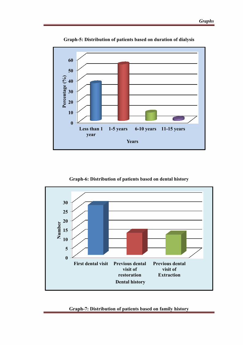
Graphs
Graph-5: Distribution of patients based on duration of dialysis
Graph-6: Distribution of patients based on dental history
Graph-7: Distribution of patients based on family history
0
10
20
30
40
50
60
Less than 1
year
1-5 years 6-10 years 11-15 years
Per
cen
tage
(%)
Years
0
5
10
15
20
25
30
First dental visit Previous dental
visit of
restoration
Previous dental
visit of
Extraction
Nu
mb
er
Dental history

Graphs
Graph-8: Distribution of patients based on the status of oral hygiene
Graph-9: Distribution of patients based on oral lesions
0
10
20
30
40
50
60
70
No relevant family
history
Diabetes mellitus Hypertension
Per
cen
tage
(%)
Family history
0 10 20 30 40
No abnormality detected
Mobility
Dental caries
Calculus
Generalized attrition
Missing tooth
Root stumps
Ulcer on buccal mucosa
Pain
Percentage (%)
Ora
l h
yg
ien
e

Graphs
Graph-10: Distribution of patients based on Candida species
0 10 20 30 40 50 60 70
No abnormality detected
Acute pseudomembranou s…
Chronic erythematous candidiasis
Angular cheilitis
Uremic stomatitis
Chronic erythematous…
Percentage (%)
Ora
l le
sion
s
No Candida
species (64.00
%)
C. albicans
(22.00 %)
C. tropicalis
(14.00 %)

DISCUSSION

Discussion
64
Great physician Sir William Osler quoted “Mouth is the mirror to the body”. I
would like to go one step further and state that the mouth mirrors what the body wants
to show.
Oral physicians can play a pivotal role in diagnosing the undiagnosed
cases of certain disease on the basis of the oral signs and symptoms. The study of oral
health abnormalities in chronic renal failure patient is important due to need of greater
knowledge as well conflicting results in regards to the prevalence of oral
abnormalities in literature as well as the fact that renal failure is a worldwide health
problem.
The present study was undertaken to determine the prevalence of candidiasis,
uremic stomatitis and uremic frost and Candida subspecies in chronic renal failure
patients. This study was conducted in the Department Of Nephrology and Department
of Oral Medicine and Radiology, Sree Mookambika Institute of Medical Sciences,
Kulasekaram, Kanyakumari district, Tamil Nadu. The total sample size was 50
chronic renal failure patients undergoing hemodialysis in the Department Of
Nephrology of Sree Mookambika Institute of Medical Sciences, Kulasekaram,
KanyaKumari district, Tamil Nadu. Samples were selected based on our preformatted
inclusion and exclusion criteria.
This is one of the first studies to assess the presence of yeasts in the oral cavity
and associate the results with the presence of oral lesions with the use of KB006
HiCandida TM
Identification Kit in patients with CKD undergoing HT. The results
revealed that among the 50 patients studied, 18 (36%) were colonized by yeasts, all of
them belonging to the genus Candida, with the predominance of C. albicans.

Discussion
65
To avoid bias concerning treatment method and its possible complications, all
patients in this study came from the same reference hospital. Although the number of
participants was higher than the calculated minimum sample size, it was expected that
a higher number of patients would be interested in participating in the study. The
unwillingness demonstrated by patients to have the oral cavity examined may have
been due to a series of reasons: the need to undergo hemodialysis three times a week,
the considerable amount of time spent in hospital, their depressive state, the anxiety
involved in waiting for organ transplant, or simply their general indisposed condition
due to the constant alterations in their clinical status. None of the volunteers enrolled
in this study had malignancy at the time samples were collected. Other confounding
factors, such as the use of immunosuppressant, steroid, or history of previous oral
candidiasis, were not taken into consideration, as all participants have been exposed to
these variables at some point in their recent medical history.
The rate of colonization by yeasts found in this study was almost similar to
that found in a previous study performed with 146 patients receiving hemodialysis
treatment (HT) by Godoy et al where there was 57 patients colonized with yeast with
variability in species distribution4 lower than that found among 400 patients with head
and neck cancer before treatment by Schelenz et al .The majority of infections in
these cancer patients were caused by C . albicans and almost one third of patients
harbored non C.albicans strains such as C.glabrata.7 The rate of colonization by yeasts
found in this study was lower than that found in kidney transplant recipients by
Ahmadieh et al8; and two times higher than that previously detected in healthy
subjects by Yang et al 40
. However, the incidence was lower than that found for HIV
positive individuals by Junqueira et al 29.

Discussion
66
As a matter of fact, there is a great variability concerning the number of
people that carry yeasts in the oral cavity. The high incidence rate found in this study
in patients with CKD undergoing HT seems to indicate that some kind of interference
in the host/Candida spp relationship may exist, which may favor the colonization by
Candida spp. The importance of these results lies in the risk that these
microorganisms might represent. Yeasts may become agents of serious and fatal
infections, as seen in patients with cancer, in whom the prevalence of candidemia is
high and the colonization rate is increased by the treatment itself.
In this study, C. albicans represented 75% of all species isolated, which is in
accordance with results found in previous studies. Even though no association
between C. albicans and oral lesions (odds ratio) was found, our results reinforce what
has been observed in daily dental practice, with C. albicans being the most commonly
isolated species of yeasts found in the oral cavity, with incidences ranging from
33.3% to 84.62%, of both healthy and ailing subjects.
However, it is also important to call attention to the presence of non-albicans
Candida species isolated both in the absence and in the presence of oral lesions,
including those suggestive of candidiasis. Although the number of non albicans (C.
tropicalis) species found in this study was small, their presence is still of great
relevance from a clinical point of view.
Some species of Candida such as C. tropicalis are not only highly virulent, but
they also do not usually respond well to the treatment with antifungals. As a result,
the incidence of these fungal species in systemic infections involving high levels of
morbidity and mortality has dramatically increased in the last decades. Thus, further

Discussion
67
investigations associating non-albicans species and the presence of oral lesions could
bring some relevant information on the impact these microorganisms may have on
patients with CKD undergoing HT.
Analyzing the possible variables associated with colonization rate (42.3%) by
yeasts found in patients with CKD undergoing HT, our results are in accordance with
other groups of volunteers previously studied such as smokers, patients with DM.
The most common symptom patients complained was burning sensation but some of
them were asymptomatic too. Lamster et al22
indicated that the presence of candidiasis
is a common manifestation of immuno compromised state.
A study done in edentulous chronic renal failure patients by Bobkowska and
Ziolkiewicz46
has shown that nearly 67 % had denture stomatitis which is a form of
candidiasis. Another study done by Bartholomew and Rodu37
in chronic renal failure
patients states that there is predisposition to oral candidiasis and is independent of
creatinine and blood urea nitrogen level. But the studies done by Knight and Fletcher
38 states that decreased saliva output promote overall growth of candida in these
patients. The results of our study is consistent with this observation in the sense that
the number of patients affected by candidiasis is higher in edentulous chronic renal
failure patients.
In this study, a correlation between oral lesions and the presence of yeasts was
clear. Patients with chronic erythematous were 15 times more likely to present yeasts
in the oral cavity. Although expected, to the best of our knowledge, this is the first
time that this finding has been estimated, which can be of relevance in handling
fungal infections in patients with CKD undergoing HT. As they become more

Discussion
68
common with age, removable dental prostheses can exert pressure on the subjacent
mucosa, causing trauma and favoring the colonization by yeasts. Besides, the acrylic
base can easily become a reservoir for microorganisms and be contaminated by
yeasts, a scenario that may lead to oral infections, especially in immunosuppressed
patients . The high chance of colonization by yeasts in patients with CKD receiving
HT who are dental prostheses users found in this study suggests that the different
members of the medical team responsible for these patients should be aware of this
condition. This information indicates that continuous oral assessments, as well as
reinforcement of oral and prostheses hygiene, should be performed, especially in the
pre-transplant period.
Angular chelitis (fissuring of the corners of mouth) was noticed in 20% of
these population examined which also represents a form of candidiasis. This condition
is of higher prevalence in renal failure patients as were in our study also.
Guggenheimer et al 42
also reported a high prevalence of angular cheilitis associated
with the presence of Candida.
In this study the most important health complication noticed was Periodontitis.
The prevalence of periodontal disease was found to be 70%. It was found that long
duration of disease can present with severe manifestations of periodontitis like
recession, furcation involvement ,mobility and pus exudation from periodontal
pockets. The present study is consistent with study by Brian et al among 109 renal
failure patients, the prevalence of periodontal disease was high , incidence of which
being 2-6 fold higher.23
A study was done by Tsai et al24
in which it was proved that
patients with chronic renal failure had a significantly higher prevalence of severe
periodontitis. The level of creatinine and blood urea nitrogen control is of key

Discussion
69
importance in determining increased risk. Maike et al25
from his study suggested that
incidence and severity of periodontitis are influenced by presence or absence of
disease. Improving the creatinine and blood urea nitrogen level is likely to reduce
risk and severity of periodontitis. Periodontal therapy showed marked improvement
in these patients .
The relationship between dental caries and renal failure is complex. literature
states that patients with renal failure are susceptible to oral infection that leads to
tooth decay and loss. Ship32
has stated in his review that he was not able to find any
specific association of occurrence of dental caries in renal failure patients which is
quite contradictory to our study which shows that prevalence of decay and tooth loss
is high. Slight increase in prevalence of DMFT scores was observed in these patients.
Halitosis was also noted in the study population though it was not a parameter
in our study. About 21% of the population examined had halitosis. It is primarily
caused by bacterial putrefactions and generation of volatile sulphur compound.
It is interesting to observe that the amount of yeasts isolated from patients with
oral lesions (31.19 CFU/ml) was three times higher than that found in patients without
oral lesions (9.50 CFU/ml) . These results are not surprising, as yeasts are either the
agents responsible for the lesions themselves or opportunistic elements, taking
advantage of tissue alterations to multiply and thus evolving from colonization to
infection. Nonetheless, the number found was much lower than the 300 CFU/ml
previously found in patients with lesions that were clinically suggestive of
candidiasis. This difference may be due to the fact that results in this study reflect

Discussion
70
mean CFU/ml detected in all patients, including all types of lesions, and not only in
those clinically suggestive of fungal infection.
Almost half of the population in this study (18) presented oral lesions,
confirming its high prevalence in kidney patients. All these patients, supplied positive
cultures for yeasts, presenting lesions clinically suggestive of oral candidiasis. Most
yeast isolates were recovered from patients with chronic–atrophic– candidiasis-type
lesions (nine lesions), from whom two species of yeasts were isolated. In addition to
that, the yeasts isolated from these lesions were those recognized as being the main
agents of lesions in the oral mucosa, that is, C. albicans and C. tropicalis , the first and
the second most common species, respectively. This correlation reinforces the
recommendation made by Sahebjamee et al.54
, who suggested that, due to the
incidence and the high likelihood of serious infections, any type of oral lesion
evidenced in kidney patients should be treated with antifungals before kidney
transplant. Within this context, Ahmadieh et al. 34
also suggested that when detecting
an oral lesion, systemic investigation aiming at improving the prognosis would be
opportune, with detection and treatment of the infection being initiated as early as
possible.
This study identified a high prevalence of oral mucosal lesions (candidal
lesions) among patients with chronic renal failure. The lesions commonly noticed in
Kanyakumari population were acute pseudomembranous candidiasis, denture
stomatitis, median rhomboid glossitis, angular chelitis, geographic tongue, fissured
tongue. Due to the high prevalence of these lesions in the population, oral
physicians should be aware of the clinical appearance, etiology, diagnosis and the
required treatment for the lesion.

Discussion
71
The prevalent mucosal lesion recorded was denture stomatitis (24.14%) But it
was also observed in our study that people in Kanyakumari district has higher
incidence of geographic tongue but due to the lack of similar studies in South
Indian population, no conclusion can be drawn regarding the exact prevalence in this
region. Most of the patients were asymptomatic .Very few patients were aware of the
lesions. Males were more frequently to be affected than females in our study. Male
predominance has been reported by Vörös-Balog et al.39
However, a large discrepancy was observed in this study that uremic
stomatitis and uremic frost were not seen contradicting the expectations. Thus, it
seems that clinical assessments of a lesion should be confirmed by laboratory tests
through the isolation and identification of yeasts. Even then, cultures positive for
yeasts obtained from oral samples need to be interpreted with care, due to the
possibility of a positive culture representing just an asymptomatic colonization
without clinical implications.
In this study, 64% of samples collected from patients were negative for yeasts,
and curiously, 36% of lesions were suggestive of candidiasis (acute erythematous
candidiasis, chronic atrophic candidiasis, or angular cheilitis). Apart from the fact
that the presence of yeasts may just indicate colonization, other diagnostic hypotheses
must be considered for the presence of oral lesions, such as reactions induced by
drugs, dietary habits, and malnutrition.
The results of this study suggests, therefore, that close supervision concerning
the use of dental prostheses and the occurrence of oral lesions, together with the
performance of mycological tests, could ensure that patients comply with the pre-

Discussion
72
surgical requirements and are free of infection before transplant . Besides, in the face
of the high occurrence of post-transplant oral candidiasis, these simple procedures
could also enable the physician to individualize the antifungal prophylaxis, often
empirically started . These conducts should all have in mind the high mortality rates
and loss of the transplanted grafts owing to fungal infections, rendering early
diagnosis and antifungal treatment mandatory in immunosuppressed patients.

SUMMARY AND CONCLUSION
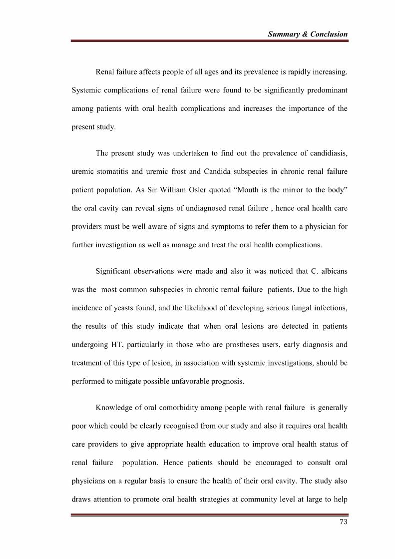
Summary & Conclusion
73
Renal failure affects people of all ages and its prevalence is rapidly increasing.
Systemic complications of renal failure were found to be significantly predominant
among patients with oral health complications and increases the importance of the
present study.
The present study was undertaken to find out the prevalence of candidiasis,
uremic stomatitis and uremic frost and Candida subspecies in chronic renal failure
patient population. As Sir William Osler quoted “Mouth is the mirror to the body”
the oral cavity can reveal signs of undiagnosed renal failure , hence oral health care
providers must be well aware of signs and symptoms to refer them to a physician for
further investigation as well as manage and treat the oral health complications.
Significant observations were made and also it was noticed that C. albicans
was the most common subspecies in chronic rernal failure patients. Due to the high
incidence of yeasts found, and the likelihood of developing serious fungal infections,
the results of this study indicate that when oral lesions are detected in patients
undergoing HT, particularly in those who are prostheses users, early diagnosis and
treatment of this type of lesion, in association with systemic investigations, should be
performed to mitigate possible unfavorable prognosis.
Knowledge of oral comorbidity among people with renal failure is generally
poor which could be clearly recognised from our study and also it requires oral health
care providers to give appropriate health education to improve oral health status of
renal failure population. Hence patients should be encouraged to consult oral
physicians on a regular basis to ensure the health of their oral cavity. The study also
draws attention to promote oral health strategies at community level at large to help

Summary & Conclusion
74
people to understand the relationship that exhibits between renal failure and oral
health as their lack of awareness on oral cavity problems among common people.
The oral cavity exhibits the first sign of an undiagnosed renal failure , hence
oral health care providers must be well aware of signs and symptoms to refer them to
a physician for further investigation as well as manage and treat the oral health
complications. Oral health providers can play an important role by identifying and
treating oral diseases, by encouraging the renal failure individual to maintain rigorous
oral hygiene and follow proper diet.
There is need for future longitudinal studies among renal failure patients to
elicit accurate knowledge regarding the development and progression of oral diseases
as well as results of interventional procedures and treatment (both medical and
dental) instituted in the patients.
We conclude our study by stating that oral physicians can play pivotal role to
identify, treat various oral health problems, encourage patient to receive routine
stomatological care thereby we can enhance the quality of life for patients with
chronic renal failure.

BIBLIOGRAPHY

Bibliography
ix
1. Gautam NR, Gautam NS, Alamanda M.Effect of end- stage renal disease on
oral health in patients undergoing renal dialysis: A cross – sectional study. J
Int Soc Prev Community Dent.2014 Sep-Dec; 4(3):164-9.
2. Williams DW, Kuriyama T,Silva S, Malic S, Lewis MA .Candida biofilms and
oral candidosis: treatment and prevention . Periodontology 2000.2011; 55:
250–65.
3. Pieralisi N, Godoy J, Yamada S, Rosangela, SvidzinskiT. Oral lesions and
colonization of yeasts in hemodialysis patients.J Oral Pathol Med. 2015;
44:585-90.
4. Gogoy JS, de Souza Bonfim- Mendonca P,Nakamura SS,yamada SS,
Shinobu- Mesquita C,Pieralisi N, Fiorini A, Svidzinski TI.Colonization of the
oral cavity by yeasts in patients with chronic renal failure undergoing
hemodialysis. J Oral Pathol Med.2013; 42(3): 229–34.
5. De la Rosa-García E, Miramontes-Zapata M, Sánchez-Vargas
LO, Mondragón-Padilla A. Oral colonization and infection by Candida sp.in
diabetic and nondiabetic patients with chronic kidney disease on dialysis
Nefrologia.2013; 33(6):764-70
6. Premkumar J, Ramani P, Chandrasekar T, Natesan A, Premkumar P.Detection
of species diversity in oral candidacolonization and antifungal susceptibility
among oral habit adult diabetic patients. J Nat Sci Biol Med .2014; 5(1):
148-54.
7. Schelenz S, Abdallah S, Gray G, Stubbings H, Gow I, Baker P, Hunter PR.
Epidemiology of oral yeast colonization and infection in patients with

Bibliography
x
hematological malignancies, head neck and solid tumors .J Oral Pathol
Med.2011; 40(1): 83–9.
8. Ahmadieh A, Baharvand M, Fallah F, Djaladat H, Eslani M. Oral Microflora
in Patients on Hemodialysis and Kidney Transplant Recipients. Iran J kidney
Dis.2010; 4(3):227-31.
9. Mehta R, Anupama S., Wyawahare. Evaluation of Hicrome Candida
Differential Agar for Species Identification of Candida Isolates from Various
Clinical Samples. International Journal of Contemporary Medical Research.
2016; 3(4):1219-22
10. Pyrgos V, Ratanavanich K, Donegan N, Veis J, Walsh TJ, Shoham S. Candida
bloodstream infection in hemodialysis Recipients. Med Mycol.2009;
47(5):463-7.
11. De Rossi SS, Glick M. Dental considerations for patients with renal disease
receiving hemodialysis. J Am Dent Assoc. 1996;127:211–219.
12. Ganesh SK, Hulbert-Shearon TE, Port FK, et al. Mortality differences by
dialysis modality among incident ESRD patients with and without coronary
artery disease. J Am Soc Nephrol. 2003;14:415–428.
13. Ledebo I, Lamiere N, Charra B, et al. Improving the outcome of dialysis—
opinion vs Scientific evidence. Nephrol Dial Transplant. 2000;15:1310–1316.
14. Orth SR. Effects of smoking on systemic and intrarenal hemodynamics:
influence on renal function. J Am Soc Nephrol. 2004;15(suppl1):S58-S63.
15. Pereira BJ. Optimization of pre-ESRD care; the key to improved dialysis
outcomes. Kidney Int. 2000;57:351–365.

Bibliography
xi
16. Singh AK, Szczech L, Tang KL, et al.; CHOIRInvestigators. Correction of
anemia with epoetin alfa in chronic kidney disease. N Engl J Med.
2006;355(20):2085–2098.
17. St Peter WL, Obrador GT, Roberts TL, Collins AJ. Trends in intravenous iron
use among dialysis patients in the United States (1994–2002). Am J Kidney
Dis. 2005;46:650–660.
18. Strippoli GF, Palmer SC, Ruospo M, et al. Oral disease in adults treated with
hemodialysis:prevalence, predictors, and association with mortality and
adverse cardiovascular events: the rationale and design of the ORAL Diseases
in hemodialysis (ORAL-D) study, a prospective, multinational, longitudinal,
observational, cohort study. BMC Nephrol. 2013;14:90.
19. Chattopadhyay A, Caplan DJ, Slade GD, et al. Incidence of oral candidiasis
and oral hairy leukoplakia in HIV-infected adults in North Carolina. Oral
Surg Oral Med Oral Pathol Oral Radiol Endod. 2005;99:39–47.
20. Alousi AM, Bolaños-Meade J, Lee SJ. Graft-versus-host disease: state of the
science. Biol Blood Marrow Transplant. 2013;19:S102-S108.
21. Atsuta Y, Suzuki R, Yamashita T, et al.; Japan Society for Hematopoietic
Cell Transplantation. Continuing increased risk of oral/esophageal cancer after
allogeneic hematopoietic stem cell transplantation in adults in association with
chronic graft-versus-host disease. Bone Marrow Transplant. 2013;48(8):
1123-8.
22. Lamster JW, Fassil H, Edwards DA, et al. Oral chronic graft-versushost
disease: current pathogenesis, therapy, and research. Oral Dis.
2013;19(4):327–346.

Bibliography
xii
23. Brian RC, Lopes AA, Thom_e FS, Lugon JR, Watanabe Y, Santos DR.
Chronic dialysis in Brazil: report of the Brazilian dialysis census, 2011. J Bras
Nefrol 2012; 34: 272–7.
24. Tsai MA, Diekema DJ. Epidemiology of invasive candidiasis: a persistent
public health problem. Clin Microbiol 2007; 20: 133–63.
25. Maike DW, Kuriyama T, Silva S, Malic S, Lewis MA. Candida biofilms and
oral candidosis: treatment and prevention. Periodontol 2000 2011; 55: 250–65.
26. Bassetti M, Taramasso L, Nicco E, Molinari MP, Mussap M, Viscoli C.
Epidemiology, species distribution, antifungal susceptibility and outcome of
nosocomial candidemia in a tertiary care hospital in Italy. PLoS ONE 2011; 6:
e24198.
27. Wisplinghoff H, Bischoff T, Tallent SM, Seifert H, Wenzel RP, Edmond MB.
Nosocomial bloodstream infections in US hospitals: analysis of 24,179 cases
from a prospective nationwide surveillance study. Clin Infect Dis 2004; 39:
309–17.
28. Colombo AL, Nucci M, Park BJ, et al. Epidemiology of candidemia in Brazil:
a nationwide sentinel surveillance of candidemia in eleven medical centers. J
Clin Microbiol 2006; 44: 2816–23.
29. Junqueira JC, Vilela SF, Rossoni RD, et al. Oral colonization by yeasts in
HIV-positive patients in Brazil. Rev Inst Med Trop Sao Paulo 2012; 54: 17–
24.
30. Mubareka S, Vinh DC, Sanche SE. Candida dubliniensis bloodstream
infection: a fatal case in a lung transplant recipient. Transpl Infect Dis 2005; 7:
146–9.

Bibliography
xiii
31. Kramer IR, Pindborg JJ, Bezroukov V, Infirri JS. Guide to epidemiology and
diagnosis of oral mucosal diseases and conditions. World Health Organization.
Community Dent Oral Epidemiol 1980; 8: 1–26.
32. Ship, Dongari-Bagtzoglou A, Dwivedi P, Ioannidou E, Shaqman M, Hull D,
Burleson J. Oral Candida infection and colonization in solid organ transplant
recipients. Oral Microbiol Immunol 2009; 24: 249–54.
33. Schelenz S, Abdallah S, Gray G, et al. Epidemiology of oral yeast
colonization and infection in patients with hematological malignancies, head
neck and solid tumors. J Oral Pathol Med 2011; 40: 83–9.
34. Ahmadieh A, Baharvand M, Fallah F, Djaladat H, Eslani M. Oral microflora
in patients on hemodialysis and kidney transplant recipients. Iran J Kidney Dis
2010; 4: 227–31.
35. Larone DH. Medically important fungi. A guide to identification, 5th edn.
Washington, DC: ASM Press, 2011.
36. Godoy JS, de Souza Bonfim-Mendonc_a P, Nakamura SS, et al. Colonization
of the oral cavity by yeasts in patients with chronic kidney failure undergoing
hemodialysis. J Oral Pathol Med 2013; 42: 229–34.
37. Bartholomew DW, Rodu S. Applied logistic regression, 2nd
edn. New York,
USA: Wiley, 2000.
38. Knight H, Fletcher SM, Hazarey VK, Wanjare VS. Oropharyngeal candidosis
relative frequency in radiotherapy patient for head and neck cancer. J Oral
Maxillofac Patho 2012; 16: 31–7.
39. Voras, Balog, Al-Attas SA, Amro SO. Candidal colonization, strain diversity,
and antifungal susceptibility among adult diabetic patients. Ann Saudi Med
2010; 30: 101–8.

Bibliography
xiv
40. Yang YL, Leaw SN, Wang AH, Chen HT, Cheng WT, Lo HJ.
Characterization of yeasts colonizing in healthy individuals. Med Mycol 2011;
49: 103–6.
41. Pires-Gonc_alves RH, Miranda ET, Baeza LC, Matsumoto MT, Zaia JE,
Mendes-Giannini MJ. Genetic relatedness of commensal strains of Candida
albicans carried in the oral cavity of patients’ dental prosthesis users in Brazil.
Mycopathologia 2007; 164: 255–63.
42. Guggenheimer V, Ratanavanich K, Donegan N, Veis J, Walsh TJ, Shoham S.
Candida bloodstream infections in hemodialysis recipients. Med Mycol 2009;
47: 463–7.
43. Goregen M, Miloglu O, Buyukkurt MC, Caglayan F, Aktas AE. Median
rhomboid glossitis: a clinical and microbiological study. Eur J Dent 2011; 5:
367–72.
44. de la Rosa Garc_ıa E, Mondrag_on Padilla A, Aranda Romo S, Bustamante
Ram_ırez MA. Oral mucosa symptoms, signs and lesions, in end stage kidney
disease and non-end stage kidney disease diabetic patients. Med Oral Patol
Oral Cir Bucal 2006; 11: E467–73.
45. Kadir T, Pisiriciler R, Aky€uz S, Yarat A, Emekli N, Ipb€uker A. Mycological
and cytological examination of oral candidal carriage in diabetic patients and
non-diabetic control subjects: thorough analysis of local aetiologic and
systemic factors. J Oral Rehabil 2002; 29: 452–7.
46. Ziolkiewiz AL, Bobkowska MEF, de Carli ML, Rabelo GD, Nunes TB, da
Silveira FR. Candida albicans isolation from buccal mucosa of patients with
HIV wearing removable dental prostheses. Int J Prosthodont 2012;
25: 127–31.

Bibliography
xv
47. Pires FR, Santos EB, Bonan PR, de Almeida OP, Lopes MA. Denture
stomatitis and salivary Candida in Brazilian edentulous patients. J Oral
Rehabil 2002; 29: 1115–9.
48. Yazdani M, Jahanshahi GR, Mahdavi KN, Asadian F. Study of the
relationship between oral Candida colony counts and time on hemodialysis at
Khorshid Hospital, Isfahan, Iran. Transplant Proc 2003; 35: 2580–1.
49. Castillo A, Mesa F, Li_ebana J, et al. Periodontal and oral microbiological
status of an adult population undergoing haemodialysis: a cross-sectional
study. Oral Dis 2007; 13: 198–205.
50. Mandali G, Sener ID, Turker SB, Ulgen H. Factors affecting the distribution
and prevalence of oral mucosal lesions in complete denture wearers.
Gerodontology 2011; 28: 97–103.
51. Pathak AK, Sharma S, Shrivastva P. Multi-species biofilm of Candida
albicans and non-Candida albicans Candida species on acrylic substrate. J
Appl Oral Sci 2012; 20: 70–5.
52. Thorman R, Neovius M, Hylander B. Clinical findings in oral health during
progression of chronic kidney disease to endstage kidney disease in a Swedish
population. Scand J Urol Nephrol 2009; 43: 154–9.
53. Dirschnabel AJ, Martins AES, Dantas SA, et al. Clinical oral findings in
dialysis and kidney-transplant patients. Quintessence Int 2011; 42: 127–33.
54. Sahebjamee M, Shakur Shahabi M, Nikoobakht MR, Momen Beitollahi J,
Mansourian A. Oral lesions in kidney transplant patients. Iran J Kidney Dis
2010; 4: 232–6.
55. Cabello Benavente R, Quicios Dorado C, L_opez Martin L, Sim_on
Rodr_ıguez C, Charry G_onima P, Gonz_alez Enguita C. The candidate for

Bibliography
xvi
kidney transplantation work up: medical, urological and oncological
evaluation. Arch Esp Urol 2011; 64: 441–60.
56. Badiee P, Alborzi A. Invasive fungal infections in kidney transplant
recipients. Exp Clin Transplant 2011; 9: 355–62.
57. Cohen D, Galbraith C. General health management and longterm care of the
kidney transplant recipient. Am J Kidney Dis 2001; 38(6 Suppl 6): S10–24.
58. Avery RK. Infectious disease following kidney transplant: core curriculum
2010. Am J Kidney Dis 2010; 55: 755–71.

ANNEXURE


CONSENT FORM
PART 1 OF 2
INFORMATION FOR PARTICIPANTS OF THE STUDY
1. Name of the Principal Investigator:
Divya N.L.
Second Year Post Graduate student
Department of Oral Medicine and Radiology
Sree Mookambika Institute of Dental Sciences,
Kulasekharam
1. Name of the Guide : Dr. J. Eugenia Sherubin MDS.
Reader,
Department of oral medicine and Radiology,
Sree Mookambika Institute of Dental Sciences,
Kulasekharam, KanyaKumari District-629161.
Dear Volunteers,
We welcome you and thank you for your keen interest in participation in this research project.
Before you participate in this study, it is important for you to understand why this research is being carried
out. This form will provide you all the relevant details of this research. It will explain the nature, the
purpose, the benefits, the risks, the discomforts, the precautions and the information about how this project
will be carried out. It is important that you read and understand the contents of the form carefully. This
form may contain certain scientific terms and hence, if you have have any doubts or if you want more
information, you are free to ask the study personnel or the contact person mentioned below before you
give your consent and also at any time during the entire course of the project.

2. Name of the Co-Guide : Dr. Tatu Joy. E MDS
Professor and Head,
Department of oral medicine and Radiology,
Sree Mookambika Institute of Dental Sciences,
Kulasekharam, KanyaKumari District-629161.
3. Name of the Co-Guide : Dr. Beena Unnikrishnan MD,DM
Associate Professor
Department of Nephrology
Sree Mookambika Institute of Medical
Sciences,
Kulasekharam, KanyaKumari District-629161.
4. Name of the Co-Guide : Dr. R.V. Mookambika MD,DM
Assistant Professor
Department of Nephrology
Sree Mookambika Institute of Medical Sciences,
Kulasekharam, KanyaKumari District-629161
5. Institute:
Sree Mookambika Institute of Dental Sciences,
V.P.M Hospital complex, Padanilam,
Kulasekharam,
Kanyakumari – 629161
Tamil nadu
2. Title of the study: “Prevalence of Candidasis, Uremic Stomatitis, Uremic
frost and Colonization of Candidal subspecies in hemodialysis patients”

3. Background information:
Chronic kidney disease is a slow irreversible and progressive process that
occurs over a period of years leading to death in the absence of medical
intervention. Patients with chronic kidney disease, undergoing hemodialysis
treatment are frequently colonized by yeasts of the genus Candida spp, with high
chance of developing fungal infections. Oral candidiasis is an intermediary form
between yeast colonization and invasive candidiasis .Although oral candidiasis is
a superficial oral infection that is clinically detectable, it should be taken
seriously due to its high potential to develop into a systemic infection in
immune- compromised patients.
4. Aims and Objectives:
o To assess the presence of candidiasis, Uremic Stomatitis, Uremic
frost in patients undergoing hemodialysis.
o To assess the presence of various subspecies of Candida in patients
with chronic renal disease undergoing hemodialysis in the
Department Of Nephrology in Sree Mookambika Institute of
Medical Sciences.
5. Scientific justification of the study:
With impaired renal function, decrease GFR and accumulation and
retention of various products of renal failure the oral cavity may shows a variety of
changes as the body progresses through an azotemic to uremic state. In 90% of
hemodialysis patient oral manifestation are evident which may be due to disease
process or as a result of treatment. Studies have been conducted regarding
colonization of yeast in hemodialysis patients. However there is a gap in the
knowledge regarding assessment of Candida subspecies and its correlation with oral
lesions in hemodialysis patient which aid in prophylactic treatment. Prevalence of

Candidiasis, Uremic Stomatitis, Uremic frost and colonization of Candida
Subspecies will open eyes in this new research.
6. Procedure for the study:
1. Obtain clinical records regarding data such as gender,history of systemic
diseases,hemodialysis time of each patients.
2. An informed consent was obtained from all participants.
3. Patients were asked to rinse the oral cavity with 10ml of distilled water
for 30sec.
4. Then patient is asked to spilt into a beaker to obtain the sample.
5. The samples are transported within 2 hrs to the laboratory for culture
and identification of candida subspecies.
6. A complete intra oral examination of the participants is carried out.
7. Once a lesion and candida subspecies detected, patients are referred to
the department of Oral Medicine And Radiology and treated with
antifungal agents.
7. Expected risks for the participants:
NIL
8. Expected benefits of research for the participants:
You will get a better knowledge about your oral health condition and
various treatment and preventive options available to improve your oral
health .
9. Maintenance of confidentiality:
a. You have the right to confidentiality regarding the privacy of your medical
information
(Personal details, results of physical examinations, investigations, and your
medical
history).

b. By signing this document, you will be allowing the research team
investigators, other study Personnel, sponsors, institutional ethics committee
and any person or agency required by law to view your data, if required.
c. The results of study performed as part of this research may be included in
your medical record.
d. The information from this study, if published in scientific journals or
presented at scientific meetings, will not reveal your identity.
10. Why have I been chosen to be in this study?
a. Chosen because of grouping under the inclusion and exclusion criteria
b. Need of good sampling size
c. No invasive procedure that harm your health and helps in diagnosis and
helpful for the society
11. How many people will be in the study? 50
12. Agreement of compensation to the participants (In case of a
study related injury):
No related injury anticipated. Patient will be taken care in case of complication
and
medical treatment will be provided.
13. Anticipated prorated payment, if any, to the participant(s) of the
study: Not applicable.
14. Can I withdraw from the study at any time during the study
period?
The participation in this research is purely voluntary and you have the right
to withdraw from this study at any time during the course of the study
without giving any reasons.
However, it is advisable that you talk to the research team prior to stopping
information.
15. If there is any new findings/information, would I be informed?
Yes

16. Expected duration of the participant’s participation in the study:
1 day
17. Any other pertinent information:
No other information
18. Whom do I contact for further information?
Place:
Date: Signature of Principal Investigator
Signature of the participant
For any study related queries, you are
free to contact :
Dr.Divya N.L.,
Post graduate student,
Department of oral Medicine and Radiology
Sree Mookambika Institute of Dental Sciences
Kulasekharam,
KanyaKumari District-629161
9841144518
divya_nl @ yahoo.com

CONSENT FORM
PART 2 OF 2
PARTICIPANTS CONSENT FORM
The details of the study have been explained to me in writing and the details have
been fully explained to me. I am aware that the results of the study may not be
directly beneficial to me but will help in the advancement or medical sciences. I
confirm that I have understood the study and had the opportunity to ask questions. I
understand that my participation in the study is voluntary and that I am free to
withdraw at any time, without giving any reason, without the medical care that will
normally be provided by the hospital being affected. I agree not to restrict the use of
any data or results that arise from this study provided such a use is only for scientific
purpose(s). I have been given an information sheet giving details of the study. I fully
consent to participate in the study titled
“Prevalence of Candidasis, Uremic Stomatitis, Uremic frost and Colonization
of Candidal subspecies in hemodialysis patients”
Serial no / Reference no:
Name of the participant:
Address of the participant:
Contact number of the participant:
Signature / thumb impression of the participant / Legal guardian Witnesses:
1.
2.
Date:
Place:

Jl×Rp YôdÏêXm
ØRp TôLm
BWônf£«p TeÏùTßúYôÚdLô] RLYp ϱl×
BWônf£«u RûXl×:
ÏÚ§d árUl TÏl× Øû\ ùNnÙm úSôVô°L°p HtTÓm úLu¥¥V£v,
Ùìªd vùPUûPh¥v, Ùìªd ×ú\ôv égûN Eh£\l©]Øm LiP±Ùm BnÜ.
RûXûU BnYô[o : PôdPo. §qVô, Gu.Gp.
Rϧ : ØÕLûX UôQYo, (Gm.¥.Gv) ©¬Ü : J\p ùU¥£u & úW¥úVô[´ Õû\,
¨ßY]m : c êLôm©Lô Cuv¥hëh Bl ùPuPp NVu^v CPm : ÏXúNLWm - 629 161
Y¯Lôh¥ : PôdPo. ë´]ô úNÚ©u MDS Rϧ : ÃPo
©¬Ü : J\p ùU¥£u & úW¥úVô[´ Õû\, ¨ßY]m : c êLôm©Lô Cuv¥hëh Bl ùPuPp NVu^v
CPm : ÏXúNLWm - 629 161
CûQ Y¯Lôh¥ : PôdPo. PôhÓ ú_ôn. C MDS
Rϧ : úTWô£¬Vo, Õû\ úUXô[o, ©¬Ü : J\p ùU¥£u & úW¥úVô[´ Õû\,
¨ßY]m : c êLôm©Lô Cuv¥hëh Bl ùPuPp NVu^v CPm : ÏXúNLWm - 629 161
CûQ Y¯Lôh¥ : PôdPo. À]ô Eu² ¡ÚxQu CûQ úTWô£¬Vo,
¨lWôX´ Õû\, c êLôm©Lô Cuv¥hëh Bl ùPuPp NVu^v
ÏXúNLWm - 629 161
CûQ Y¯Lôh¥ : PôdPo. Bo.®. êLôm©Lô ER® úTWô£¬Vo,
¨lWôX´ Õû\, c êLôm©Lô Cuv¥hëh Bl ùPuPp NVu^v ÏXúNLWm - 629 161
ùLôsLs Utßm úSôdLeLs :
ÏÚ§ árUl TÏl×Øû\ ùNnÙm úSôVô°L°p HtTÓm égûN
Eh£\l©]m LiP±Rp.
ÏÚ§d árUl TÏl×Øû\ ùNnÙm úSôVô°L°p HtTÓm úLu¥¥V£v,
Ùìªd vùPUûPh¥v, Ùìªd ×ú\ôv LiP±Rp.

Bn®u ùNVp Øû\ :
úSôVô°«u RLYpLs UÚjÕY BYQeL°u êXm GÓjÕ
ùLôs[lTÓ¡\Õ.
CkR BWônf£«p Es[ úSôVô°L°Pm RLYpLû[ á± AàU§ Gݧ
YôeLlTÓm.
úSôVô°Ls 10 Gm.Gp Y¥¿Wôp YôûV 30 ®]ô¥ Su\ôL AXN úYiÓm.
AûR JÚ LiQô¥ ÏYû[«p úNL¬dL úYiÓm.
CkR TRdáû\ 2 U¦ úSWjÕdÏs BnÜdáPj§p Aàl© égûN
LiP±V úYiÓm.
úSôVô°L°u Yô«u TϧûV Su\ôL BnÜ ùNnV úYiÓm.
Yôn £ûRÜ Utßm égûN Eh£\l©]Øm LiP±kRôp JWp ùU¥£u
Utßm úW¥VôX´ ©¬®p úSôVô°Lû[ ùLôiÓ ùNuß B]¥@TeLp
UÚkûR ùLôÓjÕ ÏQlTÓjR úYiÓm.
BWônf£«u G§oTôodLlTÓm TeÏLôX A[Ü? JÚ Sôs
TeúLtTô[oLÞdÏ G§oTôodLlTÓm TVuLs?
SpX JÚ UÚjÕYm úSôVô°LÞdÏ A°dLlTÓm.
Bn®p LXkÕ ùLôsTYoLÞdÏ G§oTôodLlTÓm BTjÕdLs?
G§oùLôs[lTÓm BTjÕLs úSôVô°«Pm ùR¬VlTÓjÕm.
CWL£VjRuûU LôjRp?
EeL°Pm CÚkÕ úNL¬jR GkR ®TWØm CWL£VUôL ûYdLlTÓm.
CRuêXm ¡ûPdÏm ×s°®TWm UhÓm ùY°«PlTÓm Ut\T¥ R²ST¬u ùNôkR
®TWeLs ùY°«PlTPUôhPôÕ.
CkR Bn®u êXm HúRàm ©u®û[ÜLs HtThPôp BWônf£Vô[o ùTôßl×
HtTôWô? BWônf£Vô[o ùTôÚ[ôRôW A[®p ùTôßlúTtTôo
CkR BWônf£«p TeÏùTßúYôÚdÏ Gq®R ùRôûLÙm YZeLlTÓUô? CpûX

CkR Bn®u êXm HúRàm ©u®û[ÜLs HtThPôp BWônf£Vôp TeÏ
ùTßúYôÚdÏ Gq®R ùRôûLLs YZeLlTÓUô? ©u®û[ÜLs HtTPôÕ.
Sôu CkR BWônf£«ÚkÕ ®ÚlTlThPôp GkR LôXLhPj§Ûm ®XLXôUô?
úSôVô°«u GkR JÚ LhÓlTôÓ, ¨TkRû]L°u ¸r CkR Bn®tÏ
EhTÓjRlTP®pûX. AYoL°u ØÝ JjÕûZl× Utßm NmURj§u úT¬p
UhÓúU TeùLÓjÕs[]o.
úYß HúRàm ùTôÚjRUô] ®TWeLs EiPô? CpûX
CqYôWônf£ûVl Tt±V ®YWeLû[ Vô¬Pm úLhÓ ùR¬kÕd ùLôsYÕ?
RûXûU BnYô[o : PôdPo. §qVô, Gu.Gp
Rϧ : ØÕLûX, (Gm.¥.Gv)
©¬Ü : J\p ùU¥£u & úW¥úVô[´ Õû\ ¨ßY]m : c êLôm©Lô Cuv¥hëh Bl ùPuPp NVu^v
CPm : ÏXúNLWm - 629 161 ûLúT£ : 9841144915
ªu AgNp : [email protected]
CPm : ØRuûU BWônf£Vô[¬u
úR§ : ûLùVôlTm

Jl×Rp T¥Ym
TôLm : 2
ÏÚ§d árUl TÏl×Øû\ ùNnÙm úSôVô°L°p HtTÓm úLu¥¥V£v,
Ùìªd vùPUûPh¥v, Ùìªd ×ú\ôv égûN Eh£\l©]Øm LiP±Ùm BnÜ.
TeÏ ùLôsTY¬u ùTVo :
18 YV§tÏ ¸r Es[YoLÞdÏ TôÕLôYX¬u ûLùVôlTm :
ØLY¬ :
ùRôûX ùRôPo× Gi :
CkR BWônf£«u RLYpLs Aû]jÕm Gu²Pm ùR°YôL GÝjÕêXm
®[dLlThÓs[Õ. CkR BWônf£«u Ø¥ÜLs G]dÏ úSW¥VôL
TVuùT\ô®hPôÛm UÚjRYjÕû\«u Øuú]t\j§tÏ TVuTÓm GuTûR
A±úYu. CqYôWônf£ûVl Tt± Sôu ùR°YôL ׬kÕd ùLôiÓsú[u. Sôu
Rô]ôL ØuYkÕ C§p TeÏl ùTß¡ú\u GuTûR A±úYu. C§ÚkÕ GkR
úSWØm GdLôWQØm á\ôUp YkRôÛm CkR UÚjÕYUû]«p G]dÏ ¡ûPdÏm
UÚjÕY ER® Gq®Rj§Ûm Tô§dLlTPôÕ GuTûRÙm A±úYu.
CqYôWônf£«u êXm YÚm Ø¥ÜLs Utßm RLYpLû[ A±®VpÕû\«u
TVuTôÓLÞdÏ (UhÓúU) ETúVôLlThÓdùLôs[ NmU§d¡ú\u. G]dÏ
CqYôWônf£ûVl Tt±V ®¬Yô] RLYpLs APe¡V T¥Ym RWlThÓs[Õ.
TeÏ ùLôsTY¬u ùRôPo, UÚjÕY Gi :
TeÏ ùLôsTYo TWôU¬lTYo ûLùVôlTm / ùTÚ®Wp ÑYÓ : Sôs :
Nôh£ 1
Nôh£Vô[¬u ûLùVôlTm Utßm ØLY¬ Sôs :
Nôh£ 2
úR§ :
CPm : ÏXúNLWm
RûXûU BnYô[¬u ûLùVôlTm

k½X ]{Xw þ `mKw þ 1
]T\hpambn klIcn¡p¶ hyànIfpsS Adnhntebv¡v
{]nbs¸« k¶² tkhItc,
R§Ä \n§sf kzmKXw sN¿p¶p. AtXmsSm¸w Cu ]T\hpambn klIcn¡m\pÅ k¶²XtbmSv \µn tcJs¸Sp¯p¶p. \n§Ä Cu ]T\¯n ]s¦Sp¡p¶Xn\p ap³]v Cu ]T\w F´n\mWv \S¯s¸Sp¶Xv F¶v AndtbXpv. AXn\m Cu t^md¯n KthjW ]T\¯nsâ hnhc§fpw aäpw hniZambn tcJs¸Sp¯nbncn¡p¶p. Cu ]T\¯nsâ coXn, Dt±iw, {]tbmP\w, A]ISkm²yX, t¢iw, ap³IcpXÂ, F§s\ Cu ]T\w ap³t]m«p sImpt]mIp¶p F¶n§s\ FÃm hnhc§fpw t^md¯n tcJs¸Sp¯nbncn¡p¶p. kZbw Cu hnhc§Ä hmbn¨p a\Ênem¡phm³ A`yÀ°n¡p¶p. Cu hnhc§fn imkv{X]camb ]Z§Ä DÅXn\m kwib\nhmcW¯n\p {][m\ ]T\IÀ¯mhnt\mtSm Xmsg tcJs¸Sp¯n bncn¡p¶ hyànItfmtSm t^mdw H¸nSp¶Xn\p ap³t]m Asæn Cu ]T\¯nsâ Imemh[n Xocp¶Xphtctbm kao]n¡mhp¶XmWv.
1. ]T\ hnjbw:
càip²oIcW¯nteÀs¸« tcmKnIfpsS hmbn Im³UnUnbmknkv bpdoanIv sÌma¡vssSänkv bpdqanIv t{^mÌv tI³UnUm k_vkv]ojkv Idnbp¶ ]T\w.
2. apJy KthjI³ : tUm. Znhy. F³.FÂõ cmw hÀjw t]mÌp{KmPpthäv Un¸mÀ«vsaâv Hm^v Hmd saUnkn³ & tdUntbmfPn, {io aqImw_nI C³Ìnäyq«v Hm^v sUâ kb³kv, IpetiJcw þ 629 161. {][m\ amÀ¤ZÀin : tUm.bqPn\nb sjdq_n³.
s{]m^kÀ & slUv Hm^v Zn Un¸mÀ«psaâv, Un¸mÀ«vsaâv Hm^v Hmd saUnkn³ & tdUntbmfPn {io aqImw_nI C³Ìnäyq«v Hm^v sUâ kb³kkv, IpetiJcw.
kl amÀ¤ ZÀin : tUm. Smäp tPmbn doUÀ
Un¸mÀ«vsaâv Hm^v Hmd saUnkn³ & tdUntbmfPn {io aqImw_nI C³Ìnäyq«v Hm^v sUâ kb³kkv, IpetiJcw.
kl amÀ¤ ZÀin : tUm. _o\m D®nIrjvW³
AtkmÊntbäv s{]m^kÀ Un¸mÀ«vsaâv Hm^v s\t{^mfPn Un¸mÀ«vsaâv {io aqImw_nI C³Ìnäyq«v Hm^v saUn¡Â kb³kkv, IpetiJcw.

. kl amÀ¤ ZÀin : tUm. BÀ.hn. aqImw_nI
AknÌâv s{]m^kÀ Un¸mÀ«vsaâv Hm^v s\t{^mfPn Un¸mÀ«vsaâv {io aqImw_nI C³Ìnäyq«v Hm^v saUn¡Â kb³kkv, IpetiJcw.
3. ]Tt\mt±iyw
I\ymIpamcn PnÃbnse Z´mip]{Xnbn sNÃp¶ càip²oIcW¯nteÀs¸« tcmKnIfn Im³UnUnb k_vkv]ojknsâ B[nIyw Idnbmw.
cà ip²oIcW¯nteÀs¸« tcmKnIfn hmbv£X§fpsS càip²oIcW¯nteÀs¸« tcmKnIfpsS hmbn Im³UnUnbmknkv bpdoanIv sÌma¡vssSänkv bpdqanIv t{^mÌv tI³UnUm k_vkv]ojkv Idnbp¶ B[nIyw Idnbmw.
4. ]T\coXn
1. tcmKnbpsS hyànhnhc§Ä Bip]{XntcJIfn \ns¶Sp¡Ww.
2. NnInÕmcoXnIÄ tcmKn¡v hyàam¡pIbpw AhcpsS k½Xw tcJs¸Sp¯pIbpw thWw
3. tcmKnIÄ 10 Fw.F UnÌnÂUv shůn hmb 30 sk¡âv IgpIWw
4. tcmKn AXv Hcp NjI¯n tiJcn¡pI.
5. tiJcn¨h 2 aWn¡qdn\pÅn ]cntim[\ tI{µ¯n Ab¡pI
6. tcmKnIfpsS hmb ]cntim[n¡pI
7. hmbv£Xtam Im³UnUnb k_vkv]ojnbknsâtbm km¶n²yw Dd¸ph¶m tcmKnsb Un¸mÀ«psaâv Hm v̂ Hmd saUnkn³ B³Uv tdUntbmfPnbn Ab¨v Bâvdn^wK GPâv sImSp¯v NnInÕn¡mw. ]T\aqew ]s¦Sp¡p¶ BÄ¡v DmIm³ CSbpÅ A]IS km[yX þ A]IS km²yX CÃ
5. Cu ]T\¯nsâ kab ssZÀLyw F{XbmWv ? þ Hcp Znhkw
6. tcmKnIÄ¡v {]Xo£n¡p¶ KpW§Ä ?
tcmKn Bhiys¸SpIbmsW¦n Cu ]T\¯ns\mSphn Gähpw \à NnInÕmcoXn \nÀt±in¡p¶XmWv.
7. ]T\^eambn tcmKn¡v Fs´¦nepw £Xw DmIm³ km²yX Dtm ? CÃ
8. hnhc§Ä clkyambn kq£n¡ptam ? þ AsX
9. ]T\w aqeapmIp¶ £X§Ä¡v \ã]cnloc¯n\pÅ k½Xw ?
10. GsX¦nepw hn[¯n thX\w e`n¡ptam? þ CÃ
11. ]T\w aqeapmIp¶ £X§Ä¡v \ã]cnloc¯n\pÅ k½Xw ?

12. Ft¸mÄ thWsa¦nepw F\n¡v Cu ]T\¯n \n¶v ]n·mdmtam ?
þ kz´w XmÂ]cy{]Imcw ]T\¯n \n¶v Ft¸mÄthWsa¦nepw ]n·mdmhp¶XmWv. ImcW§Ä \ÂImsX Xs¶. F¶ncp¶mepw KthjW kwLt¯mSv ]n·mdp¶Xn\p ap³]v kwkmcn¡phm³ R§Ä \n§tfmSv A`yÀ°n¡p¶p.
13. Cu KthjW¯nsâ ^eambn ]pXnb Fs´¦nepw Is¯epIfps¦n AXv Fs¶ Andbn¡ptam þ AsX
14. IqSpX hnhc§Ä¡mbn Xmsg ]dbp¶hsc \n§Ä¡v _Ôs¸Smhp¶XmWv.
tUm. Znhy. F³.FÂõ cmw hÀjw t]mÌp{KmPpthäv Un¸mÀ«vsaâv Hm^v Hmd saUnkn³ & tdUntbmfPn, {io aqImw_nI C³Ìnäyq«v Hm^v sUâ kb³kv, IpetiJcw þ 629 161. samss_ \¼À : 9841144915 Cþsabn sFUn: [email protected] Øew: {]Ya At\zjIsâ H¸v XobXn : ]s¦Sp¡p¶ Bfnsâ H¸v

k½X]{Xw
`mKw þ 2
]T\¯nsâ Xes¡«v :
càip²oIcW¯nteÀs¸« tcmKnIfpsS hmbn Im³UnUnbmknkv bpdoanIv sÌma¡vssSänkv bpdqanIv t{^mÌv tI³UnUm k_vkv]ojkv Idnbp¶ ]T\w.
]s¦Sp¡p¶ Bfnsâ t]cv:
taÂhnemkw:
Cu ]T\s¯ ]änbpÅ FÃm Imcy§fpw F\n¡v ]dªv a\Ênem¡n XcnIbpw AXnsâ Hcp ]IÀ¸v F\n¡p \ÂIpIbpw sNbvXn«pv. Cu ]T\w KthjW¯n\mbn DÅXmsW¶pw F\n¡v CXn \n¶v t\cn«v Hcp ^ehpw DmInsöpw Rm³ a\Ênem¡p¶p. Cu ]T\¯nsâ coXnbpw Dt±ihpw F\n¡v a\Ênem¡n X¶n«pv. AXp t]mse F\n¡v kwib§Ä tNmZn¡m³ Ahkc§Ä e`n¨n«papv. CXn ]s¦Sp¡m\pw ]s¦Sp¡mXncn¡m\pw DÅ AhImiw F\n¡ps¶pw AXpt]mse ]T\¯nsâ GXp L«¯nepw CXn \n¶v ]n³h§m\pÅ kzmX{´yhpw F\n¡ps¶v Rm³ a\Ênem¡p¶p. Cu ]T\¯n ]s¦Sp¡p¶Xp sImtm, ]s¦Sp¡m¯XpsImtm Fsâ aäp NnInÕIsf _m[n¡p¶Xsöv Rm³ Andbp¶p.
]s¦Sp¡p¶ Bfnsâ ssIs¿m v̧ : XobXn:
km£nbpsS ssIs¿m¸w : XobXn:
km£nbpsS t]cv aäpw taÂhnemkw: XobXn:
apJyAt\zjIsâ ssIs¿m¸v: XobXn

SREE MOOKAMBIKA INSTITUTE OF DENTAL SCIENCE
DEPARTMENT OF ORAL MEDICINE AND RADIOLOGY
Prevalence of Candidiasis Uremic stomatitis Uremic Frost and
colonization of candidal subspecies in Hemodialysis patients
CASE SHEET PROFORMA FOR ASSESSING ORAL LESIONS IN
HEMODIALYSIS PATIENTS
OP NO
NAME-
AGE -
SEX -
OCCUPATION-
ADDRESS-
SOCIOECONOMIC STATUS-
CHIEF COMPLAINT-

MEDICATION/TREATMENT-
MEDICAL HISTORY-
DENTAL HISTORY -
FAMILY HISTORY:

GENERAL EXAMINATION:
Built Nourishment
VITAL SIGNS:
Pulse rate
Respiratory rate
Temperature
BP
Signs Yes No
Pallor
Jaundice
Cyanosis
Clubbing
Pedal oedema
Regional lymphadenopathy

LOCAL EXAMINATION
a. Oral hygiene- Good/Fair/Poor
b. Candidiasis-
1. Acute pseudomembranous candidiasis
2. Chronic erythematosus candidiasis
3. Angular cheilitis
4. Uremic stomatitis
5. Uremic frost
c. Taste Alteration (dysgeusia)- NCI common terminology criteria
for adverse events -
Grade I-Altered taste but no change in diet
Grade II-Altered taste with change in diet (e.g., oral supplements); noxious
or unpleasant taste; loss of taste.
Grade 1 Grade 2

d. Other observations:
PROVISIONAL DIAGNOSIS:
DIFFERENTIAL DIAGNOSIS:
INVESTIGATIONS:
FINAL DIAGNOSIS: