presenilin 1 in somite segmentation - development
TRANSCRIPT
INTRODUCTION
In vertebrates, somite formation precedes the segmentalappearance of the vertebral column, trunk musculature andperipheral nerves. The somites are generated by segmentationof the paraxial mesoderm on either side of the neural tube andnotochord, where they form as pseudoepithelial spheres.Newly formed somites are a mosaic of presumptive rostral andcaudal cells. A number of differences exist between the rostraland caudal sclerotome halves, reflected, that is, by the specificexpression of several marker genes (Stern and Keynes, 1987;Yamaguchi et al., 1992; Candia et al., 1992; Bettenhausen etal., 1995; Rovescalli et al., 1996; Mansouri et al., 1997; Sagaet al., 1997). The rostrocaudal polarity of each somite isalready established within the presomitic mesoderm. Thespecification of the rostral and caudal compartments is playsan important role in the formation of segment boundaries(Aoyama and Asamoto, 1988; Swiatek et al., 1994; Conlon etal., 1995; Hrabe de Angelis et al., 1997; Saga et al., 1997).
Insight into the segmentation of the somites has recentlybeen obtained from observations of the rhythmic expression of
hairy-related genes (chick hairy1and chick hairy2, avianhomologues of mouse Hes1), of lunatic fringe (Lfng) in chickpresomitic mesoderm, and of Hes1and Lfng in mousepresomitic mesoderm (Palmeirim et al., 1997; McGrew et al.,1998; Aulehla and Johnson, 1999; Jouve et al., 2000). Sincethe Lfng gene product is essential for normal segmentation ofsomites, a molecular clock represented by the cyclic expressionof Lfng seems to be involved in somite segmentation (Zhanget al., 1998; Evrard et al., 1998). The Lfng gene productapparently functions via the Notch signalling pathway duringsegmentation of the somitic mesoderm for the followingreasons: First, fringe products are required for the Delta-dependent proteolytic activation of Notch by presenilins inboth invertebrates and vertebrates (Panin et al., 1997; Cohen etal., 1997; Irvine 1999; Moloney et al., 2000; Brückner et al.,2000; Hicks et al., 2000). Second, segmentation of the somiticmesoderm is affected in Notch1, Delta-like1(Dll1), Dll3and presenilin 1 (Ps1; encoded by Psen1– Mouse GenomeInformatics) -deficient mice (Swiatek et al., 1994; Conlon etal., 1995; Hrabe de Angelis et al., 1997; Wong et al., 1997;Shen et al., 1997; Kusumi et al., 1998). Therefore, the Notch
1391Development 128, 1391-1402 (2001)Printed in Great Britain © The Company of Biologists Limited 2001DEV2688
The Notch signalling pathway plays essential roles duringthe specification of the rostral and caudal somite halves andsubsequent segmentation of the paraxial mesoderm. Wehave re-investigated the role of presenilin 1 (Ps1; encodedby Psen1) during segmentation using newly generatedalleles of the Psen1mutation. In Psen1-deficient mice,proteolytic activation of Notch1 was significantly affectedand the expression of several genes involved in the Notchsignalling pathway was altered, including Delta-like3, Hes5,lunatic fringe (Lfng) and Mesp2. Thus, Ps1-dependentactivation of the Notch pathway is essential for caudalhalf somite development. We observed defects in Notchsignalling in both the caudal and rostral region of thepresomitic mesoderm. In the caudal presomitic mesoderm,Ps1 was involved in maintaining the amplitude of cyclicactivation of the Notch pathway, as represented bysignificant reduction of Lfng expression in Psen1-deficientmice. In the rostral presomitic mesoderm, rapid
downregulation of the Mesp2expression in the presumptivecaudal half somite depends on Ps1 and is a prerequisite forcaudal somite half specification. Chimaera analysisbetween Psen1-deficient and wild-type cells revealed thatcondensation of the wild-type cells in the caudal half somitewas concordant with the formation of segment boundaries,while mutant and wild-type cells intermingled in thepresomitic mesoderm. This implies that periodic activationof the Notch pathway in the presomitic mesoderm is stilllatent to segregate the presumptive rostral and caudalsomite. A transient episode of Mesp2expression might beneeded for Notch activation by Ps1 to confer rostral orcaudal properties. In summary, we propose that Ps1is involved in the functional manifestation of thesegmentation clock in the presomitic mesoderm.
Key words: Somite, Presenilin 1, Mesp2, Notch1, Lunatic fringe,Dll1, Segmentation, Mouse
SUMMARY
The role of presenilin 1 during somite segmentation
Ken-ichi Koizumi 1,2, Mitsunari Nakajima 3, Shigeki Yuasa 4, Yumiko Saga 5, Tsuyoshi Sakai 1,Takayuki Kuriyama 2, Takuji Shirasawa 3 and Haruhiko Koseki 1,*1Department of Molecular Embryology, Graduate School of Medicine, Chiba University, Chuo-ku, Chiba 260-8670, Japan2Department of Chest Medicine, Graduate School of Medicine, Chiba University3Department of Molecular Genetics, Tokyo Metropolitan Institute of Gerontology, Tokyo 173-0015, Japan4Department of Anatomy & Developmental Biology, Graduate School of Medicine, Chiba University, 5Cellular & Molecular Toxicology Division, National Institute of Health Sciences, 1-18-1 Kamiyohga, Setagaya-ku, Tokyo158, Japan*Author for correspondence (e-mail: [email protected])
Accepted 5 February; published on WWW 22 March 2001
1392
pathway is essential for the establishment of rostrocaudalpolarity under the control of the segmentation clock.
Psen1has been identified as a major causal gene of early-onset familial Alzheimer’s disease (Sherrington et al., 1995).Ps1 processes the amyloid precursor protein (APP) and Notchfamily proteins as a γ-secretase (De Strooper et al., 1998; DeStrooper et al., 1999; Wolfe et al., 1999; Struhl and Greenwald,1999; Ye et al., 1999). Ps1 regulates the nuclear translocationof the Notch intracellular domain (NICD) via the proteolyticcleavage of the C-terminal fragment (CTF) of Notch in a Delta-and fringe-dependent manner. In Psen1deficient mice, thespecification of the caudal half somite is affected and,subsequently, segmentation of the somitic mesoderm isimpaired (Wong et al., 1997; Shen et al., 1997). In our previousstudies, we showed that Mesp2, which encodes a bHLHtranscriptional regulator expressed in the rostral region ofthe presomitic mesoderm is required for specification of therostral half somite (Saga et al., 1997). Phenotypic analyses ofMesp2/Psen1double mutants suggest that the sequentialrepression and activation of the Notch pathway by Mesp2 andPs1, respectively, coordinates the establishment of the rostraland caudal halves of the somites (Takahashi et al., 2000).Although a functional antagonism between Mesp2 and Ps1seems to be essential for somite segmentation, the molecularbasis for this antagonism is not well understood.
In this study, we report the generation of a new allele ofthe Psen1 mutation with slightly hypomorphic skeletalphenotypes, and suggest that Ps1 is involved in the repressionof Mesp2expression in the presumptive caudal somite halves.Chimaera analysis revealed the segregation of Psen1-deficientand wild-type cells immediately after the transient expressionof Mesp2and before the formation of the segment boundaries,suggesting a requirement of Mesp2 expression for Notchactivation.
MATERIALS AND METHODS
Generation of a new allele of the Psen1 mutation A genomic clone covering exon 9 of Psen1gene was isolated fromthe 129/SvJ mouse genomic library in λFIXII (Stratagene). The 6 kbgenomic fragment containing exons 7 and 8 and a 2.1 kb fragmentamplified from a genomic clone by PCR were used for long and shortarms, respectively (Fig. 1A). A NotI/KpnI restricted insert fragmentcontaining the long arm fragment with the neo-resistance gene (Neor)cassette derived from the pMC1Neo and DT-A(B) vector wereintroduced to allow negative and positive selection (Oriental Kobo,Japan, Yagi et al., 1993). The targeting vector was linearised with NotIand used for the electroporation of embryonic stem (ES) cells.Genomic DNA from the 264 G418-resistant clones was digested withBglII and analyzed by Southern blot using a 442 base pair (bp) 3′probe (nucleotides 45509-45950; GenBank, AF007560) (Fig. 1A,B).Four homologous recombinants were obtained and germlinechimaeras were generated from ES#174 as described (Nagy et al.,1993). Resulting male chimaeras were mated to C57BL/6 females,and their offspring were examined by DNA blot and PCR analyses.Primers used for the PCR were Ps1/PCR#1, 5′-GAGCAATGCTG-TGTTAGTGCA-3′; Ps1/PCR#2, 5′-CTAGGAGACTCTCGTGATA-TC-3′; and Ps1/PCR#3; 5′-CTCGTGCTTTACGGTATCG-3′ (Fig.1A,C).
The following primers were used for RT-PCR (as shown in Fig.2A). Upstream primer on Psen1exon 7, 5′-TAAGACCTACAATG-TCGCCG-3′; and downstream primer on Psen1exon 11, 5′-
CCCAGTCCAAGTTTTACTCC-3′were used. For G3PDH, upstreamprimer, 5′-TCGGTGTGAACGGATTTGGC-3′; and downstreamprimer, 5′-ATTTCTCGTGGTTCACACCC-3′ were used. Theexpected product size is 402 bp.
Western blot analysisR007 and R006 are rabbit affinity-purified antibodies against thehuman Ps1 peptide spanning amino acids 331-360 and 1-25,respectively. Thus, R007 and R006 recognise approximately 20 kDaCTF and 30 kDa N-terminal fragment (NTF) of mouse and humanPs1, respectively. An affinity-purified polyclonal goat antibodyagainst CTF of Notch1 was purchased from Santa Cruz Biotec (sc-6014; CA). Approximately 100 µg protein of the embryonic extractswere resolved by 12.5% SDS-polyacrylamide gel electrophoresis,transferred to PVDF membrane (Millipore, MA), probed withappropriate antibodies and detected using ECL (Pharmacia, Biotech,Uppsala, Sweden).
Skeletal preparation Skeletal preparations were performed with 17.5 days post coitus (dpc)foetuses (Kessel and Gruss, 1990).
Whole-mount in situ hybridization Whole mount in situ hybridization was performed using digoxigenin-labelled riboprobes (Wilkinson, 1992) with minor modifications. Thefollowing cDNA probes were used in this study: Uncx4.1 (Mansouriet al., 1997), Notch1 (Reaume et al., 1992), Notch2 (Weinmaster etal., 1992), Dll1 (Bettenhausen et al., 1995), Dll3 (Dunwoodie et al.,1997), Hes5 (Akazawa et al., 1992), paraxis (Burgess et al., 1995),Fgfr1 (Yamaguchi et al., 1992), Fgf18 (unpublished, S. Takada),Wnt3a (Takada et al., 1994), Lfng (Evrard et al., 1998) and Mesp2(Saga et al., 1997).
Detection of proliferating and apoptotic cellsProliferating cells in 11.5 dpc embryos were labelled withBromodeoxyuridine (BrdU; Sigma) and immunodetected withanti-BrdU antibody (clone B44; Becton Dickinson), as describedpreviously (Wallin et al., 1994).
For detection of programmed cell death, 11.5 dpc embryos werefixed in 4% paraformaldehyde in PBS, embedded in paraffin wax andsectioned at 6 µm. The in situ TdT-mediated dUTP-fluorescein-labelled nick end labelling (TUNEL) was performed on sections usingcell death detection kits (Boehringer Mannheim).
In vitro culture of explantsEmbryos at 9.5 dpc were harvested and the tail region wasexcised in 100% foetal calf serum. Then the tail region was bisectedalong the midline with a microscalpel. One half was fixedimmediately and the other half was cultured for 60-120 minutes at37°C in DMEM supplemented with 20% foetal calf serum in thehanging drops.
Generation of the chimaeric embryos and β-galactosidasestainingChimaeras were generated as depicted in Fig. 8A. Briefly, eight-cellembryos of the mutant mice were obtained from crosses of Psen1∆E9
heterozygous (Psen1+/∆E9;ROSA26+/+) with Psen1null mutantheterozygous (Psen1+/−;ROSA26+/+). Eight-cell embryos of wild typewere obtained from crosses of males heterozygous for ROSA26genetrap insertion with BDF1 females (Zambrowicz et al., 1997).Chimaeric embryos were generated by aggregating eight-cell embryosof wild type with and prospective mutant mice, harvested at 11.5 dpc,and fixed in 1% formaldehyde, 0.2% glutaraldehyde and 0.02% NP-40 in PBS for 30 minutes at 4°C, followed by two washes with PBSfor 30 minutes at room temperature. β-galactosidase staining wascarried out for 2 days at 37°C in PBS containing 1 mg/ml X-Gal, 5mM K3Fe(CN)6, 5 mM K4Fe(CN)6, 2 mM MgCl2 and 0.02% NP-40.
K. Koizumi and others

1393Presenilin 1 in somite segmentation
Stained embryos were refixed in 4% paraformaldehyde and embeddedin paraffin wax and sectioned.
RESULTS
Generation of a new allele of the Psen1 mutationlacking exon 9We have generated a new allele of the Psen1mutation lackingexon 9, which encodes a proteolytic cleavage site in the Ps1protein, and designate this mutant allele as Psen1∆E9 (Fig. 1A;Wolfe et al., 1999). In the offspring of heterozygousintercrosses, 4.2 kb wild-type and 6.0 kb targeted alleles weredetected by a 3′probe following BglII digestion (Fig. 1B).Mutant allele-specific bands were amplified by PCR fromgenomic DNA of Psen1+/∆E9 and Psen1∆E9/∆E9 foetuses (Fig.1C).
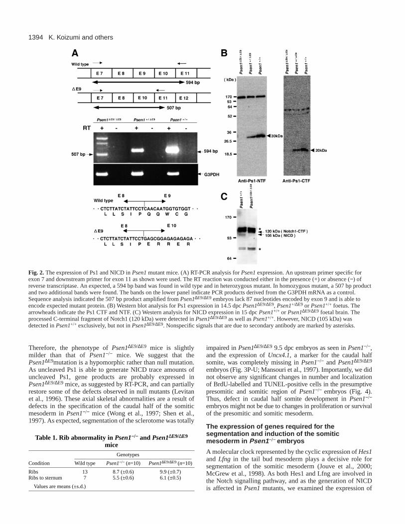
The expression of Psen1or Psen1∆E9 transcripts wasexamined by RT-PCR. As expected, 507 bp fragmentsrepresenting Psen1∆E9 transcriptswere amplified from Psen1∆E9/∆E9
embryonic RNA, a while 594 bpfragment was amplified from thewild type (Fig. 2A). Two additionalfragments seen in the Psen1∆E9/∆E9
embryo encoded abnormally splicedtranscripts from exon 8 to the Neor
cassette (K. K. and H. K., unpublished).Sequence analysis of the 507 bpfragment revealed splicing of Psen1mRNA from exon 8 to exon 10 withoutframeshift (Fig. 2A). However, theexpression of Psen1∆E9 transcripts wasmuch lower than that of wild-typetranscripts because the 507 bp PCRfragment was hardly visible inPsen1+/∆E9 embryos. Western blotanalysis using antisera against NTF andCTF of Ps1 revealed that the expressionof the 30 kDa NTF and 20 kDa CTFwere totally abolished, and that nouncleaved Ps1 protein with theexpected molecular mass of about 50kDa was detectable in Psen1∆E9/∆E9
foetal tissues (Fig. 2B). In Psen1+/∆E9
foetuses, the expression of NTF andCTF was half of that in the wild type.Therefore, the expression of theuncleaved Ps1 protein derived from thePsen1∆E9 locus could not be detected,and the Psen1∆E9 mutation wassuggested to be almost equivalent to anull mutation. Indeed, the generation ofNICD was significantly impaired inPsen1∆E9/∆E9 foetal brain (Fig. 2C). Thetrace amounts of NICD observed inPsen1∆E9/∆E9 foetuses might be due toresidual Ps1∆E9 or presenilin 2 (Ps2)gene products (Levitan et al., 1996;Donoviel et al., 1999; Herreman et al.,1999).
Somite segmentation defects in Psen1−/− andPsen1∆E9/∆E9 micePsen1+/∆E9 mice were externally normal and fertile.Psen1∆E9/∆E9 pups died shortly after birth, similar to thePsen1−/− pups described previously (Wong et al., 1997; Shenet al., 1997). Psen1−/− and Psen1∆E9/∆E9 pups were recognizedby considerable truncation of the axis due to severe axialskeletal malformations (Fig. 3A-F). We observed umbilicalherniation in all Psen1−/− pups, but not in any of thePsen1∆E9/∆E9 pups (Fig. 3C,E). Skeletal preparations of 17.5dpc embryos revealed multiple defects in the vertebral columnand ribs in Psen1−/− and Psen1∆E9/∆E9 mice. Most importantly,the pedicles of the neural arches and the proximal parts of theribs were missing in Psen1−/− and Psen1∆E9/∆E9 mice, whilethe vertebral bodies and laminae were maintained, althoughirregularly shaped (Fig. 3G-L). Deformities of the rib cagewere more prominent in Psen1−/− mice than in Psen1∆E9/∆E9
mice (Fig. 3M-O). The number of ribs was partially restoredin Psen1∆E9/∆E9 mice compared with Psen1−/− mice (Table 1).
Fig. 1.Generation of a new allele of Psen1mutation lacking exon 9. (A) Maps of the wild-type Psen1locus, the targeting vector, Psen1∆E9 allele and Psen1null allele. Black boxesrepresents the exons in Psen1locus. A white box indicates Neor cassette. The targeting vectorshows the replacement of exon 9 and flanking genomic sequences by the Neor cassette (neo-tk). Arrows indicate the sites for PCR primers used for genotyping of wild type, Psen1∆E9 andPsen1null alleles, and grey bars represent expected size of PCR products from each allele. TheDNA fragment used for external probe is indicated by a horizontal black bar (probe). EcoRI,RI; HindIII, H; BamHI, B; BglII, Bgl. (B) DNA blot analysis by BglII digestion clearlydistinguished homozygous, heterozygous and wild-type embryos. The external probe detects4.2 kb BglII fragment in wild-type allele and 6.0 kb in Psen1∆E9 allele. (C) PCR analysis alsodistinguished homozygous, heterozygous and wild-type embryos. Genotyping was performedby PCR using three primers: Ps1/PCR#1 and Ps1/PCR#2 for wild type (giving 656 bpproduct) and Ps1/PCR#1 and Ps1/PCR#3 for Psen1∆E9 allele (giving 251 bp PCR product).
1394
Therefore, the phenotype of Psen1∆E9/∆E9 mice is slightlymilder than that of Psen1−/− mice. We suggest that thePsen1∆E9mutation is a hypomorphic rather than null mutation.As uncleaved Ps1 is able to generate NICD trace amounts ofuncleaved Ps1, gene products are probably expressed inPsen1∆E9/∆E9 mice, as suggested by RT-PCR, and can partiallyrestore some of the defects observed in null mutants (Levitanet al., 1996). These axial skeletal abnormalities are a result ofdefects in the specification of the caudal half of the somiticmesoderm in Psen1−/− mice (Wong et al., 1997; Shen et al.,1997). As expected, segmentation of the sclerotome was totally
impaired in Psen1∆E9/∆E9 9.5 dpc embryos as seen inPsen1−/−,and the expression of Uncx4.1, a marker for the caudal halfsomite, was completely missing in Psen1−/− and Psen1∆E9/∆E9
embryos (Fig. 3P-U; Mansouri et al., 1997). Importantly, we didnot observe any significant changes in number and localizationof BrdU-labelled and TUNEL-positive cells in the presumptivepresomitic and somitic region of Psen1−/− embryos (Fig. 4).Thus, defect in caudal half somite development in Psen1−/−
embryos might not be due to changes in proliferation or survivalof the presomitic and somitic mesoderm.
The expression of genes required for thesegmentation and induction of the somiticmesoderm in Psen1−/− embryos
A molecular clock represented by the cyclic expression of Hes1and Lfng in the tail bud mesoderm plays a decisive role forsegmentation of the somitic mesoderm (Jouve et al., 2000;McGrew et al., 1998). As both Hes1 and Lfng are involved inthe Notch signalling pathway, and as the generation of NICDis affected inPsen1mutants, we examined the expression of
K. Koizumi and others
Fig. 2.The expression of Ps1 and NICD inPsen1mutant mice. (A) RT-PCR analysis for Psen1expression. An upstream primer specific forexon 7 and downstream primer for exon 11 as shown were used. The RT reaction was conducted either in the presence (+) or absence (−) ofreverse transcriptase. An expected, a 594 bp band was found in wild type and in heterozygous mutant. In homozygous mutant, a 507 bp productand two additional bands were found. The bands on the lower panel indicate PCR products derived from the G3PDH mRNA as a control.Sequence analysis indicated the 507 bp product amplified from Psen1∆E9/∆E9 embryos lack 87 nucleotides encoded by exon 9 and is able toencode expected mutant protein. (B) Western blot analysis for Ps1 expression in 14.5 dpc Psen1∆E9/∆E9, Psen1+/∆E9 or Psen1+/+ foetus. Thearrowheads indicate the Ps1 CTF and NTF. (C) Western analysis for NICD expression in 15 dpc Psen1+/+ or Psen1∆E9/∆E9 foetal brain. Theprocessed C-terminal fragment of Notch1 (120 kDa) were detected in Psen1∆E9/∆E9 as well as Psen1+/+. However, NICD (105 kDa) wasdetected in Psen1+/+ exclusively, but not in Psen1∆E9/∆E9. Nonspecific signals that are due to secondary antibody are marked by asterisks.
Table 1. Rib abnormality in Psen1−/− and Psen1∆E9/∆E9
miceGenotypes
Condition Wild type Psen1−/− (n=10) Psen1∆E9/∆E9 (n=10)
Ribs 13 8.7 (±0.6) 9.9 (±0.7)Ribs to sternum 7 5.5 (±0.6) 6.1 (±0.5)
Values are means (±s.d.)

1395Presenilin 1 in somite segmentation
genes involved in the Notch signalling pathway, includingNotch1, Notch2, Dll1, Dll3, Hes1, Hes5, Lfng and Mesp2, inPsen1−/− embryos. The expression of Notch1, Notch2and Dll1was not significantly affected in Psen1−/− embryos, althoughthe anterior boundaries of Notch1and Notch2expression werediffuse (Fig. 5A-D). Dll3 expression in the presomiticmesoderm was clearly increased in Psen1−/− embryos (Fig.5G,H). A similar observation has been reported in Dll1-deficient mice (Barrantes et al., 1999). The expression of Hes5,one of the genes responsive to the Notch signalling pathway,was totally lacking. However, we could not detect expressionof Hes1 in the presomitic mesoderm of wild-type embryos(Fig. 5I,J; K. K. and H. K., unpublished; Nishimura et al.,1998). This might be due to a problem in the sensitivity of ourexperiments. The expression of Lfng and Mesp2 were alsosignificantly reduced in the presomitic mesoderm in Psen1−/−
embryos (Figs 6A, 7A). Therefore, the expression of severalgenes involved in the Notch signalling pathway is affected inthe presomitic mesoderm of Psen1−/− embryos.
We further investigated the expression of other classes ofgenes involved in segmentation and induction of the somitic
mesoderm. Inductive signals from the surface ectodermrequired for segmentation are represented by the expressionof Paraxis (Sosic et al., 1997). Paraxis expression is notsignificantly affected in Psen1−/− embryos (Fig. 5K,L).Inductive signals mediated by the FGF family proteins are alsoknown to play an essential role during segmentation of thesomitic mesoderm (Yamaguchi et al., 1994; Deng et al., 1994).The expression of Fgfr1and Fgf18 was not significantlyaffected in Psen1−/− embryos (Fig. 5M-P). We also investigatedthe expression of genes during the induction of presomiticmesoderm, because cyclic expression of Hes1and Lfnghasbeen described in mesenchymal cells in the tail bud region(McGrew et al., 1998; Aulehla and Johnson, 1999; Jouve et al.,2000). We have looked at the expression of several genes inthe tail bud and early somitic mesoderm, including Wnt3a,Evx1, Hoxb4and Mfh1. The expression of these genes is notaffected in Psen1−/− embryos (Fig. 5Q,R; K. K. and H. K.,unpublished). Thus, the signals required for early induction ofthe somitic mesoderm or for segmentation emanating from thesurface ectoderm or mediated by FGF family proteins areapparently not affected in Psen1−/− embryos. We suggest that
Fig. 3. Segmentation defects in Psen1−/− andPsen1∆E9/∆E9 mice. (A,C,E) Lateral views of17.5 dpc wild-type (A), Psen1−/− (C) andPsen1∆E9/∆E9 (E) foetuses. (B,D,F) Lateralviews of skeletal preparation of 17.5 dpc wild-type (B), Psen1−/− (D) andPsen1∆E9/∆E9
(F) foetuses. (G-I) Higher magnification viewsof lumbar region of 17.5 dpc wild-type(G), Psen1−/− (H) andPsen1∆E9/∆E9 (I) foetuses.Note lack of pedicles of the neural arches inPsen1−/− (H) andPsen1∆E9/∆E9 (I) foetuses asindicated by arrows. (J-L) Dorsal views of thethoracic region of 17.5 dpc wild type (J),Psen1−/− (K) andPsen1∆E9/∆E9 (L) foetuses.Note lack of proximal regions of ribs asindicated by arrowheads. (M-O) Ventral viewsof rib cage of 17.5 dpc wild-type (M), Psen1−/−
(N) andPsen1∆E9/∆E9 (O) foetuses. (P-R)Parasagittal sections stained with Hematoxylinand Eosin of 9.5 dpc wild-type (P), Psen1−/−
(Q) andPsen1∆E9/∆E9 (R) embryos. The rostralis right. (S-U) The expression of Uncx4.1in 9.5dpc wild-type (S), Psen1−/− (T) andPsen1∆E9/∆E9 (U) embryos.

1396
defects in the proteolytic activation of Notch family proteinsby Ps1 could be causative factors for the defects insegmentation in Psen1-deficient mice.
Lfng expression in the presomitic mesoderm isdependent on the Ps1 gene productThe expression of Lfngin the paraxial mesoderm of wild-typeembryos demarcates mutually distinct portions of the
presomitic mesoderm because of its cyclic expression asreported previously with 30 minutes chromogenic reaction(Fig. 6A, parts a-d) (McGrew et al., 1998; Aulehla andJohnson, 1999). However, we could not see Lfng expressionin Psen1−/− embryos following the same chromogenic reaction(Fig. 6A, parts e-h). Thus, Lfng expression in the presomiticmesoderm is significantly downregulated in Psen1−/−
embryos. Interestingly, we did not observe the reduction of
K. Koizumi and others
Fig. 4.Localization of proliferating and apoptotic cells in the presomitic and somitic mesoderm of Psen1−/− mice. (A) Localization of BrdU-labelled cells in wild-type (a) and Psen1−/− (c) mice. An arrowhead indicates a newly generated segment boundary. (b,d) Higher magnificationviews of as indicated by boxes in (a) and (c), respectively. (B) Localization of TUNEL-positive cells in wild type (a) and Psen1−/− (c) mice.(b,d) Higher magnification views as indicated by boxes in (a) and (c), respectively. Scale bars: 100 µm.
Fig. 5.The expression of mesodermal markers in wild type andPsen1−/− embryos. (A,B) Notch1 expression in 9.5 dpc embryos. (C,D)Notch2expression in 9.5 dpc embryos. (E,F)Dll1 expression in 9.5 dpc embryos. (G,H) Dll3 expression in 9.5 dpc embryos. Dll3 is significantly increasedin the presomitic mesoderm ofPsen1−/− embryos. (I,J) Hes5 expression in 11.5 dpc embryos. (K,L) Paraxis expression in 9.5 dpc embryos. (M,N)Fgfr1 expression in 9.5 dpc embryos. (O,P) Fgf18 expression in 10.5 dpc embryos. (Q,R) Wnt3aexpression in 9.5 dpc embryos.

1397Presenilin 1 in somite segmentation
Lfng expression in the neural tube ofPsen1−/− embryos (K. K. and H. K.,unpublished). Chromogenic reaction forover 2 hours allowed us to see Lfngexpression in Psen1−/− embryos (Fig. 6B,parts e-h). Specimens were aligned toshow the rostrally directed progressionof the wave front of Lfngexpression. Theanteriorly directed progression of Lfngexpression was seen in wild-type andPsen1−/− embryos. This suggests that thecyclic expression of Lfng in thepresomitic mesoderm is retained inPsen1−/− embryos. We further compared the duration of onecycle of Lfng expression between wild-type and Psen1−/−
embryos. After bisection of the caudal regions of 9.5 dpcembryos at the midline with a microscalpel, one side wasimmediately fixed and the other was cultured for variousperiods from 60 to 120 minutes as reported previously(Aulehla and Johnson, 1999). For wild-type samples, morethan ten specimens were examined for every time point. After110 minutes of culture, four out of ten specimens exhibitedalmost identical expression patterns between uncultured andcultured specimens, while the position of the Lfngstripe in therostral region of the somitic mesoderm shifted caudally (Fig.6C, part b). After both 100 and 120 minutes in culture, oneout of ten specimens exhibited identical patterns (Fig. 6C,parts a,c). We conclude that Lfngexpression oscillates every110 minutes on average under our culture conditions.Similarly, two out of six specimens obtained from Psen1−/−
embryos exhibited almost identical expression patterns after110 minutes in culture (Fig. 6C, part e) but identical patternwas not seen after 100 and 120 minutes (Fig. 6C, parts d,f).Thus, the amplitude of oscillation of Lfngexpression is
significantly affected while the duration is unaffected inPsen1−/− embryos.
Rapid repression of Mesp2 expression in thepresumptive caudal half somite is dependent on Ps1Mesp2is also involved in the Notch pathway and is essentialfor the specification of the rostral half somite, and subsequentlyfor segmentation (Saga et al., 1997). Mesp2expression firstappears one segment in width, and subsequently the expressionin the presumptive caudal half somite is rapidly repressed.Rapid repression of Mesp2expression has been suggested tobe important for the establishment and maintenance of Ps1-dependent activation of the Notch signalling pathway(Takahashi et al., 2000). This led us to re-examine Mesp2expression inPsen1−/− embryos. In 9.5 dpc wild-type embryos,Mesp2expression in the presomitic mesoderm varies in widthand strength due to the rapid downregulation in thepresumptive caudal half somite (Fig. 7A, parts a-d). InPsen1−/−
embryos, Mesp2 expression appears at almost the samepositions as in the wild type, but the level is significantlyreduced (Fig. 7A, parts e-h). Importantly, the width of the
Fig. 6.Cyclic expression of Lfngin thepresomitic mesoderm was retained butsignificantly reduction in Psen1−/− embryos.(A) The expression of Lfngin the presomiticmesoderm of the wild-type (a-d) andPsen1−/− (e-h) embryos. To allow thequantitative comparison, chromogenicreaction was limited to 30 minutes. Underthis condition, Lfngexpression could not beobserved in any of Psen1−/− embryos (15specimens), while dynamic expression ofLfng was clearly seen in the wild type.(B) Progression of wave front of cyclicexpression of Lfngin the wild type (a-d) andPsen1−/− (e-h) embryos. Two hourschromogenic reaction allowed us to see Lfngexpression in the presomitic mesoderm ofPsen1−/− embryos (e-h). Specimens arealigned to show the rostrally directedprogression of the wave front of Lfngexpression as indicated by brackets. For wild-type embryos (left), specimens after 30minutes reactions were shown.(C) Comparison of Lfngexpression betweenuncultured (left) and cultured (right) halveswith various times as shown. In Psen1−/−
embryos (d-f), Lfngexpression is indicatedby brackets.

1398
Mesp2stripe does not vary while the strength of the stripevaries in Psen1−/− embryos. This suggests that the rapidrepression ofMesp2expression in the presumptive caudal halfsomite might be impaired in Psen1−/− embryos. To examinethis possibility, we examined changes in the Mesp2stripe after60 and 120 minutes of culture in 24 wild-type or Psen1+/−
embryos, and ninePsen1−/− embryos. In the wild type, thewidth and strength of the Mesp2stripe changed clearly within60 or 120 minutes (Fig. 7B, parts a-d), and in some cases thestripe shifted posteriorly (Fig. 7B, parts a,d). A rapid repressionin the caudal half of the stripe is obvious as shown in Fig. 7B,part c. In contrast, we found no significant alterations in thewidth of Mesp2stripe in Psen1−/− embryos while strength ofthe stripe changed (Fig. 7B, parts e-h). The position of thestripe shifted posteriorly in some cases (Fig. 7B, parts e,h). Inconclusion, the rapid repression of Mesp2expression in thepresumptive caudal half somite is affected in Psen1−/−
embryos, while the Mesp2stripe appears at an almost normalposition.
Distribution of Psen1∆E9/− cells in the somitic andpresomitic mesoderm of chimaeric embryos
Although impairment in the Notch signalling is obvious in thetail bud mesoderm of Psen1-deficient mice, it has not yet beendetermined when it is functionally manifested. To addressthis, we have generated chimaera composed of Psen1∆E9/−
and wild-type cells and, have analyzed chimaerism in the
presomitic and somitic mesoderm. Thewild-type cells were genetically labelledby β-galactosidase expression derivedfrom the ROSA26locus (Fig. 8A). Theavailability of Psen1∆E9 and Psen1null
alleles allowed us to distinguishPsen1∆E9/−chimaera from Psen1+/∆E9 orPsen1+/− chimaeras (Fig. 8B). Wegenerated 74 chimaeras, seven of whichwere Psen1∆E9/− chimaeras with β-galactosidase expression. The tailregions of the chimaeras were dissectedand subjected to a histological analysisin order to examine the formation ofsegment boundaries. The newly formedsomites and presumptive somitesundergoing segmentation are designatedS0 and S-1, respectively (Fig. 8C). Inthis definition, the region of the Mesp2stripe corresponds to S-2. Five out ofseven of the Psen1∆E9/− chimaerasexhibited a moderate contribution ofPsen1∆E9/−cells (Fig. 8D, parts a,e). Twoshowed a strong contribution ofPsen1∆E9/− cells (Fig. 8D, part h). In allof the moderate Psen1∆9/−chimaeras, weobserved stripes of β-galactosidase-positive cells derived from the wild typein the somitic mesoderm, while thestriped distribution was never seenin Psen1+/∆E9, Psen1+/− or wild-typechimaeras (Fig. 8D, parts a, e, E, partsa,e; K. K. and H. K., unpublished).
β-Galactosidase-positive cells were metamerically distributedin the somitic mesoderm of moderate Psen1∆E9/− chimaerasas confirmed by low magnification histological analyses (Fig.8D, parts b,f). In strong chimaeras, a metameric distributionof β-galactosidase-positive cells was vaguely visible, andhistological investigation revealed the periodic appearance ofcolonies of β-galactosidase-positive cells (Fig.8D, parts h,i).Stripes of β-galactosidase-positive cells were seenexclusively in the segmented region, but not in the presomiticmesoderm of either moderate or strong chimaeras.Interestingly, three out of five moderate chimaeras weresegmented in the somitic mesoderm, but two were not(compare Fig. 8D, parts c,g). The segmentedPsen1∆E9/−
chimaeras allowed us to examine the localizationof β-galactosidase-positive cells in each segment. β-Galactosidase-positive cells, indeed, clearly distributed to thecaudal region of each segment while the segment borderswere less obvious than in wild-type chimaeras (Fig. 7D, partc, E, part c). Within the newly segmented somite (S0), β-galactosidase-positive cells were clearly distributed in thecaudal region, suggesting the exclusion of Psen1∆E9/− cellsfrom the caudal region, while Psen1∆E9/− and wild-type cellsstill intermingle in the S-1 and S-2 regions (Fig. 8D, partd). In Psen1+/∆E9, Psen-1+/− or wild-type chimaeras, β-galactosidase-positive cells were distributed almost evenlywithin each segment (Fig. 8E, parts c,d,g). Another piece ofevidence obtained from the trunk region of strong chimaerasalso suggests the predominant localization of wild-type cells
K. Koizumi and others
Fig. 7. Impairment of rapid downregulationof Mesp2expression in the presumptivecaudal half somite inPsen1−/− embryos.(A) Mesp2expression in the presomiticmesoderm of 9.5 dpc wild-type (a-d) andPsen1−/− (e-h) embryos. (B) Comparison ofMesp2expression between uncultured (left)and cultured (right) halves with 60 or 120minutes culture of 9.5 dpc wild-type (a-d) and Psen1−/− (e-h) embryos.

1399Presenilin 1 in somite segmentation
in the caudal half somite. β-Galactosidase-positive cells in thestrong chimaeras localise exclusively in the neural archanlage, which are known to be derived from the caudal halfsomite (Fig. 8D, part k) (Goldstein and Kalcheim, 1991). This
implies that the colonies of β-galactosidase-positive cellsseen in the sclerotomal region of strong chimaeras mightpossess cellular properties specific for the caudal half somite,although the segment boundaries are not formed.
Fig. 8.Distribution ofPsen1-deficient cells inPsen1∆E9/−↔Psen1+/+ chimaericembryos. (A) Schematicrepresentation of the experimentalprocedures for generation ofPsen1∆E9/−↔Psen1+/+ chimaeras.Head region was used forgenotyping. (B) The genotypes ofchimaeric embryos were determinedby PCR using three primers shownin Fig. 1A. The 656 bp, 328 bp and251 bp PCR products represent thewild-type, Psen1null and Psen1∆E9
alleles, respectively. (C) Thesegmentation of somites in theparaxial mesoderm as described byTakahashi et al., 2000. S1 indicatesthe most recently formed completesomite. S0 indicates a formingsomite. S-1 indicated presomiticregion forming next somite. Theblack and white arrowheads show aforming and formed segmentalborders, respectively, in C, D and E.(D) Dorsal views and sections ofPsen1∆E9/−; ROSA26+/+↔Psen1+/+;ROSA26Tg/+ chimaeric embryos.Three representative specimens, amoderate chimaera with theformation of segment boundaries(a-d), moderate chimaera without theformation of segment boundaries(e-g) and chimaera with predominantcontribution of Psen1∆E9/−cells (h-k)are shown. (a) Dorsal view of thecaudal region of a moderatechimaera with the formation ofsegment boundaries. (b) Lowermagnification view of a frontalsection of an identical embryo asshown in (a). (c) Highermagnification view of S4 and S5region as shown by a box in (b).(d) Higher magnification view of S0,S-1 and S-2 region as shown by abox in (b). (e) Dorsal view of thecaudal region of a moderatechimaera without the formation ofsegment boundaries. (f) Lowermagnification view of an identicalembryo as shown in (e). (g) Higher magnification view of S4 and S5 region as shown by the box in (f). (h) Dorsal view of the caudal region of achimaera with predominant contribution of Psen1∆E9/−cells. (i) Lower magnification view of a frontal section of an identical embryo as shownin (h). (j) Higher magnification view of presumptive segmented region as shown by a box in (i). (k) Parasaggital section of prospectivethoracolumbar region of an identical embryo as shown in above. Dorsal root ganglia are indicated by arrows and prospective neural arches byasterisks. (E) Dorsal views and sections of Psen1+/∆E9; ROSA26+/+↔Psen1+/+; ROSA26Tg/+ chimaeric embryos. Two representative specimenswith moderate and weak contribution of ROSA26Tg/+ cells are shown (a,e). (a) Dorsal view of the caudal region of a moderate chimaera.(b) Lower magnification view of a frontal section of an identical embryo as shown in (e). (c) Higher magnification view of S2 to S4 region asshown by a box in (b). (d) Higher magnification view of S-2 to S1 region as shown by a box in (b). (e) Dorsal view of the caudal region of achimaera with weak contribution of ROSA26Tg/+ cells. (f) Lower magnification view of a frontal section shown in (e). (g) Higher magnificationview of S3 and S4 region as shown by a box in (f). Scale bars, 100 µm.

1400
DISCUSSION
Periodic activation of the Notch pathway in thepresomitic mesodermIn the present study, we generated an allelic mutation of Psen1that shows slightly hypomorphic phenotypes in the axialskeleton. Establishment of the caudal half somites andsubsequent segmentation were totally impaired, as reportedpreviously (Wong et al., 1997; Shen et al., 1997; Takahashiet al., 2000). Defects in the generation of NICD and mis-expression of Dll3, Hes5andLfng in the presomitic mesodermin Psen1-deficient mice suggest that the defects in the somitesegmentation are most probably due to impairment in theNotch signalling pathway.
The significant reduction of Lfngexpression in Psen1mutant embryos indicates that the Ps1 gene product isessential for maintaining the amplitude of Lfngexpression inthe presomitic mesoderm. Thus, the expression of Lfngissuggested to be under the control of NICD. As the fringe geneproduct is needed for the generation of NICD, the molecularcircuitry, including the Notch family proteins, Ps1 and Lfng,and presumably Delta family proteins, seem to be importantfor the maintenance of the amplitude of the periodicactivation of the Notch pathway in the presomitic mesoderm(Panin et al., 1997; Cohen et al., 1998; Brückner et al., 2000;Hicks et al., 2000). We have also demonstrated that theduration of cyclic activation of the Notch pathway, asrepresented by Lfngexpression, is not altered significantlyin Psen1 mutants. Thus, the molecular mechanisms thatgenerate the periodic expression of Lfng, which presumablyinclude the clock mechanism, are apparently independent ofthe Ps1 gene product. Importantly, the dynamic expression ofHes1 and Lfng is lost in the presomitic mesoderm of Dll1mutants (Barrantes et al., 1999; Jouve et al., 2000). As bothHes1 and Lfng expression are regulated by the molecularclock linked to segmentation, the initial oscillation of thesegmentation clock could be dependent on Notch signallingin the presomitic mesoderm. Because Ps2, a homologue ofPs1, is also able to function as a γ-secretase and is expectedto play redundant roles, the remaining activity of Lfngexpression might be due to the function of Ps2 (Donoviel etal., 1999; Herreman et al., 1999). It will be necessary toexamine Lfngexpression in Psen1/Psen2double homozygousembryos.
Specification of the caudal half somite involves aPs1-dependent rapid downregulation of Mesp2Mesp2 is a transcriptional regulator essential for specificationof the rostral half somite as revealed by experiments involvingMesp2-deficient mice (Saga et al., 1997). The induction ofMesp2 expression in the S-2 region is, at least in part,dependent on Notch signals, because Mesp2 expression issignificantly downregulated not only in Psen1- but also Dll1-,Notch1-, RBPJκ- and Dll3-deficient mice (this study, Barranteset al., 1999; Y. Takahashi and Y. S., unpublished). Based onthe present study, we suggest that the rapid downregulation ofMesp2 in the presumptive caudal half is also dependent onNotch activation mediated by Ps1. Derepression of Mesp2expression in the presumptive caudal half somites seems to beinvolved in the development of caudal half somite defects inPsen1mutants because the effects of the Psen1mutation can
be restored by a Mesp2mutation, as previously observed inPsen1/Mesp2double homozygotes (Takahashi et al., 2000). AsMesp2 has been demonstrated to repress Dll1 expression, therapid repression of Mesp2mediated by Ps1 might allow theestablishment and stabilisation of the molecular circuitryessential for caudal half somite specification, including Dll1,Notch1 and Ps1.
A previous study using the Mesp2lacZ allele demonstratedthat the downregulation of Mesp2in the segmented region isdependent upon Mesp2 itself, not Ps1 (Takahashi et al., 2000).The downregulation of Mesp2in the presumptive rostral andcaudal halves might therefore be dependent on Mesp2 and Ps1,respectively.
Functional manifestation of the Notch activationinduces homophilic condensation of like half somitecellsThe chimaera assay revealed that segment border formation isconcordant with the condensation of wild-type cells into thecaudal compartment of forming somites while Psen1-deficientand wild-type cells intermingle in the presomitic region. Thisimplies that segregation of presomitic cells into prospectiverostral and caudal half compartments takes place in the S-1region of chimaeras because the transient expression of Mesp2in the S-2 region is essential for the rostral half specification(Takahashi et al., 2000). This is in good agreement with theobservation that striped expression of Dll1 in the caudal halfsomites is first established in the S-1 region (Takahashi et al.,2000). Since Ps1 is not essential for the proliferation or survivalof presomitic and somitic mesoderm, exclusion ofPsen1-deficient cells from the prospective caudal half somite mightbe due to cell-sorting machinery, rather than defects inproliferation or survival. This is supported by previousobservations that indicate counteracting cell adhesiveproperties of rostral and caudal half cells (Stern and Keynes,1987). Thus, Dll1- and Ps1-dependent activation of the Notchpathway might induce or stabilise the homophilic condensationof caudal half somite cells, and, at the same time, excludePsen1-deficient Dll1-negative cells from the caudal half. Thisis reminiscent of recent observations on avian trunk neuralcrest cells that neurogenic precursors expressing Notch1proteins in nascent crest populations are eliminated by Dll1-expressing cells (Wakamatsu et al., 2000; Maynard et al.,2000). In contrast, cyclic activation of the Notch pathway inthe caudal region of presomitic mesoderm, as represented bycyclic expression of Lfng, is still latent in the segregation ofPsen1-deficient and wild-type cells. A transient episode ofMesp2 expression might be needed for the functionalmanifestation of Notch activation to segregate prospectiverostral or caudal half somite cells.
We thank Drs R. Conlon (Notch1 and Fgfr1), A. Gossler (Dll1), P.Gruss (Uncx4.1), R. Kageyama (Hes5), R. Beddington (Dll3), A.Rawls (paraxis), S. Takada (Fgf18 and Wnt3a), and R. Johnson (Lfng)for providing us with reagents. We are also grateful to Dr R. Ballingfor critical reading of the manuscript, and to Ms Sanae Takeda, MisaoUchida and Mr Shozo Sugimori for their help in many respects. Thiswork was supported by grants from Ministry of Education ofJapan, the Naito Foundation, the Mochida Foundation, the KanaeFoundation, the Uehara Memorial Foundation, the SpecialCoodination Funds of the Ministry of Education, Culture, Sports,Science and Technology, and by the Japanese Government.
K. Koizumi and others

1401Presenilin 1 in somite segmentation
REFERENCES
Akazawa, C., Sasai, Y., Nakanishi, S. and Kageyama, R. (1992). Molecularcharacterization of a rat negative regulator with a basic helix-loop-helixstructure predominantly expressed in the developing nervous system. J. Biol.Chem. 267, 21879-21885.
Aoyama, H. and Asamoto, K. (1988). Determination of somite cells:independence of cell differentiation and morphogenesis. Development104,15-28.
Aulehla, A. and Johnson, R. L. (1999). Dynamic expression of lunatic fringesuggests a link between notch signaling and an autonomous cellularoscillator driving somite segmentation. Dev. Biol. 207, 49-61.
Barrantes, I. B., Elia, A. J., Wunsch, K., De Angelis, M. H., Mak, T. W.,Rossant, J., Conlon, R. A., Gossler, A. and de la Pompa, J. L. (1999).Interaction between Notch signalling and Lunatic fringe during somiteboundary formation in the mouse. Curr. Biol. 9, 470-480.
Bettenhausen, B., Hrabe de Angelis, M., Simon, D., Guenet, J. L. andGossler, A. (1995). Transient and restricted expression during mouseembryogenesis of Dll1, a murine gene closely related to Drosophila Delta.Development121, 2407-2418.
Brückner, K., Perez, L., Clausen, H. and Cohen, S. (2000).Glycosyltransferase activity of Fringe modulates Notch-Delta interactions.Nature406, 411-415.
Burgess, R., Cserjesi, P., Ligon, K. L. and Olson, E. N. (1995). Paraxis: abasic helix-loop-helix protein expressed in paraxial mesoderm anddeveloping somites. Dev. Biol. 168, 296-306.
Candia, A. F., Hu, J., Crosby, J., Lalley, P. A., Noden, D., Nadeau, J. H.and Wright, C. V. (1992). Mox-1 and Mox-2 define a novel homeobox genesubfamily and are differentially expressed during early mesodermalpatterning in mouse embryos. Development 116, 1123-1136.
Cohen, B., Bashirullah, A., Dagnino, L., Campbell, C., Fisher, W. W.,Leow, C. C., Whiting, E., Ryan, D., Zinyk, D., Boulianne, G., Hui, C. C.et al. (1997). Fringe boundaries coincide with Notch-dependent patterningcentres in mammals and alter Notch-dependent development in Drosophila.Nat. Genet. 16, 283-288.
Conlon, R. A., Reaume, A. G. and Rossant, J. (1995). Notch1 isrequired for the coordinate segmentation of somites. Development. 121,1533-1545.
Deng, C. X., Wynshaw-Boris, A., Shen, M. M., Daugherty, C., Ornitz, D.M. and Leder, P. (1994). Murine FGFR-1 is required for earlypostimplantation growth and axial organization. Genes Dev. 8, 3045-3057.
De Strooper, B., Saftig, P., Craessaerts, K., Vanderstichele, H., Guhde, G.,Annaert, W., Von Figura, K. and Van Leuven, F. (1998). Deficiency ofpresenilin-1 inhibits the normal cleavage of amyloid precursor protein.Nature 391, 387-390.
De Strooper, B., Annaert, W., Cupers, P., Saftig, P., Craessaerts, K.,Mumm, J. S., Schroeter, E. H., Schrijvers, V., Wolfe, M. S., Ray,W. J. et al. (1999). A presenilin-1-dependent gamma-secretase-likeprotease mediates release of Notch intracellular domain. Nature398, 518-522.
Donoviel, D. B., Hadjantonakis, A. K., Ikeda, M., Zheng, H., Hyslop, P. S.and Bernstein, A. (1999). Mice lacking both presenilin genes exhibit earlyembryonic patterning defects. Genes Dev. 13, 2801-2810.
Dunwoodie, S. L., Henrique, D., Harrison, S. M. and Beddington, R. S.(1997). Mouse Dll3: a novel divergent Delta gene which may complementthe function of other Delta homologues during early pattern formation inthe mouse embryo. Development 124, 3065-3076.
Evrard, Y. A., Lun, Y., Aulehla, A., Gan, L. and Johnson, R. L. (1998).lunatic fringe is an essential mediator of somite segmentation and patterning.Nature394, 377-381.
Goldstein, R. S. and Kalcheim, C. (1991). Normal segmentation and size ofthe primary sympathetic ganglia depend upon the alternation of rostrocaudalproperties of the somites. Development 112, 327-334.
Herreman, A., Hartmann, D., Annaert, W., Saftig, P., Craessaerts, K.,Serneels, L., Umans, L., Schrijvers, V., Checler, F., Vanderstichele, H.et al. (1999). Presenilin 2 deficiency causes a mild pulmonary phenotypeand no changes in amyloid precursor protein processing but enhances theembryonic lethal phenotype of presenilin 1 deficiency. Proc. Natl. Acad. Sci.USA96, 11872-11877.
Hicks, C., Johnston, S. H., diSibio, G., Collazo, A., Vogt, T. F. andWeinmaster, G. (2000). Fringe differentially modulates Jagged1 and Delta1signalling through Notch1 and Notch2. Nat. Cell Biol. 2, 515-520.
Hrabe de Angelis, M., McIntyre, J., II and Gossler, A. (1997). Maintenance
of somite borders in mice requires the Delta homologue DII1. Nature386,717-721.
Irvine, K. D. (1999). Fringe, Notch, and making developmental boundaries.Curr. Opin. Genet. Dev. 9, 434-441.
Jouve, C., Palmeirim, I., Henrique, D., Beckers, J., Gossler, A., Ish-Horowicz, D. and Pourquie, O. (2000). Notch signalling is required forcyclic expression of the hairy-like gene HES1 in the presomitic mesoderm.Development127, 1421-1429.
Kessel, M. and Gruss, P. (1991). Homeotic transformations of murinevertebrae and concomitant alteration of Hox codes induced by retinoic acid.Cell 67, 89-104.
Kusumi, K., Sun, E. S., Kerrebrock, A. W., Bronson, R. T., Chi, D. C.,Bulotsky, M. S., Spencer, J. B., Birren, B. W., Frankel, W. N. andLander, E. S. (1998). The mouse pudgy mutation disrupts Delta homologueDll3 and initiation of early somite boundaries. Nat. Genet. 19, 274-278.
Levitan, D., Doyle, T. G., Brousseau, D., Lee, M. K., Thinakaran, G., Slunt,H. H., Sisodia, S. S. and Greenwald, I. (1996). Assessment of normal andmutant human presenilin function in Caenorhabditis elegans. Proc. Natl.Acad. Sci. USA93, 14940-14944.
Mansouri, A., Yokota, Y., Wehr, R., Copeland, N. G., Jenkins, N. A. andGruss, P. (1997). Paired-related murine homeobox gene expressed in thedeveloping sclerotome, kidney, and nervous system. Dev. Dyn. 210, 53-65.
Maynard, T. M., Wakamatsu, Y. and Weston, J. A. (2000). Cell interactionswithin nascent neural crest cell populations transiently promote death ofneurogenic precursors. Development 127, 4561-4572.
McGrew, M. J., Dale, J. K., Fraboulet, S. and Pourquie, O. (1998). Thelunatic fringe gene is a target of the molecular clock linked to somitesegmentation in avian embryos. Curr Biol. 8, 979-982.
Moloney, D. J., Panin, V. M., Johnston, S. H., Chen, J., Shao, L., Wilson,R., Wang, Y., Stanley, P., Irvine, K. D., Haltiwanger, R. S. and Vogt, T.F. (2000). Fringe is a glycosyltransferase that modifies Notch. Nature406,369-375.
Nagy, A., Rossant, J., Nagy, R., Abramow-Newerly, W. and Roder, J. C.(1993). Derivation of completely cell culture-derived mice fromearly-passage embryonic stem cells.Proc. Natl. Acad. Sci. USA90, 8424-8428.
Nishimura, M., Isaka, F., Ishibashi, M., Tomita, K., Tsuda, H., Nakanishi,S. and Kageyama, R. (1998). Structure, chromosomal locus, and promoterof mouse Hes2 gene, a homologue of Drosophila hairy and Enhancer ofsplit. Genomics 49, 69-75.
Palmeirim, I., Henrique, D., Ish-Horowicz, D. and Pourquie, O. (1997).Avian hairy gene expression identifies a molecular clock linked to vertebratesegmentation and somitogenesis. Cell 91, 639-648.
Panin, V. M., Papayannopoulos, V., Wilson, R. and Irvine, K. D. (1997).Fringe modulates Notch-ligand interactions. Nature387, 908-912.
Reaume, A. G., Conlon, R. A., Zirngibl, R., Yamaguchi, T. P. and Rossant,J. (1992). Expression analysis of a Notch homologue in the mouse embryo.Dev Biol. 154, 377-387.
Rovescalli, A. C., Asoh, S. and Nirenberg, M. (1996). Cloning andcharacterization of four murine homeobox genes. Proc. Natl. Acad. Sci. USA93, 10691-10696.
Saga, Y., Hata, N., Koseki, H. and Taketo, M. M. (1997). Mesp2: a novelmouse gene expressed in the presegmented mesoderm and essential forsegmentation initiation. Genes Dev.11, 1827-1839.
Shen, J., Bronson, R. T., Chen, D. F., Xia, W., Selkoe, D. J. and Tonegawa,S. (1997). Skeletal and CNS defects in Presenilin-1-deficient mice. Cell 89,629-639.
Sherrington, R., Rogaev, E. I., Liang, Y., Rogaeva, E. A., Levesque, G.,Ikeda, M., Chi, H., Lin, C., Li, G., Holman, K. et al. (1995). Cloning ofa gene bearing missense mutations in early-onset familial Alzheimer’sdisease. Nature 375, 754-760.
Sosic, D., Brand-Saberi, B., Schmidt, C., Christ, B., Olson, E. N. (1997).Regulation of paraxis expression and somite formation by ectoderm- andneural tube-derived signals. Dev. Biol. 185, 229-243.
Stern, C. D. and Keynes, R. J. (1987). Interactions between somite cells: theformation and maintenance of segment boundaries in the chick embryo.Development 99, 261-272.
Struhl, G. and Greenwald, I. (1999). Presenilin is required for activity andnuclear access of Notch in Drosophila. Nature 398, 522-525.
Swiatek, P. J., Lindsell, C. E, del Amo, F. F., Weinmaster, G. and Gridley,T. (1994). Notch1 is essential for postimplantation development in mice.Genes Dev.8, 707-719.
Takada, S., Stark, K. L., Shea, M. J., Vassileva, G., McMahon, J. A. and

1402
McMahon, A. P. (1994). Wnt-3a regulates somite and tailbud formation inthe mouse embryo. Genes Dev. 8, 174-189.
Takahashi, Y., Koizumi, K., Takagi, A., Kitajima, S., Inoue, T., Koseki, H.and Saga, Y. (2000). Mesp2 initiates somite segmentation through theNotch signalling pathway. Nat. Genet. 25, 390-396.
Wakamatsu, Y., Maynard, T. M. and Weston, J. A. (2000). Fatedetermination of neural crest cells by NOTCH-mediated lateral inhibitionand asymmetrical cell division during gangliogenesis. Development 127,2811-2821.
Wallin, J., Wilting, J., Koseki, H., Fritsch, R., Christ, B. and Balling, R.(1994). The role of Pax-1 in axial skeleton development. Development 120,1109-1121.
Weinmaster, G., Roberts, V. J. and Lemke, G. (1992). Notch2: a secondmammalian Notch gene. Development 116, 931-941.
Wilkinson, D. G. (1992). In Situ Hybridization: A Practical Approach.Oxford: Oxford University Press.
Wolfe, M. S., Xia, W., Ostaszewski, B. L., Diehl, T. S., Kimberly, W. T. andSelkoe, D. J. (1999). Two transmembrane aspartates in presenilin-1 requiredfor presenilin endoproteolysis and gamma-secretase activity. Nature 398,513-517.
Wong, P. C., Zheng, H., Chen, H., Becher, M. W., Sirinathsinghji, D. J.,Trumbauer, M. E., Chen, H. Y., Price, D. L., Van der Ploeg, L. H. and
Sisodia, S. S. (1997). Presenilin 1 is required for Notch1 and DII1expression in the paraxial mesoderm. Nature387, 288-292. Yagi, T., Nada, S., Watanabe, N., Tamemoto, H., Kohmura, N., Ikawa,Y. and Aizawa, S. (1993). A novel negative selection for homologousrecombinants using diphtheria toxin A fragment gene. Anal. Biochem. 214,77-86.
Yamaguchi, T. P., Conlon, R. A. and Rossant, J. (1992). Expression of thefibroblast growth factor receptor FGFR-1/flg during gastrulation andsegmentation in the mouse embryo. Dev. Biol. 152, 75-88.
Yamaguchi, T. P., Harpal, K., Henkemeyer, M. and Rossant, J. (1994). fgfr-1 is required for embryonic growth and mesodermal patterning duringmouse gastrulation. Genes Dev. 8, 3032-3044.
Ye, Y., Lukinova, N. and Fortini, M. E. (1999). Neurogenic phenotypes andaltered Notch processing in Drosophila Presenilin mutants. Nature398, 525-529.
Zambrowicz, B. P., Imamoto, A., Fiering, S., Herzenberg, L. A., Kerr, W.G. and Soriano, P. (1997). Disruption of overlapping transcripts in theROSA beta geo 26 gene trap strain leads to widespread expression of beta-galactosidase in mouse embryos and hematopoietic cells. Proc. Natl. Acad.Sci. USA94, 3789-3794.
Zhang, N. and Gridley, T. (1998). Defects in somite formation in lunaticfringe deficient mice. Nature394, 374-377.
K. Koizumi and others