preferred responses
TRANSCRIPT

8/2/2019 Preferred Responses
http://slidepdf.com/reader/full/preferred-responses 1/6
PREFERRED RESPONSES: Part 1-Baseline
CONTINUUM: Lifelong Learning in Neurology
December 2008; Volume 14(6) Acute Ischemic Stroke; pp 197-204
The following are the preferred responses accompanied by comments and pertinent links to materialpresented within this issue of Continuum for the Quintessentials Acute Ischemic Stroke Part 1-Baseline
Questionnaire. The cases and questions are repeated, and the preferred responses are indicated in bold.On the Continuum pages referenced in this section you will see yellow-shaded text including materialspecific to the question. No score will be assigned to the questionnaire you complete since the emphasis of this program is on self-assessment. You are encouraged to review the responses and explanations carefullyand consider making changes in your practice. One month after you submit the Part 1-BaselineQuestionnaire online, you will receive an email reminder to complete Part 2-Follow-up Questionnaire, whichwill help you assess whether you have adjusted your practice behavior based on the learning pointsprovided in the Part 1 preferred responses. If you submitted the questionnaire by mail or fax, you will receivePart 2 by mail.
Case 1 (Acute Ischemic Stroke-Eligible for IV Recombinant Tissue-TypePlasminogen Activator)
History: A 72-year-old man is gardening with his wife when he develops trouble speaking. She asks him aquestion several times, but he does not respond. He appears unsteady on his feet and drops his spade from
his right hand. His wife is able to help him to the ground, and she notes that he is unable to move his rightside. He does not lose consciousness but appears "dazed," and his wife is unable to communicate with himdespite repeated efforts. After several minutes, she calls 911, and the patient arrives in the local emergencydepartment 45 minutes after symptom onset.
Past Medical History: Hypertension, hyperlipidemia, osteoarthritis.
Past Surgical History: Appendectomy, lumbar laminectomy.
Medications: Amlodipine 10 mg daily, atorvastatin 20 mg daily, aspirin 81 mg daily, chondroitin sulfate 1200mg daily.
Family History: No family history of stroke or neurologic disease. Mother and brother with hypertension.
Social History: Retired civil engineer. Married for 51 years. Remote tobacco use; quit more than 30 yearsago. No regular alcohol use.
During the initial clinical assessment of this patient, how likely are you to perform the following?
The time-sensitive nature of acute stroke therapy necessitates an expeditious examination focused onidentifying signs of lateralized hemispheric or brainstem dysfunction suggestive of focal cerebral ischemia(page 15). Many elements of the full neurologic examination require a degree of patient cooperation andattention that may be difficult to achieve in a high-acuity setting (ie, emergency department).
The NIH Stroke Scale (NIHSS) is a standardized clinical assessment tool that measures key aspects of neurologic function often involved in stroke syndromes. The NIHSS can be performed rapidly, and providerscan be trained to reliably perform the assessment in a short period of time. The NIHSS allows quantitativemeasurement of stroke severity, facilitates communication between providers regarding a patient'sneurologic status, and can capture changes in neurologic status over time (page 16.)
Examination: Temperature 37.1°C, blood pressure 179/89 mm Hg, pulse 91 and irregular, respirations 16.He is awake, alert, and in no distress. The neck is supple. No cervical bruits or cardiac murmurs are present.He is unable to state his age or the current month. He is able to close his eyes to command but protrudes
his tongue when asked to make a fist with his left hand. He is unable to read or name objects. There is a left

8/2/2019 Preferred Responses
http://slidepdf.com/reader/full/preferred-responses 2/6
gaze preference that can be overcome with vigorous stimulation from the right side. No appreciable visualfield deficit is present. The motor examination demonstrates dense hemiparesis involving the right lower face, arm, and leg. There are trace distal movements in the right hand and foot. The sensory examination isremarkable for decreased pin sensation in the right face, arm, and leg. There is no evidence of ataxia or hemispatial neglect.
How likely are you to order the following tests in the evaluation of this patient?
Guidelines recommend routine testing of blood glucose in patients with suspected stroke (page 50; Table 3-2). Hypoglycemia and hyperglycemia may present with strokelike symptoms, and these conditions can bereadily identified by measurement of finger-stick blood glucose. A blood glucose concentration of greater than 50 mg/dL (2.7 mmol/L) is necessary for IV recombinant tissue-type plasminogen activator (rt-PA)eligibility (page 51; Table 3-3).
Guidelines recommend routine testing of the complete blood count in patients with suspected stroke ( page50; Table 3-2). Thrombocytopenia may increase the risk of hemorrhagic complications with reperfusion
therapy, and eligibility criteria for IV rt-PA include a platelet count of greater than 100,000 mm3 ( page 51;Table 3-3).
The routine evaluation of a patient with stroke does not include lumbar puncture unless symptomssuggestive of subarachnoid hemorrhage are present (page 14). Subarachnoid hemorrhage should beconsidered in a patient with a severe headache that reaches maximal intensity at onset or within seconds of onset. If such a history is elicited, lumbar puncture may be warranted to exclude a small subarachnoid bleedfrom an unruptured intracranial aneurysm (ie, sentinel leaks) when blood is not evident on routinenoncontrast head CT.
Routine laboratory testing of the aPTT and international normalized ratio (INR) are
recommended in patients with suspected stroke (page 50; Table 3-2). Eligibility criteria for IV rt-PA include aPTT in the normal range and an INR less than or equal to 1.7 (page 51;
Table 3-3).
The utility of routine chest radiography as part of the acute stroke evaluation is limited (page 17). Performinga chest radiograph without a specific indication is likely to introduce unnecessary delays into the evaluationand treatment of eligible patients with stroke.
Brain imaging is the only reliable means to differentiate between ischemic and hemorrhagic stroke prior toadministration of thrombolytic therapy (pages 17-18). Noncontrast head CT is sensitive to intracranial blood,
is widely available, and can be performed rapidly as part of the acute stroke evaluation. For these reasons,

8/2/2019 Preferred Responses
http://slidepdf.com/reader/full/preferred-responses 3/6
CT remains the most widely used imaging modality in the acute setting. MRI is sensitive to acute cerebralischemia and intracranial hemorrhage and is an acceptable modality to evaluate patients with acute strokewhen readily available.
After obtaining normal laboratory and noncontrast head CT results, how likely are you to administer the following medications to this patient 100 minutes after symptom onset?
Current evidence does not support the use of urgent anticoagulation to prevent early recurrent stroke or neurologic worsening or to improve outcomes after stroke (page 57).
IV rt-PA is the standard of care for patients with acute ischemic stroke within 3 hours of symptom onset(page 47). Patients with a clinical syndrome consistent with acute stroke and no laboratory or radiographiccontraindications should receive treatment after discussing the risks and benefits with the patient and family.
A negative CT scan in the acute setting should not cast doubt on a clinical diagnosis of stroke in theappropriate clinical setting.
How likely are you to administer IV rt-PA to an otherwise eligible patient with the following findingson noncontrast head CT?
An intraparenchymal hyperintensity on noncontrast head CT is indicative of acute hemorrhage and is acontraindication to administration of thrombolytic therapy (page 18).
Loss of differentiation of the gray-white matter interface in the insula or lentiform nucleus and sulcaleffacement due to focal tissue edema are early CT indicators of ischemia ( page 18). These so-called earlyischemic changes are often identified with the assistance of a radiologist and can be helpful in confirming anearly diagnosis of ischemic stroke (page 18). In the pivotal clinical trial demonstrating the efficacy of rt-PA,the presence of early ischemic changes was not independently associated with adverse outcome after rt-PAtreatment. Therefore, early ischemic changes should not directly influence therapeutic decisions.
In contrast to subtle early ischemic changes, a clearly delineated area of hypodensity with associated masseffect is a contraindication to IV rt-PA (page 51; Table 3-3). A distinct hypodensity in a clinically relevantbrain region is unlikely to develop within 3 hours of stroke onset and should prompt reconsideration of thetime of symptom onset.
Prior to administration of IV rt-PA to an eligible patient with stroke, how likely are you to obtain thefollowing?

8/2/2019 Preferred Responses
http://slidepdf.com/reader/full/preferred-responses 4/6
Testing for stool guaiac is not routinely recommended unless a specific indication such as melena or hematochezia exists (page 17).
IV rt-PA is the standard of care for acute ischemic stroke, and written consent is not required prior totreatment (page 53). However, a full discussion of the potential risks, benefits, and alternatives should bediscussed with the patient and/or family when possible.
Case 2 (Acute Ischemic Stroke-Postthrombolysis)History: A 53-year-old man arrives in the emergency department by ambulance 1 hour after he suddenlydeveloped slurred speech and difficulty walking. His son was with him when the symptoms began, and hereports that his father was "staggering" and appeared "sweaty." The patient reports no headache, visualchanges, or loss of feeling. There was no witnessed loss of consciousness and no prior history of stroke or TIA.
Past Medical History: Coronary artery disease, peripheral vascular disease, hypertension, non-insulindependent diabetes, hyperlipidemia.
Past Surgical History: Percutaneous coronary angioplasty and two stents.
Medications: Aspirin 325 mg daily, clopidogrel 75 mg daily, amlodipine 10 mg daily, hydrochlorothiazide 25mg daily, lisinopril 10 mg daily, simvastatin 20 mg daily.
Family History: Mother with diabetes. Father deceased with stroke. Two "healthy" brothers.
Social History: Smokes 1.5 packs of cigarettes daily. Drinks two to three beers nightly.
Examination: Temperature 37.6°C, blood pressure 200/99 mm Hg, pulse 95 and regular, respirations 18.The general physical examination is remarkable for a left cervical bruit and a 2/6 systolic ejection murmur heard best at the right upper sternal border with radiation to the suprasternal notch. He is able to correctlystate his age and the current month. There is no evidence of aphasia or neglect. There is no gazepreference or visual field deficit. Ocular motility is full with persistent nystagmus on left lateral gaze. Speechis markedly dysarthric. The motor examination is normal without evidence of drift. The sensory examinationshows no evidence of sensory asymmetry to pin or hemispatial neglect. There is marked ataxia on finger-to-nose testing of the left arm and heel-to-shin testing of the left leg.
Management: The patient undergoes a noncontrast head CT that shows no evidence of acute intracranialhemorrhage. The finger-stick glucose level is 121. The remainder of the laboratory evaluation is within
normal limits. There is no past history of intracranial hemorrhage, recent head trauma, recent surgicalprocedures, gastrointestinal or genitourinary hemorrhage, or recent arterial puncture. A bolus of IV labetalol20 mg is administered, and the blood pressure 5 minutes later is 176/78 mm Hg. The risks, benefits, andalternatives of IV rt-PA are discussed with the patient and his family. Alteplase 0.9 mg/kg is ordered; 10% of the dose is given as a bolus over 1 minute, and the remainder of the dose is started as a continuous infusionover the next hour.
How likely are you to recommend continued care for this patient in the following setting?
The available resources on a general medical ward are not typically able to provide the required frequentmonitoring of vital signs and clinical status during and after rt-PA infusion ( page 53). Additionally, a generalmedical ward may not able to provide the resources and monitoring necessary for administration of IV bloodpressure medications when clinically indicated.
Observation of a patient in an intensive care unit setting for the first 24 hours after rt-PA administration isgenerally accepted. The intensive care unit setting provides the support necessary to follow therecommended regimen of clinical and vital sign monitoring every 15 minutes during the rt-PA infusion, every30 minutes for the next 7 hours, and every hour for the following 16 hours (page 53).

8/2/2019 Preferred Responses
http://slidepdf.com/reader/full/preferred-responses 5/6
Following transfer to an appropriate unit, how likely are you to recommend the following for thispatient?
Placement of intraarterial pressure catheters, indwelling bladder catheters, and nasogastric tubes should bedelayed at the time of treatment with IV rt-PA ( Adams et al, 2007). The purpose of this recommendation is toreduce the risk of hemorrhage associated with invasive procedures in the immediate period surrounding rt-PA administration. Although frequent monitoring of blood pressure is necessary during and after rt-PA
administration, noninvasive cuff measurements should be sufficient in most clinical situations.
Although the use of low-intensity anticoagulation for deep venous thrombosis prophylaxis is recommendedfor all immobilized patients with stroke (page 63), anticoagulants should not be used during the initial 24-hour period after administration of thrombolytic therapy.
Although insufficient evidence exists to support the routine use of mechanical compressive devices as thesole method for deep venous thrombosis prophylaxis in acute stroke (pages 62-63), the use of thesedevices is appropriate in the initial 24-hour period postthrombolysis when anticoagulants arecontraindicated.
Investigations: Thirty minutes after initiation of rt-PA therapy a blood pressure of 235/110 mm Hg isobtained.
How likely are you to administer the following agents to this patient?
Blood pressure greater than 180/105 mm Hg may be a significant risk factor for symptomatic intracranialhemorrhage after rt-PA treatment and warrants aggressive treatment (page 53). However, sodiumnitroprusside is not considered a first-line agent in the management of hypertension during and after rt-PAtreatment (page 52; Table 3-4). Sodium nitroprusside lacks a smooth dose response and may causeprecipitous declines in blood pressure and excess hypotension, particularly in older adults. Additionally,sodium nitroprusside is a potent arterial and venodilator that may cause an undesired increase in intracranialpressure.
Nicardipine is recommended as a first-line agent in the management of hypertension during and after rt-PAtreatment (page 52; Table 3-4). Nicardipine is an arterial vasodilator with a predictable dose-responserelationship and is less likely to cause precipitous changes in blood pressure. The recommended initialinfusion rate is 5 mg/h and can be titrated to the desired effect by increasing by 2.5 mg/h every 5 minutes upto a maximum of 15 mg/h. The goal of therapy should be to keep the blood pressure under the acceptableupper limits rather than achieving normotension.
Interval Case History: Shortly after the elevated blood pressure measurement, there is a change in thepatient's neurologic condition. The patient reports a severe occipital headache, and his speech is moredysarthric. He vomits once, and the headache continues to worsen.
In response to the change in the patient's clinical condition, how likely would you be to perform thefollowing?
8/2/2019 Preferred Responses
http://slidepdf.com/reader/full/preferred-responses 6/6
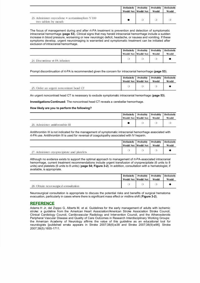
The focus of management during and after rt-PA treatment is prevention and detection of symptomaticintracranial hemorrhage (page 53). Clinical signs that may herald intracranial hemorrhage include a suddenincrease in blood pressure, worsening or new neurologic deficit, headache, or nausea and vomiting. If thesesymptoms develop, urgent neuroimaging is warranted and symptomatic treatment can be initiated after exclusion of intracranial hemorrhage.
Prompt discontinuation of rt-PA is recommended given the concern for intracranial hemorrhage (page 53).
An urgent noncontrast head CT is necessary to exclude symptomatic intracranial hemorrhage (page 53).
Investigations Continued: The noncontrast head CT reveals a cerebellar hemorrhage.
How likely are you to perform the following?
Antithrombin III is not indicated for the management of symptomatic intracranial hemorrhage associated withrt-PA use. Antithrombin III is used for reversal of coagulopathy associated with IV heparin.
Although no evidence exists to support the optimal approach to management of rt-PA-associated intracranialhemorrhage, current treatment recommendations include urgent transfusion of cryoprecipitate (6 units to 8units) and platelets (6 units to 8 units) (page 54; Figure 3-2). In addition, consultation with a hematologist, if available, is appropriate.
Neurosurgical consultation is appropriate to discuss the potential risks and benefits of surgical hematomaevacuation, particularly in cases where there is significant mass effect or midline shift (Figure 3-2).
REFERENCE Adams H Jr, del Zoppo G, Alberts M, et al. Guidelines for the early management of adults with ischemicstroke: a guideline from the American Heart Association/American Stroke Association Stroke Council,Clinical Cardiology Council, Cardiovascular Radiology and Intervention Council, and the AtheroscleroticPeripheral Vascular Disease and Quality of Care Outcomes in Research Interdisciplinary Working Groups:the American Academy of Neurology affirms the value of this guideline as an educational tool for neurologists [published errata appears in Stroke 2007;38(6):e38 and Stroke 2007;38(9):e96]. Stroke
2007;38(5):1655-1711.