predictors of important ct findings and … · kecil untuk menjustifikasikan kegunaan rutin imbasan...
TRANSCRIPT

PREDICTORS OF IMPORTANT CT FINDINGS AND NEUROSURGICAL INTERVENTION IN
MINOR HEAD INJURY
By Dr. LAlLI SURIANI AB. LA TIP
Dissertation Submitted in Partial Fulfillment Of The Requirements For The Degree Of Master Of Medicine
(Radiology)
Universiti Sains Malaysia May 2002
Supervisor: Assoc. Prof. Dr Hj Nurul Azman Bin Ahmad Alias

To
My Husband: Dr Abd. Rahim Bin Abd. Ghani
My sons: Muhamad Luqman & Muhamad Ashraff,
My parents: Hj Abd. Latip & Hjj Maimunah
With Love
ii

Acknowledgements
The author would like to thank the following individuals for their advice, guidance,
comments and support during the preparation of this dissertation and also during the
whole duration she was in the Radiology Department Hospital Universiti Sains Malaysia.
• Associate Prof. Dr. Haji Nurul Azman Bin Ahmad Alias. Supervisor of this study
and Lecturer I Radiologist, HUSM for his guidance during the course of this study
• Dr Abdul Rahman Mohd. Ariff, Lecturer/Radiologist, HUSM for his valuable
time as a second observer.
• My dear teachers: Associate Prof Dr. Ibrahim Lutfi Shuib, Dr Mahayidin
Muhamad, Dr Abdul Kareem, Dr Noreen Norfaraheen Lee Abdullah, Dr Latifah
Mohd Basheer, Lecturers I Radiologist, HUSM and Dr Haji Md Ariff Abas, Ketua
Jabatan, Jabatan Pengimejan Dan Diagnostik Hospital Kota Bharu.
• Dr Syed Hatim@ Nyi Nyi Naing, Head, Unit of Biostatistic I Lecturer.
Department of Community Medicine, for giving invaluable advice on data
analysis.
• Associate Prof. Dr Jafri Malin, the neurosurgeon and his team for the patient's
clinical data.
iii

• Inspector Hashim from Jabatan Traftk, Ibupejabat Polis Kontigen Kelantan, Kota
Bharu for his information on the statistic of road traffic accident in Kelantan.
• Dr Malik Mumtaz, Lecturer I Nuclear Physician, HUSM for reading and
providing comments on the manuscript.
• Colleagues and all staff in the Radiology Department, HUSM; individuals in the
Department of Diagnostic & Imaging, Hospital Kota Bharu and Radiology
Department, Hospital Kuala Lumpur.
• All staff from the record office, HUSM
• Last but not least Dr Abdul Rahim Bin Abdul Ghani, my beloved husband who '
has been very supportive and understanding.
iv

Acknowledgement
List of tables
List of figures
Abbreviations
Abstract
Bahasa Malaysia
English
Section One: Introduction
Introduction
Section Two: Literature review
2.1 Literature review
CONTENTS
2.2 Neuroimaging and minor head injury
Section Three: Aim and Objectives
3.1 Aim of study
3.2 Objectives
3.3 Hypothesis
Section Four: Methodology
4.1 Methodology
4.2 Criteria
4.3 Exlusion criteria
4. 4 Cranial CT scan
v
111
lX
X
X1
Xll
XV
1
3
10
22
22
23
24
24
25
25
4.5 Data collection
4.6 Definition of clinical parameters
4. 7 Classification and defmition of CT findings
4.9 Statistical analysis
Section Five: Results
5.1 General results
5.2 Demography
5.3 Clinical parameters
5.4 Outcome measured
5.5 Univariate Analysis
5.5a Important CT fmdings
5.5b Neurosurgical Intervention
5.6 Multivariate Analysis
5.6a Outcome One
5.6b Outcome Two
Section Six: Discussion
6.1 Demography- age
6.2 Demography- sex
6.3 Demography - racial distribution
6.4 Mechanism of injury
6.5 GCS score
6.6 Outcome measured
6. 6a Important CT findings
vi
26
26
27
35
37
37
43
45
48
48
52
56
56
57
59
60
62
63
63
65
66
66

6.6b Neurosurgical Intervention
6.7 CT findings
6.8 Predictors ofCT findings and neurosurgical Intervention
6.8aGCS
6.8b Skull fracture
6.8c Mechanism of injury
6.9 Significant clinical parameter on univariate only
6.9a Post-traumatic vomiting
6.9b Memory loss I amnesia
6.9c Cranial soft tissue injury
6.9d Abnormal neurological examination
6.10 Other clinical variables
6.10a Loss of consciousness
6.10b Headache
6.1 Oc Seizure
6.1 Od Clinical sign of basal skull fracture
6.1 Oe Drug I Alcohol intoxication
Section Seven: Summary & Conclusion
Summary & Conclusion
Section Eight: Problems and Limitation
Problems & Limitation
Section Nine: Recommendations
Recommendations
vii
69
70
71
71
74
76
78
78
80
81
82
83
83
84
85
86
87
89
92
94

Section Ten: References & Appendices
References
Appendix One: Questionaire
viii
97
102

LIST OF TABLES
Table 2.1: Summary of studies involving patients with minor
head injury
Table 2.2: Summary of studies involving clinical risk factors
in head injury I minor head injury
Table 5.1: Age of patients in years
Table 5.2: Mechanism of injury
Table 5.3: CT interval
Table 5.4: Clinical Parameters of patients with minor head injury
Table 5.5: Frequency of skull fracture
Table 5.6: Association between clinical parameters and
Important CT findings on Univariate analysis
Table 5.7: Association between clinical parameters and
9
20
37
40
42
43
44
50
Neurosurgical Intervention on Univariate analysis 54
Table 5.8: Multiple Logistic Regression On Predictors of Important CT
Findings and the need of neurosurgical Intervention 58
ix

LIST OF FIGURES
Figure 4.1: CT scan showing extradural haemorrhage 30
Figure 4.2: CT scan showing subdural haemorrhage and pneumocephalus 30
Figure 4.3: CT scan showing subarachnoid haemorrhage 31
Figure 4.4: CT scan showing contusion 31
Figure 4.5: CT scan showing intraparenchymal haemorrhage 32
Figure 4.6: CT scan showing midline shift 32
Figure 4.7: CT scan showing non-depressed linear skull fracture 33
Figure 4.8: CT scan showing depressed skull fracture 33
Figure 5.1: Pie chart- racial distribution 38
Figure 5.2: Pie chart- sex distribution 39
Figure 5.3: Bar chart- distribution ofGCS 41
Figure 5.4: Bar chart- Various type of skull fracture 44
Figure 5.5: Pie chart- Percentage of Important CT findings 45
Figure 5.6: Bar chart- Percentage of Important CT findings 46
Figure 5.7: Pie chart- rate of neurosurgical intervention in minor head injury 47
X
ABBREVIATIONS
CONT. Contusion
CT Computed Tomography
CNS Central Nervous System
ED Emergency department
EDH Extradural haemorrhage
GCS Glasgow Coma Scale
IPB Intra-parenchymal haemorrhage
MLS Midline shift
MRI Magnetic Resonance Imaging
No. Number
PNEUMO Pneumocephalus
SAD Subarachnoid haemorrhage
SDB Subdural haemorrhage
Xl

ABSTRAK
Bahasa Malaysia
Tajuk: Faktor klinikal yang meramal penemuan penting dalam imbasan tomografi
berkomputer and keperluan intervensi neuro bagi pesakit kecederaan kepala ringan.
Latarbelakang: Di antara semua kecederaan kepala, kecederaan kepala nngan
merupakan suatu kumpulan kecederaan kepala yang paling kerap berlaku dimana ia
menyumbang sebanyak 70 - 80 % kes yang ditemui di Jabatan Kecemasan. Kecederaan
kepala ringan didefinasikan sebagai seseorang yang mengalami hentakan di kepala yang
mengakibatkan pengsan dan I atau hilang ingatan. Sementara itu, skala koma Glasgow
pula adalah di antara 13 - 15. Penggunaan imbasan tomografi berkomputer ke atas semua
pesakit yang mengalami kecederaan kepala ringan adalah suatu perkara yang kontroversi.
Ini disebabkan peratusan keputusan penting yang di perolehi dari imbasan berkenaan
adalah kecil berbanding dengan kos tinggi yang membabitkan penggunaan imbasan
tomografi berkomputer. Peratusan pesakit yang menjalani intervensi neuro juga amat
kecil untuk menjustifikasikan kegunaan rutin imbasan tomografi berkomputer.
Penggunaan faktor klinikal untuk meramal samada pesakit perlu untuk menjalani
pemeriksaan imbasan tomografi berkomputer dilihat sebagai suatu perkara yang
berpatutan untuk mengurangkan kadar penggunan imbasan tomografi berkomputer ke
atas semua pesakit kecederaan kepala ringan. Faktor-faktor klinikal ini boleh digunakan
oleh para doktor untuk meramalkan penemuan penting basil dari imbasan tomografi
berkomputer and keperluan intervensi neuro.
xii

Objectif & Kaedah: Tujuan penyelidikan ini adalah untuk mengenal-pasti faktor -
faktor klinikal yang boleh meramal penemuan penting basil daripada imbasan tomografi
berkomputer dan keperluan intervensi neuro di kalangan pesakit kecederaan kepala
ringan. Sebanyak 15 faktor klinikal telah di siasat hubung-kaitnya dengan penemuan
penting imbasan tomografi berkomputer dan keperluan intervensi neuro. Data pesakit di
rekodkan samada secara retrospektif atau prospektif. Ramalan faktor klinikal kemudian
dianalisis berdasarkan kaedah analysis "univariate'' yang kemudiannya dilanjutkan
kepada 'Multiple Logistic Regression'.
Keputusan: 94 orang pesakit prospektif dan retrospektif yang mengalami kecederaan
kepala ringan telah dikenalpasti. Daripada jumlah tersebut, 50 kes (53.2%) didapati
mempunyai penemuan penting dari imbasan tomografi berkomputer manakala 19 pesakit
(20.2%) telah dikenal pasti memerlukan intervensi neuro. Terdapat dua faktor klinikal
yang terbukti signifikan di dalam menentukan keputusan imbasan tomografi
berkomputer. Skala koma Glasgow yang kurang dari 15 boleh meramalkan keputusan
penting imbasan tomografi berkomputer sebanyak 18.7 kali lebih berbanding dengan
pesakit yang mempunyai skala coma Glasgow yang penuh. Begitu juga dengan kehadiran
retak tempurung kepala yang boleh meramalkan keputusan penting imbasan tomografi
berkomputer iaitu sebanyak 56 kali lebih berbanding dengan pesakit yang tidak
mengalami retak. Kaedah kecederaan pula memainkan peranan yang penting untuk
meramalkan keperluan intervensi neuro. Kaedah kecederaan selain daripada kemalangan
kenderaan (yakni yang meliputi kecederaan pejalan kaki, jatuh dari ketinggian, terhentak
xiii

objek dan kes 'assault') merupakan faktor klinikal penting yang berkaitan dengan
keperluan intervensi neuro.
Kesimpulan: Skala koma Glasgow kurang daripada 15 dan retak tempurung kepala
merupakan dua faktor klinikal yang penting untuk meramal penemuan penting imbasan
tomografi berkomputer. Saranan telah dibuat supaya kecederaan kepala ringan perlu
untuk direklassifikasikan semula berdasarkan kepada skala koma Glasgow. Kecederaan
kepala ringan perlu hanya untuk pesakit yang mempunyai skala koma Glasgow penuh
iaitu 15 manakala kecederaan kepala ringan yang berisiko tinggi untuk pesakit yang
mengalami skala koma Glasgow antara 13 dan 14. Sekiranya pesakit mengalami
keretakan tempurung kepala atau pesakit yang mendapat kaedah kecederaan seperti yang
tersebut diatas, pesakit adalah berisiko tinggi dan perlu menjalani imbasan tomografi
berkomputer segera jika keadaannya stabil.
xiv

ABSTRACT
English
Topic: Predictors of Important CT fmdings and Neurosurgical Intervention in minor head
InJury.
Background: Of all patients with head injury, minor head injury represents the most
common visitors of the emergency department. It contributed to about 70 - 80 o/o of all
head injury cases that are seen in the Accident and Emergency department. Minor head
injury is generally defined as those with history of blow to the head in which has resulted
in loss of consciousness and I or amnesia with Glasgow Coma Score of 13 to 15. It is
controversial whether cranial CT scan should be performed on all patients, as the yield of
positive CT scan is low as opposed to its high cost. The rate of neurosurgical intervention
is even lower to justify the routine use of CT scan on this patient. The use of clinical
variables as a screening tool before deciding to embark on CT scan is appropriate in order
to reduce the number of CT scan performed on all patients with minor head injury. The
clinical variables can than be used by the attending doctors in predicting important CT
findings and the need of neurosurgical intervention.
Objectives and Method: The aim of this study is to evaluate clinical variables that can
be used to predict important CT findings and the need of neurosurgical intervention
among patient who presented with minor head injury. A descriptive study was conducted
on 94 patients who had history of blow to the head, with Glasgow Coma Score of 13 to
15 and had cranial CT scan examination. There were a total of 15 clinical variables that
XV

were correlated with two outcomes. The outcomes were important CT findings and the
need of neurosurgical intervention. The predictors of important CT findings and the need
of neurosurgical intervention were derived after performing initial univariate analysis,
which was then followed by Multiple Logistic Regression.
Result: There were a total of 94 retrospective and prospective patients with minor head
injury. 50 patients (53.2 %) had important CT findings and 19 patients (20.2 %) had
neurosurgical intervention. There were two significant predictors of important CT
findings. The presence of GCS score of 14 and 13 were associated with nearly 19-fold
increase in the risk of developing important CT findings compared to patients with full
GCS score. 3 out of 4 patients with GCS score of 14 or 13 developed clinically important
brain injury on CT scan examination. Similarly, the present of skull fracture was
associated with 56-time increase risk of developing important CT fmdings. 8 out of 10
patients with skull fracture developed clinically important CT fmdings. Mechanism of
injury was the only predictor of neurosurgical intervention. Non-motor vehicular accident
in which included pedestrian injury, fall from height, falling object, assault cases and
others were associated with four-fold increase risk of needing neurosurgical intervention.
Conclusion: Glasgow Coma Score of less of 15 and skull fractures were significant
clinical variables in predicting important CT findings in patients with minor head injury.
Therefore, proposal was made for reclassifying minor head injury in which it should be
based on the GCS score. Patients with full GCS score of 15 were classified as mild head
injury, while patients with GCS score of 13 and 14 were at higher risk of developing
xvi

brain injury and therefore categorized as "high-risk mild head injury". Those patients
with skull fracture regardless of other clinical findings would automatically qualified the
patient into high-risk group. The high-risk group needed urgent cranial CT scan
examination. Particular mechanism of injury which included pedestrian injury, fall from
height, falling object and assaulted cases predisposed the patient for neurosurgical
intervention and therefore classified as high-risk mild head injury as well.
xvii

SECTION ONE
INTRODUCTION

INTRODUCTION
Head injury is common in modem society. Of all patients with head injury, minor
head injury represents the most common attenders of the emergency department.
Generally, minor head injury is defined as any patient with a history of blow to the head
resulting in loss of consciousness and amnesia with GCS score of 13 - 15. Although
minor head injury represents the majority of head trauma patients, the management of
minor head injury remains controversial and confusing.
There has been much debate on whether to perform cranial CT scan on every
patient with minor head injury. The decision to obtain cranial CT scan in every patient
with minor head injury is due to the anxiety of the clinician missing potentially curable
intracranial haematoma as weli as for the medico-legal consequences. Although most
patients with minor head injury can be safely discharged without sequelae after a period
of observation, there is a group of patients who has significant intracranial injury that
necessitate neurosurgical intervention. Early neurosurgical intervention is important in
improving the prognosis of such patient. Therefore, cranial CT scan is essential to
identify such patient. However, one can argue that the blanket rule of performing cranial
CT scan on every patient with minor head injury is unjustifiable as the rate of positive CT
findings and neurosurgical intervention are low. The low yield of CT scan findings
among patients with minor head injury suggests great potential for reducing the use of
CT. It is clear that CT scanning is an expensive modality used as screening tools and
therefore, more selective use for patient with minor head injury could lead to large
reductions in health-care cost.
1

There is a clear need for a valid and reliable clinical guidelines to allow doctors to
be more selective in the use of cranial CT without compromising the care of the patients
with minor head injury. The used of clinical variables in deciding whether to perform
cranial CT scan is appropriate in order to reduce the number of performing CT scan on all
patients with minor head injury. Clinical variables are identified based on history and
physical examination. A patient who presented with minor head injury can be screened
using the clinical variables. This set of clinical variables can be used by the attending
doctors to predict which patients that need urgent cranial CT assessment. By utilizing this
clinical variable, the patient that needs neurosurgical intervention can be identified. This
rule will allow doctors in emergency department to order cranial CT for their patients
based upon strong evidence and to provide consistent management without jeopardizing
optimum patient care. Without selective guideline, there is very strong evident that the
use of cranial CT for minor head injury will be markedly increase in the years to come
(Stiell et al., 2001 ).
The aim of this study was to identify clinical variables in minor head
injury that could be used to predict significant intracranial injury and the need for
neurosurgical intervention. The main outcome measured the need of neurosurgical
intervention and clinically important brain injury on CT scan.
2

SECTION TWO
LITERATURE
REVIEW

LITERATURE REVIEW
Head injury is a common problem in emergency department. It constitutes about
20- 30o/o of all trauma admission (Fisher et al., 1981, Taheri et al., 1993). In the United
States of America and other western countries, head injury does not only represent a
serious cause of loss of life, it also adds to financial burden of the health care system. The
financial burden is due to the increase in the resources in caring and treating these
patients. In the United States of America, more than 500,000 Americans suffer from head
injury per year (Stein et al., 1993). Similarly, head injury is also a common problem in
United Kingdom where more than 150,000 patients with head injury are admitted to
hospital each year (Thornhill et al., 2000).
In Malaysia, head injury is also a serious health problem. In the year 1998, the
incidence rate of motor vehicle accident in Peninsular Malaysia was 112.3 per 10,000
population (KKM., 2000). The number of casualties due to motor vehicle accident in the
year 1998 was estimated to be around 52,218 (KKM., 2000). From the year 1997- 1998,
accident was the third principal cause of hospitalization in MOH (Ministry of Health)
hospital (KKM., 2000). Accident was the fourth principle cause of death in the year 1997
behind heart disease, septicaemia and cerebrovascular accident (KKM., 2000). In
Hospital Universiti Sains Malaysia, which is a tertiary referral center with a neurosurgical
unit, a total of 570 head injured patients were seen in the year 1999 (Record Office,
HUSM). In the year 2001, a total of 1382 cases were reported to involve in motor vehicle
accident in Kelantan in which 428 cases were fatal (Jabatan Polis Trafik, 2001 ). From
3

this number, 382 cases were reported to suffer from head injury (Jabatan Polis Trafik,
2001).
Serious head injury occurs in only 3 % of non-vehicular and 15% of vehicular
injuries (Lee et al., 1999). Most cases of head injury in the emergency department are
classified as minor or mild head injury. This is the biggest group of patients that will be
seen by the casualty officer. The proportion of patients with minor head injury are
different in different hospital with inter-study variability among different researchers
depending on the population studied and the definition of minor head injury. The
proportion of patients with minor head injury ranges from 70 - 90 % of all the head
injured patient (Kraus et al., 1988, Livingston et al., 1991, Mittl et al., 1994, Gomez et
al., 1996 and Nagy et al., 1999).
In the past 20 years, there have been a lot of controversies regarding minor head
injury on the literature review. Among the controversy is in defining minor head injury
(Stein et al., 1993). The authors had commented in their article that there was no
consensus on the term of the least severe fonn of head injury - whether the least severe
form should be termed as mild, minor, grade 1, low risk or class 1. Some studies defined
mild head injury as a brief loss of consciousness after a blow to the head, whereas other
studies graded the degree of injury by the length of posttraumatic amnesia (Hsiang.,
1997).
4

Generally, minor head injury is defined as a patient with a blow to the head with a
history of loss of consciousness, amnesia or disorientation and Glasgow Coma Score of
13- 15 (Shackford et al., 1992, Stein et al., 1992, Mittl et al., 1994, Gomez et al., 1996,
Borczuk., 1997 and Nagy et al., 1999). Therefore a patient with a Glasgow Coma Score
of 13 to 15 is classified as having minor head injury. However, this term is arbitrary and
misleading (Hsiang et al., 1997). This is because, the term minor head injury should not
be associated with significance consequences and long-term sequelae. In reality, studies
have proven that there is convincing evidence that considerable amount of disability can
occur after the so-called "minor head injury" (Hsiang et al., 1997). It is misleading to
label all of these patients with minor head injury because the word minor means not
serious (Hsiang et al., 1997). There is heterogeneous pathophysiology among patients
with GCS score of 13 to 15 (William et al., 1990, Culotta et al., 1996, Hsiang et al.,
1997). The classification of minor head injury has been constantly revised and in 1997,
Hsiang had proposed that the definition of minor head injury should be divided into mild
head injury and high-risk mild head injury (Hsiang et al., 1997).
Patients with high-risk mild head injury may be at an increased risk of
developing complications that may need neurosurgical intervention Patients with GCS
score of 13 or 14 and those with scores of 15 who exhibited acute radiographic
abnormalities according to the above authors should be classified as high .. risk mild head
injury (Hsiang et al., 1997). This is in agreement with previous study done by William in
1990 that reclassified minor head injury into complicated and uncomplicated depending
5

on the GCS score and radiographic findings (which include skull fracture and focal brain
lesion).
The importance of proper classification of minor head injury has implication to
the management of minor head injury. Patients classified as mild or minimal head injury
can be discharged home while patients with high-risk mild head injury should be
admitted to the hospital (Hsiang et al., 1997). By using this system, the authors argued
that tremendous amount of resources could have been saved (Hsiang et al., 1997).
Although minor head injury represents the most common group of head injury
that is seen in the casualty, the management of minor head injury is still confusing. There
has been a lot of argument in the literature on how to manage minor head injury. The goal
of any management scheme should be to minimize mortality and morbidity at a
reasonable cost and effort (Stein et al., 1993). In any head injury patient, the management
is directed towards identifying which patient that need prompt surgical intervention and
therefore urgent referral to the neurosurgical team. Because missed intracranial
haematomas are potentially devastating, the aim is to identify these as early and as
accurate as possible (Stein et al., 1993). The other factor is the severity of the medico-
legal consequences should the identification of the neurosurgical candidate is not timely.
There is no argument that patient with moderate and severe head injury will need
urgent Computed Tomography assessment to exclude any intracranial haemorrhage
(Stein et al., 1992). However, thes~ population only represents 10- 15 o/o of all patient \
6

with head injury (Fisher., 1981) As mentioned earlier, the main bulk of the head injured
patients are those who sustained minor head injury. It is not cost effective to perform
Computed Tomography on all patients with minor head injury because of the low yield in
the positive intracranial findings. The rate of those with minor head injury that need
neurosurgical intervention is even lower to justify the blanket rule of doing Computed
Tomography on all patients. On the other hand, excess mortality from head injury
actually came from the lowest risk group i.e. those patients with minor head injury
(Klauber et al., 1989). The fore mention authors argued that the improvements in
mortality would not come from added technology and "advance care'' of patients with
severe head injury, but from identifying and preventing deterioration in patients who
appear to be at low risk (Klauber et al., 1989).
The rate of positive CT findings in patient with minor head injury varies in
different studies. It ranges from 3% to 41 %(Dacey et al., 1986, Feuerman et al., 1988,
Livingston et al., 1991, Mohanty et al., 1991, Harad et al., 1992, Mikhail et al., 1992,
Shackford et al., 1992, Stein et al., 1992, Jeret et al., 1993, Borczuk., 1995, Miller et al.,
1996, Nagy et al., 1999, Haydel et al., 2000 and Stiell et al., 2001). Refer to table 2.1 for
summary of the studies in minor head injury. Comparison between different studies is
difficult because of the different in study design. The definition of the studied population
with minor head injury is also not uniform, therefore making comparison difficult. For
example, some of the authors ((Dacey et al., 1986, Feuerman et al., 1988, Harad et al.,
1992, Mikhail et al., 1992, Stein et al., 1992, Shackford et al., 1992, Borczuk., 1995 and
Stiell et al., 2001) included all patient with GCS score of 13- 15 while other researchers
7

only included GCS score of 15 (Jeret et al., 1993, Nagy et al., 1999, Haydel et al., 2000).
Similarly, another group of researchers only included GCS score of 14 -15 (Livingston et
al., 1991 and Mohanty et al., 1991). Computed Tomography was not performed on all of
the patients with minor head injury. The range of performing CT scan in these patients
ranged from as low as 11% to 1 OOo/o of their entire head injury patients. Because not all
of the patients with minor head injury were studied with CT scan it might therefore
under-estimated the true rate of positive CT findings.
8
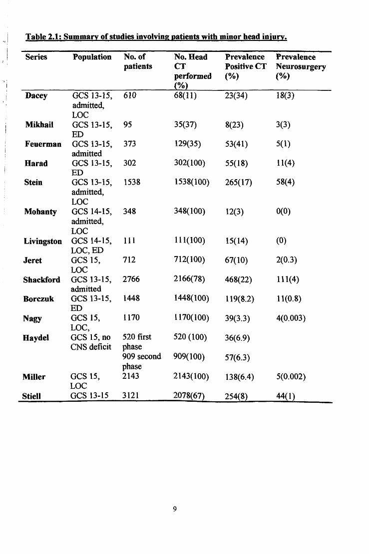
Table 2.1: Summaa of studies involving J!&tients with minor head injun:.
Series Population No. of No. Bead Prevalence Prevalence patients CT PositiveCT Neurosurgery
., '
performed (%) (%) %
Dacey GCS 13-15, 610 68(11) 23(34) 18(3) admitted, LOC
Mikhail GCS 13-15, 95 35(37) 8(23) 3(3) ED
Feuerman GCS 13-15, 373 129(35) 53(41) 5(1) admitted
Harad GCS 13-15, 302 302(100) 55(18) 11(4) ED
Stein GCS 13-15, 1538 1538(100) 265(17) 58(4) admitted, LOC
Mohanty GCS 14-15, 348 348(100) 12(3) 0(0) admitted, LOC
Livingston GCS 14-15, Ill 111(100) 15(14) (0) LOC,ED
Jeret GCS 15, 712 712(100) 67(10) 2(0.3) LOC
Shackford GCS 13-15, 2766 2166(78) 468(22) 111(4) admitted
Borczuk GCS 13-15, 1448 1448(100) 119(8.2) 11(0.8) ED
Nagy GCS 15, 1170 1170(100) 39(3.3) 4(0.003) LOC,
Haydel GCS 15, no 520 first 520 (100) 36(6.9) CNS deficit phase
909 second 909(100) 57(6.3) phase
Miller GCS 15, 2143 2143(100) 138(6.4) 5(0.002) LOC
Stiell GCS 13-15 3121 2078(67) 254(8) 44(1)
9

2.2 NEUROIMAGING AND MINOR HEAD INJURY.
In minor head injury, history and physical examination still remain the most
important means of identifying those at risk for a significant head injury. However,
imaging of the skull and brain can offer detail information about the anatomic site of
injury and prognosis (Yealy et al., 1991). Roentgenography was first developed in 1895.
Since than until 1970s, imaging of head trauma patient had been relied on plain skull
radiograph. In 1920's pneumocephalography became available to help diagnose and
localize intracranial lesions, although this technique was only helpful in the presence of a
large mass lesions. With the start of CT era in 1970s, the utilization of plain skull
radiograph has reduced and pneumocephalography has become historic.
The use of plain radiograph in minor head injury has received much attention in
the literature review. Plain radiograph of the skull is widely available and relatively in
expensive than CT scan. It is a useful imaging tool with good sensitivity and specificity
to diagnose bony fracture (Y ealy et al., 1991 ). In many emergency rooms, skull
radiographs are taken routinely in almost all patients who sustained acute head injury
regardless of their clinical condition. Depressed or open skull fracture is often well
identified by plain skull radiograph. Aside from the basilar fracture, skull radiograph can
visualized most non-displaced linear fracture when interpreted by an experienced
radiologist or emergency doctor. However, some researchers argued the usefulness of
performing routine skull radiography on every patient with head injury. This group of
researchers argued that by identifying skull vault fracture, it is often necessary to also
10

perform CT scan on these patients. Skull radiograph would result in delaying CT
examination on these patients. Furthermore, skull radiograph does not provide details on
the intracranial injury and the anatomical site to guide the neurosurgeon should the
patients need neurosurgical intervention (Yealy et al., 1991).
An imaging procedure that is more rapid such as skull radiograph but insensitive
to provide evident for parenchyma brain damage only delays the diagnosis and treatment
of the patient (Lee et al., 1999). Although patients who sustained skull fracture are at
increase risk of developing an intracranial injury, it is also common to find patients with
normal skull radiograph and yet developed intracranial haematoma on CT scan.
Therefore, there is limited role of skull radiography as a screening tool in head injury.
Furthermore, the discovery of incidental fracture on skull radiograph in a neurologically
intact patient rarely changes the treatment (Perini et al., 1984).
The reason why plain skull radiography is used in patient with minor head injury
is due to the following reason. In patient with no impairment of consciousness, a normal
physical examination and a normal skull series, the risk of intracranial injury is less than
0.001o/o (Yealy et al., 1991). This data has been used to justify the routine use of plain
skull radiograph in all patients with head injury in order to identify those who can safely
discharged home (Yealy et al., 1991).
After the advent of CT scan, the role of skull radiograph has become less popular.
c·1 scan has become the imaging modality of choice in acute head injured patient. It is
i i

highly specific for bony and intracranial lesions (Perini et al., 1984 and Y ealy et al.,
1991 ). Its role in identifying those lesions, which requires urgent referral to the
neurosurgical team and urgent neurosurgical intervention, are well established. CT scan
is also useful as an imaging tool to determine the prognosis of patients who involved in
head injury. A nonnal CT scan in patients with a nonnal neurological examination has an
excellent negative predictive value for delayed neurological complication. Another
advantage of CT scan is that it showed basilar skull fracture that is not demonstrable on
plain skull radiograph (Lee et al., 1999). It is also the method of choice for demonstrating
fracture of the facial bones, including the paranasal sinus and orbits (Lee et al., 1999). In
a typical head injury, window levels are optimized for the brain (brain window), used to
assess the presence of blood adjacent to the inner table of the skull (subdural or epidural
haematoma in subdural window) and lastly bone window to see if there is any skull vault
fracture (Lee et al., 1999).
The role of cranial CT scan in minor head injury remains controversial and
confusing. Some authors claim there should be blanket rule of performing CT scanning in
all patients involved in minor head injury. This group of authors argued that because the
rate of positive CT findings can be as high as 20o/o, CT scan must be performed on every
one who presented with minor head injury. Other investigators argued that because the
rate of neurosurgical intervention is actually low, the universal approached of performing
CT scan is not cost effective. The increase in the demand of CT request will put an extra
load to the radiology deparbnent.
12

There are many studies performed to look at the indication of CT scanning in
minor head injury. Some of the studies are summarised in Table 2.1. The following
authors have supported the notion of performing Computer Tomography on every minor
head injury patients. For example, Dacey et al. in 1981 prospectively evaluated 610
patients with a transient loss of consciousness and GCS score of 13 and above. Only 11 o/o
of the patients underwent CT scanning. In the above study, 34% of the patients had
positive CT findings and 3% required neurosurgical intervention. This is the largest
percentage of positive CT findings, but it must be understood that it represents a selection
bias in test ordering (Borczuk, 1997). The conclusion from this study was that -
discharging all patients with a GCS score of 15 and a normal CT were much cheaper that
routinely admitting these patients with mild head injury.
Stein et al ( 1991, 1992, 1993) were strong advocates for obtaining a cranial CT in
patients with mild head trauma and LOC, claiming the test serves to identify the patients
requiring surgery, as well as define the low-risk patients who do not need hospital
admission for observation. The authors reviewed 1538 patients who were admitted to the
hospital in a 2 years period (Stein et al., 1992). Contrary to Dacey et al, the above authors
performed CT scanning on all patients who presented with history of LOC or amnesia, no
matter how brief. Positive CT findings were identified in 17.2% of their population. The
patients who had GCS score of 13 had the highest rate of positive CT findings. The rate
of positive CT findings for patients with GCS score of 13, 14 and 15 were 37.5o/o, 24.2o/o
and 13.2% respectively. Of these patients, 4% required neurosurgical intervention (Stein
et al., 1992). The authors emphasized that because CT scan can provide more anatomic
13

information than radiographs, radiographs were not indicated in the evaluation of mild
head injury (Stein et al., 1992). Similar to Dacey, the population of patients in the study
done by Stein et al (1992) represented only those who were admitted to the hospital and
therefore, may not actually reflected the true number of patients who were discharged and
not admitted.
Other studies that were supportive of routinely performing CT scan on minor head
injury are similar in term of the rate of positive CT findings. This study was done by
Harad et al. (1992) who reported abnormal CT findings in 18 % and neurosurgical
intervention in 4% of the total patients. Similarly Livingston et al. (1991) evaluated 111
patients who had GCS score of 14 and 15, a history of LOC and underwent cranial CT
scanning. 14% of the patients with minor head injury in their population had positive CT
findings. Again the above authors concluded that their study supported the use of cranial
CT scan on all patients with minor head injury in order to identify those who required
neurosurgical intervention and those who could be safely discharged home. Because there
was no predictive characteristic that could identify the 14o/o of their patients with positive
CT findings, the author concluded that CT scan should be perfonned on all patients with
LOC or amnesia. In addition, they recommended that patient with normal neurological
examination and negative CT findings does not need admission at all. This observation is
supported by Klauber et al. ( 1989) who found that observation in the hospital and
frequent neurological checks were actually not practiced. As high as 50o/o of their patients
admitted to the surgical floors had no records of checks being done. In addition, the
14

frequency of observation was frequently insufficient to detect deterioration (Klauber et
al., 1989).
Jeret and colleagues in 1993 conducted a study on 712 patients with minor head
injury who presented with LOC and normal GCS score. The authors concluded that
regardless of age, mechanism of injury or clinical findings, intracranial lesions could not
be completely excluded clinically on bead-trauma patients who had loss consciousness or
amnesia, even if the GCS score was normal (Jeret et al., 1993). Again the above authors
advocated for a routine CT scanning in minor head injury.
Another investigator further emphasized the need of cranial CT scanning in minor
head injury as a screening tool to determine which patient can be safely discharged.
Shackford et al. (1992) studied 2766 patients with isolated mild head injury who
presented to seven trauma centers. They concluded that because 1 in 5 patients with
minor head injury developed positive CT findings, cranial CT scanning should be used
routinely. CT scan was essential especially to patients with GCS score of 13 because 1 in
3 would have positive CT findings and about 1 in 10 would require craniotomy. They
also supported the idea of discharging patients with normal neurological examination and
negative CT scan, as it was more cost-effective than routinely admitting these patients
(Shackford et al., 1992).
Contrary to the idea of performing universal cranial CT scan on all minor head
injury patients, there are few others who disagree that patients with minor head injury
15

would require cranial CT scan at all. Mohanty in 1991 studied which group of patients
with minor head injury that did not require cranial CT scanning. In the above study,
patient included was limited to those with age 18 years and above, had a history of minor
head injury, remains neurologically stable for 20 minutes on arrival to the casualty and
had GCS score of 13 with no evident of basal skull fracture. Only 0.03% of the patients
exhibited abnormal CT scan and all had uneventful hospital admission. Because CT scan
did not have any prognostic or therapeutic on the above patients, the author concluded
that routine CT scan for minimal head injury was an inefficient use of personnel and
equipment. The above patient could have gone home, and been observed instead of
undergoing CT scan, because there were no intervention other than observation. Taheri et
al. (1993) also advocated discharge from the casualty department without radiologic
evaluation. In the above study, all the neurosurgical patients with initial GCS score of 15
(5 out of 310 or 0.016%) were identified by abnormal neurological deficit or a positive
skull radiograph. Thus, the above authors believed it would be safe to discharge a patient
with GCS score of 15, a negative skull radiograph and no neurological deficit.
Duss et al. in 1994 studied a large number of patients in Denmark in which the
authors concluded that it was safe to exclude neuroimaging in primary assessment of
patients with minor head injury and rely instead on clinical criteria (Duss et a., 1994 ).
Minor head injury in the above study was defined as individual who can talk and walk
that presented to the hospital. This entry criterion makes it difficult to compare with other
study that uses GCS score or LOC as main criteria of minor head injury.
16

So far, what have been discussed were two different schools of thought regarding
the usage of cranial CT in minor head injury. The first half of the discussion are the group
of researchers whom advocated routine cranial CT scan for every minor head injury and
the second group of researchers dismissed the idea of performing routine CT scanning. In
evaluating patients with minor head injury, doctors need to define the goal that they wish
to achieve (Borczuk., 1997). If the goal is to identify patients who need neurosurgical
intervention, than based on the low prevalence of neurosurgical intervention of< 0.1% in
patients with GCS score of 15, than one can argued that there is no benefit of performing
routine cranial CT scan (Borczuk., 1997). On the other hand, if the goal is to identify
those patients with abnormal CT findings, with the purpose of prescribing anti-convulsant
therapy or other medical therapy, clearance for general anesthesia or post-concussion
rehabilitation program, then routine CT scanning will need to be performed (Borczuk.,
1997).
There are other researchers who rely on clinical decision criteria to order CT scan
in minor head injury. Many studies have looked into this issue in order to identify
significant clinical variable that can predict positive CT findings. The clinical variables or
parameters that were chosen are derived either from the history or physical examination.
These clinical variables are than studied for their sensitivity or specificity in predicting
positive CT findings. The summary of studies done by different researchers is shown in
Table 2.2. Reinus et al. reviewed 373 head trauma patients and identified the following
combination that had association with positive CT findings: 1) positive neurological
examination, 2) intoxication, 3) history of amnesia and 4) history of a focal deficit. By
17
using these clinical variables, the author claimed that they were able to predict positive
intracranial findings with sensitivity of 90.9% and specificity of 65.6o/o and a negative
predictive value of98.1% (Reinus et al., 1993).Jeret et al. in 1993 also looked at clinical
variables that were associated with positive CT findings. The author found that older age,
white race; signs of basilar skull fracture, pedestrian involved in head injury and
assaulted cases were the significant risk factors that predict positive intracranial findings.
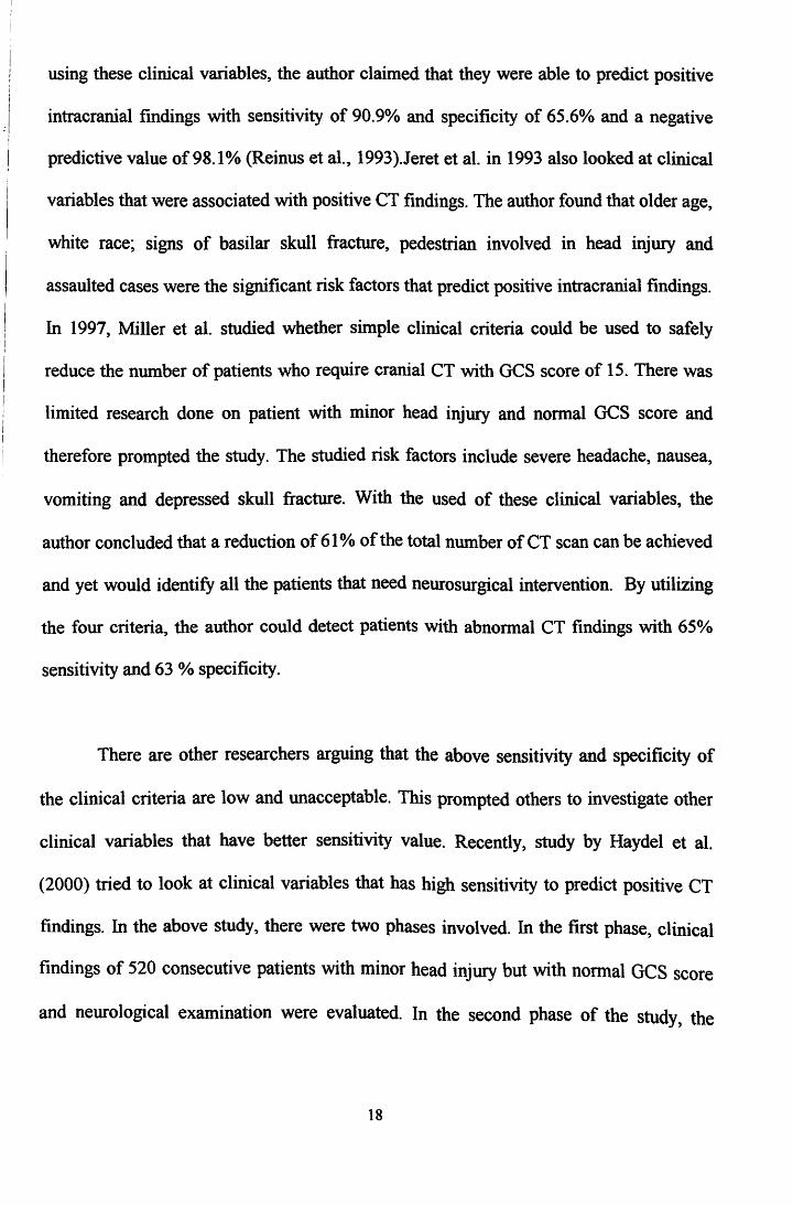
In 1997, Miller et al. studied whether simple clinical criteria could be used to safely
reduce the number of patients who require cranial CT with GCS score of 15. There was
limited research done on patient with minor head injury and normal GCS score and
therefore prompted the study. The studied risk factors include severe headache, nausea,
vomiting and depressed skull fracture. With the used of these clinical variables, the
author concluded that a reduction of 61% of the total number of CT scan can be achieved
and yet would identify all the patients that need neurosurgical intervention. By utilizing
the four criteria, the author could detect patients with abnormal CT findings with 65o/o
sensitivity and 63% specificity.
There are other researchers arguing that the above sensitivity and specificity of
the clinical criteria are low and unacceptable. This prompted others to investigate other
clinical variables that have better sensitivity value. Recently, study by Haydel et al.
(2000) tried to look at clinical variables that has high sensitivity to predict positive CT
findings. In the above study, there were two phases involved. In the first phase, clinical
findings of 520 consecutive patients with minor head injury but with normal GCS score
and neurological examination were evaluated. In the second phase of the study, the
18

sensitivity and specificity of the criteria for predicting a positive CT scan were evaluated
in 909 patients. Patients with positive CT scans had one or more of the following seven
findings: headache, vomiting, age over 60 years, drug or alcohol intoxication, physical
evidence of trauma above clavicle and seizure. Using these clinical variables, the
sensitivity of 100% was achieved which was far higher than the previous study.
Therefore, the author concluded that in evaluating patients with minor head injury, the
used of cranial CT scan could be safely limited to those who had certain clinical findings
(Haydel et al., 2000).
Similar study, which showed high rate of sensitivity, was just published recently.
The study was conducted in emergency department of ten large Canadian hospitals
involving a large population of minor head injury patients (Stiell et al., 2001). The author
had developed the Canadian CT head rule, which identified five high risk factors,
associated with positive CT findings. The rule consists of failure to reach GCS of 15
within 2 hours, suspected open skull fracture, any sign of basal skull fracture, vomiting of
more than 2 episodes, or age of more than 65 years. The authors also identified medium
risk factors, which included amnesia before impact of more than 30 minutes and
dangerous mechanism of injury. These high risk factors were 100% sensitive for
predicting the need of neurosurgical intervention and would require only 32% of patients
to undergo CT. Please refer to Table 2.2 for the summary of the studies investigating the
clinical risk factors.
19
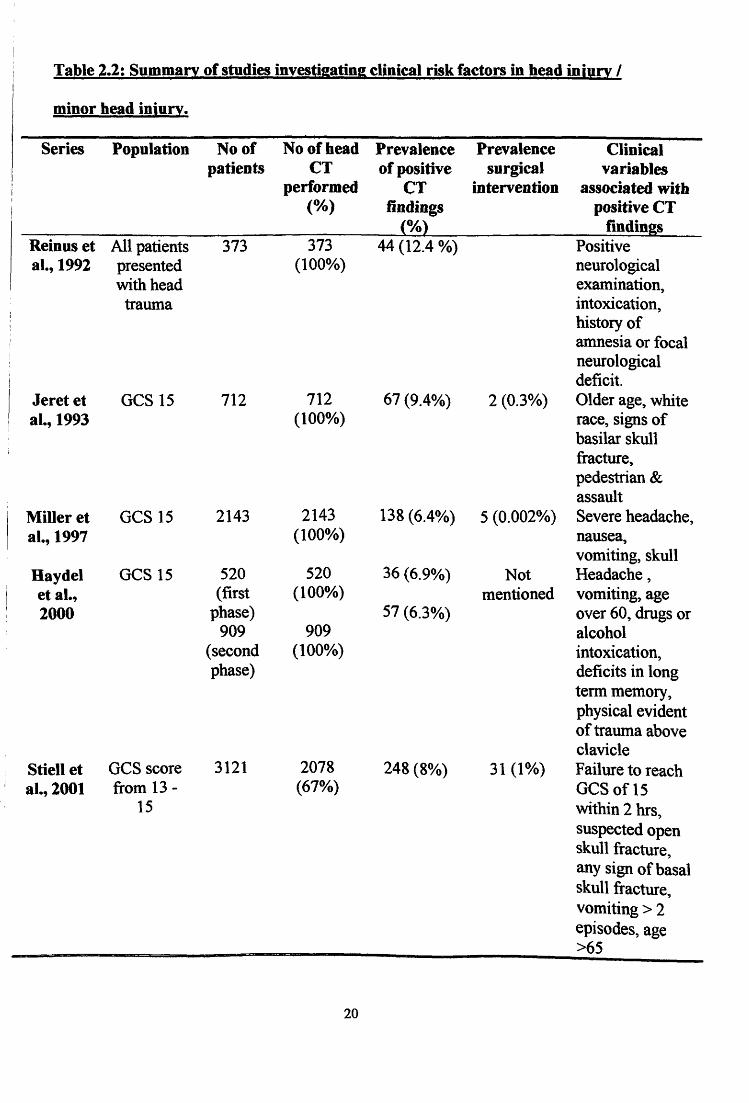
Table 2.2: Summary of studies investigating clinical risk factors in head injury I
minor head injun.
Series Population No of Noofhead Prevalence Prevalence Clinical patients CT of positive surgical variables
performed CT intervention associated witb (%) findings positive CT
{%) findings Reinus et All patients 373 373 44 (12.4 %) Positive al., 1992 presented (100%) neurological
with head examination, trawna intoxication,
history of amnesia or focal neurological deficit.
Jeret et GCS15 712 712 67 (9.4%) 2 (0.3°/o) Older age, white al., 1993 (lOOo/o) race, signs of
basilar skull fracture, pedestrian & assault
Miller et GCS15 2143 2143 138 (6.4o/o) 5 (0.002%) Severe headache, al., 1997 (100%) nausea,
vomiting, skull Haydel GCS15 520 520 36 (6.9%) Not Headache, et al., (first (100%) mentioned vomiting, age 2000 phase) 57 (6.3%) over 60, drugs or
909 909 alcohol (second (100o/o) intoxication, phase) deficits in long
term memory, physical evident of trauma above clavicle
Stiell et GCS score 3121 2078 248 (8%) 31 (1%) Failure to reach al., 2001 from 13- (67%) GCS of15
15 within 2 hrs, suspected open skull fracture , any sign of basal skull fracture, vomiting> 2 episodes, age >65
20

The role of Magnetic Resonant Imaging in minor head injury deserves to be
mentioned. Magnetic Resonant Imaging (MRI) in head injury has evolved since the past
10 years. It is valuable in imaging traumatic brain injury due to several reasons. The
multiplanar capability of MRI is useful to separate cortex from extra ... axial blood
collections in the subdural and epidural spaces that lie at the vertex and the skull base.
This area ofbleeding can sometime be missed on axial CT scanning (Lee et al., 1999). In
addition, MRI is quite useful to show non-haemorrhagic contusion that can occur in
diffuse axonal injury where CT scan is less sensitive to pick up these lesions (Lee et al.,
1999). MRI is also very sensitive to pick up small bleeding which can be missed on the
CT scan. However, MRI is inferior to CT scan in cases of acute subarachnoid
haemorrhage less than 24 hours. Skull vault fracture is also poorly detected on MRI.
Long acquisition time is another problem in acute head trauma patients. Time is a
crucial factor in head injury. Image quality is degraded by motion and the scans may be
uninterpretable (Lee et al., 1999). Motions can be controlled with sedation, however,
sedation will eliminate the ability to assess the neurological sign. When this patients need
to be intubated and paralyzed due to the head injury, monitoring equipment and
intubation devices need to be compatible with the requirement of the MR scanner and this
can be a problem as well (Lee et al., 1999). Electronic monitoring equipment may
become a problem when it causes interference to the signal of the scanner and extraneous
radio frequency source can degrades the image quality (Lee et al~ 1999).
21

SECTION THREE
AIMS
&
OBJECTIVES

3.1 AIM OF THE STUDY
The aim of this study is to identify the clinical variables that predict positive CT
findings and the need of neurosurgical intervention in patients with minor head injury
who underwent cranial CT scan examination. It is not the intention of this study to
analyse new factors that predict positive CT findings and neurosurgical intervention. The
focus of this study is to analyse the association between various clinical variables with
positive CT findings and neurosurgical intervention in reference to the local surrounding.
The results obtained were then compared to other established researchers.
3.2 OBJECTIVES
1. To determine the rate of positive CT findings and the need of neurosurgical
intervention among patients who presented with minor head injury.
2. To determine the association between a set of defined clinical variables and
positive CT findings.
3. To determine the association between a set of defined clinical variables and
neurosurgical intervention.
4. To develop a screening tool in predicting positive CT findings and the need of
neurosurgical intervention among patients with minor head injury.
22

3.3 HYPOTHESIS
Null hypothesis
1. There is no correlation between clinical variables and significant CT
findings in patients with minor head injury.
2. There is no correlation between clinical variables and the need for
neurosurgical intervention in patients with minor head injury.
23

SECTION FOU~ ~t
METHODOLOGY

4.1 METHODOLOGY
This was a descriptive study conducted on 94 cases of minor head injury
patients treated in Hospital Universiti Sains Malaysia. The period of the study was from
January 1999 till June 2001. Of the 94 cases, 17 cases were retrospective cases (from 1st
January 1999 to 31st August 1999) and the remaining numbers (77 cases) were
prospective cases. Study period for the prospective cases were taken from 1st Sept 1999
till 30th June 2001. The study sample comprised of all patients with minor head injury,
which was defined as patient with a blow to the head with initial Glasgow Coma Score of
13 - 15 and underwent cranial CT scan examination. The sampling of patients was taken
according to the Non-Probability Sampling whereby all patients who met the criteria for
the study population would be included in this study. Such patients were identified from
the Department of Radiology database. Once identified, their medical records were traced
1 from the medical record office for further data gathering.
4.2 CRITERIA
The inclusion criteria for this study were:
1. 12 years old and above
2. Patients with minor head injury who presented with history of blow to the
head and Glasgow Coma Score of 13 to 15
3. All patients had cranial CT scanning on admission.
24