pre-operative planning for mandibular reconstruction - a full digital planning workflow resulting in...
TRANSCRIPT
RESEARCH Open Access
Pre-operative planning for mandibularreconstruction - A full digital planning workflowresulting in a patient specific reconstructionHarald Essig†, Majeed Rana*†, Horst Kokemueller, Constantin von See, Martin Ruecker, Frank Tavassol andNils-Claudius Gellrich
Abstract
Objectives: Reconstruction of large mandiblular defects following ablative oncologic surgery could be done byusing vascularized bone transfer or, more often, primarily with simultaneous or delayed bone grafting, using loadbearing reconstruction plates. Bending of these reconstruction plates is typically directed along the outer contourof the original mandible. Simultaneously or in a second operation vascularized or non-vascularized bone is fixed tothe reconstruction plate. However, the prosthodontic-driven backward planning to ease bony reconstruction of themandible in terms of dental rehabilitation using implant-retained overdentures might be an eligible solution. Thepurpose of this work was to develop, establish and clinically evaluate a novel 3D planning procedure formandibular reconstruction.
Materials and methods: Three patients with tumors involving the mandible, which included squamous cellcarcinoma in the floor of the mouth and keratocystic odontogenic tumor, were treated surgically byhemimandibulectomy.
Results: In primary alloplastic mandible reconstruction, shape and size of the reconstruction plate could bepredefined and prebent prior to surgery.
Clinical relevance: This study provides modern treatment strategies for mandibular reconstruction.
Keywords: Mandibular reconstruction, backward planning, patient specific implant, computer-assisted surgery
IntroductionIndications for partial and total mandibulectomy includemalignancies especially squamous cell carcinomas(SCC), benign tumors such as ameloblastomas andsequelae of radiotherapy such as osteoradionecrosis.Defect size in ablative surgery mainly depends on dig-nity and dimensions of the pathology. Following mandi-bulectomy, reconstruction of the mandible is mainlydone with vascularized bone grafts or alloplastic materi-als (reconstruction plates). However, irrespective of thetype of primary hard tissue reconstruction is simulta-neous adequacy of the soft tissue reconstruction equallyimportant: only a well vascularized soft tissue envelope
allows for a successful hard tissue reconstruction. Evenlarge series of mandibular reconstruction after ablativesurgery or major trauma in literature show that oralrehabilitation of patients with large segmental defects isstill a challenge [1-4]. Besides soft tissue inadequaciesdue to ablative procedures and scarring, the method ofbony reconstruction plays an important role on thefunctional and aesthetic result [2,5-8]. Preferred donorsites for vascularized bone with good survival rates arethe fibula, iliac bone and scapula [9-11]. Limitations interms of either primary or secondary dental implant-based rehabilitations are especially the lack of recreatingthe alveolar height and following adverse crown-implantratio. But also the lateral and sagittal extension of thebone volume determines the success rate of dentalimplants and therefore is crucial for oral rehabilitationin cancer patients [12]. Some authors prefer secondary
* Correspondence: [email protected]† Contributed equallyDepartment of Oral and Maxillofacial Surgery, Hannover Medical School,Hannover, Germany
Essig et al. Head & Neck Oncology 2011, 3:45http://www.headandneckoncology.org/content/3/1/45
© 2011 Essig et al; licensee BioMed Central Ltd. This is an Open Access article distributed under the terms of the Creative CommonsAttribution License (http://creativecommons.org/licenses/by/2.0), which permits unrestricted use, distribution, and reproduction inany medium, provided the original work is properly cited.
mandibular reconstructions with free bone grafts whichprovide adequate volume to insert implants [12-14]. Aprerequisite to apply this method is a secure vascular-ized soft tissue bed.Pre-operative planning is highly-developed [15-17] and
image-guided surgery well-established. An ongoing issuein planning procedures is that virtual reconstruction isbased on a true-to-original outer contour reconstruction[18]. However, dimension limitations of bone grafts,especially in height and width, do not match withprosthodontic needs and often result in unavoidable mal-positioned dental implants with sequentially lateral cross-bite situation. Furthermore, dysbalance in soft-tissue-to-bone-to-implant relationship result in later periimplanta-tis with consecutive early to late implant loss. Aim of thisstudy is to implicate the ideal, i.e. prosthodontic drivenplanning and positioning of bone grafts in the planningprocedure respecting the desired implant position: back-ward planning independent of the reconstructive techni-que. This requires patient specific pre-bending of thereconstruction plate to guide the bone graft into thedesired position. So the outer contour of the mandible isnot anymore the basis for bending the plate.
Materials and methodsThree patients with tumors involving the mandible,which included squamous cell carcinoma in the floor ofthe mouth and keratocystic odontogenic tumor, weretreated surgically by hemimandibulectomy. Writteninformed consent was obtained from the patient. Thepatient’s preoperative demographic data are summarizedin Table 1.
Virtual Planning TechniqueThe imaging and planning platform used in this studywas iPlan 3.0 (Brainlab®, Feldkirchen, Germany). Thisplatform allows for alignment of the DICOM data set,viewing and analysis and provides 3D tools for the plan-ning procedure such as mirroring tools, autosegmenta-tion with atlas-function and a three-dimensionalmodification tool named SmartShaper. iPlan enablesimport of different imaging modalities and allows toautomatically image-fuse these data. That facilitates notonly the pre-operative analysis of the data but also thepost-operative quality control. Furthermore this platformallows for intra-operative navigation of the virtual plan
which is important for midface and skullbase procedures,however, this was not used in the presented cases.The basic data used for the virtual planning included the
pre-operative computed tomography (CT) imaging datawith a maximum slice thickness of 1 mm. The data weretransferred on the computer and reconstructed into 3Dimages. Different planning modalities include mirroringtools (case 1), autosegmentation tools (case 2) and thebackward planning tool (case 3). The backward planningis mainly based on size-variable ideal dental arch modelswith adjustable and truly designable virtual hollow cylin-ders. These hollow cylinders define not only the implantposition but also the needed peri-implant bone volume.Thus desired bone position as a required subvolumewithin the later bone grafts for later insertion of dentalimplants could be visualized and already considered dur-ing the primary alloplastic reconstruction of the mandible.Outcome of the virtual planning is in each case a vir-
tual template of the mandible (STL-file format) for pro-duction of a corporeal stereolithographic model. Afterprototyping of the model (Phacon GmbH, Leipzig, Ger-many) a conventional 2.4 Compact UniLOCK recon-struction plate (Synthes®, Umkirch, Germany) was bentto bridge the prospective mandible defect.After ablative surgery was performed, the preformed
reconstruction plate was used for the osteosynthesis. Atthe time of secondary reconstruction with free iliac bonegraft the individual reconstruction plate gives the surgeona clear direction where the bone should be ideally placed.The following case studies demonstrate the planning
possibilities, firstly starting with an unilateral defectusing mirroring tools and patient specific reconstructionplate (case 1), secondly using the autosegmentation tovirtual reconstruct the mandible (case 2) and finally pre-senting the prosthodontic driven “backward planning”for optimized bone graft position (case 3).
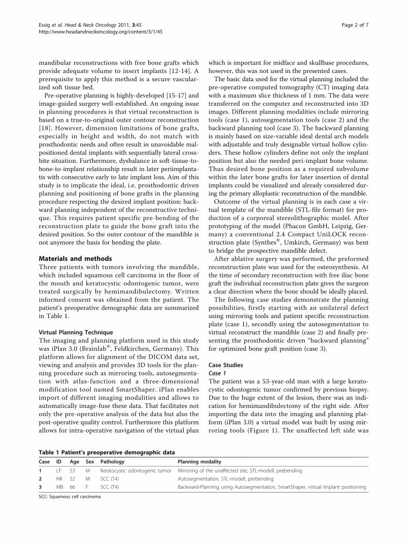
Case StudiesCase 1The patient was a 53-year-old man with a large kerato-cystic odontogenic tumor confirmed by previous biopsy.Due to the huge extent of the lesion, there was an indi-cation for hemimandibulectomy of the right side. Afterimporting the data into the imaging and planning plat-form (iPlan 3.0) a virtual model was built by using mir-roring tools (Figure 1). The unaffected left side was
Table 1 Patient’s preoperative demographic data
Case ID Age Sex Pathology Planning modality
1 LF 53 M Keratocystic odontogenic tumor Mirroring of the unaffected site, STL-modell, prebending
2 NK 52 M SCC (T4) Autosegmentation, STL-modell, prebending
3 MB 66 F SCC (T4) Backward-Planning using Autosegmentation, SmartShaper, virtual Implant positioning
SCC: Squamous cell carcinoma
Essig et al. Head & Neck Oncology 2011, 3:45http://www.headandneckoncology.org/content/3/1/45
Page 2 of 7
manually segmented and, after alignment of the data set,mirrored to the affected site. Using logical operations“union” in the advanced manipulation mode, mirroredand unaffected left side were grouped and exported asan STL-file. Stereolithographic model was fabricated andreconstruction plate bent.After ten months patient underwent secondary recon-
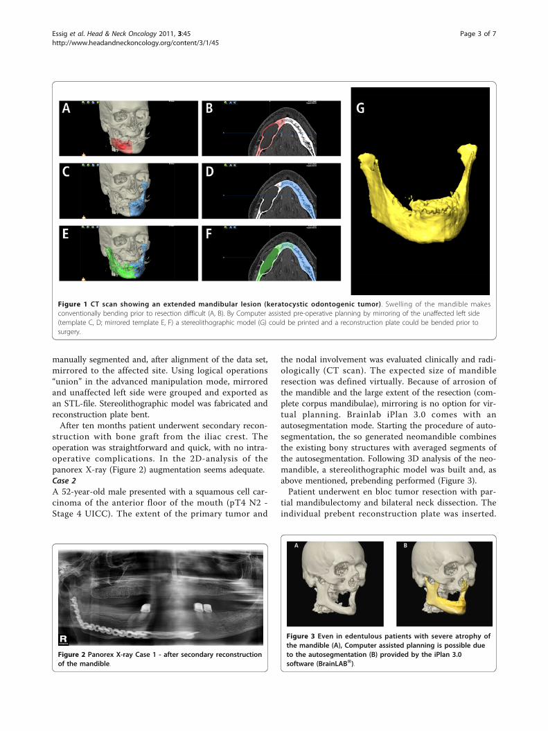
struction with bone graft from the iliac crest. Theoperation was straightforward and quick, with no intra-operative complications. In the 2D-analysis of thepanorex X-ray (Figure 2) augmentation seems adequate.Case 2A 52-year-old male presented with a squamous cell car-cinoma of the anterior floor of the mouth (pT4 N2 -Stage 4 UICC). The extent of the primary tumor and
the nodal involvement was evaluated clinically and radi-ologically (CT scan). The expected size of mandibleresection was defined virtually. Because of arrosion ofthe mandible and the large extent of the resection (com-plete corpus mandibulae), mirroring is no option for vir-tual planning. Brainlab iPlan 3.0 comes with anautosegmentation mode. Starting the procedure of auto-segmentation, the so generated neomandible combinesthe existing bony structures with averaged segments ofthe autosegmentation. Following 3D analysis of the neo-mandible, a stereolithographic model was built and, asabove mentioned, prebending performed (Figure 3).Patient underwent en bloc tumor resection with par-
tial mandibulectomy and bilateral neck dissection. Theindividual prebent reconstruction plate was inserted.
Figure 1 CT scan showing an extended mandibular lesion (keratocystic odontogenic tumor). Swelling of the mandible makesconventionally bending prior to resection difficult (A, B). By Computer assisted pre-operative planning by mirroring of the unaffected left side(template C, D; mirrored template E, F) a stereolithographic model (G) could be printed and a reconstruction plate could be bended prior tosurgery.
Figure 2 Panorex X-ray Case 1 - after secondary reconstructionof the mandible.
Figure 3 Even in edentulous patients with severe atrophy ofthe mandible (A), Computer assisted planning is possible dueto the autosegmentation (B) provided by the iPlan 3.0software (BrainLAB®).
Essig et al. Head & Neck Oncology 2011, 3:45http://www.headandneckoncology.org/content/3/1/45
Page 3 of 7
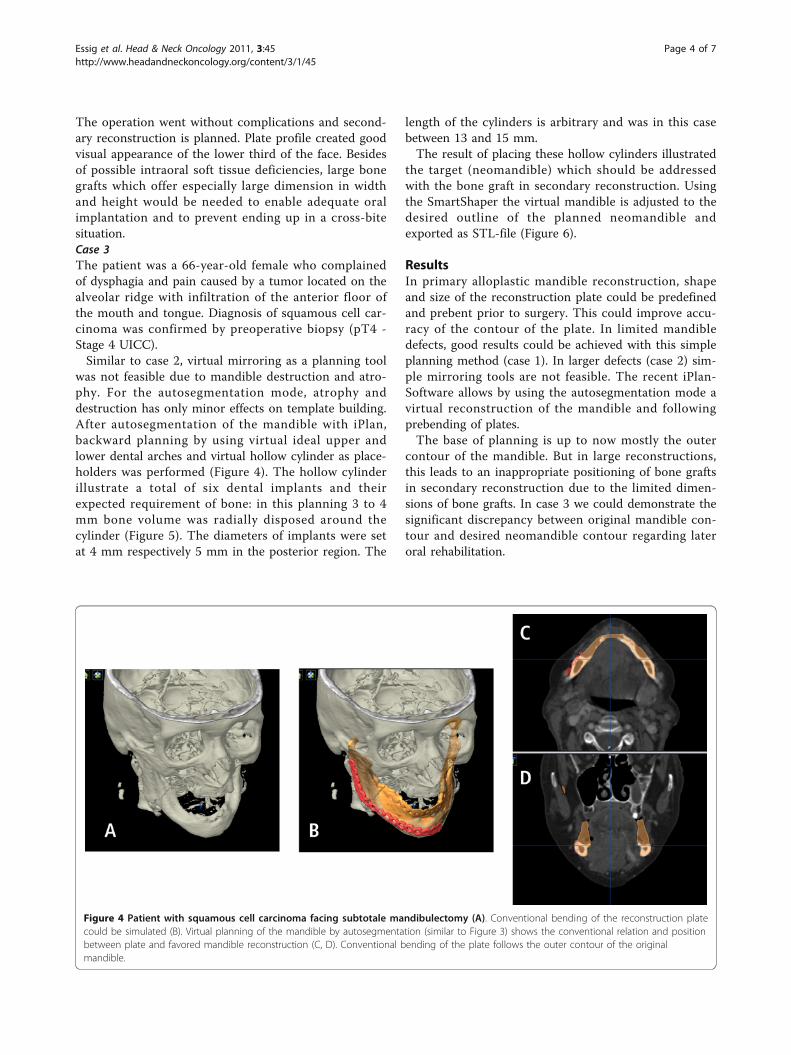
The operation went without complications and second-ary reconstruction is planned. Plate profile created goodvisual appearance of the lower third of the face. Besidesof possible intraoral soft tissue deficiencies, large bonegrafts which offer especially large dimension in widthand height would be needed to enable adequate oralimplantation and to prevent ending up in a cross-bitesituation.Case 3The patient was a 66-year-old female who complainedof dysphagia and pain caused by a tumor located on thealveolar ridge with infiltration of the anterior floor ofthe mouth and tongue. Diagnosis of squamous cell car-cinoma was confirmed by preoperative biopsy (pT4 -Stage 4 UICC).Similar to case 2, virtual mirroring as a planning tool
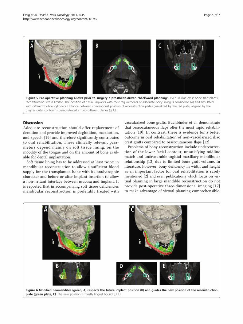
was not feasible due to mandible destruction and atro-phy. For the autosegmentation mode, atrophy anddestruction has only minor effects on template building.After autosegmentation of the mandible with iPlan,backward planning by using virtual ideal upper andlower dental arches and virtual hollow cylinder as place-holders was performed (Figure 4). The hollow cylinderillustrate a total of six dental implants and theirexpected requirement of bone: in this planning 3 to 4mm bone volume was radially disposed around thecylinder (Figure 5). The diameters of implants were setat 4 mm respectively 5 mm in the posterior region. The
length of the cylinders is arbitrary and was in this casebetween 13 and 15 mm.The result of placing these hollow cylinders illustrated
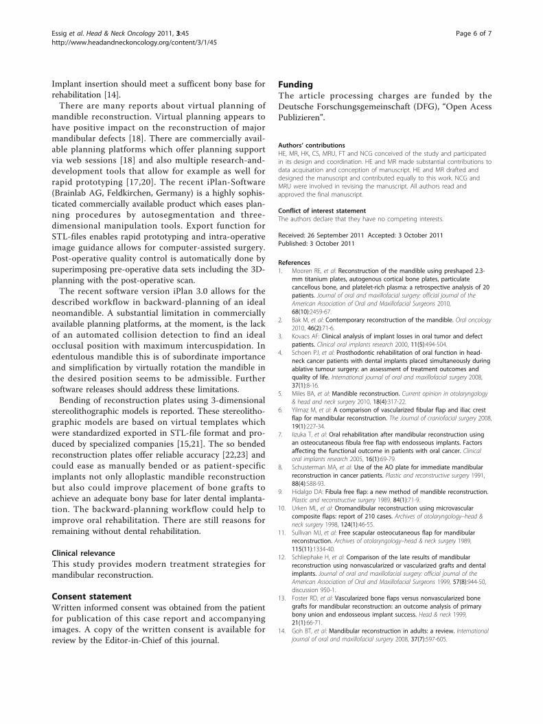
the target (neomandible) which should be addressedwith the bone graft in secondary reconstruction. Usingthe SmartShaper the virtual mandible is adjusted to thedesired outline of the planned neomandible andexported as STL-file (Figure 6).
ResultsIn primary alloplastic mandible reconstruction, shapeand size of the reconstruction plate could be predefinedand prebent prior to surgery. This could improve accu-racy of the contour of the plate. In limited mandibledefects, good results could be achieved with this simpleplanning method (case 1). In larger defects (case 2) sim-ple mirroring tools are not feasible. The recent iPlan-Software allows by using the autosegmentation mode avirtual reconstruction of the mandible and followingprebending of plates.The base of planning is up to now mostly the outer
contour of the mandible. But in large reconstructions,this leads to an inappropriate positioning of bone graftsin secondary reconstruction due to the limited dimen-sions of bone grafts. In case 3 we could demonstrate thesignificant discrepancy between original mandible con-tour and desired neomandible contour regarding lateroral rehabilitation.
Figure 4 Patient with squamous cell carcinoma facing subtotale mandibulectomy (A). Conventional bending of the reconstruction platecould be simulated (B). Virtual planning of the mandible by autosegmentation (similar to Figure 3) shows the conventional relation and positionbetween plate and favored mandible reconstruction (C, D). Conventional bending of the plate follows the outer contour of the originalmandible.
Essig et al. Head & Neck Oncology 2011, 3:45http://www.headandneckoncology.org/content/3/1/45
Page 4 of 7
DiscussionAdequate reconstruction should offer replacement ofdentition and provide improved deglutition, mastication,and speech [19] and therefore significantly contributesto oral rehabilitation. These clinically relevant para-meters depend mainly on soft tissue lining, on themobility of the tongue and on the amount of bone avail-able for dental implantation.Soft tissue lining has to be addressed at least twice: in
mandibular reconstruction to allow a sufficient bloodsupply for the transplanted bone with its bradytrophiccharacter and before or after implant insertion to allowa non-irritant interface between mucosa and implant. Itis reported that in accompanying soft tissue deficienciesmandibular reconstruction is preferably treated with
vascularized bone grafts. Buchbinder et al. demonstratethat osseocutaneous flaps offer the most rapid rehabili-tation [19]. In contrast, there is evidence for a betteroutcome in oral rehabilitation of non-vascularized iliaccrest grafts compared to osseocutaneous flaps [12].Problems of bony reconstruction include undercorrec-
tion of the lower facial contour, unsatisfying midlinematch and unfavourable sagittal maxillary-mandibularrelationship [12] due to limited bone graft volume. Inliterature, however, bony deficiency in width and heightas an important factor for oral rehabilitation is rarelymentioned [2] and even publications which focus on vir-tual planning in large mandible reconstruction do notprovide post-operative three-dimensional imaging [17]to make advantage of virtual planning comprehensible.
Figure 6 Modified neomandible (green, A) respects the future implant position (B) and guides the new position of the reconstructionplate (green plate, C). The new position is mostly lingual bound (D, E).
Figure 5 Pre-operative planning allows prior to surgery a prosthetic-driven “backward planning”. Even in iliac crest bone transplantsreconstruction size is limited. The position of future implants with their requirements of adequate bony lining is considered (A) and simulatedwith different hollow cylinders. Distance between conventional position of reconstruction plates (visualized by the red plate) aligned by theoriginal outer contour is demonstrated in two different planes (B, C).
Essig et al. Head & Neck Oncology 2011, 3:45http://www.headandneckoncology.org/content/3/1/45
Page 5 of 7
Implant insertion should meet a sufficent bony base forrehabilitation [14].There are many reports about virtual planning of
mandible reconstruction. Virtual planning appears tohave positive impact on the reconstruction of majormandibular defects [18]. There are commercially avail-able planning platforms which offer planning supportvia web sessions [18] and also multiple research-and-development tools that allow for example as well forrapid prototyping [17,20]. The recent iPlan-Software(Brainlab AG, Feldkirchen, Germany) is a highly sophis-ticated commercially available product which eases plan-ning procedures by autosegmentation and three-dimensional manipulation tools. Export function forSTL-files enables rapid prototyping and intra-operativeimage guidance allows for computer-assisted surgery.Post-operative quality control is automatically done bysuperimposing pre-operative data sets including the 3D-planning with the post-operative scan.The recent software version iPlan 3.0 allows for the
described workflow in backward-planning of an idealneomandible. A substantial limitation in commerciallyavailable planning platforms, at the moment, is the lackof an automated collision detection to find an idealocclusal position with maximum intercuspidation. Inedentulous mandible this is of subordinate importanceand simplification by virtually rotation the mandible inthe desired position seems to be admissible. Furthersoftware releases should address these limitations.Bending of reconstruction plates using 3-dimensional
stereolithographic models is reported. These stereolitho-graphic models are based on virtual templates whichwere standardized exported in STL-file format and pro-duced by specialized companies [15,21]. The so bendedreconstruction plates offer reliable accuracy [22,23] andcould ease as manually bended or as patient-specificimplants not only alloplastic mandible reconstructionbut also could improve placement of bone grafts toachieve an adequate bony base for later dental implanta-tion. The backward-planning workflow could help toimprove oral rehabilitation. There are still reasons forremaining without dental rehabilitation.
Clinical relevanceThis study provides modern treatment strategies formandibular reconstruction.
Consent statementWritten informed consent was obtained from the patientfor publication of this case report and accompanyingimages. A copy of the written consent is available forreview by the Editor-in-Chief of this journal.
FundingThe article processing charges are funded by theDeutsche Forschungsgemeinschaft (DFG), “Open AcessPublizieren”.
Authors’ contributionsHE, MR, HK, CS, MRU, FT and NCG conceived of the study and participatedin its design and coordination. HE and MR made substantial contributions todata acquisation and conception of manuscript. HE and MR drafted anddesigned the manuscript and contributed equally to this work. NCG andMRU were involved in revising the manuscript. All authors read andapproved the final manuscript.
Conflict of interest statementThe authors declare that they have no competing interests.
Received: 26 September 2011 Accepted: 3 October 2011Published: 3 October 2011
References1. Mooren RE, et al: Reconstruction of the mandible using preshaped 2.3-
mm titanium plates, autogenous cortical bone plates, particulatecancellous bone, and platelet-rich plasma: a retrospective analysis of 20patients. Journal of oral and maxillofacial surgery: official journal of theAmerican Association of Oral and Maxillofacial Surgeons 2010,68(10):2459-67.
2. Bak M, et al: Contemporary reconstruction of the mandible. Oral oncology2010, 46(2):71-6.
3. Kovacs AF: Clinical analysis of implant losses in oral tumor and defectpatients. Clinical oral implants research 2000, 11(5):494-504.
4. Schoen PJ, et al: Prosthodontic rehabilitation of oral function in head-neck cancer patients with dental implants placed simultaneously duringablative tumour surgery: an assessment of treatment outcomes andquality of life. International journal of oral and maxillofacial surgery 2008,37(1):8-16.
5. Miles BA, et al: Mandible reconstruction. Current opinion in otolaryngology& head and neck surgery 2010, 18(4):317-22.
6. Yilmaz M, et al: A comparison of vascularized fibular flap and iliac crestflap for mandibular reconstruction. The Journal of craniofacial surgery 2008,19(1):227-34.
7. Iizuka T, et al: Oral rehabilitation after mandibular reconstruction usingan osteocutaneous fibula free flap with endosseous implants. Factorsaffecting the functional outcome in patients with oral cancer. Clinicaloral implants research 2005, 16(1):69-79.
8. Schusterman MA, et al: Use of the AO plate for immediate mandibularreconstruction in cancer patients. Plastic and reconstructive surgery 1991,88(4):588-93.
9. Hidalgo DA: Fibula free flap: a new method of mandible reconstruction.Plastic and reconstructive surgery 1989, 84(1):71-9.
10. Urken ML, et al: Oromandibular reconstruction using microvascularcomposite flaps: report of 210 cases. Archives of otolaryngology–head &neck surgery 1998, 124(1):46-55.
11. Sullivan MJ, et al: Free scapular osteocutaneous flap for mandibularreconstruction. Archives of otolaryngology–head & neck surgery 1989,115(11):1334-40.
12. Schliephake H, et al: Comparison of the late results of mandibularreconstruction using nonvascularized or vascularized grafts and dentalimplants. Journal of oral and maxillofacial surgery: official journal of theAmerican Association of Oral and Maxillofacial Surgeons 1999, 57(8):944-50,discussion 950-1.
13. Foster RD, et al: Vascularized bone flaps versus nonvascularized bonegrafts for mandibular reconstruction: an outcome analysis of primarybony union and endosseous implant success. Head & neck 1999,21(1):66-71.
14. Goh BT, et al: Mandibular reconstruction in adults: a review. Internationaljournal of oral and maxillofacial surgery 2008, 37(7):597-605.
Essig et al. Head & Neck Oncology 2011, 3:45http://www.headandneckoncology.org/content/3/1/45
Page 6 of 7
15. Cohen A, et al: Mandibular reconstruction using stereolithographic 3-dimensional printing modeling technology. Oral surgery, oral medicine,oral pathology, oral radiology, and endodontics 2009, 108(5):661-6.
16. Eckardt A, Swennen GR: Virtual planning of composite mandibularreconstruction with free fibula bone graft. The Journal of craniofacialsurgery 2005, 16(6):1137-40.
17. Juergens P, et al: Computer simulation and rapid prototyping for thereconstruction of the mandible. Journal of oral and maxillofacial surgery:official journal of the American Association of Oral and Maxillofacial Surgeons2009, 67(10):2167-70.
18. Roser SM, et al: The accuracy of virtual surgical planning in free fibulamandibular reconstruction: comparison of planned and final results.Journal of oral and maxillofacial surgery: official journal of the AmericanAssociation of Oral and Maxillofacial Surgeons 2010, 68(11):2824-32.
19. Buchbinder D, et al: Functional mandibular reconstruction of patientswith oral cancer. Oral surgery, oral medicine, and oral pathology 1989, 68(4Pt 2):499-503, discussion 503-4.
20. Krol Z, et al: Surgery planning tools for the osseous grafting treatment.Biomedizinische Technik. Biomedical engineering 2002, 47(Suppl 1 Pt 1):p.97-100.
21. Derand P, Hirsch JM: Virtual bending of mandibular reconstruction platesusing a computer-aided design. Journal of oral and maxillofacial surgery:official journal of the American Association of Oral and Maxillofacial Surgeons2009, 67(8):1640-3.
22. Klug C, et al: Point-to-point computer-assisted navigation for precisetransfer of planned zygoma osteotomies from the stereolithographicmodel into reality. Journal of oral and maxillofacial surgery: official journal ofthe American Association of Oral and Maxillofacial Surgeons 2006,64(3):550-9.
23. Schicho K, et al: Accuracy of treatment planning based onstereolithography in computer assisted surgery. Medical physics 2006,33(9):3408-17.
doi:10.1186/1758-3284-3-45Cite this article as: Essig et al.: Pre-operative planning for mandibularreconstruction - A full digital planning workflow resulting in a patientspecific reconstruction. Head & Neck Oncology 2011 3:45.
Submit your next manuscript to BioMed Centraland take full advantage of:
• Convenient online submission
• Thorough peer review
• No space constraints or color figure charges
• Immediate publication on acceptance
• Inclusion in PubMed, CAS, Scopus and Google Scholar
• Research which is freely available for redistribution
Submit your manuscript at www.biomedcentral.com/submit
Essig et al. Head & Neck Oncology 2011, 3:45http://www.headandneckoncology.org/content/3/1/45
Page 7 of 7