practical 4 nervous system - fiziologie ns_eye exam.pdf · • binocular accommodation analysis,...
TRANSCRIPT

Practical 4, Nervous System:
Eye Exam
English Module 2nd Year, Physiology Dept., Carol Davila University of Medicine and Pharmacy

Eye Exam • Anamnesis, • Visual acuity, • Pupilar evaluation, • Extra ocular motility function, • Visual field exam, • Binocular accommodation analysis, • Retinoscopy & refraction, • Chromatic sight tests (Ishihara), • Exam of external ocular annexes through biomicroscopy, • Intraocular pressure measure,• Examination of eye humors • Ophthalmoscopy.

Visual acuity represents the capacity to distinguish between twoindividual luminous points in space.

Visual Acuity (VA)• VA is maximum in the macula
and decreases approximately 10 times in the peripheral retina, and that is why when trying to see an object we will move the head and eyes to project it on the macula.
• Vision decreases when the object is moving, because it cannot be analyzed as a whole by the macula. The moving object permanently moves out of the macular projection and cannot be easily examined.
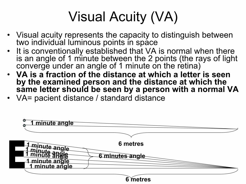
Visual Acuity (VA)• Visual acuity represents the capacity to distinguish between
two individual luminous points in space• It is conventionally established that VA is normal when there
is an angle of 1 minute between the 2 points (the rays of light converge under an angle of 1 minute on the retina)
• VA is a fraction of the distance at which a letter is seen by the examined person and the distance at which the same letter should be seen by a person with a normal VA
• VA= pacient distance / standard distance
1 minute angle
1 minute angle1 minute angle1 minute angle
1 minute angle1 minute angle 6 minutes angle
6 metres
6 metres

• VA is measured with different types of optotypes• In general the optotypes are placed at a 6 meter
distance, and for the British system at 20 feet
Examining Visual Acuity
• There are several types of optotypes:– Different characters are used:
• Snellen optotypes with letters and numbers• optotypes using the “snellen E”• optotypes using “landolt C/rings”• optotypes with drawings for small children

Faceti click pentru raspuns
If a person has a weaker vision, then, to be able to see a letter from a standard distance of 6 metres, this letter has to be magnified (the person has to come closer to see the letter).
What is the persons VA if the letter is twice the size seen by anormal person?
VA=6 metres
12 metres=6/12
6 meters
2 x 6 minutes 6 minutes
12 meters
Subject with normal VASubject with low VA
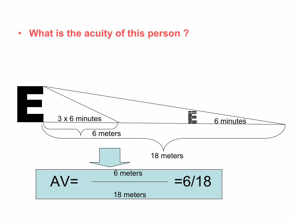
• What is the acuity of this person ?
6 meters
3 x 6 minutes 6 minutes
18 meters
AV=6 meters
18 meters=6/18

• What is the persons acuity if the letter is K times bigger than normal?
• ?
6 meters
K x 6 minutes 6 minutes
6k meters
AV=6 meters
K x 6 meters=6/6K

How to examine the VA
• Position the patient at 6 meters;• Cover the eye with supposed better acuity• Start with symbols 10 times bigger than
normal and decrease the size of the symbols gradually
• Write down the VA corresponding to the last symbol seen at that distance
• Cover the other eye and start again

• If VA is not 6/6, a stenopeic point (small opening of 1-3 mm) will be used that decreases the sphericity aberrations; you can use a sheet of paper perforated with a pen
• If the patient can see charts with smaller letters, it means that he has a refraction error
• If the patient still doesn’t see smaller letters, then it means he has an ocular pathology
Stenopeic point
1 mm


• The next 8 charts show letters for VA between 6/60 and
• Each student should test his VA separately for each eye
• For those that do not have VA = 6/6, VA will be tested with stenopeic and then with glasses.
• At the end of the testing: – VA right eye =– VA left eye=

AV 20/200 6/60

AV 20/160; 6/48

AV 20/100 6/30
A E F P C P Z

AV 20/80 6/24
F G T Z C R H D

AV 20/60 6/18
P D X G H A E F T R

AV 20/40; 6/12
A E F P D S G H X T R H

AV 20/30 6/9
A E F P D S G H X T R H D

AV 20/20 6/6A E F P D S G H X T R H D

Visual fieldPerimetry – determine the field of vision and measures the
acuity of the retina in its peripheral parts

PhototransductionLight rhodopsinstimulation (rods) decrease in cGMP
closing of Na channelshyperpolarizationinhibition of glutamate
release changes in membrane potential of upstream cells.
Similar phenomena happen in the cones, cones are much less sensitive to light.
Some bipolar cells are depolarized by glutamate, some are inhibited. The last one are then desinhibitedwhen glutamate release decreases.

Color visionDistribution of cones in the fovea
Note that there are no blue cones in the fovea.
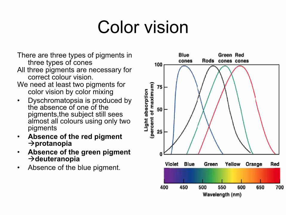
Color visionThere are three types of pigments in
three types of conesAll three pigments are necessary for
correct colour vision. We need at least two pigments for
color vision by color mixing• Dyschromatopsia is produced by
the absence of one of the pigments,the subject still sees almost all colours using only two pigments
• Absence of the red pigment protanopia
• Absence of the green pigment deuteranopia
• Absence of the blue pigment.

Color vision
• At least two types of cones must be stimulated
• The intensity of stimulation of each pigment is integrated
• Examples:– orange = 99 : 42 : 00– green = 31 : 67 : 36– blue = 0 : 0 : 97– yellow = 83 : 83 : 0
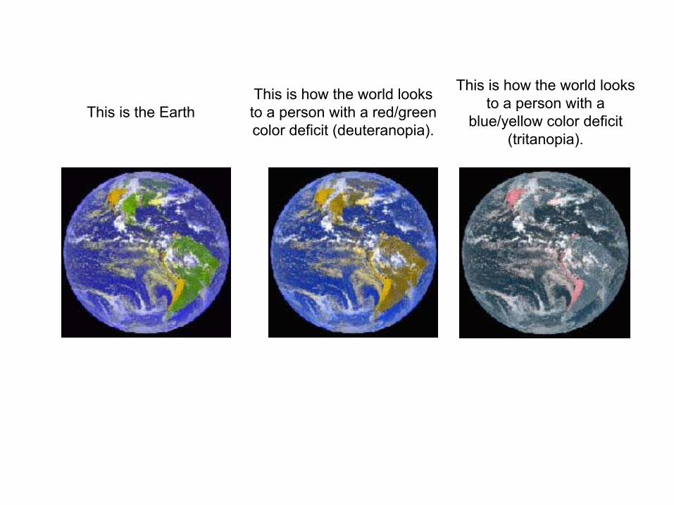
This is the EarthThis is how the world looksto a person with a red/green color deficit (deuteranopia).
This is how the world looks to a person with a
blue/yellow color deficit (tritanopia).

This is an Ishihara plate commonly used to check for
red/green color blindness
This is what a red/green colorblind person might see.
Note that the digit (3) is practically invisible.

Protanopia and protanomaliacolour blindness for red
• Because sensibility for red wavelength is absent they fail to differentiate colors that appear different for people with normal color vision.
• Colors cyan, grey andpinkish will appear identical for patients with protanopia.

Deuteranopia and deuteroanomalia colour
blindness for green• Because sensibility for green
wavelength is absent they fail to differentiate colors that appear different for people with normal color vision.
• Colors cyan, grey andpinkish will appear identical for patients with deuteranopia.

Tritanopia and tritanomaliacolour blindness for blue
• Because sensibility for blue wavelength is absent they fail to differentiate colors that appear different for people with normal color vision
• Colors yellow, greyand violet will appear identical for patients with tritanopia.

Equal saturation and luminosity Unequal saturation and luminosity
•Color blind people have the tendency to identify tones based on saturation and luminosity.
If saturation and luminosity are different, they cannot identify the colors.

Plate 1
• Number 12 can be read both by normal and color blind subjects

Plate 2
• Subjects with normal vision should read 8.
• Subjects with deficiencies in the red-green axis should see 3
• Subjects with complete color blindness are not able to identify any numbers.

Plate 3• Subjects with normal
vision should read 29. • Subjects with
deficiencies in the red-green axis should see 70.
• Subjects with complete color blindness are not able to identify any numbers.

Plate 4• Subjects with normal
vision should read 5. • Subjects with
deficiencies in the red-green axis should see 2.
• Subjects with complete color blindness are not able to identify any numbers.

Plate 5
• Subjects with normal vision should read 3.
• Subjects with deficiencies in the red-green axis should see 5.
• Subjects with complete color blindness are not able to identify any numbers.

Plate 6
• Subjects with normal vision should read 15.
• Subjects with deficiencies in the red-green axis should see 17.
• Subjects with complete color blindness are not able to identify any numbers.

Plate 7
• Subjects with normal vision should read 74.
• Subjects with deficiencies in the red-green axis should see 21.
• Subjects with complete color blindness are not able to identify any numbers.

Plate 8
• Subjects with normal vision should read 29.
• Subjects with complete or even incomplete color blindness are not able to identify any numbers.

Plate 9
• Subjects with normal vision should read 45.
• Subjects with complete or even incomplete color blindness are not able to identify any numbers.

Plate 10
• Subjects with normal vision should read 5.
• Subjects with complete or even incomplete color blindness are not able to identify any numbers.
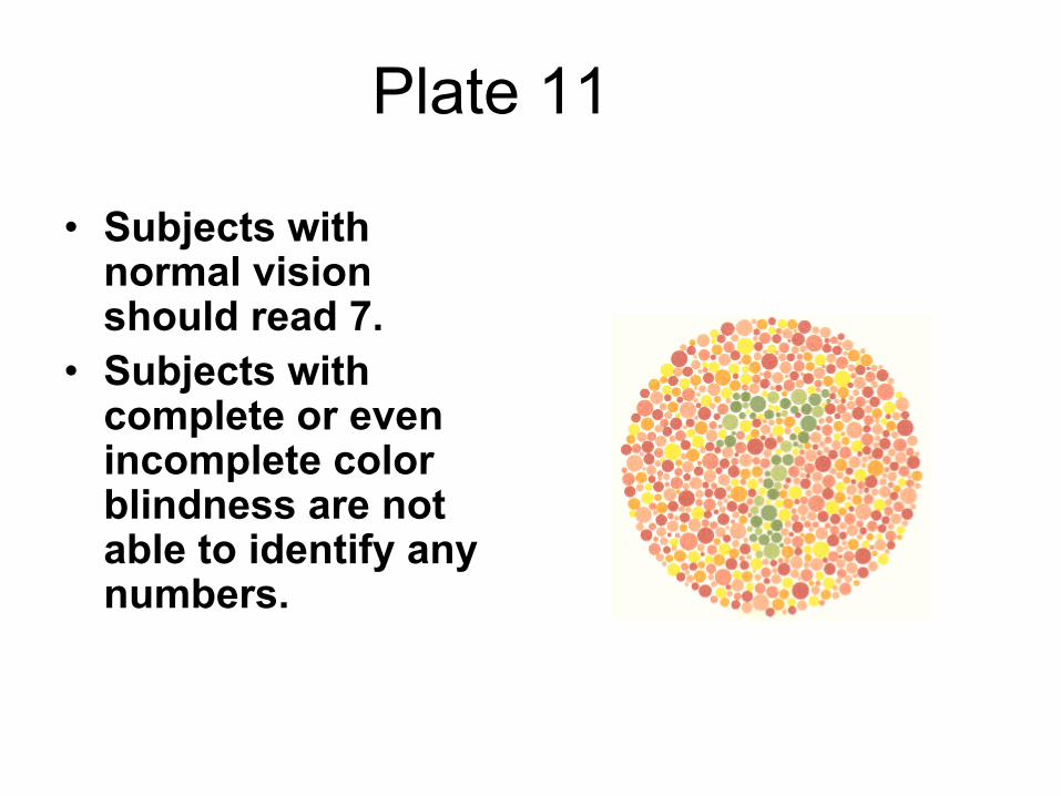
Plate 11
• Subjects with normal vision should read 7.
• Subjects with complete or even incomplete color blindness are not able to identify any numbers.

Plate 12
• Subjects with normal vision should read 16.
• Subjects with complete or even incomplete color blindness are not able to identify any numbers.

Plate 13
• Subjects with normal vision should read 73.
• Subjects with complete or even incomplete color blindness are not able to identify any numbers.

Plate 14
• Subjects with normal color vision are not able to identify any numbers.
• Subjects with color blindness should read 5.

Plate15
• Subjects with normal color vision are not able to identify any numbers.
• Subjects with color blindness should read 45.

• References & credits:- Guyton chapters 49, 50, 51- Borone chapter 13 or 15 - Berne & Levy chapter 8- most of the material for the eye exam was kindly provided by Dr. M. Ghita, Ophthalmology Dept, Bucharest Univ. Emergency Hospital & Physiology Dept., Carol Davila Univ. of Medicine and Pharmacy