potential ameliorative role of silymarin against methyl...
TRANSCRIPT

15
Vol. XVII, No. 1, Jan. 2009Mansoura J. Forensic Med. Clin. Toxicol.
Khodeary et al ...
POTENTIAL AMELIORATIVE ROLE OF SILYMARIN AGAINST METHYL PARATHION-INDUCED OXIDATIVE STRESS AND
HEPATO-RENAL TOXICITY IN ALBINO RATS
BY
Mohammed F. Khodeary, Abeer A. I. Sharaf El-Din, Shereen M. S. El Kholy,Atef A. Fouda and Essam M. Mehlab*
Departments of Forensic Medicine and Clinical Toxicology and Anatomy*
Faculty of Medicine, Benha University
ABSTRACT
Methyl parathion (MP) (C8H10NO5PS) is an organophosphate insecticide that has been used in agri-
culture for several years. The people who are at the greatest risk of being exposed to MP are farm work-
ers, chemical sprayers, and people who work in factories handling MP. The main route of human expo-
sure is inhalation, but dermal contact and inadvertent ingestion can also be substantial. The present work
was planned to study the toxic effects of MP on some antioxidative markers as well as the liver and kid-
ney. The influence of silymarin (Sily) on MP induced toxicity has not been studied, So, the possible pro-
tective effect of (Sily) had been also evaluated. The study was conducted on one hundred male albino rats
divided into five main equal groups and each of them were subdivided equally into two subgroups.
Group-I (A&B), control group: received no treatment, group-II (A&B): vehicle group (1 ml/kg/day of
corn oil, P.O.), group-III (A&B): Sily group (100 mg/kg/day, P.O.), group-IV (A&B): MP group (0.28
mg/kg/day, P.O.), group-V (A&B): Sily+MP. At the end of 4th and 8th weeks, blood samples, renal and
hepatic tissues were collected for biochemical and histological examinations. Biomarkers selected for ox-
idative stress were antioxidant defense system superoxide dismutase (SOD) and reduced glutathione
(GSH) as well as malondialdehyde (MDA). In addition, liver profile (AST, ALT, and ALP) and kidney
profile (Blood urea and creatinine) were assessed. Also, histological examination of liver and kidney was
done. The results showed that administration of MP resulted in oxidative stress as evidenced by signifi-
cant decrease in SOD activity and GSH levels accompanied with significant increase in MDA levels. At
the same time, there was hepato-nephrotoxicity as manifested by significant increase in liver and kidney
function tests when compared with control rats. Comparing with MP-treated rats, Sily supplementation to
the rats, 30 minutes before MP administration, resulted in a significant increase in SOD activity and GSH
levels. However, MDA levels, liver and kidney enzymes showed significant decrease. The histopathologi-
cal results confirmed the biochemical results. In conclusion, the obtained biochemical and histological
results of this study revealed hepatic and renal toxic effects induced by MP in a time-dependent manner.
Sily supplementation resulted in a remarkable protective effect against MP-induced oxidative stress and
hepato-renal toxicity.

Khodeary et al ...16
Vol. XVII, No. 1, Jan. 2009Mansoura J. Forensic Med. Clin. Toxicol.
manufacturers of this product may be ex-posed occupationally, although, there aresome individuals who are exposed viafood and water. Illegal application of MPincreases the risk of exposure to the chem-ical for those who do not have occupationsthat involve MP handling. Individuals re-siding near hazardous waste disposal sitesmay be subject to higher levels of MP ex-posure (ATSDR, 2001).
MP insecticidal properties reside in itsability to inhibit acetylcholinesterase activ-ity, thus enhancing endogenous acetylcho-line accumulation (Comoglio et al., 2005).
Besides their inhibitory effects onace-tylcholinesterase enzymes, OP com-pounds may induce oxidative stress lead-ing to generation of free radicals and alter-ations in antioxidants status or reactiveoxygen species (ROS) scavenging enzymes(Gultekin et al., 2000).
The lipophilic nature of OP facilitatestheir interaction with the cell membraneand leads to perturbation of the phospho-lipids bilayer structure (Videira et al.,2001). Therefore, they may enhance lipid-peroxidation (LPO) by directly interactingwith the cellular membrane and generatedROS (Aslan et al., 1997). LPO may impairantioxidant defenses, leading to cellularoxidative damage by changing the balancebetween oxidants and antioxidants (Torreset al., 2004). Some studies showed that
INTRODUCTlON
Organophosphorous (OP) compoundshave been used worldwide for pest con-trol for over 100 years. They are the insec-ticide of choice in the agricultural worldand are the most common cause ofpoisoning among the organic insecticidesas a result of their readily availability andeasy accessibility (Eddleston, 2000). Thecommon use of insecticides in publichealth and agricultural schedules hascaused severe environmental pollutionand potential health hazards includingsevere acute and chronic cases of humanand animal poisonings (Abdollahi et al.,2004).
OP compounds are known to cause in-hibition of acetylcholinesterase and pseu-docholinesterase activity (Yavuz et al.,2005). Other systems that could be affect-ed by OP compounds are urinary system(Rod-rigo et al., 2001) and liver (Kalenderet al., 2005).
Methyl parathion (MP; O, O-dimethylO-P-nitro-phenyl phosphorothioate), pop-ularly known as "cotton poison", is an or-ganophosphate insecticide licensed onlyfor agricultural use in many countries, al-though there are instances of its misuse asa domestic spray (Rubin et al., 2002).
People may be exposed to MP via anumber of several ways. Applicators and

17
Vol. XVII, No. 1, Jan. 2009Mansoura J. Forensic Med. Clin. Toxicol.
Khodeary et al ...
free radical species. This action as well asmany other beneficial intracellular reac-tions have adverted it as promising agentfor protection and treatment of the damag-ing actions of many toxic compounds(Dalmi and Sari, 1992).
The goal of the current study was toinvestigate the toxic effects of MP on someserum antioxidative markers as well as theliver and kidney of male adult albino ratsand also to evaluate the possible protec-tive role of Sily in modulating these toxiceffects.
MATERIAL AND METHODS
Animals:The study was conducted on 100
adult male albino rats with initial bodyweight ranging between 180-200 grams.All animals were housed under suitableenvironmental conditions with free accessto food and water. In order to excludefallacies induced by environmental fac-tors, the rats received standard laboratoryenvironmental conditions according toCuschieri and Backer (1977). The rats hadbeen familiarized with the environmentfor one week before the study to allowacclimatization.
Drug and Chemical :Methyl parathion (MP) was obtained
from Sigma Agency for Chemicals andPharmaceuticals, Cairo, Egypt. It was
LPO has been suggested as one of the mo-lecular mechanisms involved in OP com-pounds induced toxicity (Yamano andMorita, 1992). Hence, treatment with anti-oxidants and free radical scavengers candecrease the oxidative stress related toOP-induced toxicity(Yurumez et al., 2007).
Nutritional studies have also revealedthe crucial role of some minerals in pre-venting oxidative stress. Nutritional andbotanical supplementation was also sug-gested for chronic pesticide detoxifica-tion. A high quality multiple vitamin/mineral supplements with extra magne-sium, pyridoxine, selenium, antioxidants,and milk thistle is recommended (Walterand Crinnion, 2000). Moreover, silymar-in/silybin and their preparations are ad-vocated for the treatment of variousdiseases like cirrhosis, chronic hepatitisand diseases associated with alcohol con-sumption and environmental toxin expo-sure (Koen and Walterovab, 2005).
Silymarin (Sily) is a purified extractfrom the seeds of Silybum marinumL. (As-teraceae) which also called "milkthistle". It is shown that Sily consists ofa large number of flavolignans, includingsilybin (or silybinin), which is the most ac-tive component, isosilybin, silydianin andsilychristin (Khan et al., 2006).
Sily acts as a powerful antioxidant thatcan capture and neutralize the harmful

Khodeary et al ...18
Vol. XVII, No. 1, Jan. 2009Mansoura J. Forensic Med. Clin. Toxicol.
orally in a dose of 0.28 mg/kg bodyweight (1/50 LD50 dose orally) for 4 and 8weeks (Kalender et al., 2007).
Group-V (A & B) : Methyl parathion +Silymarin-treated group, each animal re-ceived silymarin (100 mg/kg/day, orallythrough gavage), 30 minutes before ad-ministration of MP (0.28 mg /kg /dayorally through gavage) in corn oil.
Animals were fasted for 4 hours beforegavage and received their correspondingsubstance dosage once daily.
Experimental Parameters :Biochemical parametersNo deaths were observed in any of the
experimental groups during different peri-ods.
Twenty four hours after the end of 4th
and 8th weeks, the animals were anesthe-tized with sodium pentobarbital (50 mg/kg intraperitoneally) before sacrification.Following this laparotomy was conductedand the intra-abdominal organs were ex-posed and the blood was collected fromthe descending aorta. For each animal, aportion from collected blood was trans-ferred into a heparinized centrifuge tubefor spectrophotometric determination ofplasma malondialdhyde (MDA) level (Yo-shida et al., 1980), reduced glutathione(GSH) level (Hissin and Hilf, 1976), andthe activity of superoxide dismutase(SOD) (Kakkar et al., 1984).
available in a white crystalline powderform, which was dissolved in corn oil.
Silymarin was obtained from com-mercially marketed packets containing 140mg of silymarin powder/packet (SedicoPharmaceuticals Co. Cairo). The powderwas dissolved in distilled water.
Experimental Groups:One hundred male rats were divided
equally into 2 groups, A and B, each com-promised 50 rats. Groups A and B werefurther subdivided into 5 subgroups eachconsisted of 10 rats. Animals in groups Aand B received their corresponding sub-stance for 4 and 8 weeks, respectively anddistributed as follows:
Group-I (A & B) : Control group, theanimals received no treatment, only theygavaged with 1 ml of distilled water dailyfor 4 and 8 weeks and were used to deter-mine the normal values of tested parame-ters.
Group-II (A & B) : Vehicle group, eachanimal gavaged with corn oil at a dose of1 ml/kg body weight for 4 and 8 weeks.
Group-III (A & B) : Silymarin-treatedgroup, each animal received silymarinorally in a dose of 100 mg/kg body weightfor 4 and 8 weeks (Crocenzi et al., 2003).
Group-IV (A & B) : Methyl parathiontreated group, each animal received MP

19
Vol. XVII, No. 1, Jan. 2009Mansoura J. Forensic Med. Clin. Toxicol.
Khodeary et al ...
significant changes in SOD, GSH, andMDA values, when vehicle treated groupswere compared with control groups.
I.1.B. Control Vs Sily-treated groups :Sily induced significant increase in
SOD and GSH values accompanied withsignificant decrease in MDA levels at theend of 4th and 8th weeks. These effectswere more pronounced after 8th than 4th
weeks of Sily administration to rats.
I.1.C. Control Vs MP-treated groups :In MP treated groups, SOD and GSH
values showed significant decrease, whileMDA levels showed significant increase atthe end of the 4th and 8th weeks as com-pared with control groups. These valueswere more pronounced at the end of 8th
than 4th week in MP treated groups.
I.1.D. Control Vs Sily + MP-treatedgroups :
Data analysis revealed changes in themeasured parameters in the form of signi-ficant decrease in SOD and GSH valuesaccompanied with significant increase inMDA levels at the end of the 4th and 8th
weeks in Sily+MP treated groups whencompared with control groups. However,these changes were less marked at the endof the 8th than the 4th week in Sily+MPtreated groups.
I.1.E. Sily+MP Vs MP-treated groups :Statistical analysis revealed significant
The remainder of each sample was cen-trifuged and the serum was separatedfor spectrophotometric determination ofaspartate transaminase (AST), and alanintransaminase (ALT) according to Frankeland Gradwohl, (1970) and alkaline phos-phatase (ALP) (Donald and Ralph, 1993).Also, spectrophotometric determination of(BU) and creatinine levels according toLawrence and Robert (1993).
Histopathological Examination:After the animals were sacrificed, the
liver and kidney were dissected out andfixed in 10% formol saline. Paraffin sec-tions of 5-7 µm in thickness were preparedand subjected to Hematoxylin-Eosin (Hx& E) according to Drury and Wallington(1980).
Statistical Analysis:The experimental data are expressed as
mean + S.D. The data were analyzed bythe student t-test. The differences wereconsidered to be statistically significantwhen P < 0.05.
RESULTS
I. Biochemical ResultsI.1 Oxidative stress biomarkers
(Table 1) :I.1.A. Control Vs vehicle-treated
groups :At the end of 4th and 8th weeks, the
results of the present study revealed non

Khodeary et al ...20
Vol. XVII, No. 1, Jan. 2009Mansoura J. Forensic Med. Clin. Toxicol.
groups at the end of 4th and 8th weeks.Also, marked improvement in these pa-rameters was seen at the end of 8th than4th week in Sily+MP-treated groups.
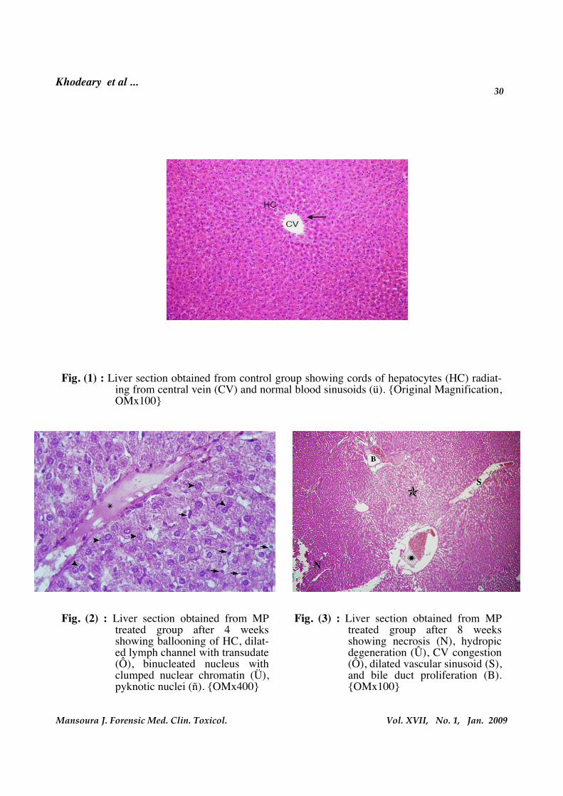
II. Liver Histopathological Results(Figures 1-5):
The histological examination of hepatictissues of the control groups as well as ve-hicle and Sily treated groups at the end of4th and 8th weeks revealed normal hepaticarchitecture with cords of hepatocytes ra-diating from the central veins. The hepa-tocytes appeared polyhedral in shape withwell defined boundaries and acidophiliccytoplasm. Each cell showed a round, ve-sicular, centrally located nucleus. The he-patic sinusoids appeared as narrow spaceslined by flattened endothelial cells andvon kupffer cells.
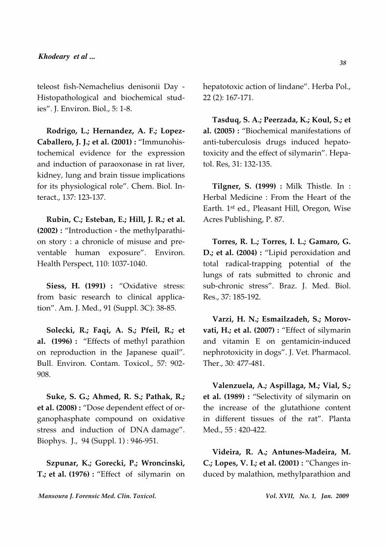
Rats given MP for 4 weeks showed sig-nificant histopathological alterations in theform of single cell necrosis of hepato-cytes, cloudy swelling, pyknotic and bi-nucleate nucleus, ballooning of hepa-tocytes, and Kupffer cell proliferation.However, in Sily+MP treated group thepreviously men-tioned histopathologicalchanges were less severe when comparedwith MP treated group.
At the end of 8th week, the liver of MPtreated rats showed higher incidence ofthe previously mentioned lesions than ratstreated for 4 weeks with focal hepatic
increase in SOD and GSH values as wellas significant decrease in MDA levels inSily+MP treated groups as compared withMP-treated groups at the end of 4th and8th weeks.
I.2 Liver and kidney functions assayparameters (Table 2) :
The results of the present study showednon significant changes in liver functiontests "AST, ALT, ALP" and kidney func-tion tests, "BU, CRE" when vehicle andSily treated groups were compared withcontrol groups at the end of 4th and 8th
weeks.
Data analysis of liver function testsshowed a significant increase in MP andSily+MP treated groups when comparedwith control group after 4 and 8 weeks oftheir administration. However, a signifi-cant decrease was found in Sily+MP treat-ed groups as compared with MP treatedgroups at the end of 4th and 8th weeks.However, marked improvement in theseparameters was seen at the end of 8th than4th week in Sily+MP treated groups.
The results obtained from the effect ofMP and Sily+MP treated groups on kid-ney function tests revealed significant in-crease as compared with those of controlgroups at the end of 4th and 8th weeks. Onthe other hand, BUN and CRE levels weresignificantly de-creased in Sily+MP treat-ed groups as com-pared with MP treated

21
Vol. XVII, No. 1, Jan. 2009Mansoura J. Forensic Med. Clin. Toxicol.
Khodeary et al ...
observed. While, 4 weeks after Sily+MPwas given to rats, interstitial inflammatorycell infiltrations were detected and someof the renal tubules were dilated. In addi-tion, 8 weeks after MP treatments to rats,hemorrhage, edema, necrosis, interstitialtissue infiltration by inflammatory cells,marked tubular dilatation, and glome-rular atrophy were observed. While, 8weeks after Sily+MP treatment to rats, cal-cification, tubular degeneration and mildmononuclear cell infiltration were report-ed. However, the incidence of all lesionswere less marked in Sily+MP treatedgroups when compared with MP treatedgroups at the end of 4th and 8th weeks butstill different when compared with controlgroup.
Marked decrease in all kidney lesionswas noticed at the end of 8th week than4th week in Sily+MP treated groups.
DISCUSSION
Organophosphorous pesticides areamong the most worldwide-used agro-chemicals; are mainly known by their neu-rotoxic effects due to the inhibition ofacetylcholinesterase by their oxygen ana-logues, which are the active metabolitesgenerated during OP metabolism (Joka-novic, 2001). Acute and chronic toxicitystudies indicate that MP is very highlytoxic to mammals. Mammals are expectedto be adversely affected by MP through
necrosis, central venous congestion, lym-phocytic infiltrations, and hydropic de-generation. While, liver examination ofrats treated with Sily+MP showed markeddecrease in the incidence of all lesionswhen compared with MP treated groupbut did not return back to normal as com-pared with control group at both periods.
Histopathologic comparison of hepa-totoxicity in Sily+MP treated groups at theend of the 4th and 8th weeks revealedmarked decrease in all lesions severity atthe end of 8th week than 4th week.
III. Kidney Histopathological Results(Figures 6-10):
Light microscopic examination of thekidney sections of control rats as well asvehicle and Sily treated rats at the end of4th and 8th weeks showed normal renalcortical and medullary structures, whichconsist of glomerulus (tuft of blood capil-laries surrounded by epithelial capsule,namely Bowman's capsule), proximal(formed of pyramidal cells with centralround nucleus and acidophilic cytoplasm)and distal (contain cubical cells with epi-cal round nucleus) convoluted tubulesand the collecting tubules (lined with cubi-cal columnar cells).
After 4 weeks of MP exposure, vas-cular dilatation, glomerular atrophy,cloudy swelling in cortical tubules, andfew foci of hydropic degeneration were

Khodeary et al ...22
Vol. XVII, No. 1, Jan. 2009Mansoura J. Forensic Med. Clin. Toxicol.
the end of 8th week than 4th week inSily+MP treated groups.
In accordance with the present workthe studies done by Suke et al. (2008) andCelik and Suzek (2008) who reported sig-nificant increase in MDA levels as well assignificant decrease in SOD activity (eryth-rocytes and lung) and GSH levels (inwhole blood and various tissue homogen-ates) of rats exposed to MP.
Also, intra-peritoneal injection of MPinto male rats showed significant decreasein erythrocyte SOD enzyme activity (Po-povici et al., 2002). In addition, López etal. (2007) reported lower levels of erythro-cyte SOD in farmers engaged in intensiveagriculture area.
However, Celik and Suzek (2008)demonstrated significant increase in SODactivity in rats’ erythrocytes. Doyotte et al.(1997) pointed out that a decreased en-zyme activity’ response may accompanya first exposure to pollutants, which canbe followed by an induction of antioxi-dant systems. Thus, the existence of an in-ducible antioxidant system may reflectan adaptation of organisms. In contrast,Dimitrova et al. (1994) suggested that thesuperoxide radicals by themselves or aftertheir transformation to H2O2 cause an oxi-dation of the cysteine in the enzyme anddecrease SOD activity. Consequently, thedecreased and increased SOD activities
oral, dermal and inhalation exposure path-ways (Garcia et al., 2003).
It has been reported that OP com-pounds induce oxidative or toxic stress inanimals and humans, both in acute andchronic poisonings (Ranjbar et al., 2002;Ghafour-Rashidi et al., 2007). These oxy-gen free radicals can be neutralized by aseries of enzymatic systems (Siess, 1991).The two main physiological antioxidantdefenses against free radicals are SOD andthe glutathione system. Both these defencemechanisms are subjected to depletionduring a period of toxic substance over-load (Wellington and Jarvis, 2001).
In the present study, MP administra-tion to rats resulted in oxidative stresswhich was manifested by significant de-crease in the mean values of SOD andRGSH as well as significant increase inMDA levels when compared with controlgroups at the end of both experimental pe-riods. However, pre-treatment with Silysupplementation resulted in modulationof these measured parameters (but stillsignificantly different as compared withcontrol groups at the end of both studiedperiods) as evidenced by significant de-crease in MDA levels as well as significantincrease in SOD activities and RGSH lev-els in Sily+MP treated groups as com-pared with MP treated groups at the endof the 4th and 8th weeks. Moreover, theseparameters were markedly improved at

23
Vol. XVII, No. 1, Jan. 2009Mansoura J. Forensic Med. Clin. Toxicol.
Khodeary et al ...
rats’ plasma (Guney et al., 2007). The in-creased MDA content might have resultedfrom an increase of free radicals as a resultof stress condition in the rats with MP in-toxication (Freeman and Crapo, 1981).
As an antioxidant, Sily and silibinin areprobably able to antagonise the depletionof SOD and GSH by acting as free radicalscavengers that reducing the free radicalload, increasing GSH levels and stimulat-ing SOD activity and/or expression (Well-ington and Jarvis, 2001). Data of thepresent work are in agreement with thesefindings.
As an anti-lipoperoxidation, Sily dis-played protection against the toxicity ofsome pro-oxidant agents that can signifi-cantly stimulate the activity of thiobarbi-turic acid reactive substance (a well-established assay for screening and moni-toring LPO). For example, Sily can reverseLPO induced by paracetamol (Nencini etal., 2007). The protective effect of Silyagainst LPO formation and elevation ofMDA may be mediated by its antioxida-tive capacity to scavenge free radicals(Mira et al., 1994). These findings go handin hand with the present study.
Data obtained from the present studyshowed that rats treated with MP andSily+MP were associated with hepatotoxicand nephrotoxic pictures as evidenced by
might have reflected a cellular oxidativestress due to MP exposure.
Della Morte et al. (1994) reported thatexposure to the pesticides, including MP,may induce an alteration of natural mech-anisms of defence against toxicants inmam-mals, including humans as evi-denced by significant depletion of GSH inrat liver fractions. This finding is in agree-ment with the present study.
Arthur (2000) has reported that theoverproduction of ROS may be associatedwith the depletion in the GSH level. Thesefindings go hand in hand with that ofMonteiro et al. (2006) who explained themechanism of GSH depletion in tissuecontent after exposure to MP as being dueto an increased utilization with subse-quent conversion into oxidized glutathi-one as well as an inefficient GSH regenera-tion. Furthermore, Della Morte et al.(1994) concluded that MP was able to de-plete GSH probably by forming GSH con-jugates.
In accordance with the increase of MDAlevels in the present work was the studydone by Kalender et al. (2007) who report-ed an elevation of MDA levels, as meas-ured in various tissue homogenates, afterexposure to MP. In addition, subchronicoral administration of MP induced signifi-cant elevation of MDA as measured in

Khodeary et al ...24
Vol. XVII, No. 1, Jan. 2009Mansoura J. Forensic Med. Clin. Toxicol.
kidney serum enzymes. Several otherstudies have demonstrated raised liver en-zymes (Hernández et al., 2006) and kid-ney enzymes (Attia, 2006) in farmers aswell as agricultural workers occupational-ly exposed to pesticides. The pronouncedincreases in the liver enzymatic activitiesinduced by exposure to MP are indicativeof the cellular toxicity of this compoundeven after its subchronic administration inlow doses for a long period (Kaur andDhanju, 2004). The cause of this rise inliver enzymes can be due to its effect onliver, muscle and/or heart (Golbs et al.,1978). These findings go hand in handwith the present results.
Dikshith et al. (1991), found that thetoxicity of repeated dermal applications ofMP to rats induced degenerative changesin the liver and kidney. Also, liver andkidney lesions were reported in humansdying of acute MP (Wofatox) intoxicationas confirmed by histopathological exami-nation. This may be a secondary effect ofhypoxia related to the neurologic effects ofMP on vascular smooth muscle and on theelectrical conduction system of the heart(Fazekas, 1971). In addition, rats treatedwith MP for 7 weeks showed higher renalhistopathological changes than those treat-ed for 4 weeks (Kalender et al., 2007).
On the contrary, adult male rats ex-posed to MP daily under the skin showed
significant changes in liver and kidney bi-ochemical enzymes as well as hepatic andrenal histopathological architecture, re-spectively, when compared with controlgroups at the end of 4th and 8th weeks.These changes were less marked inSily+MP treated groups in comparisonwith MP treated groups at the end of bothtested periods. In addition, both bio-chemical and histopathological changeswere less marked at the end of 8th weekthan 4th week in Sily+MP treated groups.
The action of the toxins on the liver andkidney might alter the enzyme activity(Dikshith et al. 1975). Also, histopathologi-cal changes were mostly confined toorgans directly involved in pesticide me-tabolism and detoxification (Rashatwarand Ilyas, 1984). The radiolabel MP wasdetected in many guinea pigs and rats’ tis-sues, among which the liver and kidneyhad the highest radioactivity (Miyamoto,1964).
Numerous experimental studies in ratsshowed liver and kidney injuries thatwere induced by MP as evidenced by bio-chemical alterations of liver (decreasedplasma protein as well as increased ASTand ALP levels) and kidney (increasedBUN and protein in the urine) functiontests (Bomhard et al., 1981). Also, toxicexposure to OP parathion (Gallo and Law-ryk, 1991) produced elevation of liver and

25
Vol. XVII, No. 1, Jan. 2009Mansoura J. Forensic Med. Clin. Toxicol.
Khodeary et al ...
bile canaliculi with subsequent elevationof ALP level, which reflects cholestatic in-jury (El Ghawaby et al., 1996). Also, themetabolic pathway of MP leads to a stateof oxidative stress with subsequent pro-duction of peroxides and free radicals thatdamage all the components of the cell, in-cluding protein, lipids, DNA and RNA. Inaddition to damaging macromolecules,ROS influence molecular and biochemicalprocesses and disrupts the function ofDNA repair proteins (Martignoni et al.,1999).
Various studies suggested that bothrenal circulation and electrolytes excretionwere under partial cholinergic control andthus exposure to cholinesterase inhibitorsmay disrupt normal renal function. In-deed, it has been shown that OP poisoningoften led to pathophysiological damagein the kidney. Also, the lipophilicity ofOPs may allow their penetration throughplasma membranes with direct access tothe intracellular space and organelles, thusrendering kidney cells susceptible to theirtoxic effects (Bloch-Shilderman and Levy,2007).
Sily-treated rats in the present studyshowed more pronounced increases inSOD and GSH values as well as more pro-nounced decrease in MDA levels at theend of 8th week than 4th week when com-pared with control groups. Administra-
slight non significant decrease in plasmaALT concentration. In addition, postmor-tem quantification of total proteins and tri-glycerides in the hepatic tissue confirm anormal hepatic function. Additionally, themean values of CRE concentrations inplasma and urine did not demonstrate anysignificant differences in treatment groupsas compared to those of controls (Castilloet al., 2002). Also, histopathological exami-nation of the liver and kidney from birds(Solecki et al., 1996) and renal system ofmice or rats (NCI, 1979) exposed to MP inthe diet showed no morphological chang-es. Paw-owska et al. (1990-1991) statedthat changes in the activities of serum liv-er enzymes depend on the dose of pesti-cides given to rats. Also, the greater thedegree of pesticide exposure the higherwould be the levels of liver enzymes (Ya-vuz et al., 2007).
Pesticide exposure causes leakage ofcytosolic enzymes from hepatocytes andother body organs (Dewan et al., 2004). Asingle dermal dose of MP caused signifi-cant inhibition of liver butyrylcholineste-rase activity (Abu-Qare et al., 2001). Theincreased acetylcholine leads to vasculardisturbance with vasogenic edema and he-patocellular damage. This may explain theelevated levels of AST and ALT after leak-age of these enzymes from the liver cellsinto the blood. Also, swelling and edemaof hepatocytes cause partial obstruction of

Khodeary et al ...26
Vol. XVII, No. 1, Jan. 2009Mansoura J. Forensic Med. Clin. Toxicol.
liver (Abascal and Yarnell, 2003). As anephro-protectant, Sily and / or silibinin(the main constituent of Sily) showedprotective or preventive effects againstnephrotoxicity in rats induced by differentoffending agents. Sily can prevent patho-logical renal damage and/or renal bio-chemical enzymatic alternations inducedby cisplatin (Gaedeke et al., 1996), andgentamicin (Varzi et al., 2007).
Sily is incorporated in cell membranesand increases the resistance of themembranes against injurious influences,probably by changing the physiochemicalproperties. It prevents the uptake of themushroom toxins amanitin and phalloidinby competitive inhibition of receptors atthe outer cell membrane and protects theliver against poisoning by orga-nophosphate insecticides. It also stimu-lates RNA polymerase A, polymerase I,which enhances ribosome protein synthe-sis and activates the regenerative capacityof the liver cells. Silibinin has been usedon laboratory rats to protect them fromglomerular and tubular damage from cis-platin. Milk thistle prevents liver damagefrom butyrophenones, phenothiazines,acetaminophen, halothane, dilantin andethanol due to membrane-stabilizing andfree radical scavenging effects of Sily(Tilgner, 1999). Thus, Sily acts by antioxi-dative, antilipid peroxidative, antifibrotic,antiinflammatory, membrane stabilizing,
tion of Sily (100 mg/kg) alone to healthyrats for 8 weeks showed increase in serumand hepatic SOD activity (Xing and He,2007). Additionally, it increases the redoxstate and the total glutathione in the liverby more than 35% and 50% in healthy sub-jects and rats, respectively (Valenzuela etal., 1989). Furthermore, mice fed milk this-tle seed oil showed increase in bodyweight without any microscopic patholog-ic changes in their livers and kidneys(Khan et al., 1986). These findings are inagreement with the present work.
Sily has been used for decades asherbal remedy and as a hepato-protectantin therapy of acute and chronic liver dis-eases (Gazak et al., 2007). As a hepato-protectant, Sily showed significant protec-tion against anti-tuberculosis drugs in-duced hepatotoxicity, as evidenced bymarked reduction of the raised serummarkers of hepatic function (Tasduq et al.,2005).
Moreover, numerous studies in ro-dents have shown that Sily provides pro-tection against the toxicity of wide rangeof hepatotoxins. For example, Sily protectsagainst liver lesions induced by CCl4(Muriel and Mourelle, 1990) and lindane(Szpunar et al., 1976).
Milk thistle’s effects on the kidneyclosely mirror the herb’s effects on the

27
Vol. XVII, No. 1, Jan. 2009Mansoura J. Forensic Med. Clin. Toxicol.
Khodeary et al ...
engaged with OPC. Pretreatment with Silyprovided protective or ameliorative effectsagainst these toxicities. Thus, Sily possessa good hepato- and nephroprotectantproperties and might be used as a detoxi-fying agents in workers chronically ex-posed to OPC.
Acknowledgement:The technical assistance of Prof. Dr.
Taghreed A. Abd El Azez, Assistant Prof.of Pathology, Faculty of Medicine, BenhaUniversity, is deeply appreciated.
immunomodulatory and liver regenerat-ing mechanisms. Also, it promotes proteinsynthesis, enhances glucuronidation andprotects against glutathione depletion(Pradhan and Girish, 2006).
In summary, rats exposed to MPshowed marked oxidative stress as well asliver and kidney injuries that were pro-nounced at the end of the 8th week. Peri-odical measurement of liver and kidneyfunction tests are required for early detec-tion of both organs impairment in workers

Khodeary et al ...28
Vol. XVII, No. 1, Jan. 2009Mansoura J. Forensic Med. Clin. Toxicol.
Table (1): The effects of Corn oil, Silymarin, MP, and Sily + MP on MDA levels, RGSHand SOD activities, as compared with control, after 4 and 8 weeks of their oraladministration to 10 normal adult male rats.
Group A (4 Weeks)MDA
(nmol/ml)SOD
(u/ml)RGSH(mg/dl)
Mean 2.9500 231.30 31.44Control
SD 0.1690 9.84 1.8682
Mean 2.9640 234.80 31.13±SD 0.1890 5.65 1.5171
P >0.40 >0.15 >0.30Corn oil
Sig. NS NS NS
Mean 2.7000 241.6 32.94±SD 0.3127 8.83 0.8861
P <0.025 <0.0125 <0.025Sily
Sig. * * *
Mean 4.5550 201.50 29.49±SD 0.4814 9.10 1.0553
P <0.0005 <0.0005 <0.01MP
Sig. *** *** **Mean 3.40 218 29.33±SD 0.5204 9.65 0.9158
P <0.10 <0.005 <0.0025Sil+MP
Sig. ** ** **
P <0.0005 <0.0005 <0.0005Sily+MP Vs MP
Sig. *** *** ***
Group B (8 Weeks)MDA
(nmol/ml)SOD
(u/ml)RGSH(mg/dl)
Mean 3.0170 233.50 31.32Control
±SD 0.5451 8.24 1.8839
Mean 2.9150 236.80 31.96±SD 0.4099 7.11 1.0049
P >0.30 >0.15 >0.15Corn oil
Sig. NS NS NS
Mean 2.54 247.30 33.16±SD 0.2162 7.62 0.8573
P <0.01 <0.0025 <0.01Sily
Sig. ** ** **
Mean 5.1750 175.50 26.35±SD 0.5946 13.84 1.7549
P <0.0005 <0.0005 <0.0005MP
Sig. *** *** ***
Mean 3.507 225.4 29.97±SD 0.3084 7.93 0.5251
P <0.0125 <0.025 <0.025Sily+MP
Sig. * * *P <0.0005 <0.0005 <0.0005
Sily+MP Vs MPSig. *** *** ***
P>0.05 = non-significant; P<0.05 = significant; NS = non-significant

29
Vol. XVII, No. 1, Jan. 2009Mansoura J. Forensic Med. Clin. Toxicol.
Khodeary et al ...
2
Table (2): The effects of Corn oil, Silymarin, MP, and Sily + MP on liver and kidneyfunction tests, as compared with control, after 4 and 8 weeks of their oral ad-ministration to 10 normal adult male rats.
Group A (4 Weeks)ALTu/l
ASTu/l
ALPu/l
BUNmg/dl
CREmg/dl
Mean 33.14 36.58 97.36 34.12 0.85Control
±SD 5.63 5.88 17.04 6.20 0.21
Mean 36.84 39.02 99.67 35.31 0.91±SD 5.95 6.67 15.46 6.86 0.24
P >0.05 >0.15 >0.35 >0.30 >0.25Corn oil
Sig. NS NS NS NS NS
Mean 32.34 35.29 98.87 32.46 0.87±SD 5.57 5.19 16.13 7.24 0.23
P >0.35 >0.30 >0.40 >0.25 >0.40Sily
Sig. NS NS NS NS NSMean 84.55 92.36 196.12 88.76 2.32±SD 24.13 26.63 29.92 25.74 0.41
P <0.0005 <0.0005 <0.0005 <0.0005 <0.0005MP
Sig. *** *** *** *** ***
Mean 40.23 44.91 121.27 43.67 1.21±SD 5.34 6.86 18.28 7.18 0.31
P <0.005 <0.005 <0.005 <0.005 <0.005Sil+MP
Sig. ** ** ** ** **
P <0.0005 <0.0005 <0.0005 <0.0005 <0.0005Sily+MP Vs MP
Sig. *** *** *** *** ***
Group B (8 Weeks)ALTu/l
ASTu/l
ALPu/l
BUNmg/dl
CREmg/dl
Mean 35.82 38.56 112.67 36.47 0.97Control
±SD 7.49 7.97 23.18 7.62 0.25Mean 36.23 38.06 114.12 37.32 0.96±SD 8.17 7.12 23.85 8.03 0.26
P >0.45 >0.40 >0.40 >0.40 >0.45Corn oil
Sig. NS NS NS NS NS
Mean 34.41 37.44 109.63 35.71 0.92±SD 7.82 7.36 24.06 7.71 0.20
P >0.30 >0.35 >0.35 >0.40 >0.30Sily
Sig. NS NS NS NS NS
Mean 138.12 157.69 298.50 178.57 4.58±SD 32.32 33.46 56.45 34.91 0.66
P <0.0005 <0.0005 <0.0005 <0.0005 <0.0005MP
Sig. *** *** *** *** ***
Mean 45.78 47.87 136.98 59.07 1.34±SD 12.21 13.66 30.48 14.14 0.41
P <0.025 <0.05 <0.05 <0.0125 <0.0125Sil+MP
Sig. * * * * *
P <0.0005 <0.0005 <0.0005 <0.0005 <0.0005Sily+MP Vs MP
Sig. *** *** *** *** ***P>0.05 = non-significant; P<0.05 = significant; NS = non-significant
Khodeary et al ...30
Vol. XVII, No. 1, Jan. 2009Mansoura J. Forensic Med. Clin. Toxicol.
Fig. (1) : Liver section obtained from control group showing cords of hepatocytes (HC) radiat-ing from central vein (CV) and normal blood sinusoids (ü). {Original Magnification,OMx100}
Fig. (2) : Liver section obtained from MPtreated group after 4 weeksshowing ballooning of HC, dilat-ed lymph channel with transudate(Ô), binucleated nucleus withclumped nuclear chromatin (Ü),pyknotic nuclei (ñ). {OMx400}
Fig. (3) : Liver section obtained from MPtreated group after 8 weeksshowing necrosis (N), hydropicdegeneration (Û), CV congestion(Ô), dilated vascular sinusoid (S),and bile duct proliferation (B).{OMx100}

31
Vol. XVII, No. 1, Jan. 2009Mansoura J. Forensic Med. Clin. Toxicol.
Khodeary et al ...
Fig. (4) : Liver section obtained fromSily+MP treated group after 4weeks showing dilated sinusoid(Ô), mild degeneration of HC,bile duct proliferation (è), andCV congestion (Û). {OMx100}
Fig. (5) : Liver section obtained fromSily+MP treated group after 8weeks showing nearly normal ap-pearance of HC with lymphocyt-ic infiltration (è) and mild CVcongestion (Ô). {OMx100}
Fig. (6) : Kidney section obtained from control group showing normal renal corpuscle (glomeru-li and Bowman’s space) (G), proximal (P) and distal (D) convoluted tubules.{OMx200}

Khodeary et al ...32
Vol. XVII, No. 1, Jan. 2009Mansoura J. Forensic Med. Clin. Toxicol.
Fig. (7) : Kidney section obtained from MPtreated group after 4 weeksshowing atrophic glomeruli withwidening of Bowman’s space(G), dilated atrophic tubules (Û),dilated blood vessel (w) withtransudate (Ô). {OMx100}
Fig. (8) : Kidney section obtained from MPtreated group after 8 weeksshowing necrosis (Ô), dilatedblood vessels (è), hydropic de-generation (H), and dilatedatrophic tubules (inset).{OMx100}
Fig. (9) : Kidney section obtained fromSily+MP treated group after 4weeks showing less dilated tu-bules (Û), mild hydropic degen-eration of tubular cells, lympho-cytic infiltration (è), minimalhemorrhage (Ô), and nearly nor-mal glomeruli (G). (OMx100)
Fig. (10) : Kidney section obtained fromSily+MP treated group after 8weeks showing inflammatorycells infiltration (Ô), tubular de-generation (Ü), macro calcifica-tions (è), and nearly normal glo-merulus. {OMx100}

33
Vol. XVII, No. 1, Jan. 2009Mansoura J. Forensic Med. Clin. Toxicol.
Khodeary et al ...
occupational exposure to pesticides”.Earth Environ. Sci., 3 : 349-362.
Bloch-Shilderman, E. and Levy, A(2007) : “Transient and reversible nephro-toxicity of sarin in rats”. J. Appl. Toxicol.,27: 189-194.
Bomhard, E.; Loser, E. and Schilde, B.(1981) : E605-methyl (Parathion -methyl),chronic toxicological study on rats. BayerAG. Study No. 9889. DPR Vols. 121-051,052, 063; #37188, 37189, #074202. AdaptedFrom: Cal/EPA (2004): Methyl Parathion,Risk Characterization Document, Dietaryand Ambient Air Exposures. KoshlukovaSE, Reed NR, Silva MH, Gee JF, Pfeifer K,and Schreider JP (Contributors). CaliforniaEnvironmental Protection Agency, De-partment of Pesticide Regulation, MedicalToxicology Branch (October 26, 2004).
Castillo, C. G.; Montante, M.; Dufour,L.; et al. (2002) : “Behavioral effects of ex-posure to endosulfan and methyl parathi-on in adult rats. Neurotoxicol”. Teratol.,24: 797-804.
Celik, I. and Suzek, H. (2008) : “Suba-cute effects of methyl parathion on antiox-idant defense systems and lipid peroxida-tion in rats”. Food Chem. Toxicol., 46:2796-2801.
Comoglio, L.; Amin, O.; Roque, A.; etal. (2005) : “Evaluation of sublethal bio-
REFERENCES
Abascal, K. and Yarnell, E. (2003) :“The many faces of Silybum marianum(milk thistle), part 1". Altern. Comple-ment. Ther., 9: 170-175.
Abdollahi, M.; Ranjbar, A.; Shadnia,S.; et al. (2004) : “Insecticides and oxida-tive stress : A review”. Med. Sci. Monit.,10 : RA141-RA147.
Abu-Qare, A. W.; Abdel-Rahman, A.;Brownie, C.; et al. (2001) : “Inhibition ofcholinesterase enzymes following a singledermal dose of chlorpyrifos and methylparathion, alone and in combination, inpregnant rats”. J. Toxicol. Environ. Health,63: 173-189.
Arthur, J. R. (2000) : “The glutathioneperoxidases”. Cell Mol. Life Sci., 57 : 1825-1835.
Aslan, R.; Sekeroglu, M. R.; Ta-rakçioglu, M.; et al. (1997) : “Investigationof malondialdehyde formation and antiox-idant enzyme activity in stored blood”.Haematologia (Budap), 28: 233-237.
ATSDR (Agency for Toxic Substancesand Disease Registry) (2001) : Toxicologi-cal Profile For Methyl Parathion. Atlanta,Georgia, USA, September.
Attia, M. A. (2006) : “Risk assessment of

Khodeary et al ...34
Vol. XVII, No. 1, Jan. 2009Mansoura J. Forensic Med. Clin. Toxicol.
Dikshith, T. S.; Raizada, R. B.; Singh,V.; et al. (1991) : “Repeated dermal toxici-ty of technical HCH and methyl parathion(50EC) to female rats (Rattus norvigicus)".Indian J. Exp. Biol., 29: 149-155.
Dimitrova, M. S. T.; Tsinova, V. andVelcheva, V. (1994) : “Combined effect ofzinc and lead on the hepatic superoxidedismutase-catalase system in carp (Cypri-nus carpio)". Comp. Bio-chem. Physiol.,108: 43-46.
Donald, W. M. and Ralph, H. (1993) :Methods of alkaline phosphatase activityand aminotransferase. In: Tietz Textbookof Clinical Chemistry. 2nd ed., WB Saun-ders Co., London, P.P. 788:832.
Doyotte, A.; Cossu, C.; Jacquin, M. C.;et al. (1997) : “Antioxidant enzymes glu-tathione and lipid peroxidation as relevantbiomarkers of experimental or field expo-sure in the gills and the digestive gland ofthe freshwater bivalve Unio tumidus”.Aquat. Toxicol., 39: 93-110.
Drury, R. A. and Wallington, E. A.(1980) : Carleton's Histological techniques.Oxford Univ. Press, London, 5th ed., P.P.241-242.
Eddleston, M. (2000) : “Patterns andproblems of deliberate self-poisoningin developing world”. QJM, 93 : 715 -731.
markers in litopenaeus vannamei on foodborn exposure to methyl parathion”. Eco-toxicol. Environ. Saf., 62: 66-67.
Crocenzi, F. A.; Sanchez Pozzi, E. J.;Pellegrino, J. M; et al. (2003) : “Preventiveeffect of silymarin against taurolithocho-late-induced cholestasis in the rat”. Bio-chem. Pharmacol., 66: 355-364.
Cuschieri, A. and Backer, P. P. (1977) :Introduction to Research in Medical Sci-ence. Churchill Livingstone Edinburgh,London, New York, P.16.
Dalmi, L. and Sari, B. (1992) : “The ef-fect of silibinin on the free radical scaven-ger mechanisms of human erythrocytes invitro”. Acta Physiol. Hung., 80: 375-380.
Della Morte, R.; Villani, G. R.; Di Mar-tino, E.; et al. (1994) : “Glutathione deple-tion induced in rat liver fractions by sevenpesticides”. Boll. Soc. Ital. Biol. Sper., 70 :185-192.
Dewan, A.; Bhatnager, V. K.; Mathur,M. L.; et al. (2004) : “Repeated episodes ofendosulphan poisoning”. Clini. Toxicol.,42: 4363-4369.
Dikshith, T. S.; Behari, J. R.; Datta, F.K. and Mathur, A. K. (1975) : “Effect of di-azinon in male rats. Histopathological andbiochemical studies”. Environ. Physiol. Bi-ochem., 5: 293-299.

35
Vol. XVII, No. 1, Jan. 2009Mansoura J. Forensic Med. Clin. Toxicol.
Khodeary et al ...
Gazak, R.; Walterova, D. and Kren, V.(2007) : “Silybin and silymarin-new andemerging applications in medicine”. Curr.Med. Chem., 14: 315-338.
Ghafour-Rashidi, Z.; Dermenaki-Farahani, E.; Aliahmadi, A.; et al. (2007) :“Protection by cAMP and cGMP phospho-diesterase inhibitors on diazinon-inducedhyperglycemia and oxidative/nitrosativestress in rat Langerhans islets cells: Molec-ular evidence for involvement of non-cholinergic mechanisms”. Pesticide Bio-chem. Physiol., 87: 261-270.
Golbs, S.; Fuchs, V.; Leipner, E.; et al.(1978) : “The effect of pesticide combina-tions in laboratory rats. III. Modification ofselected enzymes”. Arch. Exp. Veterin-armed, 32: 569-577.
Gultekin, F.; Ozturk, M. and Akdogan,M. (2000) : “The effect of organophosphateinsecticide chlorpyrifos-ethyl on lipid per-oxidation and antioxidant enzymes (in vi-tro”. Arch. Toxicol., 74: 533-538.
Guney, M.; Oral, B.; Demirin, H.; et al.(2007) : “Fallopian damage induced by or-ganophosphate insecticide methyl parathi-on, and protective effect of vitamins E andC on ultrastructural changes in rats”. Toxi-col. Ind. Health, 23: 429-438.
Hernández, A. F.; Amparo Gómez, M.;Pérez, V.; et al. (2006) : “Influence of expo-
El-Ghawaby, F. A.; El-Kolaly, H. R.and Hashem, N. A. (1996) : “Structuraland enzymatic study on the effect of anti-cholinesterases on rabbit’s liver”. Sc. J. Az.Med. Fac. (Girls), 17 (2): 323-335.
Fazekas, G. I. (1971) : “Macroscopicand microscopic changes in Wofatox(methyl parathion) poisoning”. Zeitschiftfur Rechtsmedizin, 68: 189-194.
Frankel, A. and Gradwohl, E. C.(1970) : “A colorimetric method for deter-mination of serum transaminases”. Am. J.Clin. Path., 28 : 26-34.
Freeman, B. A. and Crapo, J. D. (1981):“Hyperoxia increases oxygen radical pro-duction in rat lung and lung mitochon-dria”. J. Biol. Chem., 256: 10986-10992.
Gaedeke, J.; Fels, L. M.; Bokemeyer,C.; et al. (1996) : “Cisplatin nephrotoxicityand protection by silibinin”. Nephrol.Dial. Transplant, 11: 55-62.
Gallo, M. A. and Lawryk, N. J. (1991) :Organic phosphorus pesticides. In: Pesti-cides Studied in Man. Hayes, W. J.; Laws,(Eds.), Ch. 16, Williams & Wilkins, Balti-more, P.P. 917-1123.
Garcia, S. J.; Abu-Qare, A. W.; Meeker-O’Connell, W. A.; et al. (2003) : “MethylParathion : A review of health effects”. J.Toxicol. Env. Heal., B6: 185-210.

Khodeary et al ...36
Vol. XVII, No. 1, Jan. 2009Mansoura J. Forensic Med. Clin. Toxicol.
Khan, S. A.; Kahlid, L.; Rauf, M. A.; etal. (1986) : “Biological evaluation of Sily-bum marianum seed oil for nutritionalpurposes”. Pakistan. J. Sci. Ind. Res., 29:430-434.
Khan, S. A.; Ahmad, B. and Alam, T.(2006) : “Synthesis and antihepatotoxic ac-tivity of some new chalcones containing 1,4 - dioxane ring system”. Pak. J. Pharm.Sci., 19: 290-294.
Koen, V. and Walterovab, D. (2005) :“Silybin and silymarin - new effects andapplications”. Biomed. Papers, 149: 29-41.
Lawrence, M. and Robert, H. C. (1993) :Methods of determination of blood ureaand serum creatinine. In: Tietz Textbookof Clinical Chemistry. 2nd ed., WB Saun-ders Co., London, P. 621.
López, O.; Hernández, A. F.; Rodrigo,L; et al. (2007) : “Changes in antioxidantenzymes in humans with long-term expo-sure to pesticides”. Toxicol. Lett., 171:146-153.
Martignoni, E.; Blandini, F.; Godi, L; etal. (1999) : “Peripheral markers of oxida-tive stress in Parkinson's disease. The roleof L-DOPA”. Free Radic. Biol. Med., 27:428-437.
Mira, L; Silva, M. and Manso, C. F.(1994) : “Scavenging of reactive oxygen
sure to pesticides on serum componentsand enzyme activities of cytotoxicityamong intensive agriculture farmers”. En-viron. Res., 102: 70-76.
Hissin, P. J. and Hilf, R. A. (1976) : “Afluorometric method for determination ofoxidized and reduced glutathione in tis-sues”. Anal. Biochem., 74: 214-217.
Jokanovic, M. (2001) : “Biotransforma-tion of organophosphorous compounds”.Toxicology, 166: 139-160.
Kakkar, P.; Das, B. and Viswanathan,P. N. (1984) : “A modified spectrophoto-metric assay of superoxide dismutase”.Ind. J. Biochem. Biophys., 21: 130-132.
Kalender, S.; Ogutcu, A.; Uzunhisar-cikli, M.; et al. (2005) : “Diazinon-inducedhepatotoxicity and protective effect of vi-tamin E on some biochemical indices andultrastructural changes”. Toxicol., (211):197-206.
Kalender, S.; Kalender, Y.; Durak, D.;et al. (2007) : “Methyl parathion inducednephrotoxicity in male rats and protectiverole of vitamins C and E”. Pestic. Biochem.Phys., 88: 213-218.
Kaur, S. and Dhanju, C. K. (2004) :“Enzymatic changes induced by some or-ganophosphorus pesticides in femalerats”. Indian J. Exp. Biol., 42: 1017-1019.

37
Vol. XVII, No. 1, Jan. 2009Mansoura J. Forensic Med. Clin. Toxicol.
Khodeary et al ...
Toxic Substances and Disease Registry, Di-vision of Toxicology and EnvironmentalMedicine, Atlanta, GA (September 2001).
Nencini, C.; Giorgi, G. and Micheli, L.(2007) : “Protective effect of silymarin onoxidative stress in rat brain”. Phytomedi-cine, 14: 129-135.
Pawlowska, D.; Moniuszko-Jakoniuk,J. and Lukaszewicz-Hussain, A. (1990-1991) : “Effect of pesticides on selected bi-ochemical parameters in the serum andliver of rats”. Rocz. Akad. Med. Bialymst.,35-36: 143-161.
Popovici, I.; Ungureanu, D. and Lupu-oru, C. E. (2002) : “Reactivators of choli-nesterase activity in methylparathion ex-posed rats”. J. Prev. Med., 10: 79-84.
Pradhan, S. C. and Girish, C. (2006) :“Hepatoprotective herbal drug, silymarinfrom experimental pharmacology to clini-cal medicine”. Indian J. Med. Res., 124:491-504.
Ranjbar, A.; Pasalar, P. and Abdollahi,M. (2002) : “Induction of oxidative stressand acetylcholinesterase inhibition in or-ganophosphorous pesticide manufactur-ing workers”. Hum. Exp. Toxicol., 21: 179-182.
Rashatwar, S. S. and Ilyas, I. C. (1984):“Effect of phosphomidon in a fresh water
species by silibinin dihemisuccinate”. Bio-chem. Pharmacol., 48: 753-759.
Miyamoto, J. (1964) : “Studies on themode of ac-tion of organophosphoruscompounds. Part III. Activation and deg-radation of sumithion and methylparathi-on in mammals in vivo”. Agric. Biol.Chem., 28: 411-421.
Monteiro, D. A.; Almeida, J. A.; Ran-tin, F. T.; et al. (2006) : “Oxidative stressbiomarkers in the freshwater characid fish,Brycon cephalus, exposed to organophos-phorus insecticide Folisuper 600 (methylparathion)". Comp. Biochem. Physiol.,143 : 141-149.
Muriel, P. and Mourelle, M. (1990) :“The role of membrane composition inATPase activities of cirrhotic rat liver: ef-fect of silymarin”. J. Appl. Toxicol., 10 :281-284.
NCI (1979) : Bioassay of methyl parathi-on for possible carcinogenicity. Bethesda,MD: U.S. Department of Health, Educa-tion, and Welfare, National Institute ofHealth, National Cancer Institute, Carcin-ogenesis Testing Program. DHEW (NIH)Publication No. 79-1713; NCI-CG-TR-157,112. Adapted From : ATSDR (2001) : Toxi-cological Profile For Methyl Parathion.Wilson, J. D.; Colman, J.; Wohlers, D.; Sut-ton, C. (Contributors), Department ofHealth and Human Services, Agency for
Khodeary et al ...38
Vol. XVII, No. 1, Jan. 2009Mansoura J. Forensic Med. Clin. Toxicol.
hepatotoxic action of lindane”. Herba Pol.,22 (2): 167-171.
Tasduq, S. A.; Peerzada, K.; Koul, S.; etal. (2005) : “Biochemical manifestations ofanti-tuberculosis drugs induced hepato-toxicity and the effect of silymarin”. Hepa-tol. Res, 31: 132-135.
Tilgner, S. (1999) : Milk Thistle. In :Herbal Medicine : From the Heart of theEarth. 1st ed., Pleasant Hill, Oregon, WiseAcres Publishing, P. 87.
Torres, R. L.; Torres, I. L.; Gamaro, G.D.; et al. (2004) : “Lipid peroxidation andtotal radical-trapping potential of thelungs of rats submitted to chronic andsub-chronic stress”. Braz. J. Med. Biol.Res., 37: 185-192.
Varzi, H. N.; Esmailzadeh, S.; Morov-vati, H.; et al. (2007) : “Effect of silymarinand vitamin E on gentamicin-inducednephrotoxicity in dogs”. J. Vet. Pharmacol.Ther., 30: 477-481.
Valenzuela, A.; Aspillaga, M.; Vial, S.;et al. (1989) : “Selectivity of silymarin onthe increase of the glutathione contentin different tissues of the rat”. PlantaMed., 55 : 420-422.
Videira, R. A.; Antunes-Madeira, M.C.; Lopes, V. I.; et al. (2001) : “Changes in-duced by malathion, methylparathion and
teleost fish-Nemachelius denisonii Day -Histopathological and biochemical stud-ies”. J. Environ. Biol., 5: 1-8.
Rodrigo, L.; Hernandez, A. F.; Lopez-Caballero, J. J.; et al. (2001) : “Immunohis-tochemical evidence for the expressionand induction of paraoxonase in rat liver,kidney, lung and brain tissue implicationsfor its physiological role”. Chem. Biol. In-teract., 137: 123-137.
Rubin, C.; Esteban, E.; Hill, J. R.; et al.(2002) : “Introduction - the methylparathi-on story : a chronicle of misuse and pre-ventable human exposure”. Environ.Health Perspect, 110: 1037-1040.
Siess, H. (1991) : “Oxidative stress:from basic research to clinical applica-tion”. Am. J. Med., 91 (Suppl. 3C): 38-85.
Solecki, R.; Faqi, A. S.; Pfeil, R.; etal. (1996) : “Effects of methyl parathionon reproduction in the Japanese quail”.Bull. Environ. Contam. Toxicol., 57: 902-908.
Suke, S. G.; Ahmed, R. S.; Pathak, R.;et al. (2008) : “Dose dependent effect of or-ganophasphate compound on oxidativestress and induction of DNA damage”.Biophys. J., 94 (Suppl. 1) : 946-951.
Szpunar, K.; Gorecki, P.; Wroncinski,T.; et al. (1976) : “Effect of silymarin on

39
Vol. XVII, No. 1, Jan. 2009Mansoura J. Forensic Med. Clin. Toxicol.
Khodeary et al ...
“Hepatotoxicity of trichlorfon and dichlor-vos in isolated rat hepatocytes”. Toxicolo-gy, 76: 69-77.
Yavuz, T.; Delibas, N.; Yldrm, B.; et al.(2005) : “Vascular wall damage in rats in-duced by or-ganophosphorus insecticidemethidathion”. Toxicol. Lett., 155: 59-64.
Yavuz, Y.; Yurumez, Y.; Kücüker, H.; etal. (2007) : “Two cases of acute endosulfantoxicity”. Clin. Toxicol., 45: 530-532.
Yoshida, S.; Inoh, S.; Asano, T.; et al.(1980) : “Effect of transient ischemia onfree fatty acids and phospholipids in thegerbil brain”. J. Neurosurg., 53: 323-331.
Yurumez, Y.; Cemek, M.; Yavuz, Y.;et al. (2007) : “Beneficial effect of N-acetylcysteine against organophosphatetoxicity in mice”. Biol. Pharm. Bull., 30:490-494.
parathion on membrane lipid physico-chemical properties correlate with theirtoxicity”. Biochem. Biophys. Acta, 1511 :360-368.
Walter, J. and Crinnion, N. D. (2000) :“Environmental medicine, Part 4 : Pesti-cides-biologically persistent and ubiqui-tous toxins”. Altern. Med. Rev., 5 : 432-447.
Wellington, K. and Jarvis, B. (2001) :“Silymarin: a review of its clinical proper-ties in the management of hepatic disor-ders”. BioDrugs, 15: 465-489.
Xing, L. X. and He, Y. W. (2007) : “Ef-fects and mechanism of Silymarin on non-alcoholic steatohepatitis in rat”. ChineseJournal of Gastroenterology and Hepatol-ogy, 16: 60-62.
Yamano, T. and Morita, S. (1992) :

Khodeary et al ...40
Vol. XVII, No. 1, Jan. 2009Mansoura J. Forensic Med. Clin. Toxicol.
Èb��Q��« œUN�ù« b{ s�—ULOKO��« —UIF� WKL�;« WOMO����« …—bI�«ÊuO�«—U��« qOO WD�«u� W�b;« W�uKJ�«Ë W�b�J�« WOL��«Ë
¡UCO��« Ê«–d'« vK
Y���« v� Êu�d�A*«
s�b�« ·d‡� »U�u�«b� dO� Æœ ÈdO‡‡C‡� b‡‡‡‡�d‡� b‡‡L‡� ÆœÁœu‡‡‡� e‡‡�eF�«b� n‡‡�U Æœ v�u)« v��� bL� s�dO� Æœ
*»ö‡‡‡‡� b‡‡‡L‡� ÂU‡‡B‡ Æœ*`�dA��«Ë ¨WOJOMOK�ù« ÂuL‡��«Ë v�d‡‡A�« VD�« ÂU‡‡‡�√ s�
U‡‡‡NM� W‡‡‡F�U ≠ VD�« WOK�
bË «c?� ¨ «uM� cM� v�«—e�« ‰U:« v� U?�ü« W�?�UJ* Âb�?��?� v��« W�U?��« W�uC?F�« W�—uH?�H�« U?��d*« b?�√ ÊuO�«—U?��« qO�?O� d?��?F�‰UL� ÎUC�√Ë ÈdA(« bO�*« ‘— WOKLF� Êu�uI� s�Ë Ÿ—«e*« ‰UL� r�Ë ô√ b?O�*« «c� WOL�� W{d� d��√ ÊuJ� ”UM�« s� U�uL�� „UM� Ê≈ b Ët� bK'« ’U?B?�?�« o�d� s� Ë√ t?UAM�?�U� v�?HM��« “U?N?'« o�d� s� ÊuJ� Ê√ U?�≈ ÷d?F?��« «c� Ê≈ b Ë b?Ë t?F?OMB� v� qL?F� v��« l�U?B*«
ÆrH�« o�d� s� ÊuJ� b ÎUC�√Ë
rO?OI� v�≈ ÎU?C�√Ë Ê«d�?H�« vK�Ë b�?�Ë …b�?�_« «œUC?� ôôœ vK� ÈdA?(« bO?�*« «c?N� vL��« d?O�Q��« W?�«—œ v�≈ WO�U?(« W�«—b�« ·b?N�Ë—Q� WzU?� vK� W�«—b�« Ác� X�d? √ bË ¨lO�U�√ W?O�UL�Ë W?F�—√ Èb� vK� W�«—b�« …b?� X�U�Ë WO?L��« Ác� b?{ s�—ULOKO?��« —UI?F� vzUu�« —Ëb�«u�M?�« vK� 5�O?�d� 5��u?L�?� v�≈ XL�? rNM� W�u?L�?� q�Ë W�ËU�?�� W?O�Oz— U?�uL?�� W?�L?� v�≈ rNL?O�I� - b?Ë 5G�U��« —u?�c�« s�s� r��ØZ�± rH�« o�d� s� …—c�« X�“ U�¡UD�≈ - bË » ¨√ W?O�U��« W�uL:« ¨WD�UC�« W�uL:« v?�Ë » ¨√ v�Ë_« W�uL:« ∫ v�U��«¨ÎU?O�u� —Q?H�« Ê“Ë s� r�?�ØZ�±∞∞ rH�« o�d� s?� ÁœdH0 s�—U?L?OKO?��« —UI?� U�¡UD�≈ - b?Ë » ¨√ W?��U?��« W�u?L?:« ¨ÎUO?�u� —Q?H�« Ê“Ë- b?I� W?��U?)« W�u?L:« U?�√ ÎUO?�u� —QH�« Ê“Ë s� r�?�ØZ� ∞—≤∏ rH�« o�d� s� Êu?O�«—U��« qO?�O?� U�¡UD�≈ -Ë » ¨√ WF�«d�« W?�uL?:«l�«d�« Ÿu��ù« W�UN� v�Ë ¨U�d�– o?�U��« U�uL�LK� WK�U2 U�d�� ÊuO�«—U?��« qO�O� ¡UD�≈ s� WIOœ 5�ö� q� s�—U?LOKO��« —UI� U�¡UD�≈ U?H?�u?H�« .e�«Ë W�b?�J�« ö?UM�« b?�J�« nzU$u� ”U?O? qL?� -Ë ¨vKJ�«Ë b?�J�«Ë Âb�« UM?O� c?�√Ë Ê«d?�?H�« `�– qL?� - s�U?��« Ÿu?�?�ù«Ë.e�≈ ◊U??A� ”U??O?? v?�≈ W??�U??{ùU� «c� b�U?�b�« È«œ Êu�U*« Èu??�??�??� ”U??O?? - ÎU??C�√Ë 5?MO�U�d?J�«Ë U�—u??O�« W??�??�� v?KJ�« nzU$ËË Èu?KI�« d?N$√ b?Ë ÆvKJ�«Ë b?�J�« s� q?� vK� W?OÇu�u?�?�� W?�«—œ X�d? √ p�– vK� …Ëö?�Ë e?O�u?O?L?��œ b?O?�?�√ d�u?��« .e�≈Ë ‰e?�?<« Êu?O�U�uK'«—UI?� W�U{≈ Èœ√ b?Ë W�«—b�« …b?� …œU�e� vL��« d?O�Q��« «c� œ«“ bË Êu?O�«—U��« qO?�O* ÎU�?{«Ë ÎUO?L� ΫdO�Q� W?OÇu�u�?�N�«Ë WOzU?OL?OJ�« W�«—b�«d?��_« ‰U?L?F�« Ë√ v{d*« vK� ¡«u?� WOK�?I?��?� U?�«—œ ¡«d S� `?BM� t�S� p�– v?K�Ë WO?L?��« Ác� b{ ÿu?�K� vzU?Ë d?O�Q� v�≈ s�—UL?OKO?��«
ÆvL��« dO�Q��« «c� s� rN��UL( s�—ULOKO��« rNzUD�≈ l� bO�*« «cN� W{d�