postnatal changes renal glomerular blood flow distribution...
TRANSCRIPT

Postnatal Changes in Renal Glomerular Blood Flow
Distribution in Puppies
HERMANNOLBING, M. DONALDBLAUFOX, LoRENzo C. ASCHINBERG,GERALDrnm I. SILKALNS, JAY BERNSTEIN, ADRIAN SPITZER, andCHESTERM. EDELMANN,JR., with the technical assistance ofTHERESAC. W. LiANG
From the Departments of Pediatrics, Medicine, Radiology, and Pathology, andThe Rose F. Kennedy Center, Albert Einstein College of Medicine, Bronx,NewYork 10461; and the Department of Anatomic Pathology, WilliamBeaumont Hospital, Royal Oak, Michigan 48072
A B S T R A C T The intrarenal distribution of radionu-clide microspheres injected into the thoracic aorta wasused to examine glomerular blood flow distribution(GBFD) in 26 healthy, unanesthetized puppies, rangingin age from 5 h to 42 days, and in 5 adult dogs. Foranalysis, the cortex was divided into four equally thickzones designated zone I (subcapsular) to zone IV(juxtamedullary).
During the first 36 h of life, the highest flow rate wasin zone II, which received 35.5±2.0%/g, compared with26.8±11.4% to zone I, 23.7+1.4% to zone III, and 13.4±1.4% to zone IV. At age 6 wk, zone I had the highestrate of perfusion (48.6±2.1%, compared with 28.8±1.4%in zone II, 15.8±0.8%, in zone III, and 6.8±0.6% inzone IV). The 6-wk old animals resembled the adult ani-nials, except for relatively greater perfusion per gramof zone I in the former group. Changes in relativeGBFDdid not correlate with those in arterial pressure orperipheral hematocrit.
The distribution of glomeruli among the four zones ofthe cortex followed its own pattern of development. Atbirth and at 6 wk, the greatest density of glomeruli wasin zone I (50.6±5.4 and 42.7±3.9%/g respectively, as
This work was presented in part at the 5th Annual Meet-ing of the American Society of Nephrology, Washington,D. C., 22-23 November 1971.
Dr. Olbing was a recipient of a research and trainingscholarship by the Deutsche Forschungsgemeinschaft andDr. Aschinberg was a fellow of the National Kidney Foun-dation.
Dr. Olbing's present address is 43 Essen, Klinikum,Department of Pediatric Nephrology, West Germany.
Received for publication 13 April 1973 and in revisedform 11 June 1973.
compared with 24.1±-2.9% in adults); in adults zoneII contained the greatest density (39.1±1.6%).
At birth the relative perfusion of glomeruli in zone Iwas only one-fifth that of glomeruli in zone IV, with in-termediate values in zones II and III. By 6 wk of age, in-creased perfusion of the outer cortical glomeruli resultedin rates of flow in the four zones that did not differ sig-nificantly from each other. Relative perfusion in zone Icontinued to increase, so that in the adult animals per-fusion in that zone was significantly greater than in thethree deeper zones.
These data demonstrate the marked hemodynamicchanges that take place within the kidney during thefirst few weeks of life. The relatively greater blood flowof the most deeply situated nephrons in the early post-natal period suggests ascendancy of this population ofnephrons and may have important functional implications.
INTRODUCTIONA low rate of glomerular filtration in neonates as com-pared with adults has been documented in man (1-7), dog(8-10), guinea pig (11), monkey (12), rat (13-15),and sheep (16, 17). It has been shown in several spe-cies, including man and dog, that renal blood flow in-creases markedly during the first weeks of life (4, 14,18-22). Two lines of evidence suggest that in addition tothe absolute increases in total renal blood flow and glo-merular filtration rate, other hemodynamic changeswithin the kidney may be of considerable magnitude.Juxtamedullary glomeruli and their proximal tubules aremorphologically more developed at birth than outer
The Journal of Clinical Investigation Volume 52 November 1973@2885-2895 2885

cortical glomeruli, since structural maturation proceedsfrom the inner cortex outward (23-26). Spitzer andBrandis (27) noted in guinea pigs that during the first2 wk of life, during which time overall kidney filtrationincreased by an average of 0.96 nl/min per nephron, thesingle nephron filtration rate (SGFR)1 of the superficialcortex increased by only 0.17 nl/min, reflecting a moremarked increase in the filtration rate of the deeper units.In contrast, during the next 2 wk, the increase in wholekidney filtration rate could be accounted for entirely bythe change in SGFR in the outer cortex. Similarly, inpuppies 21-69 days of age, Horster and Valtin (10)found a 7-fold increase in SGFRof the superficial glo-meruli, whereas the filtration rate of the whole kidneyincreased only approximately 4.5-fold, reflecting the morerapid rate of change in the outer cortex of these some-what older animals. These observations suggest thatfunctional maturation, like structural maturation, pro-ceeds from the inner toward the outer cortex.
Experiments designed to evaluate whether the centri-fugal pattern of morphologic and functional maturationis paralleled by changes in intrarenal hemodynamics arefew (19, 28). Accounting in part for this was the lackof a noninvasive method for the examination of intra-renal blood flow distribution in newborns. The presentstudy was performed to investigate changes in the in-tracortical distribution of glomerular blood flow and inglomerular density in unanesthetized puppies during thefirst 6 wk of life, utilizing the radionuclide-labeled micro-sphere technique (29, 30).
METHODS
Healthy, pregnant mongrel dogs were acquired 1-2 wk be-fore whelping and were fed commercial food (Quaker CityDog Food) supplemented by milk. 26 normally delivered,well-developed puppies, with a birth weight of at least 400 g,were studied between 5 h and 42 days of age. During thefirst 4 wk of life they were suckled exclusively. Later theywere allowed to eat commercial food but were withheldfrom it for 12 h before an experiment. Suckling was avail-able to all puppies until immediately before the experiments.
Microsphere technique. The catheterization, injection,and counting techniques were modified from the method de-veloped by Milstein, Lee, Liang, and Blaufox for use inthe rat (31). During surgery the animals were placed ona heating table with a constant temperature of 37°C.Under ether anesthesia the aorta was cannulated via theantecubital artery or, if possible, a branch of it. Thetip of a PE 10 catheter was tapered to approximately one-half of its original diameter and was slightly curved tofacilitate passing the right angle at the origin from theaorta, thus permitting placement of the catheter into thearch of the aorta, close to the root. In puppies scheduled forinjections at a later age or for delayed examination, surgerywas performed under sterile conditions. The anesthesia, keptas light as possible, lasted on the average for 20 min. After
1 Abbreviations used in this paper: GBFD, glomerularblood flow distribution; SGFR, single nephron filtrationrate.
awaking, the puppies were returned to their mother's carewith the catheter in place.
3-5 h after surgery, and if necessary after normalizingbody temperature (37-37.50C), approximately 750,000 car-bonized radionuclide-labeled microspheres with a mean di-ameter of 15+5 /Am were injected over a period of 5 s.They were suspended in dextran and Tween 80 and wereshaken vigorously for 5 min before injection. The injectedvolume ranged between 0.05 and 0.15 ml. After the in-j ection the catheter was flushed immediately with 0.3-0.5ml of saline, after which it was removed and the vesselligated. Intraaortic pressure and heart rate, determinedduring each study by a Statham transducer model P23(Statham Instruments, Inc., Oxnard, Calif.) and a Grassmodel 5 polygraph recorder (Grass Instrument Co., Quincy,Mass.), were not influenced by the injection of the micro-spheres.
Studies in adult animals differed as follows. PE 20 cathe-ters, instead of PE 10, were placed under anesthesia 2days before injection of microspheres. During the 12 hbefore this procedure, the dogs were fasted, but had freeaccess to water. In all other respects the methods used in theadult animals were identical to that of the puppies.
The microspheres were labeled with .4.Ce, 'Sr, or 'Cr.The specific activity on receipt at the laboratory averaged7 cpm per microsphere for cesium, 13 for strontium, and 2for chromium. The strontium, because of its long half-life,was used in all studies, including all those in which sacrificewas delayed for 6 wk. Because of the counting difficultieswith chromium, this isotope was used soon after receipt inthe laboratory, and was used only in the four animals inwhom three injections of microspheres were made.
For the determination of the distribution of the micro-spheres within the kidney, the animals were deeply anesthe-tized, and both kidneys were removed and allowed to drainfor several minutes, while blood was drawn from the aortafor determination of hematocrit, creatinine, and urea nitro-gen. A midlongitudinal section, approximately 1.5 mmthick,was cut from each kidney. Four or five approximately 5-mmbroad segments with corticomedullary borderlines parallelto the surface were selected from each midsection, excludingmedullary rays as carefully as possible (Fig. 1). The cortexwas separated from the medulla just below the arcuatevessels, and was divided into four equally thick zones bycuts paralleling the outer surface and the corticomedullaryborderline. The subcapsular area was designated zone I andthe juxtamedullary cortex zone IV. A thin, superficial stripof medullary tissue also was obtained. All cutting was doneby hand with razor blades. It was decided to section the cor-tex into four zones because McNay and Abe showed inadult dogs that four equal-sized zones of the cortex havedifferent rates of perfusion, and that these can be changedto a remarkable degree under various conditions independ-ently one from the other (32-35). This has been confirmedby another laboratory (36, 37). Thus, this method providesmore information than examining only the outer two-thirdsand the inner one-third of the cortex, as done in all pre-viously published studies in puppies (19, 28, 38). The sepa-ration of the cortex into more than four zones, as has beendone in adult dogs (39, 40), is not applicable to the cortexof the newborn which is only 2-3 mmthick.
All kidneys had a normal appearance on the outer and cutsurfaces, except one that was excluded from further ex-amination after a macroscopic cortical scar was seen.
The corresponding slices of each area of each kidneywere pooled, weighed, and their radioactivity measured over30 min in a two-channel, well-scintillation counter (Tri-
2886 Olbing, Blaufox, Aschinberg, Silkalns, Bernstein, Spitzer, and Edelmann

Carb scintillation spectrometer, model 3002, Packard Instru-ment Co., Inc., Downers Grove, Ill.). Absolute numbers ofcounts averaged approximately 50,000/30 min for "1Ce and85Sr, and approximately 10,000/30 min for 51Cr, representinga minimum of approximately 200 microspheres per sample.Defining the sum of the activities per gram of all four cor-tical zones as 100%, the percentage of the total activity pergram was determined for each individual zone of cortex.
In two animals more than 3%o of the total activity wasfound in the area designated medulla; this was consideredas evidence of improper cutting, and these animals wereexcluded from further analysis. After injection of micro-spheres into the left renal artery 1.04% of the activity pergram of total left kidney was found per gram of ran-domly selected lung tissue; the activities in brain, heart,spleen, liver, right kidney, muscle, and skin did not exceedbackground.
Validation of mectlhods. Reproducibility of the metlhodol-ogy was evaluated as follows. In 10 puppies and 5 adultdogs, two injections of differently labeled microsphereswere performed with an interval of 5 min. Identical dis-tribution of both labels would yield ratios of relative activisyper zone of the two injections equal to unity. The meanratios of all zones in each age group were not significantlydifferent from one, and the standard errors were small.
For further validation of the methodology and demon-stration of the reproducibility of the cutting technique, com-parison was made between the left and the right kidney ineach study. Again, the means of the ratios of the distribu-tion of the microspheres were not significantly differentfrom one, and the standard errors were small.
To detect a possible effect of prior injection of micro-spheres at an earlier age on the subsequent distribution ofmicrospheres, comparison was made of eight puppies withone or two previous injections and four previously unin-jected animals. The relative distribution of microspheresinjected at 6 wk of age was identical in the two groups.
All puppies were studied under physiological conditions.They had normal values of urea-nitrogen and creatinine fortheir respective ages, and their kidneys appeared normal onmacroscopic and microscopic examination. From each litter,two puppies were spared any experimental procedures andgrew up without signs of disease. Other puppies wereallowed to survive after surgery and injection of micro-spheres; they grew at the same rate as their siblings with-out surgical intervention, although growth of the forelegwas retarded as a consequence of the vascular ligation.
Greatest possible care was taken to avoid interferencewith normal conditions at the time of injection of micro-spheres. No sequelae of the previous light anesthesia weredemonstrable. The puppies were well-acquainted with theinvestigators by daily visits and seemed to be completelyrelaxed during the injections. Pulse rate and mean systemicarterial pressure recorded at the time of injection wereconsidered as normal for the respective ages, although thereis some discrepancy with results reported from other labora-tories. The higher average values found by Arango andRowe immediately after catheterization of the aorta underlocal anesthesia may be explained by excitement (41). Theaverage systemic arterial pressures of only 40 mm Hgreported by Kleinman and Reuter (28) and Lubbe andKleinman (42) were obtained from anesthetized puppies.Horster and Valtin report exceptionally high arterial pres-sures; their pure-bred beagles had mean values of ap-proximately 80 mmHg (10) within the first 3 wk of life.
Histologic studies. Kidney tissue obtained from fiveanimals randomly selected from each age group was fixed
mr
FIGURE 1 Technique of sectioning of the kidney. See textfor details.
in 10% formialin, sectioned transversely across the longaxis, and cut perpendicular to the surface. Microscopic sec-tionis were photographed at the highest magnification thatallowed the complete thickness of the cortex to be in-cluded in the picture. The cortex in each photomicrographwas divided into four equally wide zones and the glomerulicounted. Glomeruli falling on lines dividing the corticalzones were assigned to the zone containing more than halfthe glomerulus; when a decision could not be reached byinspection, assignment was made by coin toss. Only com-pletely developed glomeruli with Bowman's capsules werecounted. Since glomerular number was counted in a singleplane, the number of glomeruli per volimie of each zonewas determined by raising the actual count to the 3/2power. The relative distribution of glomeruli could thenbe calculated as (glomeruli per zone) . 1 (glomeruli perzone) I-IV X 100.
Data from the right and left kidney of each animalwere averaged for analysis. Statistical comparisons weremade by analysis of variance. If the F ratio was statisti-cally significant, the difference between individual meanswas tested by Hartley's method of sequential analysis (43).Sets of data are expressed as the mean ±+SEM.
RESULTS
Renal histology. Except for an insignificant numberof microspheres that appeared to be located in preglo-merular arterioles, all had lodged in the glomerularcapillaries. Only a few spheres were grouped in pairs;one triplet was found. All of the kidney tissue examinedmicroscopically was normal, and no evidence of atrophyor other abnormality attributable to the microspherescould be found, even in puppies allowed to survive andgrow for 6 wk after injection.
Changes in glomerular blood flow distribution withage. From birth to 6 wk, the pattern of relative glo-merular blood flow distribution (GBFD) per gramchanged to a marked degree (Table I, Fig. 2). The moststriking changes were found within the subcapsular cor-tex (zone I), where the percent of total cortical bloodflow per gram increased from 26.8±1.4% to 48.6±2.1%,a 1.8-fold increase, and then declined to 37.1±2.6% inthe adult animals. In all other cortical areas, a decreasefrom birth to 6 wk of age was found in relative bloodflow per gram, the relative perfusion in zone II at 6 wkbeing 81% of that at birth, in zone III 67%, and in
Renal Glomerular Blood Flow Distribution in Puppies 2887

TABLE IData From Puppies Injected at Various Ages and Examined 1 Day Later and
Adult Dogs Injected and Examined 2 Days Later
Glomerular blood flow distributionAge at Mean Weight
Animal time of Body arterial Heart Plasma both Cortex zoneno. injection weight pressure rate Hct creatinine BUN kidneys I II III IV
kg mmHg % mg/1OO ml g %1 3 h 0.47 47 178 41.5 0.5 28.6 6.27 31.2 35.1 21.2 12.52 5 iI 0.42 50 192 42 0.6 35.6 6.18 25.9 36.5 24.8 12.84 7 h 0.46 43 182 41 0.4 27.4 5.07 24.5 34.5 25.1 15.86 6 h 0.46 39 170 42 0.7 27.4 6.80 25.9 46.2 16.9 11.17 3 h 0.41 45 176 40 0.5 28.6 7.04 31.3 34.0 25.8 8.98 5 h 0.43 31 174 46.5 0.4 27.4 8.05 32.9 24.4 17.5 25.19 6 h 0.50 46 166 42 0.5 35.6 7.93 22.7 27.4 32.1 11.2
11 12 h 0.45 48 168 43.5 0.7 26.5 5.93 25.1 35.7 24.9 14.212 36 Ih 0.5( 45 172 32 0.7 26.5 6.09 29.3 38.1 21.7 10.913 36 II 0.40 54 176 26 0.8 25.2 5.82 19.1 42.8 26.8 11.114 7 days 0.82 6() 160 28 0.7 26.5 10.9( 34.9 34.4 20.9 9.817 42 days 3.15 65 168 24 0.6 19.2 28.46 39.9 34.2 15.6 10.318 42 days 2.40 56 170 24.5 0.55 6.6 20.40 53.8 25.6 16.4 4.219 42 days 2.25 76 156 26 0.65 19.9 24.52 46.4 25.8 20.8 7.021 42 days 3.60 64 186 25 0.9 20.2 31.86 52.9 22.5 15.4 9.222 42 days 3.50 81 172 23.5 0.9 17.1 29.35 39.2 34.3 17.3 9.2
23 42 days 1.75 84 150 23 0.55 20.2 20.17 52.8 31.2 11.6 4.4
24 42 days 2.25 64 176 25.5 0.45 6.4 24.20 34.7 38.6 21.3 5.9
25 42 days 4.00 68 168 25.0 0.4 19.1 32.38 52.1 25.9 15.6 6.4
26 42 days 2.75 80 146 24.0 0.45 20.0 27.35 58.5 23.4 13.1 5.0
27 42 days 3.15 76 160 24.5 0.3 7.7 28.88 48.2 31.5 11.9 8.4
28 42 days 2.30 65 170 27.0 0.5 14.7 22.79 52.2 26.4 16.4 4.7
29 42 days 2.45 82 148 25.5 0.55 20.0 21.38 52.7 26.1 14.2 7.0
30 Adult 23.5 100 96 45.5 0.8 9.2 131.60 38.6 33.4 20.2 7.9
31 Adult 27.1 115 104 42.0 0.65 13.7 107.55 46.0 32.3 16.3 5.6
33 Adult 26.7 110 116 38.5 0.5 20.4 120.10 36.3 36.6 21.1 6.0
34 Adult 24.3 100 104 47 0.4 10.1 118.60 30.8 33.7 23.8 11.8
35 Adult 20.9 105 80 40.5 0.5 10.6 106.30 33.9 36.6 22.6 6.8
zone IV 51%. Statistically significant differences inrelative GBFDwere found between 6-wk old and adultanimals only in zone I.
Changes in the relative location of labeled glomeruliduring growth. 16 puppies were injected with micro-spheres within the first 36 h after birth. 10 were sacri-ficed and examined 1 day later, and 6 were allowed to
survive and to grow for 6 wk before they were-sacrificed.Although the age at the time of injection and the tech-nique of examination were identical, the relative distri-bution of the labeled glomeruli showed striking differ-ences between the two groups (Fig. 3). The percentageof total activity per gram in the subcapsular cortex was
only 3.0% when examination was deferred to permit a
6-wk period of growth, as compared with 26.8% whenexamination was performed the day after injection. Onlya slight difference was observed in zone II, whereas thepercentages in zones III and IV were greater at 6 wk,by 1.7- and 2.1-fold, respectively.
A total of 27 puppies were sacrificed and examined at
6 wk of age, after receiving microsphere injections at
birth, 1, 3, or 6 wk of age (Fig. 4). Four animals hadthree injections, six had two injections, and two were
injected only once. Of the glomeruli labeled at birth, only3.0+0.33%/g were located in cortex zone I 6 wk later,as compared with 24.8±2.1% of those labeled at 7 daysof age, 41.7±7.9% of those labeled at 3 wk of age, and48.6±2.1% of those labeled 1 day before examination at
6 wk. No differences were demonstrated in zone II in therelative percent of microspheres injected at various ages.However, the percentage of total activity per gram at 6wk of age in zone III and IV was greater for micro-
spheres injected within a few hours after birth than forthose injected 1, 3, or 6 wk later (P = 0.001). It shouldbe noted that no differences were found in any zone be-tween injections made at 21 or 42 days.
Changes of relative distribution of glomeruli per zone
of cortex with age. The relative distribution of glo-meruli in the cortex changed progressively from birth to
6 wk of age and to adulthood (Table II). The outer
cortical fraction (zone I) decreased by more than half,whereas the percentage of glomeruli per unit volume ofzone III more than doubled. The fraction found in zones
II and IV changed insignificantly.The main reason for the marked change in the frac-
tional distribution of glomeruli is the postnatal develop-
2888 Olbing, Blaufox, Aschinberg, Silkalns, Bernstein, Spitzer, and Edelmann

40
30
20 -
E 10 _ckg
CL 50
uC 40 -
Q-3Xl 300°f 20E 10 _z
O 40-U
30
20
10 _
CORTEXZONE
NEWBORN(n=10)
6 WEEKS(n=12)
- 5-36k l6r. 21 days (ln= 650-
/ days 4r U4. days 1n 12}
0;
OL 30-
00
CORTEXZONE
ADULT
I II
FIGURE 2 Relative glomerular blood flow per gram oftissue in three age groups. All animals were examined 1 or2 days after injection of microspheres. Within each agegroup the differences between the means of the four zonesare statistically significant (P < 0.05), except for zones Iand II and I and III in the newborns and I and II in theadults. In zone I, the differences between the means of thethree age groups are statistically significant (P <0.05). Nostatistically significant differences are found in zone II.The only significant differences detected in zones III and IVare between the newborn and 6-wk old groups.
ment of an aglomerular area in the outer cortex, thecortex corticis, as shown in Fig. 5. Neogenesis of glo-meruli, observed in the first days after birth, but in nocase after the 1st wk of age, was confined to the outercortex.
Changes of relative single glomerular perfusion withage. The relative perfusion of single glomeruli in each
II
FIGURE 4 Relative distribution at 6 wk of age of micro-spheres injected at 5-36 h, 7, 21, or 42 days after birth.In zones I, III, and IV, the differences between the means
(n=5) are statistically significant (P <0.05), except for the 21-and 42-day injections. No differences are observed in zoneII.
zone of the cortex was determined from the percentageof total cortical flow per gram and the relative distribu-tion of glomeruli (Fig. 6). The pattern of fractional per-fusion per glomerulus showed even more marked differ-ences between the three age groups than when expressedper gram of tissue. Between 5 and 36 h after birth, glo-meruli in the inner cortex were perfused at a rate fivetimes that of those in the outer cortex. In contrast, in6-wk old puppies blood flow through glomeruli in theouter cortex was greater than in the deeper areas, withthe pattern beginning to resemble that observed in theadult dog.
Changes of GBFDand arterial pressure. Mean sys-temic arterial pressure increased from 44.8±2.0 mmHgbetween 5 and 36 h after birth to 71.8±2.6 mmHg at 6wk of age (P < 0.01). This value is still below that of106±2.9 mmHg observed in the adult dogs (P < 0.01).At any given age, no correlation could be demonstratedbetween GBFD and svstemic arterial pressure. As a
:Ecer
k)oc
50
40
30
20
10
CORTEX IZONE I
IL
0 DAY (n 10)
3 6 WEEKS(n 6)
I1FIGURE 3 Relative distribution of microspheres injected5-36 h after birth. The left bar of each pair representsanimals examined within 1 day of injection; the right barrepresents animals examined 6 wk later. The differencesare significant; for zones I, III, and IV, P < 0.005: forzone II, P = 0.1-0.05.
TABLE IIRelative Distribution of Glomeruli Per Cortical Zone
Age 7t Zone I Zone II Zone III Zone IV
Newborn 5 .50.6 32.9 11.4 5.1(5.41) (3.16) (2.20) (0.90
6 wk 5 42.7 33.3 17.8 6.2(3.94) (3.17) (2.01) (0.9.5)
Adult 5 24.1 39.1 24.8 12.0(2.89) (1.65) (0.77) (2.53)
P Newborn vs.6 wk NS NS 0.05-0.1 NS
Newborn vs.adult <0.05 NS <0.05 NS
6 wk vs.adult <0.(5 NS 0.05 NS
Within each age group, the means of each zone are significantly differentfrom each other (P <0.05) except for the difference between III and IV inthe newborn, I and II at 6 wk, and I and III in the adults.
Renal Glomerular Blood Flow Distribution in Puppies
m
2889
gm ONII[ Iz

c
FIGURE 5 Photomicrographs of renal cortex, showing distribution of glomeruli. The cortexhas been divided into four equal zones and the glomeruli counted in each. Nephrogenesis isstill present at the time of birth (A), and glomeruli are very dense in the peripheral cortex.Differentiation of primitive metanephric vesicles from vascularized glomeruli is difficult andat times arbitrary. With increasing age and tubular growth, the glomeruli become moreseparated (B). There is also a "redistribution", in that an outer band of tubules, the cortexcorticis, lies beneath the capsule. In newborns the glomeruli of the inner zone are perceptiblylarger than in the outer zone (A), a difference that is gradually erased as the kidney grows(C). Hematoxylin and eosin stains. (A) Newborn pup, X 30; (B) 6-wk old pup, X 12; (C)adult dog, X 12.
representative example, this lack of correlation is dem-onstrated for cortex zone I in Fig. 7; further details arepresented in Table I.
Changes of GBFDand peripheral heinatocrit. Meanperipheral hematocrit between 5 and 36 h after birth(39.6±1.9%) was significantly greater than at 7 days
2890 Olbing, Blaufox, Aschinberg, Silkalns, Bernstein, Spitzer, and Edelmann
NEWBORN(n 5)
NoIn
6 WEEKS(n 4)
ADULT (n=5)
CORTEX I iI m MZONE
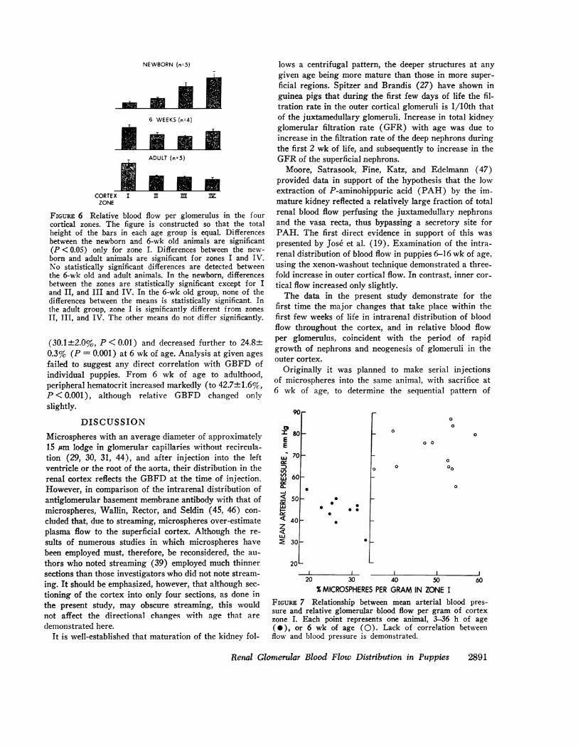
FIGURE 6 Relative blood flow per glomerulus in the fourcortical zones. The figure is constructed so that the totalheight of the bars in each age group is equal. Differencesbetween the newborn and 6-wk old animals are significant(P <0.05) only for zone I. Differences between the new-born and adult animals are significant for zones I and IV.No statistically significant differences are detected betweenthe 6-wk old and adult animals. In the newborn, differencesbetween the zones are statistically significant except for Iand II, and III and IV. In the 6-wk old group, none of thedifferences between the means is statistically significant. Inthe adult group, zone I is significantly different from zonesII, III, and IV. The other means do not differ significantly.
(30.1±2.0%, P <0.01) and decreased further to 24.8±0.3% (P = 0.001) at 6 wk of age. Analysis at given agesfailed to suggest any direct correlation with GBFD ofindividual puppies. From 6 wk of age to adulthood,peripheral hematocrit increased markedly (to 42.7±1.6%,P < 0.001), although relative GBFD changed onlyslightly.
DISCUSSION
Microspheres with an average diameter of approximately15 sm lodge in glomerular capillaries without recircula-tion (29, 30, 31, 44), and after injection into the leftventricle or the root of the aorta, their distribution in therenal cortex reflects the GBFDat the time of injection.However, in comparison of the intrarenal distribution ofantiglomerular basement membrane antibody with that ofmicrospheres, Wallin, Rector, and Seldin (45, 46) con-cluded that, due to streaming, microspheres over-estimateplasma flow to the superficial cortex. Although the re-sults of numerous studies in which microspheres havebeen employed must, therefore, be reconsidered, the au-thors who noted streaming (39) employed much thinnersections than those investigators who did not note stream-ing. It should be emphasized, however, that although sec-tioning of the cortex into only four sections, as done inthe present study, may obscure streaming, this wouldnot affect the directional changes with age that aredemonstrated here.
It is well-established that maturation of the kidney fol-
lows a centrifugal pattern, the deeper structures at anygiven age being more mature than those in more super-ficial regions. Spitzer and Brandis (27) have shown inguinea pigs that during the first few days of life the fil-tration rate in the outer cortical glomeruli is 1/10th thatof the juxtamedullary glomeruli. Increase in total kidneyglomerular filtration rate (GFR) with age was due toincrease in the filtration rate of the deep nephrons duringthe first 2 wk of life, and subsequently to increase in theGFRof the superficial nephrons.
Moore, Satrasook, Fine, Katz, and Edelmann (47)provided data in support of the hypothesis that the lowextraction of P-aminohippuric acid (PAH) by the im-mature kidney reflected a relatively large fraction of totalrenal blood flow perfusing the juxtamedullary nephronsand the vasa recta, thus bypassing a secretory site forPAH. The first direct evidence in support of this waspresented by Jose et al. (19). Examination of the intra-renal distribution of blood flow in puppies 6-16 wk of age,using the xenon-washout technique demonstrated a three-fold increase in outer cortical flow. In contrast, inner cor-tical flow increased only slightly.
The data in the present study demonstrate for thefirst time the major changes that take place within thefirst few weeks of life in intrarenal distribution of bloodflow throughout the cortex, and in relative blood flowper glomerulus, coincident with the period of rapidgrowth of nephrons and neogenesis of glomeruli in theouter cortex.
Originally it was planned to make serial injectionsof microspheres into the same animal, with sacrifice at6 wk of age, to determine the sequential pattern of
90
0I 8CEE
L; 7C
ad(I)
Y
600i< 50
LU
< 40zLU* 30
_
S
00
00
* 0
I-
.)
0
00
0
0 0
0
000
2OL__I _j
20 30 40 50 60%MICROSPHERESPER GRAMIN ZONE I
FIGuRE 7 Relationship between mean arterial blood pres-sure and relative glomerular blood flow per gram of cortexzone I. Each point represents one animal, 3-36 h of age(0), or 6 wk of age (0). Lack of correlation betweenflow and blood pressure is demonstrated.
Renal Glomerular Blood Flow Distribution in Puppies 2891
01
oD_
oF_

change in the same kidney. The results of such studiesmade it clear, however, that microspheres injected withinthe first days or weeks of life had shifted to relativelydeeper positions when examined weeks later, due to theformation of new glomeruli in the outer cortex and tochanges in the distribution of glomeruli within the cortexas a consequence of tubular growth and the developmentof an aglomerular subcapsular cortical zone. Injections ofmicrospheres at various ages and examination at 6 wkdemonstrated that most of these changes took place dur-ing the Ist wk of life, with little additional change oc-curring after the 3rd wk.
The marked increase that we observed during the first6 wk of life in relative glomerular blood flow per gramof outer cortex (zone I) thus could have been due toformation of new glomeruli, or a relative increase in flowper glomerulus, due to growth of glomeruli or changes invascular resistance. These possibilities were explored bydetermining histologically the percent of glomeruli ineach zone in the newborn, at 6 wk of age, and in theadult. The major change observed was a decrease inthe percent of glomeruli in the outermost cortex, due,as noted above, to the development of the cortex corticis.That these differential rates of growth are not completedby 6 wk of age is shown by the differences in distribu-tion of glomeruli found between the 6-wk old and theadult animals.
When the relative GBFD is factored by the relativedistribution of glomeruli per zone, it becomes apparentthat a marked increase in perfusion of the outer corticalglomeruli took place during the time period covered bythe study.' In the deepest zone of the cortex a relativedecrease in flow per glomerulus took place, reflectingthe ascendancy of the outer cortex by 6 wk of age.However, blood flow to the most superficial glomeruli at6 wk of age was only 20% greater than that of glomeruliin zone IV, and 50% greater than in zone II and thesedifferences were not statistically significant. In con-
trast, the perfusion of the outer cortical glomeruli in theadult dog is almost double that of the glomeruli in zones
II and III, and more than double that in IV.Few other data are available concerning plasma flow
in the superficial and juxtamedullary glomeruli. KUll-skog, Ulfendahl, and Wolgast (48), using a modified mi-crosphere technique in rats, found perfusion of the outercortical glomeruli to be 20% greater than the juxtamedul-lary glomeruli, a difference that was not statistically sig-nificant. Barratt, Wallin, Rector, and Seldin (49), using
'It is recognized that heterogeneity of nephrons withineach zone of the cortex and the difficulty in differentiatingperfused from nonperfused glomeruli gives this calculationonly semiquantitative significance. Nevertheless, these datado serve to demonstrate the differences in glomerular bloodflow in various levels of the cortex and the changes thattake place with age.
the antiglomerular basement membrane-antibody tech-nique, found higher single-nephron plasma flow in thejuxtamedullary cortex than in the superficial cortex ofthe hydropenic rat, with an accentuation of this differ-ence following massive volume expansion. Using thesame technique, however, Wallin et al. (45) found nodifferences in plasma flow in outer cortical, midcortical,and inner cortical glomeruli in hydropenic dogs.
In the present studies relative blood flow per glomeru-lus was found to be very low in the superficial cortex ofnewborn animals, and to increase progressively with age.Although total kidney blood flow was not determined, thevalues shown in Table III were calculated utilizing dataobtained in other puppies studied in this laboratory (50).In this fashion the magnitude of change in blood flowvper glomerulus throughout the cortex becomes apparent.From birth to 6 wk of age, the 20-fold increase in renalblood flow per kidney was associated with only a 4-foldincrease in blood flow in the deep glomeruli (zone IIIand IV), but a greater than 10-fold increase in theglomeruli of zone II, and a 25-fold increase in zone I.Examined in another way, 60% of the increase in kid-ney blood flow took place in zone I, with less than 5%being contributed by zone IV. Since these changes greatlyexceed the increase in size of glomeruli, it is apparentthat local changes in glomerular vascular resistancemust have taken place. Although, as shown in Fig. 6, thepattern of blood flow distribution at age 6 wk resemblesthat of the adult kidney, the magnitude of the quan-titative change still to occur is apparent.
The mechanism(s) controlling the distribution of bloodwithin various segments of the cortex remains to be elu-cidated. No correlation was found in the present studybetween changes in distributional flow and either periph-eral hematocrit (51, 52) or blood pressure (28). It maybe of significance that peripheral renin levels in theneonate are high (53). Additionally the role of thesympathetic nervous system has been studied by Joseet al. (54), who concluded that the pattern of intrarenalblood flow distribution may be due to a high level ofalpha adrenergic activity.
Finally, comment should be made of the possible func-tional significance of these marked hemodynamic changes.The existence of two functionally dissimilar populationsof nephrons in the outer and inner cortex of the adultanimal has been commented on recently by Jamison (55).It is recognized that the neonate responds in a limitedfashion to salt loading, a phenomenon that may reflectthe extreme immaturity and low level of filtration of the
outer cortical nephrons (56). In contrast, diluting ca-
pacity and the ability to respond to a water load are rela-
tively well-developed, reflecting the more mature func-tional level of the deep nephrons (56). It is likely that
2892 Olbing, Blaufox, Aschinberg, Silkalns, Bernstein, Spitzer, and Edelmann

TABLE II IChanges with Age in Kidney Size, Nephron Number, Glomerular Distribution, and Blood Flow
Glomerular no. Blood flow
Zone Weight total per gram per glom. per gram total
g nl/min ml/min mi/minNewborn
Cortex I 0.766 175,136 228,636 3 0.686 0.525II 0.617 91,722 148,658 5 0.743 0.459III 0.481 24,777 51,511 11 0.566 0.272IV 0.363 8,365 23,044 16 0.369 0.134
Whole kidney 3.26 300,000 - 0.426 1.39
6 WkCortex I 2.70 194,807 72,150 79 5.70 15.39
II 2.31 129,977 56,268 53 2.98 6.89III 1.94 58,349 30,077 47 1.41 2.74IV 1.61 16,867 10,476 65 0.68 1.10
Whole kidney 12.6 400,000 - 2.07 26.12
AdultCortex I 15.8 125,164 7,922 822 6.51 102.9
II 12.8 164,501 12,852 444 5.71 73.0III 10.1 82,333 8,152 420 3.42 34.6IV 7.1 28,001 3,944 350 1.38 9.8
Whole kidney 58.4 400,000 - 3.77 220.3
The weight of each zone is calculated according to the method of McNay and Abe (32), assumingthe kidney to have the shape of an ellipsoid. Although this method has serious limitations, they donot affect the conclusions drawn from the derived data. Glomerular number per gram of cortex iscalculated from the data of relative glomerular distribution (RGD) shown in Table II (see footnote)and the weight of each zone, as follows. Let Giv be the number of glomeruli per gram of zone IV,and Wiv be the weight of zone IV. It then follows that,
Gi = Giv X RGDI/RGDiv, (1)Gii = Giv X RGDII/RGDIV, (2)
Giii = Giv X RGDIII/RGDxv, (3)andGiWi + GiiWii + GiiiWiii + GivWIv = total glomerular number. (4)
By substituting equations (1)-(3) in (4), using appropriate values of RGD, and assuming totalglomerular number of 300,000 per kidney in the newborn and 400,000 per kidney for the olderanimals, (4) is solved for glomerular number per gram of zone IV. Glomerular numbers for the otherzones are then readily calculated. Blood flow data are derived in a similar fashion, using the calcu-lated glomerular number per zone and the data for relative glomerular perfusion, as shown in Fig. 7.Renal blood flow per kidney is based on unpublished data from this laboratory.
as more data are obtained concerning the differentialrates of development of the cortex, other functional char-acteristics of the immature kidney will be elucidated.
ACKNOWLEDGMENTSThe authors wish to thank Sylvia M. Wassertheil-Smoller,Ph.D., for help and advice with the statistical analyses, andthey acknowledge the excellent secretarial assistance of Mrs.Jean Massaro.
This work was supported in part by U. S. Public HealthService Grants AM 14,877, HL 5267, and HL 11,984; TheHealth Research Council of the City of New York, Inc.,I-605; and The Sylvan League, Inc.
REFERENCES1. Barnett, H. L. 1940. Renal physiology in infants and
children. I. Method for estimation of glomerular filtra-tion rate. Proc. Soc. Exp. Biol. Med. 44: 654.
2. McCance, R. A., and W. F. Young. 1941. The secretionof urine by newborn infants. J. Physiol. (Lond.). 99:265.
3. Dean, R. F. A., and R. A. McCance. 1947. Inulin, dio-done, creatinine and urea clearance in newborn infants.J. Physiol. (Lond.). 106: 431.
4. Rubin, M. I., E. Bruck, and M. Rapoport. 1949. Matu-ration of renal function in childhood. Clearance studies.J. Clin. Invest. 28: 1144.
5. Weil, W. B., Jr. 1955. Evaluation of renal function ininfancy and childhood. Am. J. Med. Sci. 229: 678.
Renal Glomerular Blood Flow Distribution in Puppies 2893

6. West, J. R., H. W. Smith, and H. Chasis. 1948. Glo-merular filtration rate, effective renal blood flow, andmaximal tubular excretory capacity in infancy. J. Pedi-atr. 32: 10.
7. Barnett, H. L., W. K. Hare, H. McNamara, and R. S.Hare. 1948. Influence of postnatal age on kidney func-tion of premature infants. Proc. Soc. Exp. Biol. Med.69: 55.
8. Dicker, E. E. 1952. Effect of diuretics in new-bornrats and puppies. J. Physiol. (Lond.). 118: 384.
9. Heller, J., and K. Capek. 1965. Changes in body watercompartments and inulin and PAH clearance in thedog during postnatal development. Physiol. Bohemoslov.14: 433.
10. Horster, M., and H. Valtin. 1971. Postnatal develop-ment of renal function: micropuncture and clearancestudies in the dog. J. Clin. Invest. 50: 779.
11. Boylan, J. W., E. P. Colbourn, and R. A. McCance.1958. Renal function in the foetal and new-born guineapig. J. Physiol. (Lond.). 141: 323.
12. Chez, R. A., F. G. Smith, and D. C. Hutchinson. 1964.Renal function in the intrauterine fetus. I. Experi-mental technique: rate of formation and chemical com-position of urine. Am. J. Obstet. Gynecol. 90: 128.
13. Falk, G. 1955. Maturation of renal function in rats.Am. J. Physiol. 181: 157.
14. Horster, M., and J. E. Lewy. 1970. Filtration fractionand extraction of PAH during neonatal period in therat. Am. J. Physiol. 219: 1061.
15. Potter, D., A. Jarrah, J. Sakai, J. Harrah, and M. A.Holliday. 1969. Character of function and size in kidneyduring normal growth of rats. Pediatr. Res. 3: 51.
16. Alexander, D. P., and D. A. Nixon. 1962. Plasma clear-ance of p-aminohippuric acid by the kidneys of foetal,neonatal and adult sheep. Nature (Lond.). 194: 483.
17. Alexander, D. P., and D. A. Nixon. 1963. Reabsorptionof glucose, fructose, and mesoinositol by the foetal andpost-natal sheep kidney. J. Physiol. (Lond.). 167: 480.
18. Calcagno, P. L., and M. I. Rubin. 1963. Renal ex-traction of para-aminohippurate in infants and chil-dren. J. Clin. Invest. 42: 1632.
19. Jose, P. A., A. G. Logan, L. M. Slotkoff, L. S. Lilien-field, P. L. Calcagno, and G. M. Eisner. 1971. Intra-renal blood flow distribution in canine puppies. Pediatr.Res. 5: 335.
20. Alexander, D. P., and D. A. Nixon. 1961. The foetalkidney. Br. Med. Bull. 17: 112.
21. Oh, W., M. A. Oh, and J. Lind. 1966. Renal functionand blood volume in newborn infant related to placentaltransfusion. Acta Paediatr. Scand. 55: 197.
22. Gruskin, A. B., C. M. Edelmann, Jr., and S. Yuan.1970. Maturational changes in renal blood flow in pig-lets. Pediatr. Res. 4: 7.
23. Peter, K. 1927. Untersuchungen uber Bau und Ent-wicklung der Niere. Gustav Fischer, Jena, East Ger-many. 2nd volume.
24. Potter, E. L., and S. T. Thierstein. 1943. Glomerulardevelopment in the kidney as an index for fetal ma-turity. J. Pediatr. 22: 695.
25. Ljundquist, A. 1963. Fetal and postnatal development ofintrarenal arterial pattern in man. A micro-angiographicand histologic study. Acta Paediatr. 52: 443.
26. Fettermann, G. H., N. A. Shuplock, F. J. Philipp, andH. S. Gregg. 1965. The growth and maturation ofhuman glomeruli and proximal convolutions from term
to adulthood. Studies by microdissection. Pediatrics. 35:601.
27. Spitzer, A., and M. Brandis. 1972. Superficial nephronand total kidney glomerular filtration rate during de-velopment. Pediatr. Res. 6: 416.
28. Kleinman, L. I., and J. H. Reuter. 1973. Maturation ofglomerular blood flow distribution in the new-born dog.J. Physiol. (Lond.). 228: 91.
29. Rudolph, A. M., and M. A. Heymann. 1967. The circu-lation of the fetus in utero. Methods for studying dis-tribution of blood flow, cardiac output and organ bloodflow. Circ. Res. 21: 163.
30. Wagner, H. N., Jr., B. A. Rhodes, Y. Sasaki, and J. P.Ryan. 1969. Studies of the circulation with radioactivemicrospheres. Invest. Radiol. 4: 374.
31. Milstein, D. M., H. B. Lee, T. Liang, and M. D.Blaufox. 1972. Glomerular blood flow distribution in therat. Preliminary observations. In Radionuclides in Ne-phrology. M. D. Blaufox, and J.-L. Funk-Brentano,editors. Grune & Stratton, Inc., New York. 17.
32. McNay, J. L., and Y. Abe. 1970. Pressure-dependentheterogeneity of renal cortical blood flow in dogs.Circ. Res. 27:571.
33. McNay, J. L., and Y. Abe. 1970. Redistribution ofcortical blood flow during renal vasodilitation in dogs.Circ. Res. 27: 1023.
34. McNay, J. L., and Y. Abe. 1970. Effects of acute andchronic salt loading on the distribution of renal corticalblood flow. Clin. Res. 18: 510. (Abstr.)
35. Miyazaki, M., and J. McNay. 1971. Redistribution ofrenal cortical blood flow during ureteral occlusion andrenal constriction. Proc. Soc. Exp. Biol. Med. 138: 454.
36. Stein, J. H., T. F. Ferris, J. E. Huprich, T. C. Smith,and R. W. Osgood. 1971. Effect of renal vasodilatationon the distribution of cortical blood flow in the kidneyof the dog. J. Clin. Invest. 50: 1429.
37. Bay, W. H., J. H. Stein, J. B. Rector, R. W. Osgood,and T. F. Ferris. 1972. Redistribution of renal corticalblood flow during elevated ureteral pressure. Am. J.Physiol. 222: 33.
38. Jose, P., L. Slotkoff, L. Lilienfield, S. Counts, P. Cal-cagno, and G. Eisner. 1971. Adrenergic activity andsalt excretion. Clin. Res. 19: 534. (Abstr.)
39. Katz, M. A., R. C. Blantz, F. C. Rector, Jr., and D. W.Seldin. 1971. Measurement of intrarenal blood flow. I.Analysis of microsphere method. Am. J. Physiol. 220:1903.
40. Blantz R. C., M. A. Katz, F. C. Rector, Jr., and D. W.Seldin. 1971. Measurement of intrarenal blood flow. II.Effect of saline diuresis in the dog. Am. J. Physiol.220: 1914.
41. Arango, A., and M. I. Rowe. 1971. The neonatal puppyas an experimental subject. Biol. Neonate. 18: 173.
42. Lubbe, R. J., and L. I. Kleinman. 1969. Relationshipbetween GFR and blood pressure in newborn puppies.Physiologist. 12: 289.
43. Snedecor, G. W. 1956. Statistical Methods Applied toExperiments in Agriculture and Biology. Iowa StateUniversity Press, Ames.
44. Slotkoff, L. M., A. Logan, P. Jose, J. D'Avella, andG. M. Eisner. 1971. Microsphere measurement of intra-renal circulation of the dog Circ. Res. 28: 158.
45. Wallin, J. D., F. C. Rector, Jr., and D. W. Seldin.1971. Measurement of intrarenal plasma flow with anti-glomerular basement-membrane antibody Am. J. Phys-iol. 221: 1621.
2894 Olbing, Blaufox, Aschinberg, Silkalns, Bernstein, Spitzer, and Edelmann

46. Wallin, J. D., F. C. Rector, Jr., and D. W. Seldin.1972. Effect of volume expansion on intrarenal distribu-tion of plasma flow in the dog Am. J. Physiol. 223:125.
47. Moore, E. S., S. S. Satrasook, B. P. Fine, M. C.Katz, and C. M. Edelmann, Jr. 1969. Renal PAH ex-traction in puppies. 39th Annual Meeting of the So-ciety of Pediatric Research, Atlantic City, N. J., 2, 3May. 114. (Program and Abstr.)
48. Killskog, O., H. R. Ulfendahl, and M. Wolgast. 1972.Single glomerular blood flow as measured with carbon-ized "41Ce labelled microspheres. Acta Physiol. Scand.85: 408.
49. Barratt, L. J., J. D. Wallin, F. C. Rector, Jr., andD. W. Seldin. 1973. Influence of volume expansion onsingle-nephron filtration rate and plasma flow in therat. Am. J. Physiol. 224: 643.
50. Aschinberg, L. C., D. I. Goldsmith, H. Olbing, M. A.Hardy, A. Spitzer, C. M. Edelmann, Jr., and M. D.Blaufox. 1973. Neonatal changes in renal blood flowdistribution in puppies. Pediatr. Res. 7: 183. (Abstr.)
51. Nashat, F. S., and R. W. Portal. 1967. The effects ofchanges in haematocrit on renal function. J. Physiol.(Lond.). 193: 513.
52. Schrier, R. W., and L. E. Earley. 1970. Effects of acutechanges of hematocrit on renal hemodynamics and so-dium excretion in hydropenic and volume-expandeddogs. J. Clin. Invest. 49: 1656.
53. Kotchen, T. A., A. L. Strickland, T. W. Rice, andD. R. Walters. 1972. A study of the renin-angiotensinsystem in newborn infants J. Pediatr. 80: 938.
54. Jose, P. A., A. Logan, L. M. Slotkoff, L. S. Lilien-field, P. L. Calcagno, and G. M. Eisner. 1972. Intra-renal blood flow distribution in the maturing kidney. InRadionuclides in Nephrology. M. D. Blaufox, and J.-L.Funck-Brentano, editors. Grune & Stratton, Inc., NewYork 87.
53. Jamison, R. L. 1973. Intrarenal heterogeneity. The casefor two functionally dissimilar populations of nephronsin the mammalian kidney. Am. J. Med. 54: 281.
56. Nash, M. A., and C. M. Edelmann, Jr. 1973. The de-veloping kidney. Immature function or inappropriatestandard. Nephron. 11: 71.
Renal Glomerular Blood Flow Distribution in Puppies 2895