mechanismof preservation of glomerular perfusion...
TRANSCRIPT
Mechanism of Preservation of Glomerular Perfusion and Filtrationduring Acute Extracellular Fluid Volume DepletionImportance of Intrarenal Vasopressin-Prostaglandin Interaction for Protecting
Kidneys from Constrictor Action of VasopressinAida Yared, Valentina Kon, and lekuni IchikawaLaboratory of Renal Physiology, Department of Medicine, The Children's Hospital and Department of Pediatrics,Harvard Medical School, Boston, Massachusetts 02115
Abstract
Glomerular circulatory dynamics were assessed in 60 adultanesthetized rats, which were either deprived or not deprivedof water for 24-48 h. Water-deprived rats (a = 21) were char-acterized by a depressed level of single nephron glomerularfiltration rate (SNGFR) when compared with nonwater-deprivedcontrols (a = 8) (23.2±1.3 vs. 44.8±4.1 nl/min). This wasprimarily due to decreased glomerular plasma flow rate (71±5vs. 169±23 nl/min) and glomerular capillary ultrafiltrationcoefficient (0.028+0.003 vs. 0.087±0.011 nl/Is-mmHgj). In-fusion of saralasin to these water-deprived rats resulted insignificant increases in plasma flow rate and ultrafiltrationcoefficient, and decline in arteriolar resistances. Consequently,SNGFRincreased by .50% from pre-saralasin levels. Whenwater-deprived saralasin-treated rats were given a specificantagonist to the vascular action of arginine vasopressin (AVP),d(CH2)5Tyr(Me)AVP, a fall in systemic blood pressure oc-curred, on average from 102±5 to 80±5 mmHg,unaccompaniedby dilation of renal arterioles, so that both plasma flow rate(129±8 vs. 85±13 nl/min) and SNGFR(31.0±2.9 vs. 18.2±4.4nl/min) decreased.
This more selective extrarenal constrictor action of AVPwas further documented in additional studies in which cardiacoutput and whole kidney blood flow rate were simultaneouslymeasured. In water-diuretic rats, administration of a moderatelypressor dose of AVP (4 mU/kg per min) resulted in asignificant rise in kidney blood flow rate (from 8.8±1.2 to9.6±13 ml/min). The higher kidney blood flow rate occurreddespite a fall in cardiac output (from 111±7 to 98±9 ml/min),and was associated with a significant increase in the ratio ofsystemic vascular to renal vascular resistance (on average from0.083±0.014 to 0.106±0.019). Furthermore, infusion ofd(CH2)ATyr(Me)AVP to water-deprived animals (a = 6) toantagonize endogenous AVP resulted in systemic but not renalvasodilation, so that kidney blood flow rate fell (by -30%),as did systemic-to-renal resistance ratio (by -30%). When the
Portions of these studies were presented at the annual meeting of theAmerican Federation for Clinical Research, Washington, DC, 1983; atthe American Society of Nephrology, Washington, DC, 1983; andwere published in abstract form (Clin. Res. 31:431, 1983; Kidney Int.25:293, 1984.
Address correspondence to Dr. Ichikawa.Received for publication 19 January 1984 and in revised form 3
January 1985.
above two experiments were repeated in indomethacin-treatedanimals, exogenous AVP administration in water-diuretic rats(a = 6) and antagonism of endogenous AVP in water-deprivedrats (a = 7) caused, respectively, parallel constriction anddilation in systemic and renal vasculatures. The net effect wasunaltered systemic to renal vascular resistance ratio in bothcases. These results indicate that (1) unlike angiotensin II,AVP maintains glomerular perfusion and filtration in acuteextracellular fluid volume depletion by a more selective con-striction of the extrarenal vasculature. (2) The relative renalinsensitivity to the vasoconstrictor action of AVP appears tobe due to an AVP-induced release of a potent renal vasodilator,sensitive to indomethacin, presumably prostaglandins.
Introduction
Recent evidence indicates that arginine vasopressin (AVP),' inplasma concentrations within the physiological range, exerts acritically important role in the maintenance of blood pressureand systemic circulatory dynamics (1-4). The availability ofseveral forms of synthetic analogues to the vascular action ofthe hormone (1-7) has allowed experimental demonstrationof this role.
In addition to its systemic effect, AVP possesses a directvasoconstrictor action on the glomerular microcirculation. Anon-pressor dose of AVP leads to a profound fall in theglomerular capillary ultrafiltration coefficient (Kf) (8), presum-ably by inducing mesangial cell contraction (9). Moreover, inour recent experiments with two-kidney Goldblatt hypertension,an experimental model characterized by a high circulatinglevel of AVP (10), a specific AVP antagonist was shown tomarkedly raise Kf.
Of related interest are the observations (11-13) that therenal vasculature is less sensitive to the constrictor action ofexogenous AVP than are some extrarenal vascular beds. Thepresent study aims at defining the role of endogenously released
1. Abbreviations used in this paper: All, angiotensin II; AP, meansystemic arterial pressure; TA, systemic plasma oncotic pressure; AVP,arginine vasopressin; CA, systemic plasma protein concentration; CE,efferent arteriolar plasma protein concentration; CO, cardiac output;ECF, extracellular fluid; TE, efferent arteriolar plasma oncotic pressure;Kf, glomerular capillary ultrafiltration coefficient; non-WD, nonwater-deprived rats; PE, efferent arteriolar hydraulic pressure; PGC, mean
glomerular capillary hydraulic pressure; PT, proximal tubule hydraulicpressure; AP, mean glomerular transcapillary hydraulic pressure differ-ence; QA, initial glomerular plasma flow rate; RA, afferent arteriolarresistance; RBF, whole kidney blood flow rate; RE, efferent arteriolarresistance; RVR, renal vascular resistance; SNFF, single nephronfiltration fraction; SNGFR, single nephron glomerular filtration rate;SVR, systemic vascular resistance; WD, water-deprived rats.
Renal Hemodynamics in Acute Extracellular Fluid Volume Depletion 1477
J. Clin. Invest.C The American Society for Clinical Investigation, Inc.0021-9738/85/05/1477/11 $ 1.00Volume 75, May 1985, 1477-1487
AVP in determining the prevailing levels of renal perfusionand filtration during acute extracellular fluid (ECF) volumedepletion. The overall functional effect of AVP on renalcirculation under a given set of physiological circumstances isnot readily predictable, since renal circulatory dynamics aredetermined by the balance between intrinsic renal and extrare-nal vascular tones, as well as the level of cardiac output (CO).Wetherefore studied the effects of specific antagonism to thevascular action of AVP, by measuring various renal andsystemic circulatory indices in water-deprived animals. Todocument the uniqueness of AVP action, the results werecompared with the vasodilatory influence of angiotensin II(AII) inhibition, as well as that of hydralazine, a drug knownto dilate both renal and extrarenal vascular beds ( 14). It shouldbe noted that, although the sympathetic nervous system andcirculating catecholamines likely contribute to the prevailingrenal hemodynamics in volume-depleted animals, their rolewill not be addressed in the present study.
Methods
General. Experiments were performed in nine groups of adult malerats. Munich-Wistar rats were used for the experiments involvingmicropuncture (groups 1-5), and Sprague-Dawley rats in studieswithout micropuncture (groups 6-9). Before study, the animals weremaintained on standard rat pellet chow. In groups 1-4, 6, and 7, acuteECF depletion was induced by withholding drinking water 24-48 hbefore experiment. Rats in groups 5, 8, and 9 were allowed free accessto water until the time of study.
At the time of study, the animals were anesthetized with Inactin(Byk, Federal Republic of Germany; 100 mg/kg, i.p.), placed on atemperature-regulated table, and subjected to tracheostomy. Indwellingpolyethylene catheters were inserted into the left and/or right jugularveins for infusion of various intravenous solutions as specified below.The left femoral artery was also catheterized for periodic bloodsampling and estimation of mean arterial pressure (AP). The AP wasmonitored with an electronic transducer (model p23Db, StathamInstruments Div., Gould, Inc., Oxnard, CA) connected to a direct-writing recorder (model 2200, Gould Inc.). In group 6-9 animals, acatheter was inserted into the left ventricle through the left carotidartery for injection of radioactive microspheres. Correct placement ofthe catheter tip was confirmed by pressure tracing. The left kidney wasexposed through a left subcostal incision and gently separated fromthe adrenal gland and the surrounding perirenal fat. In group 1-5studies, the kidney was suspended on a Lucite holder, and its surfaceilluminated with a fiberoptic light source and bathed with isotonicNaCl solution heated to 350-370C. A 0.5-ml bolus intravenous injectionof 10% inulin in 0.9% NaCl was given, followed by a continuousinfusion at the rate of 0.6 ml/h (groups 1-4) or 1.2 ml/h (group 5).
Since the plasma volume of rats prepared surgically in the abovefashion is substantially reduced from the level prevailing in theconscious state (14), estimated plasma losses were replaced in group1-9 animals with use of the formula described in detail previously(15). The amount of iso-oncotic plasma used was -1% of body weightin group 5, 8, and 9 animals, and 0.5-0.7% in group 1-4, 6, and 7animals.
Micropuncture measurements. In group 1-5 animals, micropuncturemeasurements were carried out as follows: exactly timed (1-2 min)samples of tubule fluid were collected from surface proximal convo-lutions of two or three nephrons for determination of flow rate andinulin concentration. These measurements permitted calculation ofsingle nephron glomerular filtration rate (SNGFR). Coincident withthese tubule fluid collections, two or three samples of femoral arterialblood were obtained in each period for determination of arterialhematocrit and plasma concentrations of protein and inulin. Timeaveraged hydraulic pressures were measured in surface glomerular
capillaries (PFc), proximal tubules (PT), and surface efferent arterioles(PE) with a continuous recording, servo-null micropipette transducersystem (model 3, Instrumentation for Physiology and Medicine, SanDiego, CA). Micropipettes with outer tip diameters of 1-2 gm andcontaining 2.0 Msodium chloride were used. Hydraulic output fromthe servo-nulling system was coupled electronically to a second channelof the recorder.
Colloid osmotic pressure (7r) of plasma entering and leaving glo-merular capillaries was estimated from values for protein concentration(C) in femoral arterial (CA) and surface efferent arteriolar (CE) plasmasamples by using the equation derived by Deen et al. (16). Values forCA, and thus systemic plasma oncotic pressure (rA), in femoral arterialplasma were taken as representative of values for C and wr at theafferent end of the glomerular capillary network. These estimates ofpreglomerular and postglomerular plasma protein concentrations per-mitted calculation of single nephron filtration fraction (SNFF) andinitial glomerular plasma flow rate (QA). Kf, as well as resistances ofsingle afferent (RA) and efferent (RE) arterioles, were calculated byusing the equations given elsewhere (17).
Cardiac output and whole kidney blood flow rate (RBF) measure-ments. These two indices were measured in group 6-9 studies.
CO was determined with carbonized microspheres (3M Co., St.Paul, MN), 15±0.6 Mmin diameter, labeled with 5"Cr. An isotonicsaline solution in a volume of 50 til, containing -35,000 microsphereswas placed into an 8-cm length of Silastic tube (ID 0.04 in., DowChemical Co., Midland, MI). The tube was capped at both ends. Itsradioactivity was measured immediately before use in a Packard Tri-Carb solid crystal gammaradiation counter. At the time of study, themicrospheres were disaggregated and flushed (with 0.3 ml isotonicsaline) into the left ventricular cavity through an indwelling catheterover a 20-s period. Concurrently, arterial femoral blood (-1.5 ml)was collected for 30 s by unclamping the femoral arterial catheter andallowing the blood to flow freely into a graduated test tube. Replacementtransfusion was simultaneously performed, using whole blood obtainedfrom littermates of the experimental animals. For group 8 and 9studies, the blood was diluted with 0.9% NaCl to achieve a low serumprotein concentration (4.5-5.0 g/dl) comparable with that of therecipient animals, while keeping the hematocrit unchanged. No fluc-tuation of arterial pressure occurred during the procedure. Residualradioactivity in the Silastic tube and its caps was measured. Cardiacoutput was calculated as: CO = (counts injected into left ventricle)/(counts collected in femoral arterial blood X femoral arterial bloodflow rate).
Left renal arterial blood flow rate was measured with an electro-magnetic flow probe (model FP402, Carolina Medical Electronics, Inc.,King, NC, 2.0 mmin circumference) connected to an electromagneticflow meter (model 501, Carolina Medical Electronics, Inc.). This flowmeter system was calibrated in vivo (16) before use. Systemic (SVR)and renal (RVR) vascular resistances were calculated as the ratio ofAP to COand RBF, respectively.
Experimental groups. Animals were divided into nine groups.Groups 1-5 underwent micropuncture studies as described above,groups 1-4 being deprived of water for 24-48 h, while group 5 wasallowed free access to water until the time of study. Groups 6-9 hadsimultaneous measurement of AP, CO, and RBF; again, groups 6 and7 were water deprived for 24-48 h, while groups 8 and 9 were allowedfree access to water until the time of study. Individual protocols aredescribed in detail below, and summarized in Table I.
Group 1 (seven Munich-Wistar rats): The time course of renalmicrocirculatory and systemic dynamics of acutely water-deprived rats(WD) was examined by micropuncture. The micropuncture measure-ments and collections were obtained 90 and 120 min after inductionof anesthesia, and completed within 30 min. These measurements andcollections were repeated =70 min later, which was a timing similarto the third study period in group 2-4 animals.
Group 2 (eight Munich-Wistar rats): In WDof group 2, the effectof AVP inhibition after All blockade was studied. Baseline micro-puncture measurements and collections were performed as in the first
1478 A. Yared, V. Kon, and I. Ichikawa

Table I. Summary of Individual Protocols for Group 1-9 Experimental Animals
Fluid intake before study First study period Second study period Third study period
Group 1 24-48-H water deprivation No treatment No treatment(7 MWrats)
Group 2 24-48-H water deprivation No treatment Saralasin Saralasin(8 MWrats) + d(CH2)5Tyr(Me)AVP
Group 3 24-48-H water deprivation No treatment d(CH2)5Tyr(Me)AVP d(CH2)5Tyr(Me)AVP(6 MWrats) + saralasin
Group 4 24-48-H water deprivation Saralasin Saralasin + hydralazine(6 MWrats)
Group 5 Free access to water No treatment Saralasin(8 MWrats)
Group 6 24-48-H water deprivation Teprotide Teprotide(6 SD rats) + d(CH2)5Tyr(Me)AVP
Group 7 24-48-H water deprivation Indomethacin + teprotide Indomethacin + teprotide(7 SD rats) + d(CH2)5Tyr(Me)AVP
Group 8 Free access to water No treatment AVP(6 SD rats)
Group 9 Free access to water Indomethacin Indomethacin + AVP(6 SD rats)
Dosages employed are 0.3 mg/kg per h (saralasin), 20 ,g (d[CH2]5Tyr[Me]AVP), 0.05 mg (hydralazine), 4 mU/kg per min (AVP), 6 mg/kg perh (teprotide), and 2 mg/kg (indomethacin, group 7) and 2 mg/kg + 2 mg/kg per h (indomethacin, group 9). Munich-Wistar and Sprague-Dawley rats are abbreviated as MWand SD rats, respectively.
period of group 1 animals. At the end of this first study period, anintravenous infusion of saralasin (0.3 mg/kg per h), an All antagonist,was started and continued throughout the rest of the experiment. Aftera 40-min equilibration period, measurement of the SNGFRand itsdeterminants was repeated. Immediately after completion of thissecond study period, each rat received a 20-tg i.v. bolus injection of along acting specific vasopressin vascular antagonist (7), [1 -((-mercapto-f,(,-cyclopentamethylenepropionic acid) 2-(o-methyl) tyrosine] AVP,or d(CH2)JTyr(Me)AVP, and micropuncture measurements and collec-tions were again repeated in the subsequent 30 min.
Group 3 (six Munich-Wistar rats): In group 3 WD, we first studiedthe effect of d(CH2)5Tyr(Me)AVP alone, followed by the addition ofsaralasin in the third study period. In these rats, the first study periodwas carried out as in group 2. Subsequently, d(CH2)5Tyr(Me)AVP wasinjected as in the third study period of group 2. 20 min later,micropuncture was repeated during the next 30 min. Immediatelyafter this second study period, infusion of saralasin was begun at arate identical to group 2. After a 40-min equilibration period, mea-surements and collections again were performed and completed in 30min (third study period).
Group 4 (six Munich-Wistar rats): In this group of WD, the effectof a nonspecific vasodilator on renal microcirculatory hemodynamicswas compared with the effect of AVP antagonist-induced vasodilation(group 2). In this group of animals, the time course described forgroup 2 was duplicated, except that hydralazine (0.05 mg i.v.) wassubstituted for the AVP antagonist, and that measurements andcollections before saralasin infusion were omitted.
Group 5 (eight Munich-Wistar rats): In order to identify thespecificity of endogenous All action in WDanimals, the effect ofsaralasin on the systemic and renal cortical circulations was examinedin eight Munich-Wistar rats allowed free access to tap water until thetime of the study. In these rats, the protocol for the first and secondstudy periods of group 2 animals was duplicated.
Group 6 (six Sprague-Dawley rats): In these WD, the effect of AVPantagonism in the presence of renin-angiotensin inhibition was studied.An intravenous infusion of teprotide (6 mg/kg per h), an angiotensinI converting enzyme inhibitor, was started immediately after inductionof anesthesia, and continued throughout the duration of the experiment.Measurement of AP, CO, and RBFwas begun '90 min after surgicalpreparation, and completed within 15 min. At the end of this firststudy period, rats received 2O-,g i.v. bolus injection ofd(CH2)5Tyr(Me)AVP. 20 min after injection of the AVP antagonist,measurement of AP, CO, and RBFwas repeated (second study period).
Group 7 (seven Sprague-Dawley rats): Seven additional WDwereused to examine the effect of prostaglandin inhibition on the AVPantagonist-induced changes in AP, CO, and RBF. In this group, theprotocol described for group 6 was duplicated except that, in additionto continuous teprotide infusion, group 7 rats received a 2-mg/kg i.v.injection of indomethacin at the start of surgical preparation.
Group 8 (six Sprague-Dawley rats): In these water-diuretic rats, theeffect of a moderately pressor dose of exogenous vasopressin onsystemic and renal circulatory dynamics was examined. In order toachieve hyposthenuria and suppress endogenous AVP release, the ratswere given a continuous hypotonic fluid infusion. Immediately afterrestoration of the surgical plasma loss, an intravenous infusion (0.83%dextrose, 0.3% NaCl) was started at a rate of 60 ml/kg per h. Wepreviously found this regimen to effectively maintain urine hypotonicity(osmolality below 170 mosmol/kg or specific gravity below 1.005)without significantly changing the serum level of glucose (8). Approx-imately 2 h after surgical preparation, measurements of AP, CO, andRBF were begun and completed within 15 min. At the end of thisfirst study period, a continuous intravenous infusion of AVP (4 mU/kg per min, Pitressin, Parke, Davis & Co., Detroit, MI) was started. 5min later, after a steady state was reached for AP, the above-mentionedmeasurements and collections were again performed within a 15-mininterval (second study period).
Renal Hemodynamics in Acute Extracellular Fluid Volume Depletion 1479
Group 9 (six Sprague-Dawley rats): Using additional water-diureticrats, the influence of prostaglandin inhibition on the vasopressin-induced changes in AP, CO, and RBF was examined. The protocoldescribed for group 8 was duplicated, except that group 9 animalsreceived indomethacin (2-mg/kg i.v. bolus at the start of surgicalpreparation, followed by 2 mg/kg per h continuous infusion).
Analytical. The volume of fluid collected from individual proximaltubules was estimated from the length of the fluid column in aconstant-bore capillary tube of known internal diameter. The concen-tration of inulin in tubule fluid was measured, usually in duplicate, bythe microfluorescence method of Vurek and Pegram (18). Inulinconcentration in plasma was determined by the macroanthrone methodof Fuhr et al. (19). CE and CA were determined, usually in duplicate,by the fluorometric method of Viets et al. (20).
Analysis of variance was used to determine statistical significanceof differences in groups 1-5. Paired and unpaired t test was used forgroups 6-9. Statistical significance was defined as P < 0.05. Significancevalues are given at the levels of <0.05 and <0.01.
Results
Base-line renal microcirculatory indices, measured in 21 water-deprived rats (groups 1-3) and 8 nonwater-deprived rats (non-WI) (group 5) are presented in Table II. The body weight ofgroup 1-3 animals averaged 276±6 g before water deprivation,which corresponded to a 12±1% loss of body weight duringthe deprivation. At the time of study, therefore, there was asignificant difference in body weight between WDand non-WDanimals. Mean arterial pressure was comparable in thetwo groups. Mean glomerular capillary hydraulic pressure,PGC, was significantly elevated after water deprivation, whileproximal tubule hydraulic pressUre, PT, was unchanged, leadingto a significantly higher value for the mean glomerular tran-scapillary hydraulic pressure difference, AP. Efferent arteriolarhydraulic pressure (PE) was comparable in the two groups.Hemoconcentration after water deprivation was reflected by asignificant elevation in plasma protein concentration, CA, andhence systemic plasma oncotic pressure, WrA, as well as anincrease in hematocrit. Both SNGFRand QA were decreasedto a similar extent (- 50%) after water deprivation, leading tonear-constancy in SNFF. The marked decrease in SNGFRwas, therefore, attributed largely to the marked decrease in QAand a simultaneously observed marked decrease in the ultra-filtration coefficient, Kf, as well as, but to a lesser extent, theincrease in systemic plasma oncotic pressure, WA. These changeswere partly offset by the increase in AP, the latter tending to
prevent an even greater reduction in SNGFR. The decrease inQA, in turn, was attributed largely to a marked increase in RAand RE. The increase in RE was proportionately more markedthan RA, thus accounting for the observed increase in PGC.
In the absence of pharmacologic intervention (group 1rats), all hemodynamic and renal microcirculatory indicesremained essentially unchanged with time, as shown in Ta-ble III.
Administration of saralasin in the second period to group2 water-deprived rats led only to a slight decrease in AP, onaverage by 6 mmHg(Table III). Saralasin treatment inducedprofound changes in several renal microcirculatory parameters:PGCdecreased while PT remained unchanged, leading to a fallin AP. CA and 7rA were unaffected by angiotensin II inhibition,while both CE and rE decreased. A dramatic increase in QAwas observed; its value doubled after saralasin infusion. SNGFRalso increased by 11 nl/min. The relatively greater increase inQA than SNGFRwas reflected by a fall in SNFF. It is clearfrom Table III that the increase in QAwas the consequence ofa profound fall in both RA and RE. The decrease in RA wasproportionately less than the decrease in RE, which accountedfor the observed decrease in PGCduring saralasin infusion. Inaddition to the increase in QA, Kf was found to markedlyincrease. Overall, saralasin treatment tended to partially correctthe abnormalities seen during water deprivation by modulatingthe levels of Kf, RA, and RE-
After pretreatment with saralasin, administration of theAVPantagonist d(CH2)5Tyr(Me)AVP to group 2 water-deprivedrats led to a substantial reduction in AP, on average by 22mmHg(Table III). Both PGc and AP decreased, while PTremained constant. CA and WrA remained unchanged, whileboth CE and rE decreased. Marked reductions occurred in QAas well as SNGFR, the latter primarily due to the decreases inQA and AP, since values for Kf and CA were essentiallyunaffected, as shown. Contrasting to treatment with saralasinalone, afferent arteriolar resistance was unchanged afterd(CH2)5Tyr(Me)AVP addition, and RE increased slightly.
When d(CH2)5Tyr(Me)AVP, a specific vascular antagonist,was given without simultaneous inhibition of All (group 3,second period), the systemic hemodynamic and renal micro-circulatory parameters were only minimally affected. Thus,the AVP antagonist exerted only a mild depressor effect onsystemic circulation, as indicated by an average fall of AP of7 mmHg,and was without effect on RA, RE, and Kf. Additional
Table II. Summary of Renal Cortical Microcirculatory Indices Measured Under Basal Conditions in WDand Non- WDRats
HctBW AP PuC PT AP PE CA CE HA H1E SNFF SNGFR QA RA RE Kf (Vol)
g mmHg mmHg mmHg mmHg mmHg gidl gidl mmHg mmHg nl/mi nl/min X10° dyn s* cm'n1/(s *mmHg) %
WDrats(n = 21) 238 109 64 14 50 19 6.8 9.7 25 47 0.32 23.2 71 2.43 2.90 0.028 55.5(±) 3 2 1 1 1 1 0.1 0.4 1 2 0.01 1.3 5 0.20 0.24 0.003 0.4
Non-WD rats(n = 8) 264 111 48 12 36 18 5.8 8.1 19 32 0.28 44.8 169 1.68 0.97 0.087 48.2(±) 8 2 2 1 2 1 0.1 0.2 1 1 0.02 4.1 23 0.18 0.14 0.011 1.0
P value* <0.01 NS <0.01 NS <0.01 NS <0.01 <0.01 <0.01 <0.01 NS <0.01 <0.01 <0.05 <0.01 <0.01 <0.01
Values are expressed as mean± 1 SE. Minimum Kf values were calculated when filtration pressure equilibrium was reached (i.e., HIE AP). Unique Kf values werecalculated when filtration pressure equilibrium was not reached (i.e., IIE < AP). To calculate mean Kf values, unique Kf values (14 in WDand 6 in non-WD rats)were pooled within each group. * Calculated using variance analysis. NS denotes P > 0.05.
1480 A. Yared, V. Kon, and I. Ichikawa

00 - en - r4 0oN a% o00tf -4 so -: trn C 4a 6cl C
- i 0 00008- 06C;
WI)0
z z z z z z v V)z
~O en ID~' Nn 000 (O1Nr M~0r-Nr 00~ OON0 000n e'n rO~-l,)rsenco 'IO tn 46 o 00 6 00 00
O2 0 00>O0O O( Q OOO v 0Z 0
0): r ri- O- o0 O4- 0- - 0 6 00 6 00oooooooooo6oooooo6-6oooo~i6-6 oooo~6.-~6- Z V V Z V Z V V
C- 0 ~ Ci 00qen0On O~W
x en Oe 0oren -0 0r rt ' i4- O- -N0C O en O O 0 0 00 0c OrO 0 CD -6-0 6O 0O COO O O O ZO O(6 6 Z V V Z V Z Z V0
Z~~~.~~~Ot-%0 e O S0U -00r-o 0rO 't~0o0e -000000n 0 0 0
0 0 N 00 N 00-N. WN00r- e I --n0C0 0 0 00 6 0 0 00
CIo C4ro_o4_o _o1_o _ _ o o _ Z Z ZV Z Z Z Z Z
,>~~~~~~- - --z VZV VV Z
0~~~~~~~~~~~~~~~~~~~~~
E °aors°s °. o a r°. N i or ir ^ o eoo00rx 00(9n 9oo_9oo . o
4 ! el enIo C-i oo i t biq Ot biq r - O^ CI o- ~ oi "It eno o o o CoO ON 0 O a,00i~rooCrN O o6-Q 0r. r- O 0 O06i6i 00 0 0 0 0 0 00; 00(
0 ri ri ri r' _ ri ri ri r'^ ri t- reo Z V V Z V V Z Z
00 ,O (7., N or - nr wMu-- N-Nre 00 r4 0 0 0 0e°
6666 666666 66o6o66 o66 6ooo Z V VZ V VZZVZ Z V
oo~~~~~~~~~~~~~~~~~~~~~~~~~~~~~~~~~~~~C
0- -- x00 00- oo N -0 -Tx 0 O 9 9 9°
2 b ~ e.~x.b.eoeso ri ro Z V- Z V >V V
etn
sS~~~~~~~~~~~~~~~~~~~~~~~~~~~~~~~~~0 C> '1qeO.n
.oi~e~6 A6.o*6.oi6 ee -ON 6O-O0000 00 00M 0 00°C
2 ki en o.r e Nr en e4n ,Z Z Z Z Z
l la
.6 > > >t , en
Eo v) o v b v q ^ e Z bZ Vqi
t-00 0
tC 06
S£££o t £ £ m e t t t N Z _~~~~~~~~~~~~~~~~~~~V -V&Z Z &ZV Z Z Z
16E~~~~~~~~~~~~~~~~~~~~~~~~~~~~~~~~~~~~~~~~~~~~~~~~~~~~~~~~~~~~~~~~~~~~~~~~~~~~~~~~~~~~~~~~~~~~~~~~~~~~~~
la~~~~~
n en v- 00 in en kn 't an rq as 4 En Co.2- 00-2- 00-00-- Ne fr2 > ir2- 000r 00A~ 0 000c0< 00 00
v~~~~ ~ ~ ~ ~ ~ ~~~ ~ ~~~~~~~~~~~~~~~~~~~~~~~~~~~'L Z Z V
RnHmyaciAtEtcllFu VlmDli
.*00
-4 0 -n -) -e C- - 00CnC. 0e Te n c, (' r o0 0
00 a~~~~~~~~0C 0 a r Z ZZ V Z0 >0
CL. E>~~~~~~.00000 0 > ~~ ~~~~~>> i:U
C~~~~ ~ ~ ~ ~ ~ ~ ~ ~ ~ r 0a t1~~i> I .. 0
C4 Cn o~~~~~00.c.o 0. M0
0 0~~~~a06 6 .' - 'LO >
riririri 00~~~~~ 04000 Z Z~ii 0e r 00r C Q 000sCO 00s > U0 6o >~
en64en 0 CI4 en 0 C400)en ui .2 U5 < i
RenaHeodynmic in cut Exraela Fli oue elto
.0-
.00_0.00
.0
0._
20C
e.0
C:00
D
D)E00
00
Q
00
9r0A
0)
0=0
00z
ui
00O.
00
A00*0
C.)
E._00
.2.=000>0
1481
N- W - so 00 M
U4 W6 mi
IO It I"
"C 01 00I
t
cr Woo;4, as "o*t _ .i
~zgu
0u
Q
Q
tt
AL
Q.)
~.)1
0..)
0.St
0.
1
'I "C en WI tf I" IR
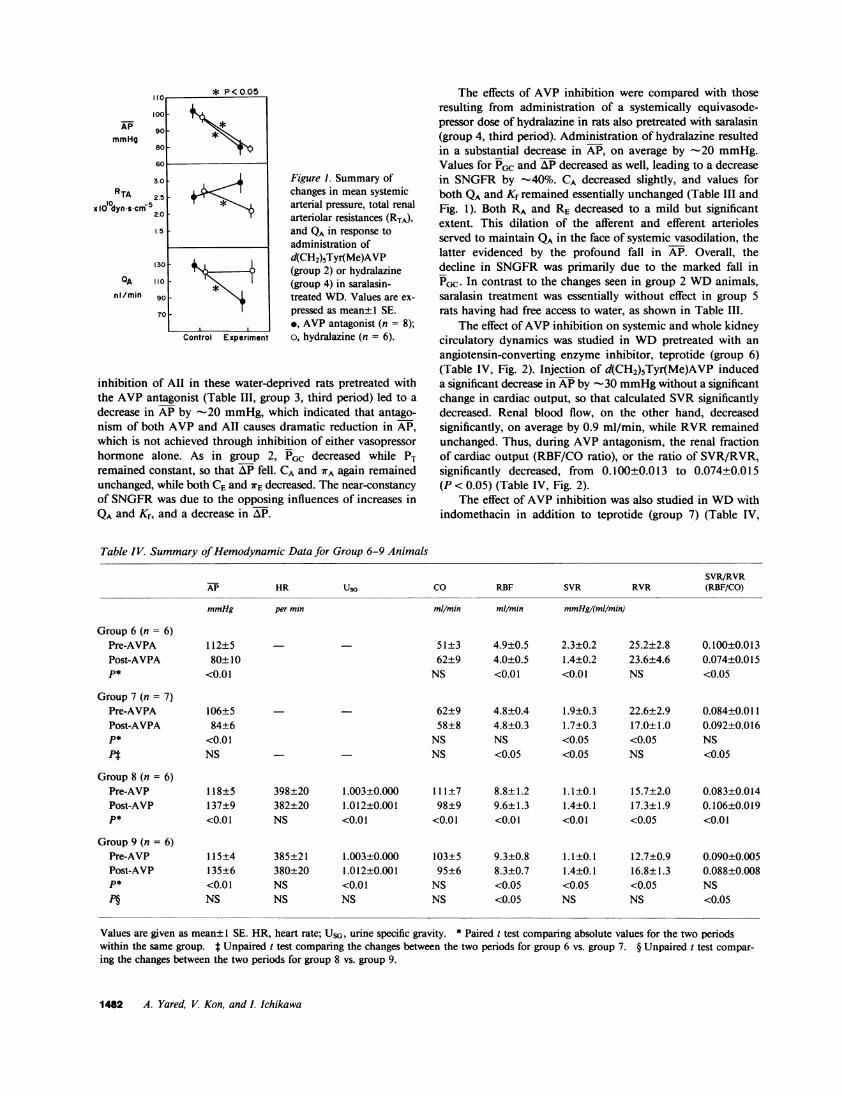
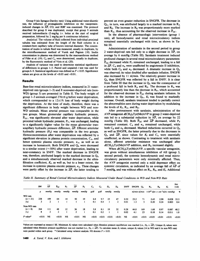
Figure 1. Summary ofchanges in mean systemicarterial pressure, total renalarteriolar resistances (RTA),and QA in response toadministration ofd(CH2)5Tyr(Me)AVP(group 2) or hydralazine(group 4) in saralasin-treated WD. Values are ex-pressed as mean± 1 SE.*, AVP antagonist (n = 8);o, hydralazine (n = 6).
inhibition of All in these water-deprived rats pretreated withthe AVP antagonist (Table III, group 3, third period) led to a
decrease in AP by -20 mmHg, which indicated that antago-nism of both AVP and All causes dramatic reduction in AP,which is not achieved through inhibition of either vasopressor
hormone alone. As in group 2, PGC decreased while PTremained constant, so that AP fell. CA and WA again remainedunchanged, while both CEand WrE decreased. The near-constancyof SNGFRwas due to the opposing influences of increases inQA and Kf, and a decrease in AP.
The effects of AVP inhibition were compared with thoseresulting from administration of a systemically equivasode-pressor dose of hydralazine in rats also pretreated with saralasin(group 4, third period). Administration of hydralazine resultedin a substantial decrease in AP, on average by -20 mmHg.Values for PGC and AP decreased as well, leading to a decreasein SNGFRby -40%. CA decreased slightly, and values forboth QAand Kf remained essentially unchanged (Table III andFig. 1). Both RA and RE decreased to a mild but significantextent. This dilation of the afferent and efferent arteriolesserved to maintain QA in the face of systemic vasodilation, thelatter evidenced by the profound fall in AP. Overall, thedecline in SNGFRwas primarily due to the marked fall inPGC. In contrast to the changes seen in group 2 WDanimals,saralasin treatment was essentially without effect in group 5rats having had free access to water, as shown in Table III.
The effect of AVP inhibition on systemic and whole kidneycirculatory dynamics was studied in WDpretreated with anangiotensin-converting enzyme inhibitor, teprotide (group 6)(Table IV, Fig. 2). Injection of d(CH2)5Tyr(Me)AVP induceda significant decrease in APby - 30 mmHgwithout a significantchange in cardiac output, so that calculated SVR significantlydecreased. Renal blood flow, on the other hand, decreasedsignificantly, on average by 0.9 ml/min, while RVRremainedunchanged. Thus, during AVP antagonism, the renal fractionof cardiac output (RBF/CO ratio), or the ratio of SVR/RVR,significantly decreased, from 0.100±0.013 to 0.074±0.015(P < 0.05) (Table IV, Fig. 2).
The effect of AVP inhibition was also studied in WDwithindomethacin in addition to teprotide (group 7) (Table IV,
Table IV. Summary of Hemodynamic Data for Group 6-9 Animals
SVR/RVRAP HR USG Co RBF SVR RVR (RBF/CO)
mmHg per min mIl/min ml/min mmHg/(mi/mnin)
Group 6 (n = 6)Pre-AVPA 112±5 - 51±3 4.9±0.5 2.3±0.2 25.2±2.8 0.100±0.013Post-AVPA 80±10 62±9 4.0±0.5 1.4±0.2 23.6±4.6 0.074±0.015P* <0.01 NS <0.01 <0.01 NS <0.05
Group 7 (n = 7)Pre-AVPA 106±5 62±9 4.8±0.4 1.9±0.3 22.6±2.9 0.084±0.011Post-AVPA 84±6 58±8 4.8±0.3 1.7±0.3 17.0±1.0 0.092±0.016P* <0.01 NS NS <0.05 <0.05 NSPt NS - NS <0.05 <0.05 NS <0.05
Group 8 (n = 6)Pre-AVP 118±5 398±20 1.003±0.000 111±7 8.8±1.2 1.1±0.1 15.7±2.0 0.083±0.014Post-AVP 137±9 382±20 1.012±0.001 98±9 9.6±1.3 1.4±0.1 17.3±1.9 0.106±0.019P* <0.01 NS <0.01 <0.01 <0.01 <0.01 <0.05 <0.01
Group 9 (n = 6)Pre-AVP 115±4 385±21 1.003±0.000 103±5 9.3±0.8 1.1±0.1 12.7±0.9 0.090±0.005Post-AVP 135±6 380±20 1.012±0.001 95±6 8.3±0.7 1.4±0.1 16.8±1.3 0.088±0.008P* <0.01 NS <0.01 NS <0.05 <0.05 <0.05 NSP§ NS NS NS NS <0.05 NS NS <0.05
Values are given as mean± 1 SE. HR, heart rate; USG, urine specific gravity. * Paired t test comparing absolute values for the two periodswithin the same group. t Unpaired t test comparing the changes between the two periods for group 6 vs. group 7. § Unpaired t test compar-ing the changes between the two periods for group 8 vs. group 9.
1482 A. Yared, V. Kon, and I. Ichikawa
APmmHg
RTA
OAnl/min

+ 20 .
a co+/10 omI /min 0
10II
+
ARBF 0T
ml/minmL*
-2
A SVR/RVR
Figure 2. Summary of changes in CO, RBF, and RBF/CO (or SVR/RVR) in response to d(CH2)5Tyr(Me)AVP administration in tepro-tide-treated WD. Animals were pretreated with either indomethacin(group 7, shaded bars) or vehicle (group 6, open bars). * indicatesstatistical significance of the changes seen, in each group, within thesame animals. When comparison was made between changes ingroup 8 vs. group 9 animals, ARBFand ARBF/COwere statisticallydifferent.
Fig. 2). In these rats, base-line AP and RBF/CO ratio tendedto be lower and COhigher than in nonindomethacin-treatedrats; however, the differences in these values did not reachstatistical significance. After administration of AVPantagonist,a significant decrease in AP was again observed, on average
by -20 mmHg,without a significant change in cardiac output,so that SVR significantly decreased. However, in contrast tothe pattern seen in group 6 rats, RVRsignificantly decreased,so that both RBFand RBF/CO ratio remained unchanged.
The effect of exogenous AVPwas studied in water-loadedrats (i.e., suppressed endogenous release of the hormone)(group 8) (Table IV, Fig. 3). In these animals, the administrationof AVP led to a significant increase in AP, on average by -20mmHg. Cardiac output significantly decreased in associationwith an increase in SVR. RBF, on the other hand, increased,on average by 0.8 ml/min, despite an increase in RVR. Theincrease in RBF was due to a proportionately lesser AVP-induced increase in renal as compared with systemic vascularresistance, since RBF/CO or SVR/RVRratio increased signif-icantly, on average from 0.083±0.014 to 0.106±0.019 (P< 0.01).
In a separate group of water-diuretic rats, AVPwas admin-istered while potential stimulation of prostaglandin release byAVPwas blocked by pretreating the animals with indomethacin(group 9) (Table IV, Fig. 3). In these animals, base-linehemodynamic parameters were not significantly different fromthose of group 8 animals (Table IV). Administration of AVPinduced an increase in AP, on average by -20 mmHg, a
magnitude similar to that seen in group 8 animals. Both SVRand RVRincreased. However, in contrast to group 8 animals,AVP induced a comparable increase in both SVR and RVRin these group 9 indomethacin-pretreated animals, so thatRBF/CO or SVR/RVR ratio remained essentially unchanged.In association with a uniform (though statistically insignificant)reduction in CO, RBF significantly fell by 1.0 ml/min.
Figure 3. Summary of changes in CO, RBF, and RBF/CO (or SVR/RVR) in response to a moderately pressor dose of AVP (4 mU/kgper min) in water-diuretic rats. Animals were pretreated with eitherindomethacin (group 9, shaded bars) or vehicle alone (group 8, open
bars). * indicates statistical significance for the changes seen, in eachgroup, within each animal. When comparison was made betweenchanges in group 8 vs. group 9 animals, both ARBFand ARBF/COwere statistically different.
Discussion
Filtration rate of superficial nephrons during water deprivationwas found to be markedly depressed, almost to half the valueof control animals. Contributing to the decrease in SNGFRduring water deprivation were low levels of QA (- 50% ofnormal controls) and Kf (-30% of normal controls), as well as
1 g/dl increase in systemic plasma protein concentration (TableII). A qualitatively similar pattern was previously reported inchronic forms of ECF volume depletion (21, 22), with thedifference that systemic plasma protein concentration, henceIrA, was normal in the chronically volume-contracted state.On the other hand, the glomerular capillary hydraulic pressure,
and hence the transcapillary hydraulic pressure difference, were
elevated in water-deprived rats, on average by some 15 mmHgabove the control levels. This rise in AlP partially offset thechanges in the other three determinants and tended to preserve
SNGFR.The relative contribution of each determinant to the low
value of SNGFRin WDanimals was evaluated. Theoreticalvalues of SNGFRwere computed after substituting, succes-
sively, a normal value for each of the four determinants ofSNGFR, while using the other three values obtained duringwater deprivation. These predicted values of SNGFRare givenin Table V. Not surprisingly, substituting a normal value forAP yields a SNGFRvalue even lower than actually obtainedin the WDexperimental animals. Substituting normal valuesfor either CA, QA, or Kf partially restores SNGFR. None ofthese individual substitutions, however, results in a SNGFRvalue approaching 44.8 nl/min, as measured in non-WDcontrol animals.
Although selective changes in either QA or Kf alone appearto have only minor influence on SNGFR, a dramatic improve-ment in SNGFRis predicted should both of these determinantsbe concurrently normalized. Thus, by using the values of QAand Kf measured in non-WD, and those of AlP and CA from
Renal Hemodynamics in Acute Extracellular Fluid Volume Depletion 1483
*
+ 10
Aco 0
ml/min _10
-20
A RBFml/min-
+0.0
+0.0
ASVR/RVR
-0.0
2 -
)4 *
I I
-
)2
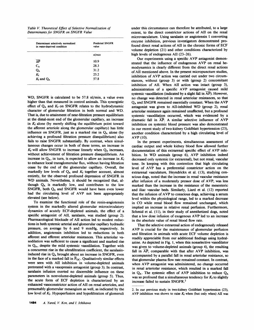
Table V. Theoretical Effect of Selective Normalization ofDeterminants for SNGFRon SNGFRValue
Determinant selectively normalized Predicted SNGFRin water-deprived condition value
nil/min
AP 10.9CA 28.3QA 31.2Kf 25.2Kf andQA 57.8
WD, SNGFRis calculated to be 57.8 nl/min, a value evenhigher than that measured in control animals. This synergisticeffect of QA and Kf on SNGFRrelates to the hydrodynamiccharacter of glomerular filtration in both normal and WD.That is, due to attainment of near-filtration pressure equilibriumat the distal-most end of the glomerular capillary, an increasein Kf alone (by merely shifting the equilibrium point towardthe afferent arteriole along the glomerular capillary) has littleinfluence on SNGFR, just as a marked rise in QA alone (byachieving a profound filtration pressure disequilibrium) alsofails to raise SNGFRsubstantially. By contrast, when simul-taneous changes occur in both of these terms, an increase inKf will allow SNGFRto increase linearly when QA increases,without achievement of filtration pressure disequilibrium. Anincrease in QA, in turn, is expected to allow an increase in Kfto enhance local transglomerular flux, without having filtrationcease by the end of the glomerular capillary. Overall, themarkedly low levels of QA and Kf together account, almostentirely, for the observed profound depression of SNGFRinWDanimals. Nevertheless, it should be recognized that, al-though QA is markedly low, and contributes to the lowSNGFR, both QA and SNGFRwould have been even lowerhad the circulating level of AVP not been appropriatelyelevated (see below).
To examine the functional role of the renin-angiotensinsystem in the markedly altered glomerular microcirculatorydynamics of acutely ECF-depleted animals, the effect of aspecific antagonist of All, saralasin, was studied (group 2).Pharmacological blockade of All action led to modest reduc-tions in both systemic arterial and glomerular capillary hydraulicpressure, on average by 6 and 9 mmHg, respectively. Inaddition, angiotensin inhibition led to reductions in bothafferent and efferent arteriolar resistances. This arteriolar va-sodilation was sufficient to cause a significant and marked risein QA, despite the mild systemic vasodilation. Together witha concurrent rise in the ultrafiltration coefficient, the saralasin-induced rise in QAbrought about an increase in SNGFR, evenin the face of a marked fall in PGc. Qualitatively similar effectswere seen with All inhibition in volume-depleted animalspretreated with a vasopressin antagonist (group 3). In contrast,saralasin infusion exerted no discernible influence on theseparameters in nonvolume-depleted animals (group 5). Thus,the acute form of ECF depletion is characterized by anenhanced vasoconstrictor action of All on renal arterioles, andpresumably glomerular mesangium as well, as indicated by thelow level of Kf. Hypoperfusion and hypofiltration of glomeruli
under this circumstance can therefore be attributed, to a largeextent, to the direct constrictor actions of All on the renalmicrovasculature. Using saralasin or angiotensin I convertingenzyme inhibition, previous investigators demonstrated pro-found direct renal actions of All in the chronic forms of ECFvolume depletion (21) and other conditions characterized byhigh levels of endogenous All (23-26).
Our experiments using a specific AVP antagonist demon-strated that the influence of endogenous AVP on renal he-modynamics is clearly different from the direct renal actionsof All mentioned above. In the present micropuncture studies,inhibition of AVP action was carried out under two circum-stances, without (group 3) or with (group 2) concomitantinhibition of All. When All action was intact (group 3),administration of a specific AVP antagonist caused mildsystemic vasodilation (indicated by a slight fall in AP). However,no change was detected in renal arteriolar resistances, so thatQAand SNGFRremained essentially constant. Whenthe AVPantagonist was given to AII-inhibited WD(group 2), renalarteriolar resistance again remained unaffected, but a profoundsystemic vasodilation occurred, which was evidenced by adramatic fall in AP. A similar selective influence of AVPinhibition on systemic blood pressure was also demonstratedin our recent study of two-kidney Goldblatt hypertension (23),another condition characterized by a high circulating level ofAVP (10).
In the present experiments, simultaneous assessment ofcardiac output and whole kidney blood flow allowed furtherdocumentation of this extrarenal specific effect of AVP inhi-bition. In WDanimals (group 6), AVP inhibition indeeddecreased only systemic (or extrarenal), but not renal, vasculartone. In keeping with this contention that high circulatinglevel of AVP has a preferential constrictor action on theextrarenal vasculature, Heyndrickx et al. (13), studying con-scious dogs, noted that the increase in renal vascular resistanceafter infusion of a moderately pressor dose of AVP, was lessmarked than the increase in the resistance of the mesentericand iliac vascular beds. Similarly, Liard et al. (12) reportedthat the infusion of AVP to conscious dogs, achieving a serumlevel within the physiological range, led to a marked decreasein CO while renal blood flow remained unchanged, whichimplied an increase in relative renal perfusion. Furthermore,Schmid et al. (1 1), in their study of anesthetized dogs, notedthat a low dose infusion of exogenous AVP led to an increasein the absolute value of renal blood flow rate.
That the selective extrarenal action of endogenously releasedAVP is crucial for the maintenance of glomerular perfusionand filtration in animals with acute ECF volume depletion isreadily appreciable from our additional findings using hydral-azine. As depicted in Fig. 1, when this nonselective vasodilatorwas given to volume-depleted animals (group 4), the resultingfall in AP, comparable with that after AVP inhibition, wasaccompanied by a parallel fall in renal arteriolar resistance, sothat glomerular plasma flow rate remained constant. In contrast,when AVP antagonist was administered, no change occurredin renal arteriolar resistance, which resulted in a marked fallin QA. The systemic effect of AVP inhibition to reduce QAwas so profound that a simultaneous tendency for Kf to slightlyincrease failed to sustain SNGFR.2
2. In our previous study in two-kidney Goldblatt hypertension (23),AVP inhibition was shown to raise Kf when (but only when) All was
1484 A. Yared, V. Kon, and I. Ichikawa

In addition to the renin-angiotensin system and vasopressin,the sympathetic nervous system is known to contribute tomaintaining the integrity of systemic circulation. Recently,Paller and Linas (3) have demonstrated in conscious rats that,even when two of these pressor systems are impaired, the levelof AP can be sustained as long as the third system remainsintact. Of note, however, is the evidence indicating that, underbarbiturate anesthesia, the ability of the sympathetic nervoussystem to adjust AP is grossly compromised (27, 28). Indeed,the rise in plasma catecholamines regularly occurring duringhemorrhage was found to be markedly blunted in pentobarbital-anesthetized dogs (29). Thus, although we did not measurecatecholamine levels, the efficiency of the sympathetic nervoussystem is likely to have been compromised by Inactin, therebyaccounting for the pronounced hypotension observed in ourstudies after antagonism of All and AVP in WDanimals.3
AVP is known to stimulate the release of vasodilatoryprostaglandins (31-33). Renal tissues, including glomeruli, areparticularly rich in enzymes that participate in the biosynthesisof prostaglandins (34-36). Oliver et al. (37) have recentlyshown that, in dogs, direct intrarenal arterial administration ofAVPaugments the renal release of prostglandin E2. Moreover,when the animals were pretreated with indomethacin, enhancedrenal constriction was noted during exogenous AVP adminis-tration. Our results in water diuretic animals (groups 8 and 9)confirm their findings: Thus, when a mildly pressor dose ofAVP was injected intravenously to rats undergoing waterdiuresis (group 8), a significant increase in renal blood flowand in RBF/CO ratio was seen. However, when potentialAVP-induced prostaglandin release was blocked by the cy-clooxygenase inhibitor indomethacin (group 9), RBFdecreasedand RBF/CO was unchanged. These results suggest that, whenthe intrarenal vasodilatory action of prostaglandins is elimi-nated, exogenous AVP is equally vasoconstrictive on renal andextrarenal vascular beds.4 Wepostulated that the absence of arenal constrictor action of AVPreleased endogenously in waterdeprivation, i.e., under physiological conditions, was similarlya consequence of enhanced release of vasodilatory prostaglan-dins, induced by AVP, and locally attenuating the directvasoconstrictor effect of the peptide hormone. Thus, adminis-tration of AVP antagonist to WDanimals led to systemichypotension and a marked fall in the renal fraction of cardiacoutput; pretreatment with indomethacin (group 7) essentially
simultaneously inhibited. In the present study, however, such a rise, ifany, could not be appreciated, due to achievement of filtration-pressureequilibrium (i.e., AP HHE). This indicates that the Krraising effect ofAVP inhibition, even if it occurred, failed to affect SNGFR, dueto the profound systemic effect of AVP inhibition, which broughtabout the fall in QA and AP, and attainment of filtration-pressureequilibrium.3. It should also be noted that the extent to which the differentialeffect of AVPon systemic vs. renal vasculature affects the overall RBF,depends on the renal capacity of "pressure" autoregulation, i.e., abilityto maintain RBF in the face of changes in AP. When this system iscompromised, as occurs in ECF volume depletion (30), the systemiceffect of AVP to maintain AP becomes a more crucial determinant forthe level of RBF.4. Our observations with indomethacin also imply that the release ofAVP-sensitive prostaglandins, at least those having vascular actions, ischanneled through receptors occupiable by d(CH2)5Tyr(Me)AVP, i.e.,vascular-type AVP receptors. This notion is supported by recentfindings in vitro (31, 38-40).
AVP |
EX TRARENA RENALr----------- ---------_i_______________
Vasoconstriction |PROSTA LANDINS Vasoconstriction
Vasodilatation
No Vascular Response
t RBF/COFigure 4. Schematic presentation of the hypothetical mechanism forinsensitivity of the renal vasculature to constrictor action of vasopres-sin (AVP). In this hypothesis, it is proposed, based on the experimen-tal observations, that vasopressin-stimulated local vasodilator actionsof prostaglandins are far more prominent in renal than extrarenalvasculature as a whole.
abolished this preferential vasodilatory action of AVPantagoniston the extrarenal vasculature, so that renal blood flow andRBF/CO ratio failed to change significantly. Although it isknown (41) that All can similarly induce the release ofvasodilatory prostaglandins, our data indicates that the vaso-constrictor action of All is far more profound than thevasodilatory effect of prostaglandins induced by All. Thus,pharmacological blockade of All was found, in our studies, toinduce a fall in the renal vascular resistance. In contrast, thevasodilatory effect of prostaglandins released in response toAVP in the dose we used, appears to be sufficient to balancethe direct renal vasoconstrictor action of the hormone, resulting,overall, in unchanged renal resistance.
Based on these results, we propose the mechanism for therenal hemodynamic action of AVPshown in Fig. 4: AVPmaypossess direct constrictor actions on both extrarenal and renalvasculatures. However, due to the release of vasodilatoryprostaglandins, the effect of which is to counteract the directconstrictive action of AVP, renal vascular resistance remainsunchanged. This scheme also provides an attractive hypothesisfor the role of endogenously released AVP. Thus, our resultsin group 6 and 7 rats point to the notion that the vasodilatoryaction of AVP-induced prostaglandins, by protecting the kidneyfrom the constrictor action of AVP, is critically important indiverting to the kidney a higher fraction of the cardiac output,the latter being profoundly depressed under conditions ofextracellular fluid depletion.5
AcknowledgmentsThe authors are grateful to Dr. Maurice Manning for the generous giftof d(CH2)5Tyr(Me)AVP, which made our studies possible. The authors
5. The interaction between AVP and prostaglandin was initiallydemonstrated for the effect of AVPon hydroosmotic water flow acrossthe terminal nephron (42-43). The sites of this interaction, bothreceptor and postreceptor levels, have since been suggested (44-48). Inview of the paucity of information regarding their interaction onvascular sites, only the interaction at the effector level is presented inour scheme. When the circulating level of AVP becomes extremelyhigh, its direct constrictor action appears to predominate. Indeed,when a markedly pressor dose of AVP was given to dogs, Schmid etal. (I 1) noted that, not only mesenteric and iliac, but also renal bloodflow declined.
Renal Hemodynamics in Acute Extracellular Fluid Volume Depletion 1485

also thank Ms. Mary L. Hughes for technical assistance, and Ms. JanetStanley for secretarial assistance.
These studies were supported largely by grants from the U. S.Public Health Service (AM-27853 and AM-32160). Dr. V. Kon is arecipient of the American Heart Association Clinician Scientist Award.
References
1. Aisenbrey, G. A., W. A. Handelman, P. Arnold, M. Manning,and R. W. Schrier. 1981. Vascular effects of arginine vasopressinduring fluid deprivation in the rat. J. Clin. Invest. 67:961-968.
2. Andrews, C. E., Jr., and B. M. Brenner. 1981. Relative contri-butions of arginine vasopressin and angiotensin II to maintenance ofsystemic arterial pressure in the anesthetized water-deprived rat. Circ.Res. 48:254-258.
3. Paller, M. S., and S. L. Linas. 1984. Role of angiotensin II, a-adrenergic system, and arginine vasopressin on arterial pressure in rats.Am. J. Physiol. 246:H25-H30.
4. Cowley, A. W., Jr., E. W. Quillen, Jr., and M. M. Skelton. 1983.Role of vasopressin in cardiovascular regulation. Fed. Proc. 42:3170-3176.
5. Manning, M., J. Lowbridge, C. T. Stier, Jr., J. Haldar, andW. H. Sawyer. 1977. [1- Deaminopenicillamine, 4-valine]-8-D- argininevasopressin, a highly potent inhibitor of the vasopressor response toarginine-vasopressin. J. Med. Chem. 20:1228-1230.
6. Lowbridge, J., M. Manning, J. Haldar, and W. H. Sawyer. 1978.[1- (fl-Mercapto-ftf4- cyclopentamethylenepropionic acid), 4-valine, 8-D-arginine] vasopressin, a potent and selective inhibitor of the vaso-pressor response to arginine-vasopressin. J. Med. Chem. 21:313-315.
7. Kruzynski, M., B. Lammek, M. Manning, J. Seto, J. Haldar,and W. H. Sawyer. 1980. [l-(j#-mercapto-BjB- cyclopentamethylene-propionic acid), 2- (o-methyl) tyrosine] arginine-vasopressin and [I-(#-mercapto-Bf6-cyclopentamethylenepropionic acid)]arginine-vasopressin,two highly potent antagonists to the vasopressor response to arginine-vasopressin. J. Med. Chem. 23:364-368.
8. Ichikawa, I., and B. M. Brenner. 1977. Evidence for glomerularactions of ADHand dibutyryl cyclic AMPin the rat. Am. J. Physiol.233:F102-F1 17.
9. Ausiello, D. A., J. I. Kreisberg, C. Roy, and M. J. Karnovsky.1980. Contraction of cultured rat glomerular cells of apparent mesangialorigin after stimulation with angiotensin II and arginine vasopressin.J. Clin. Invest. 65:754-760.
10. Mohring, J., B. Mohring, M. Petri, and D. Haack. 1978.Plasma vasopressin concentrations and effects of vasopressin antiserumon blood pressure in rats with malignant two-kidney Goldblatt hyper-tension. Circ. Res. 42:17-22.
11. Schmid, P. G., F. M. Abboud, M. G. Wendling, E. S. Ramberg,A. L. Mark, D. D. Heistad, and J. W. Eckstein. 1974. Regionalvascular effects of vasopressin: plasma levels and circulatory response.Am. J. Physiol. 227:998-1004.
12. Liard, J. F., 0. D6riaz, P. Schelling,- and M. Thibonnier. 1982.Cardiac output distribution during vasopressin infusion or dehydrationin conscious dogs. Am. J. Physiol. 243:H663-H669.
13. Heyndrickx, G. R., D. H. Boettcher, and S. F. Vatner. 1976.Effects of angiotensin, vasopressin, and methoxamine on cardiacfunction and blood flow distribution in conscious dogs. Am. J. Physiol.231:1579-1587.
14. Ablad, B. 1963. A study of the mechanism of the hemodynamiceffects of hydralazine in man. Acta Pharmacol. Toxicol. 20(Suppl. 1):1-53.
15. Ichikawa, I., D. A. Maddox, M. G. Cogan, and B. M. Brenner.1978. Dynamics of glomerular ultrafiltration in euvolemic Munich-Wistar rats. Renal Physiol. (Basel) 1: 121-131.
16. Deen, W. M., J. L. Troy, C. R. Robertson, and B. M. Brenner.1973. Dynamics of glomerular ultrafiltration in the rat. IV. Determi-nation of the ultrafiltration coefficient. J. Clin. Invest. 52:1500-1508.
17. Arendshorst, W. J., W. F. Finn, and C. W. Gottschalk. 1975.
Autoregulation of blood flow in the rat kidney. Am. J. Physiol. 228:127-133.
18. Vurek, G. G., and S. E. Pegram. 1966. Fluorometric methodfor the determination of nanogram quantities of inulin. Anal. Biochem.16:409-419.
19. Fuhr, J., J. Kazmarczyk, and C. D. Kriittgen. 1955. Eineeinfache colorimetrische Methode zur Inulinbestimmung fur Nieren-Clearanceuntersuchungen bei Stoffwechselgesunden und Diabetikern.Klin. Wochenschr. 33:729-730.
20. Viets, J. W., W. M. Deen, J. L. Troy, and B. M. Brenner.1978. Determination of serum protein concentration in nanoliter bloodsamples using fluorescamine or o-phthalaldehyde. Anal. Biochem. 88:513-521.
21. Steiner, R. W., B. J. Tucker, and R. C. Blantz. 1979. Glomerularhemodynamics in rats with chronic sodium depletion. Effect of saralasin.J. Clin. Invest. 64:503-512.
22. Schor, N., I. Ichikawa, and B. M. Brenner. 1980. Glomerularadaptions to chronic dietary salt restriction or excess. Am. J. Physiol.238:F428-F436.
23. Ichikawa, I., R. A. Ferrone, K. L. Duchin, M. Manning, V. J.Dzau, and B. M. Brenner. 1983. Relative contribution of vasopressinand angiotensin II to the altered renal microcirculatory dynamics intwo-kidney Goldblatt hypertension. Circ. Res. 53:592-602.
24. Ploth, D. W. 1983. Angiotensin-dependent renal mechanismsin two-kidney, one-clip renal vascular hypertension. Am. J. Physiol.245:F131-F141.
25. Tucker, B. J., and R. C. Blantz. 1983. Mechanism of alteredglomerular hemodynamics during chronic sodium depletion. Am. J.Physiol. 244:Fl -F18.
26. Huang, W.-C., D. W. Ploth, and L. G. Navar. 1982. Angiotensin-mediated alterations in nephron function in Goldblatt hypertensiverats. Am. J. Physiol. 243:F553-F560.
27. Tucker, B. J., 0. W. Peterson, M. G. Ziegler, and R. C. Blantz.1982. Analysis of adrenergic effects of the anesthetics Inactin and a-chloralose. Am. J. Physiol. 243:F253-F259.
28. Houck, P. C., M. J. Fiksen-Olsen, S. L. Britton, and J. C.Romero. 1983. Role of angiotensin and vasopressin on blood pressureof ganglionic blocked dogs. Am. J. Physiol. 244:H1 15-H 120.
29. Zimpfer, M., W. T. Manders, A. C. Barger, and S. F. Vatner.1982. Pentobarbital alters compensatory neural and humoral mecha-nisms in response to hemorrhage. Am. J. Physiol. 243:H713-H721.
30. Robertson, C. R., W. M. Deen, J. L. Troy, and B. M. Brenner.1972. Dynamics of glomerular ultrafiltration in the rat. III. Hemody-namics and autoregulation. Am. J. Physiol. 223:1191-1200.
31. Beck, T. R., and M. J. Dunn. 1981. The relationship ofantidiuretic hormone and renal prostaglandins. Miner. ElectrolyteMetab. 6:46-59.
32. Zusman, R. M., and H. R. Keiser. 1977. Prostaglandin biosyn-thesis by rabbit renomedullary interstitial cells in tissue culture. Stim-ulation by angiotensin II, bradykinin and arginine vasopressin. J. Clin.Invest. 60:215-223.
33. Dunn, M. J., H. P. Greely, H. Valtin, L. B. Kinter, and R.Beeuwkes III. 1978. Renal excretion of prostaglandins E2 and F2. indiabetes insipidus rats. Am. J. Physiol. 235:E624-E627.
34. Scharschmidt, L. A., and M. J. Dunn. 1983. Prostaglandinsynthesis by rat glomerular mesangial cells in culture. Effects ofangiotensin II and arginine vasopressin. J. Clin. Invest. 71:1756-1764.
35. Sraer, J., J. Foidart, D. Chancel, P. Mahieu, and R. Ardaillou.1980. Prostaglandin synthesis by rat isolated glomeruli and glomerularcultured cells. Int. J. Biochem. 12:203-207.
36. Sraer, J., W. Siess, L. Moulonguet-Doleris, J. P. Oudinet, F.Dray, and R. Ardaillou. 1982. In vitro prostaglandin synthesis byvarious rat renal preparations. Biochim. Biophys. Acta. 710:45-52.
37. Oliver, J. A., R. R. Sciacca, G. Le Cren, and P. J. Cannon.1982. Modulation by prostaglandins of the renal vascular action ofarginine vasopressin. Prostaglandins. 24:641-656.
38. Zipser, R. D., S. I. Myers, and P. Needleman. 1981. Stimulation
1486 A. Yared, V. Kon, and I. Ichikawa
of renal prostaglandin synthesis by the pressor activity of vasopressin.Endocrinology. 108:495-499.
39. Beck, T. R., A. Hassid, and M. J. Dunn. 1980. The effect ofarginine vasopressin and its analogs on the synthesis of prostaglandinE2 by rat renal medullary interstitial cells in culture. J. Pharmacol.Exp. Ther. 215:15-19.
40. Walker, L. A., A. R. Whorton, M. Smigel, R. France, andJ. C. Frolich. 1978. Antidiuretic hormone increases renal prostaglandinsynthesis in vivo. Am. J. Physiol. 235:F180-F185.
41. Danon, A., L. C. T. Chang, B. J. Sweetman, A. S. Nies, andJ. A. Oates. 1975. Synthesis of prostaglandins by the rat renal papillain vitro. Mechanism of stimulation of angiotensin II. Biochim. Biophys.Acta. 388:71-83.
42. Orloff, S., J. S. Handler, and S. Bergstrom. 1965. Effect ofprostaglandin (PGE1) on the permeability response of toad bladder tovasopressin, theophylline, and adenosine 3',5'-monophosphate. Nature(Lond.). 205:397-398.
43. Grantham, J. J., and J. Orloff. 1968. Effect of prostaglandin Elon the permeability response of the isolated collecting tubule to
vasopressin, adenosine 3',5'-monophosphate, and theophylline. J. Clin.Invest. 47:1154-1161.
44. Beck, N. P., T. Kaneko, U. Zor, J. B. Field, and B. B. Davis.1971. Effects of vasopressin and prostaglandin El on the adenyl cyclase-cyclic 3',5'-adenosine monophosphate system of the renal medulla ofthe rat. J. Clin. Invest. 50:2461-2465.
45. Marumo, F., and I. S. Edelman. 1971. Effects of Ca++ andprostaglandin El on vasopressin activation of renal adenyl cyclase. J.Clin. Invest. 50:1613-1620.
46. Omachi, R. S., D. E. Robbie, J. S. Handler, and J. Orloff.1974. Effects of ADHand other agents on cyclic AMPaccumulationin toad bladder epithelium. Am. J. Physiol 226:1152-1157.
47. Lum, G. M., G. A. Aisenbrey, M. J. Dunn, T. Berl, R. W.Schrier, and K. M. McDonald. 1977. In vivo effect of indomethacinto potentiate the renal medullary cyclic AMPresponse to vasopressin.J. Clin. Invest. 59:8-13.
48. Handler, J. S. 1981. Vasopressin-prostaglandin interactions inthe regulation of epithelial cell permeability to water. Kidney Int. 19:831-838.
Renal Hemodynamics in Acute Extracellular Fluid Volume Depletion 1487