post-graduate course handbook - ascvts 2016 2016 - post-graduate course.pdf · pg5-1 management of...
TRANSCRIPT

The 24th Annual Meeting of Asian Society for Cardiovascular and Thoracic Surgery (ASCVTS)in conjunction with 9th AATS / ASCVTS Postgraduate Course and 4th Asian Single Port VATS Symposium
April 06-10, 2016, Taipei, Taiwan
POST-GRADUATECOURSE HANDBOOK


The 24th Annual Meeting of Asian Society for Cardiovascular and Thoracic Surgery (ASCVTS)in conjunction with 9th AATS / ASCVTS Postgraduate Course and 4th Asian Single Port VATS Symposium
1
POST-GRADUATECOURSE HANDBOOK
The 24th Annual Meeting of Asian Society for Cardiovascular and Thoracic Surgery (ASCVTS)in conjunction with 9th AATS / ASCVTS Postgraduate Course and 4th Asian Single Port VATS Symposium
April 06-10, 2016, Taipei, Taiwan

The 24th Annual Meeting of Asian Society for Cardiovascular and Thoracic Surgery (ASCVTS)in conjunction with 9th AATS / ASCVTS Postgraduate Course and 4th Asian Single Port VATS Symposium
2

The 24th Annual Meeting of Asian Society for Cardiovascular and Thoracic Surgery (ASCVTS)in conjunction with 9th AATS / ASCVTS Postgraduate Course and 4th Asian Single Port VATS Symposium
3
Venue 105, 1F 201DE, 2F 201AF, 2FTime
07:30 - 08:30
08:30 - 10:00
AATS-ASCVTSPost-Graduate 1Pediatric Heart
Session 1Fallot
AATS-ASCVTSPost-Graduate 2
Thoracic Session 1Lung
AATS-ASCVTSPost-Graduate 3
Adult CardiacSession 1
Coronary Artery Disease
10:00 - 10:30
10:30 - 12:00
12:00 - 13:30 Luncheon SymposiumSponsor by Marquette
Luncheon SymposiumSponsor by Unison Surgicals
Company
Luncheon SymposiumSponsor by Edwards
Lifesciences(Taiwan) Corp.
13:30 - 15:00
15:00 - 15:30
15:30 - 17:30
17:30 - 19:00
Program at a glance
Coffee Break
Coffee Break
Welcome Reception
AATS-ASCVTSPost-Graduate 4Pediatric Heart
Session 2Vavle
AATS-ASCVTSPost-Graduate 6
Adult CardiacSession 2
Mitral Vavle and Af
AATS-ASCVTSPost-Graduate 5
Thoracic Session 2Lung II: Novel Therapies
and Technic
Congress Tour 3 (13:00 - 17:00)
AATS-ASCVTSPost-Graduate 10
Pediatric HeartSession 4Fontan
AATS-ASCVTSPost-Graduate 11Thoracic Session 4
Mediastinum & Trachea
AATS-ASCVTSPost-Graduate 12
Adult CardiacSession 4
ECMO
AATS-ASCVTSPost-Graduate 7Pediatric Heart
Session 3Borderline between Single
V and Bi-V
AATS-ASCVTSPost-Graduate 8
Thoracic Session 3Esophagus
AATS-ASCVTSPost-Graduate 9
Adult CardiacSession 3
AVR and Aorta

The 24th Annual Meeting of Asian Society for Cardiovascular and Thoracic Surgery (ASCVTS)in conjunction with 9th AATS / ASCVTS Postgraduate Course and 4th Asian Single Port VATS Symposium
4
DAILY PROGRAM
Thursday, 07 April 2016
08:30-10:00AATS-ASCVTS Post-Graduate 1
Pediatric HeartSession 1 - Fallot
105, 1F
Moderators: Duke Cameron, Johns Hopkins Hospital (USA), Hajime Ichikawa, National Cerebral and Cardiovascular Center (Japan), Yih-Sharng Chen, National Taiwan University Hospital (Taiwan)
PG1-1 MANAGEMENT OF TETRALOGY OF FALLOOT IN NEONATE AND EARLY INFANCYChristian Pizarro, Nemours Cardiac Center Alfred I Dupont Hospital for Children (USA)
PG1-2 SURGICAL TECHNIQUES FOR THE PRESERVATION OF MARGINALLY SMALL PULMONARY VALVE ANNULUS UPON THE REPAIR OF TETRALOGY OF FALLOT
Tae-jin Yun, Asan Medical Center (Korea)
PG1-3 TRANSITION OF THE TREATMENT STRATEGY FOR PULMONARY ATRESIA/VENTRICULAR SEPTUM WITH MAJOR AORTO-PULMONARY COLLATERAL ARTERIESHajime Ichikawa, Koji Kagisaki, Masatoshi Shimada, Takashi Kido, Takaya Hoashi, National Cerebral and Cardiovascular Center (Japan)
PG1-4 MANAGEMENT OF ADULT REPAIRED TETRALOGY OF FALLOT Duke Cameron, Johns Hopkins Hospital (USA)
Thursday, 07 April 2016
08:30-10:00AATS-ASCVTS Post-Graduate 2
ThoracicSession 1 - Lung
201DE, 2F
Moderators: Hiroshi Date, Kyoto University (Japan), Calvin Sze-Hang Ng, The Chinese University of Hong Kong (Hong Kong), Sanghoon Jheon, Seoul National University (Korea)
PG2-1 LUNG CANCER SCREENING: WHAT DO WE NEED TO KNOW? Sang-Hoon Jheon, Seoul National University (Korea)
PG2-2 IS THERE A ROLE FOR THE UNIPORTAL APPROACH Calvin Sze-Hang Ng, The Chinese University of Hong Kong (Hong Kong)
PG2-3 JUSTIFICATION FOR SUBLOBAR RESECTION FOR NON-SMALL CELL LUNG CANCER ≤ 2CM
Mingyon Mun, Cancer Institute Hospital (Japan)

The 24th Annual Meeting of Asian Society for Cardiovascular and Thoracic Surgery (ASCVTS)in conjunction with 9th AATS / ASCVTS Postgraduate Course and 4th Asian Single Port VATS Symposium
5
PG2-4 VIDEO-ASSISTED THORACIC SURGERY (VATS) IN ADVANCED RESECTION OF LUNG CANCERKwhanmien Kim, Seoul National University Bundang Hospital, Seoul National University College of Medicine (Korea)
PG2-5 ROLE OF SURGERY FOR RESECTION OF N2 DISEASE LUNG CANCER Hiroshi Date, Kyoto University (Japan)
Thursday, 07 April 2016
08:30-10:00AATS-ASCVTS Post-Graduate 3
Adult CardiacSession 1 - Coronary Artery Disease
201AF, 2F
Moderators: Marc R. Moon, Washington University (USA), Song Wan, The Chinese University of Hong Kong (Hong Kong), Richard L. Prager, University of Michigan Health System Cardiac Surgery (USA)
PG3-1 WHEN IS CABG CLEARLY SUPERIOR? Marc R. Moon, Washington University (USA)
PG3-2 ISCHEMIC MR: REPLACE OR REPAIRE David H. Adams, The Mount Sinai Hospital (USA)
PG3-3 CABG ON VS OFF CPB Ralph J. Damiano, Washington University in Saint Louis (USA)
PG3-4 HOW TO IMPROVE OUTCOMES AND QUALITY IN CABG? Richard L. Prager, University of Michigan Health System Cardiac Surgery (USA)
PG3-5 ARTERIAL GRAFTING IN CABG: RATIONALE VS REALITY Song Wan, The Chinese University of Hong Kong (Hong Kong)

The 24th Annual Meeting of Asian Society for Cardiovascular and Thoracic Surgery (ASCVTS)in conjunction with 9th AATS / ASCVTS Postgraduate Course and 4th Asian Single Port VATS Symposium
6
Thursday, 07 April 2016
10:30-12:00AATS-ASCVTS Post-Graduate 4
Pediatric HeartSession 2 - Valve
105, 1F
Moderators: Carl Backer, Northwestern University (USA), Qingyu Wu, The First Hospital of Tsinghua University (China), Kisaburo Sakamoto, Mt. Fuji Shizuoka Children’s Hospital (Japan)
PG4-1 ARRYTHMIA SURGERY IN PEDIATRIC CONGENITAL HEART SURGERY Carl Backer, Northwestern University (USA)
PG4-2 ATRIOVENTRICULAR VALVE REGURGITATION IN SINGLE VENTRICLE Kisaburo Sakamoto, Mt. Fuji Shizuoka Children’s Hospital (Japan)
PG4-3 MANAGEMENT OF EBSTEIN ANOMALIES IN ADULTSQingyu Wu, Hongyin Li, Mingkui Zhang, Lianyi Wang, The First Hospital of Tsinghua University (China)
PG4-4 TREATMENT OF AORTIC ANEURYSMS IN CONGENITAL HEART DISEASE Duke Cameron, Johns Hopkins Hospital (USA)
Thursday, 07 April 2016
10:30-12:00AATS-ASCVTS Post-Graduate 5
ThoracicSession 2 - Lung II: Novel Therapies and Technic
201DE, 2F
Moderators: Shaf Keshavjee, Toronto General Hospital (Canada), Kazuhiro Yasufuku, University of Toronto (Canada), Takehiro Izumo, National Cancer Center (Japan)
PG5-1 MANAGEMENT OF THE NON-PALPABLE LUNG NODULE: IMAGE GUIDED MINIMALLY INVASIVE SURGICAL TECHNIQUES
Kazuhiro Yasufuku, University of Toronto (Canada)
PG5-2 EBUS-TBNA ABOUT LUNG CANCER STAGING Takehiro Izumo, National Cancer Center (Japan)
PG5-3 LUNG REPLACEMENT THERAPIES: LUNG TRANSPLANT AND EXTRACORPOREAL LUNG SUPPORT
Shaf Keshavjee, Toronto General Hospital (Canada)
PG5-4 IMMUNE THERAPY FOR LUNG CANCER Jin-Yuan Shih, National Taiwan University Hospital (Taiwan)

The 24th Annual Meeting of Asian Society for Cardiovascular and Thoracic Surgery (ASCVTS)in conjunction with 9th AATS / ASCVTS Postgraduate Course and 4th Asian Single Port VATS Symposium
7
Thursday, 07 April 2016
10:30-12:00AATS-ASCVTS Post-Graduate 6
Adult CardiacSession 2 - Mitral Valve and Af
201AF, 2F
Moderators: Ralph J. Damiano, Washington University in Saint Louis (USA), Song Wan, The Chinese University of Hong Kong (Hong Kong), David H. Adams, The Mount Sinai Hospital (USA)
PG6-1 ROBOTIC MITRAL VALVE SURGERY – CURRENT STATUS AND FUTURE DIRECTIONS L. Wiley Nifong, East Carolina Heart Institute (USA)
PG6-2 COMPLEX BILEAFLET REPAIR: RESECT OR RESPECT? David H. Adams, The Mount Sinai Hospital (USA)
PG6-3 REPAIRING ALL TYPES OF RHEUMATIC MV: POSSIBLE AND JUSTIFIED?Taweesak Chotivatanapong, Central Chest Institute of Thailand (CCIT) (Thailand)
PG6-4 CLOSURE OF LEFT ATRIAL APPENDAGE DURING CARDIAC SURGERY: WHY, WHEN AND HOW?
Ko Bando, The Jikei University School of Medicine (Japan)
PG6-5 AF ABLATION: WHICH OPERATION? PATIENT BENEFIT? Ralph J. Damiano, Washington University in Saint Louis (USA)
Thursday, 07 April 2016
12:00-13:30 Luncheon Symposium-Marquet 105, 1F
Moderators:
Thursday, 07 April 2016
12:00-13:30 Luncheon Symposium- UnionSurgical CompanyRobotic surgery session 201DE, 2F
Moderators: Yen Chang (Taiwan), Jang-Ming Lee (Taiwan)
LS2-1 ROBOTICS IN CARDIAC SURGERY L. Wiley Nifong (USA)
LS2-2 APPLICATION OF ROBOTIC-ASSISTED SURGERY FOR COMPLEX THORACIC PROCEDURES Bernard Park (USA)

The 24th Annual Meeting of Asian Society for Cardiovascular and Thoracic Surgery (ASCVTS)in conjunction with 9th AATS / ASCVTS Postgraduate Course and 4th Asian Single Port VATS Symposium
8
Thursday, 07 April 2016
12:00-13:30
Luncheon Symposium- Sponsor by Edward Lifescience (Taiwan) Corp.
Changing Landscape in the Management of Patients with Heart Valve Disease
201AF, 2F
Moderators: Chen Yih Sharng, National Taiwan University Hospital, Taiwan (Taiwan)
LS3-1 HEART TEAM APPROACH Daniele Doyle, Holy Spirit Northside Private Hospital (Australia)
LS3-2 EXPERIENCE WITH RAPID DEPLOYMENT VALVES Frank Slachman, Mercy General Hospital (USA)
LS3-3 SURGEONS’ ROLE IN THE ERA OF TAVR Davide Gabbieri, Hesperia Hospital (Italy)
Thursday, 07 April 2016
13:30-15:00AATS-ASCVTS Post-Graduate 7
Pediatric HeartSession 3 - Borderline between SingleV and Bi-V
105, 1F
Moderators: Hiromi Kurosawa, Sakakibara Sapia Tower Clinic (Japan), Shunji Sano, Okayama University School of Medicine, Dentistry and Pharmaceutical Sciences (Japan), Chong-Yi Chang, National Taiwan University Hospital (Taiwan)
PG7-1 CONDUCTION SYSTEM IN ABNORMAL ATRIOVENTRICULAR CONNECTIONS Hiromi Kurosawa, Sakakibara Sapia Tower Clinic (Japan)
PG7-2 FONTAN OR BIVENTRICULAR REPAIR OR IN BETWEEN?Hiromi Kurosawa, Sakakibara Sapia Tower Clinic, Tokyo Women’s Medical University, Jikei University (Japan)
PG7-3 PA-IVS SURGICAL MANAGEMENTShunji Sano, Okayama University School of Medicine, Dentistry and Pharmaceutical Sciences (Japan)
PG7-4 RIGHT VENTRICULAR MORPHOLOGY AND LATE OUTCOME OF PATIENTS WITH PULMONARY ATRESIA OR CRITICAL PULMONARY STENOSIS WITH INTACT VENTRICULAR SEPTUM AFTER BIVENTRICULAR REPAIRHajime Ichikawa, Koji Kagisaki, Masatoshi Shimada, Takashi Kido, Takaya Hoashi, National Cerebral and Cardiovascular Center (Japan)
The 24th Annual Meeting of Asian Society for Cardiovascular and Thoracic Surgery (ASCVTS)in conjunction with 9th AATS / ASCVTS Postgraduate Course and 4th Asian Single Port VATS Symposium
9
Thursday, 07 April 2016
13:30-15:00AATS-ASCVTS Post-Graduate 8
ThoracicSession 3 - Esophagus
201DE, 2F
Moderators: Simon Ying-Kit Law, The University of Hong Kong (Hong Kong), Bernard J. Park, Memorial Sloan Kettering Cancer Center (USA)
PG8-1 TBC Hai-Quan Chen, Shanghai Chest Hospital (China)
PG8-2 THORACOSCOPIC ESOPHAGECTOMY IN THE PRONE POSITION Hirokazu Noshiro, Saga University (Japan)
PG8-3 NEOADJUVANT CHEMORADIATION FOR ADVANCED ESOPHAGEAL CANCER Simon Ying-Kit Law, The University of Hong Kong (Hong Kong)
PG8-4 THE FUTURE OF ROBOTIC THORACIC SURGERY Bernard J. Park, Memorial Sloan Kettering Cancer Center (USA)
Thursday, 07 April 2016
13:30-15:00AATS-ASCVTS Post-Graduate 9
Adult CardiacSession 3 - AVR and Aorta
201AF, 2F
Moderators: Joseph E. Bavaria, Hospital of the University of Pennslvania (USA), Marc R. Moon, Washington University (USA), Mattia Glauber, The Cardiothoracic Surgery Network (Italy)
PG9-1 AORTIC VALVE REPAIR DURING ROOT REPLACEMENT Joseph E. Bavaria, Hospital of the University of Pennslvania (USA)
PG9-2 CEREBRAL PROTECTION DURING ARCH SURGERY Marc R. Moon, Washington University (USA)
PG9-3 SUTURELESS AORTIC VALVE REPLACEMENT Frank N. Slachman, Mercy Medical Group (USA)
PG9-4 LEARNING CURVE IN MINIMALLY INVASIVE PROCEDURE FOR HEART VALVE SURGERY Mattia Glauber, The Cardiothoracic Surgery Network (Italy)
PG9-5 TEVAR FOR AORTIC DISSECTIONKazuo Shimamura, Toru Kuratani, Tomohiko Sakamoto, Tomoaki Kudo, Kenta Masada, Kei Torikai, Yoshiki SawaOsaka University Graduate School of Medicine (Japan)

The 24th Annual Meeting of Asian Society for Cardiovascular and Thoracic Surgery (ASCVTS)in conjunction with 9th AATS / ASCVTS Postgraduate Course and 4th Asian Single Port VATS Symposium
10
Thursday, 07 April 2016
15:30-17:00AATS-ASCVTS Post-Graduate 10
Pediatric HeartSession 4 - Fontan
105, 1F
Moderators: Christian Pizarro, Nemours Cardiac Center Alfred I Dupont Hospital for Children (USA), Tain-Yen Hsia, Great Ormond Street Hospital for Children (UK), Shu-Chien Huang, National Taiwan University Hospital (Taiwan)
PG10-1 INDIVIDUALIZED APPROACH TO HLHS MANAGEMENTChristian Pizarro, Nemours Cardiac Center Alfred I Dupont Hospital for Children (USA)
PG10-2 MODELING THE SINGLE VENTRICLE: AN UPDATE ON THE STATE OF THE ART Tain-Yen Hsia, Great Ormond Street Hospital for Children (UK)
PG10-3 LONG-TERM OUTCOME OF FONTAN CONVERSION Carl Backer, Northwestern University (USA)
PG10-4 MCS AND HTX FOR SINGLE VENTRICLE Iki Adachi, Texas Children’s Hospital (USA)
Thursday, 07 April 2016
15:30-17:00AATS-ASCVTS Post-Graduate 11
ThoracicSession 4 - Mediastinum & Trachea
201DE, 2F
Moderators: Paul De Leyn, University Hospitals Leuven (Belgium), Gunda Leschber, Berlin Chest Hospital (ELK) (Germany), Mark S. Allen, Mayo Clinic (USA)
PG11-1 WHAT IS THE BEST SURGICAL APPROACH TO TREAT THYMOMA?Ming-Ching Lee, Chung-Ping Hsu, Taichung Veterans General Hospital (Taiwan)
PG11-2 CURRENT STATUS OF INVASIVE MEDIASTINAL STAGING Paul De Leyn, University Hospitals Leuven (Belgium)
PG11-3 RADICAL LYMPH NODE DISSECTION VIA MEDIASTINOSCOPY Gunda Leschber, Berlin Chest Hospital (ELK) (Germany)
PG11-4 TRACHEAL SURGERY Mark S. Allen, Mayo Clinic (USA)

The 24th Annual Meeting of Asian Society for Cardiovascular and Thoracic Surgery (ASCVTS)in conjunction with 9th AATS / ASCVTS Postgraduate Course and 4th Asian Single Port VATS Symposium
11
Thursday, 07 April 2016
15:30-17:00AATS-ASCVTS Post-Graduate 12
Adult CardiacSession 4 - ECMO
201AF, 2F
Moderators: Roberto Lorusso, Maastricht University Medical Centre (Netherlands), Yih-Sharng Chen, National Taiwan University Hospital (Taiwan), Paul C. Jansz, St Vincent’s Hospital (Australia)
PG12-1 ECMO IN ELDERLY PATIENTS: EFFECTIVE OR FUTILE PROCEDURE? Roberto Lorusso, Maastricht University Medical Centre (Netherlands)
PG12-2 POST-OPERATIVE SUPPORT FOR CARDIOGENIC SHOCK I-wen Wang, Indiana University Health Methodist Hospital (USA)
PG12-3 ADVANCED TRENDS IN VAD Paul C. Jansz, St Vincent’s Hospital (Australia)
PG12-4 NEW TREND IN EXTRACORPOREAL CARDIOPULMONARY RESUSCITATION Yih-Sharng Chen, National Taiwan University (Taiwan)

The 24th Annual Meeting of Asian Society for Cardiovascular and Thoracic Surgery (ASCVTS)in conjunction with 9th AATS / ASCVTS Postgraduate Course and 4th Asian Single Port VATS Symposium
12
ABSTRACT
POST-GRADUATECOURSE HANDBOOK
The 24th Annual Meeting of Asian Society for Cardiovascular and Thoracic Surgery (ASCVTS)in conjunction with 9th AATS / ASCVTS Postgraduate Course and 4th Asian Single Port VATS Symposium

The 24th Annual Meeting of Asian Society for Cardiovascular and Thoracic Surgery (ASCVTS)in conjunction with 9th AATS / ASCVTS Postgraduate Course and 4th Asian Single Port VATS Symposium
13
ABSTRACT
POST-GRADUATECOURSE HANDBOOK
The 24th Annual Meeting of Asian Society for Cardiovascular and Thoracic Surgery (ASCVTS)in conjunction with 9th AATS / ASCVTS Postgraduate Course and 4th Asian Single Port VATS Symposium

The 24th Annual Meeting of Asian Society for Cardiovascular and Thoracic Surgery (ASCVTS)in conjunction with 9th AATS / ASCVTS Postgraduate Course and 4th Asian Single Port VATS Symposium
14
1
SURGICAL TECHNIQUES FOR THE PRESERVATION OF MARGINALLY SMALL PULMONARY VALVE ANNULUS UPON
THE REPAIR OF TETRALOGY OF FALLOT
Tae-Jin Yun
Asan Medical Center
From September 1997 to August 2012, total correction of ToF was performed in 170 consecutive patients by a single surgeon (Yun TJ), and data from these patients were analyzed. ToF associated with pulmonary atresia, absent pulmonary valve syndrome or atrioventricular septal defect were excluded from the study. There were 102 male and 68 female patients, and age at repair ranged from 11 days to 57 years (median: 8.1 months). Most of the patients (118/170, 69.4%) had their operations within 12 months of age. Body weight ranged from 3.1 kg to 77 kg (median: 8.1kg). Twenty patients had undergone palliative procedures before definitive surgical repair, including two modified Blalock–Taussing shunt in other institutions and three RVOT ballooning or stenting in the catheterization laboratory. Surgical techniques. The operation was carried out under moderate hypothermic (28℃) cardiopulmonary bypass with aortic and bicaval cannulation, aortic cross-clamping, and myocardial protection with intermittent infusion of cold blood cardioplegia at the aortic root. After oblique right atriotomy, parietal extension of infundibular septum was resected extensively until pulmonary valve was clearly seen from the right ventricular side. Ventricular septal defect (VSD) was repaired by trans-atrial approach in all but 6 patients who had sub-arterial type VSDs with absent infundibular septum, which were repaired through both trans-atrial andtrans-pulmonary approach. Main pulmonary artery was incised longitudinally, and morphology of the pulmonary valve was carefully inspected. Two thirds of the patients had bicuspid pulmonary valve with varying degree of degeneration and commissural fusion. Pulmonary valve commissures were sliced off the pulmonary arterial wall, and commissurotomy was performed in the majority of the AP patients. Other measures, including leaflet division (bicuspidalization or tricuspidalization), leaflet excision and pericardial patch replacement, were done to increase RVOT dimension. Then, the PVA size was measured with a Hega dilator. If it was less than -2 of Z-score, the pulmonary arteriotomy was extended down to RVOT crossing the annulus for minimal right ventriculotomy (less than 10mm) to place trans-annular patch (TAP). If it was greater than -2, inverted Y incision was made from the lower end of pulmonary arteriotomy down to the level of the annulus for extensive pulmonary annulo-arterioplasty. A Gore-Tex patch of inverted Y shape was placed to augment main pulmonary artery(Figure 4). After coming off cardiopulmonary bypass, the ratio of right ventricular pressure to left ventricular pressure (PRV/LP) and the pressure gradient between RV and pulmonary artery (PA) were directly measured. If PRV/LP was greater than 0.8 with significant gradient between RV and PA, the patient was put back on CPB and mini-infundibular incision (10 mm) was made for RVOT muscle resection and
2
infundibular patch (IP) placement (Figure 5). If PRV/LP was still higher than 0.8 after the placement of IP, the patient went back on CPB again for trans-annular patch (TAP). Various techniques of peripheral pulmonary angioplasty (Patch angioplasty, Carinoplasty in the bifurcation of main pulmonary artery, LPA wedge resection and repair to prevent a kinking and stenosis of LPA…) were employed, if indicated.
PG1-2

The 24th Annual Meeting of Asian Society for Cardiovascular and Thoracic Surgery (ASCVTS)in conjunction with 9th AATS / ASCVTS Postgraduate Course and 4th Asian Single Port VATS Symposium
15
1
SURGICAL TECHNIQUES FOR THE PRESERVATION OF MARGINALLY SMALL PULMONARY VALVE ANNULUS UPON
THE REPAIR OF TETRALOGY OF FALLOT
Tae-Jin Yun
Asan Medical Center
From September 1997 to August 2012, total correction of ToF was performed in 170 consecutive patients by a single surgeon (Yun TJ), and data from these patients were analyzed. ToF associated with pulmonary atresia, absent pulmonary valve syndrome or atrioventricular septal defect were excluded from the study. There were 102 male and 68 female patients, and age at repair ranged from 11 days to 57 years (median: 8.1 months). Most of the patients (118/170, 69.4%) had their operations within 12 months of age. Body weight ranged from 3.1 kg to 77 kg (median: 8.1kg). Twenty patients had undergone palliative procedures before definitive surgical repair, including two modified Blalock–Taussing shunt in other institutions and three RVOT ballooning or stenting in the catheterization laboratory. Surgical techniques. The operation was carried out under moderate hypothermic (28℃) cardiopulmonary bypass with aortic and bicaval cannulation, aortic cross-clamping, and myocardial protection with intermittent infusion of cold blood cardioplegia at the aortic root. After oblique right atriotomy, parietal extension of infundibular septum was resected extensively until pulmonary valve was clearly seen from the right ventricular side. Ventricular septal defect (VSD) was repaired by trans-atrial approach in all but 6 patients who had sub-arterial type VSDs with absent infundibular septum, which were repaired through both trans-atrial andtrans-pulmonary approach. Main pulmonary artery was incised longitudinally, and morphology of the pulmonary valve was carefully inspected. Two thirds of the patients had bicuspid pulmonary valve with varying degree of degeneration and commissural fusion. Pulmonary valve commissures were sliced off the pulmonary arterial wall, and commissurotomy was performed in the majority of the AP patients. Other measures, including leaflet division (bicuspidalization or tricuspidalization), leaflet excision and pericardial patch replacement, were done to increase RVOT dimension. Then, the PVA size was measured with a Hega dilator. If it was less than -2 of Z-score, the pulmonary arteriotomy was extended down to RVOT crossing the annulus for minimal right ventriculotomy (less than 10mm) to place trans-annular patch (TAP). If it was greater than -2, inverted Y incision was made from the lower end of pulmonary arteriotomy down to the level of the annulus for extensive pulmonary annulo-arterioplasty. A Gore-Tex patch of inverted Y shape was placed to augment main pulmonary artery(Figure 4). After coming off cardiopulmonary bypass, the ratio of right ventricular pressure to left ventricular pressure (PRV/LP) and the pressure gradient between RV and pulmonary artery (PA) were directly measured. If PRV/LP was greater than 0.8 with significant gradient between RV and PA, the patient was put back on CPB and mini-infundibular incision (10 mm) was made for RVOT muscle resection and
2
infundibular patch (IP) placement (Figure 5). If PRV/LP was still higher than 0.8 after the placement of IP, the patient went back on CPB again for trans-annular patch (TAP). Various techniques of peripheral pulmonary angioplasty (Patch angioplasty, Carinoplasty in the bifurcation of main pulmonary artery, LPA wedge resection and repair to prevent a kinking and stenosis of LPA…) were employed, if indicated.
PG1-2

The 24th Annual Meeting of Asian Society for Cardiovascular and Thoracic Surgery (ASCVTS)in conjunction with 9th AATS / ASCVTS Postgraduate Course and 4th Asian Single Port VATS Symposium
16
TRANSITION OF THE TREATMENT STRATEGY FORPULMONARY ATRESIA/VENTRICULAR SEPTUM WITHMAJOR AORTO-PULMONARY COLLATERAL ARTERIES
Hajime Ichikawa, Koji Kagisaki, Masatoshi Shimada, Takashi Kido, Takaya Hoashi
Department of pediatric cardiovascular surgery, National Cerebral and Cardiovascular Center, Suita, Osaka, Japan
OBJECTIVES:In 1980s, unifocalization (UF) of major aorto-pulmonary collateral arteries (MAPCA) aiming definitive repair for pulmonary atresia with ventricular septal defect (PAVSD) had been mostly done through thoracotomy. They required multiple staged operations, which is not favorable choice for the patients. In 1990s, We have changed this strategy to one-stage UF and RV-PA conduit with or without VSD closure. The long term outcome of both strategies are discussed. METHOD:Patients followed-up more than 3 years are included in this presentation. From 1982 to 2012, UF with or without other procedure was done 283 times in 100 patients. Mean follow-up period is 16.0 ± 7.6 years. The follow-up rate is 100%. Palliative staged UF through thoracotomy, one stage UF, SP shunt and palliative RVOTR were done 119, 31, 41 and 22 times, respectively.RESULTS:Survival after UF was 74 % and 82 % in staged group and one-stage group, respectively Completion of definitive repair (DR) was achieved in 70 patients. DR was done in 67% of patient with staged UF and 82% with one-stage UF. The mean age at DR in staged UF and one-stage UF was 5.5 and 2.6, respectively. The survival rate 10 and 20 years after DR was 77.0 and 70.0 % in staged group, whereas it was 94.4 and 94.4 % in one-stage group. Reoperations after DR includes 22 right ventricular outflow and 4 aortic valve replacement The requirement of catheter intervention occurred after UF was 27% in staged-group and 66% in one-stage group, which reflect the difference in era. CONCLUSION: One-stage UF provided early and high rate of successful DR. However, since this type of operation is still relatively new, careful follow-up is mandatory for an early detection of unknown complications they may encounter.
MANAGEMENT OF ADULT REPAIRED TETRALOGY OFFALLOT
Duke Cameron, MD
Division of Cardiac SurgeryThe Johns Hopkins Medical Institutions
Baltimore, MD, USA
Seventy two years have passed since the first palliative surgery for tetralogy of Fallot (TOF) and 62 years since the first total correction. These years have witnessed more than three generations who have survived an otherwise fatal congenital heart lesion. Early survival rates are now excellent and more and more patients are surviving into adulthood to reveal the late “unnatural” history of corrected TOF. Right heart failure, atrial and ventricular dysrhythmias, pulmonary valve insufficiency and ascending aortic aneurysm have become the major concerns. Though it is hoped that the current surgical strategies such as early repair, pulmonary valve preservation and avoidance of ventriculotomy will minimize these late issues, there is still uncertainty about their effect on the late course. Indications for pulmonary valve replacement and prophylactic replacement of the dilated aorta are still controversial. This presentation will review the evidence for these interventions and their outcomes.
PG1-3

The 24th Annual Meeting of Asian Society for Cardiovascular and Thoracic Surgery (ASCVTS)in conjunction with 9th AATS / ASCVTS Postgraduate Course and 4th Asian Single Port VATS Symposium
17
MANAGEMENT OF ADULT REPAIRED TETRALOGY OFFALLOT
Duke Cameron, MD
Division of Cardiac SurgeryThe Johns Hopkins Medical Institutions
Baltimore, MD, USA
Seventy two years have passed since the first palliative surgery for tetralogy of Fallot (TOF) and 62 years since the first total correction. These years have witnessed more than three generations who have survived an otherwise fatal congenital heart lesion. Early survival rates are now excellent and more and more patients are surviving into adulthood to reveal the late “unnatural” history of corrected TOF. Right heart failure, atrial and ventricular dysrhythmias, pulmonary valve insufficiency and ascending aortic aneurysm have become the major concerns. Though it is hoped that the current surgical strategies such as early repair, pulmonary valve preservation and avoidance of ventriculotomy will minimize these late issues, there is still uncertainty about their effect on the late course. Indications for pulmonary valve replacement and prophylactic replacement of the dilated aorta are still controversial. This presentation will review the evidence for these interventions and their outcomes.
PG1-4

The 24th Annual Meeting of Asian Society for Cardiovascular and Thoracic Surgery (ASCVTS)in conjunction with 9th AATS / ASCVTS Postgraduate Course and 4th Asian Single Port VATS Symposium
18
LUNG CANCER SCREENING: WHAT DO WE NEED TO KNOW?
Sanghoon Jheon, MD, PhD1, 2
1Department of Thoracic and Cardiovascular Surgery, Seoul National University Bundang Hospital, Gyeonggi, Korea
2Department of Thoracic and Cardiovascular Surgery, Seoul National University College of Medicine, Seoul, Korea
Early detection of suspicious pulmonary lesions is a major concern for reducing cancer mortality. Despite noteworthy advances in treatment modalities for cancers including chemo-radiotherapy and surgery, lung cancer still remains the leading cause of cancer mortality, mainly because most of lung cancers are at an advanced stage when diagnosed. However, no standard screening strategy for lung cancer has been established. Regular screening test with chest radiography or sputum cytology is no longer recommended. Screening with low dose chest tomography (LDCT) has been reported to be effective in reducing lung cancer mortality with few harmful result, but only when applied in carefully selected patients who have sufficient risk factors and acceptable comorbidities. Although LDCT is the only potential way of improving lung cancer mortality by
early detection, it still has not been introduced as a standard tool for lung cancer detection. Most randomized controlled trials for demonstration of the effectiveness of LDCT as a screening modality are either ongoing or have shown inconsistent results, and it could be effective only when it is performed within a structured programs with selection, evaluation and management strategies.There are novel tools for lung cancer screenings which have been intensively researched. Breathing gas analysis, serum biomarkers using free circulating DNA and RNA, exosomal microRNA, circulating tumor cells and various lung cancer specific antigens are examples which have been studied extensively with encouraging results. No matter what kinds of tools are chosen for lung cancer screening, they should have prognostic benefits with scientific evidence and should not increase the chances of harmful effects.
IS THERE A ROLE FOR THE UNIPORTAL APPROACH
Calvin S.H. Ng,
The Chinese University of Hong Kong
The history of uniportal VATS goes back more than a decade, when the more basic thoracic procedures such as sympathectomy or pleural biopsies were performed. Subsequently, intermediate procedures of uniportal VATS pleurodesis, wedge resection, pericardial window were successfully performed, mostly pioneered by Rocco’s team. During the same period, Thomas D’Amico from Duke was developing the 2-port VATSmajor lung resection technique, and more recently the modified uniportal VATS approach. Dr Diego Gonzalez-Rivas initially adopted the 2-port VATS technique, and in2010, he performed the first uniportal VATS lobectomy for left lower lobe tumour through a 4 cm incision. Over the following years, other complex thoracic procedures, including uniportal VATS segmentectomy, pneumonectomy, sleeve resection, lobectomy with chest wall resection, pulmonary artery resection and reconstruction have been successfully accomplished.
The major advantage of the uniportal VATS technique is that surgical access involves less intercostal spaces. Apart from potentially reducing the total length of the incision(s) compared with conventional VATS, the uniportal approach reduces the number of intercostal nerve at risk of injury. Therefore, pain and the incidence of intercostal neuralgia may be reduced. Studies so far comparing uniportal VATS versus 3-port VATS techniques in procedures such as pleurodesis seem to support this. In addition, when uniportal VATS sympathectomy was compared with 2-port VATS, post-operative pain was also less in the uniportal group. The ultra-minimal invasive approach of uniportal VATS has also made it particularly popular in the Asia countries where patients demand surgical procedures with the least surgical access trauma. The interest in the region can clearly be seen in the Asian Single Port VATS Symposium (ASPVS) series with the most recent held in Hong Kong in 2015 attended by over 220 thoracic surgeons from over 18 countries throughout the world. The appetite for the uniportal VATS technique in Asia seems insatiable, at least for the foreseeable years to come.
The development of uniportal VATS major lung resection in the last 5 years or more has had an additional unexpected effect. Perhaps not since the surgical evolution from open thoracotomy to conventional 3-port VATS of the 1990s have we seen so much interest in collaboration from surgeons and industry in developing novel, smaller, more specialized procedure specific instruments for minimally invasive thoracic surgery.Further refinement of uniportal VATS instruments, angulated and narrower endo-staplers, and improvements in video-camera systems including 3D systems, and 120 degrees articulating lens that allow uniportal VATS major lung resection easier to perform and learn. These advances will no doubt facilitate all forms of minimally invasive thoracic surgery. In addition, the ethos of performing lung resection through a single incision has led to the development of subcostal and e-NOTES access, which may further reduce access trauma and complications of intercostal neuralgia. The spirit for reinvention has extended into multidisciplinary collaboration with the anesthetists in the form of non-intubated uniportal VATS major lung resection, which may herald a new era for patient
PG2-1

The 24th Annual Meeting of Asian Society for Cardiovascular and Thoracic Surgery (ASCVTS)in conjunction with 9th AATS / ASCVTS Postgraduate Course and 4th Asian Single Port VATS Symposium
19
IS THERE A ROLE FOR THE UNIPORTAL APPROACH
Calvin S.H. Ng,
The Chinese University of Hong Kong
The history of uniportal VATS goes back more than a decade, when the more basic thoracic procedures such as sympathectomy or pleural biopsies were performed. Subsequently, intermediate procedures of uniportal VATS pleurodesis, wedge resection, pericardial window were successfully performed, mostly pioneered by Rocco’s team. During the same period, Thomas D’Amico from Duke was developing the 2-port VATSmajor lung resection technique, and more recently the modified uniportal VATS approach. Dr Diego Gonzalez-Rivas initially adopted the 2-port VATS technique, and in2010, he performed the first uniportal VATS lobectomy for left lower lobe tumour through a 4 cm incision. Over the following years, other complex thoracic procedures, including uniportal VATS segmentectomy, pneumonectomy, sleeve resection, lobectomy with chest wall resection, pulmonary artery resection and reconstruction have been successfully accomplished.
The major advantage of the uniportal VATS technique is that surgical access involves less intercostal spaces. Apart from potentially reducing the total length of the incision(s) compared with conventional VATS, the uniportal approach reduces the number of intercostal nerve at risk of injury. Therefore, pain and the incidence of intercostal neuralgia may be reduced. Studies so far comparing uniportal VATS versus 3-port VATS techniques in procedures such as pleurodesis seem to support this. In addition, when uniportal VATS sympathectomy was compared with 2-port VATS, post-operative pain was also less in the uniportal group. The ultra-minimal invasive approach of uniportal VATS has also made it particularly popular in the Asia countries where patients demand surgical procedures with the least surgical access trauma. The interest in the region can clearly be seen in the Asian Single Port VATS Symposium (ASPVS) series with the most recent held in Hong Kong in 2015 attended by over 220 thoracic surgeons from over 18 countries throughout the world. The appetite for the uniportal VATS technique in Asia seems insatiable, at least for the foreseeable years to come.
The development of uniportal VATS major lung resection in the last 5 years or more has had an additional unexpected effect. Perhaps not since the surgical evolution from open thoracotomy to conventional 3-port VATS of the 1990s have we seen so much interest in collaboration from surgeons and industry in developing novel, smaller, more specialized procedure specific instruments for minimally invasive thoracic surgery.Further refinement of uniportal VATS instruments, angulated and narrower endo-staplers, and improvements in video-camera systems including 3D systems, and 120 degrees articulating lens that allow uniportal VATS major lung resection easier to perform and learn. These advances will no doubt facilitate all forms of minimally invasive thoracic surgery. In addition, the ethos of performing lung resection through a single incision has led to the development of subcostal and e-NOTES access, which may further reduce access trauma and complications of intercostal neuralgia. The spirit for reinvention has extended into multidisciplinary collaboration with the anesthetists in the form of non-intubated uniportal VATS major lung resection, which may herald a new era for patient
PG2-2

The 24th Annual Meeting of Asian Society for Cardiovascular and Thoracic Surgery (ASCVTS)in conjunction with 9th AATS / ASCVTS Postgraduate Course and 4th Asian Single Port VATS Symposium
20
care in terms of quicker recovery and shorter hospital stay. Future investigations should also focus on the use of hybrid operating theatre cone beam CT for image guided uniportal VATS procedures to improve surgical accuracy.
1
JUSTIFICATION FOR SUBLOBAR RESECTION FOR NON-SMALL CELL LUNG CANCER ≦ 2CM
Mingyon Mun, MD, PhD
Department of Thoracic Surgical Oncology, The Cancer Institute Hospital, 3-10-6Ariake, Koto-ku, Tokyo, Japan
Cahan reported radical lobectomy for early stage non-small cell lung cancer (NSCLC) in 1960, since then, standard procedure for early stage lung cancer is lobectomy with mediastinal lymph node dissection. However, recently, peripheral small-sized lung cancer such as a ground glass nodule (GGN) has been frequently detected by low dose CT (LDCT) screening. So the indication of sublobar resection has been increasing.
In 1995, Ginsburg and Rubinstein reported the results of the Lung Cancer Study Group (LCSG) which was the only randomized controlled study comparing lobectomy and sublobar resection. Despite a tripling of the local recurrence rate in patients after sublobar resection, the differences in overall and cancer specific survival did not achieve statistical significance. Since then, the standard procedure for early stage NSCLC is lobectomy. However, this trial had some limitations, its small sample size (247 patients), the large number of wedge resection in the sublobar group, and 2-3 cm tumors included in the trial. So, in 2009, large randomized phaseIII trial (JCOG0802) started in Japan comparing lobectomy to segmentectomy in patients with peripheral small (≦ 2cm) NSCLC in whom there is no intraoperative evidence of metastases to the hilar or mediastinal lymph nodes.For the patients with almost pure GGN, phase II trial (JCOG0804) of limited surgical resection (wide wedge resection) for peripheral early lung cancer defined with thin-section CT (TSCT) started also in 2009.
At our institution, sublobar resection is conducted by thoracoscopic surgery (TS). When we perform TS sublobar resection, we need to do some ingenuity. When we perform TS wedge resection (TS-W), preoperative marking may be required for hardly palpate nodule. Now we perform non-invasive computed tomography-guided marking without puncturing the visceral pleura. To perform TS segmentectomy (TS-S), preoperatively, we decide the indication of the TS-S not only TSCT but three-dimensional CT reconstruction to know anatomical variances and the surgical margin from the tumor. Intraoperatively, after the target segmental artery and bronchus were divided, and intravenous systemic injection of indocyanine green (ICG, 0.25 mg/kg), ICGF of the non-target segments was observed using infrared
PG2-2

The 24th Annual Meeting of Asian Society for Cardiovascular and Thoracic Surgery (ASCVTS)in conjunction with 9th AATS / ASCVTS Postgraduate Course and 4th Asian Single Port VATS Symposium
21
care in terms of quicker recovery and shorter hospital stay. Future investigations should also focus on the use of hybrid operating theatre cone beam CT for image guided uniportal VATS procedures to improve surgical accuracy.
1
JUSTIFICATION FOR SUBLOBAR RESECTION FOR NON-SMALL CELL LUNG CANCER ≦ 2CM
Mingyon Mun, MD, PhD
Department of Thoracic Surgical Oncology, The Cancer Institute Hospital, 3-10-6Ariake, Koto-ku, Tokyo, Japan
Cahan reported radical lobectomy for early stage non-small cell lung cancer (NSCLC) in 1960, since then, standard procedure for early stage lung cancer is lobectomy with mediastinal lymph node dissection. However, recently, peripheral small-sized lung cancer such as a ground glass nodule (GGN) has been frequently detected by low dose CT (LDCT) screening. So the indication of sublobar resection has been increasing.
In 1995, Ginsburg and Rubinstein reported the results of the Lung Cancer Study Group (LCSG) which was the only randomized controlled study comparing lobectomy and sublobar resection. Despite a tripling of the local recurrence rate in patients after sublobar resection, the differences in overall and cancer specific survival did not achieve statistical significance. Since then, the standard procedure for early stage NSCLC is lobectomy. However, this trial had some limitations, its small sample size (247 patients), the large number of wedge resection in the sublobar group, and 2-3 cm tumors included in the trial. So, in 2009, large randomized phaseIII trial (JCOG0802) started in Japan comparing lobectomy to segmentectomy in patients with peripheral small (≦ 2cm) NSCLC in whom there is no intraoperative evidence of metastases to the hilar or mediastinal lymph nodes.For the patients with almost pure GGN, phase II trial (JCOG0804) of limited surgical resection (wide wedge resection) for peripheral early lung cancer defined with thin-section CT (TSCT) started also in 2009.
At our institution, sublobar resection is conducted by thoracoscopic surgery (TS). When we perform TS sublobar resection, we need to do some ingenuity. When we perform TS wedge resection (TS-W), preoperative marking may be required for hardly palpate nodule. Now we perform non-invasive computed tomography-guided marking without puncturing the visceral pleura. To perform TS segmentectomy (TS-S), preoperatively, we decide the indication of the TS-S not only TSCT but three-dimensional CT reconstruction to know anatomical variances and the surgical margin from the tumor. Intraoperatively, after the target segmental artery and bronchus were divided, and intravenous systemic injection of indocyanine green (ICG, 0.25 mg/kg), ICGF of the non-target segments was observed using infrared
PG2-3

The 24th Annual Meeting of Asian Society for Cardiovascular and Thoracic Surgery (ASCVTS)in conjunction with 9th AATS / ASCVTS Postgraduate Course and 4th Asian Single Port VATS Symposium
22
2
thoracoscopy (KARL STORZ Endoskope Japan K.K., Tokyo, Japan). We marked the border between target and non-target segments with electrocautery and divided the lung parenchyma along this border using electrocautery or staples. As a result, from April 2008 to December 2014, we performed TS-W in 172 patientsand TS-S in 132 patients with favorable outcomes.
VIDEO-ASSISTED THORACIC SURGERY (VATS)IN ADVANCED RESECTION OF LUNG CANCER
Kwhanmien Kim, MD
Department of Thoracic and Cardiovascular SurgerySeoul National University Bundang Hospital
Seoul National University College of Medicine
Since the first VATS lobectomy was performed in lung cancer patient, VATS techniques have been evolved and their applications have been expanded continuously. Although VATS was not admitted as a standard procedure in lung cancer treatment in 1990’s, nowadays it has become the first choice in early stage lung cancer surgery.
The safety and feasibility of VATS lobectomy in the early stage lung cancer surgery have been already well known. However, in advanced lung cancer with central location originating from the bronchus or with metastatic lymph nodes, chest wall invasion and pleural seeding, the efficacy and safety of VATS are not established yet.
With increasing experience and technological developments, we have tried to expand the application of VATS in more complicated lung cancer patients, such as who are expected to undergo pneumonectomy, or bronchoplasty, and who need neoadjuvant therapy because of metastatic mediastinal lymph nodes. And, intraplueral perufusion hyperthermic chemotherapy (IPHC) can be applied to the patients who have unexpectedly minimal pleural seeding, after pulmonary resection. VATS for expanded indications in lung cancer surgery can be applied in carefully selected patients with acceptable postoperative outcomes, if it is performed by an expert surgeon.
PG2-3

The 24th Annual Meeting of Asian Society for Cardiovascular and Thoracic Surgery (ASCVTS)in conjunction with 9th AATS / ASCVTS Postgraduate Course and 4th Asian Single Port VATS Symposium
23
VIDEO-ASSISTED THORACIC SURGERY (VATS)IN ADVANCED RESECTION OF LUNG CANCER
Kwhanmien Kim, MD
Department of Thoracic and Cardiovascular SurgerySeoul National University Bundang Hospital
Seoul National University College of Medicine
Since the first VATS lobectomy was performed in lung cancer patient, VATS techniques have been evolved and their applications have been expanded continuously. Although VATS was not admitted as a standard procedure in lung cancer treatment in 1990’s, nowadays it has become the first choice in early stage lung cancer surgery.
The safety and feasibility of VATS lobectomy in the early stage lung cancer surgery have been already well known. However, in advanced lung cancer with central location originating from the bronchus or with metastatic lymph nodes, chest wall invasion and pleural seeding, the efficacy and safety of VATS are not established yet.
With increasing experience and technological developments, we have tried to expand the application of VATS in more complicated lung cancer patients, such as who are expected to undergo pneumonectomy, or bronchoplasty, and who need neoadjuvant therapy because of metastatic mediastinal lymph nodes. And, intraplueral perufusion hyperthermic chemotherapy (IPHC) can be applied to the patients who have unexpectedly minimal pleural seeding, after pulmonary resection. VATS for expanded indications in lung cancer surgery can be applied in carefully selected patients with acceptable postoperative outcomes, if it is performed by an expert surgeon.
PG2-4

The 24th Annual Meeting of Asian Society for Cardiovascular and Thoracic Surgery (ASCVTS)in conjunction with 9th AATS / ASCVTS Postgraduate Course and 4th Asian Single Port VATS Symposium
24
1
1
ROLE OF SURGERY FOR RESECTION OF N2 DISEASE LUNG CANCER
i
Hiroshi Date, MD
Department of Thoracic Surgery, Kyoto University Graduate School of Medicine, Kyoto Japan
Prognosis of completely resected pathological (p) N2 stage IIIA non-small cell lung cancer (NSCLC) is still unsatisfactory as a result of a high incidence of metastasis or recurrence of tumor. In our retrospective study on 496 patients with pathological N2 disease who underwent complete resection, patterns of lymphnode metastases (skip metastases), as well as pT factor, sex, and PS, were significant prognostic factors for disease-free and overall survival of pN2 stage IIIA NSCLC.A meta-analysis on cisplatin-based adjuvant chemotherapy after initial resection including ALPI, BLT, IALT, JBR.10, and ANITA studies has confirmed to improve survival rate of patients with completely resected stage II and III NSCLC. Vinorelbine and cisplatin is recognized as standard regimen and is recommended for completely resected N2 disease as adjuvant chemotherapy. Several studies have suggested that induction therapy followed by surgical resection may improve the outcome in patients with stage IIIa (N2) non-small-cell lung cancer (NSCLC), however, optimal induction strategy has not been defined. INT0139 study failed to demonstrate survival benefit by tri-modality therapy as compare with chemo-radiotherapy in phase III trial. However, high rate of pneumonectomy and high mortality rate after pneumonectomy in this study have been criticized. In our experience at Universities of Okayama and Kyoto, the 5 year survival rate exceeded 50% for NSCLC patients with pre-determined pathological N2 disease who underwent complete resection after induction chemo-radiotherapy.
i
WHEN IS CABG CLEARLY SUPERIOR?
Marc R. Moon, M.D.
Washington University School of Medicine, Saint Louis, Missouri
Since coronary bypass grafting first became popular to treat coronary atherosclerosis in the 1960’s, there have been countless studies comparing surgical revascularization to medical therapy and percutaneous interventions. This presentation will summarizehistoric randomized controlled trials, including the GABI, MASS II, RITA-I, ARTS, BARI, CABRI, EAST and others. The combined endpoint of death and repeat revascularization favors CABG (10%) over PCI (25%) while the hazard ratio for death alone favors CABG only for patients with diabetes and those greater than 65 years of age. The impact of diabetes on survival increases over time. Propensity-matched registries have demonstrated a decreased reintervention rate with CABG and an absolute survival benefit with CABG for patients with 3-vessel disease and these benefits remain significant even when comparing CABG to drug-eluding stents (82% survival at 5 years vs. 74%, p<0.001). The definitive SYNTAX trails determined the impact of the complexity of coronary artery disease on MACCE. Patients with low complexity CAD had similar outcomes with CABG and PCI, while patients with high complexity CAD had much better outcomes with CABG. Most studies support that CABG is superior with left main disease and 3-V disease.
PG2-5

The 24th Annual Meeting of Asian Society for Cardiovascular and Thoracic Surgery (ASCVTS)in conjunction with 9th AATS / ASCVTS Postgraduate Course and 4th Asian Single Port VATS Symposium
25
WHEN IS CABG CLEARLY SUPERIOR?
Marc R. Moon, M.D.
Washington University School of Medicine, Saint Louis, Missouri
Since coronary bypass grafting first became popular to treat coronary atherosclerosis in the 1960’s, there have been countless studies comparing surgical revascularization to medical therapy and percutaneous interventions. This presentation will summarizehistoric randomized controlled trials, including the GABI, MASS II, RITA-I, ARTS, BARI, CABRI, EAST and others. The combined endpoint of death and repeat revascularization favors CABG (10%) over PCI (25%) while the hazard ratio for death alone favors CABG only for patients with diabetes and those greater than 65 years of age. The impact of diabetes on survival increases over time. Propensity-matched registries have demonstrated a decreased reintervention rate with CABG and an absolute survival benefit with CABG for patients with 3-vessel disease and these benefits remain significant even when comparing CABG to drug-eluding stents (82% survival at 5 years vs. 74%, p<0.001). The definitive SYNTAX trails determined the impact of the complexity of coronary artery disease on MACCE. Patients with low complexity CAD had similar outcomes with CABG and PCI, while patients with high complexity CAD had much better outcomes with CABG. Most studies support that CABG is superior with left main disease and 3-V disease.
PG3-1

The 24th Annual Meeting of Asian Society for Cardiovascular and Thoracic Surgery (ASCVTS)in conjunction with 9th AATS / ASCVTS Postgraduate Course and 4th Asian Single Port VATS Symposium
26
HOW TO IMPROVE OUTCOMES AND QUALITY IN CABG
Richard L. Prager
University of Michigan Health System Cardiac Surgery
Utilizing STS database variables as well as other variables, cardiac surgeons in the State of Michigan in the United States, created a Quality Improvement Collaborative Group over 10 years ago as a part of a professional society for Cardiac and Thoracic Surgeons in Michigan called the Michigan Society of Thoracic and Cardiovascular Surgeons ( The MSTCVS). Using these data and meeting four times a year, this collaborative group has reviewed data from each site, eventually doing so in a transparent , non-blinded way, and has created focused improvement approaches in many areas of adult cardiac surgery. This approach has led to areas of improved outcomes, saved money for the hospitals and insurers, helped to create more efficiencies in hospitals and care, and led to greater collaboration among surgeons throughout the state of Michigan.
PG3-4
ARTERIAL GRAFTING IN CABG: RATIONALE VS REALITY
Song Wan, MD, FRCS
The Chinese University of Hong Kong, Prince of Wales Hospital,Hong Kong, China
Coronary artery bypass grafting (CABG) is one of the most thoroughly studied operations in the history of surgery. Over the past two decades, parallel to the rapid development of percutaneous coronary intervention, critical issues such as improving long-term graft patency have been the focus of intense investigations worldwide.Even with a growing body of evidence over the past decade, clinical decision-making in surgical myocardial revascularization apparently remains a complex process. In the real world, it is surprising that some surgical choices are still not “evidence-based”(i.e., as what should be expected). For instance, despite the eminent value of arterial grafting, less than 10% isolated CABG cases are actually carried out with total arterial grafting in North America.
Meanwhile, no doubt saphenous vein conduits will continually be used in CABG until acceptable alternative approaches are evaluated. The late failure of vein grafts,however, remains a major clinical burden. Identifying novel strategies to prevent neointimal thickening is of utmost importance. To date, apart from lipid-lowering drugs,no other pharmacological agents are effective in preventing early vein graft remodelling or subsequent accelerated atherosclerosis. As such, the fate of vein grafts remains largely unchanged even after decades of technical progress. Gene therapy is attractive in this setting as an ex vivo technology to genetically manipulate the conduit prior to grafting. The use of safe and efficient vectors for delivery is a necessity strategy to improve graft patency in the long term.

The 24th Annual Meeting of Asian Society for Cardiovascular and Thoracic Surgery (ASCVTS)in conjunction with 9th AATS / ASCVTS Postgraduate Course and 4th Asian Single Port VATS Symposium
27
ARTERIAL GRAFTING IN CABG: RATIONALE VS REALITY
Song Wan, MD, FRCS
The Chinese University of Hong Kong, Prince of Wales Hospital,Hong Kong, China
Coronary artery bypass grafting (CABG) is one of the most thoroughly studied operations in the history of surgery. Over the past two decades, parallel to the rapid development of percutaneous coronary intervention, critical issues such as improving long-term graft patency have been the focus of intense investigations worldwide.Even with a growing body of evidence over the past decade, clinical decision-making in surgical myocardial revascularization apparently remains a complex process. In the real world, it is surprising that some surgical choices are still not “evidence-based”(i.e., as what should be expected). For instance, despite the eminent value of arterial grafting, less than 10% isolated CABG cases are actually carried out with total arterial grafting in North America.
Meanwhile, no doubt saphenous vein conduits will continually be used in CABG until acceptable alternative approaches are evaluated. The late failure of vein grafts,however, remains a major clinical burden. Identifying novel strategies to prevent neointimal thickening is of utmost importance. To date, apart from lipid-lowering drugs,no other pharmacological agents are effective in preventing early vein graft remodelling or subsequent accelerated atherosclerosis. As such, the fate of vein grafts remains largely unchanged even after decades of technical progress. Gene therapy is attractive in this setting as an ex vivo technology to genetically manipulate the conduit prior to grafting. The use of safe and efficient vectors for delivery is a necessity strategy to improve graft patency in the long term.
PG3-5

The 24th Annual Meeting of Asian Society for Cardiovascular and Thoracic Surgery (ASCVTS)in conjunction with 9th AATS / ASCVTS Postgraduate Course and 4th Asian Single Port VATS Symposium
28
ARRHYTHMIA SURGERY IN PEDIATRIC CONGENITAL HEART SURGERY
Carl L. Backer, M.D.
Division of Cardiovascular-Thoracic Surgery, Ann & Robert H. Lurie Children’s Hospital of Chicago, and Department of Surgery, Northwestern University Feinberg
School of Medicine, Chicago, IL, USA
BACKGROUND: Arrhythmia surgery and pacing systems have favorably impacted the clinical course of patients undergoing pediatric congenital heart surgery. We reviewed the outcomes of arrhythmia surgery, pacemaker placement, and AICD placement at our institution.METHODS: Between 1987 and 2011, 231 patients underwent open heart surgical procedures that included arrhythmia surgery as a part of the operation. During the same time period 552 patients underwent 804 pacemaker related procedures.RESULTS: Forty-one patients underwent an operation for atrial reentry tachycardia. These patients had a modified right atrial maze procedure. Recurrence rate was 14%. Twelve patients underwent operation for atrial fibrillation. These patients had a Cox-maze III procedure. Recurrence rate was 16%. Nineteen patients underwent an operation for an accessory connection. These patients underwent either a left or right free wall dissection. Recurrence rate was 18%. Five patients had an operation for automatic atrial tachycardia, 7 patients for AV nodal reentry tachycardia, and 14 patients for ventricular tachycardia. One-hundred and 33 patients underwent Fontan conversion; 65 of these patients had atrial fibrillation, 45 had right atrial reentry tachycardia, 17 had left atrial reentry tachycardia. Our pacemaker strategy was to avoid transvenous placement until the patient weighed over 40 kg. There were 451 epicardial systems, 188 transvenous systems, and 87 AICD placements (13 were epicardial and 74 were transvenous systems). This also included 17 dual-chamber pacemakers in neonates.CONCLUSION: Results of surgical intervention for arrhythmias in pediatric congenital heart surgery are very good. The most common operations were the right-sided maze procedure, cox-maze III, and device placement. Consideration for arrhythmia surgery should be incorporated into any planned surgical reoperation of patients with congenital heart disease (especially adults). Pacemaker placement using a strategy of epicardial systems until the patients are 40 kg and transvenous systems after that have been quite successful. Dual-chamber pacing in neonates is very effective therapy. The key to success for the above involves a joint cardiovascular surgery-electrophysiology team.
PG4-1
ATRIOVENTRICULAR VALVE REGURGITATION IN SINGLE VENTRICLE
Kisaburo Sakamoto
Department of Cardiovascular Surgery in Cardiovascular Center, Mt. Fuji Shizuoka Children’s Hospital
Atrio-ventricular (AV) valve regurgitation has been recognized one of the mostimportant risk factors to bring patients with single ventricle to Fontan circulation, however it is not easy to manage it acceptably. The infants with more than moderate common AV valve regurgitation before Glenn stage are, especially, difficult even to rescue, because they have a dysplastic valve composed of multiple, thin and fragile leaflets difficult to repair and have to bear postoperative volume-loading condition. Today, I show our recent strategy and the experience for them.
[Our current strategy] 1) Evaluation & primary planning mainly using echo: Not only the grade of the regurgitation but also the 4-D condition of the valve are checked and a primary plan to repair is considered in the heart team. 2) Reproduction of the preoperative valve condition during surgical intervention and final planning:Common AV valves are so easily deformed during cardiac arrest that the evaluationseems to be misled. This step of reproduction gives us appropriate information to make the right valve repair without discrepancy between preoperative findings. 3)Repair based on the concept of leaflet apposition: After confirming potential of each leaflet, acceptably-functioning leaflets are preserved and apposed apposition to make better coaptation, using localized annuloplasty on commissures and bridging anuuloplasty. 4) Staged annuloplasty: The annular diameter or opening of the AV valve is controlled by following limitation; more than 100% of normal TV diameter at palliative stage, more than 100% of MV at Glenn stage and more than 80% of MV at Fontan stage.

The 24th Annual Meeting of Asian Society for Cardiovascular and Thoracic Surgery (ASCVTS)in conjunction with 9th AATS / ASCVTS Postgraduate Course and 4th Asian Single Port VATS Symposium
29
ARRHYTHMIA SURGERY IN PEDIATRIC CONGENITAL HEART SURGERY
Carl L. Backer, M.D.
Division of Cardiovascular-Thoracic Surgery, Ann & Robert H. Lurie Children’s Hospital of Chicago, and Department of Surgery, Northwestern University Feinberg
School of Medicine, Chicago, IL, USA
BACKGROUND: Arrhythmia surgery and pacing systems have favorably impacted the clinical course of patients undergoing pediatric congenital heart surgery. We reviewed the outcomes of arrhythmia surgery, pacemaker placement, and AICD placement at our institution.METHODS: Between 1987 and 2011, 231 patients underwent open heart surgical procedures that included arrhythmia surgery as a part of the operation. During the same time period 552 patients underwent 804 pacemaker related procedures.RESULTS: Forty-one patients underwent an operation for atrial reentry tachycardia. These patients had a modified right atrial maze procedure. Recurrence rate was 14%. Twelve patients underwent operation for atrial fibrillation. These patients had a Cox-maze III procedure. Recurrence rate was 16%. Nineteen patients underwent an operation for an accessory connection. These patients underwent either a left or right free wall dissection. Recurrence rate was 18%. Five patients had an operation for automatic atrial tachycardia, 7 patients for AV nodal reentry tachycardia, and 14 patients for ventricular tachycardia. One-hundred and 33 patients underwent Fontan conversion; 65 of these patients had atrial fibrillation, 45 had right atrial reentry tachycardia, 17 had left atrial reentry tachycardia. Our pacemaker strategy was to avoid transvenous placement until the patient weighed over 40 kg. There were 451 epicardial systems, 188 transvenous systems, and 87 AICD placements (13 were epicardial and 74 were transvenous systems). This also included 17 dual-chamber pacemakers in neonates.CONCLUSION: Results of surgical intervention for arrhythmias in pediatric congenital heart surgery are very good. The most common operations were the right-sided maze procedure, cox-maze III, and device placement. Consideration for arrhythmia surgery should be incorporated into any planned surgical reoperation of patients with congenital heart disease (especially adults). Pacemaker placement using a strategy of epicardial systems until the patients are 40 kg and transvenous systems after that have been quite successful. Dual-chamber pacing in neonates is very effective therapy. The key to success for the above involves a joint cardiovascular surgery-electrophysiology team.
ATRIOVENTRICULAR VALVE REGURGITATION IN SINGLE VENTRICLE
Kisaburo Sakamoto
Department of Cardiovascular Surgery in Cardiovascular Center, Mt. Fuji Shizuoka Children’s Hospital
Atrio-ventricular (AV) valve regurgitation has been recognized one of the mostimportant risk factors to bring patients with single ventricle to Fontan circulation, however it is not easy to manage it acceptably. The infants with more than moderate common AV valve regurgitation before Glenn stage are, especially, difficult even to rescue, because they have a dysplastic valve composed of multiple, thin and fragile leaflets difficult to repair and have to bear postoperative volume-loading condition. Today, I show our recent strategy and the experience for them.
[Our current strategy] 1) Evaluation & primary planning mainly using echo: Not only the grade of the regurgitation but also the 4-D condition of the valve are checked and a primary plan to repair is considered in the heart team. 2) Reproduction of the preoperative valve condition during surgical intervention and final planning:Common AV valves are so easily deformed during cardiac arrest that the evaluationseems to be misled. This step of reproduction gives us appropriate information to make the right valve repair without discrepancy between preoperative findings. 3)Repair based on the concept of leaflet apposition: After confirming potential of each leaflet, acceptably-functioning leaflets are preserved and apposed apposition to make better coaptation, using localized annuloplasty on commissures and bridging anuuloplasty. 4) Staged annuloplasty: The annular diameter or opening of the AV valve is controlled by following limitation; more than 100% of normal TV diameter at palliative stage, more than 100% of MV at Glenn stage and more than 80% of MV at Fontan stage.
PG4-2

The 24th Annual Meeting of Asian Society for Cardiovascular and Thoracic Surgery (ASCVTS)in conjunction with 9th AATS / ASCVTS Postgraduate Course and 4th Asian Single Port VATS Symposium
30
MANAGEMENT OF EBSTEIN ANOMALY IN ADULTS
Qingyu Wu, Hongyin Li, Mingkui Zhang, Lianyi Wang
First Hospital of Tsinghua University, Beijing, China
Objective: Ebstein anomaly (EA) is one type of complex congenital heart disease,with an incidence of around 1% of all congenital heart disease. The pathological changes have a big variety and involve not only tricuspid valve, but also the right ventricle and even the whole heart. The surgical strategy has to be individualized. This paper is to report our experience of surgery for adults with Ebstein’s anomaly with improved results.Methods: Between March 2004 and December 2015, 107 consecutive adult patients(19 re-operations) with EA underwent surgery in our Heart Center. In this group, there were 38 male , 69 female patients, aged 18 to 63 years , body weight 36-88kg (60.2±10.9kg). All had palpitations and shortness of breath, with activity limitation symptoms. Diagnosis was confirmed by ECG, X-Ray (C/T ratio 0.44-0.9, mean 0.63)and echocardiography.
89 patients (male 28, female 61) underwent anatomical repair. Among them,tricuspid incompetence was moderate in 13 patients and severe in 76 patients. 76patients had an atrialized right ventricle. Associated defects were ASD in 32, VSD in 1, PECD in 1, RVOTS in 4, PFO in 20, and mitral valve incompetence in 1 patient. Classification of Heart function(NYHA)were class II in 38, and III in 52 patients. All of the patients had an enlarged tricuspid annulus, hypoplastic and downward displaced leaflets as well as tricuspid incompetence. Anatomical repair technique includes excision of the atrialized right ventricle, detachment and reimplantation of the new leaflet which was created with autologous hypoplastic leaflet tissue or autologous pericardium, transposition of the chordae or papillary muscles, and tricuspid valve annulus plication.
11 patients underwent 1 1/2 ventricular repair. Other procedures were tricuspidvalve plasty (TVP) in 1 and tricuspid valve replacement (TVR) in 6 patients. Results: 104 patients recovered without major complication, 3 patient died (2.8%). 2of low cardio output syndrome 3 days after surgery, 1 of refractory arrhythmia.Follow-up period is (2 month to 11 years (mean 65±33 months )), echocardiography showed tricuspid competence in 70 patients, mild regurgitation in 21 patients, moderate regurgitation in 1 patients, and severe regurgitation in 5 patients. Revision surgery was performed in 5 patients (4.7%), including TVR in 1 and annular plication in 4(due to leaflet/suture tears). 1 patient died suddenly 2 years after TVR, possibly due to valvular thrombosis.Conclusions: Individualized strategies should be adopted in the surgical treatment ofEA, because of the varied malformations and patient status. Anatomical repair procedures are based on understanding of structures and function of the right atrium, tricuspid valve, and right ventricle. 94% of the patients in our series could avoid TVR,
PG4-3
which would require anticoagulation or revision surgery. Excellent results can be achieved. Surgical results of 1 1/2 ventricular repair are highly acceptable, TVR is asuboptimal technique.

The 24th Annual Meeting of Asian Society for Cardiovascular and Thoracic Surgery (ASCVTS)in conjunction with 9th AATS / ASCVTS Postgraduate Course and 4th Asian Single Port VATS Symposium
31
MANAGEMENT OF EBSTEIN ANOMALY IN ADULTS
Qingyu Wu, Hongyin Li, Mingkui Zhang, Lianyi Wang
First Hospital of Tsinghua University, Beijing, China
Objective: Ebstein anomaly (EA) is one type of complex congenital heart disease,with an incidence of around 1% of all congenital heart disease. The pathological changes have a big variety and involve not only tricuspid valve, but also the right ventricle and even the whole heart. The surgical strategy has to be individualized. This paper is to report our experience of surgery for adults with Ebstein’s anomaly with improved results.Methods: Between March 2004 and December 2015, 107 consecutive adult patients(19 re-operations) with EA underwent surgery in our Heart Center. In this group, there were 38 male , 69 female patients, aged 18 to 63 years , body weight 36-88kg (60.2±10.9kg). All had palpitations and shortness of breath, with activity limitation symptoms. Diagnosis was confirmed by ECG, X-Ray (C/T ratio 0.44-0.9, mean 0.63)and echocardiography.
89 patients (male 28, female 61) underwent anatomical repair. Among them,tricuspid incompetence was moderate in 13 patients and severe in 76 patients. 76patients had an atrialized right ventricle. Associated defects were ASD in 32, VSD in 1, PECD in 1, RVOTS in 4, PFO in 20, and mitral valve incompetence in 1 patient. Classification of Heart function(NYHA)were class II in 38, and III in 52 patients. All of the patients had an enlarged tricuspid annulus, hypoplastic and downward displaced leaflets as well as tricuspid incompetence. Anatomical repair technique includes excision of the atrialized right ventricle, detachment and reimplantation of the new leaflet which was created with autologous hypoplastic leaflet tissue or autologous pericardium, transposition of the chordae or papillary muscles, and tricuspid valve annulus plication.
11 patients underwent 1 1/2 ventricular repair. Other procedures were tricuspidvalve plasty (TVP) in 1 and tricuspid valve replacement (TVR) in 6 patients. Results: 104 patients recovered without major complication, 3 patient died (2.8%). 2of low cardio output syndrome 3 days after surgery, 1 of refractory arrhythmia.Follow-up period is (2 month to 11 years (mean 65±33 months )), echocardiography showed tricuspid competence in 70 patients, mild regurgitation in 21 patients, moderate regurgitation in 1 patients, and severe regurgitation in 5 patients. Revision surgery was performed in 5 patients (4.7%), including TVR in 1 and annular plication in 4(due to leaflet/suture tears). 1 patient died suddenly 2 years after TVR, possibly due to valvular thrombosis.Conclusions: Individualized strategies should be adopted in the surgical treatment ofEA, because of the varied malformations and patient status. Anatomical repair procedures are based on understanding of structures and function of the right atrium, tricuspid valve, and right ventricle. 94% of the patients in our series could avoid TVR, which would require anticoagulation or revision surgery. Excellent results can be achieved. Surgical results of 1 1/2 ventricular repair are highly acceptable, TVR is asuboptimal technique.
PG4-3
The 24th Annual Meeting of Asian Society for Cardiovascular and Thoracic Surgery (ASCVTS)in conjunction with 9th AATS / ASCVTS Postgraduate Course and 4th Asian Single Port VATS Symposium
32
MANAGEMENT OF THE NON-PALPABLE LUNG NODULE: IMAGE GUIDED MINIMALLY INVASIVE SURGICAL
TECHNIQUES
Kazuhiro Yasufuku MD, PhD
Director of Endoscopy, University Health NetworkDirector, Interventional Thoracic Surgery Program
Division of Thoracic Surgery, Toronto General HospitalAssociate Professor of Surgery, University of Toronto
The National Lung Cancer Screening Study which was a multicentre, randomized controlled lung cancer screening trial comparing low-dose CT versus chest x-ray in current and former heavy smokers showed that there were 20% fewer lung cancer deaths seen among those who were screened with low-dose CT than with chest x-ray [1]. Over the years, centers around the world have implemented lung cancer CT screening programs, thus physicians and surgeons are now asked to diagnose, manage, and treat small lung nodules discovered on CT. Immediate biopsy is justified if the likelihood of cancer is high, but if the likelihood is low or intermediate, a period of observation by CT is appropriate [2]. The current biopsy techniques widely performed for small lung nodules are bronchoscopic biopsy, percutaneous needle biopsy, and surgical resection either by video-assisted thoracoscopic surgery (VATS) or thoracotomy. Introduction of new technology (endobronchial ultrasound and navigational bronchoscopy) has increased the diagnostic yield of transbronchial biopsy of peripheral pulmonary nodules, but still has not reached the yield of CT guided percutaneous needle biopsy [3-5]. However, CT-guided needle biopsy of small peripheral lung nodules can be difficult and the tissue obtained may be non-diagnostic [5]. Surgical resection of these small lung nodules will lead to a definitive diagnosis. In general, complications are reduced when the lung nodules are resected by minimally invasive surgery (MIS). If the nodules are found to be localized lung cancer, a complete resection by lobectomy and lymph node dissection can be performed in the same setting.
Peripheral small lung nodules are often times visible with the thoracoscope if they lie within 5mm of the visceral pleural surface, but if they are located deeper in the lung, they need to be palpated prior to resection [6]. Moreover, non-solid nodules, often times characterized as ground glass opacity (GGO) on chest CT, may not be palpable. It is for these reasons that there have been extensive investigations into localization techniques to assist in resection of small lung nodules by VATS. Localizing techniques of small lung
TREATMENT OF AORTIC ANEURYSMS IN CONGENITAL HEART DISEASE
Duke Cameron, MD
Division of Cardiac SurgeryThe Johns Hopkins Medical Institutions
Baltimore, MD, USA
Aortic aneurysms may be important components of congenital heart disease. These are seen most frequently among patients with connective tissue disorders (Marfan syndrome, Loeys-Dietz syndrome, Turner’s syndrome, Ehlers Danlos syndrome), bicuspid aortic valve syndrome, and post-surgical scenarios (Ross procedure, repair of hypoplastic left heart syndrome, and after repair of conotruncal defects such as tetralogy of Fallot, transposition of the great arteries, truncus arteriosus and double outlet right ventricle). Morphology and the indications for surgical intervention vary considerably;thresholds are lowest for Loeys-Dietz syndrome because of the proclivity toward rupture at early age and small aortic diameters, and highest for repaired conotruncal defects, but specific evidence-based guidelines are lacking for many of these aneurysms. Results of aneurysm surgery are generally excellent, but co-morbidities in previously operated patients can be significant. Careful imaging, preoperative planning and a thorough background and expertise in congenital heart disease are essential to favorable outcomes.
PG4-4

The 24th Annual Meeting of Asian Society for Cardiovascular and Thoracic Surgery (ASCVTS)in conjunction with 9th AATS / ASCVTS Postgraduate Course and 4th Asian Single Port VATS Symposium
33
MANAGEMENT OF THE NON-PALPABLE LUNG NODULE: IMAGE GUIDED MINIMALLY INVASIVE SURGICAL
TECHNIQUES
Kazuhiro Yasufuku MD, PhD
Director of Endoscopy, University Health NetworkDirector, Interventional Thoracic Surgery Program
Division of Thoracic Surgery, Toronto General HospitalAssociate Professor of Surgery, University of Toronto
The National Lung Cancer Screening Study which was a multicentre, randomized controlled lung cancer screening trial comparing low-dose CT versus chest x-ray in current and former heavy smokers showed that there were 20% fewer lung cancer deaths seen among those who were screened with low-dose CT than with chest x-ray [1]. Over the years, centers around the world have implemented lung cancer CT screening programs, thus physicians and surgeons are now asked to diagnose, manage, and treat small lung nodules discovered on CT. Immediate biopsy is justified if the likelihood of cancer is high, but if the likelihood is low or intermediate, a period of observation by CT is appropriate [2]. The current biopsy techniques widely performed for small lung nodules are bronchoscopic biopsy, percutaneous needle biopsy, and surgical resection either by video-assisted thoracoscopic surgery (VATS) or thoracotomy. Introduction of new technology (endobronchial ultrasound and navigational bronchoscopy) has increased the diagnostic yield of transbronchial biopsy of peripheral pulmonary nodules, but still has not reached the yield of CT guided percutaneous needle biopsy [3-5]. However, CT-guided needle biopsy of small peripheral lung nodules can be difficult and the tissue obtained may be non-diagnostic [5]. Surgical resection of these small lung nodules will lead to a definitive diagnosis. In general, complications are reduced when the lung nodules are resected by minimally invasive surgery (MIS). If the nodules are found to be localized lung cancer, a complete resection by lobectomy and lymph node dissection can be performed in the same setting.
Peripheral small lung nodules are often times visible with the thoracoscope if they lie within 5mm of the visceral pleural surface, but if they are located deeper in the lung, they need to be palpated prior to resection [6]. Moreover, non-solid nodules, often times characterized as ground glass opacity (GGO) on chest CT, may not be palpable. It is for these reasons that there have been extensive investigations into localization techniques to assist in resection of small lung nodules by VATS. Localizing techniques of small lung
PG5-1
TREATMENT OF AORTIC ANEURYSMS IN CONGENITAL HEART DISEASE
Duke Cameron, MD
Division of Cardiac SurgeryThe Johns Hopkins Medical Institutions
Baltimore, MD, USA
Aortic aneurysms may be important components of congenital heart disease. These are seen most frequently among patients with connective tissue disorders (Marfan syndrome, Loeys-Dietz syndrome, Turner’s syndrome, Ehlers Danlos syndrome), bicuspid aortic valve syndrome, and post-surgical scenarios (Ross procedure, repair of hypoplastic left heart syndrome, and after repair of conotruncal defects such as tetralogy of Fallot, transposition of the great arteries, truncus arteriosus and double outlet right ventricle). Morphology and the indications for surgical intervention vary considerably;thresholds are lowest for Loeys-Dietz syndrome because of the proclivity toward rupture at early age and small aortic diameters, and highest for repaired conotruncal defects, but specific evidence-based guidelines are lacking for many of these aneurysms. Results of aneurysm surgery are generally excellent, but co-morbidities in previously operated patients can be significant. Careful imaging, preoperative planning and a thorough background and expertise in congenital heart disease are essential to favorable outcomes.

The 24th Annual Meeting of Asian Society for Cardiovascular and Thoracic Surgery (ASCVTS)in conjunction with 9th AATS / ASCVTS Postgraduate Course and 4th Asian Single Port VATS Symposium
34
nodules prior to VATS can be classified into three types [7]. The first is intraoperative imaging, either by intraoperative ultrasound [8-10] or intraoperative CT [11, 12]. However, ultrasound is difficult since the lung needs to be completely collapsed to visualize the nodules. Experience with CT guided VATS resection is still limited and requires surgical procedures within the CT suite. The second targeting techniques include percutaneous injection of different substances prior to VATS. Different substances include contrast media, radionuclides, colored adhesive agents, or dyes [13-16]. Although they are effective for localization, complications cannot be ignored. The third localization techniques utilize percutaneous hook wire or wire coil placement [17-19]. Besides intraoperative imaging, most procedure requires collaboration with the interventional radiologist for preoperative localization usually in the CT suite. Coordination may sometimes be challenging. At the Toronto General Hospital a 1200 sq. ft. hybrid OR (Guided Therapeutic OR: GTX OR) has been built which contains state-of-the art imaging equipment including a robotic cone-beam CT and a dual source-dual energy CT scanner, complete high definition MIS OR with high-end endoscopic technology. In the GTx OR, translational studies of image guided minimally invasive surgeries are being performed. It is an ideal OR, since procedures can be performed without any delays between image-guided interventions. It will be important for thoracic surgeons to be up-to-date with the current technology in MIS, intraoperative imaging and transbronchial access to peripheral nodules in the lung.
References1. National Lung Screening Trial Research Team. Reduced lung-cancer mortality with
low-dose computed tomographic screening. N Engl J Med 2011; 365: 395-409.2. Libby DM, Smith JP, Altorki NL, et al. Managing the small pulmonary nodule
discovered by CT. Chest. 2004; 125: 1522-15293. Y Asahina H, Yamasaki K, Onodera Y, et al. Transbronchial biopsy using
endobronchial ultrasonography with a guide sheath and virtual bronchoscopic navigation. Chest 2005; 128: 1761-1765.
4. Eberhardt R, Anantham D, Herth F, Feller-Kopman D, Erant A. Electromagnetic navigation diagnostic bronchoscopy in peripheral lung lesions. Chest 2007; 131: 1800-1805.
5. Li H, Boiselle PM, Shepard JO, et l. Diagnostic accuracy and safety of CT-guided percutaneous needle aspiration biopsy of the lung: comparison of small and large pulmonary nodules. AJR Am J Roentgenol. 1996; 167: 105-109
6. Suzuki K, Nagai K, Yoshida J, et al. Video-assisted thoracoscopic surgery for small
indeterminate pulmonary nodules: indications for preoperative marking. Chest. 1999; 115: 563-568.
7. Dendo S, Kanazawa S, Ando A, et al. Preoperative localization of small pulmonary lesions with a short hook wire and suture system: experience with 168 procedures. Radiology. 2002; 225: 511-518.
8. Santambrogio R, Montorsi M, Bianchi P, et al. Intraoperative ultrasound during thoracoscopic procedures for solitary pulmonary nodules. Ann Thorac Surg. 1999; 68(1): 218-222.
9. Matsumoto S, Hirata T, Ogawa E, et al. Ultrasonographic evaluation of small nodules in the peripheral lung during video-assisted thoracic surgery (VATS). Eur J Cardiothorac Surg. 2004; 26(3): 469-473.
10. Greenfield AL, Steiner RM, Liu JB, et al. Sonographic guidance for the localization of peripheral pulmonary nodules during thoracoscopy. AJR Am J Roentgenol. 1997; 168: 1057-1060.
11. Laura-Guerra H, Kalloger SE, Powell T, et al. Tomographic comparison of ventilation techniques for CT-guided thoracoscopic staple excision of subcentimeter lung nodules. J Invest Surg. 2006; 19(3): 185-191.
12. Akamatsu H, Sunamori M, Katsuo K. Thoracoscopic lung resection for extremely small nodular lesions using simultaneous intraoperative real-time computed tomography. Thorac Cardiovasc Surg. 2000; 48: 34-35.
13. Choi BG, Kim HH, Kim BS, et al. Pulmonary nodules: CT-guided contrast material localization for thoracoscopic resection. Radiology. 1998; 208: 399-401.
14. Chella A, Lucchi M, Ambrogi M, et al. A pilot study of the role of TC-99 radionuclide in localization of pulmonary nodular lesions for thoracoscopic resection. Eur J Cardio-Thoracic Surg. 2000; 18: 17-21.
15. Yoshida J, Nagai K, Nishimura M, et al. Computed tomography-fluoroscopy guided injection of cyanoacrylate to mark a pulmonary nodule for thoracoscopic resection. Jpn J Thorac Cardiovasc Surg. 1999; 47: 210-213.
16. Vandoni RE, Cuttat JF, Wicky S, et al. CT-guided methylene-blue labelling before thoracoscopic resection of pulmonary nodules. Eur J Cardiothorac Surg. 1998; 14: 265-270.
17. Powell TI, Jangra D, Clifton JC, et al. Peripheral lung nodules: Fluoroscopically guided video-assisted thoracoscopic resection after computed tomography-guided localization using platinum microcoils. Ann Surg. 2004; 240: 481-489.
18. Mayo JR, Clifton JC, Powell TI, et al. Lung nodules: CT-guided placement of microcoils to direct video-assisted thoracoscopic surgical resection. Radiology. 2009;
PG5-1

The 24th Annual Meeting of Asian Society for Cardiovascular and Thoracic Surgery (ASCVTS)in conjunction with 9th AATS / ASCVTS Postgraduate Course and 4th Asian Single Port VATS Symposium
35
nodules prior to VATS can be classified into three types [7]. The first is intraoperative imaging, either by intraoperative ultrasound [8-10] or intraoperative CT [11, 12]. However, ultrasound is difficult since the lung needs to be completely collapsed to visualize the nodules. Experience with CT guided VATS resection is still limited and requires surgical procedures within the CT suite. The second targeting techniques include percutaneous injection of different substances prior to VATS. Different substances include contrast media, radionuclides, colored adhesive agents, or dyes [13-16]. Although they are effective for localization, complications cannot be ignored. The third localization techniques utilize percutaneous hook wire or wire coil placement [17-19]. Besides intraoperative imaging, most procedure requires collaboration with the interventional radiologist for preoperative localization usually in the CT suite. Coordination may sometimes be challenging. At the Toronto General Hospital a 1200 sq. ft. hybrid OR (Guided Therapeutic OR: GTX OR) has been built which contains state-of-the art imaging equipment including a robotic cone-beam CT and a dual source-dual energy CT scanner, complete high definition MIS OR with high-end endoscopic technology. In the GTx OR, translational studies of image guided minimally invasive surgeries are being performed. It is an ideal OR, since procedures can be performed without any delays between image-guided interventions. It will be important for thoracic surgeons to be up-to-date with the current technology in MIS, intraoperative imaging and transbronchial access to peripheral nodules in the lung.
References1. National Lung Screening Trial Research Team. Reduced lung-cancer mortality with
low-dose computed tomographic screening. N Engl J Med 2011; 365: 395-409.2. Libby DM, Smith JP, Altorki NL, et al. Managing the small pulmonary nodule
discovered by CT. Chest. 2004; 125: 1522-15293. Y Asahina H, Yamasaki K, Onodera Y, et al. Transbronchial biopsy using
endobronchial ultrasonography with a guide sheath and virtual bronchoscopic navigation. Chest 2005; 128: 1761-1765.
4. Eberhardt R, Anantham D, Herth F, Feller-Kopman D, Erant A. Electromagnetic navigation diagnostic bronchoscopy in peripheral lung lesions. Chest 2007; 131: 1800-1805.
5. Li H, Boiselle PM, Shepard JO, et l. Diagnostic accuracy and safety of CT-guided percutaneous needle aspiration biopsy of the lung: comparison of small and large pulmonary nodules. AJR Am J Roentgenol. 1996; 167: 105-109
6. Suzuki K, Nagai K, Yoshida J, et al. Video-assisted thoracoscopic surgery for small
indeterminate pulmonary nodules: indications for preoperative marking. Chest. 1999; 115: 563-568.
7. Dendo S, Kanazawa S, Ando A, et al. Preoperative localization of small pulmonary lesions with a short hook wire and suture system: experience with 168 procedures. Radiology. 2002; 225: 511-518.
8. Santambrogio R, Montorsi M, Bianchi P, et al. Intraoperative ultrasound during thoracoscopic procedures for solitary pulmonary nodules. Ann Thorac Surg. 1999; 68(1): 218-222.
9. Matsumoto S, Hirata T, Ogawa E, et al. Ultrasonographic evaluation of small nodules in the peripheral lung during video-assisted thoracic surgery (VATS). Eur J Cardiothorac Surg. 2004; 26(3): 469-473.
10. Greenfield AL, Steiner RM, Liu JB, et al. Sonographic guidance for the localization of peripheral pulmonary nodules during thoracoscopy. AJR Am J Roentgenol. 1997; 168: 1057-1060.
11. Laura-Guerra H, Kalloger SE, Powell T, et al. Tomographic comparison of ventilation techniques for CT-guided thoracoscopic staple excision of subcentimeter lung nodules. J Invest Surg. 2006; 19(3): 185-191.
12. Akamatsu H, Sunamori M, Katsuo K. Thoracoscopic lung resection for extremely small nodular lesions using simultaneous intraoperative real-time computed tomography. Thorac Cardiovasc Surg. 2000; 48: 34-35.
13. Choi BG, Kim HH, Kim BS, et al. Pulmonary nodules: CT-guided contrast material localization for thoracoscopic resection. Radiology. 1998; 208: 399-401.
14. Chella A, Lucchi M, Ambrogi M, et al. A pilot study of the role of TC-99 radionuclide in localization of pulmonary nodular lesions for thoracoscopic resection. Eur J Cardio-Thoracic Surg. 2000; 18: 17-21.
15. Yoshida J, Nagai K, Nishimura M, et al. Computed tomography-fluoroscopy guided injection of cyanoacrylate to mark a pulmonary nodule for thoracoscopic resection. Jpn J Thorac Cardiovasc Surg. 1999; 47: 210-213.
16. Vandoni RE, Cuttat JF, Wicky S, et al. CT-guided methylene-blue labelling before thoracoscopic resection of pulmonary nodules. Eur J Cardiothorac Surg. 1998; 14: 265-270.
17. Powell TI, Jangra D, Clifton JC, et al. Peripheral lung nodules: Fluoroscopically guided video-assisted thoracoscopic resection after computed tomography-guided localization using platinum microcoils. Ann Surg. 2004; 240: 481-489.
18. Mayo JR, Clifton JC, Powell TI, et al. Lung nodules: CT-guided placement of microcoils to direct video-assisted thoracoscopic surgical resection. Radiology. 2009;
PG5-1

The 24th Annual Meeting of Asian Society for Cardiovascular and Thoracic Surgery (ASCVTS)in conjunction with 9th AATS / ASCVTS Postgraduate Course and 4th Asian Single Port VATS Symposium
36
250(2): 576-85.19. Kha LT, Hanneman K, Donahoe L, et al. Safety and Efficacy of Modified
Preoperative Lung Nodule Microcoil Localization Without Pleural Marking: A Pilot Study. J Thorac Imaging. 2015 Oct 23. [Epub ahead of print].
EBUS-TBNA ABOUT LUNG CANCER STAGING
Takehiro Izumo, MD, PhD, FCCP1,2,3
Head of Respiratory Endoscopy Division, Department of Endoscopy, National Cancer Center, Japan1; Visiting Professor, Shanghai Chest Hospital, Shanghai Jiaotong
University, China2; Visiting Professor, Tongde Hospital of Zhejiang Province, China3
Endobronchial ultrasound guided transbronchial needle aspiration (EBUS-TBNA) is a widely used minimally invasive procedure that has been shown to have a high sensitivity and diagnostic yield for detecting metastasis to hilar and mediastinal lymph nodes (LNs). Recently, elastography, a new ultrasonography-associated technology that measures tissue compressibility, was introduced. In principle, pathophysiological processes, such as malignancy,make tissues less deformable or stiff. Elastography, is a new non-invasive EBUS modality that is hypothesized to predict mediastinal and hilar nodal metastasis based on hardness of tissue. Based on our results, we propose a simple EBUS elastography classification that could predict with 96.7% accuracy the presence or absence of mediastinal and hilar nodal metastasis. Type 1 (predominantly non-blue) indicates a benign pathology; Type 2 (part blue, part non-blue) is equivocal; and Type 3 (predominantly blue) indicates malignancy. EBUS elastography is a useful tool with very high sensitivity, specificity and accuracy for differential diagnosis of mediastinal and hilar LNs. Aside from providing complementary information to conventional EBUS imaging, it may potentially increase the diagnostic yield of EBUS-TBNA and reduce the number of unnecessary biopsies.
References:1. Izumo T, Sasada S, Chavez C, Matsumoto Y, Tsuchida T. Endobronchial
ultrasound elastography in the diagnosis of mediastinal and hilar lymph nodes. Jpn J Clin Oncol. 2014;44(10):956-62
PG5-1

The 24th Annual Meeting of Asian Society for Cardiovascular and Thoracic Surgery (ASCVTS)in conjunction with 9th AATS / ASCVTS Postgraduate Course and 4th Asian Single Port VATS Symposium
37
250(2): 576-85.19. Kha LT, Hanneman K, Donahoe L, et al. Safety and Efficacy of Modified
Preoperative Lung Nodule Microcoil Localization Without Pleural Marking: A Pilot Study. J Thorac Imaging. 2015 Oct 23. [Epub ahead of print].
EBUS-TBNA ABOUT LUNG CANCER STAGING
Takehiro Izumo, MD, PhD, FCCP1,2,3
Head of Respiratory Endoscopy Division, Department of Endoscopy, National Cancer Center, Japan1; Visiting Professor, Shanghai Chest Hospital, Shanghai Jiaotong
University, China2; Visiting Professor, Tongde Hospital of Zhejiang Province, China3
Endobronchial ultrasound guided transbronchial needle aspiration (EBUS-TBNA) is a widely used minimally invasive procedure that has been shown to have a high sensitivity and diagnostic yield for detecting metastasis to hilar and mediastinal lymph nodes (LNs). Recently, elastography, a new ultrasonography-associated technology that measures tissue compressibility, was introduced. In principle, pathophysiological processes, such as malignancy,make tissues less deformable or stiff. Elastography, is a new non-invasive EBUS modality that is hypothesized to predict mediastinal and hilar nodal metastasis based on hardness of tissue. Based on our results, we propose a simple EBUS elastography classification that could predict with 96.7% accuracy the presence or absence of mediastinal and hilar nodal metastasis. Type 1 (predominantly non-blue) indicates a benign pathology; Type 2 (part blue, part non-blue) is equivocal; and Type 3 (predominantly blue) indicates malignancy. EBUS elastography is a useful tool with very high sensitivity, specificity and accuracy for differential diagnosis of mediastinal and hilar LNs. Aside from providing complementary information to conventional EBUS imaging, it may potentially increase the diagnostic yield of EBUS-TBNA and reduce the number of unnecessary biopsies.
References:1. Izumo T, Sasada S, Chavez C, Matsumoto Y, Tsuchida T. Endobronchial
ultrasound elastography in the diagnosis of mediastinal and hilar lymph nodes. Jpn J Clin Oncol. 2014;44(10):956-62
PG5-2

The 24th Annual Meeting of Asian Society for Cardiovascular and Thoracic Surgery (ASCVTS)in conjunction with 9th AATS / ASCVTS Postgraduate Course and 4th Asian Single Port VATS Symposium
38
LUNG REPLACEMENT THERAPIES: LUNG TRANSPLANT AND EXTRACORPOREAL LUNG SUPPORT
Shaf Keshavjee MD MSc FRCSC FACS
Surgeon in Chief, University Health NetworkJames Wallace McCutcheon Chair in SurgeryDirector, Toronto Lung Transplant Program
Professor Division of Thoracic SurgeryVice Chair, Innovation, Department of Surgery
University of Toronto
Lung replacement therapies for the treatment of end stage lung failure continue to evolve. Currently lung transplantation is the standard of care and artificial lung support is applied primarily as a bridge to recovery or to lung transplantation.
Since the world’s first clinical success in lung transplantation in Toronto in 1983, lung transplantation has become widely applied to save the lives of patients with end stage lung disease. The application of lung transplantation is limited primarily by the availability of donor organs. A number of innovations such as ex vivo lung perfusion and strategies to enhance utilization of otherwise non-used lungs, such as donation after cardiac death, continue to effectively increase the utilization of the existing donor lung pool.
A “Personalized Medicine” approach to the management of donor lungs will provide the opportunity to treat donor lungs in a targeted way to significantly improve both utilization as well as safety and durability of the lung graft. Ultimately advanced pharmacologic and biologic therapies such as gene therapy and stem cell therapy will create the ability to pre-modify organs before implantation. Such interventions could immunologically pre-prepare the organ for immunologic tolerance in the recipient. The is continued progress in the area of development of xeno–lungs; that is, pig lungs that are “humanized” to ultimately be successfully transplanted into patients, although this remains a challenge.
Advances in artificial lung technology continue to move the field forward. Improved devices and improved understanding of the physiology of ECLS (Extracorporeal Lung Support) have made it possible now to have patients bridged to lung transplantation for over six months. Ambulatory ECLS continues to evolve such that a significant number of patients today can be extubated and ambulatory in the hospitalwhile on artificial lung support. True mobile ECLS devices are in the pipeline envisioning more independent ambulation of patients on lung replacement devices for longer durations and outside the hospital. Another area of active research and development is in the area of bioartificial lung development. That is, the use of a mechanical device that is lined with the patient’s own cells to create a less hostile and more compatible man-machine interface.
IMMUNE THERAPY FOR LUNG CANCER
Jin-Yuan Shih, MD, PhD
Department of Internal Medicine, National Taiwan University Hospital, Taipei, Taiwan
Modulation of the immune system to combat cancers has a long story. However, the cancer has immunosuppressive environment and vaccination has so far been ineffective in lung cancer treatment. More recently, inhibitors of immunecheckpoints—such as pembrolizumab, and nivolumab (PD1 antibodies)— haveearned approval for the second-line treatment of advanced non-small cell lung cancer, because of the significant overall survival improvement. The PD-L1 antibodies are also under the phase III clinical trials. There are still many questions to be answered by further clinical trials. Are there suitable biomarkers to select the patients for immunotherapy? Is it feasible to use the combination of these inhibitors (PD-1/PD-L1 inhibitors) with another immune checkpoint inhibitor of CTLA-4 in lung cancer patients? How to combine the use of immune checkpoint inhibitor with target therapy (TKIs) , chemotherapy and radiotherapy in lung cancer treatment? In addition,immune checkpoint inhibitors have unique toxicity profiles due to a nonspecificimmunologic activation. The clinician should be aware of the adverse effects before the application of this kind of treatment..
Despite these challenges, the promise of immunotherapy has promptedabundant enthusiasm in the treatment of lung cancer patients.
PG5-3

The 24th Annual Meeting of Asian Society for Cardiovascular and Thoracic Surgery (ASCVTS)in conjunction with 9th AATS / ASCVTS Postgraduate Course and 4th Asian Single Port VATS Symposium
39
LUNG REPLACEMENT THERAPIES: LUNG TRANSPLANT AND EXTRACORPOREAL LUNG SUPPORT
Shaf Keshavjee MD MSc FRCSC FACS
Surgeon in Chief, University Health NetworkJames Wallace McCutcheon Chair in SurgeryDirector, Toronto Lung Transplant Program
Professor Division of Thoracic SurgeryVice Chair, Innovation, Department of Surgery
University of Toronto
Lung replacement therapies for the treatment of end stage lung failure continue to evolve. Currently lung transplantation is the standard of care and artificial lung support is applied primarily as a bridge to recovery or to lung transplantation.
Since the world’s first clinical success in lung transplantation in Toronto in 1983, lung transplantation has become widely applied to save the lives of patients with end stage lung disease. The application of lung transplantation is limited primarily by the availability of donor organs. A number of innovations such as ex vivo lung perfusion and strategies to enhance utilization of otherwise non-used lungs, such as donation after cardiac death, continue to effectively increase the utilization of the existing donor lung pool.
A “Personalized Medicine” approach to the management of donor lungs will provide the opportunity to treat donor lungs in a targeted way to significantly improve both utilization as well as safety and durability of the lung graft. Ultimately advanced pharmacologic and biologic therapies such as gene therapy and stem cell therapy will create the ability to pre-modify organs before implantation. Such interventions could immunologically pre-prepare the organ for immunologic tolerance in the recipient. The is continued progress in the area of development of xeno–lungs; that is, pig lungs that are “humanized” to ultimately be successfully transplanted into patients, although this remains a challenge.
Advances in artificial lung technology continue to move the field forward. Improved devices and improved understanding of the physiology of ECLS (Extracorporeal Lung Support) have made it possible now to have patients bridged to lung transplantation for over six months. Ambulatory ECLS continues to evolve such that a significant number of patients today can be extubated and ambulatory in the hospitalwhile on artificial lung support. True mobile ECLS devices are in the pipeline envisioning more independent ambulation of patients on lung replacement devices for longer durations and outside the hospital. Another area of active research and development is in the area of bioartificial lung development. That is, the use of a mechanical device that is lined with the patient’s own cells to create a less hostile and more compatible man-machine interface.
IMMUNE THERAPY FOR LUNG CANCER
Jin-Yuan Shih, MD, PhD
Department of Internal Medicine, National Taiwan University Hospital, Taipei, Taiwan
Modulation of the immune system to combat cancers has a long story. However, the cancer has immunosuppressive environment and vaccination has so far been ineffective in lung cancer treatment. More recently, inhibitors of immunecheckpoints—such as pembrolizumab, and nivolumab (PD1 antibodies)— haveearned approval for the second-line treatment of advanced non-small cell lung cancer, because of the significant overall survival improvement. The PD-L1 antibodies are also under the phase III clinical trials. There are still many questions to be answered by further clinical trials. Are there suitable biomarkers to select the patients for immunotherapy? Is it feasible to use the combination of these inhibitors (PD-1/PD-L1 inhibitors) with another immune checkpoint inhibitor of CTLA-4 in lung cancer patients? How to combine the use of immune checkpoint inhibitor with target therapy (TKIs) , chemotherapy and radiotherapy in lung cancer treatment? In addition,immune checkpoint inhibitors have unique toxicity profiles due to a nonspecificimmunologic activation. The clinician should be aware of the adverse effects before the application of this kind of treatment..
Despite these challenges, the promise of immunotherapy has promptedabundant enthusiasm in the treatment of lung cancer patients.
PG5-4

The 24th Annual Meeting of Asian Society for Cardiovascular and Thoracic Surgery (ASCVTS)in conjunction with 9th AATS / ASCVTS Postgraduate Course and 4th Asian Single Port VATS Symposium
40
REPAIRING ALL TYPE OF RHEUMATIC MV : POSSIBLE AND JUSTIFIED?
Taweesak Chotivatanapong, MD.
Central Chest Institute of Thailand, Nonthaburi, Thailand.
Repairing of rheumatic MV is a challenge for cardiac surgeons. Although repairing of rheumatic MR has proven to be an effective with good long term result. However, for those more complex types of rheumatic MV, eg. MS MR, MS or post PBMV etc., it is still a debatable area of intense discussion. This is due to complexity of pathology , technically more demanding surgery and hence the doubt of long term outcomes in this group of patients. Although MV replacement with prosthetic valve may offer immediate good results but several disadvantages exist and need careful decision . Acute catastrophic complication from mechanical valve is not uncommon and often disastrous. MV repair in rheumatic MV although is difficult but with better understanding of MV complex and its dynamics both in normal and pathological condition, surgeons can tackle problems and repair rheumatic MV better.
In this presentation, rheumatic MV repair in patients with MS MR, MS , post PBMV Restenosis and Redo MV repair will be shown , Surgical techniques based on MV pathology will be elaborately depicted. The goals of MV rapier is to restore normal DIASTOLIC as well as SYSTOLIC function as much as possible. Although repairing of rheumatic MV is challenging, current understanding of MV complex and its dynamics allows surgeons to repair with encouraging results. In conclusion, based on our results, repairing all types of rheumatic MV is feasible, safe ,reproducible with acceptable durability.
CLOSURE OF LEFT ATRIAL APPENDAGE DURING CARDIACSURGERY: WHY, WHEN AND HOW?
Ko Bando, MD, PhD
Department of Cardiac SurgeryThe Jikei University School of Medicine
Atrial fibrillation (AF) is a common cardiac arrhythmia, present in 1-2% of the general population1. The risk of developing stroke is at least 5-fold higher in patients with AF than in those without AF2. The left atrial appendage (LAA) has been identified as the source of up to 90% of LA thrombi in nonvalvular AF and 57% in valvular AF3. Closure of LAA at the time of surgery is a simple solution to reducing the risk of stroke, especially in an aged population who may be at high risk if they receive anticoagulation therapy. At the same time, we have to remind ourselves that closure of LAA does not totally eliminate stroke either during the perioperative period or late after surgery. Other causes of ischemic stroke, such as intracerebral vascular disease, carotid artery stenosis, atherosclerosis of the proximal aorta, and heart failure, should also be taken into account. The recent report indicated that addition of closure of LAA did not increase the
mortality or re-exploration rate related to bleeding and did not affect the length of ICU stay4. The question is whether we should perform closure of LAA in patients who have no history of pre-operative AF. Since adding the closure of LAA procedure did not increase the risk of perioperative co-morbidity and perioperative AF could occur in patients without a history of pre-operative AF, such an aggressive approach may be justified only when the closure of LAA can be completed without leaving a significant remnant of the LAA and when there is no residual communication between the left atrium and the closed/amputated LAA. Unfortunately, there remains a major concern regarding late incomplete closure of
the LAA since significant variation exists among AF patients in LAA size, wall thickness and morphology. Partially closed LAA is more likely to produce a thrombus, as surrounding blood is more stagnant5. Thus, incomplete LAA closure may be worse than no closure at all and long-term follow-up is warranted to ensure the completeness of LAA closure especially when termination of anticoagulation is planned. References
1. Go AS, Hylek EM, Phillips KA, Chang Y, Henault LE, Selby JV, et al. Prevalence of diagnosed atrial fibrillation in adults: national implications for rhythm management and stroke prevention: the AnTicoagulaiton and Risk Factors in Atrial Fibrillation (ATRIA) Study. JAMA. 2001;285:2370-5.
2. Wolf PA, Abbott RD, Kannel WB. Atrial fibrillation as an independent risk factor for stroke: the Framingham Study. Stroke. 1991;22:983-8.
3. Lloyd-Jones DM, Adams RJ, Brown TM, Carnethon M, Dai S, De Simone G et al. Heart disease and stroke statistics – 2010 update: a report from the American Heart Association. Circulation. 2010;121:e46-e215. doi:10.1161/CIRCULATIONAHA.109.192667
4. Kato TS, Iwamura T, Endo D, Yokoyama Y, Oishi A, Shimada A et al. Left atrial appendage closure reduces the incidence of post-operative cerebrovascular accidents in patients undergoing cardiac surgery. Circulation Journal (in press).
5. Kanderian AS, Gillinov AM, Pettersson GB, Blackstone E, Klein AL. Success of surgical left atrial appendage closure. J Am Coll Cardiol 2008;52:924-9.
PG6-3

The 24th Annual Meeting of Asian Society for Cardiovascular and Thoracic Surgery (ASCVTS)in conjunction with 9th AATS / ASCVTS Postgraduate Course and 4th Asian Single Port VATS Symposium
41
CLOSURE OF LEFT ATRIAL APPENDAGE DURING CARDIACSURGERY: WHY, WHEN AND HOW?
Ko Bando, MD, PhD
Department of Cardiac SurgeryThe Jikei University School of Medicine
Atrial fibrillation (AF) is a common cardiac arrhythmia, present in 1-2% of the general population1. The risk of developing stroke is at least 5-fold higher in patients with AF than in those without AF2. The left atrial appendage (LAA) has been identified as the source of up to 90% of LA thrombi in nonvalvular AF and 57% in valvular AF3. Closure of LAA at the time of surgery is a simple solution to reducing the risk of stroke, especially in an aged population who may be at high risk if they receive anticoagulation therapy. At the same time, we have to remind ourselves that closure of LAA does not totally eliminate stroke either during the perioperative period or late after surgery. Other causes of ischemic stroke, such as intracerebral vascular disease, carotid artery stenosis, atherosclerosis of the proximal aorta, and heart failure, should also be taken into account. The recent report indicated that addition of closure of LAA did not increase the
mortality or re-exploration rate related to bleeding and did not affect the length of ICU stay4. The question is whether we should perform closure of LAA in patients who have no history of pre-operative AF. Since adding the closure of LAA procedure did not increase the risk of perioperative co-morbidity and perioperative AF could occur in patients without a history of pre-operative AF, such an aggressive approach may be justified only when the closure of LAA can be completed without leaving a significant remnant of the LAA and when there is no residual communication between the left atrium and the closed/amputated LAA. Unfortunately, there remains a major concern regarding late incomplete closure of
the LAA since significant variation exists among AF patients in LAA size, wall thickness and morphology. Partially closed LAA is more likely to produce a thrombus, as surrounding blood is more stagnant5. Thus, incomplete LAA closure may be worse than no closure at all and long-term follow-up is warranted to ensure the completeness of LAA closure especially when termination of anticoagulation is planned. References
1. Go AS, Hylek EM, Phillips KA, Chang Y, Henault LE, Selby JV, et al. Prevalence of diagnosed atrial fibrillation in adults: national implications for rhythm management and stroke prevention: the AnTicoagulaiton and Risk Factors in Atrial Fibrillation (ATRIA) Study. JAMA. 2001;285:2370-5.
2. Wolf PA, Abbott RD, Kannel WB. Atrial fibrillation as an independent risk factor for stroke: the Framingham Study. Stroke. 1991;22:983-8.
3. Lloyd-Jones DM, Adams RJ, Brown TM, Carnethon M, Dai S, De Simone G et al. Heart disease and stroke statistics – 2010 update: a report from the American Heart Association. Circulation. 2010;121:e46-e215. doi:10.1161/CIRCULATIONAHA.109.192667
4. Kato TS, Iwamura T, Endo D, Yokoyama Y, Oishi A, Shimada A et al. Left atrial appendage closure reduces the incidence of post-operative cerebrovascular accidents in patients undergoing cardiac surgery. Circulation Journal (in press).
5. Kanderian AS, Gillinov AM, Pettersson GB, Blackstone E, Klein AL. Success of surgical left atrial appendage closure. J Am Coll Cardiol 2008;52:924-9.
PG6-4

The 24th Annual Meeting of Asian Society for Cardiovascular and Thoracic Surgery (ASCVTS)in conjunction with 9th AATS / ASCVTS Postgraduate Course and 4th Asian Single Port VATS Symposium
42
CONDUCTION SYSTEM IN ABNORMAL ATRIOVENTRICULAR CONNECTIONS
Hiromi Kurosawa, M.D.
Sakakibara Sapia Tower Clinic, Tokyo Women’s Medical University, Jikei University
The development of the conduction system is highly related to the alignment of atrial and ventricular septum. Concordant atrioventricular (AV) connections provide an alignment of atrial and ventricular septum and hence AV conduction axis is usually posterior or postero-lateral. Atrioventricular septal defect has an unique configuration‘scooping’ of ventricular septum which results in a long nonpenetrating-nonbranching bundle in the setting of posterior AV conduction axis. Tricuspid atresia has also unique configuration, ‘fused’ AV conduction axis. Discordant AV connections mostly have amalalignment of atrial and ventricular septum resulting in anterior AV node and bundle as in congenitally corrected transposition and double inlet left ventricle with left anterior small right ventricle. However discordant AV connections with astraddling of right sided mitral valve provides less malaligment of atrial and ventricular septum and hence both anterior and posterior AV node and bundle exist at once resulting in a sling of the conduction system. Furthermore double inlet right ventricle with right posterior small left ventricle provides alignment of atrial and ventricular septum which results in only posterior AV node and bundle despite of discordant AV connection.Precise understanding of AV conduction axis in abnormal AV connections is useful for surgeon to prevent heart block in VSD closure, AV valve repair, double switchoperation, Fontan procedure and ventricular septation.
FONTAN OR BIVENTRICULAR REPAIR OR IN BETWEEN?
Hiromi Kurosawa, M.D.
Sakakibara Sapia Tower Clinic, Tokyo Women’s Medical University, Jikei University
Essential factors to chose Fontan or biventricular repair are size of ventricles, size of atrioventricular(AV) valves, size of VSD and mode of AV connections.Congenitally corrected TGA is usually repaired by double switch operation. However if this anomaly has pulmonary stenosis and small right ventricle or small VSD, it can be repaired by double switch operation by using Rastelli procedure and bidirectional Glenn. Otherwise Fontan is a choice of surgery.Anatomically corrected malposition of the great arteries with small right ventricle can be repaired by atrioventricular groove patch plasty with bidirectional Glenn as one and one half repair.Fontan is a choice for severe straddling of AV valve and atretic or very small AV valve Asplenia usually has unusual AV connection similar to complete atrioventricular septal defect and right ventricular type large ventricle and hence this particular anomaly is simply a candidate for Fontan.Double inlet left ventricle with left anterior small right ventricle is only one suitable candidate for ventricular septation.
PG7-1

The 24th Annual Meeting of Asian Society for Cardiovascular and Thoracic Surgery (ASCVTS)in conjunction with 9th AATS / ASCVTS Postgraduate Course and 4th Asian Single Port VATS Symposium
43
FONTAN OR BIVENTRICULAR REPAIR OR IN BETWEEN?
Hiromi Kurosawa, M.D.
Sakakibara Sapia Tower Clinic, Tokyo Women’s Medical University, Jikei University
Essential factors to chose Fontan or biventricular repair are size of ventricles, size of atrioventricular(AV) valves, size of VSD and mode of AV connections.Congenitally corrected TGA is usually repaired by double switch operation. However if this anomaly has pulmonary stenosis and small right ventricle or small VSD, it can be repaired by double switch operation by using Rastelli procedure and bidirectional Glenn. Otherwise Fontan is a choice of surgery.Anatomically corrected malposition of the great arteries with small right ventricle can be repaired by atrioventricular groove patch plasty with bidirectional Glenn as one and one half repair.Fontan is a choice for severe straddling of AV valve and atretic or very small AV valve Asplenia usually has unusual AV connection similar to complete atrioventricular septal defect and right ventricular type large ventricle and hence this particular anomaly is simply a candidate for Fontan.Double inlet left ventricle with left anterior small right ventricle is only one suitable candidate for ventricular septation.
PG7-2

The 24th Annual Meeting of Asian Society for Cardiovascular and Thoracic Surgery (ASCVTS)in conjunction with 9th AATS / ASCVTS Postgraduate Course and 4th Asian Single Port VATS Symposium
44
RIGHT VENTRICULAR MORPHOLOGY AND LATE OUTCOMEOF PATIENTS WITH PULMONARY ATRESIA OR CRITICAL
PULMONARY STENOSIS WITH INTACT VENTRICULARSEPTUM AFTER BIVENTRICULAR REPAIR
Hajime Ichikawa, Koji Kagisaki, Masatoshi Shimada, Takashi Kido, Takaya Hoashi
National Cerebral and Cardiovascular Center, Suita, Osaka Japan
OBJECTIVE:The late outcome of the patients with pulmonary atresia with intact ventricular septum (PA/IVS) or critical pulmonary stenosis (cPS) after biventricular repair (BVR) was investigated according to the preoperative right ventricular (RV) end-diastolic volume (RVEDV) findings.METHODS:Since 1985, 23 of 73 patients with PA/IVS (n=22) or cPS (n=1) with a tripartite RV and without major sinusoidal communication underwent BVR with a hybrid approach. The mean age and weight at BVR were 1.4±2.1 years and 6.9±5.9 kg, respectively. Mean follow-up was 10.1±6.4 years (range, 1.1 to 24.6 years).RESULTS:Overall survival, reintervention-free, and arrhythmia-free rates at 20 years were 90.6%, 75.4%, and 50.4%, respectively. In 19 patients with preoperative RVEDV of 60% to 120% of normal, echocardiography at 10 years after BVR showed well-maintained RV systolic function. However, RV volume was quantitatively dilated in 16 (88.9%) due to moderate or greater tricuspid regurgitation in 8 (44.4%), pulmonary regurgitation in 12 (66.7%), or both, which caused arrhythmia in 3 patients more than 10 years after BVR. Two patients with preoperative RVEDV of greater than 120% of normal required tricuspid valve replacement after BVR, after which refractory atrial tachyarrhythmia developed in both patients. Furthermore, 2 patients with preoperative RVEDV of less than 60% of normal showed a cardiac index value within 2.5 L/min/m2 at 1 year after BVR, which did not improve.CONCLUSIONS:Patients with PA/IVS or cPS and adequately sized RV showed good late clinical features after BVR. However, long-term follow-up examinations are necessary for RV dilatation and late-onset arrhythmia.
THORACOSCOPIC ESOPHAGECTOMY IN THE PRONE POSITION
Hirokazu Noshiro
Department of Surgery, Faculty of Medicine Saga University
Due to extremely aggressive characteristics of lymph node metastasis in esophageal cancer, esophagectomy with extensive lymph node dissection as the optimal management is required in most cases. Despite improvements in the survival rate, however, the procedure is associated with significant operative morbidity and mortality rates owing to the extreme invasiveness of extensive dissection of the mediastinal lymph nodes. Because the use of minimally invasive surgery reduces both pain and the systemic inflammatory response, minimally invasive esophagectomy was introduced in an obvious attempt to reduce the incidence of complications. In addition, the magnified views by thoracoscopy accelerate recognition for the fine surgical anatomy of mediastinum. Thoracoscopic mobilization of the esophagus as part of a three-stage procedure was reported in the early 1990s. This procedure was originally performed in the left lateral decubitus position. Recently, thoracoscopic esophageal mobilization in the prone position has been spread. The prone position had many advantages over the lateral position. Enhanced visualization and improved ergonomics for surgeons in the prone position provide higher-quality mobilization and lymphadenectomy and contribute to enhancement of the learning curve. In this presentation, the surgical procedures and results of thoracoscopic esophagectomy in the prone position are demonstrated.
PG7-4

The 24th Annual Meeting of Asian Society for Cardiovascular and Thoracic Surgery (ASCVTS)in conjunction with 9th AATS / ASCVTS Postgraduate Course and 4th Asian Single Port VATS Symposium
45
THORACOSCOPIC ESOPHAGECTOMY IN THE PRONE POSITION
Hirokazu Noshiro
Department of Surgery, Faculty of Medicine Saga University
Due to extremely aggressive characteristics of lymph node metastasis in esophageal cancer, esophagectomy with extensive lymph node dissection as the optimal management is required in most cases. Despite improvements in the survival rate, however, the procedure is associated with significant operative morbidity and mortality rates owing to the extreme invasiveness of extensive dissection of the mediastinal lymph nodes. Because the use of minimally invasive surgery reduces both pain and the systemic inflammatory response, minimally invasive esophagectomy was introduced in an obvious attempt to reduce the incidence of complications. In addition, the magnified views by thoracoscopy accelerate recognition for the fine surgical anatomy of mediastinum. Thoracoscopic mobilization of the esophagus as part of a three-stage procedure was reported in the early 1990s. This procedure was originally performed in the left lateral decubitus position. Recently, thoracoscopic esophageal mobilization in the prone position has been spread. The prone position had many advantages over the lateral position. Enhanced visualization and improved ergonomics for surgeons in the prone position provide higher-quality mobilization and lymphadenectomy and contribute to enhancement of the learning curve. In this presentation, the surgical procedures and results of thoracoscopic esophagectomy in the prone position are demonstrated.
PG8-2

The 24th Annual Meeting of Asian Society for Cardiovascular and Thoracic Surgery (ASCVTS)in conjunction with 9th AATS / ASCVTS Postgraduate Course and 4th Asian Single Port VATS Symposium
46
NEOADJUVANT CHEMORADIATION FOR ADVANCED ESOPHAGEAL CANCER
Simon Law
Department of Surgery, The University of Hong Kong
Introduction: Multimodality strategies have become standard-of-care for esophageal cancer. In Asia, where squamous cell cancers predominate, neoadjuvant chemotherapy (based on JCOG 9907) or chemoradiation (based on CROSS trial) are widely practiced. The performance of videothoracoscopic (VATS) esophagectomy in this context is controversial.
Methods: From 1994 to 2013, patients with squamous cell cancer of the esophagus who underwent VATS esophagectomy were studied. Patients were divided into two groups: (1) VATS esophagectomy and (2) CRT followed by VATS esophagectomy. Patients’ demographics, clinical-pathological data, postoperative outcome and long-term prognosis were compared.
Results: A total of 189 patients were studied; 84 in the VATS and 105 in the CRT + VATS group. Patients’ demographics did not differ. CRT +VATS group had longer operating duration (460 vs. 383 mins) as well as thoracoscopy time (180 vs. 156 mins). Postoperatively, VATS group had higher incidence of pneumonia (25% vs. 9.5%, p=0.005), and tracheostomy rate (19% vs. 7.6%, p=0.027). However when only when patients from 2006 onwards were included, pneumonia rates were 20.4% vs. 9.2% (p=0.07) and tracheostomy rates were 7.4% vs. 6.1% (p=0.7). Other complications did not differ. Hospital mortality rate were both at 6%. CRT + VATS group sampled more lymph nodes (median 33.8 (range 1-78) vs. 26 (range 2-72), p=0.006. However again when patients from 2006 were included, the respective numbers were (median 34 (4-78) vs. 29 (2-72), p=0.371). CRT resulted in a 32.4% pCR rate, with consequent lower stage distribution compared to VATS group. Multivariate analysis identified pT-stage, number of involved nodes, and R-category as independent prognostic factors. In the VATS group, only the number of involved nodes and R-category were prognostic.
Conclusions: VATS esophagectomy appeared safe after CRT. pT-stage, number of involved lymph nodes and R-category were determinant of long-term prognosis.
AORTIC VALVE REPAIR DURING ROOT REPLACEMENT
Joseph E. Bavaria, MD
Hospital of the University of Pennslvania
BackgroundThe spectrum of bicuspid aortic valve (BAV) disease may clinically manifest as an isolated pathology of the aortic valve, aortic root, ascending aorta, or as any combination of these entities. Operative management of BAV aortic insufficiency (AI) and root dilation may be a Bentall or a valve sparing root reimplantation. The fundamental dilemma for surgeons today worldwide is the decision to either repair an insufficient BAV or perform a classic full root replacement (Bentall). There are no randomized series or single institution prospective studies comparing theBentall to the valve sparing root reimplantation in BAV patients in current literature. In a series published by Griepp’s group of 109 BAV patients who underwent Bentall operation, in-hospital mortality was 2.9% and stroke rate was 1.9%. Long-term survivalwas 93% and freedom from aortic reoperation was 100% at 5 years (mean 5.6 yearsfollow up). In a similar report on 93 BAV patients undergoing the Bentall operation by David’s group, in-hospital mortality was 2% and rate of exploratory reoperation for bleeding was 8%. Median follow-up was 11.5 years. There were 10 (11%) deaths and 13 (14%) patients required aortic reoperation. These studies show that the Bentall operation can be safely performed in BAV patients with good outcomes.
El Khoury’s group reported on 53 BAV patients (average age, 44 years) who had valve sparing root reimplantation. Preoperative mean SOV and STJ were 41.5 mm and 38 mm respectively. There was zero in-hospital mortality. 15% of patients had exploratory reoperations for bleeding, 8% of patients required permanent pacemaker and 4% of patients had a CVA. Postoperatively, 98% (52) of patients had zero-trace AI and 42 (81%) had a peak gradient <20 mmHg. 13% (7) of patients had a peak gradient >21-30 mmHgand 7% (3) of patients had a peak gradient >30 mmHg. Median follow-up was 34 months.Mortality was 2% (1) and there was no aortic reoperation. Freedom from >1+ AI was 95% and one patient had a mean gradient >40 mmHg. These findings suggest that valve-sparing root reimplantation can also be performed in BAV patients with good outcomes.
In bicuspid aortic valve (BAV) patients presenting with aortic insufficiency (AI) and root aneurysm, we assessed whether outcomes of primary valve repair with root reimplantation were equivalent to the gold standard Bentall aortic root replacement. The comparison between the three groups was extremely homogeneous. All had primary AI, BAV, and proximal aneurysm.
Methods. From 2002 to 2014, 710 BAV patients underwent aortic root procedures. Of these, only patients presenting with non-calcified type I BAV with AI (n=165) were included to maintain anatomic and physiologic homogeneity between the groups. Aortic stenosis, endocarditis, redo root, emergency cases were excluded. Patients undergoing valve sparing root reimplantation (VSRR group, n=45) were retrospectively compared to those undergoing Bentall root replacement (Bentall group, n=120).
PG8-3
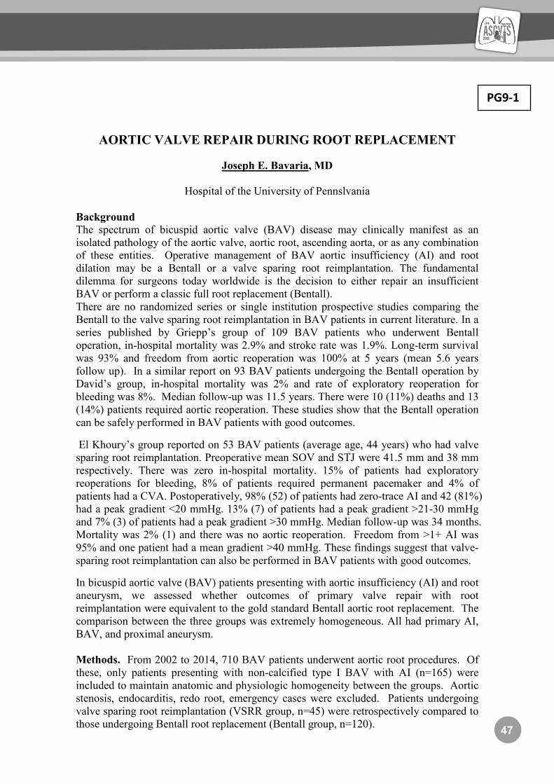
The 24th Annual Meeting of Asian Society for Cardiovascular and Thoracic Surgery (ASCVTS)in conjunction with 9th AATS / ASCVTS Postgraduate Course and 4th Asian Single Port VATS Symposium
47
AORTIC VALVE REPAIR DURING ROOT REPLACEMENT
Joseph E. Bavaria, MD
Hospital of the University of Pennslvania
BackgroundThe spectrum of bicuspid aortic valve (BAV) disease may clinically manifest as an isolated pathology of the aortic valve, aortic root, ascending aorta, or as any combination of these entities. Operative management of BAV aortic insufficiency (AI) and root dilation may be a Bentall or a valve sparing root reimplantation. The fundamental dilemma for surgeons today worldwide is the decision to either repair an insufficient BAV or perform a classic full root replacement (Bentall). There are no randomized series or single institution prospective studies comparing theBentall to the valve sparing root reimplantation in BAV patients in current literature. In a series published by Griepp’s group of 109 BAV patients who underwent Bentall operation, in-hospital mortality was 2.9% and stroke rate was 1.9%. Long-term survivalwas 93% and freedom from aortic reoperation was 100% at 5 years (mean 5.6 yearsfollow up). In a similar report on 93 BAV patients undergoing the Bentall operation by David’s group, in-hospital mortality was 2% and rate of exploratory reoperation for bleeding was 8%. Median follow-up was 11.5 years. There were 10 (11%) deaths and 13 (14%) patients required aortic reoperation. These studies show that the Bentall operation can be safely performed in BAV patients with good outcomes.
El Khoury’s group reported on 53 BAV patients (average age, 44 years) who had valve sparing root reimplantation. Preoperative mean SOV and STJ were 41.5 mm and 38 mm respectively. There was zero in-hospital mortality. 15% of patients had exploratory reoperations for bleeding, 8% of patients required permanent pacemaker and 4% of patients had a CVA. Postoperatively, 98% (52) of patients had zero-trace AI and 42 (81%) had a peak gradient <20 mmHg. 13% (7) of patients had a peak gradient >21-30 mmHgand 7% (3) of patients had a peak gradient >30 mmHg. Median follow-up was 34 months.Mortality was 2% (1) and there was no aortic reoperation. Freedom from >1+ AI was 95% and one patient had a mean gradient >40 mmHg. These findings suggest that valve-sparing root reimplantation can also be performed in BAV patients with good outcomes.
In bicuspid aortic valve (BAV) patients presenting with aortic insufficiency (AI) and root aneurysm, we assessed whether outcomes of primary valve repair with root reimplantation were equivalent to the gold standard Bentall aortic root replacement. The comparison between the three groups was extremely homogeneous. All had primary AI, BAV, and proximal aneurysm.
Methods. From 2002 to 2014, 710 BAV patients underwent aortic root procedures. Of these, only patients presenting with non-calcified type I BAV with AI (n=165) were included to maintain anatomic and physiologic homogeneity between the groups. Aortic stenosis, endocarditis, redo root, emergency cases were excluded. Patients undergoing valve sparing root reimplantation (VSRR group, n=45) were retrospectively compared to those undergoing Bentall root replacement (Bentall group, n=120).
PG9-1

The 24th Annual Meeting of Asian Society for Cardiovascular and Thoracic Surgery (ASCVTS)in conjunction with 9th AATS / ASCVTS Postgraduate Course and 4th Asian Single Port VATS Symposium
48
Results. Bentall patients were older (52 + 13 vs 46 + 12 years, p=<0.01), had lower EF (53 + 12% vs. 58 + 8%, p<0.01), but LV diastolic diameter was similar (58 + 10 mm vs. 57 + 9 mm, p=0.5). 30-day/ in-hospital mortality and stroke was zero; permanent pacemaker rate was similar (6% (n=7) vs 0, p=0.2). On discharge echocardiography, AI grade<1+ (100%, p=1) and transvalvular gradients (mean gradient 7+3 versus 6+3 mmHg, p=0.14) were similar.
Mean follow-up was 7.5 + 3.2 and 3.4 + 2.9 years (p<0.001). There were 14 TIA/ stroke events in Bentall group; none in VSRR group. One patient in each group developed AI>3+. 5-year actuarial survival (100 vs 98+2%, p=0.8) and freedom from aortic reoperation (98+2% versus 100%, p=0.8) were similar.
Conclusions. In patients with BAV AI with root aneurysm, primary cusp repair with root reimplantation achieves equivalent midterm outcomes compared to Bentall root replacement.
PG9-1
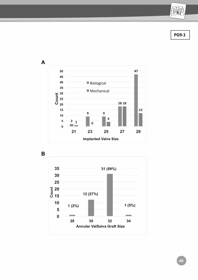
The 24th Annual Meeting of Asian Society for Cardiovascular and Thoracic Surgery (ASCVTS)in conjunction with 9th AATS / ASCVTS Postgraduate Course and 4th Asian Single Port VATS Symposium
49
Results. Bentall patients were older (52 + 13 vs 46 + 12 years, p=<0.01), had lower EF (53 + 12% vs. 58 + 8%, p<0.01), but LV diastolic diameter was similar (58 + 10 mm vs. 57 + 9 mm, p=0.5). 30-day/ in-hospital mortality and stroke was zero; permanent pacemaker rate was similar (6% (n=7) vs 0, p=0.2). On discharge echocardiography, AI grade<1+ (100%, p=1) and transvalvular gradients (mean gradient 7+3 versus 6+3 mmHg, p=0.14) were similar.
Mean follow-up was 7.5 + 3.2 and 3.4 + 2.9 years (p<0.001). There were 14 TIA/ stroke events in Bentall group; none in VSRR group. One patient in each group developed AI>3+. 5-year actuarial survival (100 vs 98+2%, p=0.8) and freedom from aortic reoperation (98+2% versus 100%, p=0.8) were similar.
Conclusions. In patients with BAV AI with root aneurysm, primary cusp repair with root reimplantation achieves equivalent midterm outcomes compared to Bentall root replacement.
PG9-1

The 24th Annual Meeting of Asian Society for Cardiovascular and Thoracic Surgery (ASCVTS)in conjunction with 9th AATS / ASCVTS Postgraduate Course and 4th Asian Single Port VATS Symposium
50
PG9-1

The 24th Annual Meeting of Asian Society for Cardiovascular and Thoracic Surgery (ASCVTS)in conjunction with 9th AATS / ASCVTS Postgraduate Course and 4th Asian Single Port VATS Symposium
51
PG9-1

The 24th Annual Meeting of Asian Society for Cardiovascular and Thoracic Surgery (ASCVTS)in conjunction with 9th AATS / ASCVTS Postgraduate Course and 4th Asian Single Port VATS Symposium
52
CEREBRAL PROTECTION DURING ARCH SURGERY
Marc R. Moon, M.D.
Washington University School of Medicine, Saint Louis, Missouri
Since Dr. Randall Griepp first reported four patients undergoing prosthetic replacement of the aortic arch under profound hypothermic circulatory arrest in 1975, there has been unparalleled progress in the techniques for cerebral protection during surgery on the arch. Prevention of stroke and preservation of neurocognitive function postoperatively is the ultimate goal and this is achieved through strict attention to detail. Surgeons need to minimize embolization, select appropriate cannulation strategies, avoid atheroma, minimize clamping of diseased aortic segments, maintain cerebral perfusion when possible and monitor cerebral flow to ensure adequate protection. Crawford’s classic dictum is that with hypothermic circulatory arrest alone, the stroke rate increases when HCA times exceed 40 minutes, while the death rate increases when HCA times exceed 60 minutes. With modern cerebral protection techniques, cerebrovascular accident occurs in 2-4% while transient neurologic deficits appear in 10-25%. Adjuncts to HCA include retrograde cerebral perfusion and antegrade cerebral perfusion. ACP maintains brain cooling, retrograde flushing of debris, and allows a “no touch” technique, but only provides 10-15% of nutrient flow. Antegrade cerebral perfusion can provide nearly normal nutrient flow, potentially increasing “safe” circulatory arrest times at warmer perfusion levels. Aortic arch replacement remains one of the most challenging cardiovascular disease processes and requires careful preoperative and intraoperative planning. Success rates have improved with better perfusion methods, neuroprotective strategies, and technical advances.
SUTURELESS AORTIC VALVE REPLACEMENT
Frank N. Slachman
Mercy Medical Group
We are observing an increased incidence of Aortic Valve Stenosis in our aging population. More than one in eight people over the age of 75 have moderate to severe aortic valve stenosis. With an increased awareness of the true incidence and poor natural history of this disease process, technologic advances such as catheter based therapies (TAVI ) have been developed to help treat this aging population, with their corresponding co-morbidities, and associated risks. Another such technology, a ‘rapid deployment’, sutureless valve has been developed for the treatment of patients who are considered acceptable candidates for conventional surgical valve replacement. Potential advantages include the reliable implant of a robust valve, in a well debrided annulus, with exceptional hemodynamics and low incidence of paravalvular leak, while reducing cross-clamp, and bypass times, and simplifying the implantation from a minimally invasive platform.
PG9-2

The 24th Annual Meeting of Asian Society for Cardiovascular and Thoracic Surgery (ASCVTS)in conjunction with 9th AATS / ASCVTS Postgraduate Course and 4th Asian Single Port VATS Symposium
53
CEREBRAL PROTECTION DURING ARCH SURGERY
Marc R. Moon, M.D.
Washington University School of Medicine, Saint Louis, Missouri
Since Dr. Randall Griepp first reported four patients undergoing prosthetic replacement of the aortic arch under profound hypothermic circulatory arrest in 1975, there has been unparalleled progress in the techniques for cerebral protection during surgery on the arch. Prevention of stroke and preservation of neurocognitive function postoperatively is the ultimate goal and this is achieved through strict attention to detail. Surgeons need to minimize embolization, select appropriate cannulation strategies, avoid atheroma, minimize clamping of diseased aortic segments, maintain cerebral perfusion when possible and monitor cerebral flow to ensure adequate protection. Crawford’s classic dictum is that with hypothermic circulatory arrest alone, the stroke rate increases when HCA times exceed 40 minutes, while the death rate increases when HCA times exceed 60 minutes. With modern cerebral protection techniques, cerebrovascular accident occurs in 2-4% while transient neurologic deficits appear in 10-25%. Adjuncts to HCA include retrograde cerebral perfusion and antegrade cerebral perfusion. ACP maintains brain cooling, retrograde flushing of debris, and allows a “no touch” technique, but only provides 10-15% of nutrient flow. Antegrade cerebral perfusion can provide nearly normal nutrient flow, potentially increasing “safe” circulatory arrest times at warmer perfusion levels. Aortic arch replacement remains one of the most challenging cardiovascular disease processes and requires careful preoperative and intraoperative planning. Success rates have improved with better perfusion methods, neuroprotective strategies, and technical advances.
SUTURELESS AORTIC VALVE REPLACEMENT
Frank N. Slachman
Mercy Medical Group
We are observing an increased incidence of Aortic Valve Stenosis in our aging population. More than one in eight people over the age of 75 have moderate to severe aortic valve stenosis. With an increased awareness of the true incidence and poor natural history of this disease process, technologic advances such as catheter based therapies (TAVI ) have been developed to help treat this aging population, with their corresponding co-morbidities, and associated risks. Another such technology, a ‘rapid deployment’, sutureless valve has been developed for the treatment of patients who are considered acceptable candidates for conventional surgical valve replacement. Potential advantages include the reliable implant of a robust valve, in a well debrided annulus, with exceptional hemodynamics and low incidence of paravalvular leak, while reducing cross-clamp, and bypass times, and simplifying the implantation from a minimally invasive platform.
PG9-3

The 24th Annual Meeting of Asian Society for Cardiovascular and Thoracic Surgery (ASCVTS)in conjunction with 9th AATS / ASCVTS Postgraduate Course and 4th Asian Single Port VATS Symposium
54
TEVAR FOR AORTIC DISSECTION
Kazuo Shimamura1), Toru Kuratani2), Tomohiko Sakamoto1), Tomoaki Kudo2),Kenta Masada1), Kei Torikai1), Yoshiki Sawa1)
Osaka University Graduate School of Medicine, Department of Cardiovascular Surgery1) Department of Minimally Invasive Cardiovascular Medicine2)
(Objective) The randomized trial (INSTEAD trial) showed entry closure with stent graft (TEVAR), which has been widely accepted as an effective procedure to manage acute complicated dissection, could also prevent dissection related mortality in subacute/chronic type B dissection (TBD). In this study, we investigated the impact of intervention timing in TEVAR for chronic TBD by evaluating mid-term results.(Methods) One-hundred and forty eight cases who underwent TEVAR for chronic/subacute TBD between 2005 and 2014 (Male 118(79.7%), mean age 63.4 years old) were retrospectively reviewed. The timing of intervention after dissection onset differs from 19day to 7446day (median 626day), and the mid-term results were compared in 2 groups (early (E) group; intervention within 6 month, n=43 and late (L) group; intervention after 6 month, n=105). (Results) There were no significant difference in patients preoperative background except the maximal aortic diameter (E/L=50.2/58.7mm, p<0.01), and no significant difference were observed in the early results (operative mortality (E/L=0%, 1.9%), stroke (2.3%/1.9%), spinal cord ischemia(0/0%). In average follow up of 42.3 month, overall survival and freedom from aorta-related mortality tends to be better in early group (survival; E: 93.5%/93.5% at 3/5 year, L: 89.4%/81.9% at 3/5yeaer, p=0.09). Late aortic event also tended to be well avoided in early group (freedom from aortic event: E: 84.8%/ 80.4%, L: 78.7%/64.1% at 3/5years respectively, p=0.071), with late group requiring more additional entry closure.(Conclusion) Although it could be multifactorial and further investigation is necessary, early intervention (within 6 month) could be one of significant factor to achieve better late outcomes in chronic TBD.
MODELING THE SINGLE VENTRICLE: AN UPDATE ON THE STATE OF THE ART
Tain-Yen Hsia
Great Ormond Street Hospital for Children
Single ventricle physiology and surgical palliation remain major clinical challenges. Introduced in 1996 by Prof. Marc de Leval and the group at Great Ormond Street Hospital for Children, mathematical modeling has been used as an adjunct to enhance our understanding of the unique features of single ventricle physiology and to affect opeative technique to improve outcomes. Since then, there have been substantial advancements in both engineering methodology and expansion of expertise. Recently a transatlantic collaboration between 7 American and European institutions was established under the guise of the Leducq Foundation Network of Excellence was formed in 2000 to develop a comprehensive research program to promote modeling and engineering studies in all three stage of single ventricle physiology. This talk will present the progress made by this group over the last six years and explore future endeavors that will facilitate mathematical modeling as a clinical decision support tool.
PG9-5

The 24th Annual Meeting of Asian Society for Cardiovascular and Thoracic Surgery (ASCVTS)in conjunction with 9th AATS / ASCVTS Postgraduate Course and 4th Asian Single Port VATS Symposium
55
MODELING THE SINGLE VENTRICLE: AN UPDATE ON THE STATE OF THE ART
Tain-Yen Hsia
Great Ormond Street Hospital for Children
Single ventricle physiology and surgical palliation remain major clinical challenges. Introduced in 1996 by Prof. Marc de Leval and the group at Great Ormond Street Hospital for Children, mathematical modeling has been used as an adjunct to enhance our understanding of the unique features of single ventricle physiology and to affect opeative technique to improve outcomes. Since then, there have been substantial advancements in both engineering methodology and expansion of expertise. Recently a transatlantic collaboration between 7 American and European institutions was established under the guise of the Leducq Foundation Network of Excellence was formed in 2000 to develop a comprehensive research program to promote modeling and engineering studies in all three stage of single ventricle physiology. This talk will present the progress made by this group over the last six years and explore future endeavors that will facilitate mathematical modeling as a clinical decision support tool.
PG10-2

The 24th Annual Meeting of Asian Society for Cardiovascular and Thoracic Surgery (ASCVTS)in conjunction with 9th AATS / ASCVTS Postgraduate Course and 4th Asian Single Port VATS Symposium
56
LONG-TERM OUTCOME OF FONTAN CONVERSION
Carl L. Backer, M.D.
Division of Cardiovascular-Thoracic Surgery, Ann & Robert H. Lurie Children’s Hospital of Chicago, and Department of Surgery, Northwestern University Feinberg
School of Medicine, Chicago, IL, USA
BACKGROUND: The purpose of this review was to determine long-term outcomes after Fontan conversion with arrhythmia operations and identify characteristics associated with death or need for cardiac transplantation.METHODS: The first 140 Fontan conversions with arrhythmia surgery at Ann & Robert H. Lurie Children’s of Chicago were analyzed for predictors of cardiac death or transplant and incidence of arrhythmia recurrence.RESULTS: The median age at Fontan conversion was 23.2 years (range, 2.6 to 47.3 years). Preoperative arrhythmias were present in 136 patients: right atrial tachycardia in 48 patients, left atrial tachycardia in 21, and atrial fibrillation in 67. Freedom from cardiac death or transplant was 90% at 5 years, 84% at 10 years, and 66% at 15 years. The median age at the last follow-up among survivors was 32 years (range, 15 to 61 years). By multivariable analysis, risk factors for cardiac death or heart transplantation were a right or indeterminate ventricular morphology, cardiopulmonary bypass time exceeding 240 minutes, ascites, protein-losing enteropathy, or a biatrial arrhythmia operation at the time of conversion. Freedom from recurrence of atrial tachycardia was 77% at 10 years. Among 67 patients with atrial fibrillation undergoing biatrial arrhythmia operations, none had recurrent atrial fibrillation.CONCLUSIONS: Freedom from cardiac death or transplant for patients undergoing Fontan conversion with arrhythmia surgery is 84% at 10 years. The effects of atrialarrhythmia operations are durable in most patients. Fontan conversion is effective therapy for properly selected failing atriopulmonary Fontan patients.
MCS AND HTX FOR SINGLE VENTRICLE
Iki Adachi
Texas Children’s Hospita
There has been an increasing number of patients presenting with severe heart failure after various types of single ventricle palliations. This is a challenging patient population to provide durable mechanical circulatory support for bridge-to-transplantation due to unique anatomy and physiology. In this presentation, mechanical circulatory support options for single ventricle, particularly the use of an implantable continuous-flow device, will be discussed.
PG10-3

The 24th Annual Meeting of Asian Society for Cardiovascular and Thoracic Surgery (ASCVTS)in conjunction with 9th AATS / ASCVTS Postgraduate Course and 4th Asian Single Port VATS Symposium
57
LONG-TERM OUTCOME OF FONTAN CONVERSION
Carl L. Backer, M.D.
Division of Cardiovascular-Thoracic Surgery, Ann & Robert H. Lurie Children’s Hospital of Chicago, and Department of Surgery, Northwestern University Feinberg
School of Medicine, Chicago, IL, USA
BACKGROUND: The purpose of this review was to determine long-term outcomes after Fontan conversion with arrhythmia operations and identify characteristics associated with death or need for cardiac transplantation.METHODS: The first 140 Fontan conversions with arrhythmia surgery at Ann & Robert H. Lurie Children’s of Chicago were analyzed for predictors of cardiac death or transplant and incidence of arrhythmia recurrence.RESULTS: The median age at Fontan conversion was 23.2 years (range, 2.6 to 47.3 years). Preoperative arrhythmias were present in 136 patients: right atrial tachycardia in 48 patients, left atrial tachycardia in 21, and atrial fibrillation in 67. Freedom from cardiac death or transplant was 90% at 5 years, 84% at 10 years, and 66% at 15 years. The median age at the last follow-up among survivors was 32 years (range, 15 to 61 years). By multivariable analysis, risk factors for cardiac death or heart transplantation were a right or indeterminate ventricular morphology, cardiopulmonary bypass time exceeding 240 minutes, ascites, protein-losing enteropathy, or a biatrial arrhythmia operation at the time of conversion. Freedom from recurrence of atrial tachycardia was 77% at 10 years. Among 67 patients with atrial fibrillation undergoing biatrial arrhythmia operations, none had recurrent atrial fibrillation.CONCLUSIONS: Freedom from cardiac death or transplant for patients undergoing Fontan conversion with arrhythmia surgery is 84% at 10 years. The effects of atrialarrhythmia operations are durable in most patients. Fontan conversion is effective therapy for properly selected failing atriopulmonary Fontan patients.
MCS AND HTX FOR SINGLE VENTRICLE
Iki Adachi
Texas Children’s Hospita
There has been an increasing number of patients presenting with severe heart failure after various types of single ventricle palliations. This is a challenging patient population to provide durable mechanical circulatory support for bridge-to-transplantation due to unique anatomy and physiology. In this presentation, mechanical circulatory support options for single ventricle, particularly the use of an implantable continuous-flow device, will be discussed.
PG10-4

The 24th Annual Meeting of Asian Society for Cardiovascular and Thoracic Surgery (ASCVTS)in conjunction with 9th AATS / ASCVTS Postgraduate Course and 4th Asian Single Port VATS Symposium
58
WHAT IS THE BEST SURGICAL APPROACH TO TREAT THYMOMA?
Ming-Ching Lee1,2, Chung-Ping Hsu1,3
Division of Thoracic Surgery, Department of Surgery, Taichung Veterans General Hospital, Taichung, Taiwan, Republic of China 1, Institute of Clinical Medicine, National Yang-Ming University, Taipei, Taiwan 2, School of Medicine, National
Yang-Ming University, Taipei, Taiwan, Republic of China 3
OBJECTIVE: Thymoma is the most common neoplasm in the anterior mediastinum in adults. In the past, open approaches such as median sternotomy and thoracotomy provide adequate extent of resection margin has been a standard procedure. However, many different approaches by Video-Assisted Thoracoscopic Surgery (VATS) haveevolved significantly over the last decades. The purpose of this session is to provide a succinct, practical review of the major changes that should be integrated into daily practices. This will be a fast-paced review of the relevant changes to surgical approach that have occurred over the last year. The combination of didactic lecture and case demonstration will be used to develop concepts of surgical decision makingin the treatment of thymoma.
Table 1. Clinicopathologic characteristics of the patients with and without thymoma N (%)
Number of patients 105Gender
Male 32 (31)Female 73 (69)
Age (years) 36.0±14.1 (21-80)BMI (kg/m2) 23.3±4.3 (16-38)MGFA classification
I 22 (21)II 41 (39)III 37 (35)IV 4 (4)V 1 (1)
Specimen weight (gm) 70.9±37.6 (18-310)Thymoma
yes 30 (29)no 75 (71)
Thymoma size (cm) 3.0±1.1 (1.3-6)0.1~2cm 6 (23)2.1~6cm 20 (77)
Masaoka stage (total N,%) 30 (29)I 16 (53)II a 9 (30)
II b 5 (17)WHO classification (total N,%) 27 (26)
class A 3 (11)class AB 2 (7)class B1 7 (26)class B2 10 (37)class B3 5 (19)
Operation time (min) 141.6±31.6 (80-245)Duration of chest tube (days) 3.8±1.3 (2-10)Operative complications (total N,%) 9 (9)
Conversion to open surgery 3 (3)Bleeding 2 (2)Electronic Cardioversion 4 (4)
Follow-up duration (years) 6.5±3.5 (0.2-14)Final neurologic response
Complete remission a 49 (47)Satisfactory remission b 40 (39)Unresponsive c 15 (14)
MG exacerbation d
yes 10 (9)no 95 (91)
a Complete remission presents patients need 0-1 Mestinon tab per day b Satisfactory remission presents patients need 2-3 Mestinon tab per day c Unresponsive presents patients need 4-8 Mestinon tab per day d MG exacerbation presents patients need readmission for plasmaphoresis to relief symptoms MG = myasthenia gravis
PG11-1

The 24th Annual Meeting of Asian Society for Cardiovascular and Thoracic Surgery (ASCVTS)in conjunction with 9th AATS / ASCVTS Postgraduate Course and 4th Asian Single Port VATS Symposium
59
II b 5 (17)WHO classification (total N,%) 27 (26)
class A 3 (11)class AB 2 (7)class B1 7 (26)class B2 10 (37)class B3 5 (19)
Operation time (min) 141.6±31.6 (80-245)Duration of chest tube (days) 3.8±1.3 (2-10)Operative complications (total N,%) 9 (9)
Conversion to open surgery 3 (3)Bleeding 2 (2)Electronic Cardioversion 4 (4)
Follow-up duration (years) 6.5±3.5 (0.2-14)Final neurologic response
Complete remission a 49 (47)Satisfactory remission b 40 (39)Unresponsive c 15 (14)
MG exacerbation d
yes 10 (9)no 95 (91)
a Complete remission presents patients need 0-1 Mestinon tab per day b Satisfactory remission presents patients need 2-3 Mestinon tab per day c Unresponsive presents patients need 4-8 Mestinon tab per day d MG exacerbation presents patients need readmission for plasmaphoresis to relief symptoms MG = myasthenia gravis
PG11-1
The 24th Annual Meeting of Asian Society for Cardiovascular and Thoracic Surgery (ASCVTS)in conjunction with 9th AATS / ASCVTS Postgraduate Course and 4th Asian Single Port VATS Symposium
60
CURRENT STATUS OF INVASIVE MEDIASTINAL STAGING
Prof. Dr. P. De Leyn,
Department of Thoracic Surgery, University Hospitals Leuven, Belgium
Mediastinal lymph node (LN) involvement is present in 30 to 45% of newly diagnosed lung cancers. A subgroup of patients with positive mediastinal LN’s benefitfrom surgical multimodality treatment. Therefore, mediastinal staging procedures canselect patients for resection, based on both baseline and post-induction assessment.PET scan has largely improved mediastinal LN staging. However PET positive mediastinal findings should always be cyto-histologically confirmed. Due to the high NPV of PET scan, invasive staging can be omitted in some patients with early stage NSCLC. In central tumours, PET N1 nodes or CT-enlarged LN’s and tumours larger than 3 cm (especially adenocarcinoma with high SUV) invasive staging is recommended according to the recent revised ESTS guidelines. Different techniques of invasive mediastinal staging are available. They vary in accuracy and procedure-related morbidity. Ultrasound-guided bronchoscopy with fine-needle aspiration (EBUS-FNA) and endoscopic esophageal ultrasound-guided FNA (EUS-FNA) are techniques that provide cyto-histological diagnosis and are minimally invasive. Although some studies have shown equal accuracy of EBUS and mediastinoscopy it is accepted in case of negative results of EBUS/EUS-FNA, an invasive surgical technique is indicated. A recent prospective study showed that combining endosonography and surgical staging compared with surgical staging alone resulted in greater sensitivity for mediastinal nodal metastasis end fewer unnecessary thoracotomies.Compared with classical mediastinoscopy, there are several advantages of videomediastinoscopy. These are improved teaching capacities, taping, the magnifying view and the bimanual dissection. Although visualisation is improved, there is no improvement of accuracy unless more extensive nodal dissection is performed like in VAMA or TEMLA.For restaging in a surgical multimodality regimen, invasive techniques providing cyto-histological information are advisable despite the encouraging results with the use of re-PET/CT imaging. The accuracy of remediastinoscopy and EBUS/EUS-FNAis much lower than in baseline staging. Baseline staging of mediastinal LN’s with endoscopic techniques and restaging after induction therapy with mediastinoscopy seems to be a valuable tool to select patients with N2 disease for surgical multimodality treatment.We conclude that optimal staging is a truly multidisciplinary process, with a variety of possible techniques, to be performed by experienced hands. The proficiency of the physician performing the procedure plays a role in varying sensitivity and specificity observed and each center has to adopt its own guidelines pending on their performance.
De Leyn P, Dooms C, Kuzdzal J, Lardinois D, Passlick D, Rami-Porta R, Tuma A, Van Schil P, Venuta F, Waller D, Weder D, Zielinski M. Revised ESTS guidelines for preoperative mediastinal lymph node staging for non-small cell lung cancer. Eur J Cardiothorac Surg 2014;45(5):787-98.
RADICAL LYMPH NODE DISSECTION VIA MEDIASTINOSCOPY
Gunda Leschber1
ELK Berlin Chest Hospital, Berlin, Germany1
Staging of the mediastinum is critical for choosing the best therapeutic option in lung cancer patients. Mediastinoscopy remains the gold standard for mediastinal staging despite the appearance of new radiologic or endoscopic techniques (PET-CT and EBUS/EUS-TBNA) as these methods have limited accuracy, especially in non-enlarged lymph nodes (1).Video technique has facilitated teaching of mediastinoscopy and allowed to performreal „operations“ within the mediastinum instead of simple lymph node sampling. By video-mediastinoscopy even unexperienced surgeons/trainees achieve better results compared to conventional mediastinoscopy (2). Video-assisted mediastinal lymphadenectomy (VAMLA) is a further development of video-mediastinoscopy and was developed by Martin Huertgen in 2001 (3,4). Thistechnique allows radical en bloc-resection of mediastinal lymph nodes with the surronding fatty tissue. By using a mediastinoscope with spreadable blades, bimanual dissection exposes all limiting mediastinal structures (trachea, both main bronchi, pulmonary artery, oesophagus, azygos vein, vena cava, pleura) and the lymph node compartments.
Technique:After insertion of the mediastinoscope dissection is carried down to the bifurcation and both main bronchi as well as the pulmonary artery are exposed. All lymph nodes in the subcarinal space (station 7) are dissected and removed, leaving the oesophagus, medial walls of both main bronchi as well as the mediastinal pleura clearly visible. Dissection is continued on the left side along the lateral wall of the left main bronchus for lymph nodes stations 10L and 4 L, exposing the recurrent laryngeal nerve occasionally.On the right side it is not unusual to expose the right upper bronchus and the intermediate bronchus. Following lymph node dissection in lymph node station 10R, attention is turned to the azygos vein. All fatty tissue cranially to the vein containing the lymph nodes is dissected until its confluence with the vena cava is visible. Continuing dissection upward along the pleura and vena cava until the brachiocephalic trunk (innominate artery) is reached and en bloc resection completed of lymph nodes 4R and all mediastinal fat on the right side.On the left careful exploration of paratracheal tissue for lymph nodes is done in order not to harm the recurrent laryngeal nerve.Lymph nodes of station 2 R above the crossing of the brachiocephalic artery are only removed if enlarged.
In our experience the use of bipolar coagulation (bipolar scissors) is favourable especially in the proximity of the recurrent laryngeal nerve. A suction device with a bended tip and a grasping forceps to hold the tissue is helpful. Metal clips are applied to bronchial artery branches if necessary. All instruments are 3-5 mm allowing bimanual manipulation through the scope. Video footage will demonstrate the procedure.
PG11-2

The 24th Annual Meeting of Asian Society for Cardiovascular and Thoracic Surgery (ASCVTS)in conjunction with 9th AATS / ASCVTS Postgraduate Course and 4th Asian Single Port VATS Symposium
61
RADICAL LYMPH NODE DISSECTION VIA MEDIASTINOSCOPY
Gunda Leschber1
ELK Berlin Chest Hospital, Berlin, Germany1
Staging of the mediastinum is critical for choosing the best therapeutic option in lung cancer patients. Mediastinoscopy remains the gold standard for mediastinal staging despite the appearance of new radiologic or endoscopic techniques (PET-CT and EBUS/EUS-TBNA) as these methods have limited accuracy, especially in non-enlarged lymph nodes (1).Video technique has facilitated teaching of mediastinoscopy and allowed to performreal „operations“ within the mediastinum instead of simple lymph node sampling. By video-mediastinoscopy even unexperienced surgeons/trainees achieve better results compared to conventional mediastinoscopy (2). Video-assisted mediastinal lymphadenectomy (VAMLA) is a further development of video-mediastinoscopy and was developed by Martin Huertgen in 2001 (3,4). Thistechnique allows radical en bloc-resection of mediastinal lymph nodes with the surronding fatty tissue. By using a mediastinoscope with spreadable blades, bimanual dissection exposes all limiting mediastinal structures (trachea, both main bronchi, pulmonary artery, oesophagus, azygos vein, vena cava, pleura) and the lymph node compartments.
Technique:After insertion of the mediastinoscope dissection is carried down to the bifurcation and both main bronchi as well as the pulmonary artery are exposed. All lymph nodes in the subcarinal space (station 7) are dissected and removed, leaving the oesophagus, medial walls of both main bronchi as well as the mediastinal pleura clearly visible. Dissection is continued on the left side along the lateral wall of the left main bronchus for lymph nodes stations 10L and 4 L, exposing the recurrent laryngeal nerve occasionally.On the right side it is not unusual to expose the right upper bronchus and the intermediate bronchus. Following lymph node dissection in lymph node station 10R, attention is turned to the azygos vein. All fatty tissue cranially to the vein containing the lymph nodes is dissected until its confluence with the vena cava is visible. Continuing dissection upward along the pleura and vena cava until the brachiocephalic trunk (innominate artery) is reached and en bloc resection completed of lymph nodes 4R and all mediastinal fat on the right side.On the left careful exploration of paratracheal tissue for lymph nodes is done in order not to harm the recurrent laryngeal nerve.Lymph nodes of station 2 R above the crossing of the brachiocephalic artery are only removed if enlarged.
In our experience the use of bipolar coagulation (bipolar scissors) is favourable especially in the proximity of the recurrent laryngeal nerve. A suction device with a bended tip and a grasping forceps to hold the tissue is helpful. Metal clips are applied to bronchial artery branches if necessary. All instruments are 3-5 mm allowing bimanual manipulation through the scope. Video footage will demonstrate the procedure.
PG11-3

The 24th Annual Meeting of Asian Society for Cardiovascular and Thoracic Surgery (ASCVTS)in conjunction with 9th AATS / ASCVTS Postgraduate Course and 4th Asian Single Port VATS Symposium
62
VAMLA is used for exact staging for lung cancer and as a first step to VATS lobectomy as it facilitates the complete dissection of the subcarinal area which is difficult to achieve from the thoracoscopic approach. VAMLA has been shown to increase the sensitivity and accuracy of mediastinoscopy in several studies (5-8).First studies show that survival following resection of NCSLC after radical lymph node dissection by VAMLA seems to be improved (9). VAMLA should not be confounded with video-mediastinoscopy.
References:
1. Sivrikoz CM, Ak I, Simsek FS, Döner E, Dündar E.: Is mediastinoscopy still the gold standard to evaluate mediastinal lymph nodes in patients with non-small cell lung carcinoma? Thorac Cardiovasc Surg. 2012;60:116-21
2. WallesT, Friedel G, Stegherr T, Steger V.: Learning mediastinoscopy: the need for education, experience and modern techniques—interdependency of the applied technique and surgeon’s training level. Interactive CardioVascular and Thoracic Surgery (2012) 1–5 doi:10.1093/icvts/ivs494
3. Hürtgen M, Friedel G, Toomes H, Fritz P.: Radical video-assisted mediastinoscopic lymphadenectomy (VAMLA)--technique and first results. Eur J Cardiothorac Surg.2002;21:348-51
4. Leschber G, Holinka G, Linder A.: Video-assisted mediastinoscopic lymphadenectomy (VAMLA) -a method for systematic mediastinal lymphnode dissection. Eur J Cardiothorac Surg 2003;24:192-5
5. Witte B, Wolf M, Huertgen M, Toomes H.:Video-assisted mediastinoscopic surgery: clinical feasibility and accuracy of mediastinal lymph node staging. Ann Thorac Surg. 2006;82:1821-7
6. Leschber G, Sperling D, Klemm W, Merk J.: Does video-mediastinoscopy improve the results of conventional mediastinoscopy? Eur J Cardiothorac Surg. 2008;33:289-93
7. Witte B, Messerschmidt A, Hillebrand H, Gross S, Wolf M, Kriegel E, Neumeister W, Hürtgen M.: Combined videothoracoscopic and videomediastinoscopic approach improves radicality of minimally invasive mediastinal lymphadenectomy for early stage lung carcinoma. Eur J Cardiothorac Surg. 2009;35:343-7
8. Citak N, Buyukkale S, Kok A, Celikten A, Metin M, Sayar A, Gurses A.: Does video-assisted mediastinoscopy offer lower false-negative rates for subcarinal lymph nodes compared with standard cervical mediastinoscopy. Thorac Cardiovasc Surg. 2014;62:624-30
9. Turna A, Demirkava A, Ozkul S, Oz B, Gurese A, Kaynak, K.: Video-assisted mediastinoscopic lymphadenectomy is associated with better survival than mediastinoscopy in patients with resected non-small cell lung cancer. J. Thoracic Cardiovasc Surg. 2013;146:774-80.
TRACHEAL SURGERY
Mark S. Allen
Mayo Clinic
Tracheal surgery began in the early 1960s with Dr. Grillo’s investigations at the Massachusetts General Hospital about tracheal stenosis after intubation in the ICU. The description of tracheal anatomy by Salassa and Payne revealed that the blood supply is segmental and comes into the trachea laterally; this allows a portion of the trachea to be resected and reanastomosed. Flexion of the neck and other special maneuvers allow a portion of the trachea to be removed and reconstructed without excessive tension. Typical symptoms of tracheal stenosis include wheezing, shortness of breath with exertion and cough. Stridor is a sign of severe narrowing. The flow-volume loop on pulmonary function tests will show a plateau pattern, indicating limited flow. Imaging with CT and measurement with a rigid bronchoscopy is necessary to determine how much trachea needs to be removed. Tissue diagnosis is not always necessary prior to a resection, since no matter what the histology the obstruction needs to be corrected.
Tracheal resection is performed with the patient in the supine position with the neck maximally extended. Patients are intubated so that the endotracheal tube is past the lesion. If the lesion prevents passage of the tube, rigid bronchoscopy should be used to “core out” the lesion to allow safe passage. A cervical incision is used for all tracheal lesions that are in the upper 2/3rds of the trachea. The thyroid is divided in the midline and retracted laterally and the anterior surface of the trachea is exposed. The lesion is located and the trachea is dissected circumferentially staying right on the trachea to avoid injury to the recurrent laryngeal nerves. The trachea is divided just distal to the lesion and an endotracheal tube is passed into the distal trachea and connected to the anesthesia machine to ventilate the patient. The original endotracheal tube is pulled back to just below the vocal cords. The portion of the trachea to be removed is dissected free and resected. The margins should be checked by frozen section. The ends of the trachea are then reanastomosed in an end-to-end fashion with 4-0 polyglycolic acid sutures. To reduce the tension on the anastomosis the neck is flexed. By freeing up the anterior and posterior trachea, where there is no blood supply, about 40% of the trachea can be resected in an ideal patient. The original endotracheal tube is advanced through the anastomosis to ventilate the patient. A drain is placed in the neck and the incision is closed in layers. A stitch is placed from the chin to the sternum to keep the neck flexed.
Postoperative management is usually uncomplicated. Oral intake can resume once the patient is awake and alert. A bronchoscopy is performed on postoperative day 7 to check the anastomosis and if it looks good the chin stitch can be cut and the patient discharged home.
PG11-3

The 24th Annual Meeting of Asian Society for Cardiovascular and Thoracic Surgery (ASCVTS)in conjunction with 9th AATS / ASCVTS Postgraduate Course and 4th Asian Single Port VATS Symposium
63
TRACHEAL SURGERY
Mark S. Allen
Mayo Clinic
Tracheal surgery began in the early 1960s with Dr. Grillo’s investigations at the Massachusetts General Hospital about tracheal stenosis after intubation in the ICU. The description of tracheal anatomy by Salassa and Payne revealed that the blood supply is segmental and comes into the trachea laterally; this allows a portion of the trachea to be resected and reanastomosed. Flexion of the neck and other special maneuvers allow a portion of the trachea to be removed and reconstructed without excessive tension. Typical symptoms of tracheal stenosis include wheezing, shortness of breath with exertion and cough. Stridor is a sign of severe narrowing. The flow-volume loop on pulmonary function tests will show a plateau pattern, indicating limited flow. Imaging with CT and measurement with a rigid bronchoscopy is necessary to determine how much trachea needs to be removed. Tissue diagnosis is not always necessary prior to a resection, since no matter what the histology the obstruction needs to be corrected.
Tracheal resection is performed with the patient in the supine position with the neck maximally extended. Patients are intubated so that the endotracheal tube is past the lesion. If the lesion prevents passage of the tube, rigid bronchoscopy should be used to “core out” the lesion to allow safe passage. A cervical incision is used for all tracheal lesions that are in the upper 2/3rds of the trachea. The thyroid is divided in the midline and retracted laterally and the anterior surface of the trachea is exposed. The lesion is located and the trachea is dissected circumferentially staying right on the trachea to avoid injury to the recurrent laryngeal nerves. The trachea is divided just distal to the lesion and an endotracheal tube is passed into the distal trachea and connected to the anesthesia machine to ventilate the patient. The original endotracheal tube is pulled back to just below the vocal cords. The portion of the trachea to be removed is dissected free and resected. The margins should be checked by frozen section. The ends of the trachea are then reanastomosed in an end-to-end fashion with 4-0 polyglycolic acid sutures. To reduce the tension on the anastomosis the neck is flexed. By freeing up the anterior and posterior trachea, where there is no blood supply, about 40% of the trachea can be resected in an ideal patient. The original endotracheal tube is advanced through the anastomosis to ventilate the patient. A drain is placed in the neck and the incision is closed in layers. A stitch is placed from the chin to the sternum to keep the neck flexed.
Postoperative management is usually uncomplicated. Oral intake can resume once the patient is awake and alert. A bronchoscopy is performed on postoperative day 7 to check the anastomosis and if it looks good the chin stitch can be cut and the patient discharged home.
PG11-4
The 24th Annual Meeting of Asian Society for Cardiovascular and Thoracic Surgery (ASCVTS)in conjunction with 9th AATS / ASCVTS Postgraduate Course and 4th Asian Single Port VATS Symposium
64
ECMO IN ELDERLY PATIENTS: EFFECTIVE OR FUTILE PROCEDURE?
Roberto Lorusso, MD, PhD
Euro-ELSO ChairELSO Executive and Steering Committee Member
Cardio-Thoracic Surgery Department – Heart & Vascular CentreMaastricht University Medical Centre (MUMC+)
Maastricht – The Netherlands
A recent analysis of the Nationwide Inpatient Sample (NIS) database including 1,990,486 patients with acute myocardial infarction from 2003 to 2010, showed that cardiogenic shock occurred in 8% of all cases and that patients aged > 75 years had the highest rate of acute cardiocirculatory compromise, and with a rise from 7.7% to 12.2% as compared to 5.9% to 9.4% in younger subjects during the study period. Another study has just shown that an increase in any type of mechanical circulatory support in the United States from 39.6% in the period 2004-2007, to 47.2% in the years 2008-2011, and remarkably from 6.2% to 11.9% in patients over 80 years of age. This data undoubtely confirms that conditions eligible for and application of mechanical circulatory support in advanced age is invariably increasing.Interestingly, published series show that age does not represent an absolute contraindication in case of implant in particular setting (like post-cardiotomy), indicating that ECMO is more liberally used as an ultimate rescue tool in these circumstances, at the same time underlining that worse outcome in older patients should be carefully evaluated in terms of indications and conditions at implant since more critical scenarios appear to characterize this attitude with expected worse outcome based on the more challenging patient status.Analysis from the ELSO Registry in aged patients undergoing ECMO for cardiac arrest and respiratory insufficiency have clearly shown that these patients are at higher risk of death, but that respectful survival rates can still be achieved. We conducted another investigation about elderly patients undergoing ECMO for cardiogenic shock and included in the ELSO Registry has also shown that despite higher in-hospital mortality rate (30% in the elderly patients versus 40% in the younger cohort), the survival quote is absolutely acceptable. Noteworthy, elderly patients had a significantly lower ECMO duration and experienced a higher rate of multi-organ failure.From the ELSO data, either in the challenging setting of ECPR or cardiogenic shock and in the presence of respiratory dysfunction, it is rather evident that age should not be considered a contraindication for ECMO implant, provided that careful evaluation of patient features and conditions at implant to avoid futile ECMO support.
PG12-1
POST-OPERATIVE SUPPORT FOR CARDIOGENIC SHOCK
Wen Wang, MD, PhD
Indiana University School of Medicine and Indiana University Health Methodist Hospital
Post-cardiotomy shock has multiple etiologies but always potential for high mortality and morbidity. Careful optimization for separation from cardiopulmonary bypass is crucial. Inability to wean from cardiopulmonary bypass despite the use of inotropes and intra-aortic balloon pump will require escalation to various temporary mechanical circulatory support. Base on the patient’s requirement of uni- versus biventricular support, different temporary circulatory support may be used to bridge patients to recovery. They include extracorporeal membrane oxygenation (ECMO), Impella, TandemHeart, and Centrimag temporary ventricular support. Successful outcome for post-cardiotomy shock patients depends on a multi-disciplinary approach.
Unfractionated heparin is the current anticoagulant of choice for extracorporeal membrane oxygenation (ECMO) support, offering rapid onset, reversibility, and low cost. However, risks of complications form heparin resistance secondary to anti-thrombin deficiency and the heparin-induced thrombocytopenia (10-15% of ECMO patients) are challenging. Bivalirudin, a direct thrombin inhibitor, may be a suitable choice of ECMO anticoagulation with its absence of resistance, rapid onset, and short half-life. From 2/2011-10/2013, 54 patients were supported with ECMO. 36 patients were included in this analysis: heparin = 27, bivalirudin = 9. Patientdemographics and mode of ECMO support were similar. Patients on bivalirudin wereconsistently in therapeutic range of anticoagulation more than heparin (80.2% vs 45.7%, p<.001). 9 heparin patients had suspected HIT based on HIT antibody test; heparin was discontinued in 6 patients. Risks of bleeding and thrombotic complications were not different between heparin and bivalrudin. When the added cost of correcting anti-thrombin deficiency is added to heparin therapy, bivalirudin is financially superior. Patient safety, efficacy, and cost may allow use of bivalirudin as anticoagulant of choice.

The 24th Annual Meeting of Asian Society for Cardiovascular and Thoracic Surgery (ASCVTS)in conjunction with 9th AATS / ASCVTS Postgraduate Course and 4th Asian Single Port VATS Symposium
65
POST-OPERATIVE SUPPORT FOR CARDIOGENIC SHOCK
Wen Wang, MD, PhD
Indiana University School of Medicine and Indiana University Health Methodist Hospital
Post-cardiotomy shock has multiple etiologies but always potential for high mortality and morbidity. Careful optimization for separation from cardiopulmonary bypass is crucial. Inability to wean from cardiopulmonary bypass despite the use of inotropes and intra-aortic balloon pump will require escalation to various temporary mechanical circulatory support. Base on the patient’s requirement of uni- versus biventricular support, different temporary circulatory support may be used to bridge patients to recovery. They include extracorporeal membrane oxygenation (ECMO), Impella, TandemHeart, and Centrimag temporary ventricular support. Successful outcome for post-cardiotomy shock patients depends on a multi-disciplinary approach.
Unfractionated heparin is the current anticoagulant of choice for extracorporeal membrane oxygenation (ECMO) support, offering rapid onset, reversibility, and low cost. However, risks of complications form heparin resistance secondary to anti-thrombin deficiency and the heparin-induced thrombocytopenia (10-15% of ECMO patients) are challenging. Bivalirudin, a direct thrombin inhibitor, may be a suitable choice of ECMO anticoagulation with its absence of resistance, rapid onset, and short half-life. From 2/2011-10/2013, 54 patients were supported with ECMO. 36 patients were included in this analysis: heparin = 27, bivalirudin = 9. Patientdemographics and mode of ECMO support were similar. Patients on bivalirudin wereconsistently in therapeutic range of anticoagulation more than heparin (80.2% vs 45.7%, p<.001). 9 heparin patients had suspected HIT based on HIT antibody test; heparin was discontinued in 6 patients. Risks of bleeding and thrombotic complications were not different between heparin and bivalrudin. When the added cost of correcting anti-thrombin deficiency is added to heparin therapy, bivalirudin is financially superior. Patient safety, efficacy, and cost may allow use of bivalirudin as anticoagulant of choice.
PG12-2

The 24th Annual Meeting of Asian Society for Cardiovascular and Thoracic Surgery (ASCVTS)in conjunction with 9th AATS / ASCVTS Postgraduate Course and 4th Asian Single Port VATS Symposium
66
NEW TREND IN EXTRACORPOREAL CARDIOPULMONARYRESUSCITATION
Yih-Sharng Chen, MD, PhD
National Taiwan University hospital, Taipei, Taiwan
Cardiac arrest occurs all time in and out of the hospital. CPR is the present useful maneuver in the critical condition, but the outcome is still discouraging. Several devices and trials were developed to improve the outcome, but mainly focused on the out-of-hospital CPR.
Assisted circulation (ECMO) has been advocated in recent CPR guidelines. In the early eighties, ECMO was even considered a contraindication in CPR because of the poor outcome and devastating complications. ECMO has been applied in shock rescue for 2 decades and recently extended to CPR. Nowadays, ECMO had been widely applied in resuscitation status. The survival rate was expected additional 20% survival in in-hospital cardiac arrest with cardiac origin, and the survival rate was also found improved in the category of out-of-hospital cardiac arrest in recent years. J-save data and the result from the Asian countries demonstrated the beneficial effect of ECMO in OHCA with better neurological outcome.
Therapeutic hypothermia had been advocated in out-of-hospital cardiac arrest for years. The hypothermia-related complication, such as myocardial suppression, infection or bleeding issue, sometimes may hinder to reach the much lower target temperature, although the lower temperature may have better benefit for the neuroprotection. Further Japan ECMO data revealed the additional benefit of therapeutic hypothermia for the group under CPR rescued with ECMO. Advanced approach for independent selective brain moderate deep hypothermia without systemic cooling may provide another different thinking process and another way to increase the neurological result.
ECMO had changed the outcome of the CPR and it also explores another field of cardiac surgery.
PG12-4


The 24th Annual Meeting of Asian Society for Cardiovascular and Thoracic Surgery (ASCVTS)in conjunction with 9th AATS / ASCVTS Postgraduate Course and 4th Asian Single Port VATS Symposium