poor maternal nutrition and accelerated postnatal growth...
TRANSCRIPT

RESEARCH ARTICLE SPECIAL COLLECTION: TRANSLATIONAL IMPACT OF RAT
Poor maternal nutrition and accelerated postnatal growth inducesan accelerated aging phenotype and oxidative stress in skeletalmuscle of male ratsJane L. Tarry-Adkins1,*, Denise S. Fernandez-Twinn1, Jian Hua Chen1, Iain P. Hargreaves2, Viruna Neergheen2,Catherine E. Aiken1 and Susan E. Ozanne1
ABSTRACT‘Developmental programming’, which occurs as a consequence ofsuboptimal in utero and early environments, can be associated withmetabolic dysfunction in later life, including an increased incidence ofcardiovascular disease and type 2 diabetes, and predisposition ofolder men to sarcopenia. However, the molecular mechanismsunderpinning these associations are poorly understood. Manyconditions associated with developmental programming are alsoknown to be associated with the aging process. We therefore utilizedour well-established rat model of low birth weight and acceleratedpostnatal catch-up growth (termed ‘recuperated’) in this study toestablish the effects of suboptimal maternal nutrition on age-associated factors in skeletal muscle. We demonstratedaccelerated telomere shortening (a robust marker of cellular aging)as evidenced by a reduced frequency of long telomeres (48.5-8.6 kb)and an increased frequency of short telomeres (4.2-1.3 kb) in vastuslateralis muscle from aged recuperated offspring compared tocontrols. This was associated with increased protein expression ofthe DNA-damage-repair marker 8-oxoguanine-glycosylase (OGG1)in recuperated offspring. Recuperated animals also demonstratedan oxidative stress phenotype, with decreased citrate synthaseactivity, increased electron-transport-complex activities of complexI, complex II-III and complex IV (all markers of functionalmitochondria), and increased xanthine oxidase (XO), p67phox andnuclear-factor kappa-light-chain-enhancer of activated B-cells (NF-κB). Recuperated offspring also demonstrated increased antioxidantdefense capacity, with increased protein expression of manganesesuperoxide dismutase (MnSOD), copper-zinc superoxide dismutase(CuZnSOD), catalase and heme oxygenase-1 (HO1), all of which areknown targets of NF-κB and can be upregulated as a consequence ofoxidative stress. Recuperated offspring also had a pro-inflammatoryphenotype, as evidenced by increased tumor necrosis factor-α(TNFα) and interleukin-1β (IL1β) protein levels. Taken together, wedemonstrate, for the first time to our knowledge, an accelerated agingphenotype in skeletal muscle in the context of developmental
programming. These findings may pave the way for suitableinterventions in at-risk populations.
KEY WORDS: Skeletal muscle, Oxidative stress, Mitochondria,Developmental programming
INTRODUCTIONFor over 25 years, it has been known that a suboptimal in uteroenvironment is strongly associated with increased risk ofdevelopment of age-associated disease in later life, includingcardiovascular disease (CVD) (Barker et al., 1989) and type 2diabetes (T2D) (Barker et al., 1993). These findings have beenrobustly confirmed in both humans and animals (Tarry-Adkins andOzanne, 2014; Zambrano et al., 2016), and these studies support the‘thrifty phenotype hypothesis’ (Hales and Barker, 1992), whichstates that, under conditions of suboptimal nutrition, the fetuspermanently alters its organ structure, metabolism and function toensure immediate survival of the organism. Although beneficialin continued conditions of poor postnatal nutrition, such‘developmental programming’ is known to be detrimental inpostnatal conditions of adequate or over-nutrition, both of whichcan cause accelerated postnatal growth.
The development of skeletal muscle is especially vulnerable tonutritional deficiency compared to other tissues, owing to musclemass being lost at the expense of brain-sparing in utero (Desai et al.,1996). Indeed, maternal nutrient restriction, a widely used model ofdevelopmental programming, is known to reduce offspring birthweight due to reductions in fetal circulating amino acids (Janssonet al., 2006; Pantham et al., 2015). This is highlighted in studies ofovine fetuses (Zhu et al., 2004) and offspring (Zhu et al., 2006)exposed to a suboptimal in utero environment, which demonstratelow birth weight as well as dysregulation of muscle development,including changes in the number and composition of myofibers.Numbers of myofibers and neuromuscular junctions are also alteredin a rat model of maternal protein restriction (Confortim et al., 2016)and this effect is long-lasting, into old age (Confortim et al., 2015).There is also evidence that a suboptimal early environment (nutrientrestriction) in the mouse can impact on muscle metabolism/functionand molecular changes, including decreased mitochondrial content(Beauchamp et al., 2015) and reduced expression of mitochondrialgenes, especially those involved in oxidative phosphorylation(Mortensen et al., 2010). Decreased muscle fiber score has also beenobserved in vastus lateralis muscle of low-birth-weight elderly men(Patel et al., 2012).
Muscle mass is known to decline with age, which cancontribute to age-associated muscular dysfunction; however, therate of decline shows great inter-individual variation (Sayer et al.,2010). With age, skeletal muscle can accumulate oxidative stress,Received 16 June 2016; Accepted 18 August 2016
1University of Cambridge Metabolic Research Laboratories and MRC MetabolicDiseases Unit, Wellcome Trust-MRC Institute of Metabolic Science, Addenbrooke’sTreatment Centre, Addenbrooke’s Hospital, Hills Road, Cambridge CB2 OQQ, UK.2Neurometabolic Unit, National Hospital, University College London, LondonWC1N 3BG, UK.
*Author for correspondence ( [email protected])
J.L.T., 0000-0001-9569-6132
This is an Open Access article distributed under the terms of the Creative Commons AttributionLicense (http://creativecommons.org/licenses/by/3.0), which permits unrestricted use,distribution and reproduction in any medium provided that the original work is properly attributed.
1221
© 2016. Published by The Company of Biologists Ltd | Disease Models & Mechanisms (2016) 9, 1221-1229 doi:10.1242/dmm.026591
Disea
seModels&Mechan
isms

which can cause issues such as a reduction in force generation andmuscle atrophy. Muscle atrophy contributes to progressiveweakness and an increased risk of mobility impairment, fallsand physical frailty in very advanced age (Cruz-Jentoft et al.,2010). Among the most frequently implicated mechanisms ofaging muscle atrophy is mitochondrial dysfunction, which leads toincreased reactive oxygen species (ROS) generation (Marzettiet al., 2013).Oxidative stress accumulation occurs when cellular ROS
overwhelm the endogenous antioxidant defense capacity andthus redox homeostasis is not maintained (Ray et al., 2012). Thisexcess ROS generation can cause macromolecular damage toproteins, lipids and DNA (Valko et al., 2007). Telomeres (hexamerrepeats of DNA: [TTAGGG]n), which are found at the ends ofchromosomes, are particularly susceptible to ROS damagebecause of their guanine-rich sequences (Oikawa and Kawanishi,1999). In normal somatic cells, telomeres shorten with everycellular division. This makes telomere length measurement arobust marker of aging in many species, including humans androdents, and this has been shown to be associated with longevity(Haussman et al., 2003; Heidinger et al., 2012). It is known thatsuboptimal in utero nutrition can lead to accelerated aging in anumber of tissues (Tarry-Adkins and Ozanne, 2014). The highmetabolic activity of skeletal muscle renders it particularlysusceptible to oxidative stress; however, accelerated aging inskeletal muscle as a consequence of developmental programminghas never been explored.This study therefore aimed to investigate the effects of a poor
maternal diet followed by accelerated postnatal growth upon skeletalmuscle (vastus lateralis) of aging male rat offspring, focusingspecifically upon telomere length, and indices of oxidative stress,antioxidant defense capacity and inflammation.
RESULTSIn all cases, the reported data are expressed as mean±s.e.m.
Anthropometrical dataRecuperated offspring were significantly (P<0.001; 6.3±0.3 g)smaller compared to controls (7.4±0.2 g) on day 3, and remainedsignificantly (P<0.001) smaller at day 7 (13.4±0.6 vs 16.8±0.8 g).By 14 days of age, the recuperated offspring had undergone rapidpostnatal catch-up growth and so were similar in weight to controloffspring (33.7±0.7 g vs 34±1.7 g), and this was maintained atweaning (52.2±0.9 g vs 50.7±1.2 g) and at 12 months of age(920±29 g vs 956±25 g). These values reflect average male pupweight in the litter.
Poor maternal nutrition and accelerated postnatal growthaccelerated telomere shortening and increased DNAdamageVastus lateralis muscle from recuperated animals had fewer longtelomeres and more short telomeres compared to control animals,with significant effects of both maternal diet (P<0.05) and telomerelength category (P<0.01) (Fig. 1A). There was no significant effectof maternal diet upon mRNA expression of the telomere-lengthmaintenance proteins Ku70 and Ku80 (Fig. 1B). However,increased (P<0.01) protein expression of the base excision-repairprotein 8-oxoguanine-glycosylase (OGG1) was observed inrecuperated animals compared to controls (Fig. 1C). There was noeffect of maternal diet upon the mRNA levels of markers of cellularsenescence, p53 (776±119 vs 789±99) or p21 (531±88 vs 370±32)(average copy number, control versus recuperated).
Poor maternal nutrition and accelerated postnatal growthlead to increased skeletal-muscle oxidative stress(a) NF-κB:There was a significant effect of in uteromaternal diet upon proteinexpression of NF-κB, with increased (P<0.001) NF-κB proteinlevels in recuperated offspring compared to controls (Fig. 2A).There was, however, no effect of maternal diet (control 442±60 vsrecuperated 377±60 copy number) upon Nf-κB1 gene expression.This suggests that the mechanism underlying the NF-κBdysregulated expression involves post-transcriptional regulation.
(b) NADPHoxidase 2 (NOX2), xanthine oxidase (XO) and cytochromec:XO protein expression was significantly (P<0.05) increased inrecuperated offspring compared to controls and there was a trendtowards an increase in P67phox (P=0.1) (Fig. 2B); howevercytochrome c protein expression was similar between groups(Fig. 2B). mRNA levels of the NOX2 protein-complex componentsGp91phox (P<0.05) and P22phox (P<0.05) were significantlyincreased in recuperated animals compared to controls (Fig. 2C).Expression levels of P67phox and Nox4 were unchanged betweengroups (Fig. 2C). Gene expression of Xo was also unchangedbetween groups (control 444±54, recuperated 344±40 copynumber).
(c) Mitochondrial indices of ROS:Levels of citrate synthase (CS), a marker of functionalmitochondria, were decreased (P<0.05) in recuperated offspringcompared to controls (Fig. 2D). Increased levels of complex I(P<0.001), linked complex II-III (P<0.01) and complex IV(P<0.001) activities were observed in recuperated offspringcompared to controls (Fig. 2E). The increased complex II-IIIactivity was not associated with any differences in coenzyme Q9
(CoQ9) (control 9675±660 vs recuperated 8410±695 pmol/mgprotein). mRNA levels of Cytochrome c oxidase 1 (Cox1), asubunit of complex IV, was increased (P<0.05) in recuperatedoffspring compared to controls (Fig. 2F).
(d) Direct indices of Reactive Oxygen Species (ROS):Markers of lipid peroxidation (4-Hydroxynonenal) and proteintyrosination (3-nitrotyrosine) were undetectable in skeletal muscle.
Poor maternal nutrition and accelerated postnatal growthaltered skeletal-muscle antioxidant defense capacityProtein expression of the antioxidant enzymes manganesesuperoxide dismutase (MnSOD) (P<0.05), copper-zincsuperoxide dismutase (CuZnSOD) (P<0.05), catalase (P<0.05)and heme oxygenase-1 (HO1) (P<0.05) were increased inrecuperated offspring compared to controls (Fig. 3A). Proteinexpression of peroxiredoxin-1 (PRDX1), peroxiredoxin-3 (PRDX3)and glutathione reductase (GR) were unaffected by maternal diet(Table 1). mRNA expression of MnSOD, CuZnSOD, extracellularsuperoxide dismutase (ECSOD), catalase and Hmox1 wereunaffected by maternal diet (Fig. 3B).
NF-κB1 correlates with antioxidant defense and oxidantcapacityPositive correlations were observed between NF-κB1 proteinexpression and XO (P=0.05; r2=0.3596), MnSOD (P=0.0432;r2=0.3805), CuZnSOD (P=0.0181; r2=0.4801), catalase (P=0.05;r2=0.3585), HO1 (P=0.048; r2=0.3590) and IL1β (P=0.029;r2=0.4277). The correlation between Gp91phox and NF-κB1 wasnon-significant (P=0.1619; r2=0.2051) (Fig. 4A-F).
1222
RESEARCH ARTICLE Disease Models & Mechanisms (2016) 9, 1221-1229 doi:10.1242/dmm.026591
Disea
seModels&Mechan
isms

Poor maternal nutrition and accelerated postnatal growthlead to altered markers of inflammationTNFα (P=0.05) and IL1β (P<0.001) protein levels were increased inrecuperated offspring compared to controls; however, IL6 proteinexpression was similar between groups (Fig. 5A). No effect ofmaternal diet was observed upon Tnfα or Il6 mRNA levels;however, there was a trend toward increased Tgfβ1 mRNAexpression levels (P=0.11) in recuperated offspring compared tocontrols (Fig. 5B).
DISCUSSIONThe aging process is associated with a decline in muscle ‘fitness’,with distinct muscle mass decline and loss of muscle strengthoccurring from 40 years of age in humans (Keller., 2013). This isbecoming of increasing concern in an aging population. Theenvironment to which an individual is exposed in utero and in earlylife has also been shown to have an effect on skeletal muscle in oldage; skeletal muscle from elderly monozygotic twins, in which thelower-birth-weight twin developed type 2 diabetes (TD2) in laterlife, had perturbations in glycogen metabolism and insulinresistance, which are not apparent in young monozygotic twins(Poulsen et al., 2007). An accelerated postnatal growth trajectory inlow-birth-weight children is also known to reduce physical fitness inpre-pubescent children (van Deutekom et al., 2015).We have shown
previously using an animal model that low birth weight followed byaccelerated postnatal growth leads to a reduction in lifespan (Haleset al., 1996). Low birth weight alone without postnatal catch-upgrowth did not impact on lifespan (Hales et al., 1996). It is unknownwhether low birth weight and accelerated early postnatal growthimpacts on skeletal muscle aging. We addressed this knowledge gapusing a well-established rat model of maternal protein restriction,which generates low-birth-weight offspring and acceleratedpostnatal growth (recuperated), by cross-fostering to control-fedmothers.
Accelerated telomere shortening, a robust marker of cellularaging, was observed in aged skeletal muscle from recuperated ratscompared to control animals, suggesting that low birth weight andrapid postnatal growth causes accelerated skeletal-muscle aging.This was not associated with premature cellular senescence: twomarkers of cellular senescence, p53 and p21, were unaltered at themRNA level; however, changes at the protein level cannot bedisregarded. Ku70 and Ku80, two of the major telomere-lengthmaintenance proteins, which are also instrumental in non-homologous end-joining (NHEJ) DNA repair (a mechanism thatrepairs double-stranded DNA breaks), were unchanged at themRNA level. However, we cannot discount the possibility thatchanges may occur at the protein level: there is a lack of antibodiesto these molecules that work well in muscle. OGG1 the major
Fig. 1. Telomere length and markers of DNA damage. The effect of in utero protein restriction and accelerated postnatal growth upon (A) telomere length (thepercentage of telomeres at each length is shown), (B) mRNA expression of telomere-length maintenance proteins (Ku70 and Ku80) and (C) protein expression ofDNA-damage-repair protein (OGG1) in vastus lateralis skeletal muscle of 12-month-old male rats (shown as a percentage of the total amount in control rats).Results are expressed as mean±s.e.m. **P<0.01 (control versus recuperated). Statistics were calculated using a Student’s t-test (two-tailed) and a linearregression model was used to analyze the telomere length data, which included effects of maternal diet (P<0.05), category of telomere length (P<0.001). C,control; R, recuperated. n=6 per group for telomere-length analysis and protein expression; n=8 per group for mRNA expression.
1223
RESEARCH ARTICLE Disease Models & Mechanisms (2016) 9, 1221-1229 doi:10.1242/dmm.026591
Disea
seModels&Mechan
isms

enzyme involved in the excision of 8-oxo-7,8-dihydroguanine (8-oxodG) DNA base lesions via the base excision repair (BER)mechanism, which is key in repairing oxidative base damagespecifically in telomeres, was increased at the protein level inrecuperated offspring. It is worthwhile to note that we previouslyreported an increase in OGG1 protein expression in hearts fromrecuperated rats (Tarry-Adkins et al., 2012).The increase in skeletal-muscle OGG1 is particularly noteworthy
given that recuperated offspring demonstrated a strong oxidativestress phenotype with increased XO protein expression, elevatedcomponents of the NADPH oxidase-2 (Gp91phox, P22phox andP67phox) protein complex at both the protein and mRNA level andincreased NF-κB protein expression. This suggests that the
observed upregulation of OGG1 is a compensatory mechanism toattempt to repair the oxidative base damage.
Skeletal muscle is one of the most aerobically and metabolicallyactive tissues in the body, and is therefore extremelymitochondrially rich, and a major source of oxidative stress.Therefore, we investigated muscle mitochondria as a potentialsource of ROS in vastus lateralis muscle of the recuperatedoffspring. Evidence of mitochondrial dysfunction was observed inthe muscle of recuperated offspring by a reduction in CS activity (amarker for functional intact mitochondria), and increased complexI, linked complex II-III and complex IV electron transport chain(ETC) activity. It has been shown that, in states of ‘mitochondrialhyperactivity’ (in which ETC activities are upregulated), the ETC
Fig. 2. Oxidative stressmarkers. The effect of in utero protein restriction and accelerated postnatal growth upon protein expression of (A) NF-κB, (B) markers ofoxidative stress (XO, Gp91phox, P67phox and cytochrome c), (C) mRNA expression of Gp91phox, P22phox, P67phox and Xo, (D) ETC complex activity, (E) citratesynthase (CS) activity and (F)Cox1mRNAexpression in vastus lateralis skeletal muscle in 12-month-old male rats. Results in A and B are shown as a percentageof the total amounts in control rats. Results are expressed as mean±s.e.m. *q<0.05 and **P<0.01, ***P<0.001 (control versus recuperated). C, control; CS, citratesynthase; R, recuperated. n=6 per group for protein expression; n=8 per group for mRNA expression analysis; n=10 per group for ETC complex activity analysis.
1224
RESEARCH ARTICLE Disease Models & Mechanisms (2016) 9, 1221-1229 doi:10.1242/dmm.026591
Disea
seModels&Mechan
isms

generates pathologically high ΔΨm levels, and this mitochondrialhyperpolarization leads to an exponential increase in ROSgeneration at membrane potentials exceeding 140 mV. This hasbeen demonstrated in many pathologies, including lupuserythematosus (Doherty et al., 2014), rubella infection (Claus et al.,2013) and in multiple sclerosis lesions (Mahad et al., 2009). mRNAlevels of Cox1, one of the three mitochondrially encoded subunits ofcomplex IV of the ETC (Heilbronn et al., 2007), was also upregulatedin the skeletal muscle of aged recuperated offspring. Taken together,evidence of a reduced number of functional mitochondria andincreased ETC activity suggests that the recuperated musclemitochondria might have to compensate for fewer mitochondria by
increasing the activity of ETC complexes to generate sufficient ATP,which in turn produces more ROS. This ‘vicious cycle’ of events ishypothesized to occur in the normal aging process (Wang et al.,2013), which is accelerated further in skeletal muscle of recuperatedoffspring. As a point of interest, vastus lateralis muscle is reported tobe either white or a mixture of red andwhite fibers (both fast and slowtwitch) and therefore has differences in mitochondrial densitycompared to other muscle types, such as the soleus muscle whichis predominantly made from red (slow twitch) muscle fibers;therefore, investigations into different muscle types with regards totheir mitochondrial content and fuel partitioning profiles would be ofgreat interest for future study.
Recuperated offspring also showed consistently upregulatedantioxidant defense capacity, particularly at the protein level;however, MnSOD was also increased at the mRNA level inrecuperated offspring. Given the increased oxidative stress, it ishighly likely that this increase is a compensatory mechanism to dealwith the increased oxidative damage. We observed a similarcompensatory phenotype of increased antioxidant capacity in heartsfrom recuperated rat offspring (Tarry-Adkins et al., 2012).
NF-κB is a transcription factor whose activation causes severemuscle atrophy in mice (Cai et al., 2004). It is a master regulator ofROS and is known to drive the upregulation of many pro-oxidants,including Gp91phox (which also might be involved in a positive-feedback loop in which NF-κB activation by oxidative stress leadsto further radical production via NADPH oxidase) (Anrather et al.,2006) and XO (Xu et al., 1996). One of the most important ways inwhich NF-κB activity influences ROS levels is via increasedexpression of antioxidant enzymes (Morgan and Lui, 2011).MnSOD is the most well-known NF-κB target (Kairisalo et al.,2007); however, CuZnSOD has also been implicated as being anNF-κB target (Rojo et al., 2004) and HO1 is also upregulated byNF-κB in situations of increased ROS (Lavrovsky et al., 1994).Consistent with NF-κB being a driver of the increase in antioxidantdefense capacity, we demonstrated significant positive correlationsbetween NF-κB versus XO, MnSOD, CuZnSOD and HO1 proteinexpression in this study.
The skeletal muscle from recuperated offspring alsodemonstrated a pro-inflammatory phenotype, with increasedTNFα and IL1β protein expression and increased Tgfβ1 mRNAexpression. NF-κB is also a master regulator of inflammation and,interestingly, TNFα and IL1β can regulate MnSOD and can alsocause rapid activation and nuclear translocation of NF-κB (Beget al., 1993; Jones et al., 1997).
In conclusion, we have shown evidence for accelerated aging as aconsequence of suboptimal nutrition and provide a molecular basisthrough which this can occur. This includes accelerated telomereshortening and increased DNA damage, which was associated witha strong oxidative stress phenotype, a compensatory increasedantioxidant defense capacity and inflammation – all of which maybe regulated by NF-κB signaling. These findings provide anexplanation of why some individuals are at greater risk ofdeveloping age-associated muscular dysfunction than others.Given that oxidative stress is a major phenotype in this model,this study provides a strong rationale for a targeted postnatalantioxidant intervention as a potentially safe and cost-effectivetherapy in at-risk individuals.
MATERIALS AND METHODSAnimal experimentationAll procedures involving animals were conducted under the British Animals(Scientific Procedures) Act (1986) and underwent ethical review by the
Fig. 3. Markers of antioxidant enzyme defense mechanisms. The effect ofin utero protein restriction and accelerated postnatal growth upon (A) proteinexpression (shown as a percentage of the total amounts in control rats) and (B)mRNA expression of antioxidant defense capacity in vastus lateralis skeletalmuscle of 12-month-old male rats. Results are expressed as mean±s.e.m.*q<0.05 (control versus recuperated). Statistics were calculated usingStudent’s t-test (two-tailed) and are reported after correction for multiplehypothesis testing where appropriate. C, control; R, recuperated. n=6 pergroup for protein analysis and n=8 per group for mRNA analysis.
Table 1. Effect of maternal diet upon antioxidant defense capacity
Protein Control (%) Recuperated (%)
GR 100±6 95±4PRDX1 100±9 112±15PRDX3 100±10 134±21
Control values are set at 100% and other values are relative to the controlvalue.Results are expressed as mean±s.e.m. and n=6 per group.GR, glutathione reductase.
1225
RESEARCH ARTICLE Disease Models & Mechanisms (2016) 9, 1221-1229 doi:10.1242/dmm.026591
Disea
seModels&Mechan
isms

Fig. 4. Correlations of NF-κB protein expression with markers of oxidative stress, antioxidant enzymes and markers of inflammation. The effect ofin utero protein restriction and accelerated postnatal growth upon correlations of protein expression of NF-κB versus (A) Gp91phox, (B) XO, (C) MnSOD, (D)CuZnSOD, (E) catalase, (F) HO1 and (G) IL1β in vastus lateralis skeletal muscle of 12-month-old male rats. P-values are shown in the graphs (NF-κB versusantioxidants). Statistics were calculated using a Student’s t-test (two-tailed). Results are expressed as mean±s.e.m. n=6 per group. IDV, integrated density value.
1226
RESEARCH ARTICLE Disease Models & Mechanisms (2016) 9, 1221-1229 doi:10.1242/dmm.026591
Disea
seModels&Mechan
isms

University of Cambridge Animal Welfare and Ethical Review Board. Stockanimals were purchased from Charles River. Dams were produced from in-house breeding from stock animals, and each was paired with a differentstock male for mating. Pregnant Wistar rats (rattus norvegicus) weremaintained at room temperature in specific pathogen-free (SPF) housingusing individually ventilated cages with environmental enrichment. Thedams were maintained on a 20% protein diet (control) or, an isocaloric lowprotein (LP) (8%) diet, as previously described (Snoeck et al., 1990). Accessto diets and water was provided ad libitum. All animals used in this studywere SPF-housed individually at 22°C on a controlled 12:12-h light-darkcycle. Diets were purchased from Arie Blok (Woerden, The Netherlands).
The day of birth was recorded as day 1 of postnatal life. Pups born to LPdiet-fed dams were cross-fostered to control-fed mothers on postnatal day 3,in order to create a recuperated litter. Each recuperated litter wasstandardized to four male pups at random to maximize their plane ofnutrition. The control group was the offspring of mothers fed the 20%protein diet and suckled by 20% protein-fed dams. Each control litter wasculled to eight pups as a standard. Animals in this group were suckled bytheir own dams. Tominimize stress to the animals when cross-fostered, pupswere transferred with some of their own bedding. Body weights wererecorded at postnatal days 3, 7, 14 and 21, and at 12 months. For time pointsup until 21 days of age, these reflect average male pup weight in the litter. At21 days, two males per litter were weaned in their home-cage onto standardlaboratory chow fed ad libitum (Special Diet Services) and were maintainedon this diet until 12 months of age. All animals were killed by CO2
asphyxiation at approximately 10 am. At post-mortem, vastus lateralis tissuewas removed, weighed and snap-frozen in liquid nitrogen and then stored at−80°C until analysis. Ten litters per group were used in this study; this wasbased on power calculations. In all cases, n refers to the number of litters(with one animal used from each litter).
ReagentsAll general reagents for western blotting were purchased from Sigma (Poole,UK), except for the antibodies, which are detailed in the Protein analysissection. All general reagents for gene expression analysis were purchasedfrom Applied Biosystems (Warrington, UK) and all general reagents forCoQ9 and ETC activities were sourced from Sigma (Poole, UK).
Protein analysisProtein was extracted from samples of vastus lateralis muscle tissue andassayed as described previously (Tarry-Adkins et al., 2016). Protein (20 µg)was loaded onto 10%, 12% or 15% polyacrylamide gels, dependent upon themolecular weight of the protein to be measured. The samples wereelectrophoresed and transferred to polyvinylidene fluoride membranes(Tarry-Adkins et al., 2016), and detected using the following dilutions ofprimary antibody: OGG1 (Novus Biologicals, Abingdon, UK; cat. no.:NB100-106, 1:500), XO (Santa-Cruz, Wimbledon, Middlesex, UK; cat. no.:SC-20991, 1:200), Gp91phox (ProteinTech, Cambridge, UK; cat. no.: 19013-1-AP, 1:1000), P67phox (ProteinTech, Cambridge, UK; cat. no.: 15551-1-AP,1:1000), cytochrome c (Abcam, Cambridge, UK; cat. no.: Ab90529,1:2000), MnSOD (Upstate, Watford, UK; cat. no.: 06-984, lot 26654,1:1000), CuZnSOD (ProteinTech, Cambridge, UK; cat. no.: 10269-1-AP,1:1000), TNFα (Cell Signaling Technology, Danvers, MA, USA; cat. no.:11948S). NF-κB (cat. no.: Ab89060), catalase (cat. no.: Ab1877-10), GR(cat. no.: Ab16801), PRDX1 (cat. no.: Ab15571), PRDX3 (cat. no.: 6751),IL6 (cat. no.: Ab6672), IL1β (cat. no.: Ab9722) and HO1 (cat. no.: Ab6672)were all diluted 1:1000 and purchased from Abcam (Cambridge, UK). Allantibodies used anti-rabbit IgG secondary antibodies from Cell SignalingTechnology (Danvers, MA, USA) at a dilution of 1:2000. Equal proteinloading was confirmed by staining electrophoresed gels with Coomassie blue(Bio-Rad, Hemel Hempstead, UK) to visualize total protein.
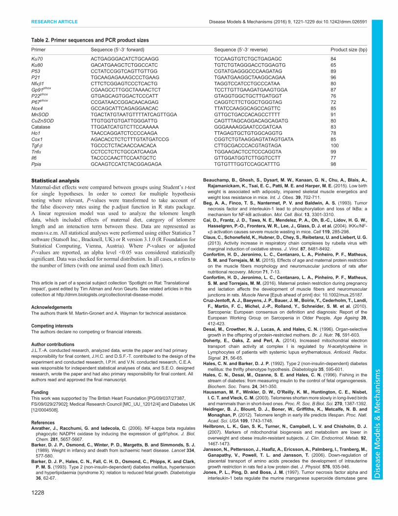
Gene expressionRNA was extracted using an RNeasy Plus mini kit (Qiagen, Manchester,UK) following the manufacturer’s instructions. A DNase digestion step wasperformed in order to ensure no genomic DNA contamination. RNA (1 µg)was used to synthesize cDNA using oligo-dT primers and M-MLV reversetranscriptase (Promega, Southampton, UK). Gene expression wasdetermined using custom-designed primers (Sigma, Poole, UK) andSYBR Green reagents (Applied Biosystems, Warrington, UK). Primersequences are presented in Table 2. Quantification of gene expression wasperformed using a Step One Plus RT-PCR machine (Applied Biosystems,Warrington, UK). Equal efficiency of the reverse transcription of RNA fromall groups was confirmed through quantification of expression of thehousekeeping gene Ppia. Expression of Ppia did not differ between groups(effect of maternal diet P=0.99; control 37±6, recuperated 39±6 averagecopy number). Sample sizes were n=8 per group.
Mitochondrial ETC complex activities and CoQ measurementActivities of complex I (NADH: ubiquinone reductase; EC 1.6.5.3),complex II-III (succinate: cytochrome c reductase; EC 1.3.5.1+EC 1.10.2.2)and complex IV (cytochrome oxidase; EC 1.9.3.1) as well as citrate synthase(CS; EC 1.1.1.27) were assayed as described previously (Tarry-Adkinset al., 2016). Vastus lateralis CoQ9 was quantified by reverse phase high-performance liquid chromatography (HPLC) with UV detection at 275 nmas described previously (Tarry-Adkins et al., 2016).
4-hydroxynonenal (4-HNE) and 3-nitrotyrosine (3-NT) analysisProtein nitrotyrosination was assayed using a 3-Nitrotyrosine ELISA kit(MitoSciences, Cambridge, UK), according to the manufacturer’sinstructions. 4-HNE (a marker of lipid peroxidation) was analyzed usingan OxiSelect HNE Adduct ELISA kit (Cambridge Biosciences), accordingto the manufacturer’s instructions.
Fig. 5. Inflammatory markers. The effect of in utero protein restriction andaccelerated postnatal growth upon (A) protein expression of inflammationmarkers (TNFα, IL1β and IL6; shown as a percentage of the total amounts incontrol rats) and (B) mRNA expression of Tnfα, Il6 and Tgfβ1 in vastus lateralisskeletal muscle of 12-month-old male rats. Results are expressed asmean±s.e.m. ***q<0.001 (control versus recuperated). Statistics werecalculated using Student’s t-test (two-tailed) and are reported after correctionfor multiple hypothesis testing where appropriate. C, control; R, recuperated.n=6 per group for protein expression and n=8 per group for mRNA expression.
1227
RESEARCH ARTICLE Disease Models & Mechanisms (2016) 9, 1221-1229 doi:10.1242/dmm.026591
Disea
seModels&Mechan
isms
Statistical analysisMaternal-diet effects were compared between groups using Student’s t-testfor single hypotheses. In order to correct for multiple hypothesistesting where relevant, P-values were transformed to take account ofthe false discovery rates using the p.adjust function in R stats package.A linear regression model was used to analyze the telomere lengthdata, which included effects of maternal diet, category of telomerelength and an interaction term between these. Data are represented asmean±s.e.m. All statistical analyses were performed using either Statistica 7software (Statsoft Inc., Bracknell, UK) or R version 3.1.0 (R Foundation forStatistical Computing, Vienna, Austria). Where P-values or adjustedP-values are reported, an alpha level <0.05 was considered statisticallysignificant. Data was checked for normal distribution. In all cases, n refers tothe number of litters (with one animal used from each litter).
This article is part of a special subject collection ‘Spotlight on Rat: TranslationalImpact’, guest edited by Tim Aitman and Aron Geurts. See related articles in thiscollection at http://dmm.biologists.org/collection/rat-disease-model.
AcknowledgementsThe authors thank M. Martin-Gronert and A. Wayman for technical assistance.
Competing interestsThe authors declare no competing or financial interests.
Author contributionsJ.L.T.-A. conducted research, analyzed data, wrote the paper and had primaryresponsibility for final content, J.H.C. and D.S.F.-T. contributed to the design of theexperiment and conducted research, I.P.H. and V.N. conducted research, C.E.A.was responsible for independent statistical analyses of data, and S.E.O. designedresearch, wrote the paper and had also primary responsibility for final content. Allauthors read and approved the final manuscript.
FundingThis work was supported by The British Heart Foundation [PG/09/037/27387,FS/09/029/27902]; Medical Research Council [MC_UU_12012/4] and Diabetes UK[12/0004508].
ReferencesAnrather, J., Racchumi, G. and Iadecola, C. (2006). NF-kappa beta regulatesphagocytic NADPH oxidase by inducing the expression of gp91phox. J. Biol.Chem. 281, 5657-5667.
Barker, D. J. P., Osmond, C., Winter, P. D., Margetts, B. and Simmonds, S. J.(1989). Weight in infancy and death from ischaemic heart disease. Lancet 334,577-580.
Barker, D. J. P., Hales, C. N., Fall, C. H. D., Osmond, C., Phipps, K. and Clark,P. M. S. (1993). Type 2 (non-insulin-dependent) diabetes mellitus, hypertensionand hyperlipidaemia (syndrome X): relation to reduced fetal growth. Diabetologia36, 62-67.
Beauchamp, B., Ghosh, S., Dysart, M. W., Kanaan, G. N., Chu, A., Blais, A.,Rajamanickam, K., Tsai, E. C., Patti, M. E. and Harper, M. E. (2015). Low birthweight is associated with adiposity, impaired skeletal muscle energetics andweight loss resistance in mice. Int. J. Obes. 39, 702-711.
Beg, A. A., Finco, T. S., Nantermet, P. V. and Baldwin, A. S. (1993). Tumornecrosis factor and interleukin-1 lead to phosphorylation and loss of IkBa: amechanism for NF-kB activation. Mol. Cell. Biol. 13, 3301-3310.
Cai, D., Frantz, J. D., Tawa, N. E., Mendelez, P. A., Oh, B.-C., Lidov, H. G. W.,Hasselgren, P.-O., Frontera, W. R., Lee, J., Glass, D. J. et al. (2004). IKKκ/NF-κβ activation causes severe muscle wasting in mice. Cell 119, 285-298.
Claus, C., Schonefield, K., Hubner, D., Chey, S., Reibetanz, U. and Liebert, U. G.(2013). Activity increase in respiratory chain complexes by rubella virus withmarginal induction of oxidative stress. J. Virol. 87, 8481-8492.
Confortim, H. D., Jeronimo, L. C., Centanaro, L. A., Pinheiro, P. F., Matheus,S. M. and Torrejais, M. M. (2015). Effects of age and maternal protein restrictionon the muscle fibers morphology and neuromuscular junctions of rats afternutritional recovery. Micron 71, 7-13.
Confortim, H. D., Jeronimo, L. C., Centanaro, L. A., Pinheiro, P. F., Matheus,S. M. and Torrejais, M. M. (2016). Maternal protein restriction during pregnancyand lactation affects the development of muscle fibers and neuromuscularjunctions in rats. Muscle Nerve [Epub ahead of print] doi: 10.1002/mus.25187.
Cruz-Jentoft, A. J., Baeyens, J. P., Bauer, J. M., Boirie, Y., Cederholm, T., Landi,F., Martin, F. C., Michel, J.-P., Rolland, Y., Schneider, S. M. et al. (2010).Sarcopenia: European consensus on definition and diagnosis: Report of theEuropean Working Group on Sarcopenia in Older People. Age Ageing 39,412-423.
Desai, M., Crowther, N. J., Lucas, A. and Hales, C. N. (1996). Organ-selectivegrowth in the offspring of protein-restricted mothers. Br. J. Nutr. 76, 591-603.
Doherty, E., Oaks, Z. and Perl, A. (2014). Increased mitochondrial electrontransport chain activity at complex I is regulated by N-acetylcysteine inLymphocytes of patients with systemic lupus erythematosus. Antioxid. Redox.Signal. 21, 56-65.
Hales, C. N. and Barker, D. J. P. (1992). Type 2 (non-insulin-dependent) diabetesmellitus: the thrifty phenotype hypothesis. Diabetologia 35, 595-601.
Hales, C. N., Desai, M., Ozanne, S. E. and Hales, C. N. (1996). Fishing in thestream of diabetes: from measuring insulin to the control of fetal organogenesis.Biochem. Soc. Trans. 24, 341-350.
Haussman, M. F., Winkler, D. W., O’Reilly, K. M., Huntington, C. E., Nisbet,I. C. T. and Vleck, C. M. (2003). Telomeres shortenmore slowly in long-lived birdsand mammals than in short-lived ones. Proc. R. Soc. B Biol. Sci. 270, 1387-1392.
Heidinger, B. J., Blount, D. J., Boner, W., Griffiths, K., Metcalfe, N. B. andMonaghan, P. (2012). Telomere length in early life predicts lifespan. Proc. Natl.Acad. Sci. USA 109, 1743-1748.
Heilbronn, L. K., Gan, S. K., Turner, N., Campbell, L. V. and Chisholm, D. J.(2007). Markers of mitochondrial biogenesis and metabolism are lower inoverweight and obese insulin-resistant subjects. J. Clin. Endocrinol. Metab. 92,1467-1473.
Jansson, N., Pettersson, J., Haafiz, A., Ericsson, A., Palmberg, I., Tranberg, M.,Ganapathy, V., Powell, T. L. and Jansson, T. (2006). Down-regulation ofplacental transport of amino acids precedes the development of intrauterinegrowth restriction in rats fed a low protein diet. J. Physiol. 576, 935-946.
Jones, P. L., Ping, D. and Boss, J. M. (1997). Tumor necrosis factor alpha andinterleukin-1 beta regulate the murine manganese superoxide dismutase gene
Table 2. Primer sequences and PCR product sizes
Primer Sequence (5′-3′ forward) Sequence (5′-3′ reverse) Product size (bp)
Ku70 ACTGAGGGACATCTGCAAGG TCCAAGTGTCTGCTGAGAGC 84Ku80 GACATGAAGCTCTGGCCATC TGTCTGTAGGGACCTGGAGTG 65P53 CCTATCCGGTCAGTTGTTGG CGTATGAGGGCCCAAGATAG 89P21 TGCAAGAGAAAGCCCTGAAG TGAATGAAGGCTAAGGCAGAA 96Nfκβ1 CTTCTCGGAGTCCCTCACTG TAGGTCCATCCTGCCCATAA 80Gp91phox CGAAGCCTTGGCTAAAACTCT TCCTTGTTGAAGATGAAGTGGA 87P22phox GTGAGCAGTGGACTCCCATT GTAGGTGGCTGCTTGATGGT 76P67phox CCGATAACCGGACAACAGAG CAGGTCTTCTGGCTGGGTAG 72Nox4 GCCAGCATTCAGAGGAACAC TTATCCAAGGCAGCCAGTTC 85MnSOD TGACTATGTAATGTTTTATCAGTTGGA GTTGCTGACCACAGCCTTTT 91CuZnSOD TTGTGGTGTGATTGGGATTG CAGTTTAGCAGGACAGCAGATG 80Catalase TTGGATCATGTCTTCCAAAAA GGGAAAAGGAATCCGATCAA 83Ho1 TAACCAGGATCTCCCCAAGA TTAGAGTGCTGTGGCAGGTG 78Cox1 AGACACCTCTCTTTGTATGATCCG CGGTCTGTAAGGAGTATAGTGATA 85Tgf-β TGCCCTCTACAACCAACACA CTTGCGACCCACGTAGTAGA 100Tnfα CCTCCTCTCTGCCATCAAGA TGGAAGACTCCTCCCAGGTA 99Il6 TACCCCAACTTCCAATGCTC GTTGGATGGTCTTGGTCCTT 77Ppia GCAAGTCCATCTACGGAGAGA TGTGTTTGGTCCAGCATTTG 98
1228
RESEARCH ARTICLE Disease Models & Mechanisms (2016) 9, 1221-1229 doi:10.1242/dmm.026591
Disea
seModels&Mechan
isms

through a complex intronic enhancer involving C/EBP-beta and NF-kappaB. Mol.Cell. Biol. 17, 6970-6981.
Kairisalo, M., Korhonen, L., Blomgren, K. and Lindholm, D. (2007). X-linkedinhibitor of apoptosis protein increases mitochondrial antioxidants through NF-kappaB activation. Biochem. Biophys. Res. Commun. 364, 138-144.
Keller, K. (2013). Strength and muscle mass loss with aging process. Age andstrength loss. Muscles Tendons Ligaments J. 24, 346-350.
Lavrovsky, Y., Schwartzman, M. L., Levere, R. D., Kappas, A. and Abraham,N. G. (1994). Identification of binding sites for transcription factors NFkappa B andAP-2 in the promoter region of the human heme oxygenase 1 gene. Proc. Natl.Acad. Sci. USA 91, 5987-5991.
Mahad, D. J., Ziabreva, I., Campbell, G., Lax, N., White, K., Hanson, P. S.,Lassman, H. and Turnbull, D. M. (2009). Mitochondrial changes within axons inmultiple sclerosis. Brain 132, 1161-1174.
Marzetti, E., Calvani, R., Cesari, M., Buford, T.W., Lorenzi, M., Behnke, B. J. andLeeuwenburgh, C. (2013). Mitochondrial dysfunction and sarcopenia of aging:from signaling pathways to clinical trials. Int. J. Biochem. Cell Biol. 45, 2288-2301.
Morgan, M. J. and Lui, Z.-G. (2011). Crosstalk of reactive oxygen species and NF-κβ signaling. Cell Res. 21, 103-115.
Mortensen, O. H., Olsen, H. L., Frandsen, L., Nielsen, P. E., Nielsen, F. C.,Grunnet, N. andQuistorff, B. (2010). Amaternal low protein diet has pronouncedeffects on mitochondrial gene expression in offspring liver and skeletal muscle;protective effects of taurine. J. Biomed. Sci. 17 Suppl. 1, S38.
Oikawa, S. and Kawanishi, S. (1999). Site-specific DNA damage at GGGsequence by oxidative stress may accelerate telomere shortening. FEBS Lett.453, 365-368.
Pantham, P., Rosario, F. J., Nijland, M., Cheung, A., Nathanielsz, P. W., Powell,T. L., Galan, H. L., Li, C. and Jansson, T. (2015). Reduced placental amino acidtransport in response to maternal nutrient restriction in the baboon.Am. J. Physiol.Regul. Integr. Comp. Physiol. 309, R740-R746.
Patel, H. P., Jameson, K. A., Syddall, H. E., Martin, H. J., Stewart, C. E., Cooper,C. and Sayer, A. A. (2012). Developmental influences, muscle morphology, andsarcopenia in community-dwelling older men. J. Gerontol. A. Biol. Sci. Med. Sci.67A, 82-87.
Poulsen, P., Wojtaszewski, J. F. P., Richter, E. A., Beck-Nielsen, H. and Vaag, A.(2007). Low birth weight and zygosity status is associated with defective muscleglycogen and glycogen synthase regulation in elderly twins. Diabetes 56,2710-2714.
Ray, P. D., Huang, B.-W. and Tsuji, Y. (2012). Reactive oxygen species (ROS)homeostasis and redox regulation in cellular signaling. Cell Signal. 24, 981-990.
Rojo, A. I., Salinas, M., Martin, D., Perona, R. and Cuadrado, A. (2004).Regulation of Cu/Zn-superoxide dismutase expression via the
phosphatidylinositol 3 kinase/Akt pathway and nuclear factor-kappaB.J. Neurosci. 24, 7324-7334.
Sayer, A. A., Stewart, C., Patel, H. and Cooper, C. (2010). The developmentalorigins of sarcopenia: from epidemiological evidence to underlying mechanisms.J. Develop. Orig. Health Dis. 1, 150-157.
Snoeck, A., Remacle, C., Reusens, B. and Hoet, J. J. (1990). Effect of a lowprotein diet during pregnancy on the fetal rat endocrine pancreas. Biol. Neonate57, 107-118.
Tarry-Adkins, J. L. and Ozanne, S. E. (2014). The impact of early nutrition on theageing trajectory. Proc. Nutr. Soc. 73, 289-301.
Tarry-Adkins, J. L., Martin-Gronert, M. S., Fernandez-Twinn, D. S., Hargreaves,I., Alfaradhi, M. Z., Land, J. M., Aiken, C. E. and Ozanne, S. E. (2012). Poormaternal nutrition followed by accelerated postnatal growth leads to alterations inDNA damage and repair, oxidative and nitrosative stress, and oxidative defensecapacity in rat heart. FASEB J. 27, 379-390.
Tarry-Adkins, J. L., Fernandez-Twinn, D. S., Hargreaves, I. P., Neergheen, V.,Aiken, C. E., Martin-Gronert, M. S., McConnell, J. M. andOzanne, S. E. (2016).Coenzyme Q10 prevents hepatic fibrosis, inflammation, and oxidative stress in amale rat model of poor maternal nutrition and accelerated postnatal growth.Am. J. Clin. Nutr. 103, 579-588.
Valko, M., Leibfritz, D., Moncol, J., Cronin, M. T. D., Mazur, M. and Telser, J.(2007). Free radicals and antioxidants in normal physiological functions andhuman disease. Int. J. Biochem. Cell Biol. 39, 44-84.
van Deutekom, A. W., Chinapaw, M. J. M., Vrijkotte, T. G. M. and Gemke, R. J. B.J. (2015). The association of birth weight and infant growth with physical fitness at8-9 years of age- the ABCD study. Int. J. Obes. 39, 593-600.
Wang, C.-H., Wu, S.-B., Wu, Y.-T. andWei, Y.-H. (2013). Oxidative stress responseelicited by mitochondrial dysfunction: implication in the pathophysiology of aging.Exp. Biol. Med. 238, 450-460.
Xu, P., Huecksteadt, T. P. and Hoidal, J. R. (1996). Molecular cloning andcharacterization of the human xanthine dehydrogenase gene (XDH). Genomics34, 173-180.
Zambrano, E., Iban ez, C., Martınez-Samayoa, P. M., Lomas-Soria, C., Durand-Carbajal, M. and Rodrıguez-Gonzalez, G. L. (2016). Maternal obesity: lifelongmetabolic outcomes for offspring from poor developmental trajectories during theperinatal period. Arch. Med. Res. 47, 1-12.
Zhu, M.-J., Ford, S. P., Nathanielsz, P. W. and Du, M. (2004). Effect of maternalnutrient restriction in sheep on the development of fetal skeletal muscle. Biol.Reprod. 71, 1968-1973.
Zhu, M. J., Ford, S. P., Means, W. J., Hess, B. W., Nathanielsz, P. W. and Du, M.(2006). Maternal nutrient restriction affects properties of skeletal muscle inoffspring. J. Physiol. 575, 241-250.
1229
RESEARCH ARTICLE Disease Models & Mechanisms (2016) 9, 1221-1229 doi:10.1242/dmm.026591
Disea
seModels&Mechan
isms