polyoxygenated 24,28-epoxyergosterols inhibiting the proliferation of glioma cells from sea anemone...
TRANSCRIPT

Steroids 88 (2014) 19–25
Contents lists available at ScienceDirect
Steroids
journal homepage: www.elsevier .com/locate /s teroids
Polyoxygenated 24,28-epoxyergosterols inhibiting the proliferationof glioma cells from sea anemone Anthopleura midori
http://dx.doi.org/10.1016/j.steroids.2014.06.0130039-128X/� 2014 Elsevier Inc. All rights reserved.
⇑ Corresponding authors. Tel.: +86 571 88206577; fax: +86 571 88208891.E-mail addresses: [email protected] (X.-Y. Lian), [email protected] (Z. Zhang).
Siran Yu a, Xuewei Ye a, Lu Chen a, Xiao-Yuan Lian b,⇑, Zhizhen Zhang a,⇑a Ocean College, Zhejiang University, Hangzhou 310058, Chinab College of Pharmaceutical Sciences, Zhejiang University, Hangzhou 310058, China
a r t i c l e i n f o a b s t r a c t
Article history:Received 4 March 2014Received in revised form 11 June 2014Accepted 14 June 2014Available online 26 June 2014
Keywords:Anthopleura midoriSea anemoneEpoxyergosterolsGlioma cellsProliferation inhibition
Eleven sterols (1–11) and one carotenoid (12) were isolated and identified from sea anemone Anthopleuramidori. Compounds 1–6 are rare polyoxygenated ergosterols with a 24,28-epoxy moiety. The structures ofthese epoxyergosterols were determined by NMR and HRESIMS analyses as well as their chemical-phys-ical properties. Epoxyergosterols 1 and 2 were found to be new natural products and 3–6 are new com-pounds. Bioactive assay showed that compounds 1, 2, 3, 5, 7, 8, 11, and 12 inhibited the proliferation ofrat glioma C6 and human glioma U251 cells with IC50 in a range of 2.41–80.45 lM. Further investigationsuggested that 1 and 3 induced apoptosis in glioma cells and 1 blocked U251 cells at the G0/G1 phase.
� 2014 Elsevier Inc. All rights reserved.
1. Introduction
Gliomas are the most common and malignant brain tumors.Despite recent advances in standard therapy including surgicalresection followed by radiation and chemotherapy, the prognosisfor patients with malignant glioma remains unsatisfactory [1].Chemotherapy has played an important role as an adjuvant intreating gliomas. However, the efficacy of current drugs is limiteddue to serious side effect, poor drug delivery, and chemo-resistance[1]. There is therefore a need to develop novel anti-glioma agentsfor treating malignant gliomas. Natural products are highly signif-icant sources of novel drug leads [2]. During the course of ourongoing project for the discovery of new anti-tumor agents fromnatural sources [3–5], we investigated the bioactive constituentsagainst glioma cells from sea anemone Anthopleura midori Uchidaet Muramatsu.
The sea anemones of A. midori are mainly distributed in thecoastal areas of Pacific Ocean, Japan, and China, and usually adhereto water-beat rocks in the intertidal and subtidal zones. The entirebody of this species has been traditionally used for the treatmentof hemorrhoid and body rash by external application in China[6]. It is well known that the genus Anthopleura is a rich sourceof peptide toxins acting on ion channels [7] and also containedceramides [8], sesquiterpenes [9,10], nucleosides [11,12], sterols
[12], fatty acids [13], and guanidines [14]. The extracts and purecompounds from Anthopleura species have showed antitumor,neurotoxic, cardiotonic, and hemolytic activities [9–11]. In thisstudy, eleven sterols including six rare polyoxygenated 24,28-epoxyergosterols (1–6) and one carotenoid were isolated and iden-tified from sea anemone of A. midori. We report herein the isolationand structure elucidation of these rare epoxyergosterols and theproperties of isolated compounds against rat glioma C6 and humanglioma U251 cells.
2. Experimental
2.1. General experimental procedures
Melting points were measured with a WRX-4 microscope appa-ratus and are uncorrected. Optical rotations were measured on aJASCO DIP-370 digital polarimeter. CD spectra were recorded ona JASCO J 715 spectropolarimeter. IR spectrum was carried outon an AVATAR 370 FT-IR spectrometer (Thermo Nicolet). NMRspectra were recorded on a Bruker AV III-500 instrument at500 MHz for 1H and 125 MHz for 13C using standard pulse pro-grams and acquisition parameters. Chemical shifts were reportedin d (ppm) referencing to the NMR solvent used. HRESIMS datawere acquired on an AB Sciex Triple TOF 5600 spectrometer. Silicagel (200–300 mesh, Qingdao Oceanic Chemical Plant Branch) andoctadecyl-functionalized silica gel (ODS, Cosmosil 75C18-Prep)were used for column chromatography. HPLC separation was

20 S. Yu et al. / Steroids 88 (2014) 19–25
performed on an Agilent 1260 HPLC system with an Agilent 1260diode array detector using column A (Cosmosil 5C18-MS-II,250 � 10 mm, 5.0 lm, Nacalai Tesque), column B (ODS-2 Hypersil,250 � 4.6 mm, 5 lm, Thermo Scientific), or column C (Poroshell120 EC-C18, 150 � 4.6 mm, 2.7 lm, Agilent). Fluorescence micro-scope (Nikon SMZ1000) was used to detect apoptosis and necrosisof tumor cells stained by DAPI and PI double staining. Flow cytom-etry (Beckman Coulter, FC500MCL) was used for cell cycle assayand quantitative analysis of cell apoptosis. Rat glioma C6 andhuman glioma U251 cells were purchased from the Cell Bank ofthe Chinese Academy of Sciences. Annexin V apoptosis detectionkit was obtained from Invitrogen.
2.2. Sea anemones material
Fresh sea anemones of A. midori were collected from Turtle Isletin the South China Sea close to Shanwei City, Guangdong Province,China in October 2012 and frozen for further use. A voucher sample(SW-HK-A16) was authenticated by one of the authors (Z. Zhang)and deposited in the Laboratory of Institute of Marine Biology,Ocean College, Zhejiang University, China.
2.3. Extraction and isolation
Ten frozen whole bodies of A. midori were cut into small piecesand then percolated with MeOH for three times (first in 2000 mLfor 12 h, then each 1000 mL for 4 h). The combined MeOH solutionwas concentrated under reduced pressure to 300 mL and then par-titioned with cyclohexane to afford cyclohexane part (15.6 g). Theremaining part was evaporated to remove the organic solvents andpartitioned with EtOAc to furnish EtOAc part (1.5 g). The cyclohex-ane partition was applied to a column of silica gel (300 g) eluting inturn with cyclohexane/EtOAc (4:1, 2:1, 1:1, 1:2), EtOAc, and MeOHto give six fractions of FH-1, FH-2, FH-3, FH-4, FE-1, and FM-1. FH-2(600 mg) was further separated by a ODS (75 g) column elutingwith 100% MeOH to give 30 fractions (each 75 mL). Compounds 8(2.5 mg, Rf: 0.53, cyclohexane/EtOAc 1:2), 7 (3.8 mg, Rf: 0.51, cyclo-hexane/EtOAc 3:2), 11 (1.5 mg Rf: 0.51, cyclohexane/EtOAc 1:1), 10(200 mg, Rf: 0.45, cyclohexane/EtOAc 9:1), and 9 (2.0 mg, Rf: 0.40,cyclohexane/EtOAc 9:1) were obtained from fraction 6, fractions9–10, fraction 15, fractions 20–24, and fraction 28, respectively.Part of FH-3 (98 mg) was subjected to an ODS (75 g) column elut-ing with 100% MeOH to give 12 fractions of FH-3A1 to FH-3A12
(each 50 mL). Compound 12 (10.1 mg, tR 10.35 min) was obtainedby HPLC separation using column B (mobile phase CH3CN/H2O,75:25; flow rate 1.0 mL/min; detection 456 nm) from FH-3A3. Partof FH-4 (100 mg) was separated by HPLC using column A (mobilephase CH3CN/H2O, 90:10; flow rate 2.5 mL/min; detection203 nm) to afford a mixture (60 mg, tR 15.8 min) of 1 and 2. Partof this mixture was further separated by HPLC using column C withCH3CN/H2O (60:40, 0.7 mL/min) as mobile phase to give 1 (3.8 mg,tR 17.8 min) and 2 (3.0 mg, tR 18.6 min). FE-1 was applied to anODS (75 g) column eluting with 90% MeOH to give 16 fractions ofFE-1A1 to FE-1A16 (each 50 mL). FE-1A8 was further separated byHPLC using column C (CH3CN/H2O, 45:55, 0.6 mL/min) to give 3(2.2 mg, tR 17.8 min) and a mixture of 3 and 4 (tR 18.3 min). Furtherseparation of FE-1A11 by HPLC column C (CH3CN/H2O, 55:45,0.5 mL/min) gave 5 (2.5 mg, tR 26.3 min) and a mixture of 5 and6 (tR 27.0 min). Compounds 4 and 6 were not isolated in a pureform, and hence it would be absolutely difficult to evaluation theirchemical assignments, physical properties as well as their biologi-cal activities.
2.3.1. 24(R),28-Epoxyergost-5-en-3b-ol (1)White amorphous solid; molecular formula C28H46O2; [a]22
D
�10.3� (c 0.10, dioxane); CD (c 0.15 � 10�3 M, dioxane) [h]268
+34.88 (max); IR(KBr) kmax 3383, 2933, 1466, 1379, 1052 cm�1;13C NMR spectroscopic data (125 MHz, in pyridine-d5, see Table 1);1H NMR spectroscopic data (500 MHz, in pyridine-d5, see Table 2);HRESIMS m/z 415.3563 [M+H]+ (calcd C28H47O2 415.3576), m/z397.3457 [M-OH]+ (calcd C28H45O 397.3470), and m/z 379.3353[M-(OH-H2O)]+ (calcd C28H43 379.3365).
2.3.2. 24(S),28-Epoxyergost-5-en-3b-ol (2)White amorphous solid; molecular formula C28H46O2; [a]22
D
�8.7� (c 0.10, dioxane); CD (c 0.15 � 10�3 M, dioxane) [h]271
�55.99 (max); IR(KBr) kmax 3387, 2934, 1466, 1380, 1053 cm�1;13C NMR spectroscopic data (125 MHz, in pyridine-d5, see Table 1);1H NMR spectroscopic data (500 MHz, in pyridine-d5, see Table 2);HRESIMS m/z 415.3568 [M+H]+ (calcd C28H47O2 415.3576), m/z397.3453 [M�OH]+ (calcd C28H45O 397.3470), and m/z 379.3349[M-(OH–H2O)]+ (calcd C28H43 379.3365).
2.3.3. 24(R),28-Epoxyergost-3-one-5a,6a-diol (3)White amorphous solid; molecular formula C28H46O4; [a]22
D
�12.8� (c 0.10, dioxane); CD (c 0.15 � 10�3 M, dioxane) [h]269
+34.40 (max); IR(KBr) kmax 3431, 2939, 1710, 1467, 1383,1053 cm�1; 13C NMR spectroscopic data (125 MHz, in pyridine-d5,see Table 1); 1H NMR spectroscopic data (500 MHz, in pyridine-d5),see Table 2); HRESIMS m/z 447.3458 [M+H]+ (calcd C28H47O4
447.3474), m/z 429.3346 [M-OH]+ (calcd C28H45O3 429.3369), andm/z 411.3252 [M-(OH-H2O)]+ (calcd C28H43O2 411.3263).
2.3.4. 24(R),28-Epoxyergost-3-acetyl-3b,5a,6a-triol (5)White amorphous solid; molecular formula C30H50O5; [a]22
D
�9.5� (c 0.10, dioxane); CD (c 0.15 � 10�3 M, dioxane) [h]268
+32.98 (max); IR(KBr) kmax 3419, 2938, 1717, 1445, 1382, 1265,1029 cm�1; 13C NMR spectroscopic data (125 MHz, in pyridine-d5,see Table 1); 1H NMR spectroscopic data (500 MHz, in pyridine-d5,see Table 2); HRESIMS: m/z 507.3709 [M+H2O-H]� (calcd C30H51O6
507.3686).
2.3.5. 5a,8a-Epidioxyergosta-6,24(28)-dien-3b-ol (7)White amorphous solid; molecular formula C28H44O3; 13C NMR
spectroscopic data (125 MHz, in pyridine-d5, see Table S3); 1HNMR spectroscopic data (500 MHz, in pyridine-d5, see Table S4).
2.3.6. Cholestane-3b,5a,6a-triol (8)White amorphous solid; molecular formula C27H48O3; mp 233–
234 �C (EtOAc) (lit. 233–235 �C [15]); 13C NMR spectroscopic data(125 MHz, in pyridine-d5, see Table S3); 1H NMR spectroscopicdata (500 MHz, in pyridine-d5, see Table S4).
2.3.7. Cholest-5-en-3b-ol (9)White amorphous solid; molecular formula C27H46O; mp 150–
151 �C (EtOAc) (lit. 148–150 �C [16]); 13C NMR spectroscopic data(125 MHz, in pyridine-d5, see Table S3); 1H NMR spectroscopicdata (500 MHz, in pyridine-d5, see Table S4).
2.3.8. Ergosta-5,24(28)-dien-3b-ol (10)White amorphous solid; molecular formula C28H46O; mp 142–
143 �C (EtOAc) (lit. 140–142 �C [17]); 13C NMR spectroscopic data(125 MHz, in pyridine-d5, see Table S3); 1H NMR spectroscopicdata (500 MHz, in pyridine-d5, see Table S4).
2.3.9. Ergosta-5,22,24(28)-trien-3b-ol (11)White amorphous solid; molecular formula C28H44O; 13C NMR
spectroscopic data (125 MHz, in pyridine-d5, see Table S3); 1HNMR spectroscopic data (500 MHz, in pyridine-d5, see Table S4).
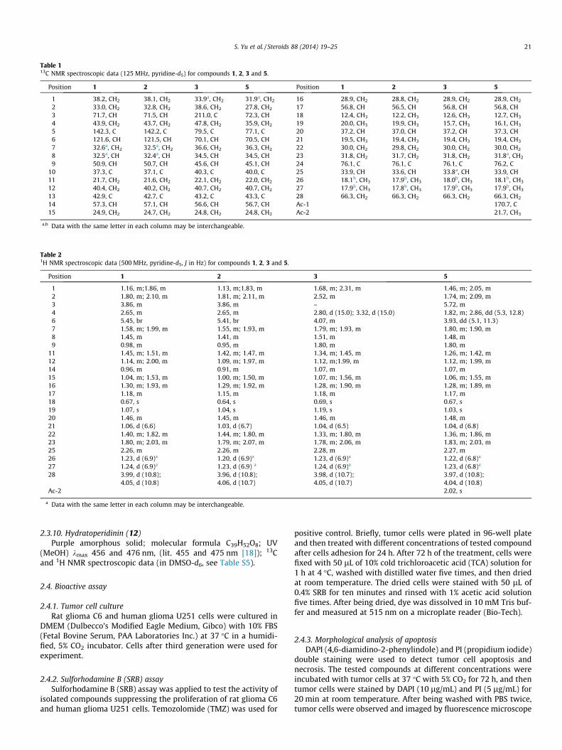
Table 113C NMR spectroscopic data (125 MHz, pyridine-d5) for compounds 1, 2, 3 and 5.
Position 1 2 3 5 Position 1 2 3 5
1 38.2, CH2 38.1, CH2 33.9a, CH2 31.9a, CH2 16 28.9, CH2 28.8, CH2 28.9, CH2 28.9, CH2
2 33.0, CH2 32.8, CH2 38.6, CH2 27.8, CH2 17 56.8, CH 56.5, CH 56.8, CH 56.8, CH3 71.7, CH 71.5, CH 211.0, C 72.3, CH 18 12.4, CH3 12.2, CH3 12.6, CH3 12.7, CH3
4 43.9, CH2 43.7, CH2 47.8, CH2 35.9, CH2 19 20.0, CH3 19.9, CH3 15.7, CH3 16.1, CH3
5 142.3, C 142.2, C 79.5, C 77.1, C 20 37.2, CH 37.0, CH 37.2, CH 37.3, CH6 121.6, CH 121.5, CH 70.1, CH 70.5, CH 21 19.5, CH3 19.4, CH3 19.4, CH3 19.4, CH3
7 32.6a, CH2 32.5a, CH2 36.6, CH2 36.3, CH2 22 30.0, CH2 29.8, CH2 30.0, CH2 30.0, CH2
8 32.5a, CH 32.4a, CH 34.5, CH 34.5, CH 23 31.8, CH2 31.7, CH2 31.8, CH2 31.8a, CH2
9 50.9, CH 50.7, CH 45.6, CH 45.1, CH 24 76.1, C 76.1, C 76.1, C 76.2, C10 37.3, C 37.1, C 40.3, C 40.0, C 25 33.9, CH 33.6, CH 33.8a, CH 33.9, CH11 21.7, CH2 21.6, CH2 22.1, CH2 22.0, CH2 26 18.1b, CH3 17.9b, CH3 18.0b, CH3 18.1b, CH3
12 40.4, CH2 40.2, CH2 40.7, CH2 40.7, CH2 27 17.9b, CH3 17.8b, CH3 17.9b, CH3 17.9b, CH3
13 42.9, C 42.7, C 43.2, C 43.3, C 28 66.3, CH2 66.3, CH2 66.3, CH2 66.3, CH2
14 57.3, CH 57.1, CH 56.6, CH 56.7, CH Ac-1 170.7, C15 24.9, CH2 24.7, CH2 24.8, CH2 24.8, CH2 Ac-2 21.7, CH3
a,b Data with the same letter in each column may be interchangeable.
Table 21H NMR spectroscopic data (500 MHz, pyridine-d5, J in Hz) for compounds 1, 2, 3 and 5.
Position 1 2 3 5
1 1.16, m;1.86, m 1.13, m;1.83, m 1.68, m; 2.31, m 1.46, m; 2.05, m2 1.80, m; 2.10, m 1.81, m; 2.11, m 2.52, m 1.74, m; 2.09, m3 3.86, m 3.86, m – 5.72, m4 2.65, m 2.65, m 2.80, d (15.0); 3.32, d (15.0) 1.82, m; 2.86, dd (5.3, 12.8)6 5.45, br 5.41, br 4.07, m 3.93, dd (5.1, 11.3)7 1.58, m; 1.99, m 1.55, m; 1.93, m 1.79, m; 1.93, m 1.80, m; 1.90, m8 1.45, m 1.41, m 1.51, m 1.48, m9 0.98, m 0.95, m 1.80, m 1.80, m
11 1.45, m; 1.51, m 1.42, m; 1.47, m 1.34, m; 1.45, m 1.26, m; 1.42, m12 1.14, m; 2.00, m 1.09, m; 1.97, m 1.12, m;1.99, m 1.12, m; 1.99, m14 0.96, m 0.91, m 1.07, m 1.07, m15 1.04, m; 1.53, m 1.00, m; 1.50, m 1.07, m; 1.56, m 1.06, m; 1.55, m16 1.30, m; 1.93, m 1.29, m; 1.92, m 1.28, m; 1.90, m 1.28, m; 1.89, m17 1.18, m 1.15, m 1.18, m 1.17, m18 0.67, s 0.64, s 0.69, s 0.67, s19 1.07, s 1.04, s 1.19, s 1.03, s20 1.46, m 1.45, m 1.46, m 1.48, m21 1.06, d (6.6) 1.03, d (6.7) 1.04, d (6.5) 1.04, d (6.8)22 1.40, m; 1.82, m 1.44, m; 1.80, m 1.33, m; 1.80, m 1.36, m; 1.86, m23 1.80, m; 2.03, m 1.79, m; 2.07, m 1.78, m; 2.06, m 1.83, m; 2.03, m25 2.26, m 2.26, m 2.28, m 2.27, m26 1.23, d (6.9)a 1.20, d (6.9)a 1.23, d (6.9)a 1.22, d (6.8)a
27 1.24, d (6.9)a 1.23, d (6.9) a 1.24, d (6.9)a 1.23, d (6.8)a
28 3.99, d (10.8);4.05, d (10.8)
3.96, d (10.8);4.06, d (10.7)
3.98, d (10.7);4.05, d (10.7)
3.97, d (10.8);4.04, d (10.8)
Ac-2 2.02, s
a Data with the same letter in each column may be interchangeable.
S. Yu et al. / Steroids 88 (2014) 19–25 21
2.3.10. Hydratoperidinin (12)Purple amorphous solid; molecular formula C39H52O8; UV
(MeOH) kmax 456 and 476 nm, (lit. 455 and 475 nm [18]); 13Cand 1H NMR spectroscopic data (in DMSO-d6, see Table S5).
2.4. Bioactive assay
2.4.1. Tumor cell cultureRat glioma C6 and human glioma U251 cells were cultured in
DMEM (Dulbecco’s Modified Eagle Medium, Gibco) with 10% FBS(Fetal Bovine Serum, PAA Laboratories Inc.) at 37 �C in a humidi-fied, 5% CO2 incubator. Cells after third generation were used forexperiment.
2.4.2. Sulforhodamine B (SRB) assaySulforhodamine B (SRB) assay was applied to test the activity of
isolated compounds suppressing the proliferation of rat glioma C6and human glioma U251 cells. Temozolomide (TMZ) was used for
positive control. Briefly, tumor cells were plated in 96-well plateand then treated with different concentrations of tested compoundafter cells adhesion for 24 h. After 72 h of the treatment, cells werefixed with 50 lL of 10% cold trichloroacetic acid (TCA) solution for1 h at 4 �C, washed with distilled water five times, and then driedat room temperature. The dried cells were stained with 50 lL of0.4% SRB for ten minutes and rinsed with 1% acetic acid solutionfive times. After being dried, dye was dissolved in 10 mM Tris buf-fer and measured at 515 nm on a microplate reader (Bio-Tech).
2.4.3. Morphological analysis of apoptosisDAPI (4,6-diamidino-2-phenylindole) and PI (propidium iodide)
double staining were used to detect tumor cell apoptosis andnecrosis. The tested compounds at different concentrations wereincubated with tumor cells at 37 �C with 5% CO2 for 72 h, and thentumor cells were stained by DAPI (10 lg/mL) and PI (5 lg/mL) for20 min at room temperature. After being washed with PBS twice,tumor cells were observed and imaged by fluorescence microscope

22 S. Yu et al. / Steroids 88 (2014) 19–25
(40� magnification). Apoptotic cells stained by DAPI displayedbright blue nuclear condensation and fragmentation, and necroticcells showed red fluorescence as stained by PI.
2.4.4. Annexin V-FITC/PI double staining assayAn Annexin V apoptosis detection kit was used for the qualifica-
tion of apoptosis and necrosis. Cells were treated with the testedcompound 72 h, and then 1 � 106 cells were harvested. The cellswere washed in cold PBS buffer and further resuspended in100 lL 1� binding buffer mixed with 5 lL Annexin V-FITC and1 lL 100 lg/mL PI working solution. Cells were incubated at roomtemperature for 15 min and then 400 lL 1� binding buffer wasadded. Flow cytometry was used to determine fluorescence usingemission wavelength at 530 nm and 575 nm and excitation wave-length at 488 nm.
2.4.5. Cell cycle assayCell cycle perturbations arrested by tested compounds were
analyzed by PI DNA staining using flow cytometry. Tumor cellswere treated with tested compound for 6 and 12 h. Detailed proto-col was referred to previous report [3].
3. Results and discussion
A cyclohexane partition from the MeOH extract of whole bodiesof A. midori was separated by silica gel and ODS chromatography,following by HPLC purification to afford twelve compounds. Onthe basis of the NMR analysis and the comparison of reported data,six known compounds were identified as 5a,8a-epidioxyergosta-6,24(28)-dien-3b-ol (7) [19], cholestane-3b,5a,6a-triol (8) [20],cholest-5-en-3b-ol (9) [21], ergosta-5,24(28)-dien-3b-ol (10) [22],ergosta-5,22,24(28)-trien-3b-ol (11) [23], and hydratoperidinin(12) [18].
Compounds 1 and 2 were initially obtained as a mixture from asignal peak of HPLC using a Cosmosil 5C18-MS-II column. Interpre-tation of extensive NMR spectroscopic data (Tables S1, S2, and Fig-ures S20–S37) of this mixture concluded that 1 and 2 were C-24isomers of ergosterols with an epoxy moiety at C-24 and C-28. Thisisomer mixture was further separated by HPLC on a column ofPoroshell 120 EC-C18 to give 1 (tR 17.8 min) and 2 (tR 18.6 min)(Figure S86). Both 1 and 2 had the same molecular formulaC28H46O2 deduced from their positive HRESIMS and 13C NMR spec-troscopic data. The structures of 1 and 2 were assigned as 24(R),28-epoxyergost-5-en-3b-ol (1) and 24(S),28-epoxyergost-5-en-3b-ol(2) by NMR spectroscopic and HRESIMS data. The 24R and 24S iso-mers could be distinguished by HPLC analysis. By use of a normalphase silica gel HPLC column, a higher tR value in the HPLC chro-matogram was corresponded to the 24R-isomer, and the lower tR
value to the 24S-isomer [24,25]. While on the reverse phase Poro-shell 120 EC-C18 HPLC column used in this study, the behaviors of24R and 24S-isomers were opposite, that is the lower tR value(17.8 min) was associated with the 24R-isomer, and the higher tR
value (18.6 min) with the 24S-isomer (Figure S86). The configura-tion at C-24 could also be assigned by CD spectra, whereby a posi-tive Cotton effect in the region 250–300 nm indicated a 24(R)-configuration, while a negative Cotton effect was suggestive of a24(S)-configuration. Epoxyergosterols 1 and 2 were previouslyobtained by synthesis [24,25] and were found to be new naturalproducts.
Compound 3 was obtained in pure form and 4 was isolated as amixture of 3 and 4, which were also C-24 isomers of 24,28-epox-yergosterols. In the 1H and 13C NMR spectra, the signals for oxyme-thine at C-3 and double bond at C-5 and C-6 in 1 and 2 weremissing in 3 and 4. Instead, the 13C NMR spectra of 3 and 4 dis-played additional signals for one ketone and two oxygenated car-
bons (Tables 1 and 2, S1, and S2). HMBC correlations as shown inthe Table S2 demonstrated the presence of the ketone group atC-3 and the two oxygenated carbons at C-5 and C-6. The relativeconfiguration for OH-5 and OH-6 of 3 and 4 was assigned as a-ori-entation because the chemical shifts for C-5 (d 79.5) and C-6 (d70.1) were very close to those for C-5 (d 77.3) and C-6 (d 70.8) ofcholestane-3b,5a,6a-triol [20], but different from those for C-5 (d76.0) and C-6 (d 76.3) of cholestane-3b,5a,6b-triol [20] and thosefor C-5 (d 76.9) and C-6 (d 76.6) of cholest-24-en-3b,5a,6b-triol[26]. The 24R and 24S of 3 and 4 were also assigned by their tR val-ues on HPLC chromatogram (Figure S87), where the lower tR value(17.8 min) was related to 24R of 3, and the higher tR value(18.3 min) to 24S of 4. The 24R of 3 was further confirmed by itspositive Cotton effect at 250–300 nm. The full assignment of 13Cand 1H NMR data (Tables 1 and 2, S1, and S2) of 3 and 4 was madeby NMR spectroscopic analysis. The structures of ergosterols 3 and4 were thus elucidated as 24(R),28-epoxyergost-3-one-5a,6a-diol(3) and 24(S),28- epoxyergost-3-one- 5a,6a-diol (4), two novelcompounds.
Compounds 5 and 6 were also a pair of C-24 isomers. Com-pound 5 was isolated in pure form, while 6 was obtained as a mix-ture of 5 and 6. The molecular formula of C30H50O5 for 5 wasdeduced from its negative HRESIMS and 13C NMR data. A detailedNMR analysis indicated that 5 and 6 were very close to 3 and 4,only different at the substitute of C-3. The ketone carbon at C-3(d 211.0) in 3 and 4 was replaced by an oxymethine carbon ataround d 72.0 in 5 and 6. In addition, signals for an acetyl moietywere observed in the NMR spectra of 5 and 6. HMBC correlationsas shown in Table S2 established this acetyl at C-3 position. Thedownfield shift at around d 5.70 (H-3) also supported the acylatedposition at C-3. The chemical shits of d 77.1 for C-5 and d 70.5 for C-6 of 5 and 6 indicated an a-orientation for both OH at C-5 and C-6[20,26]. The orientation of OAc-3 of sterols could be assigned basedon different 13C chemical shifts (d 3.5 ppm) between OAc-3b andOAc-3a [27]. The 13C NMR data for C-3, C-5 and C-6 of compounds5 and 6 are very close to those of compound 5a [28], suggesting ab-configuration for OAc-3 of 5 and 6. The 24R for 5 was assigned bythe lower tR value (26.3 min) and the 24S for 6 by the higher tR
value (27.0 min) (Figure S88). A positive Cotton effect at 250–300 nm further supported the 24R of 5. The structures of 5 and 6were assigned as 24(R),28-epoxyergost-3-acetyl-3b,5a,6a-triol(5) and 24(S),28- epoxyergost-3-acetyl-3b,5a,6a-triol (6), whichare both novel compounds (Fig. 1).
The isolated compounds were evaluated for their activity inhib-iting the proliferation of rat glioma C6 and human glioma U251cells by sulforhodamine B (SRB) assay. Temozolomide (TMZ), themost popular drug for the treatment of glioma [1], was used forpositive control. The results (Table 3 and Fig. 2) showed that com-pounds 1, 2, 5, 7, 11 and 12 showed dose-dependence activityagainst both C6 and U251 cells. Compounds 3 and 8 only inhibitedthe proliferation of C6 cells, while 9 and 10 were inactive for bothC6 and U251. Epoxyergosterols 1 and 5 were the most active com-pounds against C6 cells with an IC50 value of 2.41 lM for 1 and10.58 lM for 5, while others showed moderate or weak activitywith IC50 values in a range of 18.59–72.14 lM for C6 cells and40.94–80.45 lM for U251 cells. It seemed that most of the activecompounds were more sensitive to C6 cells than to U251 cells.The positive control TMZ showed activity against C6 cells withan IC50 value of 69.58 lM, but was inactive for U251 cells. This isprobably because U251 cells were resistant to TMZ [29].
The apoptosis and necrosis induced by 24(R),28-epoxyergost-5-en-3b-ol (1) in U251 cells were further investigated by DAPI and PIdouble staining. After 72 h of the treatment, epoxyergosterol 1(40 lM and 80 lM) induced apoptosis and necrosis in U251 cells(Fig. 3). The qualification analysis using Annexin V-FITC/PI doublestaining indicated that 1 (80 lM) caused a 21.83% increase of early

O
OHO
OH
HOO
OHO
O
O
HO
HO
O
HO
O
O
OH
HO OHOH
HO HO HO
7
12
17
20 25
18
2621
19
10
2711
22
1
1
28
2
4
8
9 10 11
OHO
O
OH5
OHO
O
O
3O
O
OH6
OHO
O
OHOH
OH5a
OHO
O
Fig. 1. Structures of compounds 1–12.
Table 3Inhibitory effects of isolated compounds on the proliferation of glioma cells (IC50: lM).
Compounds C6 U251 Compounds C6 U251
1 02.41 ± 0.52 40.94 ± 1.06 8 72.14 ± 2.70 NA2 18.59 ± 0.32 64.26 ± 3.75 9 NA NA3 43.60 ± 0.93 NA 10 NA NA5 10.58 ± 0.57 41.91 ± 2.99 11 38.38 ± 3.25 46.08 ± 1.657 38.38 ± 3.25 80.45 ± 4.07 12 18.70 ± 0.81 43.72 ± 1.73TMZ 69.58 ± 6.10 NA
NA: No inhibitory activity at concentration of 100 M.
S. Yu et al. / Steroids 88 (2014) 19–25 23
apoptotic cells from 0.56% (control, CON) to 22.39% (Fig. 4). Effectof 1 on U251 cell cycle was also evaluated by flow cytometric anal-ysis. The DNA content percentages of each phase in the cell cycleare shown in Fig. 5. The proportion of cells in the G0/G1 phase ofthe cell cycle increased 11.59% and 9.94% after 6 h and 12 h ofthe treatment of 1 (80 lM). The alternation occurring in the cellcycle suggested that 1 might arrest U251 cells at the G0/G1 phase.Epoxyergosterol 3 (40 lM) also significantly induced apoptosis in
C6 cells by 44.66% increase of early apoptotic cells and 18.41% oflate apoptotic cells (Fig. 4).
Polyoxygenated sterols are found in many marine animals suchas starfish Acodontaster conspicuus [30], sponge Luffariella c.f. vari-abilis [31], and gorgonian Menella kanisa [32]. Some polyhydroxys-terols had been showed to have anti-bacterial [30], antiviral [31],cytotoxicity [32], and anti-asthma [33] properties as well as rever-sal effect on cancer multidrug resistance [34]. Although some mar-
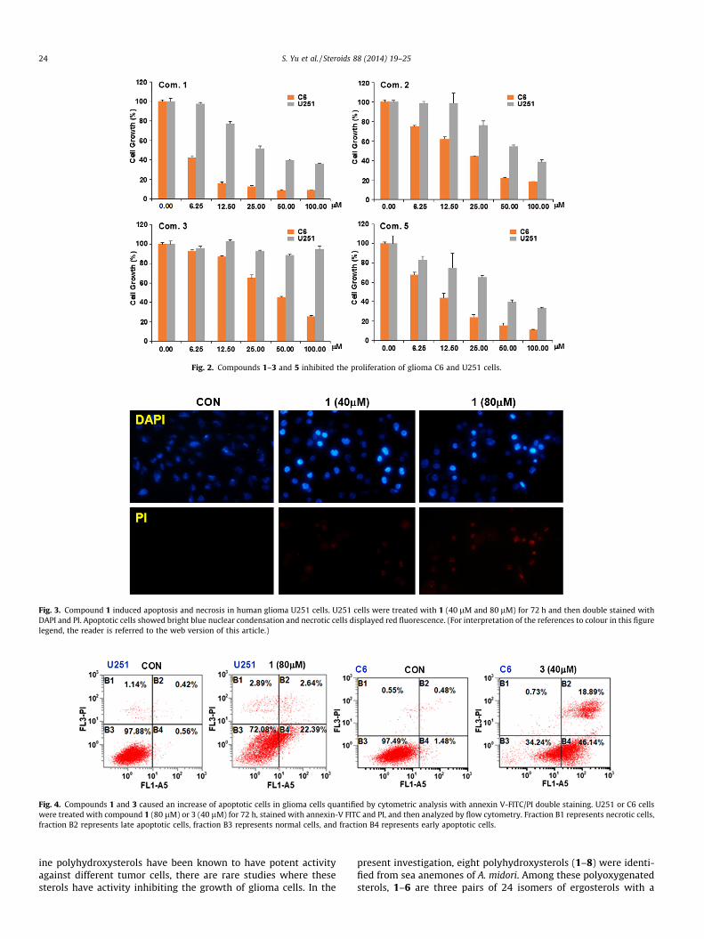
Fig. 2. Compounds 1–3 and 5 inhibited the proliferation of glioma C6 and U251 cells.
Fig. 3. Compound 1 induced apoptosis and necrosis in human glioma U251 cells. U251 cells were treated with 1 (40 lM and 80 lM) for 72 h and then double stained withDAPI and PI. Apoptotic cells showed bright blue nuclear condensation and necrotic cells displayed red fluorescence. (For interpretation of the references to colour in this figurelegend, the reader is referred to the web version of this article.)
Fig. 4. Compounds 1 and 3 caused an increase of apoptotic cells in glioma cells quantified by cytometric analysis with annexin V-FITC/PI double staining. U251 or C6 cellswere treated with compound 1 (80 lM) or 3 (40 lM) for 72 h, stained with annexin-V FITC and PI, and then analyzed by flow cytometry. Fraction B1 represents necrotic cells,fraction B2 represents late apoptotic cells, fraction B3 represents normal cells, and fraction B4 represents early apoptotic cells.
24 S. Yu et al. / Steroids 88 (2014) 19–25
ine polyhydroxysterols have been known to have potent activityagainst different tumor cells, there are rare studies where thesesterols have activity inhibiting the growth of glioma cells. In the
present investigation, eight polyhydroxysterols (1–8) were identi-fied from sea anemones of A. midori. Among these polyoxygenatedsterols, 1–6 are three pairs of 24 isomers of ergosterols with a
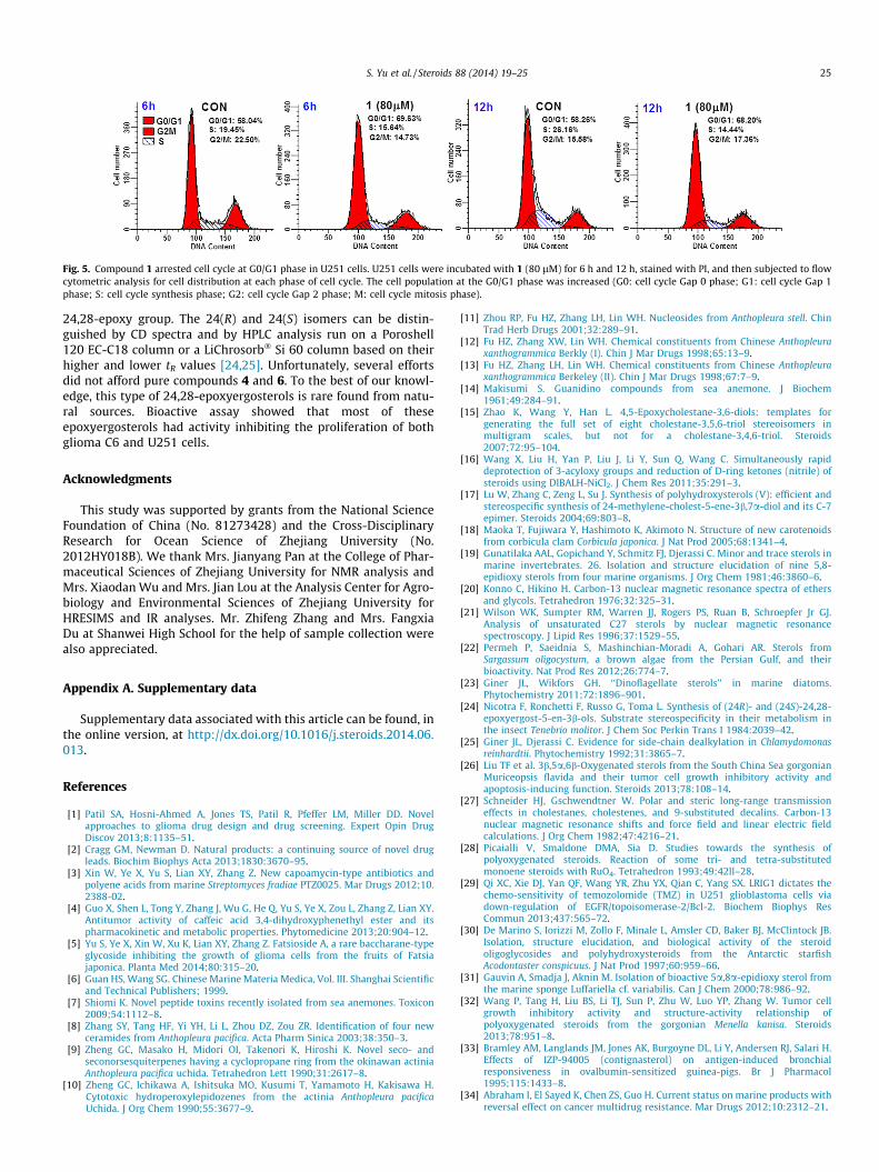
Fig. 5. Compound 1 arrested cell cycle at G0/G1 phase in U251 cells. U251 cells were incubated with 1 (80 lM) for 6 h and 12 h, stained with PI, and then subjected to flowcytometric analysis for cell distribution at each phase of cell cycle. The cell population at the G0/G1 phase was increased (G0: cell cycle Gap 0 phase; G1: cell cycle Gap 1phase; S: cell cycle synthesis phase; G2: cell cycle Gap 2 phase; M: cell cycle mitosis phase).
S. Yu et al. / Steroids 88 (2014) 19–25 25
24,28-epoxy group. The 24(R) and 24(S) isomers can be distin-guished by CD spectra and by HPLC analysis run on a Poroshell120 EC-C18 column or a LiChrosorb� Si 60 column based on theirhigher and lower tR values [24,25]. Unfortunately, several effortsdid not afford pure compounds 4 and 6. To the best of our knowl-edge, this type of 24,28-epoxyergosterols is rare found from natu-ral sources. Bioactive assay showed that most of theseepoxyergosterols had activity inhibiting the proliferation of bothglioma C6 and U251 cells.
Acknowledgments
This study was supported by grants from the National ScienceFoundation of China (No. 81273428) and the Cross-DisciplinaryResearch for Ocean Science of Zhejiang University (No.2012HY018B). We thank Mrs. Jianyang Pan at the College of Phar-maceutical Sciences of Zhejiang University for NMR analysis andMrs. Xiaodan Wu and Mrs. Jian Lou at the Analysis Center for Agro-biology and Environmental Sciences of Zhejiang University forHRESIMS and IR analyses. Mr. Zhifeng Zhang and Mrs. FangxiaDu at Shanwei High School for the help of sample collection werealso appreciated.
Appendix A. Supplementary data
Supplementary data associated with this article can be found, inthe online version, at http://dx.doi.org/10.1016/j.steroids.2014.06.013.
References
[1] Patil SA, Hosni-Ahmed A, Jones TS, Patil R, Pfeffer LM, Miller DD. Novelapproaches to glioma drug design and drug screening. Expert Opin DrugDiscov 2013;8:1135–51.
[2] Cragg GM, Newman D. Natural products: a continuing source of novel drugleads. Biochim Biophys Acta 2013;1830:3670–95.
[3] Xin W, Ye X, Yu S, Lian XY, Zhang Z. New capoamycin-type antibiotics andpolyene acids from marine Streptomyces fradiae PTZ0025. Mar Drugs 2012;10.2388-02.
[4] Guo X, Shen L, Tong Y, Zhang J, Wu G, He Q, Yu S, Ye X, Zou L, Zhang Z, Lian XY.Antitumor activity of caffeic acid 3,4-dihydroxyphenethyl ester and itspharmacokinetic and metabolic properties. Phytomedicine 2013;20:904–12.
[5] Yu S, Ye X, Xin W, Xu K, Lian XY, Zhang Z. Fatsioside A, a rare baccharane-typeglycoside inhibiting the growth of glioma cells from the fruits of Fatsiajaponica. Planta Med 2014;80:315–20.
[6] Guan HS, Wang SG. Chinese Marine Materia Medica, Vol. III. Shanghai Scientificand Technical Publishers; 1999.
[7] Shiomi K. Novel peptide toxins recently isolated from sea anemones. Toxicon2009;54:1112–8.
[8] Zhang SY, Tang HF, Yi YH, Li L, Zhou DZ, Zou ZR. Identification of four newceramides from Anthopleura pacifica. Acta Pharm Sinica 2003;38:350–3.
[9] Zheng GC, Masako H, Midori OI, Takenori K, Hiroshi K. Novel seco- andseconorsesquiterpenes having a cyclopropane ring from the okinawan actiniaAnthopleura pacifica uchida. Tetrahedron Lett 1990;31:2617–8.
[10] Zheng GC, Ichikawa A, Ishitsuka MO, Kusumi T, Yamamoto H, Kakisawa H.Cytotoxic hydroperoxylepidozenes from the actinia Anthopleura pacificaUchida. J Org Chem 1990;55:3677–9.
[11] Zhou RP, Fu HZ, Zhang LH, Lin WH. Nucleosides from Anthopleura stell. ChinTrad Herb Drugs 2001;32:289–91.
[12] Fu HZ, Zhang XW, Lin WH. Chemical constituents from Chinese Anthopleuraxanthogrammica Berkly (I). Chin J Mar Drugs 1998;65:13–9.
[13] Fu HZ, Zhang LH, Lin WH. Chemical constituents from Chinese Anthopleuraxanthogrammica Berkeley (II). Chin J Mar Drugs 1998;67:7–9.
[14] Makisumi S. Guanidino compounds from sea anemone. J Biochem1961;49:284–91.
[15] Zhao K, Wang Y, Han L. 4,5-Epoxycholestane-3,6-diols: templates forgenerating the full set of eight cholestane-3,5,6-triol stereoisomers inmultigram scales, but not for a cholestane-3,4,6-triol. Steroids2007;72:95–104.
[16] Wang X, Liu H, Yan P, Liu J, Li Y, Sun Q, Wang C. Simultaneously rapiddeprotection of 3-acyloxy groups and reduction of D-ring ketones (nitrile) ofsteroids using DIBALH-NiCl2. J Chem Res 2011;35:291–3.
[17] Lu W, Zhang C, Zeng L, Su J. Synthesis of polyhydroxysterols (V): efficient andstereospecific synthesis of 24-methylene-cholest-5-ene-3b,7a-diol and its C-7epimer. Steroids 2004;69:803–8.
[18] Maoka T, Fujiwara Y, Hashimoto K, Akimoto N. Structure of new carotenoidsfrom corbicula clam Corbicula japonica. J Nat Prod 2005;68:1341–4.
[19] Gunatilaka AAL, Gopichand Y, Schmitz FJ, Djerassi C. Minor and trace sterols inmarine invertebrates. 26. Isolation and structure elucidation of nine 5,8-epidioxy sterols from four marine organisms. J Org Chem 1981;46:3860–6.
[20] Konno C, Hikino H. Carbon-13 nuclear magnetic resonance spectra of ethersand glycols. Tetrahedron 1976;32:325–31.
[21] Wilson WK, Sumpter RM, Warren JJ, Rogers PS, Ruan B, Schroepfer Jr GJ.Analysis of unsaturated C27 sterols by nuclear magnetic resonancespectroscopy. J Lipid Res 1996;37:1529–55.
[22] Permeh P, Saeidnia S, Mashinchian-Moradi A, Gohari AR. Sterols fromSargassum oligocystum, a brown algae from the Persian Gulf, and theirbioactivity. Nat Prod Res 2012;26:774–7.
[23] Giner JL, Wikfors GH. ‘‘Dinoflagellate sterols’’ in marine diatoms.Phytochemistry 2011;72:1896–901.
[24] Nicotra F, Ronchetti F, Russo G, Toma L. Synthesis of (24R)- and (24S)-24,28-epoxyergost-5-en-3b-ols. Substrate stereospecificity in their metabolism inthe insect Tenebrio molitor. J Chem Soc Perkin Trans I 1984:2039–42.
[25] Giner JL, Djerassi C. Evidence for side-chain dealkylation in Chlamydomonasreinhardtii. Phytochemistry 1992;31:3865–7.
[26] Liu TF et al. 3b,5a,6b-Oxygenated sterols from the South China Sea gorgonianMuriceopsis flavida and their tumor cell growth inhibitory activity andapoptosis-inducing function. Steroids 2013;78:108–14.
[27] Schneider HJ, Gschwendtner W. Polar and steric long-range transmissioneffects in cholestanes, cholestenes, and 9-substituted decalins. Carbon-13nuclear magnetic resonance shifts and force field and linear electric fieldcalculations. J Org Chem 1982;47:4216–21.
[28] Picaialli V, Smaldone DMA, Sia D. Studies towards the synthesis ofpolyoxygenated steroids. Reaction of some tri- and tetra-substitutedmonoene steroids with RuO4. Tetrahedron 1993;49:42ll–28.
[29] Qi XC, Xie DJ, Yan QF, Wang YR, Zhu YX, Qian C, Yang SX. LRIG1 dictates thechemo-sensitivity of temozolomide (TMZ) in U251 glioblastoma cells viadown-regulation of EGFR/topoisomerase-2/Bcl-2. Biochem Biophys ResCommun 2013;437:565–72.
[30] De Marino S, Iorizzi M, Zollo F, Minale L, Amsler CD, Baker BJ, McClintock JB.Isolation, structure elucidation, and biological activity of the steroidoligoglycosides and polyhydroxysteroids from the Antarctic starfishAcodontaster conspicuus. J Nat Prod 1997;60:959–66.
[31] Gauvin A, Smadja J, Aknin M. Isolation of bioactive 5a,8a-epidioxy sterol fromthe marine sponge Luffariella cf. variabilis. Can J Chem 2000;78:986–92.
[32] Wang P, Tang H, Liu BS, Li TJ, Sun P, Zhu W, Luo YP, Zhang W. Tumor cellgrowth inhibitory activity and structure-activity relationship ofpolyoxygenated steroids from the gorgonian Menella kanisa. Steroids2013;78:951–8.
[33] Bramley AM, Langlands JM, Jones AK, Burgoyne DL, Li Y, Andersen RJ, Salari H.Effects of IZP-94005 (contignasterol) on antigen-induced bronchialresponsiveness in ovalbumin-sensitized guinea-pigs. Br J Pharmacol1995;115:1433–8.
[34] Abraham I, El Sayed K, Chen ZS, Guo H. Current status on marine products withreversal effect on cancer multidrug resistance. Mar Drugs 2012;10:2312–21.