pneumothorax. what is a pneumothorax? air within the pleural cavity (i.e. between visceral and...
TRANSCRIPT
Pneumothorax

What is a pneumothorax?
• Air within the pleural cavity (i.e. between visceral and parietal pleura)
• The air enters via a defect in the visceral pleura (e.g. ruptured bulla) or the parietal pleura (e.g. puncture following rib fracture)

CXR features of pneumothorax
• White line of visceral pleura parallel to chest wall
• No lung markings lateral to the line• There may be associated rib
fractures• Do not confuse the line with skin
fold or with scapula• The most sensitive test if in doubt is
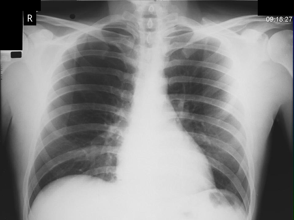
a CXR taken in expirationLook at the CXR on the next slide. Where is the pneumothorax?
R
R
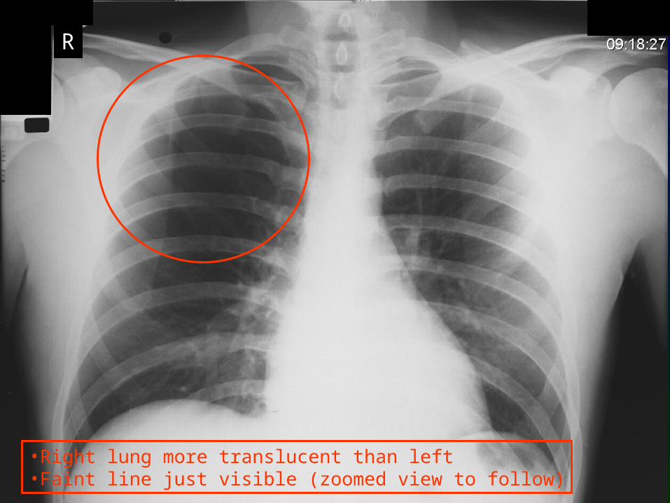
•Right lung more translucent than left•Faint line just visible (zoomed view to follow)
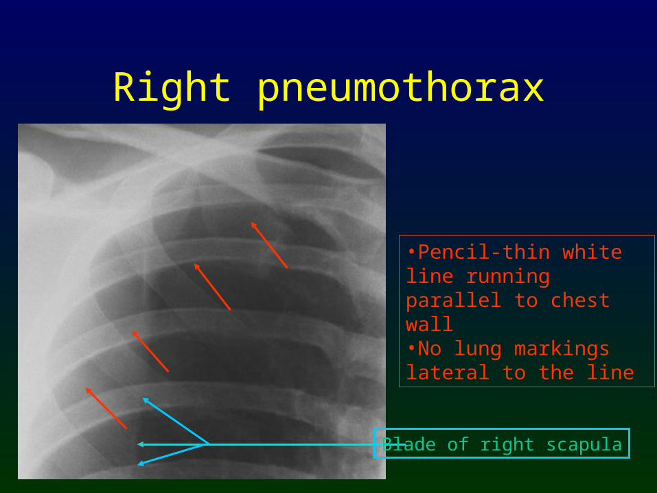
•Pencil-thin white line running parallel to chest wall•No lung markings lateral to the line
Blade of right scapula
Right pneumothorax
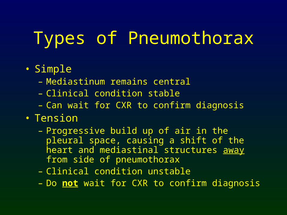
Types of Pneumothorax
• Simple– Mediastinum remains central– Clinical condition stable– Can wait for CXR to confirm diagnosis
• Tension– Progressive build up of air in the pleural
space, causing a shift of the heart and mediastinal structures away from side of pneumothorax
– Clinical condition unstable– Do not wait for CXR to confirm diagnosis

Simple Left Pneumothorax
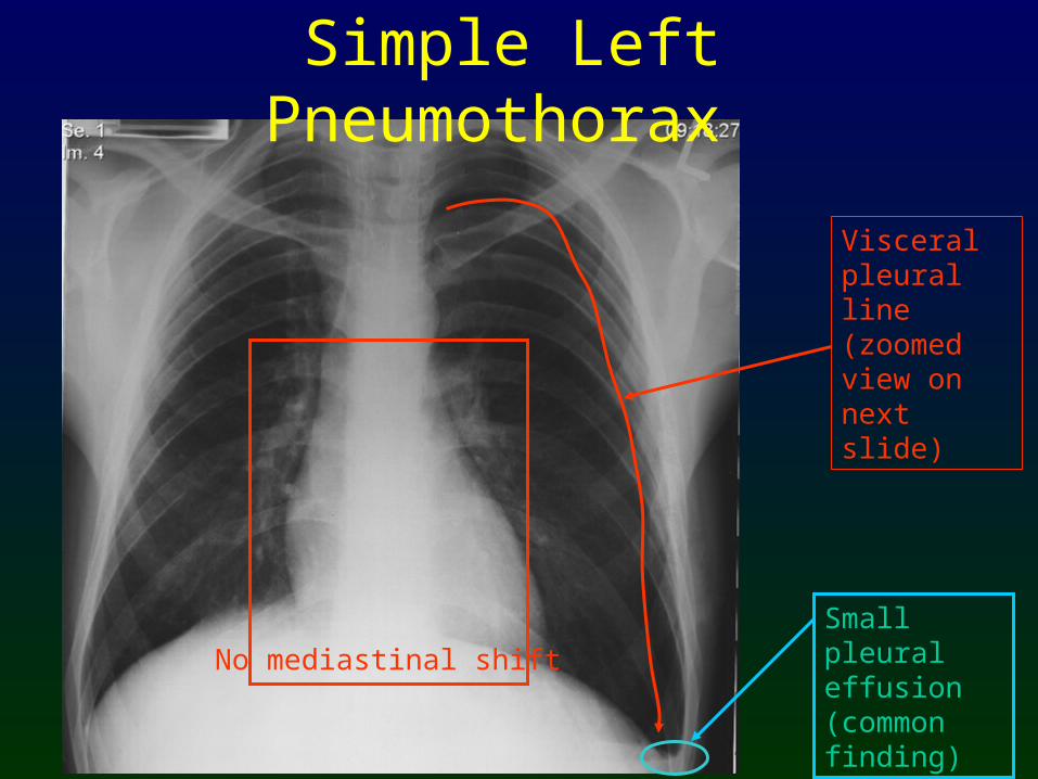
Simple Left Pneumothorax
No mediastinal shiftSmall pleural effusion (common finding)
Visceral pleural line (zoomed view on next slide)
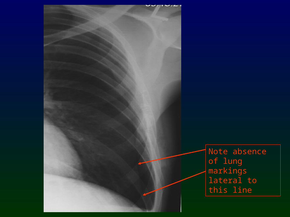
Note absence of lung markings lateral to this line
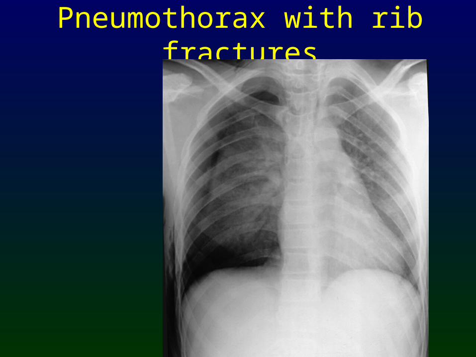
Pneumothorax with rib fractures

Pneumothorax with rib fractures
Surgical emphysema
Right pneumothorax
Rib fractures

Tension right pneumothorax
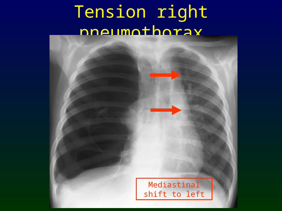
Tension right pneumothorax
Mediastinal shift to left
Causes of Pneumothorax
• Spontaneous– Rupture of an apical bleb
• Traumatic– With rib fractures– Penetrating chest trauma
• Pre-existing lung abnormality– Pulmonary fibrosis– Asthma– Vasculitis– Pulmonary metastases close to edge of lung
Other causes of absent lung markings
• Large emphysematous bullae• Large lung cysts• Pulmonary embolism
....but only pneumothorax has a white line parallel to the chest wall

Take Home Points
• Look for a pencil-thin white line parallel to the chest wall
• No lung markings lateral to the line
• Make sure the patient does not have another cause for absent lung markings before inserting a chest drain