platelets activate a pathogenic response to blood-stage
TRANSCRIPT

Platelets activate a pathogenic response to blood-stage Plasmodium infection
but not a protective immune response.
Irene Gramaglia1,*, Joyce Velez1, Valery Combes1,2, Georges E.R. Grau1,3, Melanie Wree4 and
Henri C. van der Heyde1.
1: La Jolla Infectious Disease Institute, San Diego, CA 92121. 2: School of Life Sciences,
University of Technology Sydney, Sydney, Australia. 3. Vascular Immunology Unit,
University of Sydney, Sydney, Australia. 4. University of California San Diego, San Diego CA.
Primary Scientific Category: Immunobiology
Secondary Scientific Category: Platelets and thrombopoiesis
Running title: Platelets in malaria.
*Corresponding author’s address:
Irene Gramaglia, Ph.D. La Jolla Infectious Disease Institute, 3525 Del Mar Heights Rd #453, San Diego, CA 92130. Phone: (858) 771-2007 Fax: (858) 202-1796 Email: [email protected]
Blood First Edition Paper, prepublished online January 17, 2017; DOI 10.1182/blood-2016-08-733519
Copyright © 2017 American Society of Hematology
For personal use only.on April 11, 2018. by guest www.bloodjournal.orgFrom

2
Abstract Word Count: 240 (max: 250)
Text Word Count: (4470)
Number of Figures: 7
Number of Tables: 1
Number of References: Key points:
1. Platelets are not killer cells of blood-stage Plasmodium parasites.
2. Platelets are not required to activate the protective immune response to blood-stage
Plasmodium infection in mice.
For personal use only.on April 11, 2018. by guest www.bloodjournal.orgFrom

Gramaglia et al.
3
Abstract
Clinical studies indicate that thrombocytopenia correlates with the development of severe
falciparum malaria, suggesting that platelets either contribute to control of parasite replication
possibly as innate parasite killer cells or function in eliciting pathogenesis. Removal of platelets
by anti-CD41 mAb treatment, platelet inhibition by aspirin, and adoptive transfer of WT platelets
to CD40-KO mice, which do not control parasite replication, resulted in similar parasitemia
compared with control mice. Human platelets at a physiological ratio of 1 platelet to 9 RBCs did
not inhibit the in vitro development or replication of blood-stage P. falciparum. The percentage
of iRBCs with bound platelets during the ascending parasitemia in P. chabaudi and P. berghei
infected mice and the 48 hour in vitro cycle of P. falciparum was <10%. P. chabaudi and P.
berghei iRBCs with apoptotic parasites (TdT+) exhibited minimal platelet binding (<5%), which
was similar to non-apoptotic iRBCs. These findings collectively indicate platelets do not kill
bloodstage Plasmodium at physiologically relevant effector to target ratios. P. chabaudi primary
and secondary parasitemia was similar in mice depleted of platelets by mAb-injection just prior
to infection, indicating that activation of the protective immune response does not require
platelets. In contrast to the lack of an effect on parasite replication, adoptive transfer of WT
platelets to CD40-KO mice, which are resistant to experimental cerebral malaria, partially
restored experimental cerebral malaria mortality and symptoms in CD40-KO recipients,
indicating platelets elicit pathogenesis and platelet CD40 is a key molecule.
For personal use only.on April 11, 2018. by guest www.bloodjournal.orgFrom

Gramaglia et al.
4
Introduction:
A hallmark of blood-stage Plasmodium (P) infection in humans is the development of
thrombocytopenia and a pro-coagulant state that is most pronounced in P. falciparum (Pf)
infections, the most virulent of the 5 species of Plasmodium infecting humans1,2. Levels of
thrombocytopenia and pro-coagulant state are elevated in severe Pf malaria compared with
uncomplicated malaria patients and uninfected controls1,2, suggesting that thrombocytopenia
contributes to disease. However, there is still considerable uncertainty about platelet’s role in
malaria. Thrombocytopenia could lead to increased parasite replication by: (i) decreased killing
of parasites, or (ii) decreased activation of the protective immune response controlling parasite
replication.
In support of increased parasite replication contributing to severe malaria, two groups reported
that platelets function as a critical component of innate immunity controlling parasite replication
by binding to and directly killing parasites within red blood cells (RBCs)3-5. Based on increased
mortality of aspirin-treated, Plasmodium chabaudi (Pc)-infected mice and inhibition of in vitro
killing of Pf-infected RBCs (iRBCs) by aspirin4, Greenbaum6 proposed to change clinical
practice by contra-indicating widely used non-steroidal anti-inflammatory agents with anti-
platelet activity, such as aspirin, in malaria patients.
In contrast to reported iRBC killing by platelets3-5, we reported that platelet-depletion by
antibody injection does not affect Plasmodium berghei ANKA (PbA) parasitemia7,8, which
elicits experimental cerebral malaria (eCM) in mice. Early but not late platelet depletion protects
against eCM pathogenesis by ameliorating the inflammatory response7,8. Here, we determine
whether platelets are innate killers of blood-stage Plasmodium and/or regulate either the
protective or pathogenic immune response during malaria and report that there is no evidence to
For personal use only.on April 11, 2018. by guest www.bloodjournal.orgFrom
Gramaglia et al.
5
support physiological platelet killing of blood-stage Plasmodium. Moreover, platelets do not lead
to a protective immune response that clears blood-stage Plasmodium infection, but do activate a
pathogenic response to infection.
Methods
Ethics statement. The Institutional Animal Care and Use Committee of La Jolla Infectious
Disease Institute approved all protocols and procedures.
Mouse studies. There are differences between humans and mice and between Plasmodium
species infecting humans and those infecting mice, indicating caution is needed in extrapolating
results to humans9. Nevertheless, key hallmarks of malarial thrombocytopenia, immunity, and
CM pathogenesis appear to be conserved10. We used three different strains of Plasmodium that
infect mice using standard protocols11,12. The iRBC inoculum details (0.2ml in PBS injected i.v)
and logic behind the selection of the three strains are summarized in Table 1. Day 0PI are
uninfected control mice. Between 200 and 1000 RBCs were counted in Giemsa-stained thin
blood films in experimental animals to assess parasitemia.
To assess the extent of eCM, PbA-infected mice were given neurological tests daily from day
6PI onwards. These tests comprised the sum of the righting reflex and gripping reflex each on a
scale of 1-5 with 5 exhibiting no impairment13,14. Animals with a score of <4 were moribund and
euthanized.
Treatments. Aspirin (acetylsalicylic acid; Sigma, St. Louis, MO) was injected i.p. in 0.2ml saline
(25mg/Kg). Platelets were depleted by i.p. injection with 0.1mg platelet depleting anti-CD41
mAb in 0.1ml PBS (MWReg40); control mice were injected with rat IgG mAb (Affymetrix, San
Diego, CA). Immunodeficient CD40-KO mice and C57BL/6 WT controls were cured of
For personal use only.on April 11, 2018. by guest www.bloodjournal.orgFrom

Gramaglia et al.
6
Plasmodium chabaudi adami (Pca) infection with Trimethoprim (0.4 mg/ml) and
Sulfamethoxazole (1.2 mg/ml) ad libitum for 1 week in drinking water after WT controls had
suppressed their parasitemia to undetectable levels (from >day 28PI). Cure was confirmed by 3
negative thin blood films.
Platelet analysis and isolation. Platelets and RBCs were counted by flow cytometry in 1µl of tail
vein blood as described previously8; the number of platelets/ml blood, platelet activation, and
response was similar in tail vein blood compared with blood obtained by intra-cardiac puncture
or via retro-orbital plexus8.
Purified platelets for adoptive transfer were obtained by centrifugation of whole blood at 50×g
for 20 min to generate platelet rich plasma (PRP). PRP was centrifuged at 800×g for 10 min and
platelets resuspended in Tyrode’s buffer at 0.2×10⁹ in 0.2ml saline for injection.
Flow cytometry. All antibody incubations were performed at room temperature using
antibodies purchased from Affymetrix (San Diego, CA). Anti-CD41-phycoerythrin (PE)
fluorescence labeled platelets, and anti-Ter119-PE-cyanin7 (PE-Cy7) labeled erythrocytes. Cells
were resuspended in 0.5ml PBS with 2.5×10⁴ counting beads (Spherotech, Lake Forest, IL), and
fluorescence intensities of the cells were acquired on an Accuri™ (Becton Dickinson, San Diego,
CA) as described12.
The Pca high virulence (HV) and human platelet interaction with P. falciparum iRBCs were
performed on an ImageStream MkII flow cytometer (EMD Millipore, Seattle, WA), which
distinguishes coincident events from close proximity/adhesion.
Fluorescence labeling of thin blood films. Thin blood films made on glass slides were fixed
for 1min in methanol (Sigma). TUNEL assay was performed following the manufacturer’s
instructions (EMD Millipore, Temecula, CA). Platelets were labeled with anti-CD41-APC mAb.
For personal use only.on April 11, 2018. by guest www.bloodjournal.orgFrom

Gramaglia et al.
7
Parasite DNA and RNA were fluorescence labeled with 12µM ethidium bromide (Fisher
Scientific, Fair Lawn, NJ) in PBS for 15 minutes. Slides were mounted with Vectashield
Mounting Media (Vector Laboratories, Burlingame, CA).
Pf culture and analysis. Blood-stage Pf strains DD2, FCR3, and NK54 were cultured in vitro
as described15,16. Blood was obtained from 3 volunteers who had not taken anti-inflammatory
medications for > 2 weeks before venipuncture. The blood was collected into 5ml citrate
vacutainers using a 21G needle (Becton Dickinson, San Jose, CA), processed into PRP, washed
in Tyrode’s buffer, resuspended in complete media and added at a ratio of 1 platelet per 9 RBCs
to the Pf cultures. Parasitemia of cultures with and without platelets and the percentage of RBCs
with an adherent platelet was assessed at 0, 2, 4, 6, 24, and 48h using imaging flow cytometry.
The thrombin (Fisher Scientific, Chronolog) dose response (CD62P levels) was assessed for each
donor at the above selected time points.
Statistical analysis. Analysis of variance with the Prism program (GraphPad) with Tukey’s
post-hoc test was performed to statistically compare all measurements with a P value cut-off of
0.05. The mean and standard error of the mean of the results are reported in text and figures.
Survival curves are compared with non-parametric Logrank test with a P value cut-off of 0.05
with the “neurological period” for the development of eCM occurring day 6 to 12 in C57BL/6
mice.
Results
Platelet killing of Plasmodium. Malaria in both humans and mice results in profound
thrombocytopenia, occurring during the period of ascending patent parasitemia day 4 to 7PI for
Pca in mice. Non-lethal Pca, low (LV) and high virulence (HV), elicit exponential ascending
parasitemia until close to peak parasitemia with slopes varying depending on parasite virulence
For personal use only.on April 11, 2018. by guest www.bloodjournal.orgFrom

Gramaglia et al.
8
(Table 1); after peak, parasitemia declines to undetectable levels (<0.1%) as a result of the
activation of adaptive immune responses11,17,18. When plotted linearly, thrombocytopenia
coincides with patent parasitemia and its exponential rise, suggesting that parasite replication
may be increased when circulating platelet numbers are reduced4.
Onset of thrombocytopenia exhibits no detectable effect on parasite replication. To test whether
the rate of change of parasitemia is altered when thrombocytopenia occurs (and hence decreased
platelet killing), we infected mice with Pca LV. The increase in parasitemia is concurrent with
the onset of thrombocytopenia when plotted on a linear scale (Figure 1A).
Thrombocytopenia starts on day 4PI and becomes marked during ascending and peak
parasitemia (Figure 1A). Further, in vitro cultured human platelets exhibit >80% killing of Pf-
iRBC with platelet to iRBC target ratios >160:14,5 and <10% killing at 10:15; platelets should
exhibit similar levels of killing in vivo. Based on these platelet-iRBC ratios, the period of
platelet killing of Pca-iRBCs was between day 0 and 4 PI (Figure 1); beyond day 4PI, minimal
platelet killing of Pca-iRBCs occurs.
The log(parasitemia) at each day of infection from day 0 to day 7PI (ascending parasitemia)
increases in a straight line with slope of 0.45±0.03 (R2=0.99) (Figure 1B). This slope determines
the multiplicity of infection (MOI) for Pca, which replicates nightly to produce new iRBCs.
Each cycle (i.e., day) ~3 merozoites (100.45=2.8) invade a new RBC and develop into viable
circulating iRBCs. During ascending parasitemia, there was no detectable change in slope in the
period of platelet killing of Pca-iRBCs (day 0-4PI) and the period of no killing (>day 4PI).
Hence, the MOIs were identical at 2.8.
Models indicate that thrombocytopenia should affect slope of log(parasitemia) when platelet
killing diminishes. To predict the effects of platelet killing of iRBCs on Pca parasitemia, we
For personal use only.on April 11, 2018. by guest www.bloodjournal.orgFrom
Gramaglia et al.
9
modelled its potential effects on parasitemia. There are two possible scenarios for parasitemia:
(i) iRBC sequestration does not markedly affect parasitemia, and (ii) iRBC sequestration
decreases parasitemia. We model two levels of platelet killing of iRBCs: 80% above platelet-
iRBC threshold of 160 (day 0-4PI)4,5 or a conservative estimate of 40%.
With minimal iRBC sequestration in organs, parasitemia reflects the total number of iRBCs.
During the period of platelet killing, only 20%, 60%, or 100% of iRBCs survive for 80%, 40%,
and 0% killing, respectively, where MOIs are 0.6, 1.7, and 2.8, respectively. The parasitemia on
day 0PI is calculated (Table 1) and the above MOIs are used to calculate the daily parasitemia
until day 4PI (Figure 1C). Because the period of platelet killing ends on day 4PI, the MOI for
each platelet killing scenario now equals 2.8 (0% killing) that is used to calculate the subsequent
daily parasitemia until peak (Figure 1C).
With marked iRBC sequestration, parasitemia does not reflect the total parasite burden because
sequestered iRBCs are not circulating and counted. If we assume similar levels of sequestration
throughout ascending parasitemia, then the slope of log(%parasitemia) between day 0 and 4PI
provides the “effective” rate of parasite replication in blood with sequestration and platelet
killing. The slope was measured at 0.45, so the “effective MOI” during platelet killing is
100.45=2.8. The 80%, 40%, or 0% iRBC killing by platelets has stopped from day 4-7PI, so the
“effective MOI is now: 14, 7, and 2.8, respectively. The calculated parasitemia (Figure 1D) then
results in markedly different parasitemia with 40 and 80% killing by platelets compared with 0%
(which fits the observed parasitemia). The slope of log(%parasitemia) did not change markedly
after day 4PI as predicted by modeling of parasitemia with sequestration, indicating that platelet
killing of iRBCs is minimal, if any.
For personal use only.on April 11, 2018. by guest www.bloodjournal.orgFrom

Gramaglia et al.
10
Platelet inhibition by aspirin has no detectable effect on parasite replication in blood. To
determine whether inhibition of platelet activation increases parasite replication, we injected high
dose aspirin or vehicle control starting on day-1PI and ending at peak parasitemia (day 10PI);
this aspirin regimen was chosen because it was used for the conclusion that aspirin is detrimental
in malaria patients based on this aspirin regimen’s elicitation of mortality in Pca DS strain4,6.
Ascending parasitemia was similar in high dose aspirin and vehicle control mice (Figure 2A).
The slope of log(%parasitemia) during ascending parasitemia (day 0 to 6PI) was similar
(P=0.49) in both aspirin-treated (0.45 ± 0.03; R2=0.99) and vehicle control mice (0.48 ± 0.03;
R2=0.99). No mortality was observed in either group. Aspirin did not prevent thrombocytopenia
during the course of Pca LV infection (Figure 2A).
Platelet depletion exhibits no detectable effect on parasite replication in blood. To determine
whether depletion of platelets early in infection decreases early killing of iRBCs and/or affects
the protective immune response, we injected C57BL/6 mice on day –1PI and every 2-3 days with
either anti-CD41 or isotype control mAb. Few platelets were detected by flow cytometry during
the course of Pca infection in platelet-depleted mice during ascending parasitemia whereas larger
numbers of platelets were detected in isotype controls (Figure 2B). Despite >92% platelet
depletion from day 0-4, there was no significant (P>0.05) difference in parasitemia between
platelet-depleted and control groups during the course of ascending or descending parasitemia
and suppression of the infection (Figure 2B). No mortality was observed in either group.
To determine whether the platelet depletion affects protective immune responses after a second
infection, we treated the two infected groups, with Trimethoprim-Sulfamethoxazole for 1 week
starting on day 32PI and allowed 1 week for drug clearance before injecting them and an
uninfected group i.v. with 1×10⁷ Pca iRBCs. The secondary parasitemia was similar and below
For personal use only.on April 11, 2018. by guest www.bloodjournal.orgFrom
Gramaglia et al.
11
0.1% in anti-CD41 and isotype control-injected groups whereas the primary infection controls
parasitemia peaked at 14±1% on day 10PI (Figure 2C). A maximum of 1000 RBCs were
counted resulting in a lower detection limit of 0.1% parasitemia. At least 10 pRBCs/1000 RBCs
or 1% parasitemia likely provides a reliable estimate of parasitemia. Most animals exhibited 0
pRBCs/1000 RBCs, resulting in an average parasitemia below 0.1%. Removal of platelets by
antibody depletion does not affect parasitemia at any point during primary or secondary
infections, indicating that platelets are not killing parasites and are not required for activation of
the protective immune response.
Intact platelets adoptively transferred to CD40KO recipients do not affect parasitemia or elicit a
protective immune response. To determine whether wild-type (WT) CD40+ platelets adoptively
transferred to CD40KO mice (which do not control Pca parasite replication) elicit a reduction in
the initial inoculum and/or activate a protective adaptive immune response, we injected CD40-
KO mice with 0.2×10⁹ platelets from uninfected C57BL/6 mice or saline alone on day –1 and
0PI and infected both groups plus infection control mice group (C57BL/6 ) with Pca. All groups
of mice exhibited similar ascending parasitemia; both groups of CD40KO mice exhibited
similarly high levels of unremitting parasitemia whereas the C57BL/6 mice controlled their
parasitemia to low levels (Figure 2D).
We tested the secondary protective immune response in the CD40-KO mice receiving (i)
platelets or (ii) saline and in C57BL/6 mice by drug curing the 3 groups of animals and then
infecting them plus an infection control group with Pca. The secondary parasitemia was similar
in both CD40KO groups. Both groups exhibited some initial control and then high levels of
parasitemia (Figure 2E). CD40-KO mice develop anti-parasite IgM but do not isotype switch to
IgG (unpublished results), which may account for the initial control of parasitemia in these mice.
For personal use only.on April 11, 2018. by guest www.bloodjournal.orgFrom

Gramaglia et al.
12
Increased platelet binding to iRBCs and terminal deoxynucleotidyl transferase dUTP nick end
labeled (TUNEL+) platelets is not detected in thin blood films during ascending Pca. To
analyze direct platelet cytotoxicity during ascending Pca parasitemia4,5, we counted
apoptotic/dying parasites and iRBC-platelet conjugates in thin blood films. The platelet to target
ratio was 1,070±55:1 and declined rapidly to about 1 as parasites replicated exponentially during
ascending parasitemia and circulating platelet numbers declined (Figure 3A). During ascending
parasitemia (day 0-7PI), the %parasitemia using all RBCs is similar to that seen in conjugates of
platelets and iRBCs (Figure 3B). Most platelet:RBC conjugates comprise platelets with uRBCs
rather than iRBCs (Figure 3C). Less than 10% of iRBCs exhibit an attached platelet and this
percentage does not change markedly during the course of Pca infection (Figure 3D). The
%iRBCs that are TUNEL+ is significantly greater during peak and descending parasitemia and
most apoptotic parasites do not have a platelet attached to the iRBC (Figure 3E, F). Collectively,
these findings indicate that platelet:RBC adhesion is low and few if any TUNEL+ Pca parasites
exhibit a bound platelet, indicating that direct platelet adhesion is unlikely to kill significant
numbers of parasites.
Neither platelet inhibition by aspirin nor platelet depletion affects the replication of high
virulence Pca parasites. To increase the likelihood of detecting platelet-killing of iRBCs, we
increased the initial platelet to iRBC target ratio by 100-fold and increased the time for platelets
to kill parasites by injecting 100-fold fewer Pca HV parasites into mice. We injected Pca HV-
infected mice with i) high dose aspirin, ii) saline; iii) anti-CD41 mAb or iv) isotype control until
peak parasitemia to detect changes in platelet killing of iRBCs and changes in immune response
controlling parasite replication. The parasitemia was similar at each time point during ascending
parasitemia in aspirin-treated and vehicle controls (Figure S1A) and anti-CD41 mAb-injected
For personal use only.on April 11, 2018. by guest www.bloodjournal.orgFrom

Gramaglia et al.
13
and isotype controls (Figure S1E). No mortality was observed in either aspirin or vehicle control
group. Mortality was similar in anti-CD41 mAb-injected and controls and occurred after
ascending parasitemia (Figure S1E). The parasitemia in RBC-platelet conjugates was greater
than in all RBCs (Figure S1B) but aspirin did not inhibit this. The percentage of platelet:RBC
conjugates was low (<0.1% of RBCs). The number of iRBC-platelet conjugates was low from
day 4 to day 8PI and similar to background (day 0PI), but the number of uRBC-platelet
conjugates declined (Figure S1C). The percentage of iRBCs with a bound platelet was <10%
throughout course of infection (Figure S1D). These findings indicate that neither platelet
inhibition by aspirin nor platelet depletion affects parasite replication or the activation of the
protective immune response that controls parasitemia.
Human platelets do not inhibit in vitro replication of several strains of Pf. To confirm that
human platelets do not kill Pf-iRBCs in vitro at physiological concentrations, we incubated
platelets with RBCs at a 1:9 ratio with time 0 parasitemia of Pf at trophozoite stage ranging from
4-8%. Parasitemia was similar over an entire cycle between cultures containing platelets and
those without (Figure 4A). The percent decline in parasitemia in platelet-containing cultures to
no platelet controls was minimal; at 24 and 48 hrs, the parasitemia was higher in platelet-
containing cultures, providing a negative percent decline (Figure 4B). One donor was tested
against FCR3 and DD4 strains of Pf; the parasitemia was similar at each time point in platelet-
containing cultures to no platelet controls. After excluding coincident events, the percentage of
iRBCs with bound platelet remained constant and <10%; the percentage of uRBCs with bound
platelets was lower and < 3% (Figure 4C). Platelets in the co-cultures were primarily (~90%)
unbound (Figure 4D). Platelets cultured in media alone exhibited similar thrombin dose
responses at early time points, but impaired response at 48h (Figure 4E,F).
For personal use only.on April 11, 2018. by guest www.bloodjournal.orgFrom

Gramaglia et al.
14
Platelet function in experimental cerebral malaria.
Because Pf that elicits human CM is proposed to be susceptible to platelet killing3,4, therefore,
the eCM-eliciting parasite, PbA, should also be susceptible to platelet killing. This however
appears to contradict our earlier report that platelet depletion actually protects against eCM
without affecting parasitemia7,8. We therefore reexamined platelet killing in PbA infections of
mice.
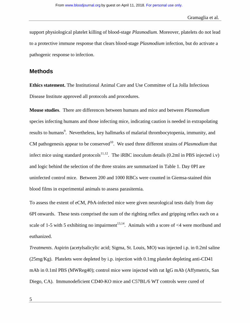
Neither increased platelet binding to iRBCs and dying (TUNEL+) platelets nor activated,
circulating platelets are detected during eCM. To determine whether platelets function in
parasite killing when eCM is elicited, we repeated the Pca analysis with PbA. PbA parasitemia
increased exponentially up to and including day 5 (R2=0.99 with slope of 0.66) when the rate of
increase slows (Figure 5A). Thrombocytopenia starts on day 4, is marked by day 5 (Figure 5A),
and occurs during the period of exponential parasite replication. PbA parasitemia in RBCs with
bound platelets is significantly higher than all RBCs (P<0.05) on day 6PI (Figure 5B). The
percentage of platelet bound iRBCs increased significantly (p<0.05) on day 6PI (Figure 5C).
However, the percentage of iRBCs at risk for platelet killing declined from day 4 to day 6PI and
was <10% (Figure 5D) with no preferential binding of platelets to apoptotic parasites (Figure
5E,F). Platelet depletion by anti-CD41 mAb does not significantly alter PbA parasitemia7,8.
To determine whether WT platelets transferred to CD40KO mice, which are eCM resistant19,
affects parasitemia and/or elicits a pathogenic response, we injected 0.2×10⁹ platelets from
uninfected mice or vehicle control into eCM-protected CD40-KO mice19 on day –1 and day 1PI.
Significantly (p<0.05) decreased clinical scores and increased mortality in CD40-KO mice
receiving platelets compared with vehicle controls (Figure 6A,B) indicate that CD40+ platelets
elicit pathogenesis in a resistant CD40KO mouse; consequently, platelet CD40 is a key molecule
For personal use only.on April 11, 2018. by guest www.bloodjournal.orgFrom

Gramaglia et al.
15
in triggering pathogenesis and platelets do not affect PbA replication in vivo (Figure
6C). Together with Pca findings, they also indicate that the pathways leading to control of
parasite replication and pathogenesis are separate and distinct.
Discussion
The explanations for thrombocytopenia correlating with severe malaria include (i) platelets kill
iRBCs, (ii) platelets activate the protective immune response, and/or (iii) absence of platelets
contributes to pathogenesis. As a consequence of (i) and (ii), thrombocytopenia would result in
increased parasitemia and disease. Two groups3-5 have proposed platelet killing of Plasmodium
in iRBCs. The first line of evidence supporting platelet killing of iRBCs is the concurrence of
thrombocytopenia during Pca infection in mice with the development of patent parasitemia and
the exponential rise in parasitemia until peak parasitemia4. However, the parasite is replicating
exponentially in the blood compartment from the time of initial infection to about peak
parasitemia; thus, the log(%parasitemia) fits with a straight line. Our data indicate that the slope
of this line does not change with the onset of thrombocytopenia, which is predicted if platelets
are controlling parasite replication. Thus, the coincidence of patent parasitemia and
thrombocytopenia is likely due to the host response the parasite has elicited rather than the direct
killing of iRBCs by platelets.
Second, depletion of platelets by mAb leads to increased parasitemia in PbA-infected mice20.
Significantly increased parasitemia occurs at a single time point (day 5PI) in PbA-infected mice
depleted of platelets with anti-CD42b mAb on day -1PI compared with isotype Ig controls20.
Parasitemia measured at a single time point by conventional flow cytometry may yield incorrect
results because some uRBCs contain nucleic acids that are then interpreted as iRBCs. In the
present study, we used a different mAb to deplete platelets (anti-CD41 mAb MWreg40) and
For personal use only.on April 11, 2018. by guest www.bloodjournal.orgFrom

Gramaglia et al.
16
verified our depletion throughout ascending parasitemia, measured parasitemia at multiple
timepoints, and tested 2 different species of Plasmodium without detecting any effect of platelet
depletion7,8. Both McMorran’s4 and our analyses of inhibition of platelet activity by high dose
aspirin indicate that this regimen does not affect Pca or PbA parasitemia. Our data therefore
indicate that neither inhibition of platelets nor depletion of platelets with mAb affects
parasitemia.
McMorran et al.4 use death rather than parasitemia to detect platelet killing of parasites because
iRBC sequestration in organs may result in parasitemia not reflecting total numbers of parasites,
resulting in an inability to detect parasite killing by platelets. However, our data (Figure 1)
indicate that the slope of log(%parasitemia) during ascending parasitemia is linear and dependent
on parasite virulence. Parasitemia and the slope of log(%parasitemia), therefore, should be
different in the presence and absence of platelets given the 80% killing observed in vitro4,5.
The use of the animal’s death as a biomarker for parasite replication maybe problematic. Death
is complex, multi-factorial, and occurs at low and high parasitemia, so death may not be directly
linked to parasite burden. Indeed, the majority of deaths reported by McMorran et al.4 occur
after ascending parasitemia at time points when parasite burden is being suppressed by the
adaptive immunity11,17,18 and thrombocytopenia has been marked for a while. Thus, there is little
evidence to indicate that an elevated parasite burden is the cause of the animal’s death.
The platelet-mediated killing of iRBCs in vivo is proposed to be contact-dependent based on: (i)
increased Pca parasitemia when platelet-RBC conjugates are counted compared with all RBCs
and (ii) significantly (p<0.05) increased % of parasites that are TUNEL+ in platelet-deficient
MPL−/− mice compared with C57BL/6 controls4. However, our analysis of platelet:xRBC (x=u
or i) conjugates during the course of Pca infection supports the conclusion that platelets are not
For personal use only.on April 11, 2018. by guest www.bloodjournal.orgFrom

Gramaglia et al.
17
cytotoxic for parasites. Our findings that most platelets adhere to uRBCs, <10% of iRBCs have
a bound platelet, and few apoptotic parasites occur in platelet:iRBC conjugates, collectively
indicate that it is unlikely that platelets are adhering to iRBCs early during the infection prior to
the onset of thrombocytopenia in order to kill the parasite within the RBC.
Others reported contact-dependent intra-Pf-RBC killing by human platelets in vitro3-5. Peyron et
al.5 reported at a platelet:Pf-iRBC ratio of 160:1 (corresponding to a platelet:RBC ratio ~1:1,
which is not physiological) results in >80% inhibition of parasite development. If this finding is
valid, then platelet inhibition should reduce this killing. However, in this study, in vitro platelet
killing of Pf-iRBC is not inhibited by 1mM aspirin or 2mM histidine. In contrast, McMorran et
al.3,4 reported that this platelet killing is abrogated by known platelet inhibitors. However, their
observation that the total parasitemia at 29h (trophozoite) and 44h (schizont) of platelet-Pf-iRBC
cocultures are similar suggests that 15h of culture have passed with no killing or inhibition of
development detected in the presence of platelets4. In the present study, we also did not detect
any change in parasitemia with Pf ring-stages maturing to schizonts in the presence or absence of
platelets. We replicated our in vivo condition as much as possible with human cells at 1:9
platelet:RBC ratio of normal blood and ~ 2:1 platelet:iRBC ratio. We tested multiple human
donors and parasite strains, confirmed platelet thrombin responses throughout the co-culture, but
did not detect inhibition of parasite replication or development by platelets. The percentage of
platelets bound to a Pf-iRBC was greater than uRBC, but these percentages were <10% and
remained constant throughout the coculture. Clinical studies report either an association of
elevated numbers of platelet-RBCs conjugates (rosettes) with severe human malaria or no
association1,2. Platelet-Pf-iRBC rosettes associate with high parasitemia and cerebral malaria21.
If platelets killed Pf-iRBCs within rosettes, then elevated rosette numbers should reduce
For personal use only.on April 11, 2018. by guest www.bloodjournal.orgFrom

Gramaglia et al.
18
parasitemia and be associated with lower parasitemia and likely with protection from severe
malaria. Collectively, these observations suggest that under physiological conditions platelets do
not inhibit the replication or intra-RBC development of Pf. Thus, there is no rationale for
avoiding the use of NSAIDs in the treatment of malaria6.
Besides their putative role as innate killer cells, platelets function in the activation of immune
responses. Indeed, CD40L+ WT platelets adoptively transferred to CD40LΚΟ mice activate the
protective immune response in virus- or bacteria-infected mice22-24. However, neither high dose
aspirin nor platelet depletion starting at day -1PI affected the primary parasitemia. Platelet
depletion during the primary infection also did not alter the secondary parasitemia. Moreover,
adoptive transfer of CD40+ WT platelets into CD40ΚΟ mice that do not control their Pca
parasitemia had no detectable effect on the primary and secondary immune responses controlling
parasitemia. These results indicate that (i) adoptive transfer of WT CD40+ platelets to CD40KO
mice does not restore immune control of primary or secondary parasitemias, as reported in some
other infections22-24, and (ii) platelets are not required for the development of a protective
primary and secondary immune responses to resolve Pca.
The observations that C57BL/6 mice depleted of platelets by anti-CD41 mAb injection early
(day 1PI) but not late (day 4 onwards) exhibit decreased inflammatory responses, decreased
acute phase proteins, and are protected from eCM7,8 indicate that the activation of the pathogenic
eCM response is impaired in the absence of platelets. Their requirement for the onset of the
pathogenic response has however not been tested. Our observation that adoptive transfer of WT
CD40+ platelets to eCM-resistant CD40ΚΟ mice partially restores mortality and clinical
symptoms of eCM supports the role of platelet CD40 in activating a pathogenic response.
For personal use only.on April 11, 2018. by guest www.bloodjournal.orgFrom

Gramaglia et al.
19
Collectively, our findings indicate that platelets (i) do not function in the killing of blood-stage
Plasmodium parasite, (ii) are not required for the activation of the protective immune response,
but (iii) are required to elicit pathogenesis. Platelets may function in part by activating the liver
to produce acute phase proteins or by activating the pathogenic immune response8,20.
For personal use only.on April 11, 2018. by guest www.bloodjournal.orgFrom

Gramaglia et al.
20
Acknowledgements.
This work was supported by an NIH grants to HH (R03AI088283, R21NS066401), IG
(R03NS081527; 5R21NS080063), and VC (R01NS079873).
Authorship.
IG, and HH developed hypotheses, designed and performed the experiments, analyzed data, and
wrote the manuscript. JV and MW performed experiments, and analyzed data. VC and GG
developed hypotheses, reviewed data, and wrote the manuscript. There are no non-author
contributions and disclosures. The authors have no conflict of interest to declare.
For personal use only.on April 11, 2018. by guest www.bloodjournal.orgFrom

Gramaglia et al.
21
Figure Legends.
Figure 1. No change in slope of log(%parasitemia) after onset of thrombocytopenia that would
suggest platelets affecting parasite replication in resolving, non-eCM Pca LV infection.
Parasitemia denoted by filled symbols (left y-axis) in A-D with corresponding platelet counts/ml
in open symbols (right y-axis) for A, B. (A): Parasitemia of Pca-infected mice (n=10) (circle)
plotted on a linear scale with latent parasitemia coinciding with thrombocytopenia. (B):
Parasitemia in (A) plotted on logarithmic scale and no change in slope occurs at the onset of
thrombocytopenia on day 4 PI. This experiment was replicated 3 times. (C): Modeling of log of
%(parasitemia) at selected rates of parasite killing by platelets between day 0 and 4 PI assuming
sequestration does not affect parasitemia. We model the Pca LV parasitemia in a 20g mouse,
which calculates the initial parasitemia (Table 1). Platelet killing is of Pf-iRBCs is ~80%4,5, and
we compare this to a more conservative 40% killing and no killing. At initiation of LV infection,
the 20g mouse has: 1×10⁶ iRBCs; 1.6×10¹⁰ RBCs; 1.6×10⁹ platelets; and a platelet effector to
iRBC target ratio > 1,000, resulting in ~80% killing of iRBCs by platelets based on in vitro
killing of Pf-iRBCs4,5. At the onset of thrombocytopenia (day 4PI: ~0.5% parasitemia; 1.6×10¹⁰
RBCs, and 1.6×10⁹ platelets), the platelet effector to target ratio is 20:1; theoretically, platelet
killing is minimal below 160:15. The period of platelet killing of iRBCs is therefore between day
0 and 4 PI (shaded, labeled “Period of platelet killing”). During ascending parasitemia (day 7PI:
~6% parasitemia; 1.6×10¹⁰ RBCs; and ~1.6×10⁸ platelets), the ratio is 1:600. The parasite
replicates each night producing new progeny, and the MOI during period of no killing and
determined from Figure 1B is 2.8. If each day during platelet killing period, 80% or 40% of
iRBCs are killed by platelets, then MOI declines to 0.6 (20% of 2.8) and 1.7 (60% of 2.8)
respectively. The measured parasitemia (Figure 1B) fits 0% line rather than 40% or 80% killing.
For personal use only.on April 11, 2018. by guest www.bloodjournal.orgFrom

Gramaglia et al.
22
(D): Modeling of log % parasitemia assuming similar degrees of iRBC sequestration throughout
ascending parasitemia and platelet killing until day 4 PI as described above in C. Because the
slope of log(%parasitemia) is linear, parasitemia likely reflects overall parasite load. The slope
of log(%parasitemia) during period of platelet killing from day 0 to 4PI is 2.8 (Panel B); this
estimates effective rate of parasitemia increase with sequestration. Because platelet killing is
minimal beyond day 4 PI, this rate of parasitemia increase should increase markedly with an
MOI from 2.8 (0% killing) to 14 (80% killing) and 7 (40% killing) after day 4 PI. The measured
parasitemia (Panel B) clearly fits 0% platelet killing best. The modeling indicates that the slope
of log(%parasitemia) should change markedly if platelet killing occurs.
Figure 2. Platelet removal or inhibition does not affect parasitemia in resolving, non eCM Pca
LV infection and does not affect activation of protective immunity. Parasitemia for each group
denoted by filled symbols (left y-axis) with corresponding platelet counts/ml in open symbols
(right y-axis). (A): Aspirin- (triangle) and saline-injected (circle) groups (n=5) of Pca-infected
mice. (B): Platelet-depleting anti-CD41 mAb (triangle) or isotype control (circle) injected on
day –1, 1, 3, and 6PI. (C): Secondary parasitemia after injection of 1×10⁷ Pca on day 0PI into
the anti-CD41 mAb and isotype control groups of mice that had resolved their primary infection;
a group of uninfected mice with primary parasitemia is infection control. (D): Primary
parasitemia in CD40KO mice that do not resolve Pca infection after iv injection of WT platelets
on day -1 and 0PI and in intact mice that resolve their Pca infection. (E): Secondary parasitemia
after injection of 1×10⁷ Pca into 3 groups of mice in Panel D; a group of uninfected mice with
primary parasitemia is infection control. Average value ± SEM are reported. *: P<0.05. The
experiment in Panel A was repeated twice, and in B was repeated once.
For personal use only.on April 11, 2018. by guest www.bloodjournal.orgFrom

Gramaglia et al.
23
Figure 3. Platelets are not associated with dying parasites during resolving, non-eCM Pca
infection in thin blood films. The analyses were performed in thin blood films fluorescence
labeled with anti-CD41 (blue; platelets), ethidium bromide (red: parasites), and TUNEL (green).
(A): Platelet effector to target (iRBC) ratios (■), platelet: uRBC ratios (□), and parasitemia (●).
Platelet:RBC is an abbreviation for a platelet RBC conjugate; the RBC maybe uninfected
(uRBC) or infected (iRBC). (B): Percent PbA parasitemia for all RBCs (□) �# ����
# ��� % � and
considering only RBCs with bound platelets (■) �# �����:����
# �����:��� % �. (C): Percentage of RBCs
with bound platelets with uninfected (□) �# �����: ���
# �����:��� % � or infected (■) RBCs:
(#�����:����
# �����:��� %�. (D): The percentage of iRBCs with an adherent platelet �# �����:����
# ����� % �.
(E): Representative thin blood film made on day 6PI and fluorescently labeled; blue arrows
indicate platelets, red arrows indicate parasites without TUNEL, and yellow arrows indicate
parasites with TUNEL (green+red). (F): Percentage on day 6, 8, and 12PI of iRBCs exhibiting
TUNEL+ labeling (gray-filled bar, x-axis) �# ������ �����
# ����� % � and TUNEL– (open bar, x-axis)
�# ������ �����
# ����� % � with the percentage of each iRBC+, TUNEL+ or iRBC+, TUNEL–
�#�����: ������ �����
# ����� % � exhibiting an adherent platelet shown as an inset (filled black bar).
This experiment was repeated twice (n=5) and verified by flow cytometry. Values are average ±
SEM.
Figure 4. Human platelets at physiological ratios do not inhibit in vitro replication of human
strains of Plasmodium falciparum. (A): Representative percent parasitemia measured by
imaging flow cytometry over the course of 48hours in cultures with platelets (□) at 1:9 RBC ratio
and without platelets (■). (B): Calculated replication inhibition of P. falciparum replication in
For personal use only.on April 11, 2018. by guest www.bloodjournal.orgFrom

Gramaglia et al.
24
platelet cultures compared with no platelet controls (n=6) at selected time points during the
development of P. falciparum parasites. (C): The percentage of uRBCs (□) �# �����: ���
# ���� % �
or iRBCs (■)�# �����:����
# ����� % � with attached platelet over course of P. falciparum
development. (D): Percentage of all platelets that are unbound (■) �# ��� �� ������
# ������ % �,
bound to uRBCs (□) �# �����: ���
# ������ % �, or bound to iRBCs (■) �# �����:����
# ������ % � during the
course of P. falciparum development. (E): Intensity of CD62P fluorescence on the surface of
platelets after stimulation with selected doses of thrombin at selected time points of culture. (F):
Calculated thrombin EC50 for platelet response at the selected time points of culture.
Figure 5. Platelets are not associated with dying parasites during non resolving, eCM-eliciting
PbA in C57BL/6 mice. The analyses were performed in thin blood films fluorescence labeled
with anti-CD41 (blue; platelets) and ethidium bromide (red: parasites) and TUNEL (green). (A):
Platelet effector to target (iRBC) ratios (■), platelet: uRBC (□) ratios and parasitemia (●) on left
axis and platelet counts/ml (○) on right axis. (B): Percent PbA parasitemia for all RBCs (□)
�# ����
# ��� % � and considering only RBCs with bound platelets (■) �# �����:����
# �����:��� % �.
Platelet:RBC is an abbreviation for a platelet RBC conjugate; the RBC maybe uninfected
(uRBC) or infected (iRBC). (C): Percentage of RBCs with bound platelets with uninfected (□)
�# �����: ���
# �����:��� % � or infected (■) RBCs: (
#�����:����
# �����:��� %�. (D): The percentage of iRBCs
with an adherent platelet �# �����:����
# ����� % �. (E): Representative thin blood film made on day
6PI and fluorescently labeled; blue arrows indicate platelets, red arrows indicate parasites
without TUNEL, and yellow arrows indicate parasites with TUNEL (green+red). (F):
Percentage on day 4 and 6PI of iRBCs exhibiting TUNEL+ labeling (gray-filled bar, x-axis)
For personal use only.on April 11, 2018. by guest www.bloodjournal.orgFrom

Gramaglia et al.
25
�# ������ �����
# ����� % � and TUNEL– (open bar, x-axis) �# ������ �����
# ����� % � with the percentage of
each iRBC+, TUNEL+ or iRBC+, TUNEL– �#�����: ������ �����
# ����� % � exhibiting an adherent
platelet shown as an inset (filled black bar). This experiment was repeated twice (n=5) and
verified by flow cytometry. Values are average ± SEM. This experiment was repeated twice
(n=5).
Figure 6. Platelets partially restore pathogenesis to eCM-protected CD40-KO mice. (A):
Clinical scores, (B): percent survival, and (C): parasitemia for groups (n=5) CD40-KO
reconstituted with WT platelets (■), CD40-KO injected with saline (□) or C57BL/6 controls (▲).
Gray shaded area indicates the time period for the development of neurological symptoms by
C57BL/6 mice. Values are average ± SEM. *: p<0.05 for comparison of CD40-KO + platelets
groups with other 2 groups; #: p<0.05 for comparison of C57BL/6 with other 2 groups.
For personal use only.on April 11, 2018. by guest www.bloodjournal.orgFrom

Gramaglia et al.
26
References 1. van der Heyde HC, Nolan J, Combes V, Gramaglia I, Grau GE. A unified hypothesis for the genesis of cerebral malaria: sequestration, inflammation and hemostasis leading to microcirculatory dysfunction. Trends Parasitol. 2006;22(11):503-508. 2. Francischetti IM, Seydel KB, Monteiro RQ. Blood coagulation, inflammation, and malaria. Microcirculation. 2008;15(2):81-107. 3. McMorran BJ, Wieczorski L, Drysdale KE, et al. Platelet factor 4 and Duffy antigen required for platelet killing of Plasmodium falciparum. Science. 2012;338(6112):1348-1351. 4. McMorran BJ, Marshall VM, de Graaf C, et al. Platelets kill intraerythrocytic malarial parasites and mediate survival to infection. Science. 2009;323(5915):797-800. 5. Peyron F, Polack B, Lamotte D, Kolodie L, Ambroise-Thomas P. Plasmodium falciparum growth inhibition by human platelets in vitro. Parasitology. 1989;99 Pt 3:317-322. 6. Greenbaum DC, FitzGerald GA. Platelets, pyrexia, and plasmodia. N Engl J Med. 2009;361(5):526-528. 7. Grau GE, Tacchini-Cottier F, Vesin C, et al. TNF-induced microvascular pathology: active role for platelets and importance of the LFA-1/ICAM-1 interaction. EurCytokine Netw. 1993;4(6):415-419. 8. van der Heyde HC, Gramaglia I, Sun G, Woods C. Platelet depletion by anti-CD41 (αIIb) mAb injection early but not late in the course of disease protects against Plasmodium berghei pathogenesis by altering the levels of pathogenic cytokines. Blood. 2005;105(5):1956-1963. 9. White NJ, Turner GD, Medana IM, Dondorp AM, Day NP. The murine cerebral malaria phenomenon. Trends Parasitol. 2010;26(1):11-15. 10. Craig AG, Grau GE, Janse C, et al. The role of animal models for research on severe malaria. PLoS Pathog. 2012;8(2):e1002401. 11. van der Heyde HC, Huszar D, Woodhouse C, Manning DD, Weidanz WP. The resolution of acute malaria in a definitive model of B cell deficiency, the JHD mouse. J Immunol. 1994;152(9):4557-4562. 12. Gramaglia I, Sahlin H, Nolan JP, Frangos JA, Intaglietta M, van der Heyde HC. Cell- rather than antibody-mediated immunity leads to the development of profound thrombocytopenia during experimental Plasmodium berghei malaria. J Immunol. 2005;175(11):7699-7707. 13. Gramaglia I, Sobolewski P, Meays D, et al. Low nitric oxide bioavailability contributes to the genesis of experimental cerebral malaria. Nat Med. 2006;12(12):1417-1422. 14. Chang WL, Jones SP, Lefer DJ, et al. CD8+-T-cell depletion ameliorates circulatory shock in Plasmodium berghei-infected mice. Infect Immun. 2001;69(12):7341-7348. 15. Daily JP, Le Roch KG, Sarr O, et al. In vivo transcriptome of Plasmodium falciparum reveals overexpression of transcripts that encode surface proteins. J Infect Dis. 2005;191(7):1196-1203. 16. Trager W. A new method for intraerythrocytic cultivation of malaria parasites (Plasmodium coatneyi and P. falciparum). J Protozool. 1971;18(2):239-242. 17. Grun JL, Weidanz WP. Immunity to Plasmodium chabaudi adami in the B-cell-deficient mouse. Nature. 1981;290(5802):143-145. 18. Roberts DW, Weidanz WP. T-cell immunity to malaria in the B-cell deficient mouse. Am J TropMed Hyg. 1979;28(1):1-3. 19. Piguet PF, Kan CD, Vesin C, Rochat A, Donati Y, Barazzone C. Role of CD40-CVD40L in mouse severe malaria. AmJPathol. 2001;159(2):733-742.
For personal use only.on April 11, 2018. by guest www.bloodjournal.orgFrom

Gramaglia et al.
27
20. Aggrey AA, Srivastava K, Ture S, Field DJ, Morrell CN. Platelet induction of the acute-phase response is protective in murine experimental cerebral malaria. J Immunol. 2013;190(9):4685-4691. 21. Chotivanich K, Sritabal J, Udomsangpetch R, et al. Platelet-induced autoagglutination of Plasmodium falciparum-infected red blood cells and disease severity in Thailand. JInfectDis. 2004;189(6):1052-1055. 22. Elzey BD, Sprague DL, Ratliff TL. The emerging role of platelets in adaptive immunity. Cell Immunol. 2005;238(1):1-9. 23. Elzey BD, Ratliff TL, Sowa JM, Crist SA. Platelet CD40L at the interface of adaptive immunity. Thromb Res. 2011;127(3):180-183. 24. Elzey BD, Schmidt NW, Crist SA, et al. Platelet-derived CD154 enables T-cell priming and protection against Listeria monocytogenes challenge. Blood. 2008;111(7):3684-3691.
For personal use only.on April 11, 2018. by guest www.bloodjournal.orgFrom

A. B.
C. D.
Figure 1
For personal use only.on April 11, 2018. by guest www.bloodjournal.orgFrom

A. B.
C. D.
E.
Figure 2
For personal use only.on April 11, 2018. by guest www.bloodjournal.orgFrom

A. B.
C. D.
E. F.
Figure 3
For personal use only.on April 11, 2018. by guest www.bloodjournal.orgFrom

A. B.
C. D.
E. F.
Figure 4
For personal use only.on April 11, 2018. by guest www.bloodjournal.orgFrom
A. B.
C. D.
E. F.
Figure 5
For personal use only.on April 11, 2018. by guest www.bloodjournal.orgFrom

A.
B.
C.
Figure 6
For personal use only.on April 11, 2018. by guest www.bloodjournal.orgFrom

doi:10.1182/blood-2016-08-733519Prepublished online January 17, 2017;
HeydeIrene Gramaglia, Joyce Velez, Valery Combes, Georges E.R. Grau, Melanie Wree and Henri C. van der infection but not a protective immune response.
PlasmodiumPlatelets activate a pathogenic response to blood-stage
http://www.bloodjournal.org/site/misc/rights.xhtml#repub_requestsInformation about reproducing this article in parts or in its entirety may be found online at:
http://www.bloodjournal.org/site/misc/rights.xhtml#reprintsInformation about ordering reprints may be found online at:
http://www.bloodjournal.org/site/subscriptions/index.xhtmlInformation about subscriptions and ASH membership may be found online at:
digital object identifier (DOIs) and date of initial publication. indexed by PubMed from initial publication. Citations to Advance online articles must include final publication). Advance online articles are citable and establish publication priority; they areappeared in the paper journal (edited, typeset versions may be posted when available prior to Advance online articles have been peer reviewed and accepted for publication but have not yet
Copyright 2011 by The American Society of Hematology; all rights reserved.Hematology, 2021 L St, NW, Suite 900, Washington DC 20036.Blood (print ISSN 0006-4971, online ISSN 1528-0020), is published weekly by the American Society of
For personal use only.on April 11, 2018. by guest www.bloodjournal.orgFrom