platelet cd40l mediates thrombotic and inflammatory - blood
TRANSCRIPT
PLATELETS AND THROMBOPOIESIS
Platelet CD40L mediates thrombotic and inflammatory processesin atherosclerosis*Dirk Lievens,1,2 *Alma Zernecke,2 Tom Seijkens,1 Oliver Soehnlein,2 Linda Beckers,1 Imke C. A. Munnix,3 Erwin Wijnands,1
Pieter Goossens,4 Roger van Kruchten,3 Larissa Thevissen,2 Louis Boon,5 Richard A. Flavell,6 Randolph J. Noelle,7,8
Norbert Gerdes,2 Erik A. Biessen,1 Mat J. A. P. Daemen,1 Johan W. M. Heemskerk,3 Christian Weber,1,2 and Esther Lutgens1,2
1Department of Pathology, Maastricht Center for Atherosclerosis Research, Cardiovascular Research Institute Maastricht, Maastricht University, Maastricht, TheNetherlands; 2Institute for Molecular Cardiovascular Research, Rheinisch-Westfalische Technische Hochschule Aachen University, Aachen, Germany;Departments of 3Biochemistry and 4Molecular Genetics, Maastricht Center for Atherosclerosis Research, Cardiovascular Research Institute Maastricht,Maastricht University, Maastricht, The Netherlands; 5Bioceros BV, Utrecht, The Netherlands; 6Department of Immunobiology and Howard Hughes MedicalInstitute, Yale University School of Medicine, New Haven, CT; 7Department of Microbiology and Immunology, Dartmouth Medical School and Norris CottonCancer Center, Lebanon, NH; and 8Medical Research Council Centre for Transplantation, King’s College London, London, United Kingdom
CD40 ligand (CD40L), identified as a co-stimulatory molecule expressed onT cells, is also expressed and functionalon platelets. We investigated the throm-botic and inflammatory contributions ofplatelet CD40L in atherosclerosis. Al-though CD40L-deficient (Cd40l�/�) plate-lets exhibited impaired platelet aggrega-tion and thrombus stability, the effects ofplatelet CD40L on inflammatory processesin atherosclerosis were more remarkable.Repeated injections of activated Cd40l�/�
platelets into Apoe�/� mice strongly de-
creased both platelet and leukocyte adhe-sion to the endothelium and decreasedplasma CCL2 levels compared with wild-type platelets. Moreover, Cd40l�/� plate-lets failed to form proinflammatory platelet-leukocyte aggregates. Expression of CD40Lon platelets was required for platelet-induced atherosclerosis as injection ofCd40l�/� platelets in contrast to Cd40l�/�
platelets did not promote lesion forma-tion. Remarkably, injection of Cd40l�/�,but not Cd40l�/�, platelets transiently de-creased the amount of regulatory T cells
(Tregs) in blood and spleen. Depletion ofTregs in mice injected with activatedCd40l�/� platelets abrogated the athero-protective effect, indicating that CD40Lon platelets mediates the reduction ofTregs leading to accelerated atherosclero-sis. We conclude that platelet CD40L playsa pivotal role in atherosclerosis, not onlyby affecting platelet-platelet interactionsbut especially by activating leukocytes,thereby increasing platelet-leukocyte andleukocyte-endothelium interactions. (Blood.2010;116(20):4317-4327)
Introduction
CD40 ligand (CD40L; CD154) and its receptor CD40, costimulatorymolecules of the tumor necrosis factor (TNF) and TNF receptor family,have important roles in modulating immune responses and inflamma-tion.1,2 In atherosclerosis, a chronic inflammatory disease of the largearteries involving multiple immune cell subsets,3,4 the cross-cellularinteraction of CD40L with CD40 plays a major role. Deficiency orinhibition of CD40L or CD40 in hyperlipidemic (apolipoprotein E[Apoe�/�] or low density lipoprotein receptor [Ldlr�/�]) mice not onlyreduced the atherosclerotic lesion formation but also resulted in aclinically favorable plaque phenotype featuring extensive fibrosis andonly a few inflammatory cells.5-10 Recently, we demonstrated that thesephenotypic changes depend on the CD40-TRAF6, but not CD40-TRAF2/3/5 axis in leukocytes.9,10
CD40L is expressed on a plethora of cell types present in oraround atherosclerotic plaques, such as T cells, macrophages,smooth muscle cells, and endothelial cells.1 In addition, it is foundon platelets, which are on activation the most important source ofcirculating, soluble CD40L.11 However, the contribution of plateletCD40L to atherosclerosis has remained unclear to date.
As early as 1998, Henn et al reported that activated plateletsexpress CD40L, which induces endothelial cells to secrete chemo-
kines and express adhesion molecules, thereby initiating an inflam-matory response of the vessel wall.11 However, later it was reportedthat platelet CD40L acts in an autocrine way by binding tointegrin-�IIb�3, contributing to the stabilization of thrombi,12,13
suggesting a role for platelet CD40L in inflammation and thrombo-sis, both imperative in atherosclerosis.
Although the role of platelets in hemostasis and thrombosis iswell established, their function as potent immune cells, capable ofinitiating and mediating inflammation in the vasculature, is justemerging.14-17 For instance, adhesion of activated platelets viaP-selectin, glycoprotein (GP)Ib�, and �IIb�3 to the endotheliuminduces expression of adhesion molecules, such as vascular cellularadhesion molecule-1 (VCAM-1), intracellular adhesion mole-cule-1, E-selectin, chemokines (eg, CCL2, CXCL4, and CCL5),and matrix metalloproteinases (MMP-1, MMP-2, and MMP-9).These factors facilitate leukocyte recruitment into the lesion, thusaccelerating atherosclerotic plaque formation.18-24
Moreover, activated platelets can indirectly support leukocyterecruitment via formation of platelet-leukocyte aggregates (PLAs).25
Through P-selectin, platelets bind to the P-selectin glycoproteinligand on leukocytes, and the multicellular conjugates produce
Submitted January 5, 2010; accepted July 22, 2010. Prepublished online asBlood First Edition paper, August 12, 2010; DOI 10.1182/blood-2010-01-261206.
*D.L. and A.Z. contributed equally to this study.
An Inside Blood analysis of this article appears at the front of this issue.
The online version of this article contains a data supplement.
The publication costs of this article were defrayed in part by page chargepayment. Therefore, and solely to indicate this fact, this article is herebymarked ‘‘advertisement’’ in accordance with 18 USC section 1734.
© 2010 by The American Society of Hematology
4317BLOOD, 18 NOVEMBER 2010 � VOLUME 116, NUMBER 20
For personal use only.on November 24, 2018. by guest www.bloodjournal.orgFrom
chemokines, such as CCL2 and CCL5, and cytokines, such asinterleukin-1� (IL-1�), to further activate leukocytes.23-28 In vitroobservations have indicated that these conjugates tether and roll onendothelial cells with a higher avidity than nonconjugated leuko-cytes, thereby enhancing endothelial activation.26-28 In addition,PLAs have been observed in prethrombotic or prothromboticclinical conditions, and may provide a suitable predictor of acutemyocardial infarction.29,30
It was demonstrated that repeated injection of thrombin-activated platelets into Apoe�/� mice resulted in acceleration ofatherosclerosis, which was caused by platelet-mediated activationof the endothelium and P-selectin-dependent formation of PLAs.23
Considering the role of platelet CD40L in both thrombosis andinflammation, we investigated the atherogenic contribution ofplatelet CD40L. We demonstrate that platelet CD40L promotesboth leukocyte and platelet adhesion to the endothelium andmediates the formation of PLAs. Repeated intravenous injection ofactivated Cd40l�/� platelets into Apoe�/� mice prevented theprofound increase of atherosclerosis and the disruption of T-cellhomeostasis (T-effector cell/Treg balance) that was observed afterinjection of activated Cd40l�/� platelets.
Methods
Mice
Cd40l�/�Apoe�/� and Cd40�/�Apoe�/� mice were generated by interbreed-ing Cd40l�/� (kind gift of R. Flavell) and Cd40�/� mice (kind gift ofR. Noelle), respectively, with Apoe�/� mice (The Jackson Laboratory), allon a C57Bl/6 background. All mice were housed and bred according toinstitutional guidelines. Experiments were approved by the MaastrichtUniversity animal experimental and care committees.
Platelet function assays
Blood was obtained from the retro-orbital plexus and collected into citratecontaining tubes. For clot retraction, platelet-rich plasma (PRP) wasprepared by centrifugation, and the platelet concentration was adjusted to2 � 108 platelets/mL with Tyrode-N-2-hydroxyethylpiperazine-N�-2-ethanesulfonic acid (HEPES) containing CaCl2 (2mM). PRP was placed ina glass tube and incubated at 37°C for 5 minutes. Thrombin (11nM,Sigma-Aldrich) was added, and clot retraction was recorded at differenttime points by photographic images. Expression levels of P-selectin andGPIb� were measured by flow cytometry (FACSCanto II, BD Biosciences).Bleeding time was assessed in 6- to 8-week-old mice. Tails were cut 2 mmfrom the end and placed in 37°C 0.9% saline solution. The times tocessation of bleeding and any rebleeding were recorded.31
Measurement of thrombus formation under flow
Platelet adhesion experiments under flow conditions were performed withmouse blood collected into D-phenylalanyl-L-prolyl-L-arginine chlorometh-ylketone (PPACK) and heparin.32 Blood was perfused over coverslipscoated with collagen or fibrinogen mounted on a transparent, parallel-plateflow perfusion chamber.33 Alternatively, coverslips were coated with mousevon Willebrand factor (VWF), using a rabbit antibody against human VWF(1:500; Dako).34 Flow chambers were perfused at a shear rate of 1000 or1700 seconds for 4 minutes. To assess platelet adhesion, microscopicphase-contrast images were recorded using a Visitech digital imagingsystem equipped with 2 intensified, charge-coupled device cameras.35
Images were captured with a 40�/1.3 numeric aperture (NA) UV-transparent objective and 1.5� optical magnification. Morphometric analy-sis of thrombus size was performed using Metamorph .5.0.0 software, usingpredefined values of platelet numbers per feature.36 Thrombus stabilitywas assessed from recorded image sequences by off-line counting ofembolizing platelets.
Platelet-leukocyte interactions
Laminar flow assays were performed as described.37 Washed Cd40l�/�
Apoe�/� or Cd40l�/�Apoe�/� platelets were immobilized at the lower wallof a flow chamber on the stage of an Olympus IX 50 inverted phase-contrastmicroscope (Olympus Optical). Adherent platelets were activated withthrombin (1.1nM) at 37°C. Leukocytes were then perfused through thechamber for 10 minutes at 1000 seconds. The number of firmly adherentcells was quantified in multiple microscopic fields by analysis of imagesrecorded with a JVC3 charge-coupled device video camera and recorder.
Platelet-leukocyte aggregate formation was also studied by flowcytometry. Washed platelets were prepared from PRP, resuspended inHEPES buffer, and activated with thrombin (1.1nM) at 37°C. Leukocyteswere isolated from the sediments obtained after centrifugation and removalof PRP. After lysis of erythrocytes (Pharmlyse kit, BD Biosciences),leukocytes were washed twice with ice-cold Hanks HEPES buffer, added tothe activated platelets, and incubated for 20 minutes at 37°C to generateplatelet-leukocyte aggregates. Samples were stained with antibodies againstCD11b, CD41, or isotype control antibodies and analyzed by flow cytometry(FACSCanto II, BD Biosciences).
Platelet-endothelium and leukocyte-endothelium interactions
Intravital microscopy was performed to monitor leukocyte-endotheliuminteractions along the atherosclerotic carotid artery. Apoe�/� mice wereanesthetized by an intraperitoneal injection of 0.15 to 0.20 mL of a mixtureof ketamine and xylazine. The left carotid artery was exposed as describedpreviously.10 Rhodamine 6G (Invitrogen) was administered intravenouslyin mice, which had been injected with activated Cd40l�/�Apoe�/� orCd40l�/�Apoe�/� platelets once every 5 days for 12 weeks. The carotidarteries were exposed, and arrest of labeled leukocytes was analyzed byepifluorescence microscopy (Zeiss Axiotech, 20�0.5 water immersion).Leukocytes were considered adherent when they remained stationary formore than 30 seconds.38
In a separate set of experiments, thrombin-activated, calcein (Invitrogen)-labeled platelets (3 � 107 in 100 �L of Tyrode-HEPES) from Cd40l�/�Apoe�/�
or Cd40l�/�Apoe�/� mice were adoptively transferred into 17-week-oldApoe�/� mice. Platelet adhesion to the external carotid artery was recorded.Subsequently, rhodamine 6G was administered and leukocyte adhesion wasvisualized. The right jugular vein was cannulated with polyethylene tubing(PE10) for intravenous administration of platelets and rhodamine 6G.Intravital microscopy was performed using an Olympus BX51 microscopeequipped with a Hamamatsu 9100-02 EM charge-coupled device cameraand a 10�/0.3 saline-immersion objective. For image acquisition andanalysis Olympus cellR software was used.
Atherosclerosis induction and measurement
Preparation of donor platelets. Blood from Cd40l�/�Apoe�/� and Cd40l�/�
Apoe�/� donor mice was obtained from the retro-orbital plexus and collected inacid-citrate-dextrose-containing tubes. PRP was prepared and platelets werewashed extensively. Washed platelets were activated with 0.5nM thrombin for15 minutes, followed by neutralization with an equimolar dose of hirudin.Activated Cd40l�/�/Apoe�/� or Cd40l�/�Apoe�/� platelets (3 � 107/20 g bodyweight) were administered every 5 days to Apoe�/� acceptor mice via tail veininjections.23
Plaque initiation. To study the effects of platelet CD40L on plaquedevelopment, injections of activated Cd40l�/�Apoe�/� platelets, Cd40l�/�
Apoe�/� platelets, or vehicle started at the age of 5 weeks (n � 11/group),when no signs of atherosclerosis were present in the aortic arch, andcontinued until 17 weeks of age, when the first fatty streak lesions appeared.The animals were fed a normal chow diet.
Advanced plaque development. Advanced atherosclerotic plaqueswere induced by placing slightly constrictive silastic collars around bothcarotid arteries in 14-week-old Apoe�/� mice,39 which were primed with a0.21% cholesterol diet for 3 weeks. Injections of activated Cd40l�/�
Apoe�/� platelets, activated Cd40l�/�Apoe�/� platelets, or vehicle (everyfifth day) started 5 days after collar placement (n � 10/group), and lastedfor 6 weeks during which time the mice continued to consume theatherogenic diet.
4318 LIEVENS et al BLOOD, 18 NOVEMBER 2010 � VOLUME 116, NUMBER 20
For personal use only.on November 24, 2018. by guest www.bloodjournal.orgFrom
Established plaques. The effect of platelet injections on establishedplaques was determined using 17-week-old Apoe�/� mice, which alreadydisplayed atherosclerotic plaques. The mice were injected with activatedCd40l�/�Apoe�/� or Cd40l�/�Apoe�/� platelets or vehicle (n � 8/group),every fifth day for 12 additional weeks. Atherosclerosis was quantified atweek 29 and compared with lesion size of 17-week-old Apoe�/� mice,which did not receive platelet injections. For all studies, plasma cholesterollevels were determined in duplicate using a colorimetric assay (CHOD-PAP, Roche Diagnostics).
Tissue processing, histology, and morphometry
At the end of the experimental period, mice were killed after 4 hours of fasting.Blood was obtained from the retro-orbital plexus. The arterial tree was perfusedfor 5 minutes with phosphate-buffered saline (PBS) containing sodium nitroprus-side (Sigma-Aldrich), followed by 1% paraformaldehyde. The aortic archincluding its main branch points was removed, fixed overnight in 1% paraformal-dehyde, longitudinally embedded in paraffin, and sectioned. Twenty consecutivesections (4 �m) representing the central area of the aortic arch with an intactmorphology of the arch and branch points were selected.6 For histologic analysisof atherosclerosis, 4 sections (20 �m apart) were stained with hematoxylin andeosin. Aortic root lesions were analyzed using serial sections of 4 �m with40-�m intervals, beginning from the onset of the aortic valves until the valveshad disappeared. In Apoe�/� mice subjected to collar placement, the right carotidartery, proximal from the collar, was analyzed. To determine plaque volume,plaque area was measured on cross sections, 100 �m apart, thereby covering theentire plaque.Atherosclerotic lesions were analyzed and classified as either initialor advanced lesions, based on histologic criteria defined by Virmani et al.40 Thenumber of atherosclerotic plaques and the presence or absence of lipid cores weredetermined. Plaque area was calculated using a Leica DM3000 light microscopeand a 10/�0.3 NA on 20/�0.5 objective (Leica Microsystems) coupled to acomputerized morphometry system (Leica Qwin, Version 3.5.1). Images werecaptured using a Leica DFC 425c camera.
(Immuno)histochemistry
Aortic sections were immunolabeled with anti-Mac3 monoclonal antibody(mAb; 1:30; BD Biosciences PharMingen) to detect macrophages, anti-CD3 (1:200; Dako) to detect T lymphocytes, anti-CD45 mAb (1:5000; BDBiosciences) to detect leukocytes, anticleaved caspase-3 mAb (1:100; CellSignaling Technology) to detect apoptosis, anti–�-smooth muscle actinmAb (1:500; Dako) as a marker for vascular smooth muscle cells, andanti-CCL2 (1:200, eBioscience) for the detection and localization of thechemokine CCL2. Antibody staining was visualized by diaminobenzidineor Vector red or blue. For CCL2, double immunohistochemistry wasperformed using the cell type-specific markers Mac3 (1:30; BD Bio-sciences) and FVIII (1:250; Dako). For visualization, 3-amino-9-ethylcarbazole and vector blue were used. Perl staining was used to detectiron deposition and Sirius red staining for analysis of collagen content.
Immune phenotyping of Apoe�/� mice
To determine potential effects of platelet injections on the immune system,cells from blood, spleen, and lymph nodes were harvested on death andanalyzed for the relative distribution of monocyte, T-cell, and B-cellsubsets. Cells were stained with antibodies against CD3, CD4, CD8, CD25,Foxp3, CD11b, Ly6C, Ly6G, CD11c, and B220 (all from BD Biosciencesor eBioscience) and analyzed by flow cytometry (FACSCanto II, BDBiosciences). Isotype IgG was used as a control.
Analysis of regulatory T-cell function
CD4�CD25� and CD4�CD25� T cells were isolated from spleens of activatedCd40l�/�Apoe�/� or Cd40l�/�Apoe�/� platelet-treated (for 5 weeks) mice usingthe Dynabeads FlowComp Mouse CD4�CD25� Treg kit (Invitrogen) accordingto the instructions of the manufacturer. CD4�CD25� T cells from both strainswere pooled and labeled using carboxyfluorescein succinimidyl ester (CFSE;Sigma-Aldrich). These responder cells were cocultured with differing dilutions ofCD4�CD25� Tregs from either Cd40l�/�Apoe�/� or Cd40l�/�Apoe�/� platelet-
treated mice. Cells were stimulated with agonistic antibodies to CD3 and CD28(eBioscience), and effector cell proliferation [CFSE] dilution) was assessed byflow cytometry.
For the analysis of Treg cell function in vivo in the presence of activatedplatelets, Apoe�/� mice (n � 30) were injected intravenously every fifth day for5 weeks with thrombin-activated Cd40l�/�Apoe�/� or Cd40l�/�Apoe�/� plate-lets and weekly with an anti-CD25 antibody (200 �g intraperitoneally, clonePC61) or phosphate-buffered saline. After the treatment period, mice were killedand atherosclerosis was analyzed as described in “Tissue processing, histology,and morphometry.”
Analysis of chemokine and cytokine profiles
Concentrations of cytokines IL-6, IL-10, CCL2, interferon-, TNF-� andIL-12p70 were measured in plasma and in the supernatant of platelet-stimulated bone marrow-derived macrophages with a Cytometric BeadArray (mouse inflammation kit; BD Biosciences) or by Luminex technol-ogy (Mouse Cytokine 20-Plex Panel; Invitrogen). Enzyme-linked immu-nosorbent assays (ELISAs) were used to quantify CCL5, IL-1�, andsVCAM levels in plasma (BD Biosciences).
Macrophage phagocytosis assay
To investigate whether platelet CD40L affects macrophage phagocytosis,bone marrow-derived macrophages were cultured as described previ-ously.10,41 Subsequently, bone marrow–derived macrophages were cocul-tured for 20 hours with or without platelets from Cd40l�/�Apoe�/� orCd40l�/�Apoe�/� mice in 24-well plates (1:20 or 3.5 � 105 macrophageswith 7 � 106 platelets per well). The platelets were washed away withphosphate-buffered saline, and the bone marrow–derived macrophageswere incubated for 3 hours in Optimem-1 (Invitrogen) with 7 � 106
fluorescently labeled beads (1:20, Invitrogen). After residual beads werewashed away, the uptake was assessed by flow cytometry.
Statistical analysis
Results are given as mean plus or minus SEM. Data from Cd40l�/�Apoe�/�
mice were compared with Cd40l�/�Apoe�/� mice, Cd40�/�Apoe�/� mice,and vehicle-treated mice by a nonparametric Mann-Whitney U test or aFisher exact test. A value of P less than .05 was considered statisticallysignificant.
Results
Platelet CD40L promotes thrombus formation
Cd40l�/�Apoe�/� and Cd40l�/�Apoe�/� mice exhibited normalplatelet counts and clotting parameters, as well as similar expres-sion of platelet surface/activation markers (supplemental Table 1,available on the Blood Web site; see the Supplemental Materialslink at the top of the online article).
Blood from these mice was used to determine the role ofCD40L in flow-dependent thrombus formation on various thrombo-genic surfaces. Perfusion of Cd40l�/�Apoe�/� or Cd40l�/�Apoe�/�
blood over immobilized VWF (Figure 1A), but not fibrinogen (notshown), at high shear rate resulted in similar platelet adhesion. Inmarked contrast, blood perfusion over type I collagen provoked theformation of multilayered platelet aggregates, which was markedlyimpaired when Cd40l�/�Apoe�/� blood was used. Analysis ofplatelet deposition showed that 32.8% 0.9% of the surface wascovered with Cd40l�/�Apoe�/� platelets, in contrast to a surfacecoverage of 11.7% 4.3% with Cd40l�/�Apoe�/� platelets (Fig-ure 1A-B). Both the height and size of Cd40l�/�Apoe�/� plateletaggregates were reduced (Figure 1B-C). The effect of CD40Ldeficiency was not the result of thrombus instability because noshedding of emboli was observed in real-time video images, but it
PLATELET CD40L IS PROATHEROGENIC 4319BLOOD, 18 NOVEMBER 2010 � VOLUME 116, NUMBER 20
For personal use only.on November 24, 2018. by guest www.bloodjournal.orgFrom
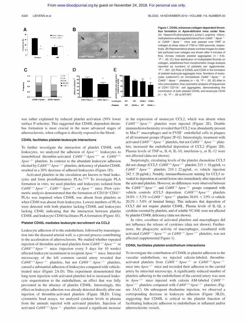
was rather explained by reduced platelet activation (50% lowersurface P-selectin). This suggested that CD40L-dependent throm-bus formation is most crucial in the more advanced stages ofatherosclerosis, when collagen is directly exposed to the blood.
CD40L facilitates platelet-leukocyte interactions
To further investigate the interaction of platelet CD40L withleukocytes, we analyzed the adhesion of Apoe�/� leukocytes toimmobilized thrombin-activated Cd40l�/�Apoe�/� or Cd40l�/�
Apoe�/� platelets. In contrast to the abundant leukocyte adhesionelicited by Cd40l�/�Apoe�/� platelets, deficiency of platelet CD40Lresulted in a 30% decrease of adhered leukocytes (Figure 1D).
Activated platelets in the circulation are known to bind leuko-cytes and form proinflammatory PLAs.23,42 To investigate PLAformation in vitro, we used platelets and leukocytes isolated fromCd40l�/�Apoe�/�, Cd40�/�Apoe�/�, or Apoe�/� mice. Flow cyto-metric analysis demonstrated that the formation of CD11b�CD41�
PLAs was impaired when CD40L was absent from platelets orwhen CD40 was absent from leukocytes. Lowest numbers of PLAswere detected with both platelets lacking CD40L and leukocyteslacking CD40, indicating that the interaction between plateletCD40L and leukocyte CD40 facilitates PLA formation (Figure 1E).
Platelet CD40L mediates leukocyte recruitment via CCL2
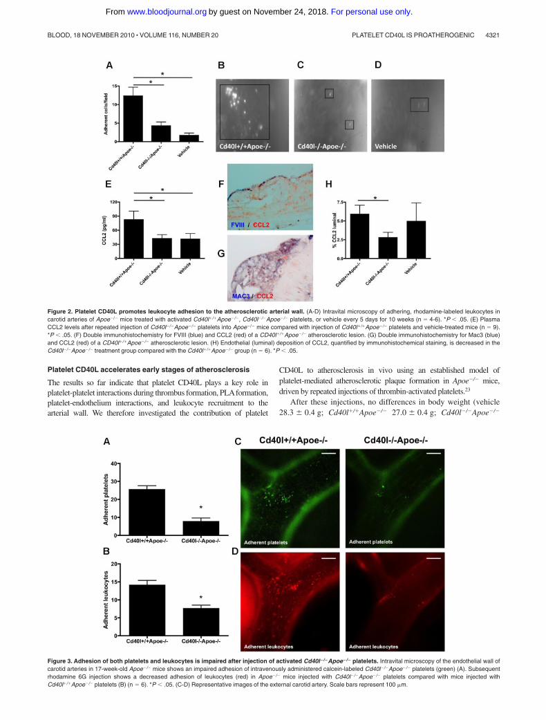
Leukocyte adhesion of to the endothelium, followed by transmigra-tion into the diseased arterial wall, is a pivotal process contributingto the acceleration of atherosclerosis. We studied whether repeatedinjection of thrombin-activated platelets from Cd40l�/�Apoe�/� orCd40l�/�Apoe�/� mice (injection every 5 days for 10 weeks)affected leukocyte recruitment in recipient Apoe�/� mice. Intravitalmicroscopy of the left common carotid artery revealed thatCd40l�/�Apoe�/� platelets, but not Cd40l�/�Apoe�/� platelets,caused a substantial adhesion of leukocytes compared with vehicle-treated mice (Figure 2A-D). This experiment demonstrated thatlong-term injection with activated platelets led to increased leuko-cyte sequestration to the endothelium, an effect that was fullyprevented in the absence of platelet CD40L. Interestingly, thiseffect on leukocyte adhesion was already detected directly after oneinjection of thrombin-activated platelets (Figure 3B,D). Usingcytometric bead assays, we analyzed cytokine levels in plasmafrom the animals injected with activated platelets. Injection ofactivated Cd40l�/�Apoe�/� platelets caused a significant increase
in the expression of monocyte CCL2, which was absent whenCd40l�/�Apoe�/� platelets were injected (Figure 2E). Doubleimmunohistochemistry revealed that CCL2 was abundantly presentin Mac3� macrophages and in FVIII� endothelial cells in plaquesof all treatment groups (Figure 2F-G). Interestingly, treatment withactivated Cd40l�/�Apoe�/�platelets, but not Cd40/�/�Apoe�/� plate-lets, increased the endothelial deposition of CCL2 (Figure 2H).Plasma levels of TNF-�, IL-6, IL-10, interferon-, or IL-12 werenot affected (data not shown).
Surprisingly, circulating levels of the platelet chemokine CCL5did not change (CCL5: Cd40l�/�Apoe�/� platelets 215 18 pg/mL vsCd40l�/�Apoe�/� platelets 210 22 pg/mL vs vehicle injection242 26 pg/mL). Notably, immunofluorescent staining for CCL5 re-vealed its deposition at carotid lesion sites immediately after injection ofthe activated platelets. However, no differences were observed betweenthe Cd40l�/�Apoe�/� and Cd40l�/�Apoe�/� groups compared withvehicle controls (CCL5 deposition: Cd40l�/�Apoe�/� platelets34.8% 5.3% vs Cd40l�/�Apoe�/� platelets 38.6% 3.9% vs vehicle20.2% 5.0% of luminal lining). This indicates that deposition ofCCL5 did not require platelet CD40L. Plasma levels of IL-1�, acytokine excreted by platelets, and of soluble VCAM, were not affectedby platelet CD40L deficiency (data not shown).
In vitro, coculture of activated platelets and macrophages didnot influence the release of cytokines (data not shown). Further-more, the phagocytic activity of macrophages, cocultured withactivated Cd40l�/�Apoe�/� or Cd40l�/�Apoe�/� platelets, was notaffected (supplemental Figure 1).
CD40L facilitates platelet-endothelium interactions
To investigate the contribution of CD40L to platelet adhesion to thevascular endothelium, we injected calcein-labeled, thrombin-activated platelets from Cd40l�/�Apoe�/� or Cd40l�/�Apoe�/�
mice into Apoe�/� mice and recorded their adhesion to the carotidartery by intravital microscopy. A significantly reduced number ofplatelets adhering to the endothelium of the carotid artery was seenin Apoe�/� mice injected with calcein AM-labeled Cd40l�/�
Apoe�/� platelets compared with Cd40l�/�Apoe�/� platelets (Fig-ure 3A,C). On subsequent rhodamine injection, we observed acorresponding decrease in leukocyte adhesion (Figure 3B,D),suggesting that CD40L is critical to the platelet function offacilitating leukocyte adhesion to endothelium in inflamed and/oratherosclerotic vessels.
Figure 1. CD40L enhances collagen-dependent throm-bus formation in Apoe-deficient mice under flow.(A) Heparin/D-phenylalanyl-L-prolyl-L-arginine chloro-methylketone-anticoagulated blood from Cd40l�/�Apoe�/�
or Cd40l�/�Apoe�/� mice was passed over VWF orcollagen at shear rates of 1700 or 1000 seconds, respec-tively. (B) Representative phase-contrast images for plate-lets perfused over collagen are shown after 4 minutes offlow. Arrows indicate platelet aggregates (n � 4-6).*P � .05. (C) Size distribution of multiplatelet thrombi oncollagen, established from morphometric image analysisreported as numbers of platelets per agglomerate.*P � .001. (D) Role of CD40L and CD40 in the formationof platelet leukocyte-aggregate mice. Numbers of leuko-cytes (cells/mm2) on immobilized Cd40l�/�Apoe�/� orCd40l�/�Apoe�/� platelets (n � 6). *P � .05. (E) After invitro coincubation, flow cytometric analysis of frequenciesof CD41�CD11b� cell aggregates, demonstrating thecontribution of both platelet CD40L and leukocyte CD40(n � 6). *P � .05 vs WT/WT.
4320 LIEVENS et al BLOOD, 18 NOVEMBER 2010 � VOLUME 116, NUMBER 20
For personal use only.on November 24, 2018. by guest www.bloodjournal.orgFrom
Platelet CD40L accelerates early stages of atherosclerosis
The results so far indicate that platelet CD40L plays a key role inplatelet-platelet interactions during thrombus formation, PLAformation,platelet-endothelium interactions, and leukocyte recruitment to thearterial wall. We therefore investigated the contribution of platelet
CD40L to atherosclerosis in vivo using an established model ofplatelet-mediated atherosclerotic plaque formation in Apoe�/� mice,driven by repeated injections of thrombin-activated platelets.23
After these injections, no differences in body weight (vehicle28.3 0.4 g; Cd40l�/�Apoe�/� 27.0 0.4 g; Cd40l�/�Apoe�/�
Figure 2. Platelet CD40L promotes leukocyte adhesion to the atherosclerotic arterial wall. (A-D) Intravital microscopy of adhering, rhodamine-labeled leukocytes incarotid arteries of Apoe�/� mice treated with activated Cd40l�/�Apoe�/�, Cd40l�/�Apoe�/� platelets, or vehicle every 5 days for 10 weeks (n � 4-6). *P � .05. (E) PlasmaCCL2 levels after repeated injection of Cd40l�/�Apoe�/� platelets into Apoe�/� mice compared with injection of Cd40l�/�Apoe�/� platelets and vehicle-treated mice (n � 9).*P � .05. (F) Double immunohistochemistry for FVIII (blue) and CCL2 (red) of a CD40l�/�Apoe�/� atherosclerotic lesion. (G) Double immunohistochemistry for Mac3 (blue)and CCL2 (red) of a CD40l�/�Apoe�/� atherosclerotic lesion. (H) Endothelial (luminal) deposition of CCL2, quantified by immunohistochemical staining, is decreased in theCd40l�/�Apoe�/� treatment group compared with the Cd40l�/�Apoe�/� group (n � 6). *P � .05.
Figure 3. Adhesion of both platelets and leukocytes is impaired after injection of activated Cd40l�/�Apoe�/� platelets. Intravital microscopy of the endothelial wall ofcarotid arteries in 17-week-old Apoe�/� mice shows an impaired adhesion of intravenously administered calcein-labeled Cd40l�/�Apoe�/� platelets (green) (A). Subsequentrhodamine 6G injection shows a decreased adhesion of leukocytes (red) in Apoe�/� mice injected with Cd40l�/�Apoe�/� platelets compared with mice injected withCd40l�/�Apoe�/� platelets (B) (n � 6). *P � .05. (C-D) Representative images of the external carotid artery. Scale bars represent 100 �m.
PLATELET CD40L IS PROATHEROGENIC 4321BLOOD, 18 NOVEMBER 2010 � VOLUME 116, NUMBER 20
For personal use only.on November 24, 2018. by guest www.bloodjournal.orgFrom
28.1 0.6 g) or in cholesterol levels (vehicle 328 28 mg/dL; Cd40l�/�
Apoe�/� 336 29 mg/dL; Cd40l�/�Apoe�/� 309 21 mg/dL) wereobserved. Moreover, no macroscopic or microscopic complications ofthe injections of activated platelets could be observed in more than20 organs analyzed, especially no signs of hemorrhage or thrombosis.
Platelet CD40L accelerates plaque development
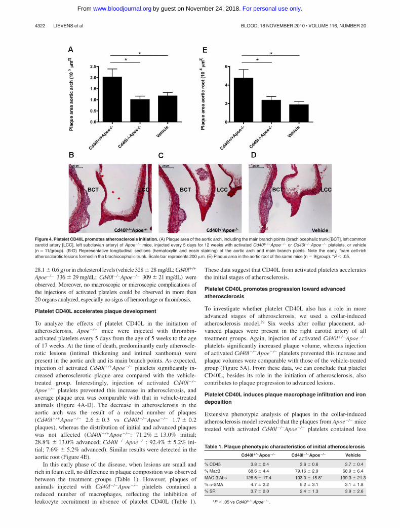
To analyze the effects of platelet CD40L in the initiation ofatherosclerosis, Apoe�/� mice were injected with thrombin-activated platelets every 5 days from the age of 5 weeks to the ageof 17 weeks. At the time of death, predominantly early atheroscle-rotic lesions (intimal thickening and intimal xanthoma) werepresent in the aortic arch and its main branch points. As expected,injection of activated Cd40l�/�Apoe�/� platelets significantly in-creased atherosclerotic plaque area compared with the vehicle-treated group. Interestingly, injection of activated Cd40l�/�
Apoe�/� platelets prevented this increase in atherosclerosis, andaverage plaque area was comparable with that in vehicle-treatedanimals (Figure 4A-D). The decrease in atherosclerosis in theaortic arch was the result of a reduced number of plaques(Cd40l�/�Apoe�/� 2.6 0.3 vs Cd40l�/�Apoe�/� 1.7 0.2plaques), whereas the distribution of initial and advanced plaqueswas not affected (Cd40l�/�Apoe�/�: 71.2% 13.0% initial;28.8% 13.0% advanced; Cd40l�/�Apoe�/�: 92.4% 5.2% ini-tial; 7.6% 5.2% advanced). Similar results were detected in theaortic root (Figure 4E).
In this early phase of the disease, when lesions are small andrich in foam cell, no difference in plaque composition was observedbetween the treatment groups (Table 1). However, plaques ofanimals injected with Cd40l�/�Apoe�/� platelets contained areduced number of macrophages, reflecting the inhibition ofleukocyte recruitment in absence of platelet CD40L (Table 1).
These data suggest that CD40L from activated platelets acceleratesthe initial stages of atherosclerosis.
Platelet CD40L promotes progression toward advancedatherosclerosis
To investigate whether platelet CD40L also has a role in moreadvanced stages of atherosclerosis, we used a collar-inducedatherosclerosis model.39 Six weeks after collar placement, ad-vanced plaques were present in the right carotid artery of alltreatment groups. Again, injection of activated Cd40l�/�Apoe�/�
platelets significantly increased plaque volume, whereas injectionof activated Cd40l�/�Apoe�/� platelets prevented this increase andplaque volumes were comparable with those of the vehicle-treatedgroup (Figure 5A). From these data, we can conclude that plateletCD40L, besides its role in the initiation of atherosclerosis, alsocontributes to plaque progression to advanced lesions.
Platelet CD40L induces plaque macrophage infiltration and irondeposition
Extensive phenotypic analysis of plaques in the collar-inducedatherosclerosis model revealed that the plaques from Apoe�/� micetreated with activated Cd40l�/�Apoe�/� platelets contained less
Figure 4. Platelet CD40L promotes atherosclerosis initiation. (A) Plaque area of the aortic arch, including the main branch points (brachiocephalic trunk [BCT], left commoncarotid artery [LCC], left subclavian artery) of Apoe�/� mice, injected every 5 days for 12 weeks with activated Cd40l�/�Apoe�/� or Cd40l�/�Apoe�/� platelets, or vehicle(n � 11/group). (B-D) Representative longitudinal sections (hematoxylin and eosin staining) of the aortic arch and main branch points. Note the early, foam cell-richatherosclerotic lesions formed in the brachiocephalic trunk. Scale bar represents 200 �m. (E) Plaque area in the aortic root of the same mice (n � 9/group). *P � .05.
Table 1. Plaque phenotypic characteristics of initial atherosclerosis
Cd40l�/�Apoe�/� Cd40l�/�Apoe�/� Vehicle
% CD45 3.8 0.4 3.6 0.6 3.7 0.4
% Mac3 68.6 4.4 79.16 2.9 68.9 6.4
MAC-3 Abs 126.6 17.4 103.0 15.8* 139.3 21.3
% �-SMA 4.7 2.2 5.2 3.1 3.1 1.8
% SR 3.7 2.0 2.4 1.3 3.9 2.6
*P � .05 vs Cd40l�/�Apoe�/�.
4322 LIEVENS et al BLOOD, 18 NOVEMBER 2010 � VOLUME 116, NUMBER 20
For personal use only.on November 24, 2018. by guest www.bloodjournal.orgFrom
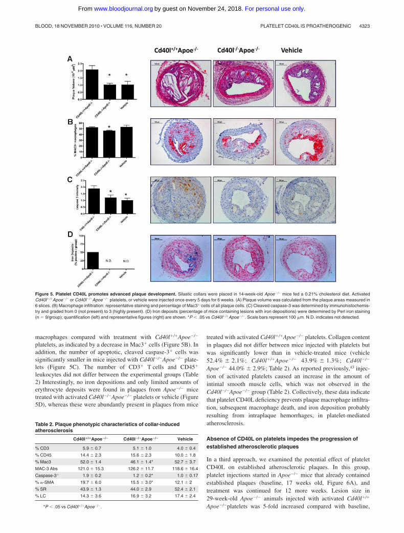
macrophages compared with treatment with Cd40l�/�Apoe�/�
platelets, as indicated by a decrease in Mac3� cells (Figure 5B). Inaddition, the number of apoptotic, cleaved caspase-3� cells wassignificantly smaller in mice injected with Cd40l�/�Apoe�/� plate-lets (Figure 5C). The number of CD3� T cells and CD45�
leukocytes did not differ between the experimental groups (Table2) Interestingly, no iron depositions and only limited amounts oferythrocyte deposits were found in plaques from Apoe�/� micetreated with activated Cd40l�/�Apoe�/� platelets or vehicle (Figure5D), whereas these were abundantly present in plaques from mice
treated with activated Cd40l�/�Apoe�/� platelets. Collagen contentin plaques did not differ between mice injected with platelets butwas significantly lower than in vehicle-treated mice (vehicle52.4% 2.1%; Cd40l�/�Apoe�/� 43.9% 1.3%; Cd40l�/�
Apoe�/� 44.0% 2.9%; Table 2). As reported previously,43 injec-tion of activated platelets caused an increase in the amount ofintimal smooth muscle cells, which was not observed in theCd40l�/�Apoe�/� group (Table 2). Collectively, these data indicatethat platelet CD40L deficiency prevents plaque macrophage infiltra-tion, subsequent macrophage death, and iron deposition probablyresulting from intraplaque hemorrhages, in platelet-mediatedatherosclerosis.
Absence of CD40L on platelets impedes the progression ofestablished atherosclerotic plaques
In a third approach, we examined the potential effect of plateletCD40L on established atherosclerotic plaques. In this group,platelet injections started in Apoe�/� mice that already containedestablished plaques (baseline, 17 weeks old, Figure 6A), andtreatment was continued for 12 more weeks. Lesion size in29-week-old Apoe�/� animals injected with activated Cd40l�/�
Apoe�/�platelets was 5-fold increased compared with baseline,
Figure 5. Platelet CD40L promotes advanced plaque development. Silastic collars were placed in 14-week-old Apoe�/� mice fed a 0.21% cholesterol diet. ActivatedCd40l�/�Apoe�/� or Cd40l�/�Apoe�/� platelets, or vehicle were injected once every 5 days for 6 weeks. (A) Plaque volume was calculated from the plaque areas measured in6 slices. (B) Macrophage infiltration: representative staining and percentage of Mac3� cells of all plaque cells. (C) Cleaved caspase-3 was determined by immunohistochemis-try and graded from 0 (not present) to 3 (highly present). (D) Iron deposits (percentage of mice containing lesions with iron depositions) were determined by Perl iron staining(n � 9/group); quantification (left) and representative figures (right) are shown. *P � .05 vs Cd40l�/�Apoe�/�. Scale bars represent 100 �m. N.D. indicates not detected.
Table 2. Plaque phenotypic characteristics of collar-inducedatherosclerosis
Cd40l�/�Apoe�/� Cd40l�/�Apoe�/� Vehicle
% CD3 5.9 0.7 5.1 1.0 4.0 0.4
% CD45 14.4 2.3 15.6 2.3 10.0 1.8
% Mac3 52.0 1.4 46.1 1.4* 52.7 3.7
MAC-3 Abs 121.0 15.3 126.2 11.7 118.6 16.4
Caspase-3� 1.9 0.2 1.2 0.2* 1.0 0.17
% �-SMA 19.7 6.0 15.5 3.0* 12.1 2
% SR 43.9 1.3 44.0 2.9 52.4 2.1
% LC 14.3 3.6 16.9 3.2 17.4 2.4
*P � .05 vs Cd40l�/�Apoe�/�.
PLATELET CD40L IS PROATHEROGENIC 4323BLOOD, 18 NOVEMBER 2010 � VOLUME 116, NUMBER 20
For personal use only.on November 24, 2018. by guest www.bloodjournal.orgFrom
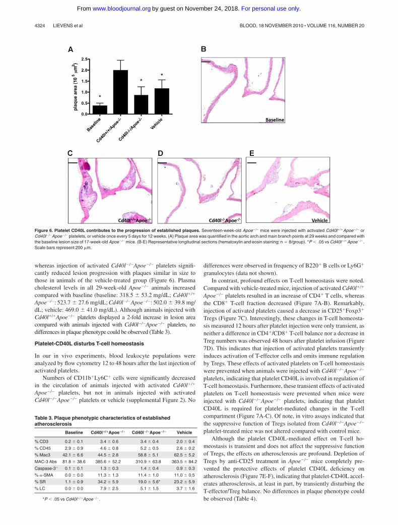
whereas injection of activated Cd40l�/�Apoe�/� platelets signifi-cantly reduced lesion progression with plaques similar in size tothose in animals of the vehicle-treated group (Figure 6). Plasmacholesterol levels in all 29-week-old Apoe�/� animals increasedcompared with baseline (baseline: 318.5 53.2 mg/dL; Cd40l�/�
Apoe�/�: 523.7 27.6 mg/dL; Cd40l�/�Apoe�/�: 502.0 39.8 mg/dL; vehicle: 469.0 41.0 mg/dL). Although animals injected withCd40l�/�Apoe�/� platelets displayed a 2-fold increase in lesion areacompared with animals injected with Cd40l�/�Apoe�/� platelets, nodifferences in plaque phenotype could be observed (Table 3).
Platelet-CD40L disturbs T-cell homeostasis
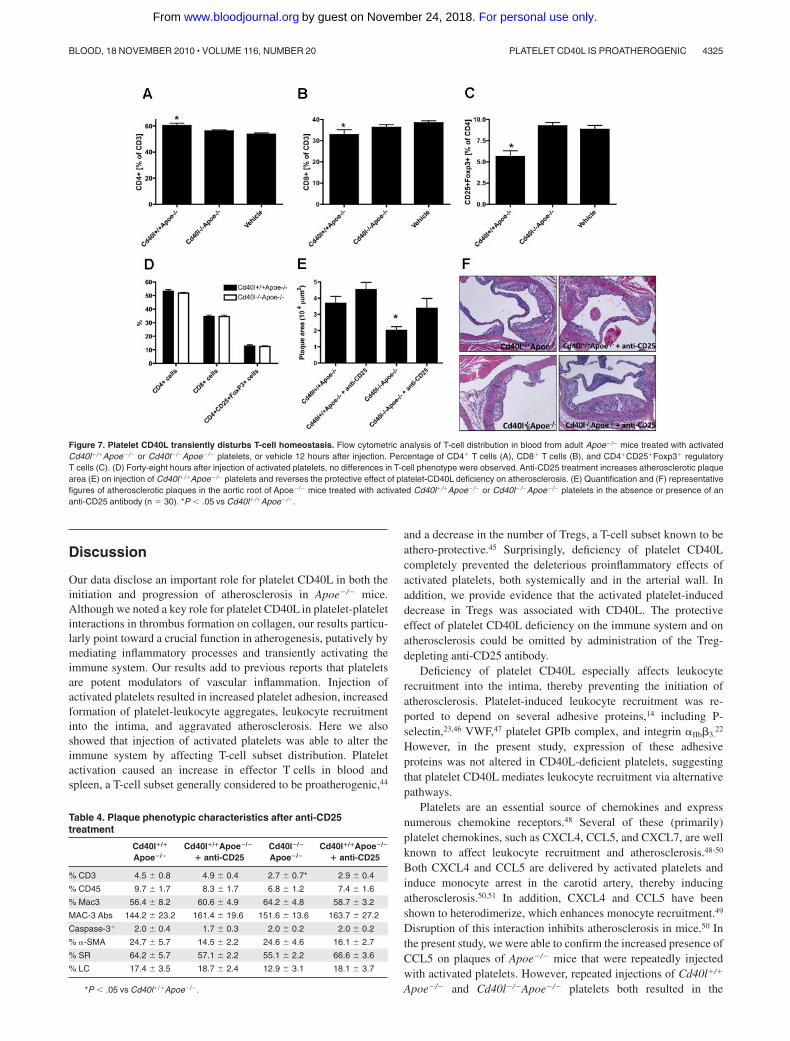
In our in vivo experiments, blood leukocyte populations wereanalyzed by flow cytometry 12 to 48 hours after the last injection ofactivated platelets.
Numbers of CD11b�Ly6C� cells were significantly decreasedin the circulation of animals injected with activated Cd40l�/�
Apoe�/� platelets, but not in animals injected with activatedCd40l�/�Apoe�/� platelets or vehicle (supplemental Figure 2). No
differences were observed in frequency of B220� B cells or Ly6G�
granulocytes (data not shown).In contrast, profound effects on T-cell homeostasis were noted.
Compared with vehicle-treated mice, injection of activated Cd40l�/�
Apoe�/� platelets resulted in an increase of CD4� T cells, whereasthe CD8� T-cell fraction decreased (Figure 7A-B). Remarkably,injection of activated platelets caused a decrease in CD25�Foxp3�
Tregs (Figure 7C). Interestingly, these changes in T-cell homeosta-sis measured 12 hours after platelet injection were only transient, asneither a difference in CD4�/CD8� T-cell balance nor a decrease inTreg numbers was observed 48 hours after platelet infusion (Figure7D). This indicates that injection of activated platelets transientlyinduces activation of T-effector cells and omits immune regulationby Tregs. These effects of activated platelets on T-cell homeostasiswere prevented when animals were injected with Cd40l�/�Apoe�/�
platelets, indicating that platelet CD40L is involved in regulation ofT-cell homeostasis. Furthermore, these transient effects of activatedplatelets on T-cell homeostasis were prevented when mice wereinjected with Cd40l�/�Apoe�/� platelets, indicating that plateletCD40L is required for platelet-mediated changes in the T-cellcompartment (Figure 7A-C). Of note, in vitro assays indicated thatthe suppressive function of Tregs isolated from Cd40l�/�Apoe�/�
platelet-treated mice was not altered compared with control mice.Although the platelet CD40L-mediated effect on T-cell ho-
meostasis is transient and does not affect the suppressive functionof Tregs, the effects on atherosclerosis are profound. Depletion ofTregs by anti-CD25 treatment in Apoe�/� mice completely pre-vented the protective effects of platelet CD40L deficiency onatherosclerosis (Figure 7E-F), indicating that platelet-CD40L accel-erates atherosclerosis, at least in part, by transiently disturbing theT-effector/Treg balance. No differences in plaque phenotype couldbe observed (Table 4).
Figure 6. Platelet CD40L contributes to the progression of established plaques. Seventeen-week-old Apoe�/� mice were injected with activated Cd40l�/�Apoe�/� orCd40l�/�Apoe�/� platelets, or vehicle once every 5 days for 12 weeks. (A) Plaque area was quantified in the aortic arch and main branch points at 29 weeks and compared withthe baseline lesion size of 17-week-old Apoe�/� mice. (B-E) Representative longitudinal sections (hematoxylin and eosin staining; n � 8/group). *P � .05 vs Cd40l�/�Apoe�/�.Scale bars represent 200 �m.
Table 3. Plaque phenotypic characteristics of establishedatherosclerosis
Baseline Cd40l�/�Apoe�/� Cd40l�/�Apoe�/� Vehicle
% CD3 0.2 0.1 3.4 0.6 3.4 0.4 2.0 0.4
% CD45 2.9 0.9 4.6 0.8 5.2 0.5 2.6 0.2
% Mac3 42.1 6.6 44.5 2.8 58.8 5.1 62.5 5.2
MAC-3 Abs 81.8 38.6 385.6 52.2 310.9 63.8 363.5 84.2
Caspase-3� 0.1 0.1 1.3 0.3 1.4 0.4 0.9 0.3
% �-SMA 0.0 0.0 11.3 1.3 11.4 1.0 11.0 0.5
% SR 1.1 0.9 34.2 5.9 19.0 5.6* 23.2 5.9
% LC 0.0 0.0 7.9 2.5 5.1 1.5 3.7 1.6
*P � .05 vs Cd40l�/�Apoe�/�.
4324 LIEVENS et al BLOOD, 18 NOVEMBER 2010 � VOLUME 116, NUMBER 20
For personal use only.on November 24, 2018. by guest www.bloodjournal.orgFrom
Discussion
Our data disclose an important role for platelet CD40L in both theinitiation and progression of atherosclerosis in Apoe�/� mice.Although we noted a key role for platelet CD40L in platelet-plateletinteractions in thrombus formation on collagen, our results particu-larly point toward a crucial function in atherogenesis, putatively bymediating inflammatory processes and transiently activating theimmune system. Our results add to previous reports that plateletsare potent modulators of vascular inflammation. Injection ofactivated platelets resulted in increased platelet adhesion, increasedformation of platelet-leukocyte aggregates, leukocyte recruitmentinto the intima, and aggravated atherosclerosis. Here we alsoshowed that injection of activated platelets was able to alter theimmune system by affecting T-cell subset distribution. Plateletactivation caused an increase in effector T cells in blood andspleen, a T-cell subset generally considered to be proatherogenic,44
and a decrease in the number of Tregs, a T-cell subset known to beathero-protective.45 Surprisingly, deficiency of platelet CD40Lcompletely prevented the deleterious proinflammatory effects ofactivated platelets, both systemically and in the arterial wall. Inaddition, we provide evidence that the activated platelet-induceddecrease in Tregs was associated with CD40L. The protectiveeffect of platelet CD40L deficiency on the immune system and onatherosclerosis could be omitted by administration of the Treg-depleting anti-CD25 antibody.
Deficiency of platelet CD40L especially affects leukocyterecruitment into the intima, thereby preventing the initiation ofatherosclerosis. Platelet-induced leukocyte recruitment was re-ported to depend on several adhesive proteins,14 including P-selectin,23,46 VWF,47 platelet GPIb complex, and integrin �IIb�3.
22
However, in the present study, expression of these adhesiveproteins was not altered in CD40L-deficient platelets, suggestingthat platelet CD40L mediates leukocyte recruitment via alternativepathways.
Platelets are an essential source of chemokines and expressnumerous chemokine receptors.48 Several of these (primarily)platelet chemokines, such as CXCL4, CCL5, and CXCL7, are wellknown to affect leukocyte recruitment and atherosclerosis.48-50
Both CXCL4 and CCL5 are delivered by activated platelets andinduce monocyte arrest in the carotid artery, thereby inducingatherosclerosis.50,51 In addition, CXCL4 and CCL5 have beenshown to heterodimerize, which enhances monocyte recruitment.49
Disruption of this interaction inhibits atherosclerosis in mice.50 Inthe present study, we were able to confirm the increased presence ofCCL5 on plaques of Apoe�/� mice that were repeatedly injectedwith activated platelets. However, repeated injections of Cd40l�/�
Apoe�/� and Cd40l�/�Apoe�/� platelets both resulted in the
Figure 7. Platelet CD40L transiently disturbs T-cell homeostasis. Flow cytometric analysis of T-cell distribution in blood from adult Apoe�/� mice treated with activatedCd40l�/�Apoe�/� or Cd40l�/�Apoe�/� platelets, or vehicle 12 hours after injection. Percentage of CD4� T cells (A), CD8� T cells (B), and CD4�CD25�Foxp3� regulatoryT cells (C). (D) Forty-eight hours after injection of activated platelets, no differences in T-cell phenotype were observed. Anti-CD25 treatment increases atherosclerotic plaquearea (E) on injection of Cd40l�/�Apoe�/� platelets and reverses the protective effect of platelet-CD40L deficiency on atherosclerosis. (E) Quantification and (F) representativefigures of atherosclerotic plaques in the aortic root of Apoe�/� mice treated with activated Cd40l�/�Apoe�/� or Cd40l�/�Apoe�/� platelets in the absence or presence of ananti-CD25 antibody (n � 30). *P � .05 vs Cd40l�/�Apoe�/�.
Table 4. Plaque phenotypic characteristics after anti-CD25treatment
Cd40l�/�
Apoe�/�Cd40l�/�Apoe�/�
� anti-CD25Cd40l�/�
Apoe�/�Cd40l�/�Apoe�/�
� anti-CD25
% CD3 4.5 0.8 4.9 0.4 2.7 0.7* 2.9 0.4
% CD45 9.7 1.7 8.3 1.7 6.8 1.2 7.4 1.6
% Mac3 56.4 8.2 60.6 4.9 64.2 4.8 58.7 3.2
MAC-3 Abs 144.2 23.2 161.4 19.6 151.6 13.6 163.7 27.2
Caspase-3� 2.0 0.4 1.7 0.3 2.0 0.2 2.0 0.2
% �-SMA 24.7 5.7 14.5 2.2 24.6 4.6 16.1 2.7
% SR 64.2 5.7 57.1 2.2 55.1 2.2 66.6 3.6
% LC 17.4 3.5 18.7 2.4 12.9 3.1 18.1 3.7
*P � .05 vs Cd40l�/�Apoe�/�.
PLATELET CD40L IS PROATHEROGENIC 4325BLOOD, 18 NOVEMBER 2010 � VOLUME 116, NUMBER 20
For personal use only.on November 24, 2018. by guest www.bloodjournal.orgFrom
accumulation of CCL5 on the plaque, suggesting that this phenom-enon is independent of CD40L. Although the paper of Huo et al23
considered CCL5 to be the key mechanism of platelet inducedatherosclerosis, we found that there are more (CD40L-dependent)factors, such as endothelial deposition of CCL2, a chemokinecrucial in monocyte recruitment throughout atherogenesis,52,53 andsystemic disruption of T-cell homeostasis.
Besides leukocyte recruitment, we found that CD40-CD40Linteractions are involved in the formation of proinflammatoryPLAs. PLAs can form when platelet P-selectin interacts withleukocyte P-selectin glycoprotein ligand.23,27 Until now, only fewmolecules are known that affect PLA formation and consequentlyprevent their proinflammatory actions. One of these molecules isgrowth arrest specific gene 6 (Gas6), which prevents PLA forma-tion by down-regulating P-selectin expression on platelets.54 Inter-estingly, it has also been reported that administration of sCD40Lincreased platelet P-selectin expression and, consequently, PLAformation in human and murine platelets.55,56 We noted a dimin-ished P-selectin exposure in CD40L-deficient thrombi on collagen,which may suggest a P-selection-dependent role of CD40-CD40Linteractions in the formation of PLAs.
Compared with complete deficiency in CD40L (Cd40l�/�
Apoe�/� mice),6 the impact of deficiency of platelet CD40L aloneon atherosclerosis is slightly less marked. However, deficiency ofplatelet CD40L still attenuates atherosclerosis progression, inducesa plaque phenotype that is low in inflammation, and contains noiron deposition, indicative of absence of intraplaque hemorrhage,but fails to induce true fibrotic plaques. This may be becauseactivated platelets impede the synthesis of collagen in the plaqueand induce the proliferation and migration of smooth musclecells,43 effects that are apparently not regulated by CD40L.However, deficiency of platelet CD40L does not result in immunesuppression and does not cause hemorrhage or bleeding in ourstudy, thereby suggesting a potential use of platelet CD40Linhibition as target in the treatment of human atherosclerosis.
The extrapolation of the animal model we used to characterizeplatelet CD40L to human vascular disease should be applied withcaution. Although patients with cardiovascular disease have anincreased number of activated platelets in their circulation,57
repeated injections of thrombin-activated platelets are not physi-ologic. However, until platelet-specific CD40L-deficient mice are
available, the currently used model is one of the best to study therole of CD40L in platelet biology in vivo in a chronic inflammatorydisease, such as atherosclerosis.
In conclusion, platelet CD40L is a potent inducer of proathero-genic inflammatory processes by enhancing the interaction be-tween platelets, leukocytes, and the endothelium. In addition,platelet CD40L disrupts T-cell homeostasis, thereby further promot-ing atherogenesis.
Acknowledgments
This work was supported by the Humboldt Foundation (SofjaKovalevskaja grant, E.L.), The Netherlands Organization forScientific Research (VIDI grant, E.L.), The Netherlands HeartFoundation (D.L. and Dr E. Dekker postdoc and establishedinvestigator grant, E.L.), the German Research Foundation DFG(FOR809, WE1913/7-2, and 10-1, C.W.; and ZE827/1-1, A.Z.),and the Interdisciplinary Center for Clinical Research BIOMATwithin the Faculty of Medicine at the Rheinisch-WestfalischeTechnische Hochschule Aachen University (VV-B113, C.W., A.Z.).
Authorship
Contribution: D.L. designed and performed research, analyzeddata, and wrote the paper; A.Z. and T.S. performed research andanalyzed data; O.S. designed experiments, performed research, andanalyzed data; L.B., I.C.A.M., E.W., P.G., R.v.K., and L.T.performed research; L.B., R.A.F., and R.J.N. contributed vital newreagents or analytical tools; N.G. designed experiments, performedresearch, and analyzed data; E.A.B., M.J.A.P.D., J.W.M.H., andC.W. designed research; and E.L. designed research and wrotethe paper.
Conflict-of-interest disclosure: The authors declare no compet-ing financial interests.
Correspondence: Esther Lutgens, Department of Pathology,Cardiovascular Research Institute Maastricht, University of Maas-tricht, P. Debeyelaan 25, 6229 HX, Maastricht, The Netherlands;e-mail: [email protected] or [email protected].
References
1. Schonbeck U, Libby P. CD40 signaling andplaque instability. Circ Res. 2001;89(12):1092-1103.
2. Schonbeck U, Libby P. The CD40/CD154 recep-tor/ligand dyad. Cell Mol Life Sci. 2001;58(1):4-43.
3. Weber C, Zernecke A, Libby P. The multifacetedcontributions of leukocyte subsets to atheroscle-rosis: lessons from mouse models. Nat Rev Im-munol. 2008;8(10):802-815.
4. Galkina E, Ley K. Immune and inflammatorymechanisms of atherosclerosis. Annu Rev Immu-nol. 2009;27:165-197.
5. Mach F, Schonbeck U, Sukhova GK, Atkinson E,Libby P. Reduction of atherosclerosis in mice byinhibition of CD40 signalling. Nature. 1998;394(6689):200-203.
6. Lutgens E, Gorelik L, Daemen MJ, et al. Require-ment for CD154 in the progression of atheroscle-rosis. Nat Med. 1999;5(11):1313-1316.
7. Lutgens E, Cleutjens KB, Heeneman S,Koteliansky VE, Burkly LC, Daemen MJ. Bothearly and delayed anti-CD40L antibody treatment
induces a stable plaque phenotype. Proc NatlAcad Sci U S A. 2000;97(13):7464-7469.
8. Schonbeck U, Sukhova GK, Shimizu K, Mach F,Libby P. Inhibition of CD40 signaling limits evolu-tion of established atherosclerosis in mice. ProcNatl Acad Sci U S A. 2000;97(13):7458-7463.
9. Donners MM, Beckers L, Lievens D, et al. TheCD40-TRAF6 axis is the key regulator of theCD40/CD40L system in neointima formation andarterial remodeling. Blood. 2008;111(9):4596-4604.
10. Lutgens E, Lievens D, Beckers L, et al. DeficientCD40-TRAF6 signaling in leukocytes preventsatherosclerosis by skewing the immune responsetoward an antiinflammatory profile. J Exp Med.2010;207(2):391-404.
11. Henn V, Slupsky JR, Grafe M, et al. CD40 ligandon activated platelets triggers an inflammatoryreaction of endothelial cells. Nature. 1998;391(6667):591.
12. Andre P, Prasad KS, Denis CV, et al. CD40L sta-bilizes arterial thrombi by a beta3 integrin-depen-dent mechanism. Nat Med. 2002;8(3):247-252.
13. Andre P, Nannizzi-Alaimo L, Prasad SK, Phillips DR.
Platelet-derived CD40L: the switch-hitting player ofcardiovascular disease. Circulation. 2002;106(8):896-899.
14. von Hundelshausen P, Weber C. Platelets as im-mune cells: bridging inflammation and cardiovas-cular disease. Circ Res. 2007;100(1):27-40.
15. Gawaz M, Langer H, May AE. Platelets in inflam-mation and atherogenesis. J Clin Invest. 2005;115(12):3378-3384.
16. May AE, Seizer P, Gawaz M. Platelets: inflamma-tory firebugs of vascular walls. ArteriosclerThromb Vasc Biol. 2008;28(3):5-10.
17. Langer HF, Gawaz M. Platelet-vessel wall inter-actions in atherosclerotic disease. Thromb Hae-most. 2008;99(3):480-486.
18. Hawrylowicz CM, Howells GL, Feldmann M.Platelet-derived interleukin 1 induces human en-dothelial adhesion molecule expression and cyto-kine production. J Exp Med. 1991;174(4):785-790.
19. Miller DL, Yaron R, Yellin MJ. CD40L-CD40 inter-actions regulate endothelial cell surface tissuefactor and thrombomodulin expression. J LeukocBiol. 1998;63(3):373-379.
4326 LIEVENS et al BLOOD, 18 NOVEMBER 2010 � VOLUME 116, NUMBER 20
For personal use only.on November 24, 2018. by guest www.bloodjournal.orgFrom
20. Kotowicz K, Dixon GL, Klein NJ, Peters MJ,Callard RE. Biological function of CD40 on hu-man endothelial cells: costimulation with CD40ligand and interleukin-4 selectively induces ex-pression of vascular cell adhesion molecule-1and P-selectin resulting in preferential adhesionof lymphocytes. Immunology. 2000;100(4):441-448.
21. von Hundelshausen P, Weber KS, Huo Y, et al.RANTES deposition by platelets triggers mono-cyte arrest on inflamed and atherosclerotic endo-thelium. Circulation. 2001;103(13):1772-1777.
22. Massberg S, Brand K, Gruner S, et al. A criticalrole of platelet adhesion in the initiation of athero-sclerotic lesion formation. J Exp Med. 2002;196(7):887-896.
23. Huo Y, Schober A, Forlow SB, et al. Circulatingactivated platelets exacerbate atherosclerosis inmice deficient in apolipoprotein E. Nat Med.2003;9(1):61.
24. Weber C. Platelets and chemokines in athero-sclerosis: partners in crime. Circ Res. 2005;96(6):612-616.
25. da Costa Martins P, van den Berk N, Ulfman LH,Koenderman L, Hordijk PL, Zwaginga JJ. Plate-let-monocyte complexes support monocyte adhe-sion to endothelium by enhancing secondarytethering and cluster formation. ArteriosclerThromb Vasc Biol. 2004;24(1):193-199.
26. Rinder HM, Bonan JL, Rinder CS, Ault KA,Smith BR. Dynamics of leukocyte-platelet adhe-sion in whole blood. Blood. 1991;78(7):1730-1737.
27. van Gils JM, da Costa Martins PA, Mol A,Hordijk PL, Zwaginga JJ. Transendothelial migra-tion drives dissociation of platelet-monocyte com-plexes. Thromb Haemost. 2008;100(2):271-279.
28. van Gils JM, Zwaginga JJ, Hordijk PL. Molecularand functional interactions among monocytes,platelets, and endothelial cells and their rel-evance for cardiovascular diseases. J LeukocBiol. 2009;85(2):195-204.
29. Lippi G, Montagnana M, Salvagno GL, et al. Riskstratification of patients with acute myocardialinfarction by quantification of circulating mono-cyte-platelet aggregates. Int J Cardiol. 2007;115(1):101-102.
30. Furman MI, Barnard MR, Krueger LA, et al. Circu-lating monocyte-platelet aggregates are an earlymarker of acute myocardial infarction. J Am CollCardiol. 2001;38(4):1002-1006.
31. Crittenden JR, Bergmeier W, Zhang Y, et al.CalDAG-GEFI integrates signaling for plateletaggregation and thrombus formation. Nat Med.2004;10(9):982-986.
32. Kuijpers MJ, Schulte V, Bergmeier W, et al.Complementary roles of glycoprotein VI and
alpha2beta1 integrin in collagen-induced throm-bus formation in flowing whole blood ex vivo.FASEB J. 2003;17(6):685-687.
33. Munnix IC, Strehl A, Kuijpers MJ, et al. The glyco-protein VI-phospholipase Cgamma2 signalingpathway controls thrombus formation induced bycollagen and tissue factor in vitro and in vivo. Ar-terioscler Thromb Vasc Biol. 2005;25(12):2673-2678.
34. Elvers M, Stegner D, Hagedorn I, et al. Impairedalpha(IIb)beta(3) integrin activation and shear-dependent thrombus formation in mice lackingphospholipase D1. Sci Signal. 3(103):ra1.
35. Heemskerk JW, Vuist WM, Feijge MA,Reutelingsperger CP, Lindhout T. Collagen butnot fibrinogen surfaces induce bleb formation,exposure of phosphatidylserine, and procoagu-lant activity of adherent platelets: evidence forregulation by protein tyrosine kinase-dependentCa2� responses. Blood. 1997;90(7):2615-2625.
36. Siljander PR, Munnix IC, Smethurst PA, et al.Platelet receptor interplay regulates collagen-induced thrombus formation in flowing humanblood. Blood. 2004;103(4):1333-1341.
37. Zernecke A, Liehn EA, Fraemohs L, et al. Impor-tance of junctional adhesion molecule-A for neo-intimal lesion formation and infiltration in athero-sclerosis-prone mice. Arterioscler Thromb VascBiol. 2006;26(2):e10-e13.
38. Bernhagen J, Krohn R, Lue H, et al. MIF is a non-cognate ligand of CXC chemokine receptors ininflammatory and atherogenic cell recruitment.Nat Med. 2007;13(5):587-596.
39. von der Thusen JH, van Berkel TJC, Biessen EAL.Induction of rapid atherogenesis by perivascular ca-rotid collar placement in apolipoprotein E-deficientand low-density lipoprotein receptor-deficient mice.Circulation. 2001;103(8):1164-1170.
40. Virmani R, Kolodgie FD, Burke AP, Farb A,Schwartz SM. Lessons from sudden coronarydeath: a comprehensive morphological classifica-tion scheme for atherosclerotic lesions. Arterio-scler Thromb Vasc Biol. 2000;20(5):1262-1275.
41. Kanters E, Pasparakis M, Gijbels MJ, et al. Inhibi-tion of NF-kappaB activation in macrophages in-creases atherosclerosis in LDL receptor-deficientmice. J Clin Invest. 2003;112(8):1176-1185.
42. Furman MI, Benoit SE, Barnard MR, et al. In-creased platelet reactivity and circulating mono-cyte-platelet aggregates in patients with stablecoronary artery disease. J Am Coll Cardiol. 1998;31(2):352-358.
43. Massberg S, Vogt F, Dickfeld T, Brand K, Page S,Gawaz M. Activated platelets trigger an inflamma-tory response and enhance migration of aorticsmooth muscle cells. Thromb Res. 2003;110(4):187-194.
44. Zhou X, Nicoletti A, Elhage R, Hansson GK.Transfer of CD4(�) T cells aggravates athero-sclerosis in immunodeficient apolipoprotein Eknockout mice. Circulation. 2000;102(24):2919-2922.
45. Ait-Oufella H, Salomon BL, Potteaux S, et al.Natural regulatory T cells control the develop-ment of atherosclerosis in mice. Nat Med. 2006;12(2):178-180.
46. Huo Y, Ley KF. Role of platelets in the develop-ment of atherosclerosis. Trends Cardiovasc Med.2004;14(1):18.
47. Theilmeier G, Michiels C, Spaepen E, et al. Endo-thelial von Willebrand factor recruits platelets toatherosclerosis-prone sites in response to hyper-cholesterolemia. Blood. 2002;99(12):4486-4493.
48. Gleissner CA, von Hundelshausen P, Ley K.Platelet chemokines in vascular disease. Arterio-scler Thromb Vasc Biol. 2008;28(11):1920-1927.
49. von Hundelshausen P, Koenen RR, Sack M, et al.Heterophilic interactions of platelet factor 4 andRANTES promote monocyte arrest on endothe-lium. Blood. 2005;105(3):924-930.
50. Koenen RR, von Hundelshausen P, Nesmelova IV,et al. Disrupting functional interactions betweenplatelet chemokines inhibits atherosclerosis in hyper-lipidemic mice. Nat Med. 2009;15(1):97-103.
51. von Hundelshausen P, Weber KSC, Huo Y, et al.RANTES deposition by platelets triggers mono-cyte arrest on inflamed and atherosclerotic endo-thelium. Circulation. 2001;103(13):1772-1777.
52. Boring L, Gosling J, Cleary M, Charo IF. De-creased lesion formation in CCR2�/� mice re-veals a role for chemokines in the initiation of ath-erosclerosis. Nature. 1998;394(6696):894-897.
53. Lutgens E, Faber B, Schapira K, et al. Gene pro-filing in atherosclerosis reveals a key role forsmall inducible cytokines: validation using a novelmonocyte chemoattractant protein monoclonalantibody. Circulation. 2005;111(25):3443-3452.
54. Tjwa M, Bellido-Martin L, Lin Y, et al. Gas6 pro-motes inflammation by enhancing interactionsbetween endothelial cells, platelets, and leuko-cytes. Blood. 2008;111(8):4096-4105.
55. Chakrabarti S, Varghese S, Vitseva O,Tanriverdi K, Freedman JE. CD40 ligand influ-ences platelet release of reactive oxygen inter-mediates. Arterioscler Thromb Vasc Biol. 2005;25(11):2428-2434.
56. Li G, Sanders JM, Bevard MH, et al. CD40 ligandpromotes Mac-1 expression, leukocyte recruit-ment, and neointima formation after vascular in-jury. Am J Pathol. 2008;172(4):1141-1152.
57. Fitzgerald DJ, Roy L, Catella F, FitzGerald GA.Platelet activation in unstable coronary disease.N Engl J Med. 1986;315(16):983-989.
PLATELET CD40L IS PROATHEROGENIC 4327BLOOD, 18 NOVEMBER 2010 � VOLUME 116, NUMBER 20
For personal use only.on November 24, 2018. by guest www.bloodjournal.orgFrom
online August 12, 2010 originally publisheddoi:10.1182/blood-2010-01-261206
2010 116: 4317-4327
Heemskerk, Christian Weber and Esther LutgensFlavell, Randolph J. Noelle, Norbert Gerdes, Erik A. Biessen, Mat J. A. P. Daemen, Johan W. M.Erwin Wijnands, Pieter Goossens, Roger van Kruchten, Larissa Thevissen, Louis Boon, Richard A. Dirk Lievens, Alma Zernecke, Tom Seijkens, Oliver Soehnlein, Linda Beckers, Imke C. A. Munnix, atherosclerosisPlatelet CD40L mediates thrombotic and inflammatory processes in
http://www.bloodjournal.org/content/116/20/4317.full.htmlUpdated information and services can be found at:
(1198 articles)Thrombosis and Hemostasis (814 articles)Platelets and Thrombopoiesis
(5640 articles)Immunobiology and Immunotherapy Articles on similar topics can be found in the following Blood collections
http://www.bloodjournal.org/site/misc/rights.xhtml#repub_requestsInformation about reproducing this article in parts or in its entirety may be found online at:
http://www.bloodjournal.org/site/misc/rights.xhtml#reprintsInformation about ordering reprints may be found online at:
http://www.bloodjournal.org/site/subscriptions/index.xhtmlInformation about subscriptions and ASH membership may be found online at:
Copyright 2011 by The American Society of Hematology; all rights reserved.of Hematology, 2021 L St, NW, Suite 900, Washington DC 20036.Blood (print ISSN 0006-4971, online ISSN 1528-0020), is published weekly by the American Society
For personal use only.on November 24, 2018. by guest www.bloodjournal.orgFrom