permanent canines. introduction “cornerstones of the mouth” - 3rd from midline (“eye-teeth”)...
TRANSCRIPT

Permanent CaninesPermanent Canines

IntroductionIntroduction
• “Cornerstones of the mouth” - 3rd from midline (“eye-teeth”)
• Situated between incisors and premolars
• Form: single cusp (middle labial lobe)• Function:
– Occlusion - piercing, tearing, lateral guidance
– Esthetics - facial support– Phonetics - speech
• Greatest combined crown-root length(in either arch)

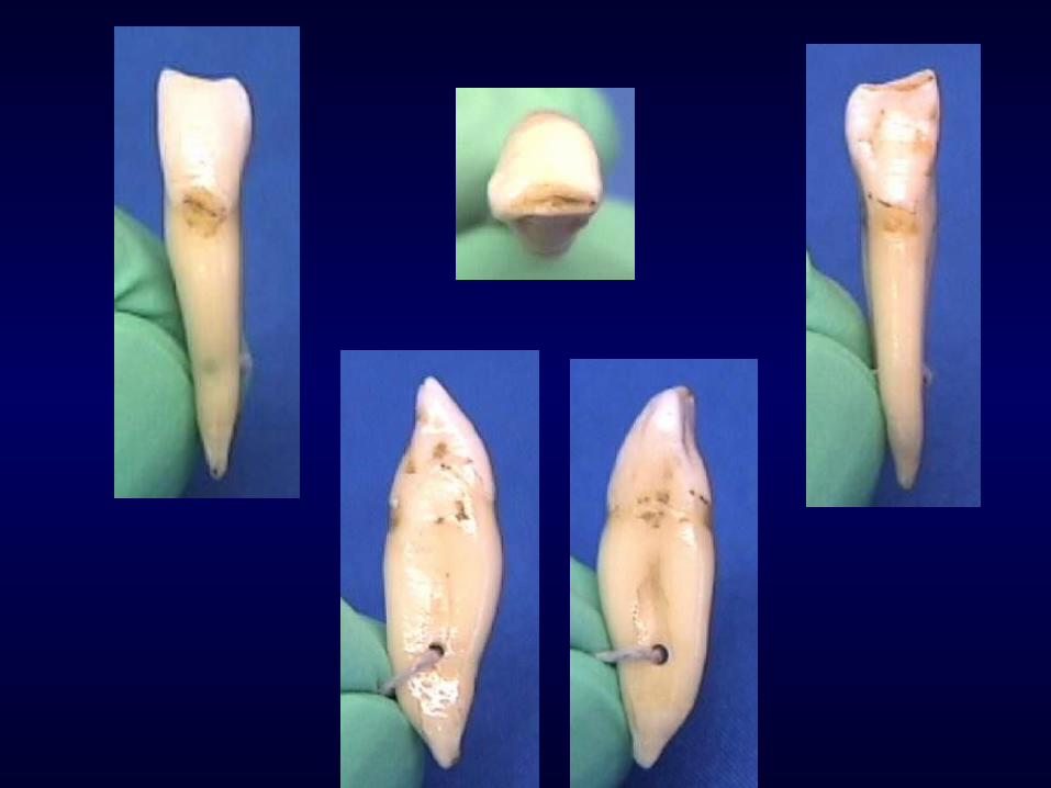
Permanent Maxillary Permanent Maxillary CanineCanine

General Characteristics:General Characteristics:
• Arch position– 3rd from midline– Between anteriors and posteriors
• Universal #6 and #11• Single facial cusp• Function: tearing, piercing, esthetics, and occlusion

Canine vs CentralCanine vs Central
• Crown length almost the same
• M-D, canine narrower (by 1 mm)• F-L, canine wider (by 1 mm)
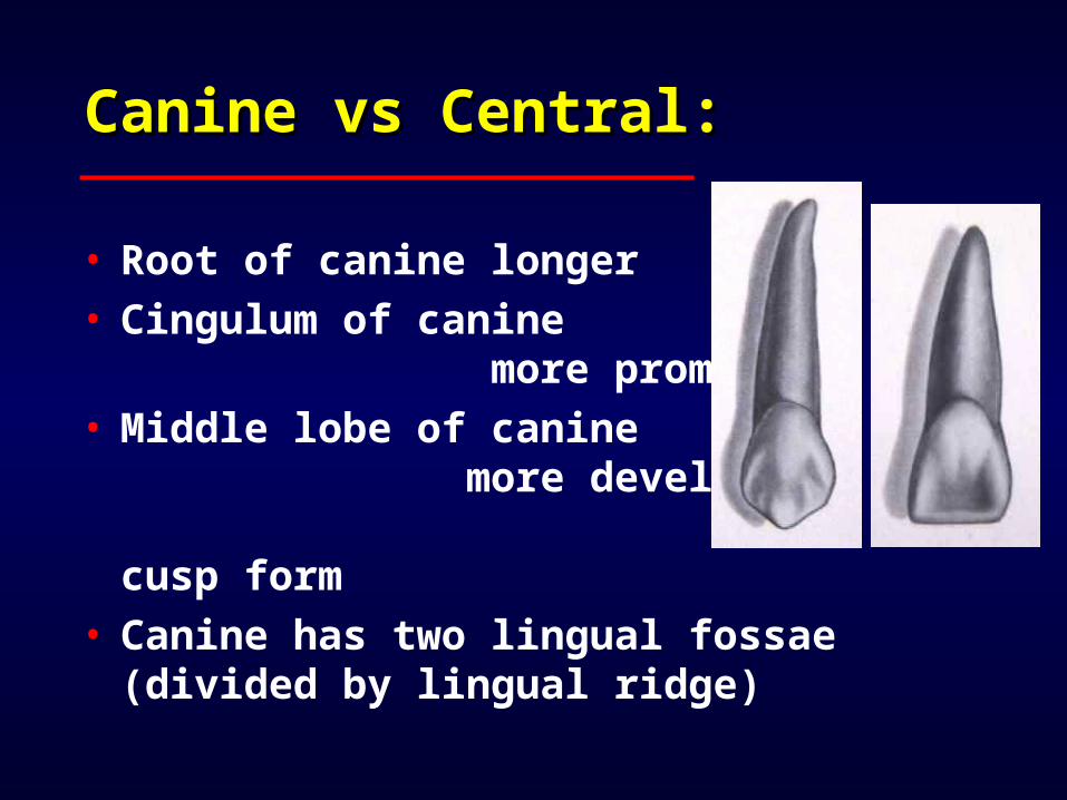
Canine vs Central:Canine vs Central:
• Root of canine longer• Cingulum of canine more prominent
• Middle lobe of canine more developed - cusp form
• Canine has two lingual fossae (divided by lingual
ridge)

Development Timeline:Development Timeline:
• Initial calcification: 4 - 5 months
• Enamel completed: 6 - 7 years• Eruption: 11 - 12 years• Root completion: 13 - 15 years
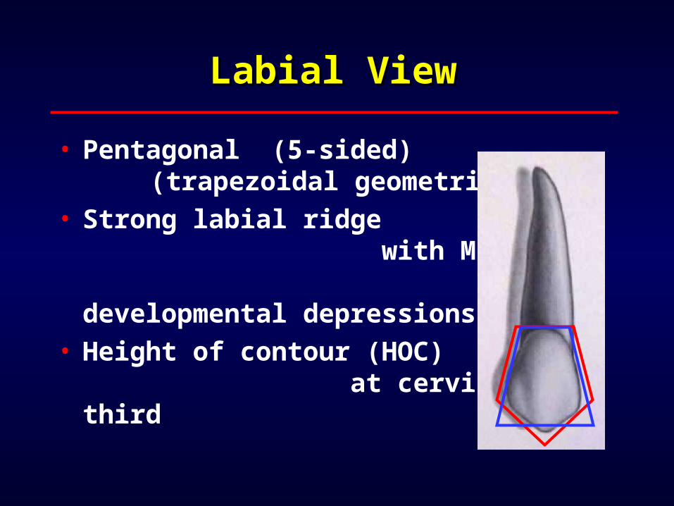
Labial ViewLabial View
• Pentagonal (5-sided) (trapezoidal geometric form)
• Strong labial ridge with MF and DF developmental depressions
• Height of contour (HOC) at cervical third

Labial view - mesial Labial view - mesial outline:outline:
• Generally convex from mesial HOC to cervical
• Contact area (HOC) at junction of incisal-middle third

Labial view - distal Labial view - distal outline:outline:
• Distal HOC at middle third
• Shorter than mesial outline from HOC to cervical
• Distal bulge with slight cervical concavity
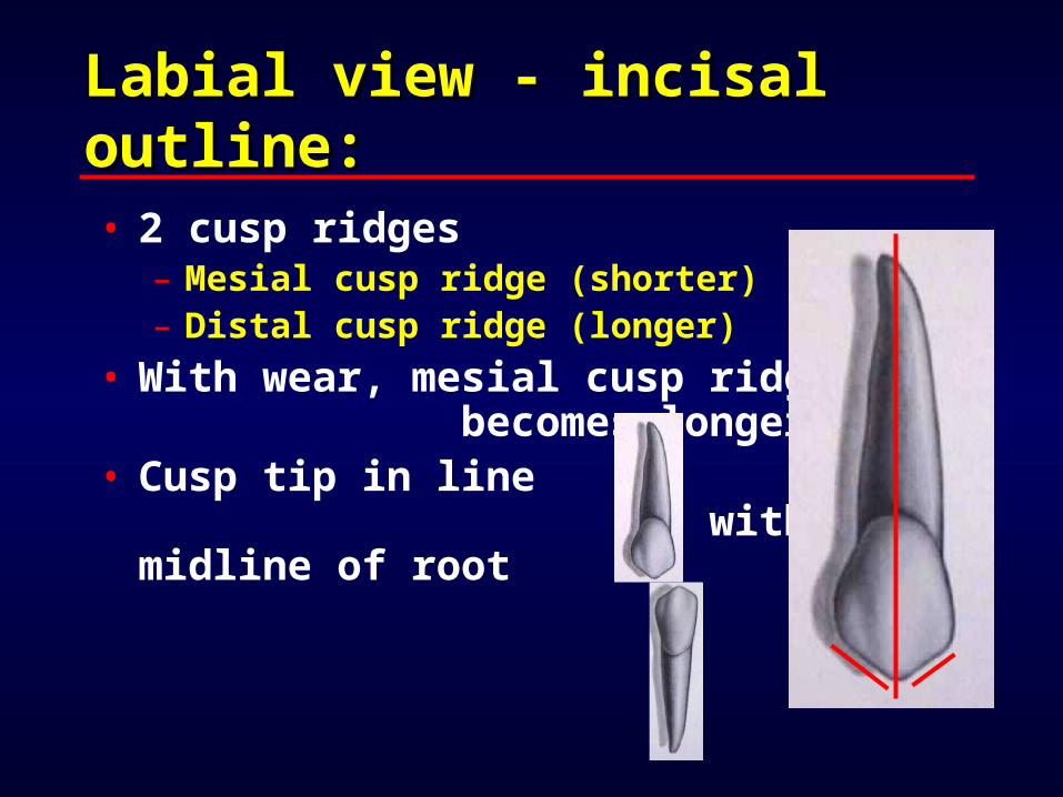
Labial view - incisal Labial view - incisal outline:outline:• 2 cusp ridges
– Mesial cusp ridge (shorter)– Distal cusp ridge (longer)
• With wear, mesial cusp ridge becomes longer*
• Cusp tip in line with midline of root

Lingual ViewLingual View
• M, D, and I outlines similar to labial view
• Bulky cingulum (bulkiest of anteriors)
• Ridges:– M and D marginal ridges– Lingual ridge
• Fossae:– ML and DL fossa

Lingual view:Lingual view:
• Uncommonly, linguogingival groove and lingual pit present
• HOC at cervical third• Cervical line slight distal offset

Mesial ViewMesial View
• Triangular crown• Wider F-L than incisors* (widest F-L of all anteriors)
• Cusp tip located just facial to long axis
• Labial outline convex, HOC at cervical third

Mesial view:Mesial view:
• Lingual outline convex at gingival 1/2 and concave at incisal 1/2
• Lingual HOC at cervical third• Mesial HOC at junction of incisal-middle thirds

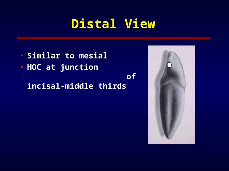
Distal ViewDistal View
• Cervical 1/2 usually concave• CE line curvature less than mesial
• HOC at middle third (most cervical of anteriors)

Incisal ViewIncisal View
• Asymmetrical diamond shape outline• Mesial half bulkier• Distal half appears “drawn” out• Cingulum offset to distal• Four cusp ridges:
– Mesial cusp ridge– Distal cusp ridge– Facial cusp ridge– Lingual cusp ridge
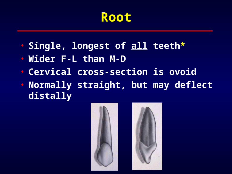
RootRoot
• Single, longest of all teeth*• Wider F-L than M-D• Cervical cross-section is ovoid• Normally straight, but may deflect distally

Permanent Mandibular Permanent Mandibular CanineCanine
• Universal #22 and #27• General form and function similar to maxillary canine

Comparisons with Comparisons with maxillary canine:maxillary canine:
• Crown as long or longer (by 1 mm)• M-D and F-L dimensions smaller (by .5 mm)

Mandibular vs Maxillary Mandibular vs Maxillary Canine:Canine:
• Total length about same*• Root length slightly shorter*• Lingual anatomy less developed

Development Timeline:Development Timeline:
• Initial calcification: 4 - 5 months
• Enamel completed: 6 - 7 years• Eruption: 9 - 10 years• Root completed: 12 - 14 years
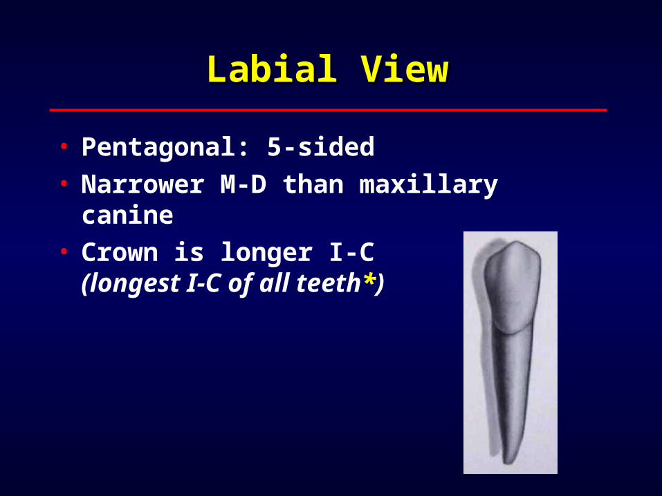
Labial ViewLabial View
• Pentagonal: 5-sided• Narrower M-D than maxillary canine
• Crown is longer I-C (longest I-C of all teeth*)
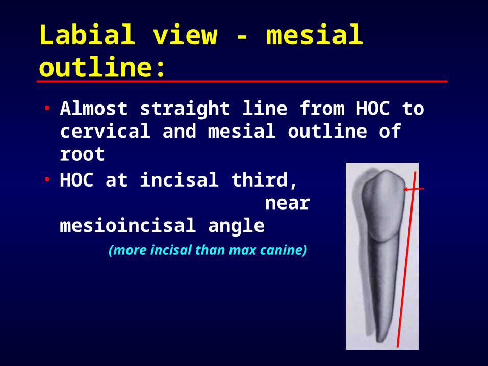
Labial view - mesial Labial view - mesial outline:outline:• Almost straight line from HOC to cervical and mesial outline of root
• HOC at incisal third, near mesioincisal angle
(more incisal than max canine)

Labial view - distal Labial view - distal outline:outline:
• More convex than mesial• Rounded distoincisal corner• HOC at junction of incisal-middle thirds

Labial view - incisal Labial view - incisal outline:outline:• D cusp ridge longer than M• With wear, D cusp ridge becomes longer*
• Cusp tip in line with root midline
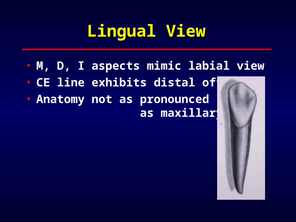
Lingual ViewLingual View
• M, D, I aspects mimic labial view• CE line exhibits distal offset• Anatomy not as pronounced as maxillary

Mesial ViewMesial View
• Labial outline convex, HOC at cervical third
• Cusp tip is lingual to root midline
• Labial outline “moonshape”

Mesial view:Mesial view:
• Lingual outline less cingulum curvature, HOC cervical third
• Contact area at incisal third
Distal ViewDistal View
• Similar to mesial• HOC at junction of incisal-middle thirds

Incisal ViewIncisal View
• Similar to maxillary canine• Bulky mesial half• Distal appears “pulled” or “pinched”
• Cingulum offset to distal

RootRoot
• Longest root in mandibular arch*• Single, straight - bifurcation possible
• Wider F-L than M-D• Cervical cross-section is “flattened” ovoid

How To Tell Maxillary How To Tell Maxillary Canine from Mandibular:Canine from Mandibular:
• M-D dimension of maxillary wider • Mesial outline of mandibular straight
• Facial outline of mandibular appears “moonshape” - location of cusp tip
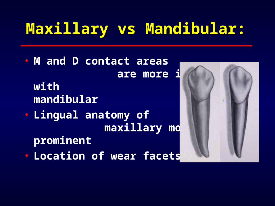
Maxillary vs Mandibular:Maxillary vs Mandibular:
• M and D contact areas are more incisal with mandibular
• Lingual anatomy of maxillary more prominent
• Location of wear facets
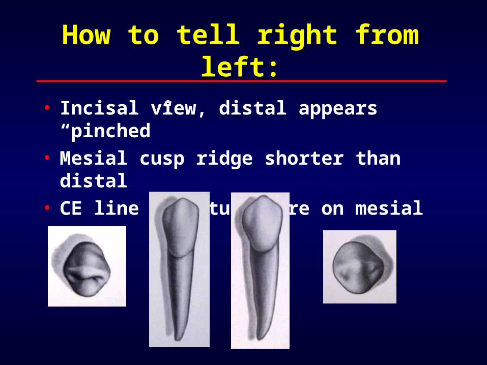
How to tell right from How to tell right from left:left:
• Incisal view, distal appears “pinched”
• Mesial cusp ridge shorter than distal
• CE line curvature more on mesial