periscopic spine surgery - department of computer science
TRANSCRIPT

Periscopic Spine SurgeryTechnology Developments in
Minimally Invasive Procedures
Kevin Cleary, PhDImaging Science and Information Systems
(ISIS) CenterComputer Assisted Interventions Laboratory
Department of RadiologyGeorgetown University Medical Center
Washington, DC
October 2002

Periscopic Spine Surgery Georgetown UniversitySlide 2
Overview of Presentation
• Background• ISIS Center / mobile CT• Medical robotics
– Review– Spinal nerve blocks at Georgetown
• Image-guided surgery (magnetic localization)
• Summary

Periscopic Spine Surgery Georgetown UniversitySlide 3
Background

Periscopic Spine Surgery Georgetown UniversitySlide 4
Georgetown University
• Located in Washington, DC• Large, private university
– Medical school– Law school– International studies
• Ranked in top 25 hospitals in U.S.• No engineering school

Periscopic Spine Surgery Georgetown UniversitySlide 5
Radiology Equipment2 MRI – Research MRI: 7T
3 fixed CT– Siemens SOMATOM Volume Zoom (CT fluoro)
Mobile CT scanner2 angiography labs (+1 soon)3 fluoroscopy units– Siemens MultiStar– Siemens NeuroStar biplane system
• 3D Virtuoso software (accelerator board)
3 general radiology rooms7 ultrasound machines5 portable C-arms (3 angio capable)1 PET scanner

Periscopic Spine Surgery Georgetown UniversitySlide 6
ISIS Center• Imaging Science and Information
Systems (ISIS)• Part of Radiology Department• Collaborations with many departments• 30 people (faculty, staff, and support)• Completely externally funded• Director: Seong Ki Mun, PhD

Periscopic Spine Surgery Georgetown UniversitySlide 7
ISIS Center Research Areas
• Image processing– Digital mammography– Computer aided diagnosis
• Telemedicine• Chronic disease: web-based monitoring• Computer aided surgery (image-guided
surgery, medical robotics, etc.)

Periscopic Spine Surgery Georgetown UniversitySlide 8
Mobile CT ScannerOperational April 1998 117 cases to date (Feb 2000)– Interventional Radiology
• Spinal interventions• Drainage procedures
– Operating Room• Complex Spinal Tumor Resections• Head and Neck surgery• Craniotomies

Periscopic Spine Surgery Georgetown UniversitySlide 9
Workstation
Mobile CT Components
Gantry and Table

Periscopic Spine Surgery Georgetown UniversitySlide 10
Interventional Suite Layout

Periscopic Spine Surgery Georgetown UniversitySlide 11
Volume Rendered
Image
(Cementin Green)

Periscopic Spine Surgery Georgetown UniversitySlide 12
Volume Rendered
Image
(CementGreen)

Periscopic Spine Surgery Georgetown UniversitySlide 13
Tumor Resection in OR with mobile CT for visualization
Spine tumor resection & stabilization

Periscopic Spine Surgery Georgetown UniversitySlide 14
Medical Robotics Review

Periscopic Spine Surgery Georgetown UniversitySlide 15
Medical Robotics
• Tremendous potential for improving the precision and capability of physicians to perform minimally invasive procedures
• At the beginning of the application of robotics to medicine
• Many questions remain as to effectiveness, safety, and cost

Periscopic Spine Surgery Georgetown UniversitySlide 16
Medical Robotics (continued)• Several commercial companies selling
medical robots– Total installed base is extremely small– Market will most likely continue to grow slowly
• While factory robotics grew rapidly during the 1970s and 1980s, medical robotics has not yet reached a critical mass
• Believed the benefits of medical robotics will become increasingly clear and this will lead to a continued rise in their use in medicine.

Periscopic Spine Surgery Georgetown UniversitySlide 17
Clinical Applications
• Neurosurgery• Orthopaedic• Urology• Maxillofacial• Opthamology• Radiosurgery• Cardiology

Periscopic Spine Surgery Georgetown UniversitySlide 18
Neurosurgery
• First recorded medical use of a robot– Kwoh 1985
• Simple positioning device to orient a brain biopsy needle
• Robot held a needle guide and surgeon inserted needle
• Not continued due to safety concerns

Periscopic Spine Surgery Georgetown UniversitySlide 19
Neurosurgery: Minerva
Periscopic Spine Surgery Georgetown UniversitySlide 20
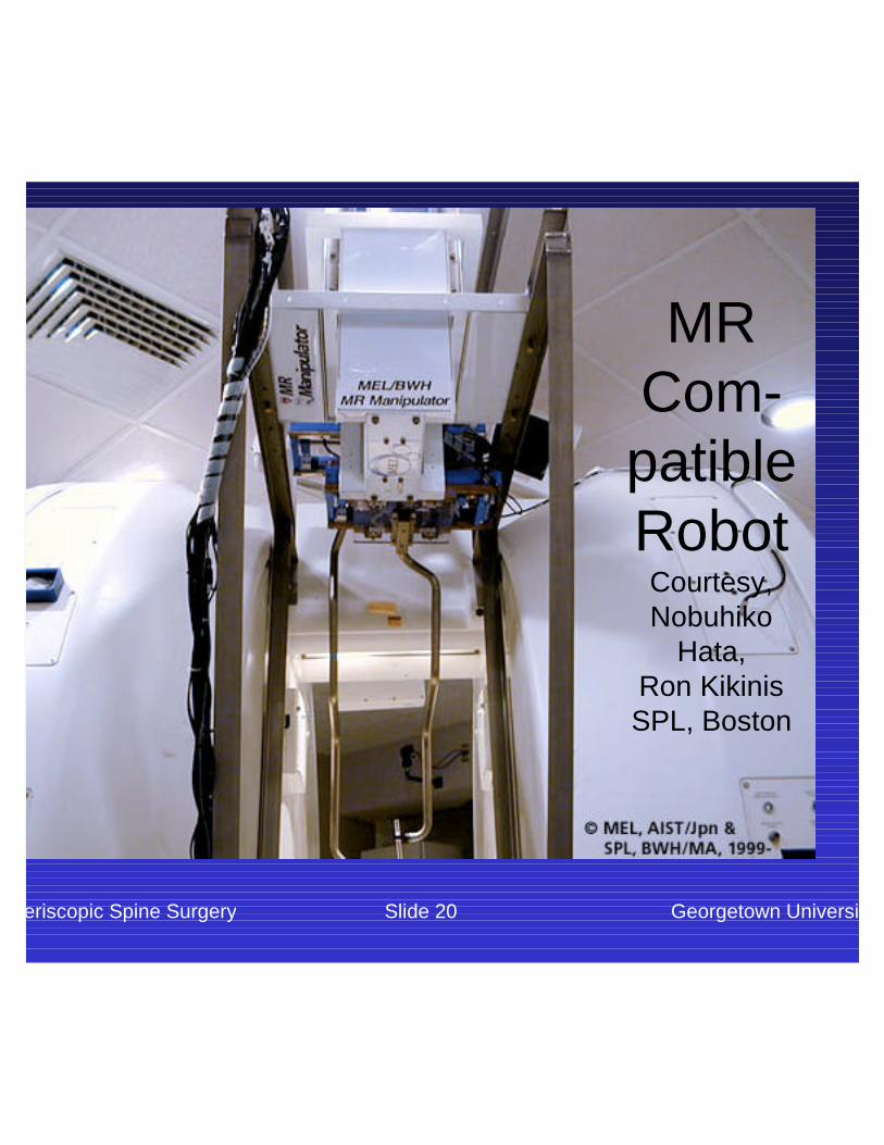
MR Com-patibleRobotCourtesy, Nobuhiko
Hata, Ron KikinisSPL, Boston

Periscopic Spine Surgery Georgetown UniversitySlide 21
Orthopaedics: Robo
DocCourtesy Integrated Surgical Systems

Periscopic Spine Surgery Georgetown UniversitySlide 22
Maxillo-facial
Tim Lueth, Charite, Berlin

Periscopic Spine Surgery Georgetown UniversitySlide 23
Radiosurgery
Accuray
(Stanford)
Cranial, spine, lung, pancreas, tracking of
respiratory motion

Periscopic Spine Surgery Georgetown UniversitySlide 24
Russ Taylor, Johns Hopkins University
Opthamalogy

Periscopic Spine Surgery Georgetown UniversitySlide 25
Cardiology: Intuitive Surgical
TelesurgerySystem
Source: (Fox news archive)
(www.intusurg.com)

Periscopic Spine Surgery Georgetown UniversitySlide 26
Intuitive Surgical Components

Periscopic Spine Surgery Georgetown UniversitySlide 27
Intuitive Surgical Console

Periscopic Spine Surgery Georgetown UniversitySlide 28
Questions
• Is there a large base of installed medical robots?
• Are there niche markets?• Will robots replace surgeons?• Is there a robot in your future?

Periscopic Spine Surgery Georgetown UniversitySlide 29
Spine Robotics at Georgetown
Robot
Mobile CT
gantry

Robot designed and constructed by Dan Stoianovici, PhD, Hopkins
URobotics LaboratoryGeorgetown

Periscopic Spine Surgery Georgetown UniversitySlide 31
Robot in Interventional Suite

Periscopic Spine Surgery Georgetown UniversitySlide 32
Robotically Assisted Nerve and Facet Blocks: Purpose of
Clinical Study
• To demonstrate that a physician controlled robotic needle driver is equivalent in safety and effectiveness to the standard manual technique for needle placement in nerve and facet blocks in the perispinal region

Periscopic Spine Surgery Georgetown UniversitySlide 33
Nerve and Facet Blocks
Typically done by interventional neuroradiologistsFor both diagnosis and therapyRequires accurately placing a thin needle (usually 22 gauge) under fluoroscopic guidanceTypical procedure time: 30 minutesHigh volume procedureNot technically demanding

Periscopic Spine Surgery Georgetown UniversitySlide 34
Cadaver Study
Purpose: evaluate feasibility of using robot to place needle for perispinal nerve and facet blocksDate: 1 September 2001

Periscopic Spine Surgery Georgetown UniversitySlide 35
Materials and Methods
• Small metal BB targets placed in lumbar spine at 3 levels
• 6 nerve block targets and 6 facet block targets
• Physician attempts to drive needle to target using joystick to control robot
• Accuracy of placement evaluated on x-ray images (goal: within 3 mm)

Periscopic Spine Surgery Georgetown UniversitySlide 36
BBs Used as Targets

Periscopic Spine Surgery Georgetown UniversitySlide 37
Placement of Target BB’s

Periscopic Spine Surgery Georgetown UniversitySlide 38
Scout CT Showing BB Targets

Periscopic Spine Surgery Georgetown UniversitySlide 39
Physician Operating Robot

Periscopic Spine Surgery Georgetown UniversitySlide 40
Robot and Needle Holder

Periscopic Spine Surgery Georgetown UniversitySlide 41
Needle Holder Close-up

Periscopic Spine Surgery Georgetown UniversitySlide 42
Anterior/Posterior Fluoro Image
BB

Periscopic Spine Surgery Georgetown UniversitySlide 43
Lateral Fluoroscopic Image of BB
BB
Needle

Periscopic Spine Surgery Georgetown UniversitySlide 44
Axial CT Image of BB & Needle Tip
BB
Needle

Periscopic Spine Surgery Georgetown UniversitySlide 45
Results & Conclusion
All 12 needles were placed within 3 mm of the target BBA joystick controlled robotic needle driver can be used by the interventionalist to accurately place needles in the nerve and facet regionsClinical studies are required to investigate the advantages and disadvantages of this system for interventional needle procedures

Periscopic Spine Surgery Georgetown UniversitySlide 46
FDA Approval Received
20 patients in initial study100 patients nextMarch 2002IRB approvals– Georgetown– Army

Periscopic Spine Surgery Georgetown UniversitySlide 47
FDA IDE clinical
trial study
outline
Meets Inclusion/Exculsion Criteria
Pre-ProceduralBaseline Screening Do Not EnrollNo
Yes
Obtain InformedConsent
Yes
Do Not Enroll
Pre-Procedural Consultation
Randomize to RoboticAssistance or NO
Robotic Assistance
PerformProcedure
Post Procedural Consultation
END
No

Periscopic Spine Surgery Georgetown UniversitySlide 48
Benefits of Robotic Guidance
• Improved path planning• More precision control of needle
trajectory• Allows operator to advance needle and
view trajectory in real-time without exposure to x-ray field

Periscopic Spine Surgery Georgetown UniversitySlide 49
And now to the video...

Periscopic Spine Surgery Georgetown UniversitySlide 50
Localization (non-line-of-sight tracking)

Periscopic Spine Surgery Georgetown UniversitySlide 51
Liver Respiratory Motion Simulator
Goal: demonstrate magnetic tracking technology for interventional proceduresInitial target: liver Collaboration with Northern Digital & Traxtal Technologies
AURORA™ magnetic tracking system Left to right: control unit, sensor
interface device, and magnetic field generator
(Courtesy of Northern Digital Inc.)

AURORA Tracking System1. Tetrahedron shaped field generator 2. The position sensor is made of a induction coil with a diameter of 0.9 mm

Periscopic Spine Surgery Georgetown UniversitySlide 53
Liver Respiratory Motion Simulator Components

Demonstration: Berlin June 2001

Periscopic Spine Surgery Georgetown UniversitySlide 55
Demonstration Baltimore April 2002

Periscopic Spine Surgery Georgetown UniversitySlide 56
Clinical Scenario
• Pre-procedure CT• Registration step
– Fiducials in CT space– Fiducials in magnetic space
• Provide image overlay and respiratory tracking during procedure
• Confirming image upon completion

Periscopic Spine Surgery Georgetown UniversitySlide 57
Interventional Suite Testing

Periscopic Spine Surgery Georgetown UniversitySlide 58
Respiratory Motion Simulator GUIPatent Pending: All Rights Reserved

Periscopic Spine Surgery Georgetown UniversitySlide 59
Results: Biopsy Experiments
Mean PlanningTime (s) ± SD
Needle ManipulationBiopsy Time (s) ± SD
Total ProcedureTime (s) ± SD
User 1 72 ± 35 79 ± 40 151 ± 59
User 2 61 ± 31 111 ± 41 172 ± 43
Overall 71 ± 36 93 ± 43 163 ± 57

Periscopic Spine Surgery Georgetown UniversitySlide 60
Results: Vessel Puncture
SuccessfulAttempts
UnsuccessfulAttempts
Mean Duration of Attempts (sec) 21.2 22
Standard Deviation 4.6 10
Mean Error (mm) 4.8 8.2
Standard Deviation 2.2 2.6

Periscopic Spine Surgery Georgetown UniversitySlide 61
Cadaver Study:
Introducing Catheter

Periscopic Spine Surgery Georgetown UniversitySlide 62
Cadaver Study: Field Generator

Periscopic Spine Surgery Georgetown UniversitySlide 63
Cadaver Study:Liver
Needle Placement

Periscopic Spine Surgery Georgetown UniversitySlide 64
Liver Simulator Demo Video

Periscopic Spine Surgery Georgetown UniversitySlide 65
Summary
• Focus on precision minimally invasive procedures
• Clinical relevance is key• Collaboration between engineer and
physician• Prospects for future are bright

Periscopic Spine Surgery Georgetown UniversitySlide 66
Acknowledgements• Clinicians
– Vance Watson, MD, Radiology– Elliot Levy, MD, Radiology– Filip Banovac, MD, Radiology– Matthew Freedman, MD, Radiology / ISIS Center
• Scientists / Researchers– David Lindisch, RT, Radiology / ISIS Center– Daigo Tanaka, MA, ISIS Center– Seong K. Mun, PhD, Radiology / ISIS Center
• Students– Sheng Xu, Johns Hopkins ERC CISST
• Collaborators– Dan Stoianovici, PhD, Johns Hopkins Urology / ERC CISST– Russell Taylor, PhD, Johns Hopkins ERC CISST – Gabor Fichtinger, PhD, Johns Hopkins ERC CISST– Charles Nguyen, PhD, Catholic University
• Funding– US Army Medical Research and Materiel Command