people “the man who smiles in the face of trouble… has found someone to blame it on.”
TRANSCRIPT

PEOPLE“The man who smiles in the face of
trouble…
Has found someone to blame it on.”

Anatomy
forebrain is responsible for "thinking," behavior, and final integration of sensory information
Supratentorial: better prognosisInfratentorial

Brain: Neoplasia
Enlarging mass in brain; causes compression of healthy tissue or replacement with cancerous tissue
• Primary: meningioma, glioma, choroid plexus papilloma, pituitary adenoma or adenocarcinoma (cushing’s), and others.
• Secondary (metastasis): hemangiosarcoma, mammary carcinoma and melanoma
• Dogs most common meningiomas and gliomas
• Sagittal MRI of the brain of an 8 year old female German Shepherd dog.
• Seizure • Well encapsulated mass in the olfactory bulb
and frontal cortex of the brain • Meningioma (histopathology)• TX: surgical removal of the tumor followed by
radiation• PX: Good not touching brain (unlike gliomas:
radiation therapy)

Hemangiosarcoma

Brain: Neoplasia• Forebrain
– Behavior• Depression, loss of
learned behavior– Seizures: acute onset, 5-6
yrs old– Pacing and circling
(Vestibular signs)– Ipsilateral vision: bump– E.g. Glioma’s and new
treatments• Brachycephalic breeds:
such as the Boxer, the Boston terrier, and the French and English bulldog

Brain - Neoplasia
• The Brainstem
– motor function (the ability to walk, CV and resp function), consciousness and balance
– 1st signs: loss of balance (vestibular signs) and paresis of one side of the body
– Dysphagia, change in voice and inability to move the eyes
– Progresses into paralysis, coma and death.
• Vestibular signs:
• Head tilt
• Leaning and falling to the side of the head tilt
• Drunken gait with loss of balance (ataxia)
• Circling to the side of the head tilt
• nystagmus
• Anorexia and vomiting
• Strabismus

• Kasey, a 10-year-old female Golden retriever
• trigeminal nerve root tumor• sensation to the face and
motor function to the muscles of mastication
• 15 months

Brain - Neoplasia
• The Cerebellum - coordination of movements and interacts closely with the vestibular system to control balance and posture. – Uncoordinated gait characterized by dramatic goosestepping
(hypermetria)
– Intention tremors: Head tremors that are worst when the animal is intent on something (i.e., food) but disappear when the animal is relaxed Swaying of the trunk
– Wide based stance
– Sometimes there can be vestibular signs such as a head tilt
– The animal's strength remains normal

Brain: Neoplasia
• Dx– Systematic screening for tumors in other organs– CBC, chem panel– Radiographs: metastasis– CSF tap to assess increased cerebral spinal
pressure– Ophthalmic exam may indicate optic nerve
edema– Computed tomography (CT) scanning or
magnetic resonance imaging (MRI) to locate tumor

CT or MRI
• MRI
– shows the brain in more detail than CT
– the test of choice when assessing for brain tumors
– more expensive test and less widely available
– CT images (more artifacts for brainstem/ cerebellum): meningiomas, choroid plexus papillomas
– MRI: gliomas, brainstem or cerebellar disease or Boston Terrier.

Brain: Neoplasia• Rx— surgical removal, radiation therapy,
chemotherapy, and palliative treatment of the symptoms
– Surgical removal of superficial single lesions
• Tumors of the brainstem pose problems
• Forebrain: you can resect certain parts of the forebrain without long-term effects.
• Meningiomas tend to be located on the surface of the brain and are therefore the best candidates for surgical removal.
• Gliomas are more difficult to remove because they lie deep within the substance of the brain.

MRI of a 6 year female old Boxeroligodendroglioma

Brain: Neoplasia
– Radiation therapy
– Chemotherapy; efficacy varies with tumor
type (lymphomas respond well; other less so)
– Palliative: Anti-seizure medication (Phenobarbital PO 2-3 times/day), Corticosteroids—prednisone
• Client info– The more severe the signs, the worse the outcome
– The larger the tumor, the worse the outcome
– Supratentorial tumors (tumors of the forebrain) have a better prognosis than infratentorial tumors (tumors of the brainstem and cerebellum)
– Radiation therapy does prolong lifespan in most cases
– Meningiomas have a better outcome than tumors that lie within the brain (e.g. gliomas)

Epilepsy
• MOA: balance within
the brain shifts too far
toward excitation,
too many cells may
become too excited and a seizure can result

Epilepsy
• Generalized/ tonic (stiff muscle) - clonic (rhythmic movements: grand mal)
• Grand mal (motor)– Prodrome
– Ictus: the seizure itself
– post-ictal (post-seizure)
• Petit mal (absence seizure): little movement, animals?
• Focal/ Partial
• focal seizure may stay localized
• may spread and affect the whole brain causing a classic, generalized, tonic-clonic seizure
• brain tumor or infection
• Simple: motor, twitching or blinking to one side of the face
• Complex: sensory. Senseless repetitive behavior: imaginary fly biting,

Minimum work-up for an epileptic
History
Your description of the character and timing of the episodes, relation to
exercise, feeding, etc.
Helps your veterinarian determine if this is indeed a seizure and what type. May
provide clues to the cause
Physicalexamination
Evaluation of the heart, lungs, abdomen, gum color, etc.
Provide clues to diseases which could cause seizures or complicate treatment
Neurologicexamination
Evaluation of behavior, coordination, reflexes and nerve functions
Provide clues to disease of the nervous system which may be causing the
seizures
Complete blood count (CBC), routine serum chemistry profile, and urine
analysis (UA)
Blood and urine samples are taken and analyzed
Rules out metabolic causes of seizures and provides baseline data to monitor
effects of medication
Bile acids assay or ammonia tolerance test
Usually, the pet is fasted and two blood samples are taken
Rules out liver problems and provides baseline data to monitor effects of
medication
Thyroid function testsBlood samples analyzed for T4 and TSH
levelsOptional, but would rule out thyroid
disease as a cause

Seizures
When the seizure begins, the dog stiffensand falls; They then begin jerking movementsThey are not in pain during the seizureand cannot control their bladder or bowels.

Epilepsy
• Signs of seizure– short aura (stare into distance,
seek comfort/protection from
someone, vocalize)
– seizure lasts 1-2 min; may consist of total body muscle twitching with extended arms and legs and arching of neck dorsally (opisthotonus)
– dog will be disoriented/blind for a few minutes
Click for video

• http://www.thepetcenter.com/gen/epilepsy.html
– may be incited by certain events
– normal at other times

Epilepsy• Dx
– CBC, chem panel—r/o metabolic diseases causing seizures• hypoglycemia• hypocalcemia• hepatic encephalopathy (failure to detox blood)
– Shunt– Cirrhosis
• Pb poisoning– Radiographs—r/o head trauma or hydrocephalus– CT scan or MRI—r/o space-occupying lesion in brain
• Rx—directed at cause if one can be found– treat if >1 every mo or two (Rx will not completely stop seizures)– Phenobarbital is TOC

Other tests that may be recommended(may require referral to a neurologist)
MRI or CT brain scan
Evaluate the structure of the brain; requires anesthesia
Rules out diseases such as brain tumors which would need to be
treated directly
Spinal tap
Spinal fluid is collected and analyzed; requires anesthesia
Looks for infectious diseases and provides clues to other brain diseases
Antibody titersBlood and/or spinal fluid is analyzed
for antibodiesIdentifies specific cause of an
infection
Toxin tests
Blood or other sample is tested for the presence of a toxin
Tells if a specific toxin is present, but usually need a clue to what toxin to
look for from the history or other test
Other laboratory testsAdvanced tests on blood, urine, or
spinal fluidFollows clues suggested by routine
tests
Electroencephalogram (EEG)Recording of brain wave to look for
the electrical stormAllows definitive diagnosis, but can
be non-diagnostic

Status Epilepticus• Signs—prolonged, uninterrupted seizures (>5-10
minutes) or cluster of seizures• Rx
– Diazepam (2-10 mg to effect); can be repeated over several minutes
• Phenobarbital - – Time to steady state blood levels: 10-14 days– Side effects: sedation, ataxia, PU/PD/PP,
hepatotoxicity, blood dyscrasias (Rare)– Establish an open airway– IV cath with IV fluids to keep an open vein– Monitor blood Ca and glucose; treat is needed– Monitor body temp; if elevated, treat appropriately– If cerebral edema is suspected, treat with mannitol (IV)– Phenobarbital—IV or IM
Status Epilepticus
• Client info—– Epilepsy is an incurable disease
– Even with treatment, animal may still seize; • goal is to reduce frequency and intensity of seizures
– Spaying/neutering will remove any hormonal influence on seizures
– Medications will probably be required for life
– Most animals that seize can live a normal life
– If seizure free for 6-9 mo, may reduced or discontinued Rx

LIFE“Life is the art of drawing without an
eraser.”
-John w. Gardner

Spinal Cord
• Function– Nerve fibers carry signals
between brain - rest of body
• Anatomy– Like brain, protected by
hard covering – dura
– Intervertebral disk (cushion):
• between vertebral body
• increases range of motion
• prevents vertebrae rubbing

Spinal Cord: Anatomy
Like brain, spinal cord enclosed in hard coveringIVDD problem in both humans and canineAnatomical differences—cervical same; lumbar—human bears weight, canine doesn’tAttached rib (thorax) helps stabilize the IV joint; worse at T-L junction (dogs)
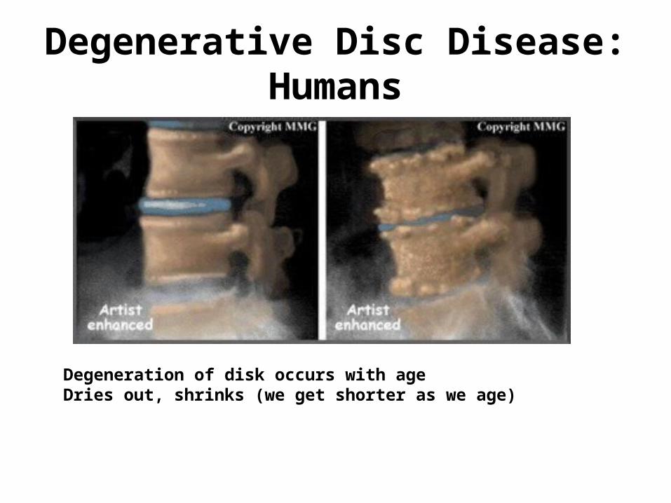
Degenerative Disc Disease: Humans
Degeneration of disk occurs with ageDries out, shrinks (we get shorter as we age)

IV Disk Disease: AnatomyNormal spinal column and disk Prolapsed disk
nucleus fibrosus
1/3 thickness

Intervertebral Disk Disease
• Etiology– IVD dries out with age → hardened, less compliant
– ↑Pressure from jumping
– Occurs most commonly in cervical, caudal thoracic, and lumbar vertebrae
– Most common spinal cord disorder in companion animals

Intervertebral Disk Disease
• Hansen TYPE I: Nucleus pulposus herniates upward; narrowest part of annulus fibrosus– TYPE I: Most common in chondrodystrophic
(“faulty development of cartilage”) breeds• Dachshunds, shih tzus, Lhasa apsos, beagles, basset
hounds (poodles also affected)
• Acute onset
• Can occur at any age, but generally younger dogs

Intervertebral Disk disease
• Hansen TYPE 2: dorsal protrusion of the annulus into the spinal canal– Common in older dogs and nonchondrodystrophic
breeds• Occurs over a longer period of time
• Clinical signs may be less severe
• Generally older dogs

Intervertebral Disk Disease• Signs:
– Pain
– Paresis/paralysis; nerve function is lost in this order:• Proprioception—largest fibers; most susceptible to pressure; signs
are ataxia
• Motor fibers—next smallest fibers; signs are weakness/paresis
• Cutaneous sensory fibers—small; require a lot of pressure to disrupt function; decreased panniculus reflex
• Deep pain fibers—smallest fibers; require the most pressure to disrupt; loss is associated with poor prognosis
• Severity of clinical signs depends on:• Speed at which disk material is deposited
• Degree of compression
• Duration of compression

IVDD – Paralysis of rear legs

Cervical IVDD

Loss of Deep Pain

IVDD Dx: Spine X-Rays
Normal horse’s head consistent IV space
Subluxation L2-3(old lesion)

IV Disk Disease: Myelogram: Definitive diagnosis
Which disk space?

IV Disk Disease: MyelogramIV Disk Disease: Myelogram
Which disk space?

Cervical IVDD
Myelogram: Disk herniation at C2-3 (narrowed IV space, narrowed spinal canal)

IVDD• Rx TYPE I, acute onset
• Medical Rx is recommended for animals, with deep pain intact, with or w/o neuro deficit– High levels of corticosteroids is CONTROVERSIAL
• Strict confinement—2 wk minimum (easy when dog hurts; not so easy after steroids/other pain medications take effect)
• Nursing care– Soft padded cage– Urinary cath or express bladder several times/day
• Surgery is recommended for– repeat offenders– No voluntary motor function– loss of deep pain (needs to be done QUICKLY!)– worsening neuro signs (poor Prognosis)

Laminectomy

IVDD: Possible sequela

IVDD

IVDD - rehabilitation
http://www.youtube.com/watch?v=7AkNVDc4lig&feature=related

IVDD – Alternative/Optional Treatment
• Methocarbamol (muscle relaxant)15-20 mg/kg q 8hr• High-dose Methylprednisolone sodium succinate
(CONTROVERSIAL!) and should be given within 8 hours– Although there is proven benefit in humans, results have
not been proven in dogs• Low dose prednisone – various regimens• NSAIDS
– Carprofen, deracoxib, etodolac• Gastroprotectants• Acupuncture

Veterinary Acupuncture
• http://www.youtube.com/watch?v=Z-JjZPnk_Mw&feature=related
• http://www.youtube.com/watch?v=vJIJDUQyOmw&feature=fvw
IVDD
• Client info• Do not let susceptible breeds get overweight
• Encourage animals to keep spine parallel to ground– No jumping on/off couch
– No begging on hind legs
– No stair climbing
• Loss of deep pain >24 h has poor prognosis
• If surgery is done soon enough, there is a good Px of recovery
• Almost half of animals treated medically will have recurrence
• Extensive home care is required for medical and surgical patients
• Severe damage to spinal cord is not reparable

Atlantoaxial Joint
Atlanto-Axial Joint

Atlantoaxial Instability (Subluxation)
• Signs– Toy and miniature breeds (<1 yr)
– Reluctance to be patted on head
– Neck pain
– May have tetraparesis (weakness in all 4 limbs) or tetraplegia (paralysis in all 4 legs)
– Sudden death due to respiratory paralysis
• Diagnosis—– Radiographs: lateral x-ray of neck in slight ventroflexion
• avoid further spinal cord damage with positioning


Atlantoaxial Instability (Subluxation)
CT scan:dens is marked by *
Normal toy breed dog Toy breed dog with atlantoaxial subluxation
X-rays of same dogs; note separation of C1 and C2 when dog’s neck is flexed in B
Narrowed spinal canal

Atlantoaxial Instability (Subluxation)
• Treatment—– Medical
• splint neck in extension with cage confinement x 6 weeks• treat like other spinal cord trauma
– Surgical (if unresponsive to medical Rx)• stabilize/decompress• attach dorsal process of axis to arch of atlas• fuse atlas and axis joint with pins and bone graft• hemilaminectomy to relieve spinal cord compression
• Client info—– prognosis is fair to good for animals with mild signs– animals should not be used for breeding; may be
hereditary
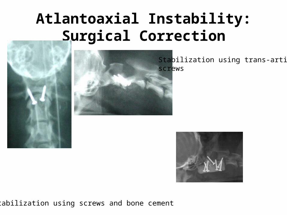
Atlantoaxial Instability: Surgical Correction
Stabilization using screws and bone cement
Stabilization using trans-articularscrews

References
• Alleice Summers, Common Diseases of Companion Animals
• http://cvm.ncsu.edu/vhc/tc/clinical_services/neuro/brain_tumor.html
• http://www.canine-epilepsy.net/basics/basics_index.html