pelvic anatomy from a laparoscopic perspective:: the ... · pelvic anatomy from a laparoscopic...
TRANSCRIPT

Pelvic Anatomy from a Laparoscopic
Perspective:: the abdominal wall &
retroperitoenum
•Tommaso Falcone
MD
•Professor &
Chairman
•Cleveland Clinic
Foundation

Disclosure
• No honoraria from Industry
• No research grants from industry
• Receive honoraria
- Editor-in-Chief of JMIG
- Section Editor- UpToDate

Learning Objectives
• Understand the causes of iatrogenic
injuries associated with laparoscopy
• Know how to diagnose and treat
laparoscopic complications
Anterior Abdominal Wall
•Relationship of the
vessels & nerves to
potential entry sites
for trocars

Relationship of the umbilicus
to vessels
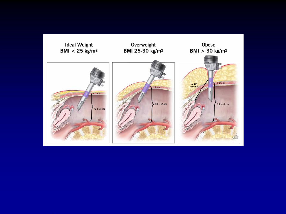

Stanhiser et al AJOG 2015

Left common iliac vein
inferior to the bifurcation of the aorta
crosses the sacrum


Left Upper Quadrant Insertion
• 2-cm below the subcostal margin mid-
clavicular line
• Organs
- Aorta-11 cm
- Spleen-12cm
- Stomach-4.4cm
- Liver-4.0cm
- Left kidney 13.2cm
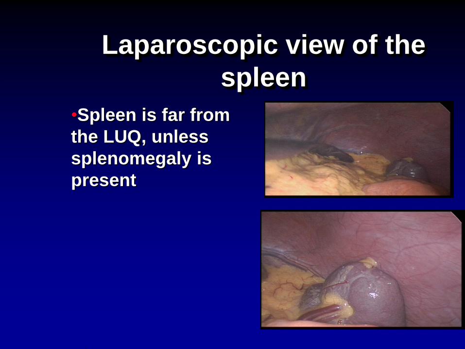
Laparoscopic view of the
spleen
•Spleen is far from
the LUQ, unless
splenomegaly is
present


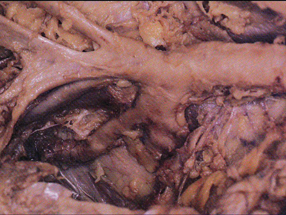
Pelvic Sidewall Anatomy
•3 layers
- Ureter
- Branches of the
int.iliac artery
- Muscle & nerve
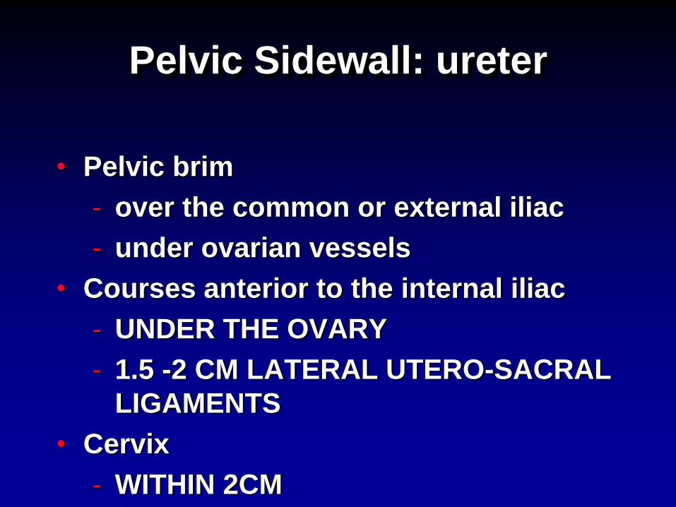
Pelvic Sidewall: ureter
• Pelvic brim
- over the common or external iliac
- under ovarian vessels
• Courses anterior to the internal iliac
- UNDER THE OVARY
- 1.5 -2 CM LATERAL UTERO-SACRAL
LIGAMENTS
• Cervix
- WITHIN 2CM


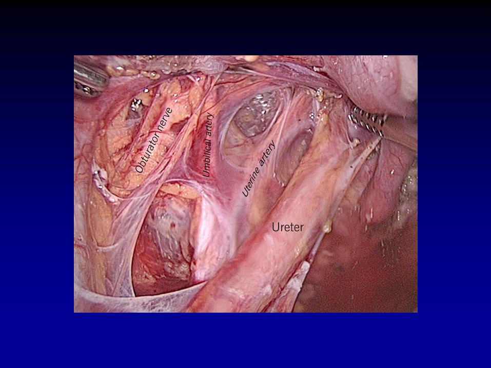
Pelvic Sidewall: Blood
vessels
• Internal iliac artery
- anterior & posterior division (trunks)
- Umbilical artery
• obliterated
• medial umbilical ligament
• relationship to the uterine artery


Pelvic
Arteriogram


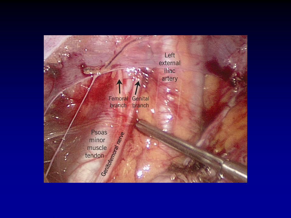
Pelvic & Inguinal Nerves
• Genito-femoral nerve
• Femoral nerve


Retropubic Space
•Anterior
- Pubic bone
•Lateral
- Obturator internus
muscle, fasciae,
neurovascular
bundle
•Posteriorly
- bladder &
pubocervical
fasciae


Pelvic Diaphragm
• Sheet of muscle (Levator ani &
coccygeus) covered on both sides by
fasciae
• From pubis to coccyx & is attached to the
lateral pelvic wall by a thickened band of
obturator fascia called arcus tendineus m.
levator ani
• Anogenital hiatus

Pelvic Diaphragm:Muscle
• Levator Ani
- Pubococcygeus (Puborectalis & pubovaginalis)
- Iliococcygeus
• Iliococcygeus portion that arises from the obturator internus muscle (arcus tendineus m. levator ani) & ischial spine
• Arcus: spine of the ischium forward & upward.


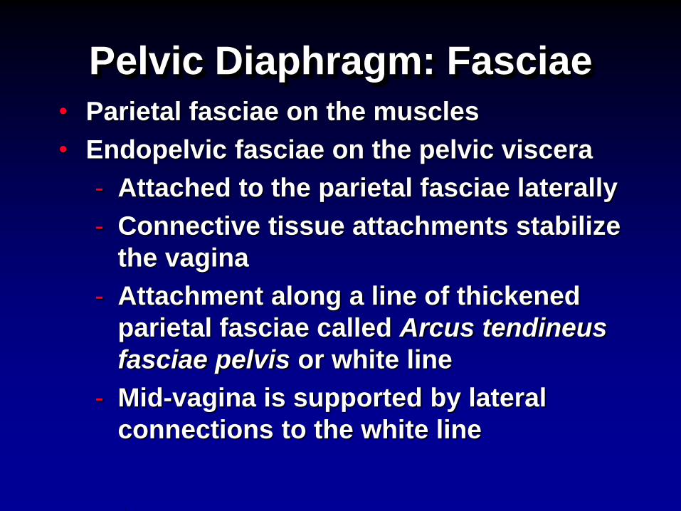
Pelvic Diaphragm: Fasciae • Parietal fasciae on the muscles
• Endopelvic fasciae on the pelvic viscera
- Attached to the parietal fasciae laterally
- Connective tissue attachments stabilize
the vagina
- Attachment along a line of thickened
parietal fasciae called Arcus tendineus
fasciae pelvis or white line
- Mid-vagina is supported by lateral
connections to the white line


Pre sacral space