pectoral nerves i and ii blocks in multimodal analgesia...
TRANSCRIPT

ULTRASOUND ARTICLE
Pectoral Nerves I and II Blocks in Multimodal Analgesia forBreast Cancer SurgeryA Randomized Clinical Trial
Ghada Mohammad Nabih Bashandy, MD, and Dina Nabil Abbas, MD
Background: The pectoral nerves (Pecs) block types I and II are noveltechniques to block the pectoral, intercostobrachial, third to sixth intercos-tals, and the long thoracic nerves. They may provide good analgesia duringand after breast surgery. Our study aimed to compare prospectively thequality of analgesia after modified radical mastectomy surgery using gen-eral anesthesia and Pecs blocks versus general anesthesia alone.Methods: One hundred twenty adult female patients scheduled for elec-tive unilateral modified radical mastectomy under general anesthesia wererandomly allocated to receive either general anesthesia plus Pecs block(Pecs group, n = 60) or general anesthesia alone (control group, n = 60).Results: Statistically significant lower visual analog scale pain scoreswere observed in the Pecs group than in the control group patients. More-over, postoperative morphine consumption in the Pecs group was lower inthe first 12 hours after surgery than in the control group. In addition, statis-tically significant lower intraoperative fentanyl consumption was observedin the Pecs group than in the control group. In the postanesthesia care unit,nausea and vomiting as well as sedation scores were lower in the Pecsgroup compared with the control group. Overall, postanesthesia care unitand hospital stays were shorter in the Pecs group than in the control group.Conclusions: The combined Pecs I and II block is a simple, easy-to-learntechnique that produces good analgesia for radical breast surgery.
(Reg Anesth Pain Med 2015;40: 68–74)
B reast cancer is the most common cancer among women. In theUnited States, 1 in 8 women develop breast cancer during
their lifetime.1,2 In Gharbiah, Egypt, increased breast cancer rateshave been recorded from 1999 to 2008. According to a recent ep-idemiologic study, higher breast cancer rates were expected be-tween 2009 and 2015.3 Acute postoperative pain is an integralrisk factor in the development of chronic postmastectomy pain;40% of women will have severe acute postoperative pain afterbreast cancer surgery, whereas 50%will develop chronic postmas-tectomy pain with impaired quality of life.4,5 Regional anesthesiatechniques have provided better-quality acute-pain control andsubsequently less chronic pain.5,6 Proposed mechanisms for de-creased persistent pain include decreased central sensitization(wind-up) and lower incidence of opioid-induced hyperalgesia.7,8
Furthermore, effective acute pain control preserves immune
From the Department of Anesthesia and PainManagement, National Cancer In-stitute, Cairo University, Cairo, Egypt.Accepted for publication August 20, 2014.Address correspondence to: GhadaMohammadNabih Bashandy,MD,National
Cancer Institute, Cairo University, Fom Al-Khalij, KasrAleiny St, Cairo,Egypt (e‐mail: [email protected]).
Funding from the National Cancer Institute, Cairo University, helped supportthis work.
Thework was presented as a poster at theWIP 2014 Congress atMaastricht, theNetherlands, May 7 to 10, 2014.
The authors declare no conflict of interest.Copyright © 2014 by American Society of Regional Anesthesia and Pain
MedicineISSN: 1098-7339DOI: 10.1097/AAP.0000000000000163
68 Regional Anesthesia a
Copyright © 2014 American Society of Regional Anesthesia and Pain
function, both by suppressing the surgical stress response and bydecreasing the need for general anesthetics and opioids. Opioids,especially morphine, inhibit both cellular and humoral immunefunctions.9 This effect may be responsible for the higher rates ofpostsurgical local recurrence and/or metastasis.10
Thoracic epidural block, thoracic paravertebral block (TPVB),interpleural block, intercostal nerve block, interscalene block, andwound infiltration have all been used in anesthesia and/or analgesiain breast cancer surgery.11–16 Thoracic paravertebral block has beenshown to provide superior analgesia, and there is some evidencesuggesting decreased cancer recurrence rates with the use ofTPVB.17,18 Nonetheless, not all anesthesiologists feel comfortableusing such invasive techniques in breast cancer surgery. The pecto-ral nerve (Pecs) block, a less invasive novel technique described byBlanco et al,19,20 is an interfascial plane block where local anes-thetic is deposited into the plane between the pectoralis major mus-cle (PMm) and the pectoralis minor muscle (Pmm) (Pecs I block)and above the serratus anterior muscle at the third rib (Pecs IIblock). These novel techniques attempt to block the pectoral;intercostobrachial; intercostals III, IV, V, VI; and long thoracicnerves.19,20 Our study prospectively compares Pecs blocks in com-bination with general anesthesia and general anesthesia alone inmodified radical mastectomy (MRM) surgery. We hypothesizedthat the Pecs blocks would provide superior postoperative analgesiafor patients undergoing mastectomy as compared with a controlgroup. Our primary outcome measure was visual analog scale(VAS) pain scores on the first postoperative day in patients whohave preoperative Pecs block compared with those having generalanesthesia alone. Secondary measures were perioperative opioidconsumption, postoperative sedation, and postoperative nauseaand vomiting (PONV).
METHODSAfter obtaining approval fromEgypt’s National Cancer Insti-
tute Institutional Review Board, we conducted this prospectiverandomized observer-blinded study from January 2013 to January2014 in the National Cancer Institute of Egypt. A continuous sam-ple of 120 American Society of Anesthesiologists (ASA) physicalstatus I and II female adult patients undergoing elective unilateralMRM under general anesthesia were recruited Written informedconsent was obtained. Exclusion criteria included declining togive written informed consent, history of allergy to the medica-tions used in the study, contraindications to regional anesthesia(including coagulopathy and local infection), prior breast surgeryexcept for diagnostic biopsies, and history of treatment for achronic pain condition and/or psychiatric disorder.
All patients included in the study were randomly assigned to1 of the 2 groups: the Pecs group receiving Pecs blocks and gen-eral anesthesia (n = 60) and a control group receiving general an-esthesia alone (n = 60). Group allocation was accomplished usinga predetermined random 1:1 sequence. All the recruited patientswere familiarized with VAS pain scoring and patient-controlledanalgesia (PCA).
nd Pain Medicine • Volume 40, Number 1, January-February 2015
Medicine. Unauthorized reproduction of this article is prohibited.

Regional Anesthesia and Pain Medicine • Volume 40, Number 1, January-February 2015 Pectoral Nerve Blocks for Breast Surgery
Preoperative ManagementAll patients were premedicated with 10 mg of oral diazepam
on the night of surgery. In the preoperative holding area, patientswere attached to standard ASAmonitors, and intravenous (IV) ac-cess was obtained. Premedication with IV 1 to 2 mg of midazolamand 10 mg of metoclopramide was administered to all patients.The patients in the control group were then transferred immedi-ately to the operating room, whereas the patients in the Pecs groupreceived an ultrasound-guided Pecs block and a 15-minute obser-vation time prior to their transfer to the operating room. One in-vestigator (G.M.N.B) did all the blocks for patients in the Pecsgroup. Anesthesia management and data collection were per-formed by personnel blinded to the treatment group.
A broadband (5–12 MHz) linear array probe of eZono 3000portable ultrasound system (eZono USA, Redmond, Washington)was used, with an imaging depth of 4 to 6 cm. After cleaning theinfraclavicular and axillary regions with chlorhexidine, the probewas placed below the lateral third of the clavicle, similar to whatis done when performing infraclavicular brachial plexus block(Fig. 1). After recognition of the appropriate anatomical struc-tures, the skin puncture point was infiltrated with 2% lignocaine,then the block was performed by using a 20-gauge Tuohy needle.The needle was advanced to the tissue plane between the PMmand Pmm at the vicinity of the pectoral branch of theacromiothoracic artery, and 10 mL of 0.25% bupivacaine was de-posited (Figs. 2, 3, and 4). In a similar manner, 20 mL was depos-ited at the level of the third rib above the serratus anterior musclewith the intent of spreading injectate to the axilla (Fig. 5).19,20
Intraoperative ManagementStandard ASA monitors were attached to the patients. Gen-
eral anesthesia was induced with fentanyl 1 to 2 μg/kg, propofol2 mg/kg, and cisatracurium 0.15 mg/kg, and the airway wassecured by endotracheal intubation. Anesthesia maintenanceconsisted of 1 MAC of isoflurane in 50% oxygen and air with ad-ditional cisatracurium at the discretion of the anesthetist. Addi-tional boluses of fentanyl were administered to maintain blood
FIGURE 1. Image of the probe position and needle direction from med
© 2014 American Society of Regional Anesthesia and Pain Medicine
Copyright © 2014 American Society of Regional Anesthesia and Pain
pressure and/or heart rate values within or 20% lower than thebaseline values. Toward the completion of the surgery, paraceta-mol (Perfalgan) 1 g/100 mL IV infusion was started, andisoflurane was discontinued. Neostigmine 0.05 mg/kg with atro-pine 0.02 mg/kg was administered IV for neuromuscular block-ade reversal as clinically relevant. After responding to verbalcommand, patients were extubated in the operating room thentransferred to the postanesthesia care unit (PACU).
Postoperative ManagementIn the PACU, patients were monitored with standard ASA
monitors. They were monitored for pain intensity using VAS painscore, degree of sedation using the Ramsay sedation scale,21 andincidence of PONVusing a 5-point scale (0–4), where 0 = no nau-sea or vomiting, 1 = mild nausea, 2 = severe nausea, 3 = vomitingonce, and 4 = vomiting more than once.
When the reported VAS score was 3 or greater, a loadingdose of 5 mg of morphine was administered through slow IVroute. Then, a PCA was administered. The PCA pump (Graseby3300 Pump; Smith Medical International, Ashford, Kent, UK)was loaded with 1 mg/mL of morphine and set to deliver on de-mand bolus doses of 1 to 2 mL based on body weight with a 5-minute lockout period. No background infusion was allowed. Oralparacetamol (1 g) and ketoprofen (100 mg), to be administered 3times daily, were prescribed to all patients as soon as oral feedingwas permitted. Ondansetron 8 mg IV was used to treat nausea andvomiting. Patients with a score of 10 in the modified Aldrete scor-ing system were considered eligible for discharge to the surgicalward.22 Patients were discharged from the hospital based on theprotocols of the surgical team, which included a pain score of lessthan 3 without morphine and PONVand sedation scores of 0.
The following data were collected: intraoperative fentanylconsumption; postoperative VAS pain scores (at 0, 3, 6, 9, and24 postoperative hours); need for PCAmorphine; time to PCA ad-ministration as well as morphine requirements at 0 to 4, 4 to 12,and 12 to 24 postoperative hours; PONV scores; sedation scores;PACU stay; and postsurgical hospital stay.
ial to lateral during Pecs I block.30
69
Medicine. Unauthorized reproduction of this article is prohibited.
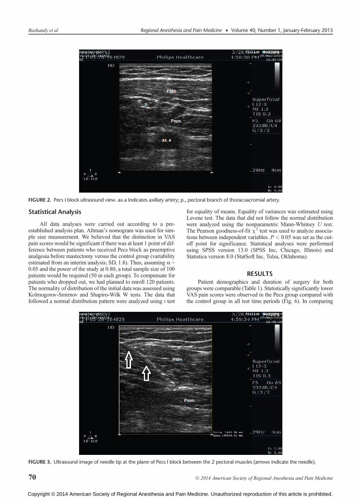
FIGURE 2. Pecs I block ultrasound view. ax.a Indicates axillary artery; p., pectoral branch of thoracoacromial artery.
Bashandy et al Regional Anesthesia and Pain Medicine • Volume 40, Number 1, January-February 2015
Statistical Analysis
All data analyses were carried out according to a pre-established analysis plan. Altman’s nomogram was used for sim-ple size measurement. We believed that the distinction in VASpain scores would be significant if there was at least 1 point of dif-ference between patients who received Pecs block as preemptiveanalgesia before mastectomy versus the control group (variabilityestimated from an interim analysis; SD, 1.8). Thus, assuming α =0.05 and the power of the study at 0.80, a total sample size of 100patients would be required (50 in each group). To compensate forpatients who dropped out, we had planned to enroll 120 patients.The normality of distribution of the initial datawas assessed usingKolmogorov-Smirnov and Shapiro-Wilk W tests. The data thatfollowed a normal distribution pattern were analyzed using t test
FIGURE 3. Ultrasound image of needle tip at the plane of Pecs I block b
70
Copyright © 2014 American Society of Regional Anesthesia and Pain
for equality of means. Equality of variances was estimated usingLevene test. The data that did not follow the normal distributionwere analyzed using the nonparametric Mann-Whitney U test.The Pearson goodness-of-fit χ2 test was used to analyze associa-tions between independent variables. P < 0.05 was set as the cut-off point for significance. Statistical analyses were performedusing SPSS version 13.0 (SPSS Inc, Chicago, Illinois) andStatistica version 8.0 (StatSoft Inc, Tulsa, Oklahoma).
RESULTSPatient demographics and duration of surgery for both
groups were comparable (Table 1). Statistically significantly lowerVAS pain scores were observed in the Pecs group compared withthe control group in all test time periods (Fig. 6). In comparing
etween the 2 pectoral muscles (arrows indicate the needle).
© 2014 American Society of Regional Anesthesia and Pain Medicine
Medicine. Unauthorized reproduction of this article is prohibited.

FIGURE 4. Local anesthetic spread between the 2 pectoral muscles. The arrow is pointing to the needle. LA indicates local anesthetic.
Regional Anesthesia and Pain Medicine • Volume 40, Number 1, January-February 2015 Pectoral Nerve Blocks for Breast Surgery
perioperative opioid needs, the intraoperative fentanyl require-ments were found to be lower in the Pecs group than in the controlgroup (115 ± 28.56 μg and 252.5 ± 44.352 μg, respectively, withP < 0.001). In addition, the total amount of postoperative mor-phine needed to keep VAS pain scores less than 3 was 2.9 ±1.714 mg and 6.9 ± 1.861 mg in the Pecs and control groups, re-spectively, and the difference was found to be statistically signifi-cant (P < 0.001). The patients in the Pecs group used lessmorphine in the first 12 hours postoperatively than did the controlgroup patients, but the morphine needs of the 2 groups were com-parable in the succeeding 12 hours (Table 2). Only 12 of 60 pa-tients in the Pecs group required morphine PCA based on theprotocol of the study, where an adequate VAS pain score of lessthan 3 was maintained only by paracetamol and nonsteroidalanti-inflammatory drug that were given to all patients in our study.
FIGURE 5. A, Image showing external probe position during Pecs II blocserratus anterior muscle; white line, needle path to deposit local anesthe
© 2014 American Society of Regional Anesthesia and Pain Medicine
Copyright © 2014 American Society of Regional Anesthesia and Pain
Conversely, 36 of 60 patients in the control group required PCAmorphine administration.
Postanesthesia care unit stay was statistically shorter in thePecs group than in the control group (14 ± 11 minutes and 28 ±12 minutes, respectively, where P = 0.012). This finding may beexplained in part by lower VAS pain scores in the Pecs group, aswell as lower PONV scores (0.15 ± .366 vs 1.65 ± 0.875, withP < 0.001). The reported lower sedation scores in the Pecs groupcompared with those in the control group are an alternative expla-nation of shorter PACU stay in the Pecs group (2.10 ± 0.308 vs3.20 ± 0.523, respectively, with P < 0.001).
Postsurgical hospital stay was shorter in the Pecs group thanthat in the control group (P < 0.001). All patients in the Pecsgroup were discharged from the hospital within 24 hours, whereasin the control group, only 12 patients left within 24 hours, 42
k.30 B, Ultrasound view of Pecs II block (r. 3 indicates 3rd rib; serr.,tic above serratus anterior muscle.
71
Medicine. Unauthorized reproduction of this article is prohibited.

TABLE 1. Demographic Data
Variable Pecs Group (n = 60) Control Group (n = 60) P
Age, y 48.65 (10.7) 50.47 (12.1) 0.74Weight, kg 75.96 (6.3) 74.34 (5.9) 0.14Height, cm 163 (6.8) 162 (7.8) 0.84Duration of surgery, min 110 (17) 109 (19) 0.34
Values are mean (SD).
P > 0.05 is statistically insignificant.
Bashandy et al Regional Anesthesia and Pain Medicine • Volume 40, Number 1, January-February 2015
patients were discharged within 48 hours, and 6 patients stayed inthe hospital for more than 48 hours.
DISCUSSIONLower pain scores were observed in patients undergoing
MRM with preemptive Pecs I and II blocks than in the controls.Perioperative opioid use, including intraoperative fentanyl as wellas postoperative morphine, was lower in the Pecs group comparedwith that in the control group. Our study also revealed loweropioid-related adverse effects with lower sedation and PONVscores in the Pecs group. Moreover, PACU and hospital stays wereshorter in the Pecs group compared with that in the control group.
To start performing the Pecs block, the neural supply ofstructures involved in breast surgery must be well understood.The pectoral nerves (PNs) show wide variability in their course.23
They are described in most textbooks as purely motor nerves, butit was suggested that they also transport proprioceptive and noci-ceptive fibers as shown in other motor nerves.24 In some patients,there might be additional innervations from the fourth intercostalnerve.25 Ameta-analysis of available literature showed that the lat-eral PN (LPN) arises most frequently with 2 branches from the
FIGURE 6. Visual analog scale scores in both study groups in different tiVAS-3,VAS-6, VAS-9, VAS-24 are VAS at 3, 6, 9, 24 hours postoperatively
72
Copyright © 2014 American Society of Regional Anesthesia and Pain
anterior divisions of the upper and middle trunks (33.8%) or asa single root from the lateral cord (23.4%). The medial PN(MPN) usually arises from the medial cord (49.3%), anterior divi-sion of the lower trunk (43.8%), or lower trunk (4.7%). The 2 PNsare usually connected by the ansa pectoralis immediately distal tothe thoracoacromial artery.26 Hoffman and Elliott27 suggestedblocking the PNs to reduce chronic postoperative pain or musclespasm after mastectomy. A denervation point for PMm targetingthe neurovascular bundle containing the LPN deep to the PMmwas identified. This point is at distances of 2.81 ± 0.33 cm verti-cally from the medial third part of the clavicle and 8.12 ± 1.09cm horizontally from the midsternal line.28 The MPN runs underthe Pmm. It crosses the Pmm in 62% of the patients to reachthe lower third of the PMm after piercing the 2 layers of theclavipectoral fascia. In the remaining 38%, it is located at the lat-eral border of the Pmm.26 Ultrasound-guided injection of 10 mLof the solution in cadavers was found to be sufficient in stainingall the medial and LPN branches without any proximal extensionto the cords of the brachial plexus.23
In MRM surgery, blocking the PNs alone is not enough. Theanterior divisions of the intercostal nerves from T2 to T6 and thelong thoracic and the thoracodorsal nerves should be blocked
me points. VAS-0: first VAS after recovery from general anesthesia., respectively.
© 2014 American Society of Regional Anesthesia and Pain Medicine
Medicine. Unauthorized reproduction of this article is prohibited.

TABLE 2. PCA Morphine Requirements
Variable Pecs Group (n = 60) Control Group (n = 60) P
Time to the first dose of morphine, min 170 (11.2) 130 (14.7) 0.008*No. PCA morphine demands 2.5 (1.2) 4.3 (1. 8) 0.04*PCA morphine, mg0–4 h 0 (0–9) 4 (2–12) 0.02*4–12 h 1 (0–4) 4 (0–10) 0.035*12–24 h 2 (0–15) 2 (0–25) 0.519
Values are means (SD) or median and range (Q1–Q3). 0–4 h, 4–12 h, and 12–24 h are postoperative hours where 0 is time of PACU admission.
*P < 0.05.
Regional Anesthesia and Pain Medicine • Volume 40, Number 1, January-February 2015 Pectoral Nerve Blocks for Breast Surgery
also.20 The intercostal nerves lie at the back between the pleuraand the posterior intercostal membrane and run in a plane betweenthe intercostal muscles as far as the sternum. They give off lateralbranches that pierce the external intercostal and the serratus ante-rior muscles at the midaxillary line to give off anterior and poste-rior terminal branches. The lateral cutaneous branch of the secondintercostal nerve does not divide in the anterior and posteriorbranches, and it is called the intercostobrachial nerve. The inter-costal nerves also give anterior branches that cross in front ofthe internal mammary artery and pierce the internal intercostalsmuscle, the intercostal membranes, and the PMm to supply themedial aspect of the breast.20,29 The long thoracic nerve arisesfrom C5 to C7, entering the axilla behind the brachial plexus rest-ing on the serratus anterior muscle. The thoracodorsal nerve is abranch of the posterior cord made up of the 3 posterior divisionsof the trunks of the brachial plexus. It follows the thoracodorsal ar-tery and innervates the latissimus dorsi in the posterior wall ofthe axilla.20
Blanco19 first described Pecs I block in 2011 as an interfascialblock to place local anesthetic into the plane between PMm andPmm. He targeted the LPN, which is consistently located adjacentto the pectoral branch of the thoracoacromial artery between thePMm and Pmm. In addition, Blanco19 stated that a catheter canreadily be placed into that interfascial plane. One year later, Blancoet al20 described a second version of the Pecs block called modifiedPecs block or Pecs block type II, another approach aiming to blockthe axilla and the intercostal nerves that are necessary for axillarynode dissection and wider excisions, respectively. In Pecs blocktype II, 20 mL of local anesthetic has to be deposited abovethe serratus anterior muscle. The local anesthetic would spread tothe axilla where the long thoracic nerve and lateral branches ofthe intercostal nerves are found as they exit at the level of themidaxillary line.20
Pérez et al also30 described a different approach for Pecs blockand reported decreased perioperative systemic analgesic requirementsand improved patient satisfaction inmajor andminor breast surgeries.The ultrasound probe is placed below the outer third of the clavicle,after identifying 4 structures: PMm, Pmm, thoracoacromial artery,and cephalic vein; the needle is introduced in plane with the ultra-sound probe frommedial to lateral. They claimed that their approachstays far from the pleura and blood vessels and avoids blocking theneedle path through the coracoid process.30
Pectoral nerves block was used in conjunction with TPVB inan observational study. The researchers compared TPVB and seda-tion with and without a Pecs block for breast augmentation surgery.Better postoperative analgesia and a lower requirement for seda-tion were observed in patients who received Pecs block.31 Theparavertebral space communicates with the epidural space, is closeto the pleural space, and contains supply arteries to the spinal cord;hence, special precautions should be adopted while performing
© 2014 American Society of Regional Anesthesia and Pain Medicine
Copyright © 2014 American Society of Regional Anesthesia and Pain
TPVB. Thoracic paravertebral block often becomes an epiduralblock and may also result in total spinal anesthesia.32,33
The Pecs block is a combination of motor and sensory nerveblocks. One advantage of Pecs block, requiring emphasis, is that itis not associated with sympathetic block as are the TPVB and epi-dural blocks. On the other hand, intravascular injection into thepectoral branch of the acromiothoracic artery is another possibil-ity that could be considered. Complications should be easilyavoidedwith proper ultrasound training and searching for the rightpattern of spread of the local anesthetic.20
LimitationsOne limitation of our study is that we did not have enough
time to assess the quality of the block before the induction of an-esthesia. Moreover, blinding the patients to the received techniqueby doing sham blocks to the control group would have made theresults more reliable. Another limitation is that, in an attempt tominimize morphine consumption, we did not offer PCAmorphineexcept for patients with VAS scores of less than 3 when we shouldhave offered it to all patients upon arrival to the PACU or as soonas they are alert enough to use it.
CONCLUSIONSThe Pecs blocks produce excellent analgesiawhen combined
with general anesthesia for breast surgery with axillary dissection.They are simple, easy-to-learn techniques, having easily identifi-able landmarks based on good anatomical and ultrasound knowl-edge, making them an excellent alternative to the conventionalthoracic paravertebral and neuraxial blocks for radical breast sur-geries. Prospective randomized studies comparing Pecs blockswith paravertebral and neuraxial blocks are recommended.
REFERENCES1. Desantis C, Ma J, Bryan L, Jemal A. Breast cancer statistics, 2013.
CACancer J Clin. 2014;64:52–62.
2. Tyczyński J, Bray F, Parkin D. Breast Cancer in Europe. Ispra, Italy: EurNetw Cancer Regist Cancer Fact Sheets; Dec 2002. Available at: http://encr.eu/images/docs/factsheets/breast-factsheets.pdf. AccessedSeptember 2, 2014.
3. Hirko KA, Soliman AS, Hablas A, et al. Trends in breast cancer incidencerates by age and sage at diagnosis in Gharbiah, Egypt, over 10 years(1999-2008). J Cancer Epidemiol. 2013;2013:916394.
4. Poleshuck EL, Katz J, Andrus CH, et al. Risk factors for chronic painfollowing breast cancer surgery: a prospective study. J Pain. 2006;7:626–634.
5. Gärtner R, Jensen M-B, Nielsen J, Ewertz M, Kroman N, Kehlet H.Prevalence of and factors associated with persistent pain following breastcancer surgery. JAMA. 2009;302:1985–1992.
73
Medicine. Unauthorized reproduction of this article is prohibited.

Bashandy et al Regional Anesthesia and Pain Medicine • Volume 40, Number 1, January-February 2015
6. Sittl R, Irnich D, Lang PM. Update on preemptive analgesia: options andlimits of preoperative pain therapy [in German]. Anaesthesist. 2013;62:789–796.
7. Richebé P, Rivat C, Liu SS. Perioperative or postoperative nerve block forpreventive analgesia: should we care about the timing of our regionalanesthesia?. Anesth Analg. 2013;116:969–970.
8. Richebé P, Pouquet O, Jelacic S, et al. Target-controlled dosing ofremifentanil during cardiac surgery reduces postoperative hyperalgesia.J Cardiothorac Vasc Anesth. 2011;25:917–925.
9. Sacerdote P, Bianchi M, Gaspani L, et al. The effects of tramadol andmorphine on immune responses and pain after surgery in cancer patients.Anesth Analg. 2000;90:1411–1414.
10. Gupta K, Kshirsagar S, Chang L, et al. Morphine stimulates angiogenesisby activating proangiogenic and survival-promoting signaling andpromotes breast tumor growth. Cancer Res. 2002;62:4491–4498.
11. Schnabel A, Reichl SU, Kranke P, Pogatzki-Zahn EM, Zahn PK. Efficacyand safety of paravertebral blocks in breast surgery: a meta-analysis ofrandomized controlled trials. Br J Anaesth. 2010;105:842–852.
12. Tahiri Y, Tran DQH, Bouteaud J, et al. General anaesthesia versus thoracicparavertebral block for breast surgery: a meta-analysis. J Plast ReconstrAesthet Surg. 2011;64:1261–1269.
13. Sundarathiti P, Pasutharnchat K, Kongdan Y, Suranutkarin P. Thoracicepidural anesthesia (TEA) with 0.2% ropivacaine in combination withipsilateral brachial plexus block (BPB) for modified radical mastectomy(MRM). J Med Assoc Thai. 2005;88:513–520.
14. Kaya M, Oğuz G, Şenel G, Kadıoğulları N. Postoperative analgesia aftermodified radical mastectomy: the efficacy of interscalene brachial plexusblock. J Anesth. 2013;27:862–867.
15. Kolawole IK, Adesina MD, Olaoye IO. Intercostal nerves block formastectomy in two patients with advanced breast malignancy. J Natl MedAssoc. 2006;98:450–453.
16. Kundra P, Varadharajan R, Yuvaraj K, Vinayagam S. Comparison ofparavertebral and interpleural block in patients undergoingmodified radicalmastectomy. J Anaesthesiol Clin Pharmacol. 2013;29:459–464.
17. Exadaktylos AK, Buggy DJ, Moriarty DC, Mascha E, Sessler DI. Cananesthetic technique for primary breast cancer surgery affect recurrence ormetastasis? Anesthesiology. 2006;105:660–664.
18. Louden K. Nerve block may reduce breast cancer recurrence and death. In:Presented at the American Society of Anesthesiology annual meeting, SanFrancisco, CA: Medscape Medical News; 2013.
74
Copyright © 2014 American Society of Regional Anesthesia and Pain
19. BlancoR. The “Pecs block”: a novel technique for providing analgesia afterbreast surgery. Anaesthesia. 2011;66:847–848.
20. Blanco R, Fajardo M, Parras Maldonado T. Ultrasound description of PecsII (modified Pecs I): a novel approach to breast surgery. Rev Esp AnestesiolReanim. 2012;59:470–475.
21. Ramsay MA, Savege TM, Simpson BR, Goodwin R. Controlled sedationwith alphaxalone-alphadolone. Br Med J. 1974;2:656–659.
22. Aldrete JA. The post-anesthesia recovery score revisited. J Clin Anesth.1995;7:89–91.
23. Desroches J, Grabs U, Grabs D. Selective ultrasound guided pectoral nervetargeting in breast augmentation: how to spare the brachial plexus cords?.Clin Anat. 2013;26:49–55.
24. Bremner-Smith AT, Unwin AJ, Williams WW. Sensory pathways in thespinal accessory nerve. J Bone Joint Surg Br. 1999;81:226–228.
25. Beheiry EE. Innervation of the pectoralis major muscle: anatomical study.Ann Plast Surg. 2012;68:209–214.
26. Porzionato A, Macchi V, Stecco C, Loukas M, Tubbs RS, Caro RD.Surgical anatomy of the pectoral nerves and the pectoral musculature. ClinAnat. 2012;25:559–575.
27. Hoffman GW, Elliott LF. The anatomy of the pectoral nerves and itssignificance to the general and plastic surgeon. Ann Surg. 1987;205:504–507.
28. Sefa Özel M, Özel L, Toros SZ, et al. Denervation point for neuromuscularblockade on lateral pectoral nerves: a cadaver study. Surg Radiol Anat.2011;33:105–108.
29. Davies F, Gladstone RJ, Stibbe EP. The anatomy of the intercostal nerves.J Anat. 1932;66:323–333.
30. Pérez MF, Miguel JG, de la Torre PA. A new approach to pectoralis block..Anaesthesia. 2013;68:430.
31. Sopena-Zubiria LA, Fernández-Meré LA, Valdés Arias C, et al. Thoracicparavertebral block compared to thoracic paravertebral block plus pectoralnerve block in reconstructive breast surgery [in Spanish]. Rev EspAnestesiol Reanim. 2012;59:12–17.
32. Norum HM, Breivik H. Thoracic paravertebral blockade and thoracicepidural analgesia: two extremes of a continuum. Anesth Analg. 2011;112:990; author reply 990–991.
33. Purcell-Jones G, Pither CE, Justins DM. Paravertebral somatic nerve block:a clinical, radiographic, and computed tomographic study in chronic painpatients. Anesth Analg. 1989;68:32–39.
© 2014 American Society of Regional Anesthesia and Pain Medicine
Medicine. Unauthorized reproduction of this article is prohibited.