update on sudden unexpected death in epilepsy … on...update on sudden unexpected death in epilepsy...
TRANSCRIPT
Update on Sudden Unexpected Death in Epilepsy (SUDEP)
Adriana Bermeo-Ovalle, MD, FACNS Assistant professor
Director of EEG laboratory Rush University Medical Center
International Epilepsy Symposium June 2016

Disclosures
• None
Case 2016
• RS 65 y/o M, Diagnosis of generalized epilepsy • Last seen in clinic 3 months before for yearly visit • No seizures since April 2014 • Current Medications:
– Lamotrigine 300 -300 – Levetiracteam 1000-1500
“Wife came home from home after work and found her husband slightly confused. He did not report any unusual events but he had a bruise on his forehead, the house was in mild disarray (milk was spilled). The baby was well. He looked different "like he was going to go in a seizure" so she asked him to lay down in the couch and gave him a valium. Shortly after that he went into a convulsion: per her description his arms were extended "out in front" and one leg was dragging and hitting the floor. He was on his side on the couch. His tongue was bleeding and he made loud and difficult breathing sounds. The seizure lasted around a minute after which he stopped convulsing and also stopped breathing suddenly. She tried to stimulate him but was unsuccessful, she tried to listen for breathing sounds but did not hear any. She called 911, her daughter and my office”.

SUDEP
• “ The sudden, unexpected, witnessed or un-witnessed, non-traumatic and non-drowning death in patients with epilepsy, with or without evidence of a seizure and excluding documented status epilepticus in which postmortem examination does not reveal a toxicological or anatomical cause of death” – Definite SUDEP – Probable SUDEP – Possible SUDEP – Unlikely SUDEP (other cause) – Near SUDEP
Nashef et al. Epilepsia 2012

SUDEP
• Risk of Sudden death is 24 times higher in patients with epilepsy
• Up to 20% of patients with childhood onset epilepsy who remain refractory will die from SUDEP
• Incidence : 1:500 – 1:1000 • Incidence in high risk groups 6.0-9.3: 1000
SUDEP
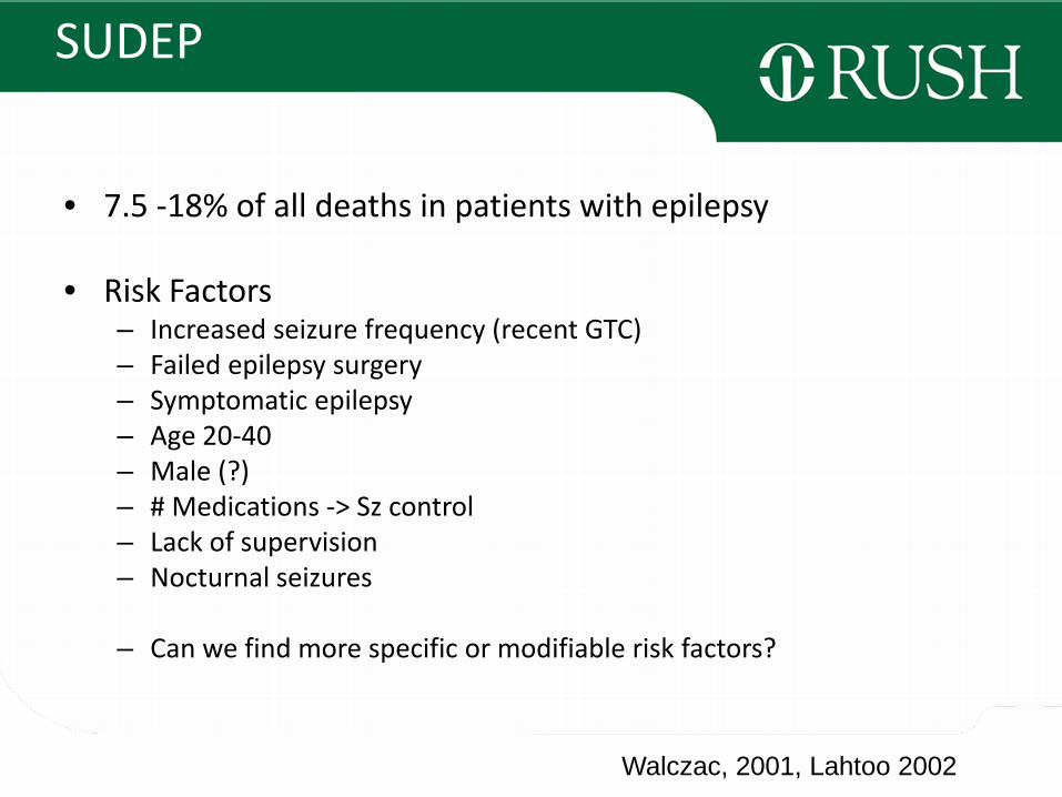
• 7.5 -18% of all deaths in patients with epilepsy
• Risk Factors – Increased seizure frequency (recent GTC) – Failed epilepsy surgery – Symptomatic epilepsy – Age 20-40 – Male (?) – # Medications -> Sz control – Lack of supervision – Nocturnal seizures – Can we find more specific or modifiable risk factors?
Walczac, 2001, Lahtoo 2002

The cascade leading to SUDEP

The heart hypothesis
• Malignant cardiac arrhythmias are the most frequent cause of Sudden death: – Coronary artery disease – Heart failure – Healthy hearts
• Acute cardiac rhythm changes are frequently seen in relation to seizures – They are usually benign – Not seen in recorded SUDEP or NSUDEP cases
Cerrone and Piori 2011, Nei et al. 2004, Oppenheimer et al. 2006

Neuro-cardiac Channelopathies
• Is there a genetic predisposition for SUDEP? – High proportion of SUDEP in pediatric epilepsy related
deaths (>20%) • Channelopathies involved in membrane excitability
could explain the association between epilepsy and increased risk for cardiac arrhythmias
• Family screening of patients with SUDEP have identified genes potentially related to epilepsy and premature death
• Mutations of these genes in 24% patients with personal or family h/o epilepsy and cardiac arrhythmias
• Mutations may predispose to excitability or alterations in cardio-respiratory homeostasis
Sillanpaa and Shinnar, 2011

Neuro-cardiac Channelopathies
• Long QT syndrome: Familiar can present as syncope, epilepsy, sudden death. Prolonged cardiac action potential. Mutations in these genes are related to LQTs. Genes encoding for: – KCNQ1 (type 1): Alpha subunit voltage gated Potassium
channel Kv7.1: Mice: abnormal neuronal repolarization, seizures, prolonged RR, inter-ictals and PVCs, asystole and autonomic dysfunction
– Types 2 and 3 reported in SUDEP cases. – KCNH2 (type 2): Protein Kv11.1. Mutations link to: Torsade
de Pointes, syncope, palpitations and SUDEP – SCN5A (type 3): Associated more frequently with seizures,
(personal or familiar) α subunit of V gated type V Na channel subunit
Sillanpaa and Shinnar, 2011

Neuro-cardiac Channelopathies
• Short QT syndrome: Increased risk for atrial arrhythmias, syncope and sudden deaths. Mutations in potassium channel genes. Leading to early repolarization. – Rufinamide may promote QT shortening – KCNH2, KCNQ1 and KCNJ2 – KCNA1:encodes for potassium channel subunit Kv1.1: mutations: severe
epilepsy, AV blocks, bradycardia, PVC. Mice have seizures and die prematurely. Kv1.1 may play a role in membrane stabilization
• Dravet syndrome – Loss of function mutation of SCN1A gene – Severe epileptic encephalopathy with high mortality rate. Loss of function
mutation of SCN1A encoding for V gated Na channel NAv1.1 that leads to reduced firing in GABAergic neurons. Expressed in brain and heart.Moels have ictal bradycardia an d premature death. Maybe a seizure induced cardiac arrhythmia?
• Brugada syndrome – Loss of function mutation of SCN5A gene – Responsible for 4-12% sudden deaths, 20% if only patients with normal hearts.
ST elevation in V1-V3, syncope, seizures and sleep abnormalities. May be unmasked by Na channel blockers, fever, alcohol, cocaine. Has been described in association with cryptogenic frontal lobe epilepsy and familiar generalized epilepsy

Other risk factors
• Catecholaminergic Polymorphic VT – Associated with cardiac death in young individuals with normal hearts and
normal QT. – Death follows tachycardia due to catecholamine release in response to
exertion or emotional stress – Sudden death can be seen following a generalized motor seizure which
triggers excessive adrenergic stimulation – Mutation in Ryanodine receptor gene RYR2 and Calsequestrin 2 (CASQ2) – Diagnosis requires 24hs monitoring showing CPVT in response to exercise.
• HR Variability – Reduced HRV in animal models and patients with chronic epilepsy – Reduction of HRV is progressive over time in pts with refractory epilepsy. – Results from recurrent ictal autonomic challenges – Unclear relation to SUDEP. But, may be a marker of autonomic vulnerability – Does not reverse after successful surgery – Reduced HRV: TLE> IGE>>controls

Other risk factors
• Autonomic dysregulation • Central regulation of cardiovascular autonomic nervous
system (Anterior insular cortex, anterior cingulate gyrus, amygdala, ventro-medial prefrontal cortex)
• Patients with refractory TLE, ictal asystole and failed epilepsy surgery have reduced postganglionic sympathetic innervation.
• Abnormal blood pressure variability and abnormal response to Valsalva in chronic epilepsy
• Sympathetic activation with parasympathetic suppression • Significant autonomic vulnerability seen in the postictal state
following a GTCS • Autonomic dysregulation is more prominent in younger
patients with GTCS and intractable epilepsy

Other risk factors
• Chronic epilepsy: higher sympathetic tone, lower parasympathetic tone
• May be related to the action of medications. • The relation of sympathetic denervation and SUDEP
assumes induced increased beta adrenergic sensitivity in the heart.
• Recurrent seizures may also induce a hyper adrenergic state, contraction band necrosis consistent with subendocardic ischemia. With predisposition for arrhythmias.
• Sudden death can be seen following a generalized
motor seizure which triggers excessive adrenergic stimulation

Medications
• Medications are related to autonomic regulation • Effects are difficult to isolate • Decreased HRV associated with polypharmacy • Special considerations with:
– Carbamazepine: Dose dependent decreased HRV, more pronounced in the elderly, reversible
– Lamotrigine: Cardiac death in association with neuro-cardiac channelopathies
– Lacosamide: Dose dependent, reversible, cardiac conduction abnormalities
– Not being treated for Epilepsy increases risk of SUDEP

Ictal cardiac events
• Ictal ECG changes are most frequent clinical manifestation of seizures.
• Most ECG changes are benign • Potentially serious arrhythmias reported in up to
6%
• 39% of pt. with refractory epilepsy have one or more peri-ictal arrhythmias: asystole, A fib, supra ventricular blockades, bundle branch blocks. More is GTCS

Ictal events
• Ictal tachycardia – 76-99% observed in EMU – May precede EEG and clinical onset – Higher maximum ictal HR and greater rate of HR increase in
SUDEP patients. (Nei et al. 2004) – IT can complicate a predisposing repolarization abnormality
• Ictal bradycardia and asystole – Ictal manifestation of focal seizures and TLE – Incidence 2-4%, IA 2.7-4.0 per 1000 patients in EMU (20% if loop
recorders implanted) – High seizure frequency and seizure clusters – Typical semiology – Recovery from IA is similar to other benign vagal-mediated
asystole events – IA seems to reduce seizure duration

Ictal events
• Ictal repolarization abnormalities – Ictal QT interval changes
• Short QT in relation to GTCS • Ictal QT prolongation and third degree AV block • Ictal repolarization abnormalities may be seen in response to
hypoxemia or hypercapnia • Malignant ictal arrhythmias
– Few cases of pure cardiac NSUDEP events reported • Ictal cardiac ischemia
– Peri-ictal ECG findings suggestive of ischemia in up to 49% following seizures
• More frequent with longer seizures, prolonged tachycardia or hypoxemia
• Subtle elevation in CK-MB • No evidence of postictal Troponin elevation if no CAD

Post Ictal EEG Suppression
• EEG may flatten after seizure termination • Generalized convulsive seizures 27-82% • Focal seizures 1-3% • PGES: Postictal unilateral or bilateral suppression > 1 second and
<10 microvolt • May be difficult to see • Patients are motionless postictally • Positive relation to the duration of tonic phase • PGES > 50 sec, may be SUDEP risk • May also be a mechanism of seizure termination • PGES: Enhanced activity of inhibitory neuronal networks,
potentially enhanced by sleep and hypoxia • May be a biomarker or predictor for SUDEP
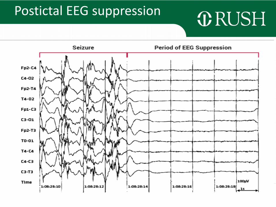
Postictal EEG suppression

Respiratory abnormalities, pulmonary edema, hypoxemia and SUDEP
• Ictal apnea and hypoxemia are frequent in GTCS • Respiratory abnormalities precede cardiac
arrhythmias in witnessed SUDEP • Strong evidence for central and obstructive
respiratory dysfunction in SUDEP models • Peri-ictal hypoxemia and hypercarbia • Early intervention reduces the duration of
postictal hypoxemia, immobility and total seizure duration
• Pulmonary Edema 50% following GTCS. It is associated with seizure duration (>100 seconds)

Serotonin (5HT)
• Link between respiratory drive, postictal EEG Suppression and Impaired Arousal
• It plays a role in arousal and respiratory control (Respiratory drive 5HT2A)
• 5-HT dysfunction is linked to SIDS • Transgenic mice lacking 5-HT neurons have
decreased respiratory output and are more vulnerable to SUDEP
• Serotoninergic interventions may be considered to decrease SUDEP risk (SSRIs)

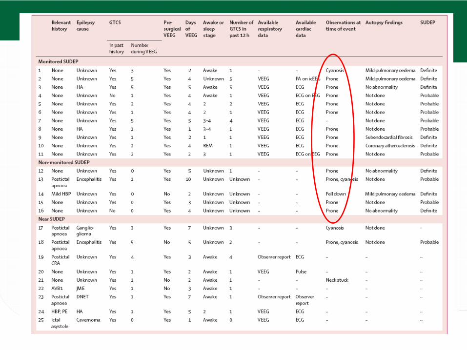
MORTEMUS
• 147 EMUs • 16 Definite SUDEP
– All after a GTCS – 14/16 At night – GTC Rapid breathing (18-50bpm) Bradycardia/apnea/PGES +/-Tachycardia
• 9 Near SUDEP – 7/9 after GTCS – CPR was successful when started within 3 minutes
Ryvlin, et al. Lancet Neurology 2013


Rajakulendran, J Clin Neurophysiol, 2015

• MORTEMUS: “SUDEP primarily follows an early postictal, centrally mediated, severe alteration of respiratory and cardiac function induced by a GTCS, leading to immediate death or a short period of partially restored cardio-respiratory function followed by terminal apnea and then cardiac arrest”

Potential prevention strategies
• Pay close attention to personal and family history of cardiac arrhythmias or sudden death. – Consider cardiac workup and ECH Holter monitoring
• Inquire about syncope, palpitations, CAD • Medication specific cardiac effects should be considered
when choosing AEDs • Consider medication interactions • Consider baseline ECG in vulnerable populations before
choosing AEDs – Poorly controlled epilepsy – Convulsive seizures during sleep
• Follow up ECG when combining medications with potential supraventricular arrythmogenic effects.
• Avoid Prone position in the postictal state

Potential prevention strategies
• In the EMU: • Multimodal cardio-respiratory monitoring • Stay with the patient until EEG is continuous or
returns to baseline • Early activation, mobilization, suctioning, oxygen,
repositioning: Reduce the duration of postictal hypoxemia, immobility and total seizure duration
• Optimize seizure management • Talk about SUDEP

Thank you!