pdf hosted at the radboud repository of the radboud ... · andy j.g. pötgens,* nicolette h....
TRANSCRIPT

PDF hosted at the Radboud Repository of the Radboud University
Nijmegen
The following full text is a publisher's version.
For additional information about this publication click this link.
http://hdl.handle.net/2066/21464
Please be advised that this information was generated on 2017-12-05 and may be subject to
change.

American Journal o f Pathology, Vol. hi(). No, I. January /W S ’c',o¡\yrii\b( (0 A m m a w Soa'ety /'or Iwvstigatitv Pathology
Vascular Permeability Factor Expression Influences Tumor Angiogenesis in Human Melanoma Lines Xenografted to Nude Mice
Andy J.G. Pötgens,* Nicolette H. Lubsen,1- Margarethe C. van Altena,*John G.G. Schoenmakers,t Dirk J. Ruiter,* and Robert M.W. de Waal*h'rom the Iustiliih vrsit)
'gen, a n d the Department o f MolecularUniversity o f
♦‘gen, ‘gen, Thi
We studied the expression o f the angiogenic fa c tor vascular permeability factor) (VPF, also called vascular endothelial growth factor), in human melanoma cells in vitro and in viva Melanoma lines that develop tumors with a low metastatic potential in nude mice were found to have low expression levels o f VPF in vitro, and the VPF expression levels in melanoma lines that
metastatic xenografts ivere high. However, in vivo the correlation between VPF mRNA levels and the frequency o f metastasis was lost; in all xenografts equally high levels o f VPF mRNA were found, independent o f the parental cell line. Hence, in vivo VPF gene expression was upregulated in the low expressing lines. The ex
maybe hypoxia, given that we found that low oxygen tension caused a. ( reversible) increase in the VPF mRNA levels in otherwise low expressing melanoma lines in vltra A melanoma line with an inducible VPF expression was engineered into a line with a constitutive VPF expression. In the xenografts from this line a change in the vascular architecture was seen, indicating that the pattern or the level o f VPF expression is important fo r tumor angiogenesis in melanoma xe-nogt s. (Am f Pathol 1995, 146:197-209)
ferent receptors for VPF have been characterized: fms-like tyrosine kinase (fit) and fetal liver kinase 1 (flk-1/kinase insert domain-containing receptor). These appear to be expressed almost specifically by endothelial cells and hematopoietic cells.3 6 VPF is an important angiogenic factor, along with other factors such as acidic and basic fibroblast growth factor (aFGF, bFGF) and transforming growth factor-/3 (TGF- /3).7,8 Apart from its expression by some normal, well- vascularized tissues and embryonic tissues, and during wound repair,9" 12 VPF has been found in many tumors and tumor cells.13 In glioblastoma VPF expression was found to be highest near necrotic areas, and clusters of newly formed capillaries were found around the sites of VPF production,14,15 suggesting that VPF can be recruited to augment angiogenesis if the tumor vasculature and therefore the oxygen supply is insufficient. The role of VPF in tumor angiogenesis was confirmed by blocking its activity in tumors by a monoclonal antibody, and by application of a dominant-negative VPF receptor mutant.m ,/ These treatments led to a decrease in angiogenesis and to slower tumor growth.
Several protein variants of VPF exist because of alternative splicing of the VPF mRNA. The molecular variants differ in their efficiency of secretion. The smaller forms ( V P F , and VPF„i;,) are efficiently secreted by the producing cells, and can easily reach their target cells. The larger forms (VPF fM<t and VPF™,;) are retained at the extracellular matrix, but biologically active parts of these proteins can be re-leased by plasmin. in ;'ii Theoretically, different celltypes may benefit from particular VPF variants. Tissue-specific expression of certain VPF messenger variants has indeed been demonstrated.19,21
As a consequence of its proposed role in tumor angiogenesis, VPF may also facilitate metastasis, as
Vascular permeability factor (VPF), also known as vascular endothelial growth factor, is a glycosylated, secreted protein factor that increases blood vessel permeability, stimulates endothelial cell division in vitro, and induces angiogenesis in wVo.1,s Two dif-
Supported by the Dutch Cancer Society (NKB grant 90-02),
Accepted far publication October 5, 1994,Address reprint requests to Dr. R, M. W. do Waal, Institute at Pa
thology, University Hospital Nijmegen, P. 0. Box 9101, N-6500HB Nijmegen, The Netherlands,
197

198 Potgens et atAJP January 1995, Vol. 146, No. I
this process is dependent on the vascular bed.22 To study the relation between VPF expression and metastasis, a panel of human melanoma cell lines was used. Xenografts of these melanoma lines in nude mice give rise to tumors with distinctly different biological behavior. Some of these lines develop into t rapidly metastasizing tumors, whereas other lines metastasize at a low frequency or very slowly (see ref. 23; J.R. Westphal, J.A.W.M. van der Laak, C.J.M. Schalkwijk, D.J. Ruiter, R.M.W. de Waal, manuscript in preparation). The metastatic phenotype of these lines was found to correlate with the expression of a series of genes or antigens, such as urokinase-plas- minogen activator and its type 1 inhibitor, various integrins, the epidermal growth factor receptor, thymosin j810, and calcyclin.24-28 We report here that low and highly metastatic melanoma lines have distinctly different expression patterns of VPF. Highly metastatic melanoma lines have constitutively high levels of VPF expression, whereas the less metastatic lines have a low level of VPF expression in culture, which is elevated in mouse xenografts. In vitro data suggest that hypoxia may be an important trigger in this upregulation of VPF gene expression. Transfection experiments show that an alteration of VPF expression in melanoma xenografts profoundly affects vascular architecture. Hence, at least in this melanoma model, the VPF expression pattern is an important determinant of angiogenesis, and possibly of angiogenesis-dependent biological behavior.
Materials and Methods
Cell Culture
Human melanoma cell lines were cultured as previously described.23 Transfected melanoma lines were cultured in medium supplemented with 200 pg/ml hygromycin B (Boehringer Mannheim, Germany). Human brain capillary pericytes were isolated and cultured as described elsewhere.20 U937 cells and Balb-3T3 cells were from American Type Culture Collection (Rockville, MD) and cultured In Dulbecco’s Modified Eagle’s Medium (DMEM, Flow Laboratories, Irvine, UK) with 10% fetal calf serum (FCS, Gibco BRL, Paisley, UK), 2 mmol/L L-glutamine and 40 jjg/ml gentamycin (Schering Corporation, Am- stelveen, The Netherlands).
Melanoma Xenografts
Human melanoma cells were trypsinized and 2 x 10⁶ cells were injected s.c. into BALB/c nu/nu mice as previously described.23 Xenografts were dissected at
different time points after inoculation, measured, rapidly frozen in liquid nitrogen, and stored at -70 C.
Isolation of RNA
RNA was isolated from cultured cells either using LiCI/ urea,30 or using guanidinium chloride.31 RNA from tumors and normal murine tissues was isolated after disruption in guanidinium isothiocyanate by CsCI centrifugation.32 RNA concentration was determined spectrophotometrically at 260 nm.32
Northern Blotting and Probes Used
Samples of total RNA were denatured in 50% form- amide in formaldehyde/phosphate buffer (65 mg/ml formaldehyde, 25 mmol/L NaH2P04/Na2HP04, pH 7.0) for 15 minutes at 55 C, run on a 1% agarose gel in formaldehyde/phosphate buffer, and blotted to nitrocellulose membranes.32 As a probe for VPF mRNA, a fragment was used which comprised the protein coding region of VPF165. To isolate this fragment, a reverse transcriptase (RT) polymerase chain reaction (PCR) was performed on RNA from phorbol 12- myristate 13-acetate-treated U937 cells,1 using reverse primer (5'-TTCCTCCTGCCCGGCTCACCG-3') and forward primer (5'-CCCGGTCGGGCCTC- CGAAACCA-3'). Blunt-ended PCR products were cloned into Smal-digested pUC19 vector DNA and characterized by sequencing. PCR products from the alternatively spliced messengers for VPF121, VPF165, and VPF189 were identified. A 0.6-kb VPF165 insert was isolated as an Xjbal/Kpnl-fragment and labeled. Control hybridizations were performed with a human ubiquitin probe,33 a human 0-actin probe,34 or with a Drosophila ribosomal RNA probe. Fragments were labeled with [cv-32P]dATP after random hexamer priming.35 Northern hybridizations were performed in 50% formamide, 6 x SSC, 0.1 % SDS, 0.1 mg/ml denatured herring sperm DNA and 5 x Denhardt's32 at 45 C, followed by washing in 0.2 x SSC, 0.1% SDS at 65 C. Intensities of hybridization bands were scanned with an Ultroscan XL Enhanced Laser Densitometer from LKB (Bromma, Sweden).
Analysis of Alternatively Spliced VPF mRNA
2 to 8 pg of total RNA isolated from cell lines or melanoma xenografts was reverse transcribed using 0.5 jjg reverse primer (5'-TTCCTCCTGCCCGGCTCA- CCG-3'), 0.4 mmol/L deoxyribonucleotides (dNTPs; Pharmacia LKB, Woerden, The Netherlands), five

VPF in Melanoma Angiogenesis 199AJPJcmitaty 1995, Vol. M6, No. i
units avian myeloblastosis virus (AMV)-RT (Strat- sity, cells were scraped and lysed, and the cellularagene, San Diego, CA), five units RNasin (Boehringer protein content was determined by a standard proteinMannheim), and reaction buffer provided by Strat- assay (Bio-rad, Veenendaal, The Netherlands).agene, in final volumes of 10 ¡jI, for 1 hour at 42 C. Based on this protein determination, samples of con-After ethanol precipitation and redissolving in 10 pi ditioned media derived from equal amounts of celldistilled water, half of the cDNA was used in PCRs material were taken, and proteins were acetone pre-containing 0.3 pg reverse primer (see above), 0.3 pg cipitated, electrophoresed on 17.5% polyacrylamideforward primer (5'-GCACCCATGGCAGAAGGA- GGA-3'), 0.2 mmol/L dNTPs, and 0.25 units Supertaq
gels under reducing conditions, and electroblotted to nitrocellulose membranes. Antisera against VPF wore
polymerase and reaction buffer provided by HT Bio- raised in rabbits using purified VPF produced in atechnology (Cambridge, UK) in final volumes of 25 pi. bacterial expression system. Antiserum was diluted20 cycles of 1 minute at 96 C, 1.5 minutes at 52 C, and 1:100 or 1:250 for detection of VPF on Western blots,4 minutes at 72 C were performed. 5 pi of every re- and the immune reactions were visualized using theaction mix was transferred to a new tube containing chemiluminescent substrate AMPPD (Tropix, West-the same reagents, for one more cycle (6 minutes at burg, Leusden, The Netherlands) following the manu-72 C). 10-pl samples of the products were separated facturer’s protocol. The crude antisera had some non-on a 1.5% agarose gel, Southern blotted to nitrocel specific reactivity toward some bands of higherlulose and probed with the VPF165 fragment in form- molecular weight than VPF on Western blots. Whereamide hybridization mix at 42 C. indicated this was diminished by affinity purification of
anti-VPF antibodies using purified Escherichia coli-
Determination of the Species Origin ofproduced VPF absorbed by nitrocellulose mem-I 0 7branes.
VPF mRNA
1 to 12 pg total RNA isolated from human cell lines, VPF Induction Experimentsmurine tissues, or melanoma xenografts was reverse transcribed using 0.5 pg reverse primer (5'-TTGGT- GAGGTTTGATCCGCAT-3'), 0.4 mmol/L dNTPs, 5 units AMV-RT, 10 units RNasin, and reaction buffer provided by the supplier of AMV-RT (Stratagene) in final volumes of 10 pi for 1 hour at 37 C. After ethanol precipitation and redissolving in 10 pi distilled water, half of the cDNA was used in PCRs containing 0.3 pg reverse primer (see above), 0.3 pg forward primer (5'-CGAAACCATGAACTTTCTGCT-3'), 0.2 mmol/L dNTPs, 0,25 units Supertaq polymerase, and reaction buffer provided by FIT Biotechnology, in final volumes of 25 pi. 20 cycles of 1 minute at 96 C, 1.5 minutes at 48 C, and 2.5 minutes at 72 C were performed. 1-pl samples of the products were transferred to new tubes with all the PCR reagents, and subjected to one more cycle (10 minutes at 72 C). PCR products were then precipitated and redissolved. One-third was digested with five units Sty\, which only cleaves the hu-man product, separated on a 1.5% agarose gel,Southern blotted, and hybridized with the VPFU;; probe.
The response to serum, fibroblast-conditioned medium, or growth factors/cytokines was tested after preculturing subconfluent melanoma cell cultures in DMEM without serum for 24 hours. Medium was then replaced for 4 or 24 hours by DMEM with 10% FCS, or by DMEM (with and without FCS) conditioned by Balb-3T3 cells for 24 hours. Alternatively, serum-free medium was replaced for 4 hours by DMEM containing 0.1% bovine serum albumin and one of the following growth factors or cytokines: 5 ng/ml EGF (Collaborative Research, Bedford, MA); 5 ng/ml TGF-<* (Bachem, Bubendorf, Switzerland): 2 ng/ml TGF-01 (R & D Systems, Minneapolis, MN); 10 ng/ml platelet- derived growth factor AA (PDGF-AA, a gift from Dr. C.H. Heldin, Uppsala, Sweden); 100 ng/ml bFGF (agift from Scios Inc., Mountain View, C A );..100 ng/mlVPFio;, (in culture supernatant of COS cells transfected with plasmid containing VPF1(iil cDNA under control of an SV40 early promoter); 50 ng/ml INF-«; 100 units/ml interleukin-4 (IL-4); 400 units/ml interferon gamma (IFN-y) (TNF-«, IL-4, and IFN-y were from Boehringer Ingelheim, Germany) 100 units/ml IL-1ß; 50 units/ml IL-2 (IL-1 ß and IL-2 were from Gen »
Analysis of Secreted VPF Protein zyme, Sanbio, Uden, The Netherlands). Hypoxia experiments were performed using subconfluent
Melanoma cells were cultured for 24 hours in serum- free medium when indicated with 100 ug/ml heparin.
melanoma cell cultures in 10 mmol/L Hepes-buffered DMEM with 10% FCS. Rubber-capped flasks were
Conditioned media were centrifuged and used for flushed for 1 hour at room temperature with 30 vol-VPF analysis. To correct for differences in cellular den- umes of nitrogen made oxygen-free using a BASF R
200 Pötgens et alA/PJcmiuuy 1995, Vol. 146, No. /
3-11 catalyst (BASF, Ludwigshafen, Germany). Oxygen-free C 02 was added up to 5%, and the flasks were then placed back at 37 C for the indicated times. At the end of each Incubation, gas samples were tested for 0 2 content by gas chromatography, and the pH of the media were determined. In some cases, the P0n of the media were also determined (CIBA-Corning
C.J.M. Schalkwijk, D J. Ruiter, R.M.W. de Waal, manuscript in preparation), and which is suitable for the examination of tumor vasculature. Also, tumor sections were stained with a polyclonal rabbit anti-mouse lamlnin antiserum (provided by Dr. J. van den Born, Department of Nephrology, Nijmegen, The Netherlands).
288 Blood Gas System, Houten, The Netherlands). Control experiments were performed (in air/5% C 02) to investigate the influence of the pH of the medium. To achieve a low final pH, cells were grown In Hepes- buffered medium for several days, and to achieve a high pH medium was replaced for 6 hours by Hepes- buffered fresh medium preincubated In air for 1 hour.
Production of Stably Transfected Melanoma Lines
The protein coding region of VPFi2i was cloned di- rectionally as a Xba\/Kpn\ fragment into the expression vector EBOpLPP,38 by which the sequence was brought under control of SV40 transcriptional elements. This plasmid also codes for the hygromycin phosphotransferase (HPH) gene. The recombinant plasmid and the empty vector were digested with Apa\, thus eliminating some nonessential sequences. The residual 8-kb fragment was transfected into Mel57 cells by calcium phosphate precipitation followed by a 3-minute shock with 25% DMS0.31 After 48 hours the cells were trypsinized, seeded sparsely in culture flasks, and selected in medium with 200 pg/ml hygromycin B (Boehringer Mannheim). Single resistant colonies were removed by scraping and grown until analysis of VPF expression, and storage of stocks in liquid nitrogen was possible.
Histological Analysis of Tumors from Transfected Melanoma Lines
Transfected melanoma lines were inoculated in nude mice as described above. Every line was injected in at least two mice on both flanks. Tumor volumes were measured weekly, and growth curves were prepared. Tumors of sizes between 150 and 800 mm3 were excised 30 to 60 days after inoculation, and cut Into three fragments. One part was formalin-fixed and used to study overall tumor morphology. The other parts were snap frozen in liquid nitrogen; one was used for RNA isolations, the other to study the vascular patterns and the organization of stroma and extracellular matrix. Cryosections were stained with monoclonal 9F1, which reacts specifically with mouse endothelium (J.R. Westphal, J.A.W.M. van der Laak,
Results
Analysis of VPF mRNA in Cultured Melanoma Cells
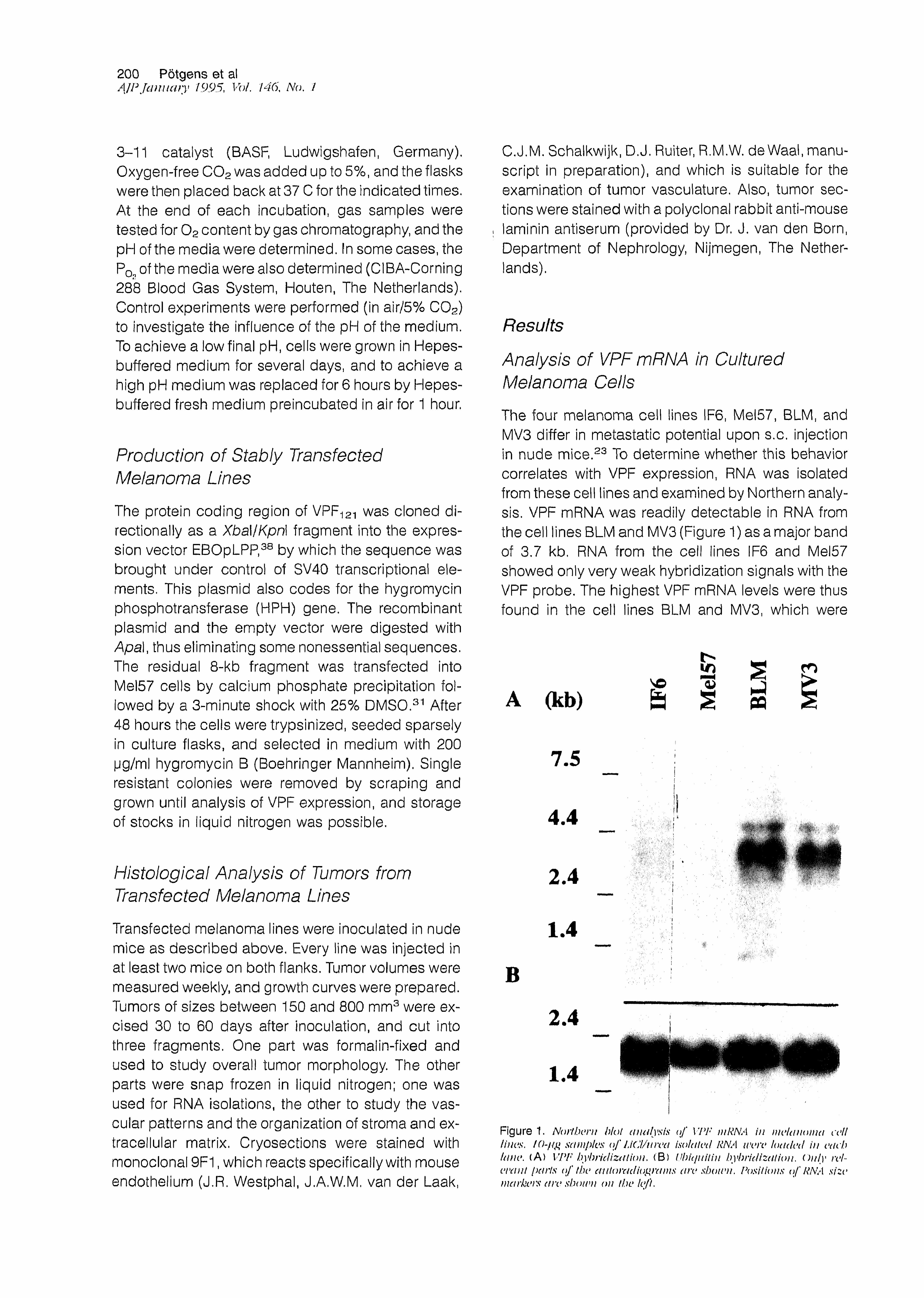
The four melanoma cell lines IF6, Mel57, BLM, and MV3 differ in metastatic potential upon s.c. Injection in nude mice.23 To determine whether this behavior correlates with VPF expression, RNA was Isolated from these cell lines and examined by Northern analysis. VPF mRNA was readily detectable In RNA from the cell lines BLM and MV3 (Figure 1) as a major band of 3.7 kb. RNA from the cell lines IF6 and Mel57 showed only very weak hybridization signals with the VPF probe. The highest VPF mRNA levels were thus found in the cell lines BLM and MV3, which were
(kb)V©
h iPQ
7.5V . V Ó . . .. -cn:. ■
4.4 if: i;:.v
; : j ‘ ‘
. . <. .• .. v . ,
; ■
2.4, •/ Ä ' j : : v .\ •; •.
• V >. •
1
I
.'I
\
VpXfs--. '•% '■ v-v-.v
■ : '■■■: /■.
1.4• ■
>
/ " - a " '■ ;. ■ ■
: r- r
V y ,f' '' ' : ■
■. : y 0 - '
‘ ‘ V V ‘ ' y
■■ vl . . .
. : * 'iïèM
B
2.4
1.4
Figure 1. Northern blot analysis of VPF mRNA in melanoma cell lines. / sampl es o f i.iCl/urea isolated RNA were loaded in each lane. (A) VPF hybridization . (B) Ubitjuiiin hybridization, Only relevant f)arts of the ciutoradiograins are shown. Positions o f RNA size markers are shown on the left.

VPF in Melanoma Angiogenesis 201AJP January 1995, Vol. 146, No. 1
shown in earlier studies to give rise to highly metastatic tumors in nude mice. The other lines, which had a low level of VPF mRNA expression, produced tumors with a much lower metastatic frequency.23
The observed difference in expression of VPF mRNA is not likely to be caused by a genomic rearrangement or an amplification of the VPF gene. Southern analysis of the genomic DNA isolated from these melanoma lines and from normal human buffy coat showed no obvious differences in intensities of the bands hybridizing with a VPF probe, nor were shifts of bands or extra bands observed in any of the melanoma lines (data not shown).
Alternatively spliced messengers of VPF cannot be distinguished by Northern blotting. To determine whether the melanoma lines differ in the ratio of these mRNA variants, an RT-PCR was performed, which amplified the alternatively spliced region of the messenger. RNA from all the melanoma cell lines yielded the same pattern in that the product from the RNA coding for VPF121 was the most prominent (52 to 70%), followed by the VPF165 product (26 to 42%). The VPF-,89 mRNA was in all cases found to be a minor species (4 to 8%), whereas no VPF206 mRNA was found in any of the melanoma cell lines (Figure 2, lanes 2 to 5). No correlation between the biological behavior of the melanoma lines and the predominance of the VPF variants produced could therefore be demonstrated. The observed ratio of splice variants does not seem to be specific for melanoma cells or even for tumor cells, as the same ratio of splice variants was found in RNA from the human lymphoma cell line U937 and in RNA from normal human pericytes (Figure 2, lanes 6 and 7).
Cell lines Xenografts
<%oc
(kb)oat
.59
.50 I*3i5 mm*
SO _£¡3 S 2
s33
x n
:.V>
VPF ,*VPF,« VPF,2|
I".**
Figure 2. Analysis of VPF mRNA splice rariants . RT-PCR was per- form ed on RNAs isolated from rations sources. amplifying the alter- natively splia *d t vgit > i1 as iudk a ted in Matt * rials an d Ah *tin uls. ( Left) Unman cell lines.' IF(h Mel 5?, A / i \ i PLM { melanoma cells), U9J? ( histiocytic lymphoma ceils), pericytes t front human brain capillar i t *v). ( R igI > t ) ,1 tela noma xent / v //As; st x * a 1st > Fig tt re i legend. The left and the right panels are from different gels, Southern blots were by t nidi zed with a VPF probe, ¡Positions a f DNA size markers are shown on the left, Expected PCR [products.* VPFij/ ( . ><S'/ bp), VPFiut ( 513 bp)t r/V*'/*,, ( W * bp), lYYvfjf, i bp), Nnte that no PCR products are expected from mouse VPF mRNA. as the primer's do not match the murine sequence completely.
Levels of Secreted VPF Protein Correlate with the VPF mRNA Levels
The amount of VPF protein secreted from the melanoma cell lines was determined by culturing the cells In serum-free medium containing heparin, to release also the longer VPF variants from the extracellular matrix.20 The proteins in the conditioned medium were Western blotted and stained with a polyclonal antibody against VPF. In the conditioned media from cell lines BLM and MV3 detectable amounts of VPF were present. Several bands with apparent molecular weights between 17 and 25 kd were identified (Figure 3), which agree with the expected sizes of nonglycosylated and glycosylated V P F i 21 andVPF165.19'20 VPF189 and VPF206 (expected around 30kd) could not be found, supporting the finding that their mRNAs are poorly expressed In melanoma cells (Figure 2; the band In the MV3 lane at 38 kd Is probably caused by a dimer of VPF12i or by a nonspecific reaction rather than by a monomeric form of one of the larger VPF variants). In conditioned media from cell lines IF6 and Mel57 no VPF could be detected, which agrees with the fact that these cells contain low amounts of VPF mRNA (Figure 1). These results in-
l >i n
kDj • • j . . 1 V ...
I .
i. ;
i. i.: r: ■ >
■■Vi- 6 6
i*
I :..v’ : '■i '-. i'/ ''. ‘ ■ ‘ ' ' ' ' ■, V : • '
• •• = -Va' •' ■ ■ " • •' '■■■.'■ /'■ V; ■ ■'] : ! : ; : i;' V '
• i,. • s ii' ' ''‘k ‘ * *
45•. r
*•'*. :-|i. ' J :.• • :i • • ! ' i • •
'■ '• ;r! J : ; V - V ■ ■ VVì V - -,:::■ A§mmM ' - . . v
31
21.5
••• :i Vi •. ; ,y. ; •:>
' •. I • 1 :l ' 14.4
Figure 3, Western blot analysis of VPF in conditioned media from melanoma cells. Gmjluent atllures of melanoma cells were grown in serum free medium with 100 ng/ml heparin for 24 hours, Precipitated proteins from 0.225 to 0,7 ml conditioned media { but derived from the same amount of cellular mass) were subjected to SDS-PAGE taider reducing conditions, Western blotted, and stained with affhiityintrified antiserum against VPF Molecular weight markers are shown on the right.
202 Pôtgens et alAJP Ja n uary 1995, Vol. 146, No. 1
dicate that the levels of VPF mRNA In melanoma cells are reflected in the levels of VPF protein expression.
Analysis o f Melanoma Xenograft RNA
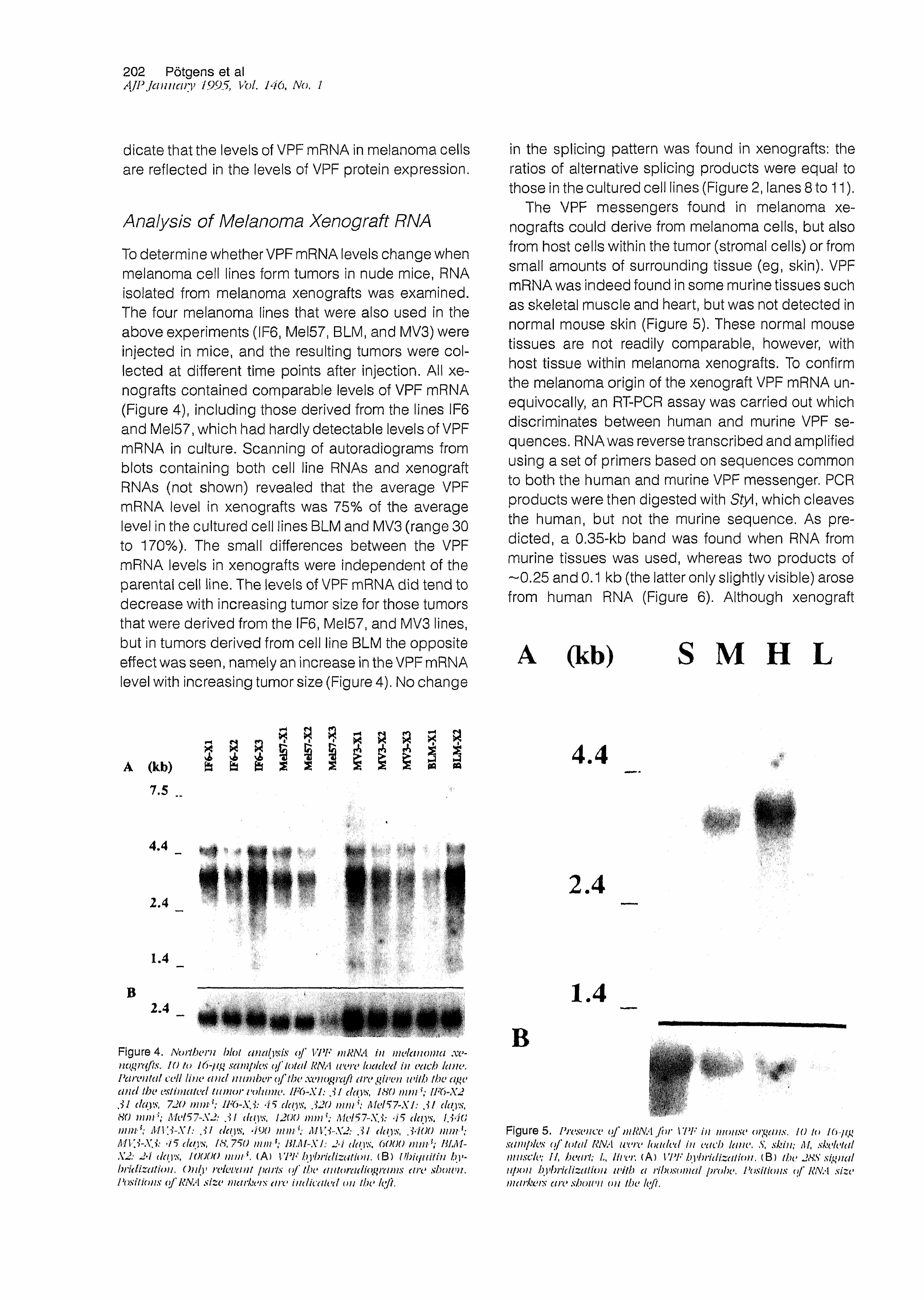
To determine whether VPF mRNA levels change when melanoma cell lines form tumors in nude mice, RNA isolated from melanoma xenografts was examined. The four melanoma lines that were also used in the above experiments (IF6, Mel57, BLM, and MV3) were injected in mice, and the resulting tumors were collected at different time points after injection. All xenografts contained comparable levels of VPF mRNA (Figure 4), including those derived from the lines IF6 and Mel57, which had hardly detectable levels of VPF mRNA in culture. Scanning of autoradiograms from blots containing both cell line RNAs and xenograft RNAs (not shown) revealed that the average VPF mRNA level in xenografts was 75% of the average level in the cultured cell lines BLM and MV3 (range 30 to 170%). The small differences between the VPF mRNA levels in xenografts were independent of the parental cell line. The levels of VPF mRNA did tend to decrease with increasing tumor size for those tumors that were derived from the IF6, Mel57, and MV3 lines, but in tumors derived from cell line BLM the opposite effect was seen, namely an increase in the VPF mRNA level with increasing tumor size (Figure 4). No change
in the splicing pattern was found in xenografts: the ratios of alternative splicing products were equal to those in the cultured cell lines (Figure 2, lanes 8 to 11).
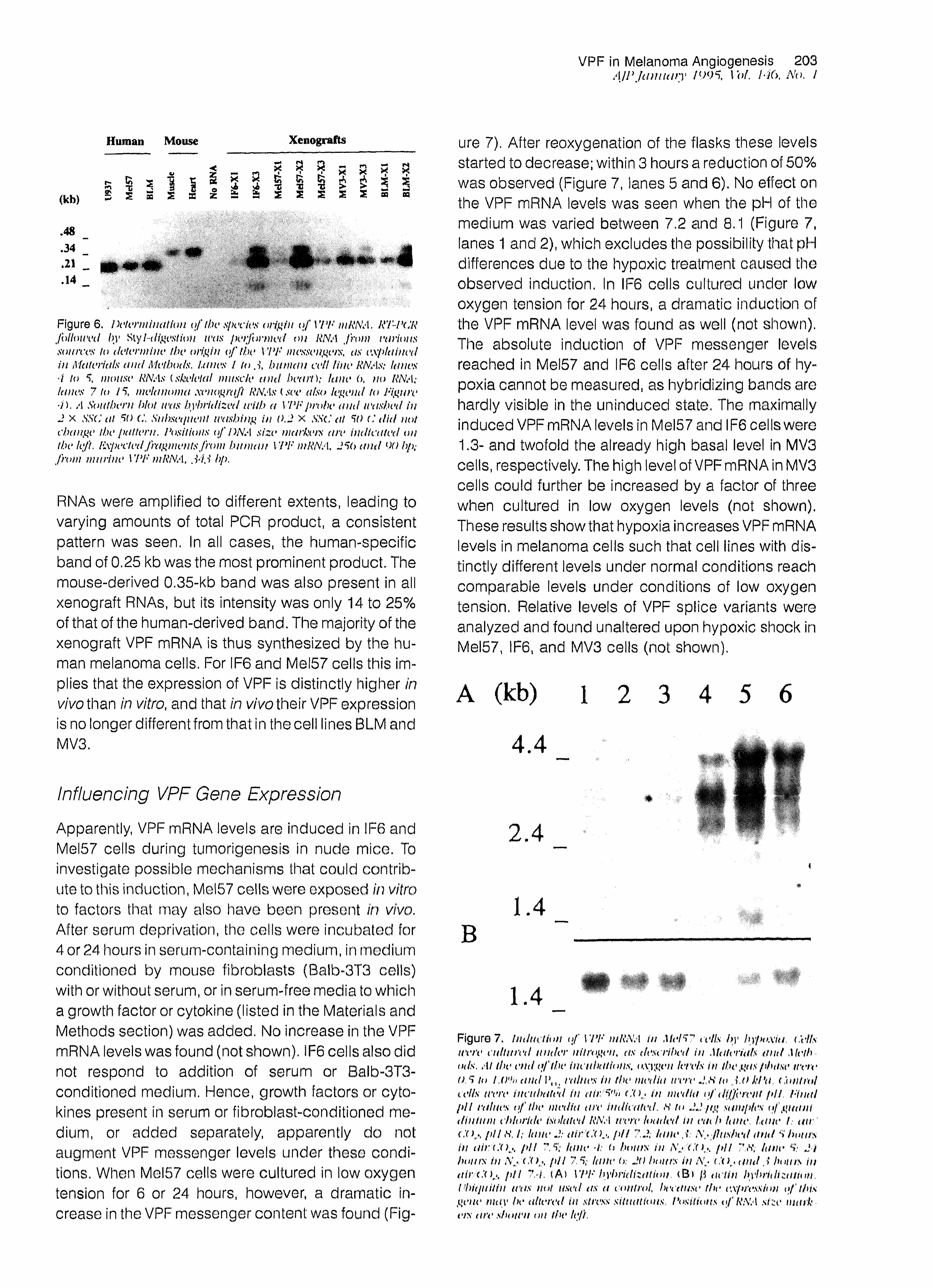
The VPF messengers found in melanoma xenografts could derive from melanoma cells, but also from host cells within the tumor (stromal cells) or from small amounts of surrounding tissue (eg, skin). VPF mRNA was indeed found in some murine tissues such as skeletal muscle and heart, but was not detected in normal mouse skin (Figure 5). These normal mouse tissues are not readily comparable, however, with host tissue within melanoma xenografts. To confirm the melanoma origin of the xenograft VPF mRNA unequivocally, an RT-PCR assay was carried out which discriminates between human and murine VPF sequences. RNA was reverse transcribed and amplified using a set of primers based on sequences common to both the human and murine VPF messenger. PCR products were then digested with Sty\, which cleaves the human, but not the murine sequence. As predicted, a 0.35-kb band was found when RNA from murine tissues was used, whereas two products of —0.25 and 0.1 kb (the latter only slightly visible) arose from human RNA (Figure 6). Although xenograft
A (kb) S M H L
A (kb)
7.5
B
4.4
2.4
1.4
2.4
• '• '*!•! \ i‘ '
1 i f/:; r \
• 'i •r • i;
.'-X.v/V .
• • . '.I. s .
:lllf . ; • • : S'
.i-,v S. .j'i'.'- '■■■. s...y
■;J y
:!^SA
'V;.';S
. ' ' : -'.I.' ‘ ‘
m i t mm
«m.-''O'-'
• :,•!! *V • S * . '
lÉICs î& û w & y - y
■mw&i'IKi
" i
. • >■>;■ - - . 'r i /
. y y $ 0 b ; i ï :<
w * *
m mlltl(kÊûi-
m m â S Ê È È m ë m Ê Ê Ê Ê a H Ê È Ë Ê È È u m. ■ x - m *' V ■ :
St<\t,(
m m WSi!
. i * . -,-v'< /hr,y ^ y i y .
?:f:îv m
!r . v ftÇj. z<>m -
Figure 4. Northern blot MuiJysis o f VPF mRNA in melanoma xe~ nog)nfts. 10 to 16-//£ sam pk*s o f total RNA 11vre k uuted i11 eacb lane. Parental cell line a n d number o f the xenograft are given with (he age a n d the estimated tumor volume. IF6-X1: 31 days , ISO mm*; ¡F6-X2 31 days, 720 mni {; IF6-X3: 4 5 days, 320 mm \ Met57-XI: 31 days , HO m m 1; Met57~X2: 31 days, 1200 m m 1; Mel57~X3: 45 days, 1340 mm {; MV3-XI; 31 days. 490 m m \ MV3-X2: 31 days, 3400 m m 1: A11 ; i' 4 5 cla) iv, / <S\ 750 m m 1; BLM~X 1: 24 cUi) '.s'. 6000 m m l; HIJ1 X2: 2* i da\ % J ()()()() mm*, (A) VI *F b ) i> / id izatU m, (B ) If hi (¡nit in h\ bridizatiou, Only relevant parts o f the autoradiograms are shoivu i'ositions o f RNA size markers are indicated on the left.
4 . 4
••'v k M \
kl̂ 'ÿy'0y <
4 V:-
2 . 4
1 . 4
ik--
■■■-■ ’.-¿x 0M&. • . . J-if /. c, , . . 'ymm
Figure 5. Pn>saice o f mRNA fo r VPF in motisi* organs, 10 to I(>■7ig samples o f total RNA were loaded in each lane, S, skin; M, skeletal miisck>; I f hea 1 */; .A, lit er, (A ) VPF by*h/idizalion. ( B ) the 2HS siguat upon hybridization with a nhosomal probe, Positions o f RNA size markets aresbotvn on the left.
VPF in Melanoma Angiogenesis 203AJPJanuary 1995, I of, I4(>, No. I
Human Mouse Xenografts
(kb)r-ma
B 5V J
S as
x aS S oZ !
.48
.34
.14
Figure 6. Determination of tin* species origin o f VI *P in RNA, R’FPCR followed by I-digest nyti was performed t>n RNA from nations sources to determine the origin o f the VPF messengers, as explained in Materials an d Methods, Lanes 1 to , i hitman veil line RNAs; lanes 4 to mouse RNAs (skeletal muscle a n d heart); lane 6, no RNA; lanes 7 to 15 melanoma xenograft RNAs (see also legend to Figure 4). A Southern hlot urn hybridized with a VPF probe and washed in 2 X SSC at 50 C. Subset¡uent trashing in 0.2 X SSC at W C d id not change the pattern. Positions o f DNA size markers are indicated on the left. Iixpevted fragments Jhtm hitman VPF mRNA. 25(> a n d 90 bp; from murine VPF mRNA, 34J bp.
RNAs were amplified to different extents, leading to varying amounts of total PCR product, a consistent pattern was seen. In all cases, the human-specific band of 0.25 kb was the most prominent product. The mouse-derived 0.35-kb band was also present in all xenograft RNAs, but its intensity was only 14 to 25% of that of the human-derived band. The majority of the xenograft VPF mRNA is thus synthesized by the human melanoma cells. For IF6 and Mel57 cells this implies that the expression of VPF is distinctly higher in wVothan in vitro, and that in vivo their VPF expression Is no longer different from that in the cell lines BLM and MV3.
Influencing VPF Gene Expression
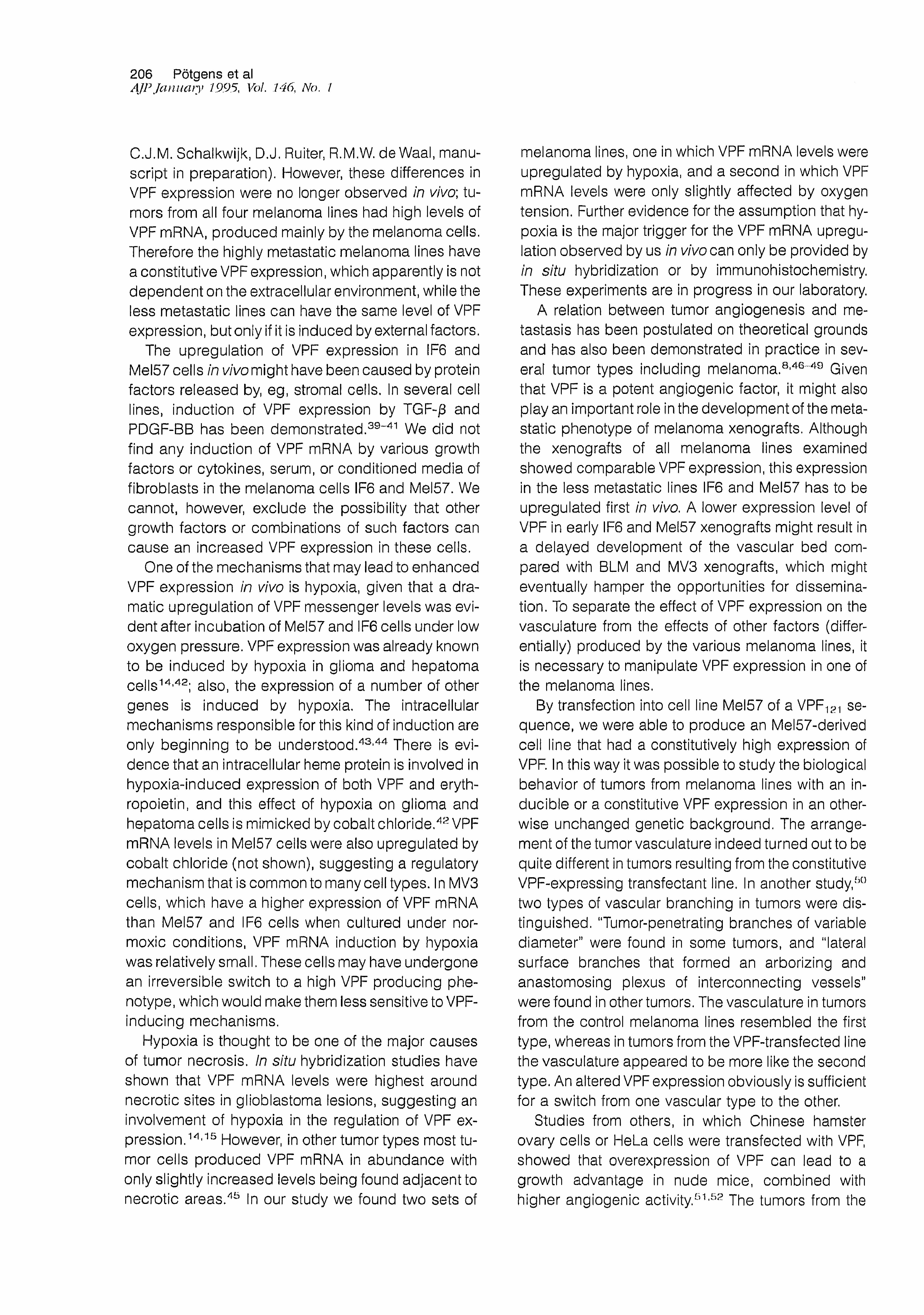
Apparently, VPF mRNA levels are induced in IF6 and Mel57 cells during tumorigenesls in nude mice. To investigate possible mechanisms that could contribute to this induction, Mel57 cells were exposed in vitro to factors that may also have been present in vivo. After serum deprivation, the cells were incubated for 4 or 24 hours in serum-containing medium, in medium conditioned by mouse fibroblasts (Balb-3T3 cells) with or without serum, or in serum-free media to which a growth factor or cytokine (listed in the Materials and Methods section) was added. No increase in the VPF mRNA levels was found (not shown). IF6 cells also did not respond to addition of serum or Balb-3T3- conditioned medium. Hence, growth factors or cytokines present in serum or fibroblast-conditioned medium, or added separately, apparently do not augment VPF messenger levels under these conditions. When Mel57 cells were cultured in low oxygen tension for 6 or 24 hours, however, a dramatic Increase in the VPF messenger content was found (Fig-
ure 7). After reoxygenation of the flasks these levels started to decrease; within 3 hours a reduction of 50% was observed (Figure 7, lanes 5 and 6). No effect on the VPF mRNA levels was seen when the pH of the medium was varied between 7.2 and 8.1 (Figure 7, lanes 1 and 2), which excludes the possibility that pH differences due to the hypoxic treatment caused the observed induction. In IF6 cells cultured under low oxygen tension for 24 hours, a dramatic induction of the VPF mRNA level was found as well (not shown). The absolute induction of VPF messenger levels reached in Mel57 and IF6 cells after 24 hours of hypoxia cannot be measured, as hybridizing bands are hardly visible in the uninduced state. The maximally induced VPF mRNA levels in Mel57 and IF6 cells were 1.3- and twofold the already high basal level in MV3 cells, respectively. The high level of VPF mRNA in MV3 cells could further be increased by a factor of three when cultured in low oxygen levels (not shown). These results show that hypoxia increases VPF mRNA levels in melanoma cells such that cell lines with distinctly different levels under normal conditions reach comparable levels under conditions of low oxygen tension. Relative levels of VPF splice variants were analyzed and found unaltered upon hypoxic shock in Mel57, IF6, and MV3 cells (not shown).
A (kb)4 . 4
mrq
' ' ' • '
2 .4■ !■: : ■
i
1 . 4 filili.
v;sV̂ :.
1 . 4
S . 'S û '
Figuro 7. Induction of 17 *F m RNA in Mel 5? a 'Ils by hypoxia. (, \ ‘Its were cultured under nitrogen, as described in Materials an d Meth (nls. At the end o f the incubations, oxygen lei vis in the gas phase were OS to 1.0% and Pn< raines in the media tecrc J.ti to J ,0 kPa . Control cells were incubated in a ir/5% C tu in media o f different fd I, Final p i i values o f the media are indicated . H to 22 pg samples o) guani din turn chloride isolated RNA teere loaded in each lane. Lane L tar C<>,\ pU S\ I; lane 2: air ' C O p H 7.2; lane .1 A,. /lushed an d 5 hoars in a i r / C O p H ?S; lane 4: U hours in Nj <,‘f k, pH 7 <v, lane ^ 24 bout's iti N,> ( '( >,\ fill 7.5 lane (>; 20 Inacrs in /Y - r ( and J bouts iti air ('(I*. pH "V/. (A) VPF hybridization. <B) ft actin byhridtzuiiun Flrùjuitiu u'as not used as a control, bectttise the cxfnvssioti o f this getw may be altered in stress situations. Positions oj RNA size m arket's are shotvn on the left,

204 Pötgens et alAJPJanuary 1995, Vol. 146, No. I
Production of a VPF-Overproducing Transfectant Melanoma Line
To determine whether the pattern of VPF expression, constitutive versus inducible, actually makes a difference in the process of angiogenesis and perhaps metastasis during the development of the melanoma xenografts, we transformed a VPF inducible line into a VPF constitutive line by stably transfecting a construct containing the protein coding region of VPF1g1, the VPF variant most abundantly expressed in melanoma cells (Figure 2), into cell line Mel57. The resulting transfectants should have a constitutive VPF expression (as in lines MV3 or BLM) instead of an inducible VPF expression, As a control, Mel57 cells were also transfected with vector DNA without a VPF sequence. A number of stable transfectant lines were examined for expression of recombinant VPF RNA, which Is easily recognizable as It is much shorterthan endogenous VPF mRNA (see Figure 8). The transfectant line I-3 had the highest level of recombinant VPF RNA and was used for further study. Medium conditioned by these cells contained clearly detectable levels of nonglycosylated and glycosylated VPF-,21 visible as bands of 17 and 20 kd on a Western blot stained with polyclonal anti-VPF. The vector- transfected line E2, which did not show any signs of (recombinant) VPF RNA or protein expression (Figure 8), was used as control in further experiments.
VPF-transfected line I-3, control transfected line E2, and the parental cell line Mel57 were injected into
Cell lines Xenografts
E2 1-3
I Akb
4.4» #
2.41.4
E2 1-3 E2 1-3 kD_I . 1II
#
2.4
B■W0/ ■fit?
66i
45
31
21.5
14.4
FI g u re 8. lixpress ion of recoin hi i ¡cm I VPF RNA a t i d prott >i 11 by ini us- fected melanoma lines. (I) RNA isolated from control- (F2) and VPF ( l\i)-transfected cell lines i left, 10 to 15 pg RNA loaded in each lane) an d from their resjK’ctiee xenografts in nude mice ( center, 22 to 30 ! °J * RNA la tided i t / ea cb lane). ( IA ) VPF hybridization (with endogenous VPF messenger bands at j . 7 kbt and recombinant VPF RNA ha ì t ds at 1 kb). (IB) Uh iq u iti n h j *h t iti izat io n. Only retei •ant pa rts < >f the Northern blots are shown. (II) Proteins from 0,2 ml scrum free co nd it io ned medi a < \f tra n sfeci ed lui os F2 and / . j were act •tot i « » p n cip ita ted, elect ropho resod on SDS-PAGlu electro! dotted, and stained with crude antiserum against VPF Specific staining fo r VPl'/jj is ob< sen v d at 17 to 20 kd. A /so so me not ispecific sta in it tg o f h igf.u v mo lea da r u vigb t ha nds Is t isi Me in In )th Ian os. 1 *t >s it U > i is o f mt >h >ci t It t r weight markers are indicated on the right,
nude mice. The tumors that developed after inoculation of line 1-3 still expressed the 1-kb recombinant VPF RNA, but the endogenous 3.7-kb VPF messenger was detectable In these tumors as well (Figure 8). The upregulatlon of VPF mRNA was only a minor contribution to the total amount of VPF RNA produced in these tumors; the band intensities of VPF mRNA were only about 50% of the intensities of the recombinant VPF RNA bands. Tumors from control transfected line E2, as expected, had elevated levels of VPF mRNA but did not show recombinant VPF RNA. Tumors from line E2 did express the vector-encoded hygromycin phosphotransferase RNA (not shown).
Vascular Architecture In Transfectant Xenografts
Tumors from the parental and the transfected cell lines did not differ significantly in growth rate, nor did they differ in their extent of necrosis (as judged from HE-stained cross sections). Staining of cross sections with the monoclonal antibody 9F1, which reacts specifically with mouse endothelium, showed that the vascular pattern in tumors from VPF-transfected line -3 was clearly different from the pattern in tumors
from the control lines. Tumors from control lines Mel57 and E2 had similar vascular patterns. Typically, in tumors from control lines blood vessels always appeared separately. Large vessels with iumina were visible as well as smaller microvessels and capillary sprouts in which no lumen could be observed upon light microscopy. Between different regions within one tumor, differences in vessel density did occur: some tumor parts were well vascularized and viable, whereas other parts were poorly vascularized and highly necrotic. In regions with a low density of vessels, as in the section shown in Figure 9A, vessels were surrounded by a layer of viable tumor cells, while at a greater distance tumor tissue was necrotic. Tumors from VPF-transfected line i-3 had quite a different arrangement of blood vessels (Figure 9B). Vessels formed a dense network around nodules of tumor cells. The endothelial staining in cross sections appeared to be largely continuous, suggesting that there is a three-dimensional plexus of blood vessels surrounding the tumor nodules, Within the tumor nodules very few or no blood vessels were present, often leading to viable layers of tumor cells surrounding necrotic centers.
Staining for the extracellular matrix component laminin showed that in tumors from both the control

VPF in Melanoma Angiogenesis 205AJPjcimuny 1995, Vol. hiù, No. I
h y •
\ % ê JFigure 9. Wsuttliuuioii o f tumor ivsscls urn! kiminin in tump's fm m transfected melanoma linos, Cross sections t ivn * shtiucd with monockmal 1>I’I /hr entlotheliumhlood vessels i A, B l magnification ttfix, or with rahhit am im ouse laminiu (C, D), magnificat km j o o x , (A, C.) r> tw sections of line l\2 tlent'cd Minot's (B. D) <>oss s a t ions of tumtus from \'l'I‘ transfected line / .!, Arrowheads ¡mint to areas o f necrosis.
lines and the VPF-overproducing line tumor nodules were surrounded and separated from each other by stromal septa (Figure 9, C and D). However, on the
larger than those in control tumors, and the stromalcomponent consisted for a large part of endothelialcells (Figure 9B), unlike the situation in control tumors, in9A), The change in the
m
cular architecture in t melanoma line.
Discussion
whether the level of VPF expression in several human melanoma lines correlated with the metastatic potential of these lines in nude mice. We did observe this correlation in cultured melanoma cells: lines IF6 and Mel57, which produce rarely or slowly metastasizing tumors, had much lower levels of VPF mRNA and secreted less VPF protein than lines BLM and MV3, which develop into highly metastatic tumors in mice (see ref. 23; J.R. Westphal, J.A.W.M. van der Laak,
206 Pötgens et alA//’January 1995, Vol. 146, No. I
C.J.M. Schalkwijk, D.J. Ruiter, R.M.W. de Waal, manuscript in preparation). However, these differences in VPF expression were no longer observed in vivo; tumors from all four melanoma lines had high levels of VPF mRNA, produced mainly by the melanoma cells. Therefore the highly metastatic melanoma lines have a constitutive VPF expression, which apparently is not dependent on the extracellular environment, while the less metastatic lines can have the same level of VPF
melanoma lines, one in which VPF mRNA levels were upregulated by hypoxia, and a second in which VPF mRNA levels were only slightly affected by oxygen tension. Further evidence for the assumption that hypoxia is the major trigger for the VPF mRNA upregu- lation observed by us in vivo can only be provided by in situ hybridization or by immunohistochemistry. These experiments are in progress in our laboratory.
A relation between tumor angiogenesis and me-expression, but only if it is induced by external factors.
The upregulation of VPF expression in iF6 and Mel57 cells in vivo might have been caused by protein factors released by, eg, stromal cells. In several cell lines, induction of VPF expression by TGF-jS and
tastasis has been postulated on theoretical grounds and has also been demonstrated in practice in several tumor types including melanoma.8,46“49 Given that VPF is a potent angiogenic factor, it might also play an important role in the development of the meta-
PDGF-BB has been demonstrated.39-41 We did not static phenotype of melanoma xenografts. Althoughfind any induction of VPF mRNA by various growth the xenografts of all melanoma lines examinedfactors or cytokines, serum, or conditioned media of showed comparable VPF expression, this expressionfibroblasts in the melanoma cells IF6 and Mel57. We in the less metastatic lines IF6 and Mel57 has to becannot, however, exclude the possibility that other upregulated first in vivo. A lower expression level ofgrowth factors or combinations of such factors can VPF In early IF6 and Mel57 xenografts might result incause an increased VPF expression in these cells. a delayed development of the vascular bed com-
One of the mechanisms that may lead to enhanced pared with BLM and MV3 xenografts, which mightVPF expression in vivo is hypoxia, given that a dra- eventually hamper the opportunities for dissemina-matic upregulation of VPF messenger levels was evl- tion. To separate the effect of VPF expression on thedent after incubation of Mel57 and IF6 cells under low vasculature from the effects of other factors (differ-oxygen pressure. VPF expression was already known entially) produced by the various melanoma lines, itto be induced by hypoxia in glioma and hepatoma is necessary to manipulate VPF expression in one ofcells14'42; also, the expression of a number of other genes is induced by hypoxia. The intracellular
the melanoma lines.By transfection into cell line Mel57 of a VPF121 se-
mechanlsms responsible for this kind of induction are quence, we were able to produce an Mel57-derivedonly beginning to be understood.43,44 There is evi- cell line that had a constitutively high expression ofdence that an intracellular heme protein is involved in VPF. In this way it was possible to study the biologicalhypoxia-induced expression of both VPF and eryth- behavior of tumors from melanoma lines with an in-ropoietin, and this effect of hypoxia on glioma and ducible or a constitutive VPF expression in an otherhepatoma cells is mimicked by cobalt chloride.42 VPF wise unchanged genetic background. The arrangemRNA levels In Mel57 cells were also upregulated by cobalt chloride (not shown), suggesting a regulatory
ment of the tumor vasculature Indeed turned out to be quite different in tumors resulting from the constitutive
mechanism that is common to many cell types. In MV3 VPF-expressing transfectant line. In another study,-’0cells, which have a higher expression of VPF mRNA two types of vascular branching in tumors were dis-than Mel57 and IF6 cells when cultured under nor- moxic conditions, VPF mRNA induction by hypoxia
tinguished. "Tumor-penetrating branches of variable diameter" were found in some tumors, and "lateral
was relatively small. These cells may have undergone surface branches that formed an arborizing andan irreversible switch to a high VPF producing phe- anastomosing plexus of interconnecting vessels"notype, which would make them less sensitive to VPF- were found in other tumors. The vasculature in tumorsinducing mechanisms. from the control melanoma lines resembled the first
Hypoxia is thought to be one of the major causes type, whereas in tumors from the VPF-transfected lineof tumor necrosis. In situ hybridization studies have the vasculature appeared to be more like the secondshown that VPF mRNA levels were highest around type. An altered VPF expression obviously is sufficientnecrotic sites in glioblastoma lesions, suggesting an for a switch from one vascular type to the other.Involvement of hypoxia in the regulation of VPF ex- Studies from others, in which Chinese hamsterpression.14,15 However, in other tumor types most tu- ovary cells or HeLa cells were transfected with VPF,mor cells produced VPF mRNA in abundance with only slightly increased levels being found adjacent to necrotic areas.46 In our study we found two sets of
showed that overexpression of VPF can lead to a growth advantage in nude mice, combined with higher angiogenic activity.51,52 The tumors from the

VPF in Melanoma Angiogenesis 207AJP January 199% Vol. 1-16, /Vo. /
VPF-transfected melanoma line used in our study did not show such behavior; they did not have a faster growth rate, and angiogenesis was qualitatively rather than quantitatively different. Obviously, angiogenesis is too complex a process to have its outcome predicted by the level of VPF expression only. Similarly, the vasculature in xenografts from transfectant line 1-3 might have been expected to be very much like that in xenografts from other constitutively VPF- producing melanoma lines such as BLM and MV3, but this was not at all the case (J. R. Westphal, J.A.W.M. van der Laak, C.J.M. Schalkwijk, D.J. Ruiter, R.M.W. de Waal, manuscript in preparation). VPF expression in line 1-3 was probably much higher than in lines BLM and MV3, and transfectant lines with a lower recombinant VPF exoression than line 1-3
■
should therefore be studied. Even then, VPF is probably not the only factor relevant to angiogenesis in these tumors; a number of other factors directly or indirectly involved in angiogenesis may be differentially expressed between BLM and MV3 on one hand and Mel57 and its tranfectants on the other hand. Differences in expression of various integrins and of proteins involved in the plasminogen activator pathway have indeed been shown between some of these melanoma lines.25,5?4 The vascular phenotype in any tumor will be the result of a large number of factors influencing angiogenesis, but our study shows that VPF is at least one of the important factors governing angiogenesis in these melanoma xenografts.
Further study has to be undertaken to completely understand the way the aberrant vascular phenotype in tumors from line 1-3 evolves. Furthermore, it is an interesting question whether the altered VPF expres-sion pattern le to a different degree of vascular
tracfil)
■tu’¿ t I V J *
for, eg, fibrinogen or foraddressing this issue are
We also hope to answer the initial question whetherDtastatic poten-
? in r V f *tial of melanomaas the constitutive expression of VPF by the
- 3 to a vascular phenotype
included in future studies on the correlation between VPF levels, vascular pattern, and metastasis,
Acknowledgments
We are grateful to Piet Daas for assistance with the hypoxia experiments; to Alex Hanssen, Ine Cornelis- sen, and Kees Jansen for their assistance with cell culture and animal experiments; and to Jaap van den Born for the anti-laminin antibody.
References
1. Keck PJ, Hauser SD, Krivi G, Sanzo K, Warren T, Feder J, Connolly DT: Vascular permeability factor, an endothelial cell mitogen related to PDGF. Science 1989, 246:1309-1312
2. Leung DW, Cachianes G, Kuang WJ, Goeddel DV, Ferrara N: Vascular endothelial growth factor is a secreted angiogenic mitogen. Science 1989, 246:1306- 1309
3. De Vries C, Escobedo JA, Ueno H, Houck K, Ferrara N, Williams LT: The fms-like tyrosine kinase, a receptor for vascular endothelial growth factor. Science 1992, 255:989-991
4. Terman Bl, Dougher-Vermazen M, Carrion ME, Dimitrov D, Armellino DC, Gospodarowicz D, Böhlen P: Identification of the KDR tyrosine kinase as a receptor for vascular endothelial growth factor. Biochern Bio- phys Res Comm 1992, 187:1579-1586
5. Quinn TP, Peters KG, De Vries C, Ferrara N, Williams LT: Fetal liver kinase-1 is a receptor for vascular endothelial growth factor and is selectively expressed in vascular endothelium. Proc Natl Acad Sei USA 1993, 90:7533-7537
6. Millauer B, Wizigmann-Voos S, Schnürch H, MartineR. Möller NPH, Risau W, Ullrich A: binding and develc
r - i t44ft
tas e major regulator of esis. Cell 1993, 72:835 846
s ist flk-1I C * eolb c ingiogen
7. Klagsbrun M: Regulators of angiogenesis: stimulators, inhibitors, and extracellular matrix. J Coll Biochern
V t % , 47: (
8. Folkman J, Shing Y: 267:10931 10934
snesis. J Biol Chem 1992,
that of other constitutive VPF-producing lines such asBLM and MVS, the metastatic frequency of line 1-3- derived tumors may not simply be expected to increase to the frequencies found for tumors from lines
St to s-3
BLM and MV3. Although it is still of in metastatic potential of
nografts from other transfectant melanoma lines withlower than inexpression levels of rec
line 1-3 perhaps will provide a better basis for comparing the metastatic potential with lines MVS. Therefore other transfectant lines will need to be
Hiliaily in
9. Berse B, Brown LF, Van De Water L, Dvorak HF, Sen ger DR: Vascular permeability factor thelial growth factor) gene is expressed normal tissues, macrophages, and tumors. Mol Biol Cell 1992, 3:211.220
10. Shweiki D, I tin A, Neufald G, Gitay-Gorsn H, Keshet E: Patterns of expression of vascular endothelial growth
receptors in mice suggest ais. J Clin In-
kfrole in hormonally vest 1993, 91:2235-2243
11. Breier G, Albrecht U, Sterrer S, Risau W: Expression of vascular endothelial growth factor during embryonic

208 Pötgens et alAJP jcmuaty 1995, Vol. 146, No. 1
angiogenesis and endothelial cell differentiation. Development 1992, 114:521-532
12. Brown LF, Yeo KT. Berse B, Yeo TK, Senger DR, Dvorak HF, Van De Water L; Expression of vascular permeability factor (vascular endothelial growth factor) by epidermal keratinocytes during wound healing. J Exp Med 1992, 176:1375-1379
13. Senger DR, Van De Water L, Brown LF, Nagy JA, Yeo KT, Yeo TK, Berse B, Jackman RW, Dvorak AM, Dvorak HF: Vascular permeability factor (VPF/VEGF) in tumor biology. Cancer Metast Rev 1993, 12:303- 324
14. Shweiki D, Itin A, Soffer D, Keshet E: Vascular endothelial growth factor induced by hypoxia may mediate hypoxia-initiated angiogenesis. Nature 1992, 359:843- 845
15. Plate KH, Breier G, Weich HA, Risau W: Vascular endothelial growth factor is a potential tumour angiogenesis factor in human gliomas in vivo. Nature 1992, 359:845-848
16. Kim KJ, Li B, Winer J, Armanini M, Gillett N, Phillips HS, Ferrara N: Inhibition of vascular endothelial growth factor-induced angiogenesis suppresses tumour growth in vivo. Nature 1993, 362:841-846
17. Mlllauer B, Shawver LK, Plate KH, Risau W, Ullrich A: Glioblastoma growth Inhibited in vivo by a dominant- negative Flk-1 mutant. Nature 1994, 367:576-579
18. Tischer E, Mitchell R, Hartman T, Silva M, Gospoda- rowlcz D, Fiddes JC, Abraham JA: The human gene for vascular endothelial growth factor. Multiple protein forms are encoded through alternative exon splicing. J Biol Chem 1991, 266:11947-11954
19. Houck KA, Ferrara N, Winer J, Cachianes G, Li B, Leung DW: The vascular endothelial growth factor family: identification of a fourth molecular species and characterization of alternative splicing of RNA. Mol Endocrinol 1991, 5:1806-1814
20. Houck KA, Leung DW, Rowland AM, Winer J, Ferrara N: Dual regulation of vascular endothelial growth factor bioavailability by genetic and proteolytic mechanisms. J Biol Chem 1992, 267:26031-26037
21. Charnock-Jones DS, Sharkey AM, Rajput-Williams J, Burch D, Schofield JP, Fountain SA, Boocock CA, Smith SK: Identification and localization of alternatively spliced mRNAs for vascular endothelial growth factor in human uterus and estrogen regulation in endometrial carcinoma cell lines. Biol Reprod 1993, 48: 1120-1128
22. Folkman J: What is the evidence that tumors are angiogenesis dependent? J Natl Cancer Inst 1990, 82:4-6
23. Van Muijen GNP, Cornelissen LMHA, Jansen CFJ, Fig- dor CG, Johnson JP, Bröcker EB, Ruiter DJ: Antigen expression of metastasizing and non-metastasizing human melanoma cells xenografted into nude mice. Clin Expl Metastasis 1991, 9:259-272
24. Quax PHA, Van Muijen GNP, Weening-Verhoeff EJD, Lund LR, Danö K, Ruiter DJ, Verheijen JH: Metastatic
*
behavior of human melanoma cell lines in nude mice correlates with urokinase-type plasminogen activator, Its type-1 inhibitor, and urokinase-mediated matrix degradation. J Cell Biol 1991, 115:191-199
25. Danen EHJ, Van Muijen GNP, Van De Wiel-Van Keme- nade E, Jansen KFJ, Ruiter DJ, Figdor CG: Regulation of Integrin-mediated adhesion to laminin and collagen in human melanocytes and in non-metastatic and highly metastatic human melanoma cells. Int J Cancer 1993, 54:315-321
26. De Wit PEJ, Moretti S, Koenders PG, Weterman MAJ, Van Muijen GNP, Glanottl B, Ruiter DJ: Increasing epidermal growth factor receptor expression in human melanocytic tumor progression. J Invest Dermatol 1992, 99:168-173
27. Weterman MAJ, Van Muijen GNP, Ruiter DJ, Bloemers HPJ: Thymosin 0-10 expression in melanoma cell lines and melanocytic lesions: a new progression marker for human cutaneous melanoma. Int J Cancer 1993, 53:278-284
28. Weterman MAJ, Stoopen GM, Van Muijen GNP, Kuznlckl J, Ruiter DJ, Bloemers HPJ: Expression of calcyclln in human melanoma cell lines correlates with metastatic behavior in nude mice. Cancer Res 1992, 52:1291-1296
29. Verbeek MM, Otte-H6ller I, Wesseling P, Ruiter DJ, De Waal RMW: Induction of a-smooth muscle actin expression in human brain pericytes by TGF/31. Am J Pathol 1994, 144:372-382
30. Auffray C, Rougeon F: Purification of mouse immunoglobulin heavy chain mRNAs from total myeloma tumor RNA. Eur J Biochem 1980, 107:303-314
31. Peek R, Van der Logt P, Lubsen NH, Schoenmakers JGG: Tissue- and species-specific promoter elements of rat -y-crystallin genes. Nucleic Acids Res 1990, 18: 1189-1197
32. Sambrook J, Fritsch EF, Maniatis T (Eds): Molecular Cloning: A Laboratory Manual, ed 2. Cold Spring Harbor, NY, Cold Spring Harbor Laboratory Press, 1989
33. Elnspanler R, Sharma HS, Schelt KH: Cloning and sequence analysis of a cDNA encoding poly-ubiquitin in human ovarian granulosa cells. Biochem Biophys Res Comm 1987, 147:581-587
34. Dodemont HJ, Soriano P, Quax WJ, Ramaekers F, Lenstra JA, Groenen MAM, Bemardi G, Bloemendal H: The genes coding for the cytoskeletal proteins actin and vimentin in warm-blooded vertebrates. EMBO J 1982, 1:167-171
35. Feinberg AP, Vogelstein B: A technique for radiolabeling DNA restriction endonuclease fragments to high specific activity. Anal Biochem 1987, 132:6-13
36. Claffey PC, Wilkinson WO, Spiegelman BM: Vascular endothelial growth factor. Regulation by cell differentiation and activated second messenger pathways. J Biol Chem 1992, 267:16317-16322
37. Hoock TC, Newcomb PM, Herman IM: 0 actin and its mRNA are localized at the plasma membrane and the
VPF in Melanoma Angiogenesis 209A//1 January / 995, Vol. M6, No. /
regions of moving cytoplasm during the cellular response to injury. J Cell Biol 1991, 112:653-664
38. Canfield V, Emanuel JR, Spickofski N, Levenson R, Margolskee RF: Ouabain-resistent mutants of rat Na,K-ATPase a2 isoform identified by using an episo- mal expression vector. Mol Cell Biol 1990, 10:1367- 1372
39. Dolecki GJ, Connolly DT: Effects of a variety of cytokines and inducing agents on vascular permeability factor mRNA levels in U937 cells. Biochem Biophys Res Comm 1991, 180:572-578
40. Pertovaara L, Kaipainen A, Mustonen T, Orpana A, Ferrara N, Saksela O, Alitalo K: Vascular endothelial growth factor is induced in response to transforming growth factor-^ in fibroblastic and epithelial cells. J Biol Chem 1994, 269:6271-6274
41. Finkenzeiler G, Marm6 D, Weich HE, Hug H: Platelet- derived growth factor-induced transcription of the vascular endothelial growth factor gene is mediated by protein kinase C. Cancer Res 1992, 52:4821-4823
42. Goldberg MA, Schneider TJ: Similarities between the oxygen-sensing mechanisms regulating the expression of vascular endothelial growth factor and erythropoietin. J Biol Chem 1994, 269:4355-4359
43. Helfman T, Falanga V: Gene expression in low oxygen tension. Am J Med Sci 1993, 306:37-41
44. Kourembanas S, McQuillan LP, Leung GK, Faller DV: Nitric oxide regulates the expression of vasoconstrictors and growth factors by vascular endothelium under both normoxia and hypoxia. J Clin Invest 1993, 92:99-104
45. Brown LF, Berse B, Jackman RW, Tognazzi K, Manseau EJ, Senger DR, Dvorak HF: Expression of vascular permeability factor (vascular endothelial growth factor) and its receptors in adenocarcinomas
of the gastrointestinal tract. Cancer Res 1993, 53: 4727-4735
46. Weidner N, Semple JP, Welch WR, Folkman J: Tumor angiogenesis and metastasis: correlation in invasive breast carcinoma. N Engl J Med 1991, 324:1-8
47. Horak ER, Leek R, Klenk N, Lejeune S, Smith K, Stuart N, Greanall M, Stepniewska K, Harris AL: Angiogenesis, assessed by platelet/endothelial cell adhesion molecule antibodies, as indicator of node metastases and survival in breast cancer. Lancet 1992, 340:1120- 1124
48. Macchiarini P, Fontanini G, Hardin MJ, Squartini F, An- geletti CA: Relation of neovascularisation to metastasis of non-small-cell lung cancer. Lancet 1992, 340: 145-146
49. Barnhill RL, Fandrey K, Levy MA, Mihm MC, Hyman B: Angiogenesis and tumor progression of melanoma. Quantification of vascularity in melanocytic nevi and cutaneous malignant melanoma. Lab Invest 1992, 67: 331-337
50. Dvorak HF, Nagy JA, Dvorak JT, Dvorak AM: Identification and characterization of the blood vessels of solid tumors that are leaky to circulating macromolecules. Am J Pathol 1988, 133:95-109
51. Ferrara N, Winer J, Burton T, Rowland A, Siegel M, Phillips HS, Terrell T, Keller GA, Levinson AD: Expression of vascular endothelial growth factor does not promote transformation but confers a growth advantage In vivo to Chinese hamster ovary cells. J Clin Invest 1993, 91:160-170
52. Kondo S, Asano M, Suzuki H: Significance of vascular endothelial growth factor/vascular permeability factor for solid tumor growth, and its inhibition by the antibody. Biochem Biophys Res Comm 1993, 194:1234- 1241