paul k. shitabata, m.d. neoplasms/cysts of the skin.pdf · epidermal cyst with effete sebacous...
TRANSCRIPT

Paul K. Shitabata, M.D.Dermatopathology Institute

Cyst vs Psuedocyst

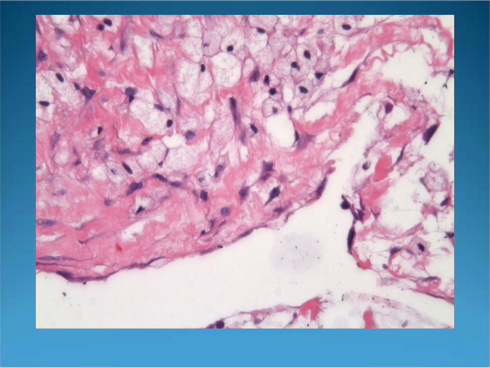
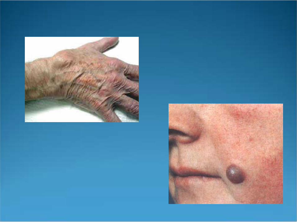
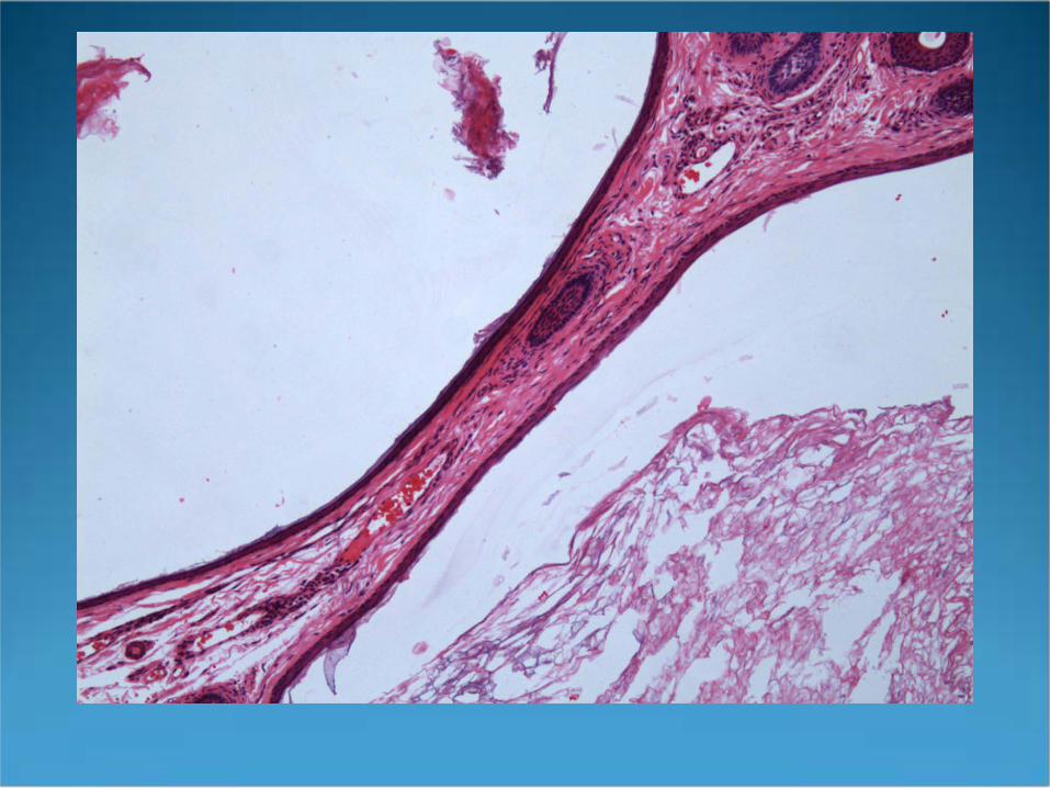
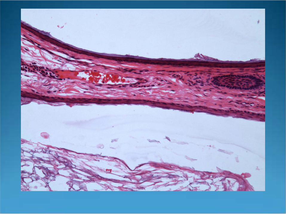
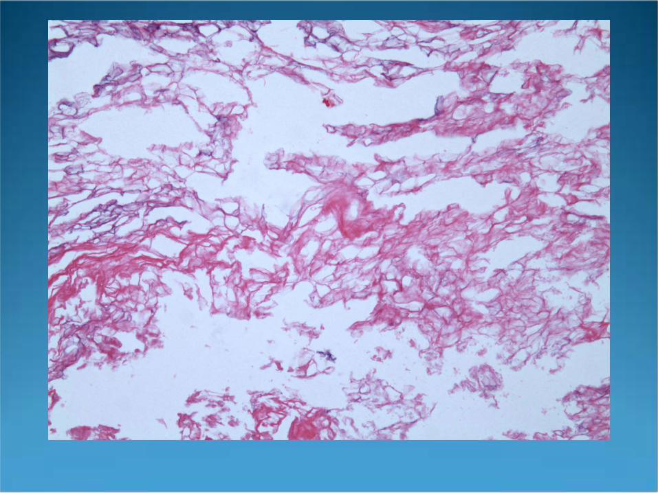
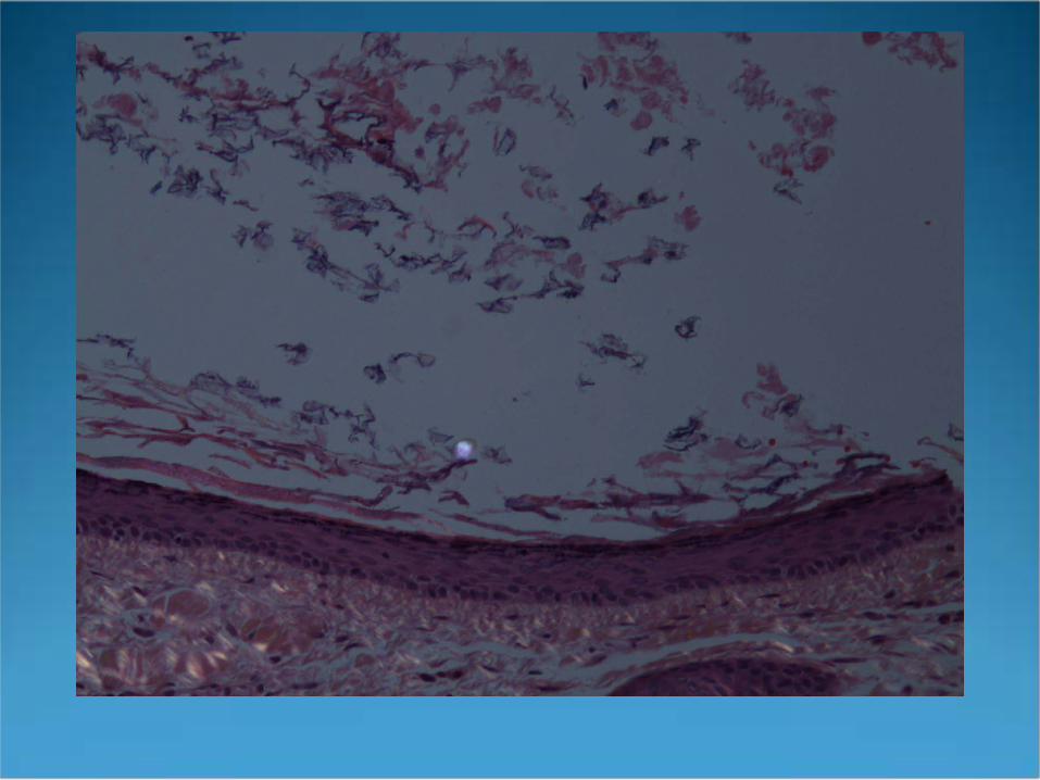
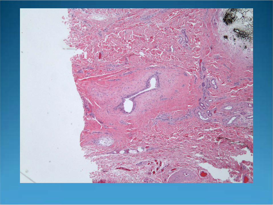
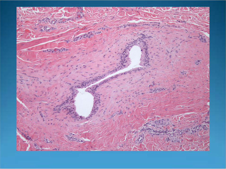
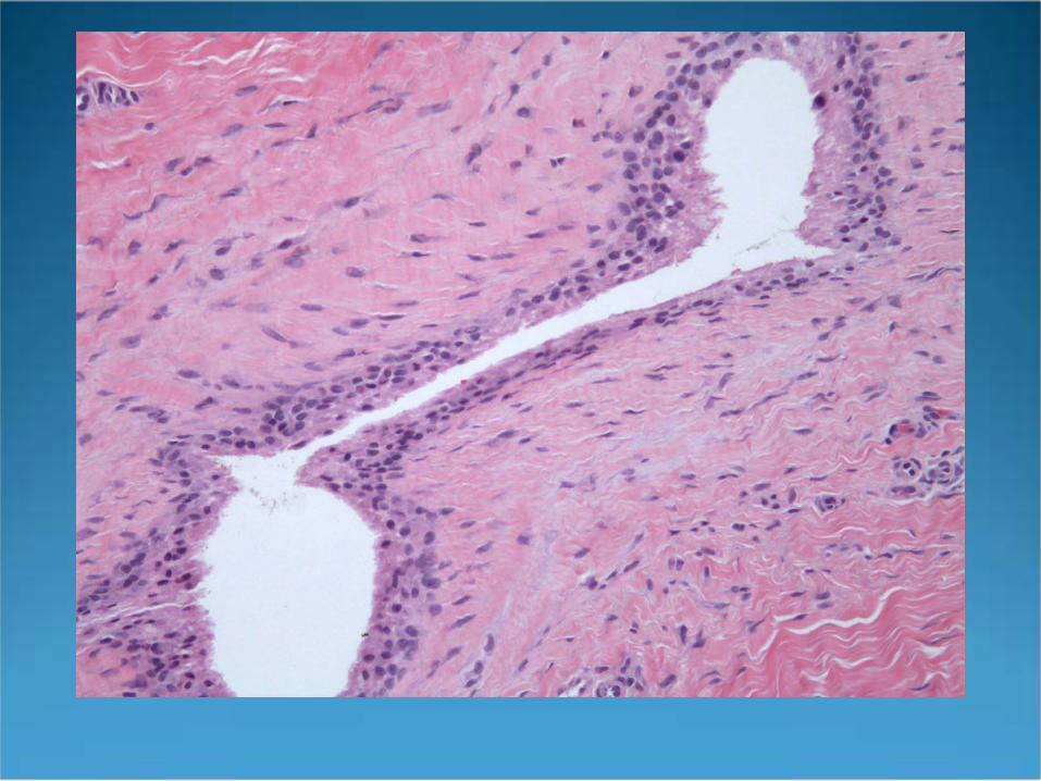
Ganglion Cyst (PseudoCyst)
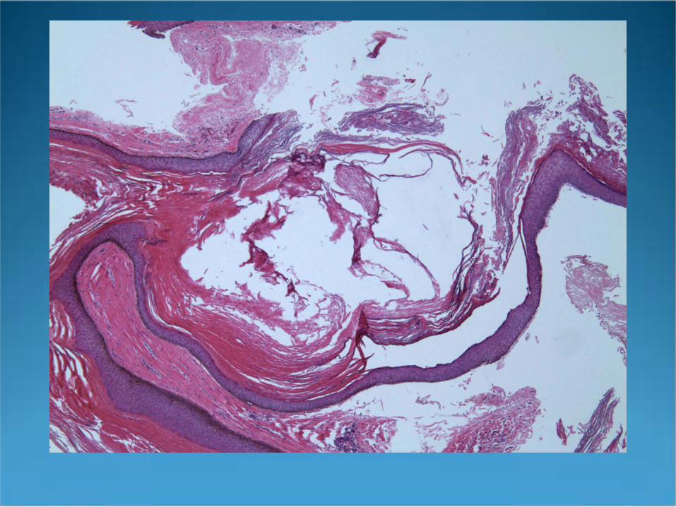
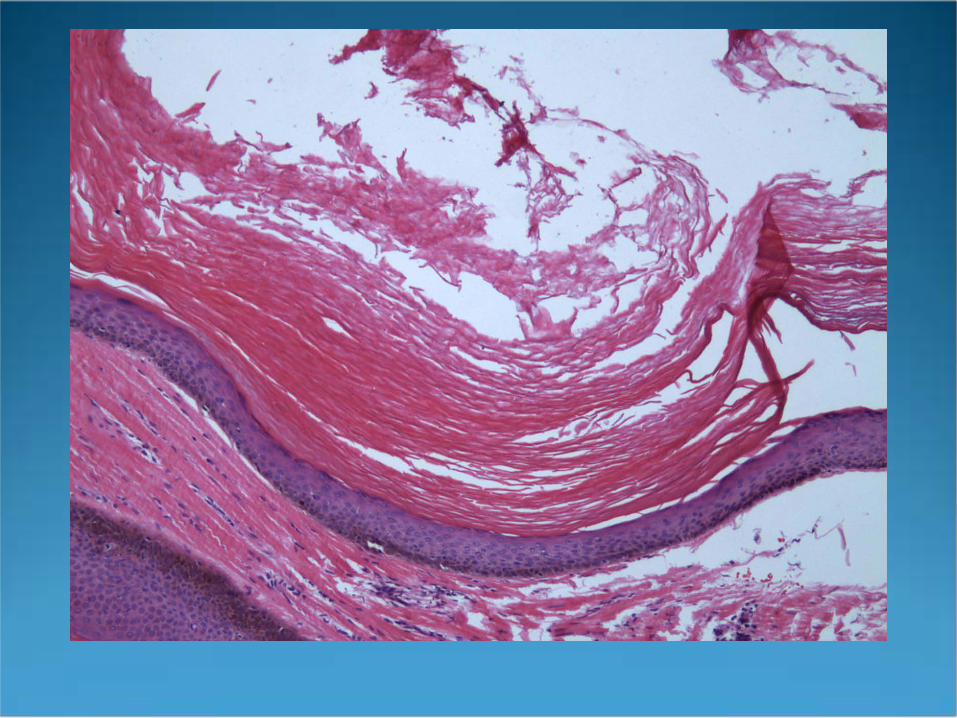
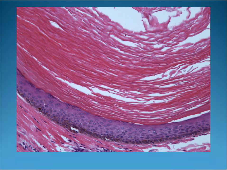
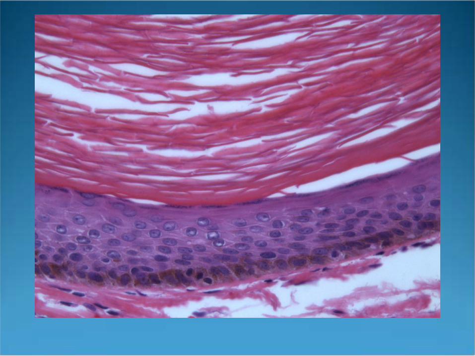
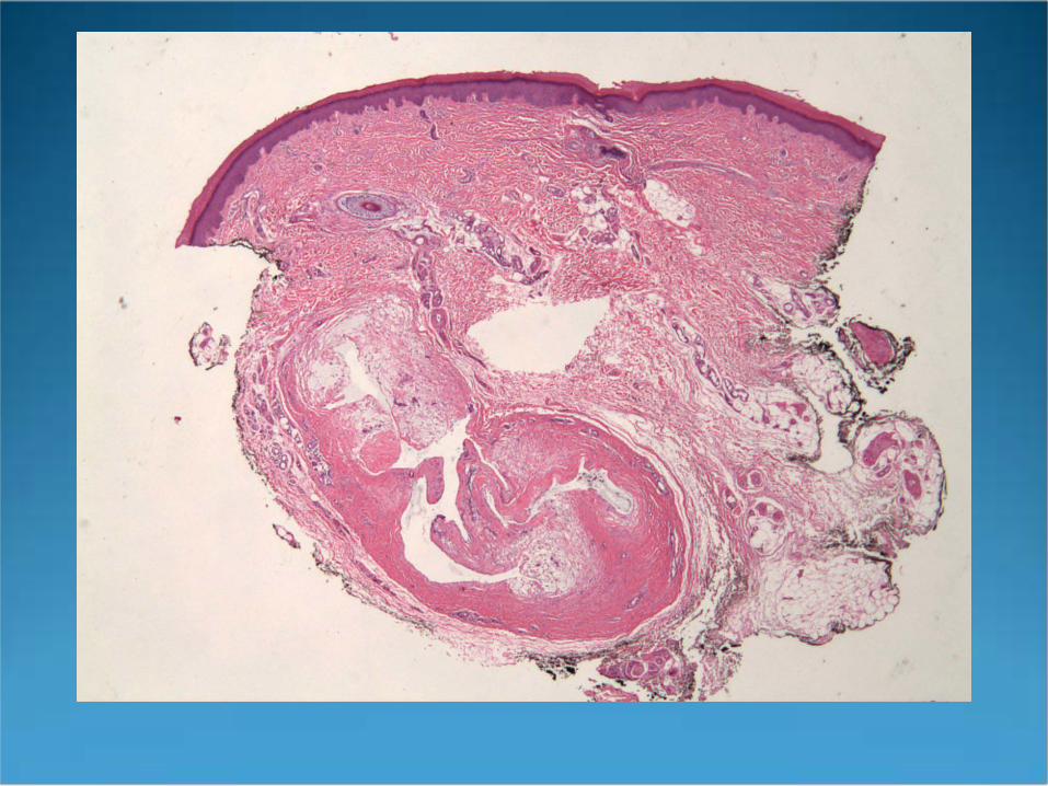
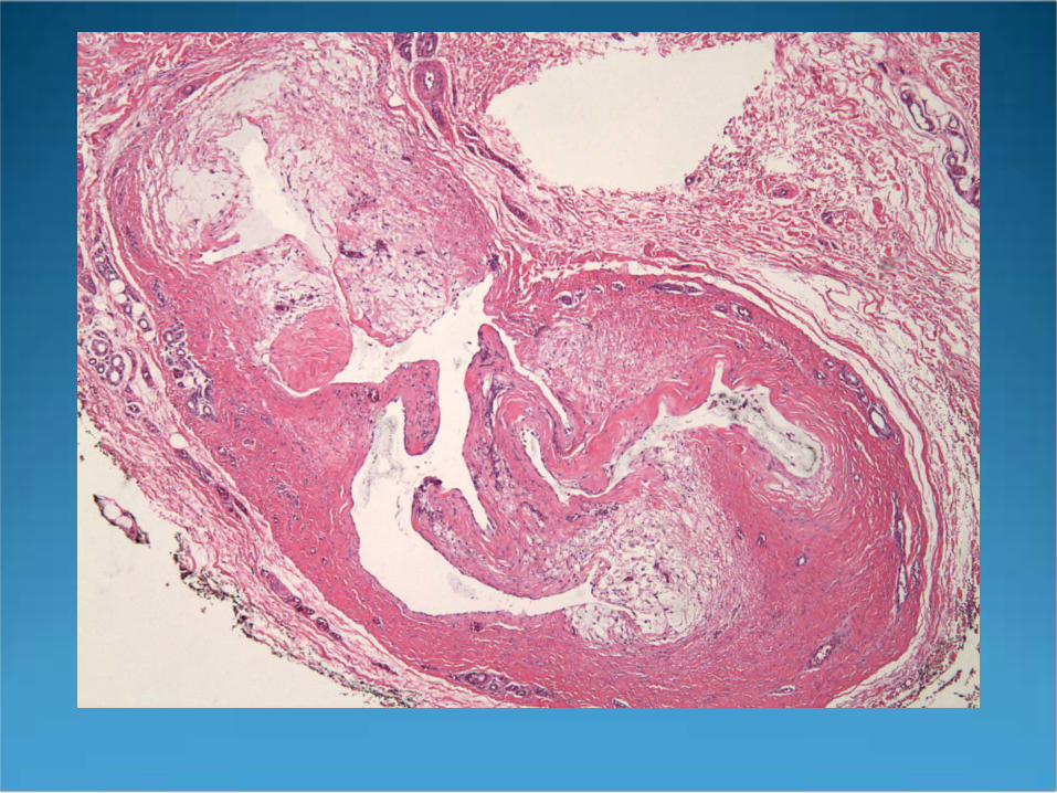
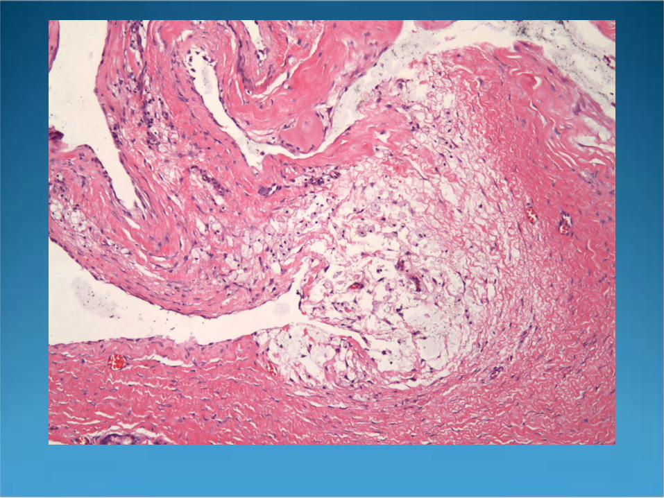
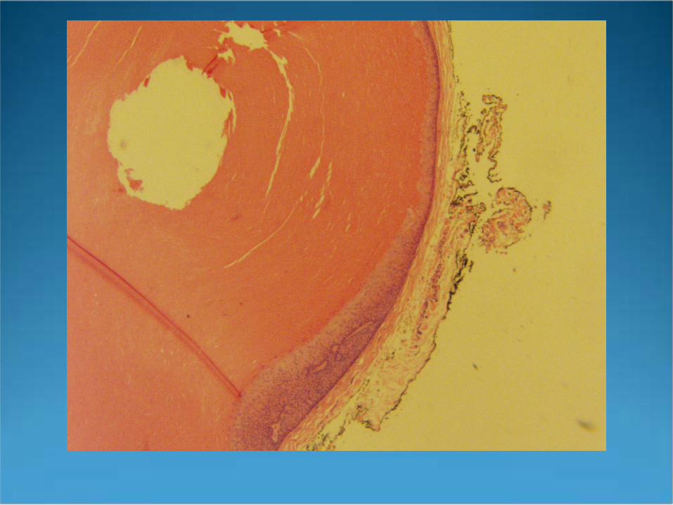
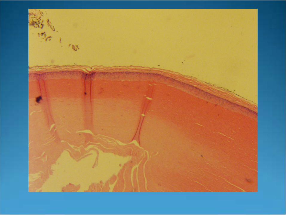
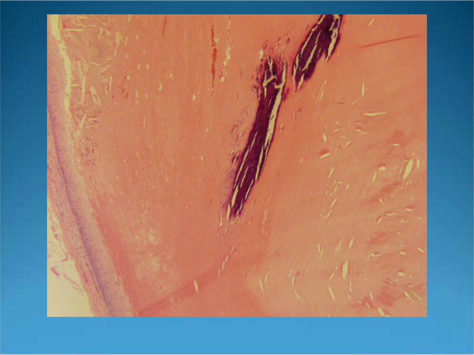
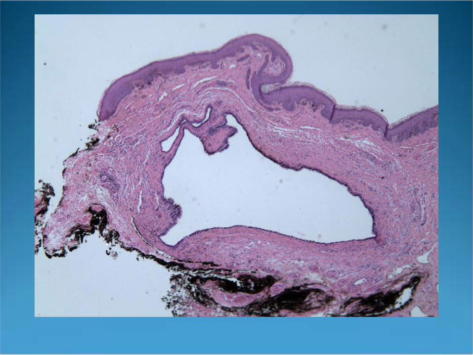
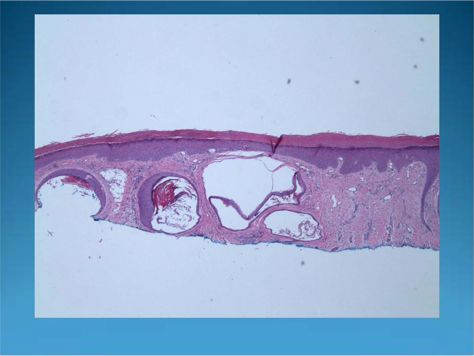
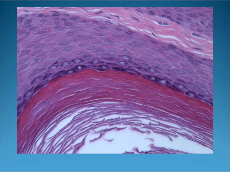
Epidermoid Cyst(Infundibular Cyst)
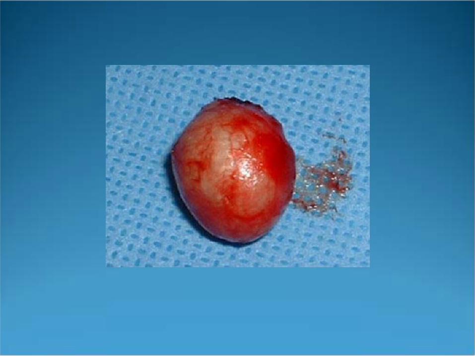
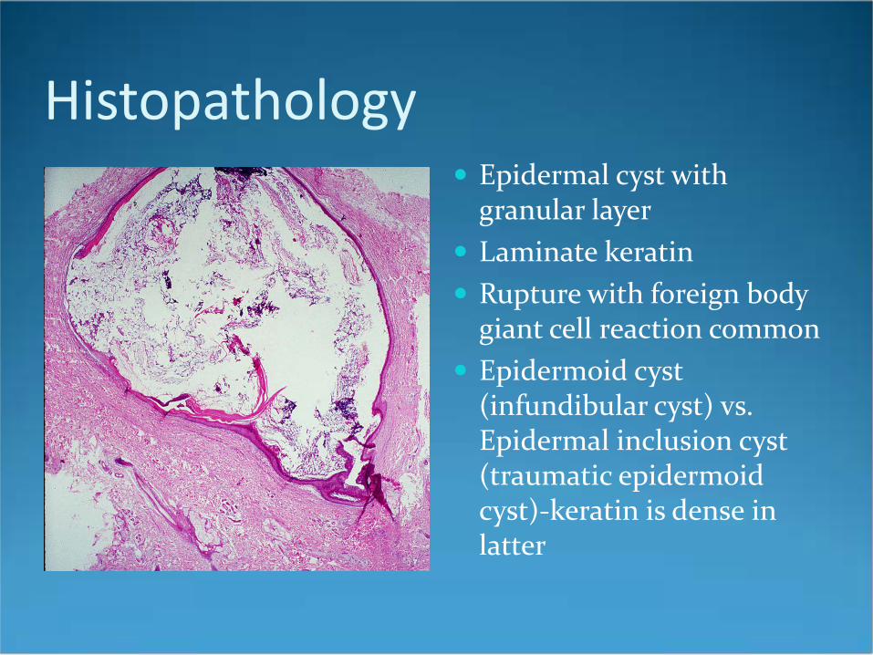
Histopathology Epidermal cyst with
granular layer Laminate keratin Rupture with foreign body
giant cell reaction common Epidermoid cyst
(infundibular cyst) vs. Epidermal inclusion cyst (traumatic epidermoid cyst)-keratin is dense in latter
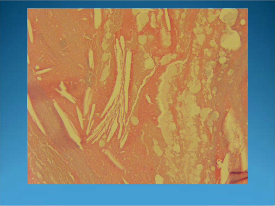

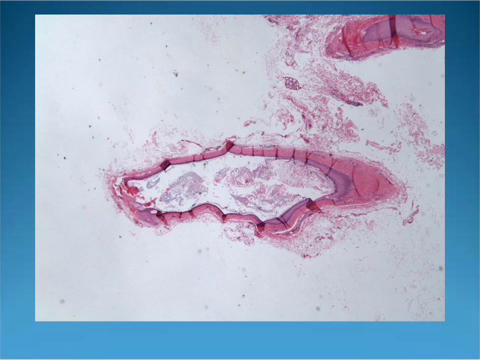
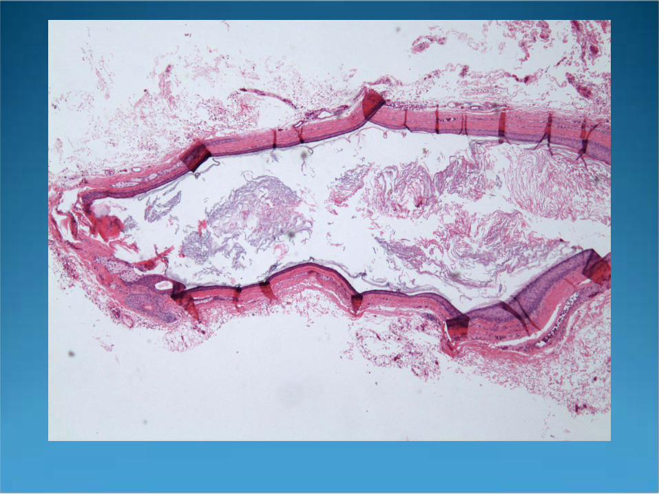
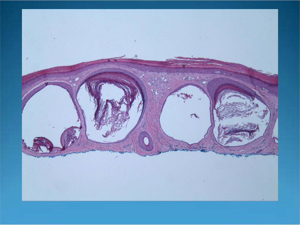
Pilar Cyst(Trichilemmal Cyst, Isthmus Catagen Cyst)
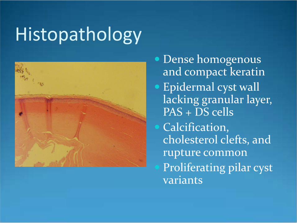
Histopathology Dense homogenous
and compact keratin Epidermal cyst wall
lacking granular layer, PAS + DS cells
Calcification, cholesterol clefts, and rupture common
Proliferating pilar cyst variants
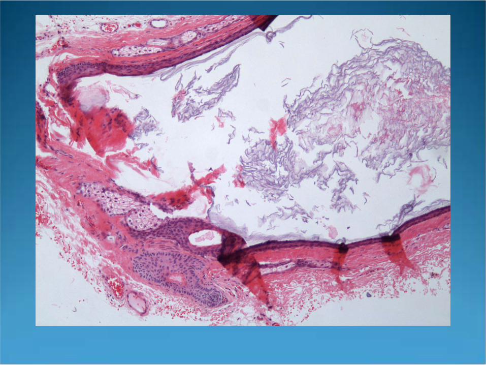
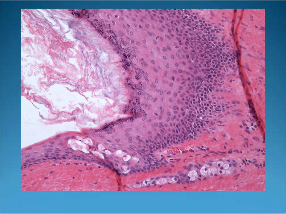
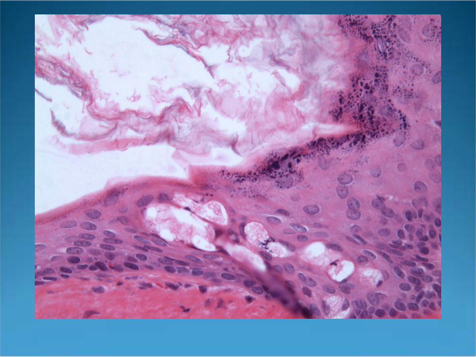
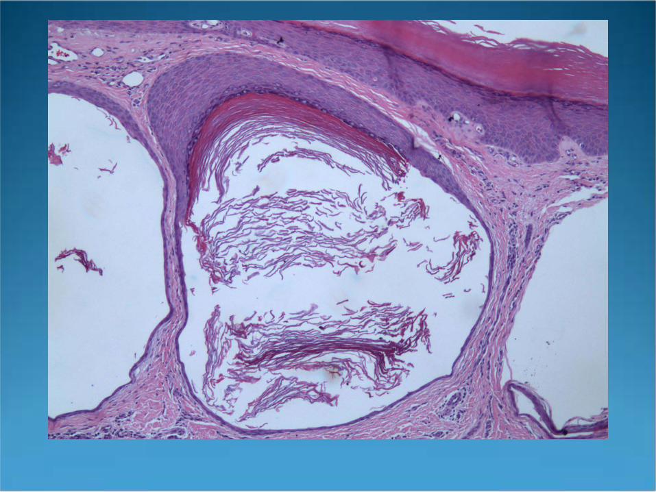
Dermoid Cyst
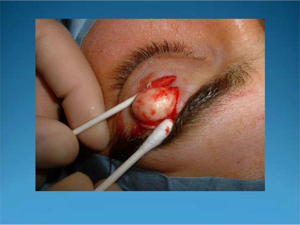
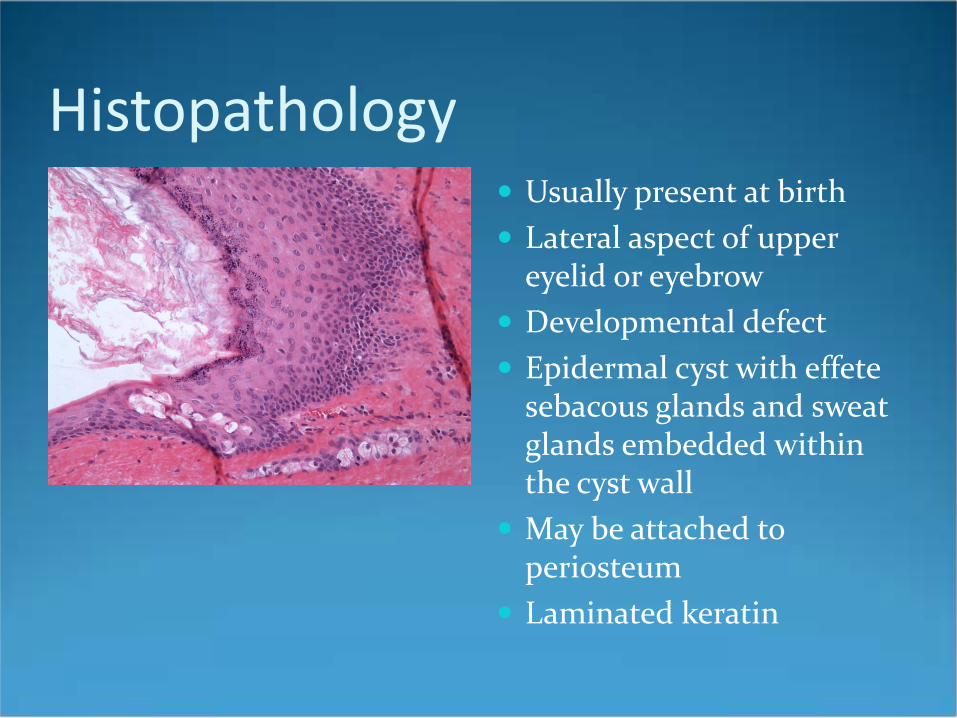
Histopathology Usually present at birth Lateral aspect of upper
eyelid or eyebrow Developmental defect Epidermal cyst with effete
sebacous glands and sweat glands embedded within the cyst wall
May be attached to periosteum
Laminated keratin
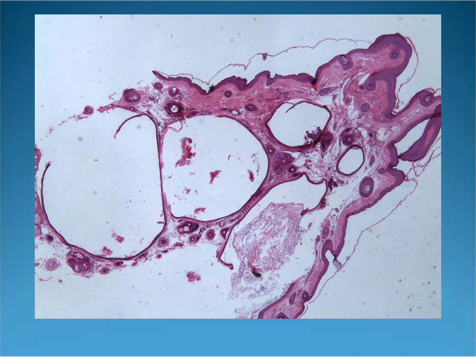
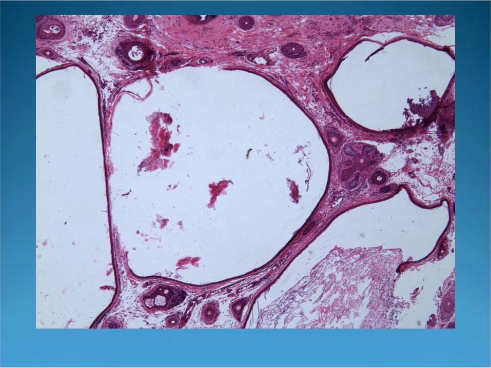
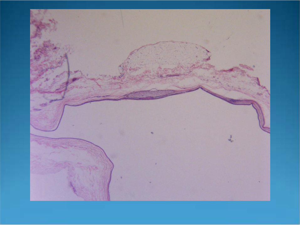
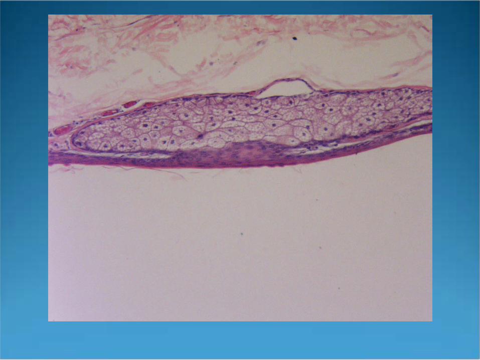
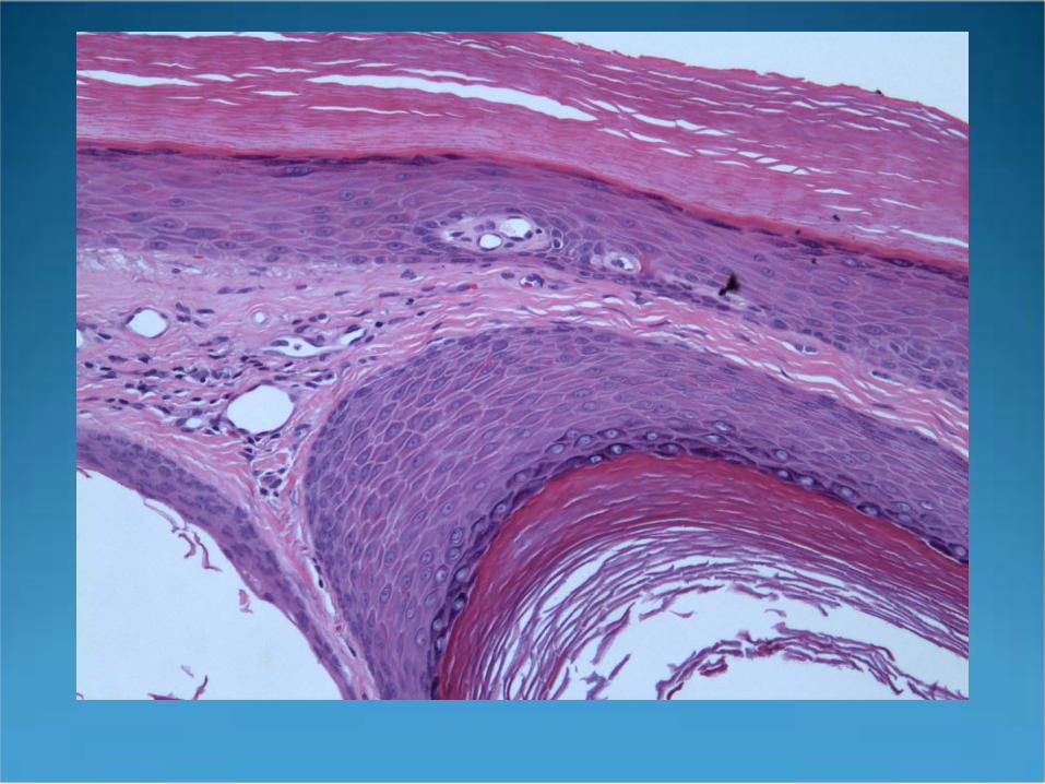
Vellus Hair Cyst
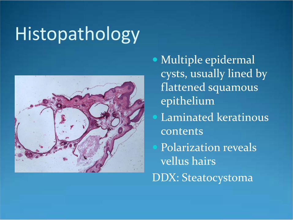
Histopathology Multiple epidermal
cysts, usually lined by flattened squamous epithelium
Laminated keratinous contents
Polarization reveals vellus hairs
DDX: Steatocystoma


Steatocystoma
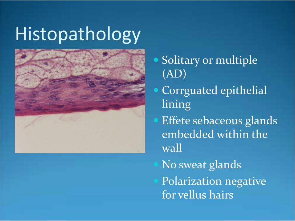
Histopathology Solitary or multiple
(AD) Corrguated epithelial
lining Effete sebaceous glands
embedded within the wall
No sweat glands Polarization negative
for vellus hairs
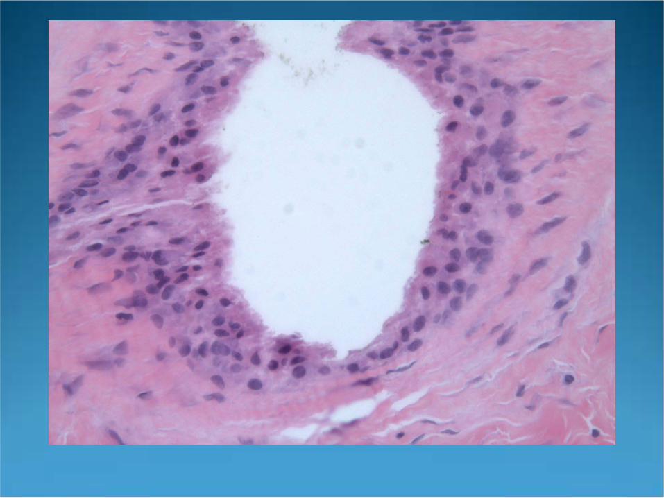
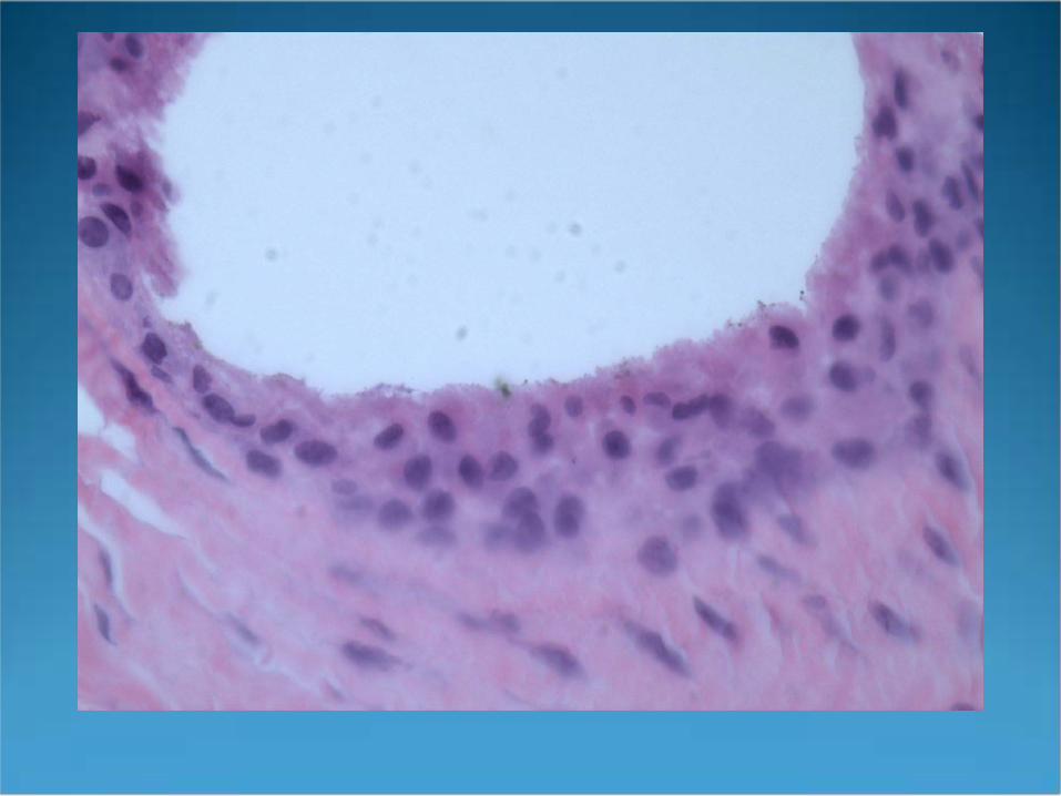
Cutaneous Ciliated Cyst
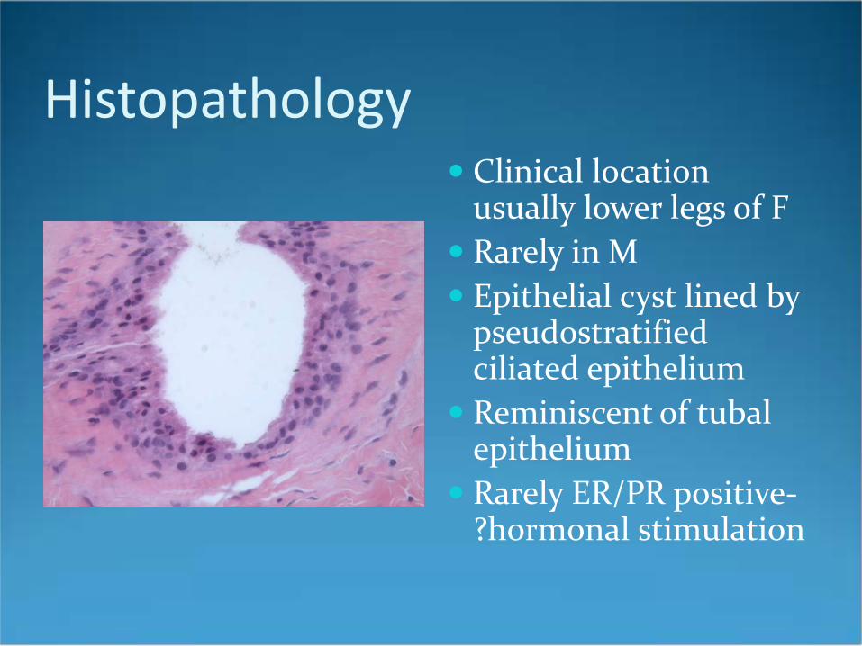
Histopathology Clinical location
usually lower legs of F Rarely in M Epithelial cyst lined by
pseudostratified ciliated epithelium
Reminiscent of tubal epithelium
Rarely ER/PR positive-?hormonal stimulation
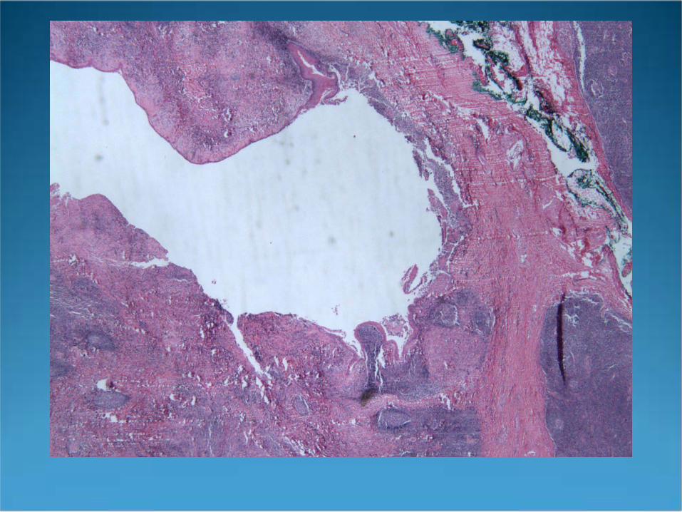
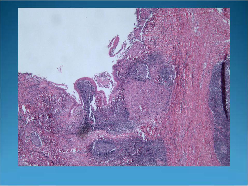
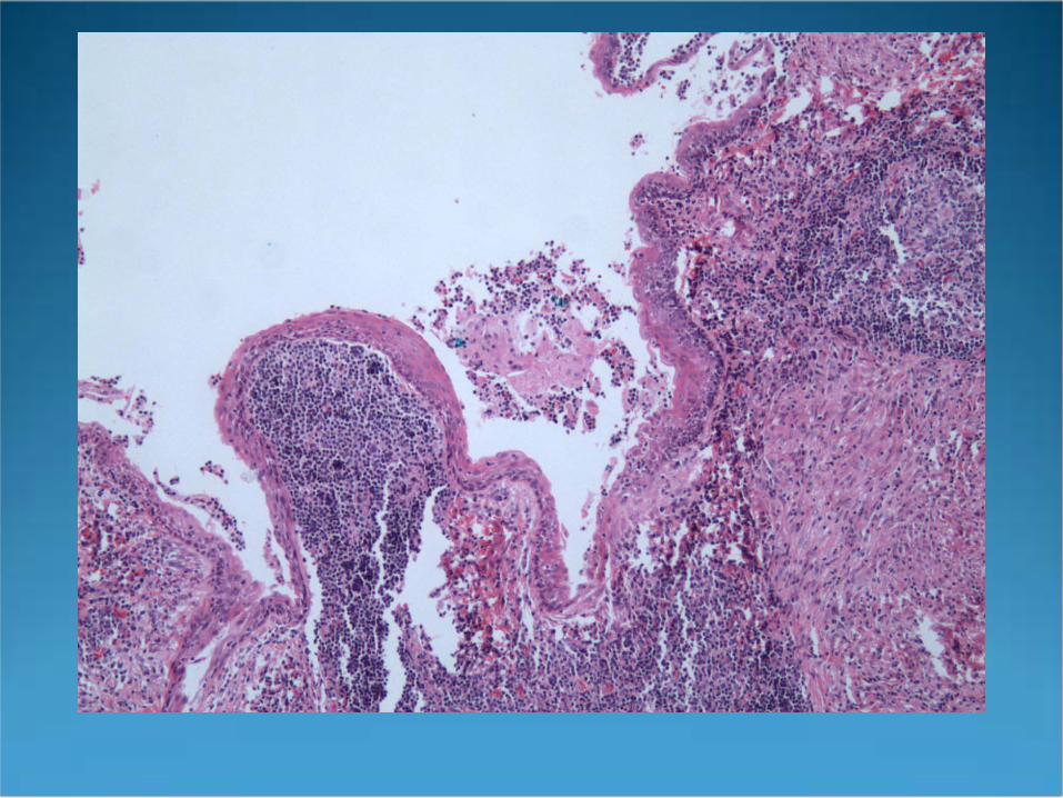
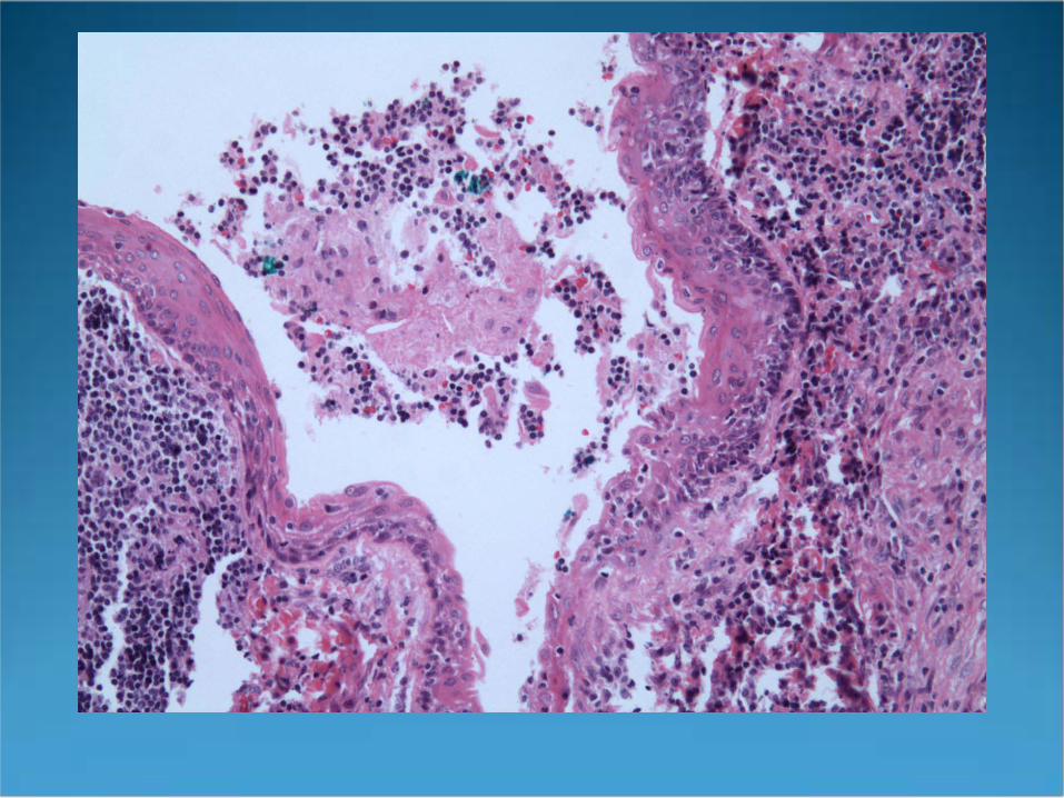

Branchial Cleft Cyst
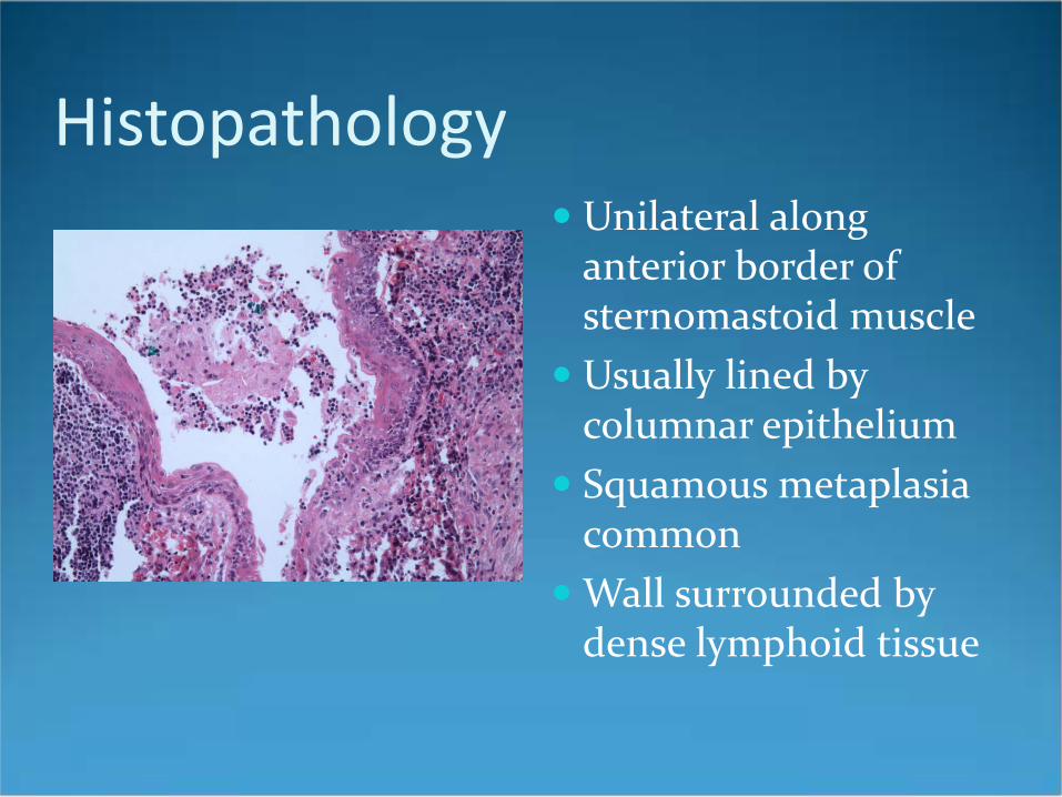
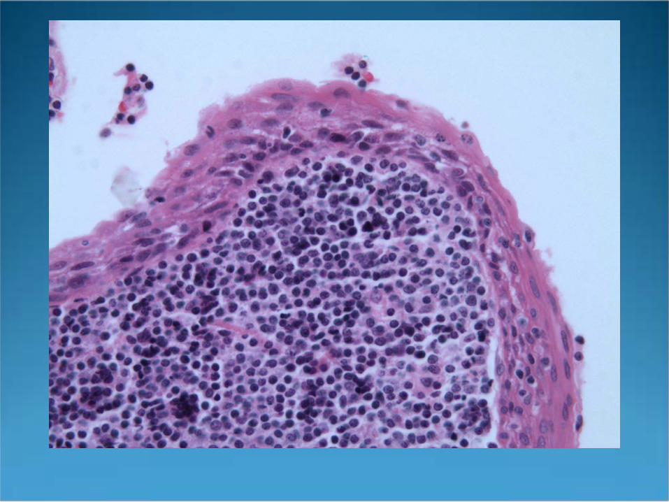
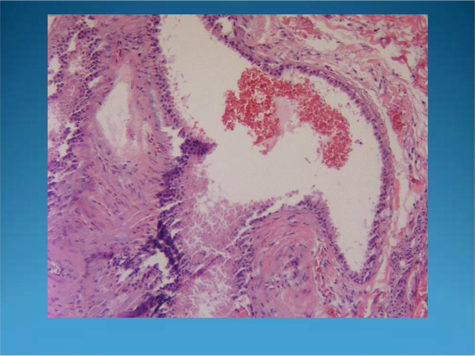
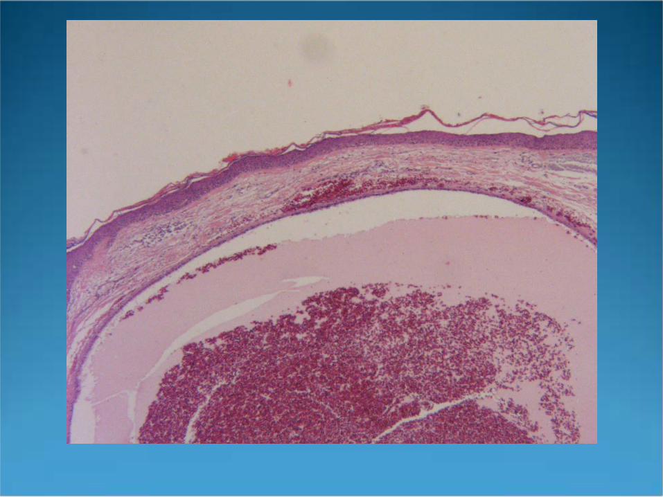
Histopathology Unilateral along
anterior border of sternomastoid muscle
Usually lined by columnar epithelium
Squamous metaplasia common
Wall surrounded by dense lymphoid tissue
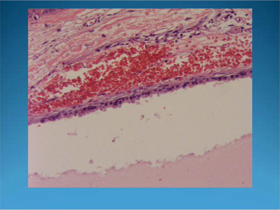
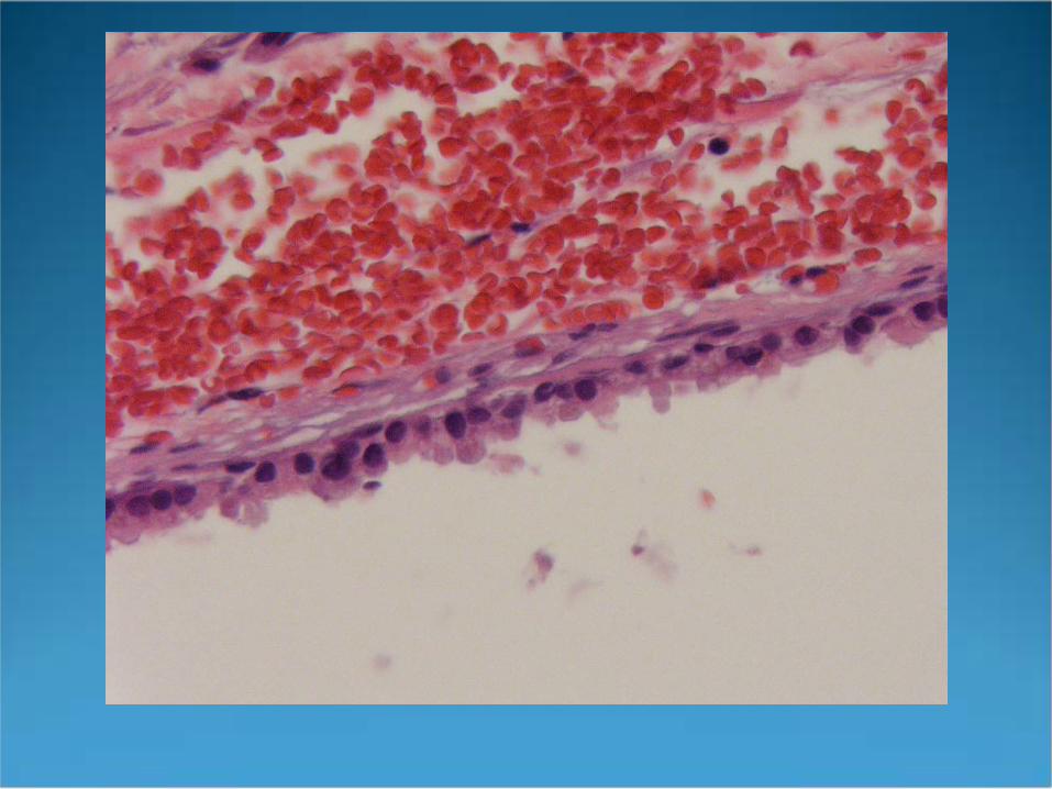

Hidrocystoma
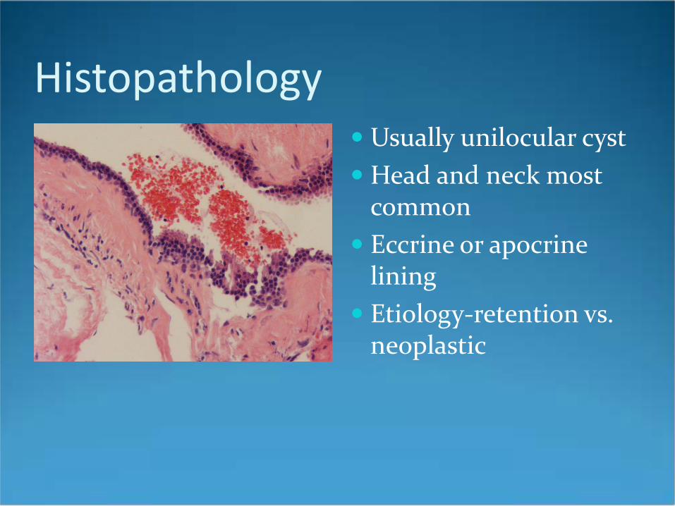
Histopathology Usually unilocular cyst Head and neck most
common Eccrine or apocrine
lining Etiology-retention vs.
neoplastic
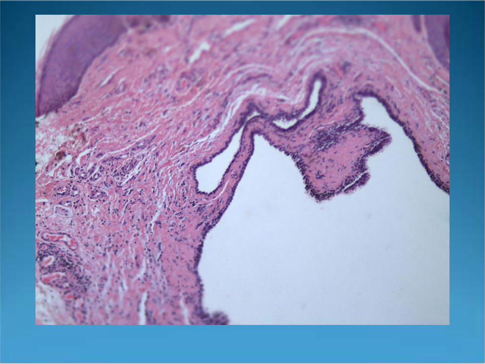
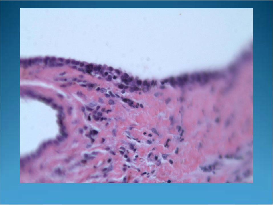

Median Raphe Cyst of the Penis
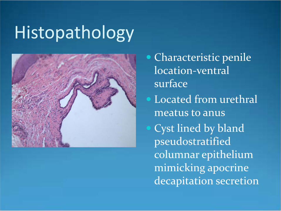
Histopathology Characteristic penile
location-ventral surface
Located from urethral meatus to anus
Cyst lined by bland pseudostratified columnar epithelium mimicking apocrine decapitation secretion

Milia en Plaque
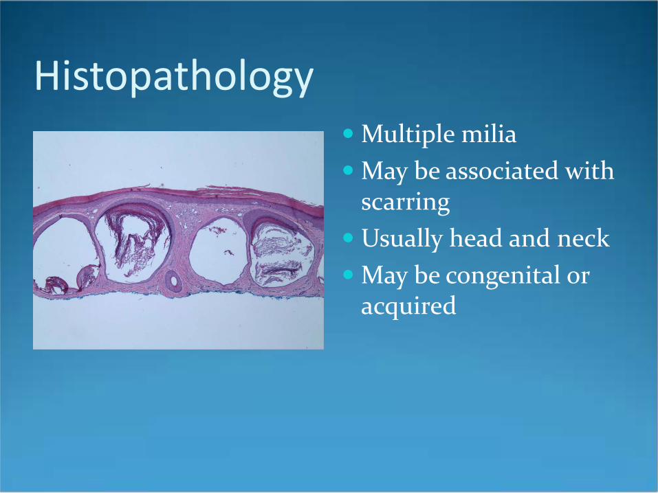
Histopathology Multiple milia May be associated with
scarring Usually head and neck May be congenital or
acquired
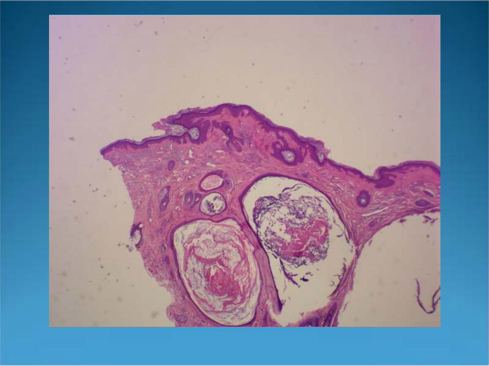
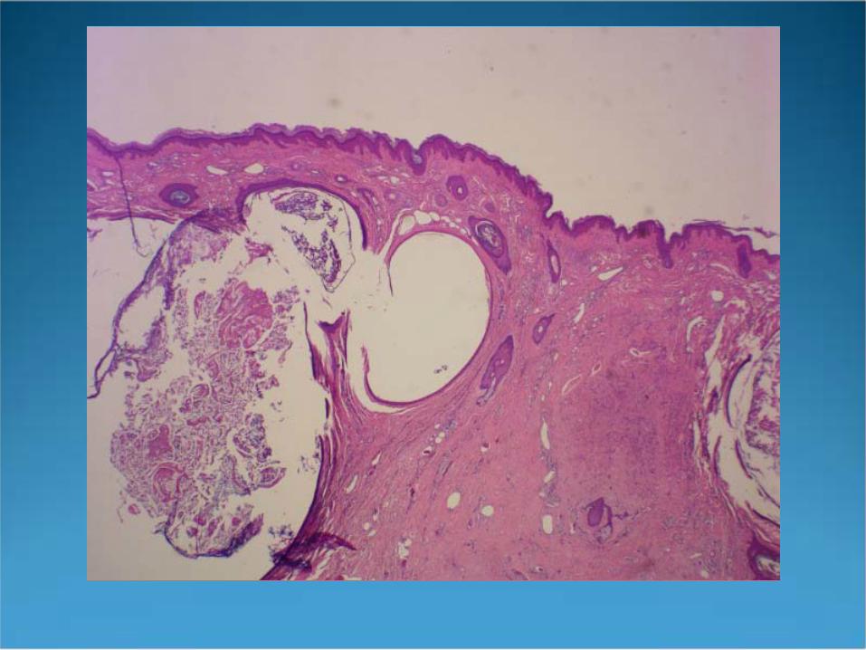
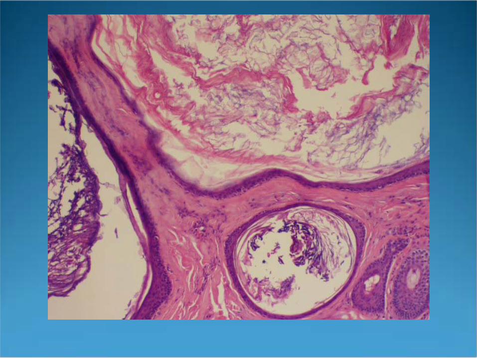
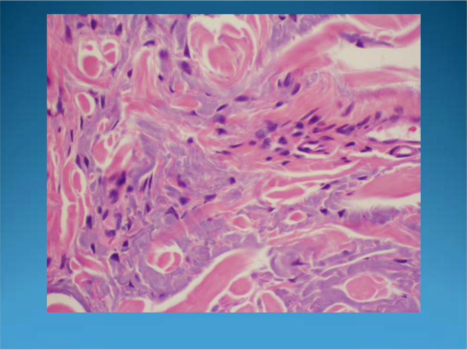

Favre-Racouchot Disease(Nodular Cysts and Comedones)
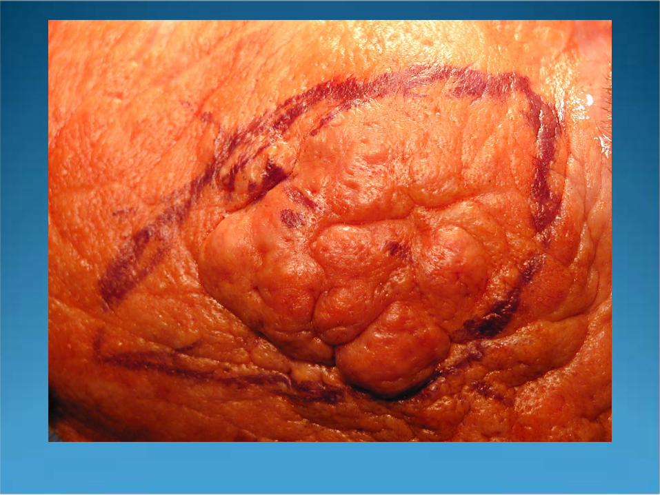
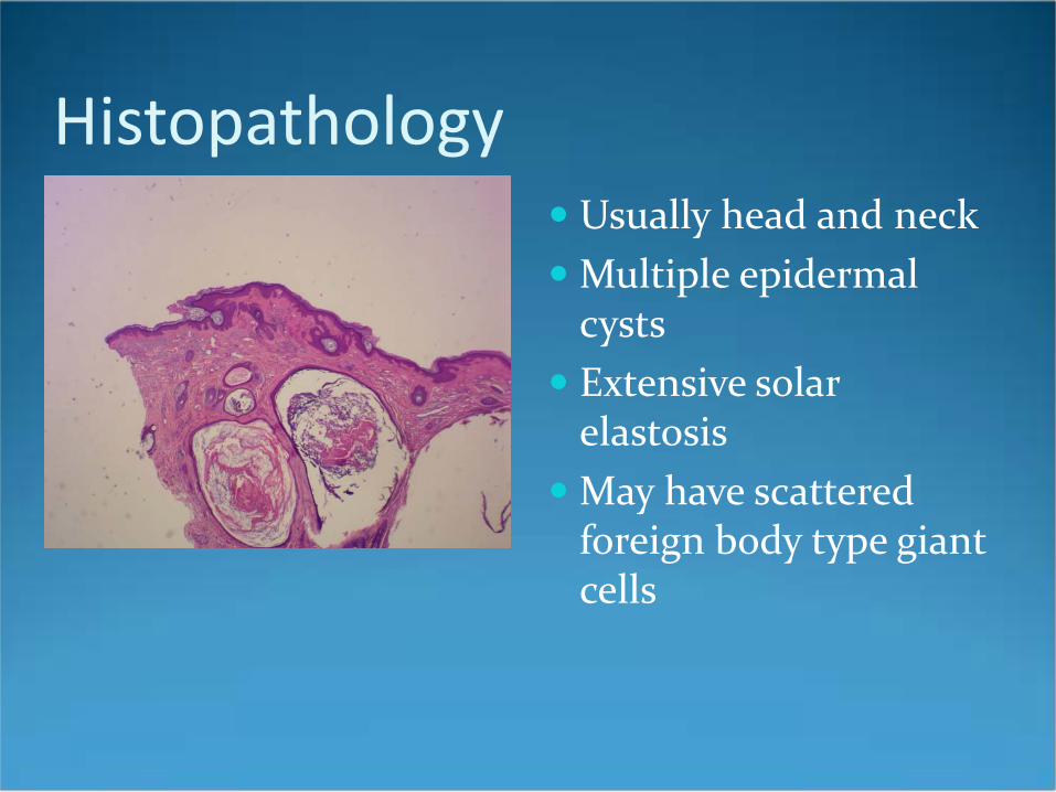
Histopathology Usually head and neck Multiple epidermal
cysts Extensive solar
elastosis May have scattered
foreign body type giant cells