clinical anatomy of the thyroid and adrenal glands · 28-10-2003 · case presentation preliminary...
TRANSCRIPT
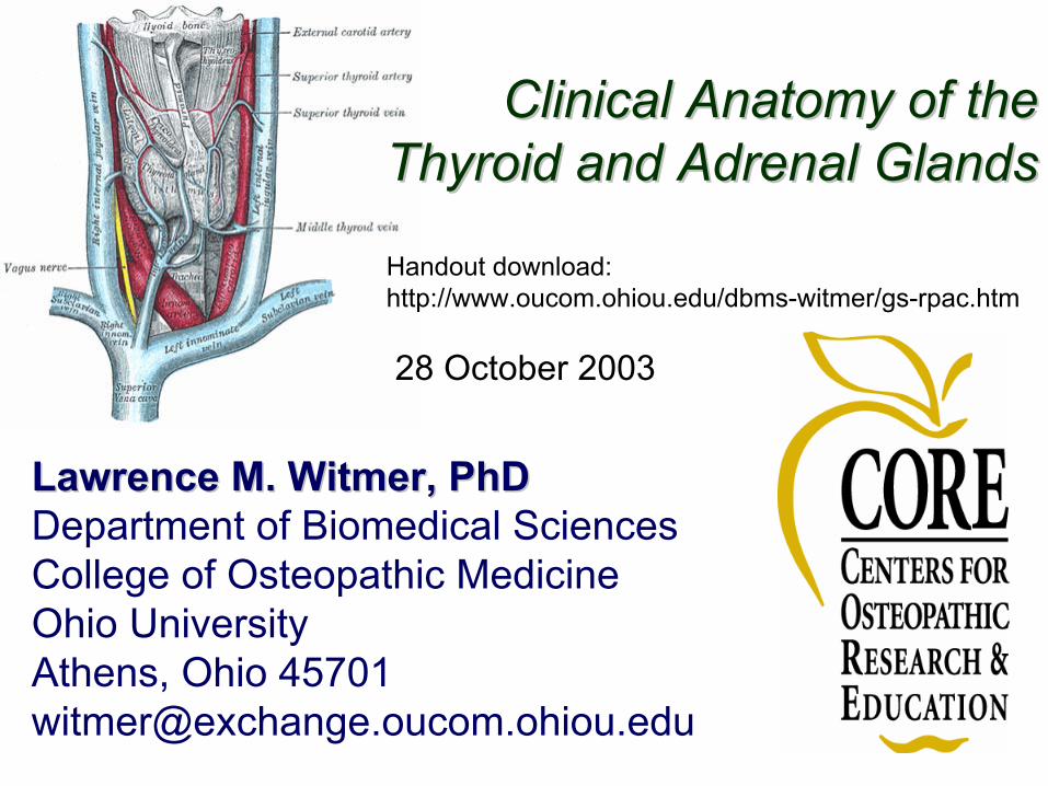
Lawrence M. Witmer, PhDLawrence M. Witmer, PhDDepartment of Biomedical SciencesCollege of Osteopathic MedicineOhio UniversityAthens, Ohio [email protected]
Clinical Anatomy of theClinical Anatomy of theThyroid and Adrenal GlandsThyroid and Adrenal Glands
Handout download:http://www.oucom.ohiou.edu/dbms-witmer/gs-rpac.htm
28 October 2003

Anatomical Overview• Right & left lobes connected
by an isthmus• Occasional pyramidal lobe• Levator glandulae thyroideae• Slightly larger in women; may
enlarge during menstruation & pregnancy
• Extends from oblique line on thyroid cartilage down to 4th
or 5th tracheal ring• Attaches to cricoid cartilage
via suspensory ligament
thyroidcartilage
commoncarotid a.
cricoidcartilage
isthmus
thyroid lobespleuralcupola
From Netter’s Atlas
variation(from
Hollinshead 1968)

Case PresentationA 32-year-old woman presents with a swelling on the anterior part of her neck. She also reports that her breathing is sometimes affected by the swelling. On examination, a single, firm, rounded mass can be felt on the left side of the laryngotracheal region. It moves up and down with swallowing. Ultrasound reveals a solid nodule in the left lobe of her thyroid gland. A needle biopsy subsequently indicates that malignant changes have taken place in the cells.
Preliminary Diagnosis:Tumor of the left lobe of the thyroid

1. Why does the mass move up and down on swallowing?
2. What can explain the difficulty breathing?
3. What structures would be endangered by subtotal or total thyroidectomy?
4. Why is the nature of the patient’s voice of interest postoperatively?
Questions

Cervical Fasciainvestingfascia pretrachealpretracheal
fascia fascia
alarfascia
prevertebralfascia
carotidsheath
buccopharyngealfascia
platysma sternohyoid
sternothyroid
thyroidthyroid
sternocleidomastoid
From Netter’s Atlas
trachea
parathyroidparathyroid
recurrentlaryngeal n.
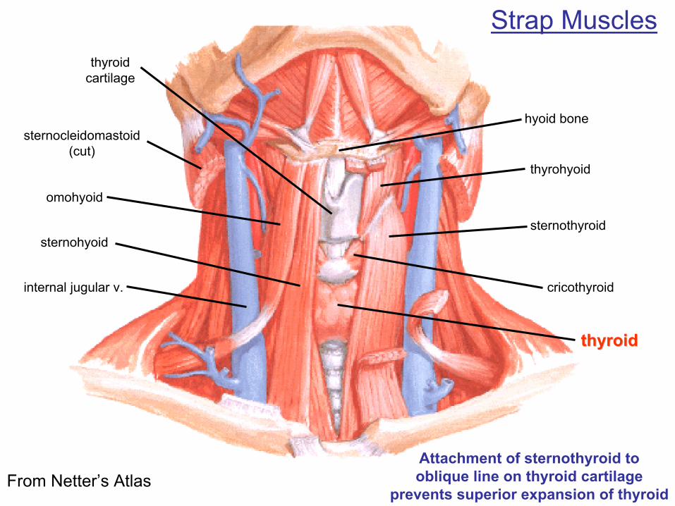
Strap Muscles
sternohyoidsternothyroid
thyroidthyroid
sternocleidomastoid(cut)
From Netter’s Atlas
thyrohyoid
omohyoid
internal jugular v. cricothyroid
hyoid bone
thyroidcartilage
Attachment of sternothyroid tooblique line on thyroid cartilage
prevents superior expansion of thyroid

Thyroid CTC7
sternocleidomastoid
trachea
C7
strap muscles
Thyroid ( )Thyroid ( )
esophagus
From Ellis et al. 1991From Ellis et al. 1991
internaljugular v.
commoncarotid a.
From web reference 1From web reference 1
displaced trachea thyroidtumor
Compression and displacementof trachea by thyroid tumor
normal
normal

superior thyroid a.
thyroidthyroid
From Netter’s Atlas
inferior thyroid a.
middle thyroid v.
inferior thyroid v.
internal jugular v.
common carotid a.
cricothyroid m.
external laryngeal n.
superior thyroid v.
recurrent laryngeal n.
pretracheal lymph node
pyramidal lobe
Vascular Supply& Relations
Anterior View

Vascular Supply& Relations
superior thyroid a.
thyroidthyroid
From Netter’s Atlas
inferior thyroid a.parathyroids
common carotid a.
inferiorconstrictor m.
external laryngeal n.
esophagusrecurrent laryngeal n.
recurrent laryngeal n.
Posterior View

Recurrent Laryngeal N.& Suspensory Lig. of Berry
From Netter’s Atlas
Variation in relationship of recurrentlaryngeal n. to inferior thyroid a.
thyroidthyroid
inferior thyroida. & branches
common carotid a.
recurrentlaryngeal n.
recurrentlaryngeal n.
inferior thyroida. & branches
(from Hollinshead 1968)

Recurrent Laryngeal N.& Suspensory Lig. of Berry
From Netter’s Atlas
Variation in relationship of recurrentlaryngeal n. to suspensory lig.
thyroidthyroid
inferior thyroida. & branches
recurrentlaryngeal n.
superior thyroida. & v. (cut)
parathyroids
susp. lig.susp. lig.
(from Hollinshead 1968)
superficial toligament
deep toligament
splits aroundligament
passes thrugland

Recurrent Laryngeal N.& Suspensory Lig. of Berry
From Sasou et al. 1998
Variation in relationship of recurrentlaryngeal n. to suspensory lig.
thyroidthyroid
inferior thyroid a.recurrentlaryngeal n.
susp. lig.susp. lig.
(from Hollinshead 1968)
superficial toligament
deep toligament
splits aroundligament
passes thrugland
trachea
suspensory ligament of Berrysuspensory ligament of Berry

Case Presentation
Preliminary Diagnosis:Thyroglossal Cyst
A 43-year-old male presents with a swelling in the front of his neck. He first noticed it 9 months ago and it has steadily grown. The lump lays near the midline and moves on swallowing. On palpation, it is firm and lays anterior to the thyroid cartilage. The mass is smooth, non-pulsatile, and non-fluctuant. The dorsum of the tongue was inspected but no thyroid tissue was observed. Ultrasound showed the mass to be cystic and separate from the thyroid gland.
cyst
thyroidcartilage
From Moore & Persaud 2003
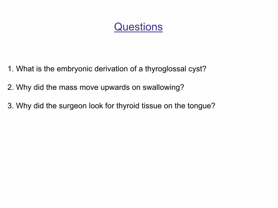
1. What is the embryonic derivation of a thyroglossal cyst?
2. Why did the mass move upwards on swallowing?
3. Why did the surgeon look for thyroid tissue on the tongue?
Questions

Thyroid Development
From Moore & Persaud 2003
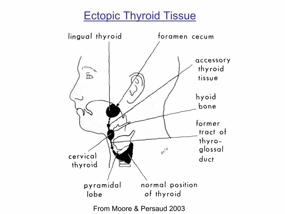
Ectopic Thyroid Tissue
From Moore & Persaud 2003

Possible Locations of Thyroglossal Duct Cysts
From Moore & Persaud 2003

Adrenal Overview
From Netter’s Atlas
spinalcord
sympathetictrunk
splanchnicnerves
preaorticganglia(celiac,
aorticorenal)
adrenaladrenalcortexcortex
adrenaladrenalmedullamedulla
T10T11T12T13
preganglionicfibers to
chromaffin cellsin medulla
— corticosteroids, androgens
— catecholamines (esp. epinephrine)
From Gray’s Atlas
From Netter’s Atlas
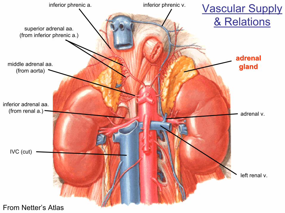
Vascular Supply& Relations
superior adrenal aa.(from inferior phrenic a.)
adrenaladrenalglandglandmiddle adrenal aa.
(from aorta)
inferior adrenal aa.(from renal a.)
inferior phrenic a. inferior phrenic v.
adrenal v.
left renal v.
IVC (cut)

From Netter’s Atlas
Perirenal fascia of Gerotaadrenaladrenalglandgland
peritoneum
L2L2L2
Gerota’sfascia
pararenalfat
perirenalfat
Toldt’s fascia(ant. layer of Gerota’s f.)
Zuckerkandl’s fascia(post. layer of Gerota’s f.)
psoasfascia
liverliver
coloncolon
kidneykidney
transversalisfascia
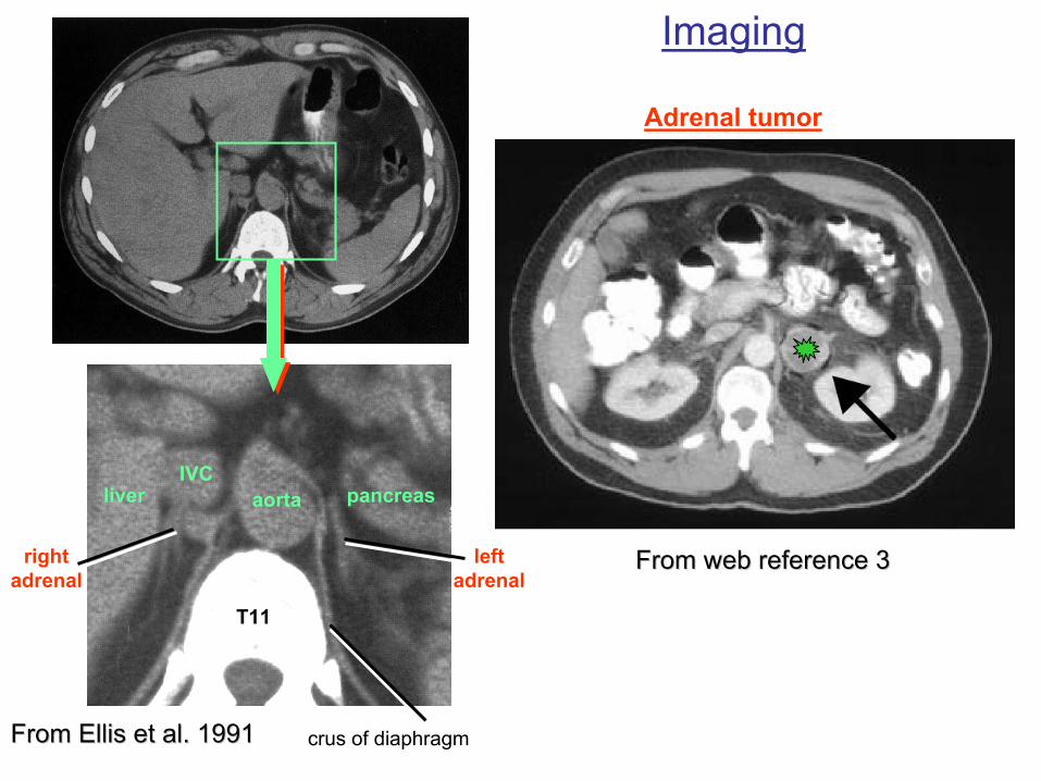
Imaging
aortaIVC
T11
pancreasliver
rightadrenal
leftadrenal
crus of diaphragmFrom Ellis et al. 1991From Ellis et al. 1991
From web reference 3From web reference 3
Adrenal tumor
ReferencesReferencesPrintEllis, H., B. Logan, and A. Dixon. 1993. Human Cross-Sectional Anatomy: Atlas of Body Sections and
CT Images. Butterworth-Heinemann, London.Hollinshead, W. H. 1968. Anatomy for Surgeons: Volume 1. The Head and Neck, Second Edition. Harper
& Row, New York.Moore, K. L. and A. F. Dalley. 1999. Clinically Oriented Anatomy. Lippincott, Williams, & Wilkins,
Baltimore.Moore, K. L. and T. V. N. Persaud. 2003. The Developing Human: Clinically Oriented Embryology.
Saunders, Philadelphia.Netter, F. H. 1987. The CIBA Collection of Medical Illustrations, Volume 8: Musculoskeletal System.
CIBA-Geigy, Summit.———. 1997. Atlas of Human Anatomy, 2nd. Ed. Novartis, East Hanover.Sasou, S., S. Nakamurak, and H. Kurihara. 1998. Suspensory ligament of Berry: its relationship to
recurrent laryngeal nerve and anatomic examination of 24 autopsies. Head & Neck 20:695–698.Younes, N. A., and D. H. Badran. 2002. The cricothyroid space: a guide for successful thyroidectomy.
Asian Journal of Surgery 25(3):226–231.
Web1. Thyroid tumor: http://www.auntminnie.com/ScottWilliamsMD2/nucmed/Tumor/Thallium/Thallium.htm2. Adrenal surgery: http://www.emedicine.com/med/topic3018.htm3. Adrenal surgery: http://www.surgery.wisc.edu/general/patients/endocrine.shtml4. Gray’s Anatomy of the Human Body: http://www.bartleby.com/107/