paul k. shitabata, m.d. dermatopathologist apmg
TRANSCRIPT

PanniculitisPanniculitis
Paul K. Shitabata, M.D.Paul K. Shitabata, M.D.DermatopathologistDermatopathologist
APMGAPMG


Are there distinguishing clinical Are there distinguishing clinical features for a features for a panniculitispanniculitis??


How do I approach a biopsy to rule How do I approach a biopsy to rule out out panniculitispanniculitis??


PathogenesisPathogenesis
Fat is divided into lobules by connective Fat is divided into lobules by connective tissue septatissue septa--Arteriole supplies the Arteriole supplies the centercenter while while venulesvenules drain the drain the septaeseptae
Arterial disorder lead to lobular Arterial disorder lead to lobular panniculitispanniculitisVenousVenous disorder lead to disorder lead to septalseptal panniculitispanniculitisSecondary to large vessel involvement (artery Secondary to large vessel involvement (artery or vein)or vein)

PatternsPatterns
Predominately lobularPredominately lobularPredominately Predominately septalseptalMixedMixed


ModifiersModifiers
VasculitisVasculitisGranulomasGranulomasInflammatory cell typeInflammatory cell typeIntracellular changesIntracellular changesTypes of necrosisTypes of necrosis
PseudomembranesPseudomembranesPseudocystsPseudocystsHyaline materialHyaline materialGhost cells without Ghost cells without vasculitisvasculitisLiquefactiveLiquefactive changeschangesNecrotizing Necrotizing granulomagranulomaBasophilic material Basophilic material

Septal
Vasculitis
Small Vessel Large Vessel
Leukocytoclastic vasculitis ThrombophlebitisPolyarteritis nodosa

Septal-No Vasculitis
Lymphocytes/Plasma cells Histiocytes/Granulomas
Necrobiosis lipoidicaScleroderma
Granuloma annulareRheumatoid nodule
Necrobiotic xanthogranuloma

Lobular-Vasculitis
Small Vessel Large Vessel
Erythema nodosum leprosumLucio’s phenomenon
Nodular vasculitisCrohn’s disease
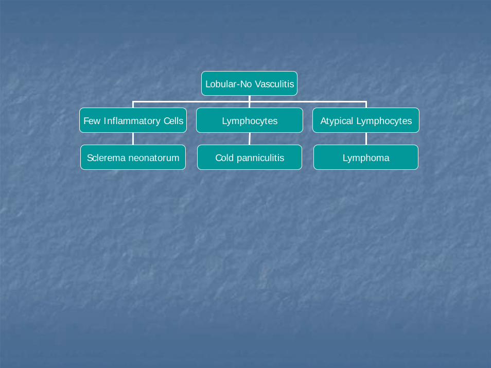
Lobular-No Vasculitis
Few Inflammatory Cells Lymphocytes Atypical Lymphocytes
Sclerema neonatorum Cold panniculitis Lymphoma

Lobular-No Vasculitis
Neutrophils Suppurative Granulomas Histiocytes/Granulomas
Pancreatic fat necrosisFactitial
Bacterial infection
MycobacterialFungal
Parasitic
SarcoidosisTrauma
LipodystrophySubcutaneous fat necrosis
Post-steroid


ErythemaErythema nodosumnodosum

ClinicalClinical
1818--34 years34 yearsMost patients resolveMost patients resolveMaleMale--toto--female 1:4female 1:4

Clinical AppearanceClinical Appearance
Red tender nodules with poorly Red tender nodules with poorly defined bordersdefined borders
Vary from 2Vary from 2--6 cm6 cmTense, hard, and painful may Tense, hard, and painful may evolve to abscess or ulcerationevolve to abscess or ulcerationNew lesions may appear for 3New lesions may appear for 3--6 6 weeksweeksAnterior leg most commonAnterior leg most commonSecond week from bright red to Second week from bright red to bluish or livid fades to a yellowish bluish or livid fades to a yellowish huehueDisappears in 1 or 2 weeks as the Disappears in 1 or 2 weeks as the overlying skin desquamates.overlying skin desquamates.
ArthralgiaArthralgia <50%<50%Ankles, knees, and wristAnkles, knees, and wristSynovitisSynovitis resolves within a few resolves within a few weeksweeks

Disease AssociationsDisease AssociationsBacteriaBacteria
Streptococcal infections*Streptococcal infections*TuberculosisTuberculosisYersiniaYersinia enterocoliticaenterocoliticaMycoplasmaMycoplasma pneumoniaepneumoniaeLeprosyLeprosyLymphogranulomaLymphogranuloma venereumvenereumSalmonellaSalmonellaCampylobacterCampylobacter
Fungal infections include the following:Fungal infections include the following:CoccidioidomycosisCoccidioidomycosisHistoplasmosisHistoplasmosisBlastomycosisBlastomycosis
DrugsDrugsSulfonamides and halidesSulfonamides and halidesOral contraceptive Oral contraceptive pillspills
UlcerativeUlcerative colitis and colitis and CrohnCrohn disease disease Hodgkin disease and nonHodgkin disease and non--Hodgkin lymphoma Hodgkin lymphoma SarcoidosisSarcoidosisBehçetBehçet disease disease PregnancyPregnancy

HistopathologyHistopathology
SeptalSeptal panniculitispanniculitis with slight with slight superficial and deep superficial and deep perivascularperivascular inflammatory inflammatory lymphocyticlymphocytic infiltrateinfiltrateSepta of subcutaneous fat Septa of subcutaneous fat usually are thickenedusually are thickenedPeriseptalPeriseptal fibrosis, giant cells, fibrosis, giant cells, and granulation tissue appearand granulation tissue appearMiescherMiescher granulomasgranulomas
Small wellSmall well--defined nodular defined nodular aggregates of aggregates of histiocyteshistiocytesaround a central around a central stellatestellate cleft cleft are scattered throughout the are scattered throughout the lesionslesions

Differential DiagnosisDifferential Diagnosis
Behcet’sBehcet’s associated associated vascopathyvascopathyPanniculitisPanniculitis in a lobular or mixed in a lobular or mixed septalseptal lobular lobular patternpatternNeutrophilsNeutrophils present in all lesions and in all stages of present in all lesions and in all stages of the disease and usually confined to the areas of fat the disease and usually confined to the areas of fat necrosis or around necrosis or around inflammedinflammed vesselsvesselsVasculitisVasculitis either either leukocytoclasticleukocytoclastic or or lymphocyticlymphocytic with with some occurring in same specimen in different vesselssome occurring in same specimen in different vessels
Am J Am J DermatopatholDermatopathol 2000;22:3792000;22:379--390390

TreatmentTreatment
SymptomaticSymptomatic--NSAIDSNSAIDSTreat underlying diseaseTreat underlying disease


ErythemaErythema induratuminduratum(Nodular (Nodular VasculitisVasculitis of of BazinBazin))

ClinicalClinical
Women aged 20Women aged 20--30 years30 yearsPast or present history of tuberculosis at an Past or present history of tuberculosis at an extracutaneousextracutaneous site ~50% of patientssite ~50% of patients
Pulmonary tuberculosis most common.Pulmonary tuberculosis most common.TuberculousTuberculous cervical lymphadenitis next most cervical lymphadenitis next most common common
Tender, Tender, erythematouserythematous nodules are present on nodules are present on the lower legsthe lower legs
Chronic, recurrent courseChronic, recurrent courseLesions heal with ulcerations or depressed scarsLesions heal with ulcerations or depressed scarsLeg edemaLeg edema

AppearanceAppearance
Crops of small, tender, Crops of small, tender, erythematouserythematous nodulesnodules
Usually shins and calvesUsually shins and calvesLower third of the legs, Lower third of the legs, especially around the especially around the anklesanklesDepressed scars or Depressed scars or pigmentation due to pigmentation due to previously active lesions previously active lesions may be presentmay be present
CauseCause
Mycobacterium tuberculosisMycobacterium tuberculosis is the cause is the cause ErythemaErythema induratuminduratum and nodular and nodular vasculitisvasculitisare a hypersensitivity reaction to endogenous are a hypersensitivity reaction to endogenous or exogenous antigens like the tubercle or exogenous antigens like the tubercle bacillusbacillusPositive tuberculin skin test result and a Positive tuberculin skin test result and a marked increase in their peripheral T marked increase in their peripheral T lymphocyte response to purified protein lymphocyte response to purified protein derivative (PPD) of tuberculin derivative (PPD) of tuberculin

HistopathologyHistopathology
Mixed Mixed septalseptal and lobular and lobular granulomatousgranulomatouspanniculitispanniculitis with with neutrophilicneutrophilic vasculitisvasculitisCaseationlikeCaseationlike necrosis necrosis may also be seenmay also be seenVary depending on the Vary depending on the age of the lesion age of the lesion VasculitisVasculitis not always not always identified and not a identified and not a requisite for the diagnosisrequisite for the diagnosis

Differential DiagnosisDifferential Diagnosis
ErythemaErythema nodosumnodosumPrimarily Primarily septalseptal
PolyarteritisPolyarteritis nodosanodosaMedium vessel Medium vessel vasculitisvasculitis with minimal lobular with minimal lobular inflammationinflammation

TreatmentTreatment
ErythemaErythema induratuminduratum of of BazinBazin: : AntituberculousAntituberculous therapy therapy Nodular Nodular vasculitisvasculitis with a negative with a negative tuberculin skin test resulttuberculin skin test result
Bed rest with systemic steroids is indicated.Bed rest with systemic steroids is indicated.Potassium iodide may be usedPotassium iodide may be used


Lupus Lupus PanniculitisPanniculitis(Lupus (Lupus ErythematosusErythematosus ProfundusProfundus))

ClinicalClinical
Women 3Women 3--5th decades, occasional infant cases5th decades, occasional infant casesHead and neck, upper arms, trunk, and buttocksHead and neck, upper arms, trunk, and buttocksTan to Tan to violaceousviolaceous plaques plaques May herald onset of LE or occur in isolation but May herald onset of LE or occur in isolation but usually occurs simultaneously with other usually occurs simultaneously with other cutaneouscutaneous and and extracutaneousextracutaneous manifestationsmanifestationsOccurs with about equal frequency with both Occurs with about equal frequency with both DLE and SLE though other studies doubt this DLE and SLE though other studies doubt this with SLEwith SLE--affects about 1affects about 1--2% of LE patients2% of LE patients

HistopathologyHistopathology
Infiltration of fat lobule by Infiltration of fat lobule by lymphocytes, lymphocytes, histiocyteshistiocytes, and , and plasma cells with interposed plasma cells with interposed zone of granular zone of granular necrobioticnecrobioticalterationalterationLymphoid follicles with Lymphoid follicles with tingibletingiblebody macrophagesbody macrophagesEndothelial necrosis, Endothelial necrosis, segmental deposits of fibrin, segmental deposits of fibrin, occlusive luminal thrombi of occlusive luminal thrombi of interstitial capillaries and interstitial capillaries and venulesvenules, , lymphocyticlymphocytic vasculitisvasculitisPositive Lupus Band with Positive Lupus Band with concomittantconcomittant SLESLE

Differential DiagnosisDifferential Diagnosis
Subcutaneous TSubcutaneous T--cell cell lymphomalymphoma
Significant percentage Significant percentage may manifest may manifest lymphoid lymphoid atypiaatypia

TreatmentTreatment
Treat underlying lupusTreat underlying lupus


Subcutaneous Fat Necrosis of the Subcutaneous Fat Necrosis of the NewbornNewborn
ClinicalClinical
Usually are healthy and fullUsually are healthy and full--term at delivery with some term at delivery with some antecedent obstetric traumaantecedent obstetric trauma
Including Including meconiummeconium aspiration, asphyxia, hypothermia, or aspiration, asphyxia, hypothermia, or peripheral hypoxemia.peripheral hypoxemia.
Hard, Hard, induratedindurated nodules and plaques with illnodules and plaques with ill--defined defined overlying overlying erythemaerythema develop on the trunk, arms, develop on the trunk, arms, buttocks, thighs, or cheeksbuttocks, thighs, or cheeks
Not warm or painfulNot warm or painfulAppear within the first several weeks of lifeAppear within the first several weeks of life
Infants usually appear well and are Infants usually appear well and are afebrileafebrileIf severe If severe hypercalcemiahypercalcemia, exam may reveal growth and , exam may reveal growth and mental retardation, hypertension, seizure activity, and mental retardation, hypertension, seizure activity, and tissue calcificationtissue calcification

CauseCause
Unknown, considered response to neonatal stress and hypothermiaUnknown, considered response to neonatal stress and hypothermiaTheoriesTheories
Underlying defect in fat composition or metabolism may be presenUnderlying defect in fat composition or metabolism may be present, t, whereby inadequately developed enzyme systems involved in fatty whereby inadequately developed enzyme systems involved in fatty acid acid desaturationdesaturation result in increased saturated fatty acids within the result in increased saturated fatty acids within the subcutaneous tissuesubcutaneous tissueNeonatal fat is composed of saturated fatty acids (Neonatal fat is composed of saturated fatty acids (stearicstearic and and palmiticpalmiticacids) with a relatively high melting pointacids) with a relatively high melting pointNeonatal stress resulting in hypothermia may induce fat to underNeonatal stress resulting in hypothermia may induce fat to undergo go crystallization, leading to necrosiscrystallization, leading to necrosis
Local pressure trauma during delivery from Local pressure trauma during delivery from macrosomiamacrosomia, forceps, or , forceps, or prolonged trauma may play a role in the induction of necrosisprolonged trauma may play a role in the induction of necrosis
Has been reported in children delivered by cesarean section, sugHas been reported in children delivered by cesarean section, suggesting gesting that pressure necrosis cannot be the only causethat pressure necrosis cannot be the only cause

Clinical AppearanceClinical Appearance
Begins as an area of Begins as an area of edema and progresses to edema and progresses to variably circumscribed variably circumscribed induratedindurated nodules and nodules and plaques plaques Skin may be red, purple, Skin may be red, purple, or fleshor flesh--colored and may colored and may look taut and shinylook taut and shinyLesions may become Lesions may become fluctuant and fluctuant and spontaneously drain spontaneously drain necrotic fatnecrotic fat
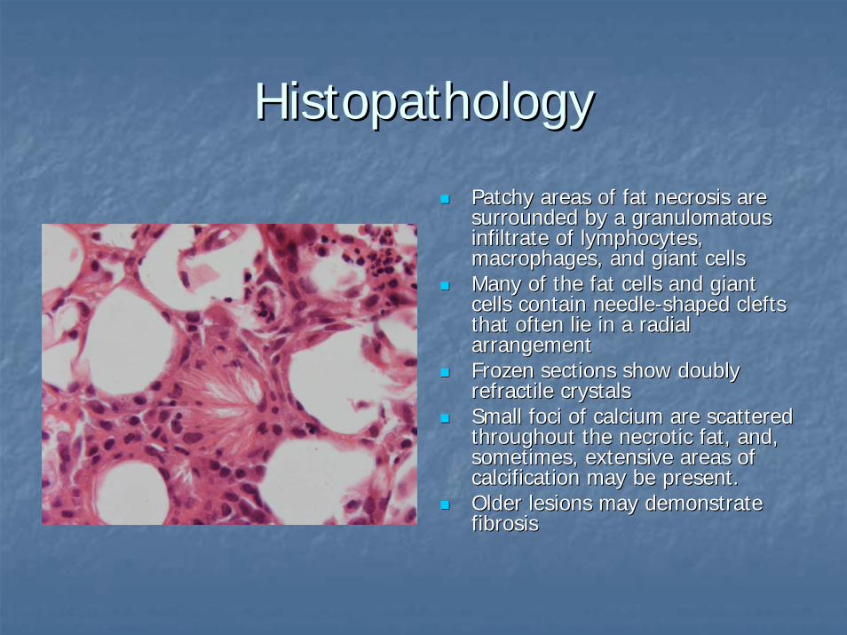
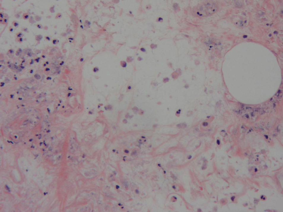
HistopathologyHistopathology
Patchy areas of fat necrosis are Patchy areas of fat necrosis are surrounded by a surrounded by a granulomatousgranulomatousinfiltrate of lymphocytes, infiltrate of lymphocytes, macrophages, and giant cellsmacrophages, and giant cellsMany of the fat cells and giant Many of the fat cells and giant cells contain needlecells contain needle--shaped clefts shaped clefts that often lie in a radial that often lie in a radial arrangementarrangementFrozen sections show doubly Frozen sections show doubly refractilerefractile crystals crystals Small foci of calcium are scattered Small foci of calcium are scattered throughout the necrotic fat, and, throughout the necrotic fat, and, sometimes, extensive areas of sometimes, extensive areas of calcification may be present. calcification may be present. Older lesions may demonstrate Older lesions may demonstrate fibrosisfibrosis

Differential DiagnosisDifferential Diagnosis
Corticosteroid withdrawalCorticosteroid withdrawalScleremaSclerema neonatorumneonatorum
Thickening of the subcutaneous fibrous septa, Thickening of the subcutaneous fibrous septa, and a radial array of fine needlelike clefts in and a radial array of fine needlelike clefts in the fat cellsthe fat cellsNo fat necrosis or inflammation No fat necrosis or inflammation
TreatmentTreatment
SupportiveSupportiveTreat symptomatic Treat symptomatic hypercalcemiahypercalcemiaaggressivelyaggressively


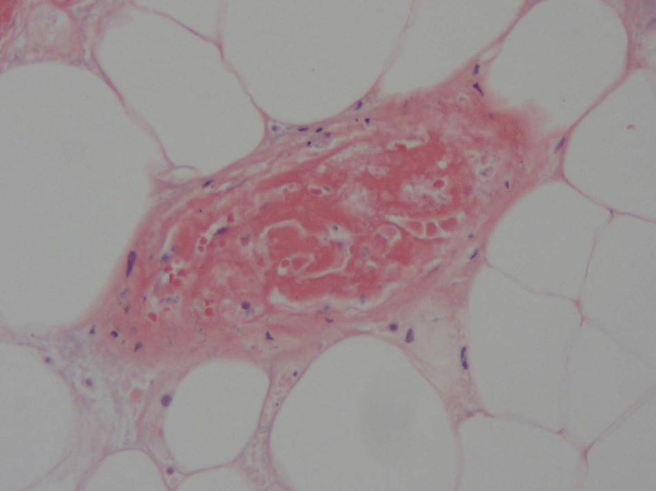


CalciphylaxisCalciphylaxis

CalciphylaxisCalciphylaxis
11--4% of the ESRD population4% of the ESRD populationProbably rare in general populationProbably rare in general population
Mortality/MorbidityMortality/MorbidityMortality rate 60Mortality rate 60--80%80%Leading cause of death is sepsis from infected, necrotic skin leLeading cause of death is sepsis from infected, necrotic skin lesionssionsMortality rate is higher in patients with proximal disease than Mortality rate is higher in patients with proximal disease than in those in those with only distal or with only distal or acralacral diseasedisease
More prevalent in whites More prevalent in whites F:M 3:1F:M 3:16 months to 83 years6 months to 83 years
Mean age of 48 years Mean age of 48 years Younger patients with longer duration of renal replacement theraYounger patients with longer duration of renal replacement therapy py more predisposed more predisposed

ClinicalClinical
Increased riskIncreased riskObesityObesity
Increased where body fat is most abundant, the Increased where body fat is most abundant, the thighs, buttocks and lower abdomenthighs, buttocks and lower abdomen
GlucocorticoidGlucocorticoid exposureexposure

PathogenesisPathogenesis
MultifactorialMultifactorialAssociated disorders chronic renal failure, Associated disorders chronic renal failure, hypercalcemiahypercalcemia, , hyperphosphatemiahyperphosphatemia, an elevated , an elevated calciumcalcium--phosphate product and secondary phosphate product and secondary hyperparathyroidismhyperparathyroidismHypercoagulableHypercoagulable conditions including protein C and conditions including protein C and protein S deficienciesprotein S deficienciesSensitizing events and agents included Sensitizing events and agents included nephrectomynephrectomyand exposure to parathyroid hormone and vitamin Dand exposure to parathyroid hormone and vitamin DChallengers included egg albumin and metallic salts Challengers included egg albumin and metallic salts


Septic Septic PanniculitisPanniculitis

What do I need to consider in What do I need to consider in diagnosing a diagnosing a panniculitispanniculitis??

ClinicalClinical
Consider in any unexplained Consider in any unexplained panniculitispanniculitisSpecial stainsSpecial stainsCulturesCultures

QuestionsQuestions
It is better to know It is better to know some of the questions some of the questions than all of the than all of the answers. answers.
James ThurberJames Thurber (1894 (1894 -- 1961)1961)