patient tetralogy of fallot: stroke in a young - … · stroke in a young patient. the patient...
TRANSCRIPT

Received 05/10/2018 Review began 05/15/2018 Review ended 05/17/2018 Published 05/31/2018
© Copyright 2018Ali et al. This is an open accessarticle distributed under the terms ofthe Creative Commons AttributionLicense CC-BY 3.0., which permitsunrestricted use, distribution, andreproduction in any medium,provided the original author andsource are credited.
Tetralogy of Fallot: Stroke in a YoungPatientHassam Ali , Shiza Sarfraz , Muhammad Sanan
1. Medical 4, Bahawal Victoria Hospital, Quaid-E-Azam Medical College, Bahawalpur, PAK 2.Department of Anesthesiology, Bahawal Victoria Hospital, Quaid-E-Azam Medical College, Bahawalpur,PAK
Corresponding author: Hassam Ali, [email protected] Disclosures can be found in Additional Information at the end of the article
AbstractTetralogy of Fallot (TOF) is a congenital birth defect of the heart which actually comprises fourindividual flaws. It causes poor flow of oxygenated blood to the organs and leads to cyanosis(blue-tinted skin, because of inadequate oxygenation). It can be recognized at birth or inadulthood. But sometimes, cases may go unnoticed, and the patient might present with somerare complications. In this case, the patient presented with an embolic infarct of the brain at theage of 25 with an undiagnosed tetralogy of Fallot.
Categories: Cardiology, Internal Medicine, NeurologyKeywords: congenital heart defects, embolic stroke, tetralogy of fallot, paralysis, cardiology,cerebrovascular accident, heart, neurology, international medicine
IntroductionTetralogy of Fallot (TOF) is a heart disease present at birth [1]. It consists of four defects [2]:
- Pulmonary stenosis
- Ventricular septal defect
- Right ventricular hypertrophy
- Aorta overriding the ventricular septal defect
Some babies, when they cry or breastfeed, may turn very blue. This is because of "a tet spell"due to the shunting of excess deoxygenated blood from the right chamber of the heart to theleft chamber. Such babies may have difficulty breathing, become limp, or even loseconsciousness [3].
TOF is treated by open-heart surgery, mostly in the first year of life. Much of the treatmentplan depends upon the individual’s signs and symptoms [4]. Most individuals can live to becomeadults, though complications may arise later—including irregular heart rates, cerebrovascularaccidents [5], and pulmonary regurgitation. We hereby present a case of a female with anundiagnosed tetralogy of Fallot, presenting with an embolic stroke of the frontoparietal regionof the brain. Informed consent was obtained from the patient.
1 2 1
Open Access CaseReport DOI: 10.7759/cureus.2714
How to cite this articleAli H, Sarfraz S, Sanan M (May 31, 2018) Tetralogy of Fallot: Stroke in a Young Patient. Cureus 10(5):e2714. DOI 10.7759/cureus.2714

Case PresentationA 25-year-old female presented to the emergency room with a complaint of left-sided bodyweakness since 12 hours. On clinical examination, the power of the left upper and lower limbswas seen to be limited to just slight movement. Planter reflex was up going on the left side(Babinski positive). Clinical anemia was also present, and the nails showed massive clubbing.According to her parents, she had a history of cyanosis since birth, but they never got treatmentfor it. There was no history of any psychiatric illness, hypertension, or diabetes.
A CT (computed tomography) scan showed no evidence of a haemorrhage, but some changes inthe temporoparietal area were observed, as shown in Figure 1. Later, an MRI (magneticresonance imaging) with contrast was advised and performed, which showed an infarct of theright temporoparietal lobe with mild brain atrophy as shown in Figure 2 and Figure 3.
FIGURE 1: Computed tomography (CT) scan showing changes
2018 Ali et al. Cureus 10(5): e2714. DOI 10.7759/cureus.2714 2 of 7
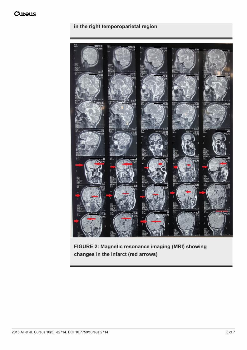
in the right temporoparietal region
FIGURE 2: Magnetic resonance imaging (MRI) showingchanges in the infarct (red arrows)
2018 Ali et al. Cureus 10(5): e2714. DOI 10.7759/cureus.2714 3 of 7

FIGURE 3: Magnetic resonance imaging report
DiscussionCerebrovascular accidents (CVAs) are not very common in young patients, but the patient wasfrom a village where medical facilities were not present. She was never diagnosed withtetralogy of Fallot, and only presented to the emergency room with left-sided paralysis as in astroke. She did have physical manifestations of chronic disease like emaciation [6] and clubbingof fingers as shown in Figure 4, which made us consider some underlying heart etiology. Later,her echocardiography report (Figure 5) confirmed a structural heart disease.
2018 Ali et al. Cureus 10(5): e2714. DOI 10.7759/cureus.2714 4 of 7

FIGURE 4: Finger nails clubbing of patient
FIGURE 5: Echocardiography report
Congenital cardiac disease can lead to the formation of a thrombus inside the heart, which can
2018 Ali et al. Cureus 10(5): e2714. DOI 10.7759/cureus.2714 5 of 7

later throw clots as emboli into the peripheral circulation; one study even suggested thatcongenital heart disease has a role in increasing thrombogenicity [7], which we also suspectedin this patient.
Alioglu et al. reported the occurrence of intra-cardiac thrombosis (two in right atrium and onein right ventricle) in three of nine children with tetralogy of Fallot [8]. Ammash et al. reportedtwo cases of cerebrovascular embolism among eight patients diagnosed with TOF over a seven-year period [9]. Ammash's study was the first to show the association between cyanoticcongenital heart disease and stroke in adults.
There are several factors that increase risk of pathogenesis of thrombosis in patients withcongenital heart disease (CHD). For example, chronic acidosis increases fibrin deposition,secondary erythrocytosis, and hypoxia/hypoxemia-induced activation of the pro-coagulantpathways, increasing tissue factor expression and impaired fibrinolysis [8]. Adults with cyanoticCHD also have an increased red blood cell (RBC) mass. This secondary erythrocytosis mayincrease blood viscosity, and it may thereby reduce cerebral blood flow, which can predisposethe patient to clot formations. Chronic hypoxemia will also activate neutrophils andmononuclear cells that release vasoactive and chemotactic factors, resulting in endothelialinjury [8]. Platelets and endothelial cells interact and activate platelets and enhanceintravascular thrombus formation by thrombin, which activates the coagulation cascade. Inaddition, an impaired fibrinolytic system due to increased plasminogen activator-1 levels cancontribute to thrombogenicity [8] and not just blood turbulence; thus, all these factors shouldalso be taken under consideration for cerebrovascular accidents in such patients. This may leadto future use of anticoagulation drugs in such patients in addition to surgical correction.
Due to advances in medical fields and paedriatic surgery, many cases of tetralogy of Fallot(TOF) undergo prompt surgical treatment. But our patient was born in a remote village withlittle or no medical facilities, and her diagnosis of TOF was delayed due to illiteracy as well asquackery in that region, though much research is needed to link causation of these factors. Forour ward, this was the first case of stroke in such a young patient due to TOF.
ConclusionsCerebrovascular accidents are usually seen in patients with a history of hypertension, diabetes,or structural cardiac anomalies—more so in older patients than in the young. Although it is notcommon, stroke in young patients can have multiple etiologies, including an undiagnosedcongenital heart disease that can lead to the formation of clots and their discharge into theperipheral circulation, leading to a blockade of that area's blood supply. Our case reportfollowed a somewhat similar course where tetralogy of Fallot was the culprit that caused astroke in a young patient. The patient presented with left-sided body paralysis, which uponinvestigation lead to a diagnosis of tetralogy of Fallot. We emphasize that structural heartdiseases should be kept in the differential for strokes, especially in younger people. It will helpin early diagnosis and treatment.
Additional InformationDisclosuresHuman subjects: Consent was obtained by all participants in this study. Quaid-eazam Medicalcollege bahawalpur issued approval 340. Infomed Consent was obtained by all participants inthis study. Conflicts of interest: In compliance with the ICMJE uniform disclosure form, allauthors declare the following: Payment/services info: All authors have declared that nofinancial support was received from any organization for the submitted work. Financialrelationships: All authors have declared that they have no financial relationships at present or
2018 Ali et al. Cureus 10(5): e2714. DOI 10.7759/cureus.2714 6 of 7

within the previous three years with any organizations that might have an interest in thesubmitted work. Other relationships: All authors have declared that there are no otherrelationships or activities that could appear to have influenced the submitted work.
AcknowledgementsTo My mother, Dr. Tahira Parveen. I am, because of you.
References1. Fallot's tetralogy. (2016). Accessed: May 30, 2018:
http://www.whonamedit.com/synd.cfm/2281.html.2. What is tetralogy of Fallot?. (2016). Accessed: May 30, 2018:
http://www.nhlbi.nih.gov/health-topics/tetralogy-fallot.3. Frederique B, Robert HA: Tetralogy of Fallot. Orphanet J Rare Dis. 2009, 4:2. 10.1186/1750-
1172-4-24. Tetralogy of Fallot. (2016). Accessed: May 30, 2018: https://www.nhlbi.nih.gov/health-
topics/tetralogy-fallot#Signs,-Symptoms,-and-Complications.5. Perloff JK, Marelli AJ, Miner PD: Risk of stroke in adults with cyanotic congenital heart
disease. Circulation. 1993, 87:1954-1959. 10.1161/01.CIR.87.6.19546. Yoshida T, Delafontaine P: Mechanisms of cachexia in chronic disease states . Am J Med Sci.
2015, 350:250-256. 10.1097/MAJ.00000000000005117. Gurgey A, Ozyurek E, Gümrük F, et al.: Thrombosis in children with cardiac pathology:
frequency of factor V leiden and prothrombin G20210A mutations. Pediatr Cardiol. 2003,24:244-248. 10.1007/s00246-002-0170-z
8. Alioglu B, Avci Z, Tokel K, Atac FB, Ozbek N: Thrombosis in children with cardiac pathology:analysis of acquired and inherited risk factors. Blood Coagul Fibrinolysis. 2008, 19:294-302.10.1097/MBC.0b013e3282fe73b1
9. Ammash N, Warnes CA: Cerebrovascular events in adult patients with cyanotic congenitalheart disease. J Am Coll Cardiol. 1996, 28:768-772.
2018 Ali et al. Cureus 10(5): e2714. DOI 10.7759/cureus.2714 7 of 7