patient guide to breast cancer surgery and treatment · although breast cancer occurs mostly in...
TRANSCRIPT

baggotstreet.mercy.net/benefits
Patient Guide to Breast CancerSurgery and TreatmentMercy Cancer ServicesYour life is our life’s work.

Patient Guide to Breast Cancer Surgery and Treatment | 1
IntroductionAn estimated one out of eight women will develop breast cancer during her lifetime. Fortunately, much progress has been made due to early detection and effective treatments.
This guide is designed to provide you with information about breast cancer and how the disease can be treated at each stage based upon the guidelines of the National Comprehensive Cancer Network (NCCN). For years, physicians have looked to the NCCN for guidance and advice on treating cancer patients. Their guidelines represent the consensus of a diverse panel of cancer experts across the nation.
Our intent is to help you understand your diagnosis and the treatments available by presenting you with basic information about breast cancer in a step-by-step fashion.
This information is not meant to replace the clinical advice of your doctor. Rather, our goal is to empower you to share your preferences and encourage you to openly discuss treatment strategies with your physician and cancer care team.

Patient Guide to Breast Cancer Surgery and Treatment | 2
What is breast cancer?
Breast cancer is a general term for cancer that originates in the breast cells.
It occurs when certain cells of the breast become abnormal and multiply out of control to form a mass called a malignant (cancerous) tumor. Sometimes these cancerous tumors locally invade (grow into) surrounding tissues or metastasize (spread) to distant parts of the body.
Although breast cancer occurs mostly in women, men can also develop the disease. It is the most common malignancy in American women, except for skin cancers.
Female Breast Anatomy
A basic understanding of the structure of breasts can help explain breast cancer.
The female breast is composed primarily of lobules (milk-producing glands), ducts (tubes that transport milk to the nipple) and stroma (fatty and connective tissues). The breast also includes lymphatic tissue, which is part of the body’s immune-fighting system.
Most breast cancers begin in the cells that line the ducts (ductal cancers). Some begin in cells that line the lobules (lobular cancers), while a small number start in the stromal tissues of the breast.
When breast cancer spreads beyond the breast, the cancer cells are generally found first in the lymph nodes under the arm.
The Lymph (lymphatic) System
One of the most common ways for breast cancer cells to spread is through the lymph system. The lymph system is a complex network of interconnected organs, vessels and nodes responsible for fighting infections.
Lymph nodes are clusters of small bean-shaped glands located throughout the body and linked together by lymphatic vessels. Lymphatic vessels are thin tubes that carry lymph, a nearly clear fluid that circulates around the tissues and into the bloodstream.
Breast cancer cells can enter lymphatic vessels, follow their paths and become trapped in lymph nodes where they can grow.
Most lymphatic vessels in the breast connect to lymph nodes under the arm (axillary nodes). Some lymphatic vessels connect to lymph nodes behind the breast bone or above or below the collarbone.
When breast cancer spreads outside the breast, the cancer cells are generally found in the axillary (underarm) nodes first. The more lymph nodes that have cancer, the greater the chance for metastases, which is the spread of cancer to distant organs.
Breast Cancer Terms
Breast cancer is described in many ways to relay specific information about the disease, including identifying its tissue type and whether it is non-invasive or invasive.
Breast Cancer Tissue TypesCarcinoma is a cancer that originates in epithelial tissues (tissues that cover body surfaces and line internal organs)

Patient Guide to Breast Cancer Surgery and Treatment | 3
Adenocarcinoma is a type of carcinoma that develops in the epithelial tissues of a gland. Glands make or secrete substances. The breast is a gland. It produces breast milk.
Breast adenocarcinoma originates in the epithelial tissues of the breast. There are two types of breast adenocarcinomas: ductal and lobular. Ductal means the cancer began in the milk ducts. Lobular means the cancer originated in the lobules, the milk-producing glands.
Sarcoma is a cancer that begins in connective tissues such as cartilage, muscle and fat. Breast sarcomas are very rare.
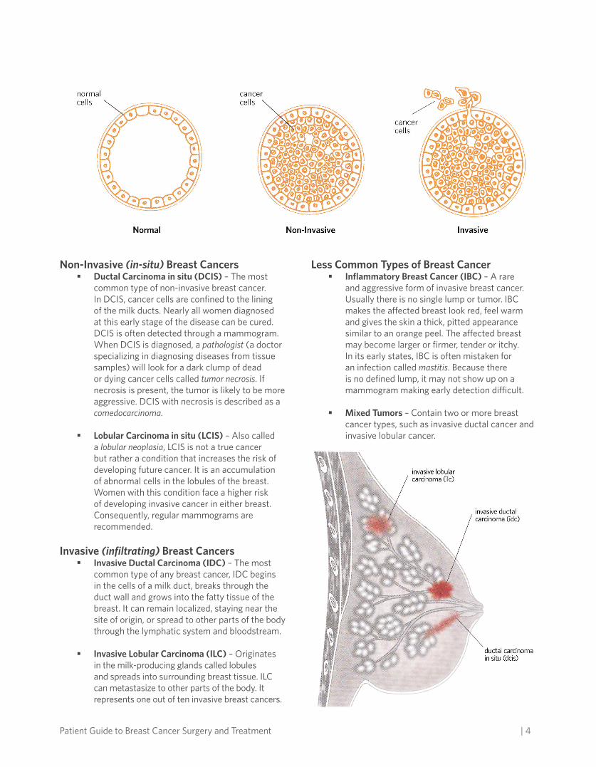
Non-invasive and Invasive CancersOne of the single-most important descriptions about breast cancer is whether it is invasive (spreading) or non-invasive.
Non-invasive (in situ) CancerNon-invasive (in situ) cancer means that the cancer cells remain in their place of origin. They are confined to the milk ducts (ductal carcinoma in situ) or the breast lobules (lobular carcinoma in situ or lobular neoplasia).
Because these cancer cells have not invaded deeper tissues of the breast or spread to other organs, they are sometimes called non-invasive breast cancers.
Invasive (infiltrating) CancerMost breast cancers are invasive; they spread.
An invasive cancer has grown beyond the layer of cells where it started and infiltrated healthy tissue adjacent to the tumor. Invasive breast cancer means that the cancer cells have broken through the milk duct and/or lobular walls.
Breast cancer can be locally invasive and not be metastatic, which refers to the spread of cancer cells to distant areas of the body through the lymph system or bloodstream.
Types of Breast Cancer
There are several types of breast cancers. Most are invasive; they spread. Some are rare. Sometimes a single tumor contains a combination of different breast cancers.
Following are brief descriptions of breast cancers beginning with the most common types.
Patient Guide to Breast Cancer Surgery and Treatment | 4
Non-Invasive (in-situ) Breast Cancers§ Ductal Carcinoma in situ (DCIS) – The most
common type of non-invasive breast cancer. In DCIS, cancer cells are confined to the lining of the milk ducts. Nearly all women diagnosed at this early stage of the disease can be cured. DCIS is often detected through a mammogram. When DCIS is diagnosed, a pathologist (a doctor specializing in diagnosing diseases from tissue samples) will look for a dark clump of dead or dying cancer cells called tumor necrosis. If necrosis is present, the tumor is likely to be more aggressive. DCIS with necrosis is described as a comedocarcinoma.
§ Lobular Carcinoma in situ (LCIS) – Also called a lobular neoplasia, LCIS is not a true cancer but rather a condition that increases the risk of developing future cancer. It is an accumulation of abnormal cells in the lobules of the breast. Women with this condition face a higher risk of developing invasive cancer in either breast. Consequently, regular mammograms are recommended.
Invasive (infiltrating) Breast Cancers§ Invasive Ductal Carcinoma (IDC) – The most
common type of any breast cancer, IDC begins in the cells of a milk duct, breaks through the duct wall and grows into the fatty tissue of the breast. It can remain localized, staying near the site of origin, or spread to other parts of the body through the lymphatic system and bloodstream.
§ Invasive Lobular Carcinoma (ILC) – Originates in the milk-producing glands called lobules and spreads into surrounding breast tissue. ILC can metastasize to other parts of the body. It represents one out of ten invasive breast cancers.
Less Common Types of Breast Cancer§ Inflammatory Breast Cancer (IBC) – A rare
and aggressive form of invasive breast cancer. Usually there is no single lump or tumor. IBC makes the affected breast look red, feel warm and gives the skin a thick, pitted appearance similar to an orange peel. The affected breast may become larger or firmer, tender or itchy. In its early states, IBC is often mistaken for an infection called mastitis. Because there is no defined lump, it may not show up on a mammogram making early detection difficult.
§ Mixed Tumors – Contain two or more breast cancer types, such as invasive ductal cancer and invasive lobular cancer.

Patient Guide to Breast Cancer Surgery and Treatment | 5
§ Medullary Carcinoma – This tumor resembles the medulla (gray matter of the brain). It occurs in the milk ducts with a well-defined boundary between the tumor and normal tissue. Its cells look, but do not act, aggressively. The outlook for medullary carcinoma is generally better than for the more common types of invasive breast cancer.
§ Metaplastic Carcinoma – Also known as carcinoma with metaplasia. These tumors begin in one type of cell and change into another type of cell. Metaplastic carcinomas are treated like invasive ductal cancers.
§ Mucinous Carcinoma – Also known as colloid carcinoma, this rare type of invasive ductal cancer contains mucus-producing cells. It tends to affect women after menopause and has a favorable prognosis (outlook) in most cases.
§ Paget Disease of the Nipple – An uncommon type of breast cancer that starts in the milk ducts, spreads to the skin of the nipple and then to the areola, which is the dark circle around the nipple. The skin of the nipple and areola appears crusted, scaly and red, with areas bleeding or oozing. Other symptoms may include itching or burning sensations. Paget disease is associated with ductal carcinoma (DCIS) or more frequently, with invasive ductal carcinoma (IDC).
§ Tubular Carcinoma – A rare type of invasive ductal carcinoma. Under a microscope, these cancer cells resemble tiny tubes. Tubular carcinomas tend to have a better prognosis than most other infiltrating ductal or lobular carcinomas.
§ Papillary Carcinoma – The cells of these cancers resemble finger-like projections when viewed under a microscope. These cancers are often considered a type of ductal carcinoma in situ (DCIS) and are treated as such. In rare cases, they are invasive and are treated like invasive ductal carcinoma although the prognosis is likely to be better. Papillary carcinomas occur most frequently in postmenopausal women.
§ Adenoid Cystic Carcinoma (adenocystic carcinoma) – A very rare form of cancer that can occur in many body sites including the head, neck or breast. These cancer cells have both glandular (adenoid) and cystic (cylinder-like)
features when seen under a microscope. They are usually not aggressive and patients tend to have a good prognosis.
§ Phyllodes Tumor – Is a very rare breast tumor that is most often benign (non-cancerous), but can become malignant (cancerous). It is called a sarcoma because it developes in the stroma (connective tissue) of the breast. Other names for these tumors include phylloides tumor and cystosarcoma phyllodes.
§ Angiosarcoma – A rare form of cancer that begins in the lining of the blood or lymph vessels in the breast or underarm. It rarely occurs in breasts, but when it does, it is usually a complication of radiation therapy and tends to develop about five to 10 years after treatment. It is an extremely rare complication of breast radiation therapy. An angiosarcoma can also occur in the arm of women who develop lymphedema (swelling due to lymph fluid build-up). These breast cancers tend to be aggressive and spread quickly.
Breast Cancer Causes
Cancer develops at a molecular level. Exactly how it occurs remains unknown. However, certain changes in DNA can cause normal breast cells to become cancerous.
DNA is the building block of an individual’s genetic makeup. We resemble our parents because they are the source of our DNA. But, DNA affects more than our appearance. It can affect our risk for developing disease, including breast cancer.
Genes, which are made of DNA, contain instructions for our cells. Some genes contain instructions that control when cells grow, divide and die. Genes that speed up cell division are called oncogenes. Genes that slow down cell division, or cause cells to die at the right time, are called tumor suppressor genes.
Cancer can be caused by DNA mutations (changes) that “turn on” oncogenes or “turn off” tumor suppressor genes. These genetic mutations can be inherited or acquired over the course of a person’s life.

Patient Guide to Breast Cancer Surgery and Treatment | 6
Inherited Gene MutationsFamilial (inherited) breast cancer is most often associated with the BRCA1 and BRCA2 genes. When functioning properly, they are tumor suppressor genes. When these genes are mutated, they no longer suppress abnormal cell growth. Consequently, cancer is more likely to develop. Mutated BRCA genes cause less than 10 percent of breast cancers.
A blood, saliva or buccal sample can identify women with inherited mutations in BRCA1 or BRCA2 genes. Having one or both of these gene mutations can predispose a woman to breast cancer. However, it does not guarantee that she will develop the disease.
Men can also inherit these genetic mutations. Although men with either mutation have only a six percent chance of developing breast cancer, they can still pass the gene to their children.
Acquired Gene MutationsMost DNA mutations related to breast cancer develop over the course of a woman’s lifetime rather than having been inherited.
These acquired mutations of oncogenes and/or tumor suppressor genes may result from a variety of factors, such as through exposure to radiation or certain chemicals. However, the cause of most acquired mutations that lead to breast cancer remains unknown.
Breast Cancer Grades
Breast cancer grade diagnosis and treatment is based upon several factors including the tumor’s grade.
Grade describes the appearance of cancer cells when viewed under a microscope. Pathologists assign one of two grade types to breast cancer: tumor and nuclear.
Tumor Grade – Describes invasive breast cancer cells. Invasive breast cancer cells are graded on a 1 to 3 scale. Invasive cancers that resemble normal breast tissue generally get a low number grade and tend to have a better prognosis.
§ Grade 1: Well-differentiated cancers with generally normal looking cells. These cancers do not grow rapidly.
§ Grade 2: Moderately differentiated cancers with cells that have features between grades 1-3 tumors. These cancers grow a bit faster.
§ Grade 3: Poorly differentiated cancers with cells that do not look normal. These cancers tend to grow fast, metastasize (spread), and be aggressive.
Nuclear Grade – Describes the cells of a non-invasive cancer, specifically ductal carcinoma in situ (DCIS). Nuclear grade refers to the rate at which DCIS cells divide to form more cancer cells. Nuclear grades can be low, intermediate or high. Cancer is likely to recur if there is a high nuclear grade, the presence of necrosis (a clump of dead cancer cells), the cancer is at or near the edge of the tissue sample, or if there are large areas of DCIS.
Breast Cancer Stages
Cancers are divided into different groups, called stages, based on whether the cancer is invasive or non-invasive, the size of the tumor, how many lymph nodes are involved, and whether there is spread to other parts of the body.
Staging a cancer is the process of finding out how far the cancer has progressed when it is diagnosed. Doctors determine the stage of a cancer by gathering information from physical examinations and tests on the tumor, lymph nodes and distant organs.
A breast cancer’s stage is one of the most important factors that may predict prognosis (outlook for cure versus the chance of cancer coming back or spreading to other organs). A cancer’s stage, therefore, is an important factor in choosing the best treatment.
Each woman’s outlook with breast cancer differs, depending on the cancer’s stage and other factors such as hormone receptors, her general state of health and her treatment.
You should talk frankly with your doctors about your cancer stage and prognosis, and how they affect treatment options.

Patient Guide to Breast Cancer Surgery and Treatment | 7
System to Define Cancer Stage
Tumor Size
5 centimters4 centimters3 centimters2 centimters1 centimters
Tumor size is usually reported in metric units (millimeters or centimeters)
The system most often used to describe the extent of breast cancer is the TNM staging system. In TNM staging, information about the tumor (T-Stage), nearby lymph nodes (N-Stage) and distant metastases (M-Stage) is combined and a stage is assigned to specific TNM groupings. The TNM stage groupings are described using Roman numerals from 0 to IV.
The clinical stage is determined by what the doctor learns from the physical examination and tests. The pathologic stage includes the findings of the pathologist after surgery. Most of the time, pathologic stage is the most important stage since involvement of the lymph nodes can only be accurately determined by examining them under a microscope.
T stands for the size of the cancer (measured in centimeters: 2.5 centimeters = 1 inch) and whether it is growing directly into nearby tissues. N stands for spread to nearby lymph nodes and M is for metastasis (spread to other parts of the body).
Categories of T, N, and M
T CategoriesT categories are based on the size of the breast cancer and whether it has spread to nearby tissue.
Tis: This is used only for carcinoma in situ or non-invasive breast cancer, such as ductal carcinoma in situ (DCIS) or lobular carcinoma in situ (LCIS).
T1: The cancer is 2 cm in diameter (about ¾ inch) or smaller.
T2: The cancer is more than 2 cm but not more than 5 cm in diameter.
T3: The cancer is more than 5 cm in diameter.
T4: The cancer is any size and has spread to the chest wall or the skin.
Breast Cancer StagesOverall Stage T category N category M categoryStage 0 Tis N0 M0Stage 1 T1 N0 M0Stage IIA T0 N1 M0 T1 N1 M0 T2 N0 M0Stage IIB T2 N1 M0 T3 N0 M0Stage IIIA T0 N2 M0 T1 N2 M0 T2 N2 M0 T3 N1 M0 T3 N2 M0Stage IIIB T4 Any N M0Stage IIIC Any T N3 M0Stage IV Any T Any N M1

Patient Guide to Breast Cancer Surgery and Treatment | 8
N CategoriesThe N category is based on which of the lymph nodes near the breast, if any, are affected by the cancer. There are two classifications used to describe N. One is clinical (before surgery) – what the doctor can feel or see on imaging studies. The other is pathological (after surgery) – what the pathologist can see in lymph nodes removed during surgery.
N0 Clinical: The cancer has not spread to lymph nodes, based on clinical exam.
N1 Clinical: The cancer has spread to lymph nodes under the arm on the same side as the breast cancer. Lymph nodes are not attached to one another or to the surrounding tissue.
N1 Pathological: The cancer is found in one to three lymph nodes under the arm.
N2 Clinical: The cancer has spread to lymph nodes under the arm on the same side as the breast cancer and lymph nodes are attached to one another or to the surrounding tissue. Or the cancer can be seen to have spread to the internal mammary lymph nodes (next to the sternum), but not to the lymph nodes under the arm.
N2 Pathological: The cancer has spread to four to nine lymph nodes under the arm.
N3 Clinical: The cancer has spread to lymph nodes above or just below the collarbone on the same side as the cancer, and may or may not have spread to lymph nodes under the arm. Or the cancer has spread to internal mammary lymph nodes and lymph nodes under the arm, both on the same side as the cancer.
N3 Pathological: The cancer has spread to 10 or more lymph nodes under the arm or also involves lymph nodes in other areas around the breast.
M CategoriesThe M category depends on whether the cancer has spread to any distant tissues and organs.
M0: No distant cancer spread.
M1: Cancer has spread to distant organs.
Stage Grouping for Breast Cancer
Breast cancer treatment includes treatment of the breast and treatment for cancer cells that may have spread to other parts of the body. The breast itself is treated by surgery, often in combination with radiation. The lymph
nodes in the armpit are also studied to see if the breast cancer has spread. The treatment for cancer cells that may have spread beyond the breast and lymph nodes in the armpit is a combination of either hormone therapy and/or chemotherapy.
Prognostic Indicators
Prognostic indicators are tests that help predict disease outcomes and determine the best treatment approach. They are performed on tumor tissue.
In breast cancer, these tests are primarily used to determine the absence or presence of special proteins on the surface of cells called receptors. Receptors respond to specific cellular signals that cause a specific reaction or result.
Estrogen Receptor (ER) and Progesterone Receptor (PR) StatusBreast cancers are tested for estrogen and progesterone receptors. When these receptors bind (attach) with estrogen or progesterone hormones, they initiate a series of events that can stimulate breast cancer cell growth. Breast cancer cells may contain one or both receptors, or they may not have either receptor.
A breast cancer is hormone receptor-positive if it shows evidence of either estrogen or progesterone receptors. A breast cancer without either receptor is called hormone-receptor negative.
Women with hormone receptor-positive cancers tend to have a better prognosis than those who are hormone-receptor negative. They are more likely to respond to hormone therapy, such as tamoxifen, than women with cancer who do not have these receptors.
All breast malignancies, with the exception of lobular carcinoma in situ (LCIS), should be tested for these hormone receptors at the time of breast biopsy or surgery. About two-thirds of breast cancers contain at least one of these receptors. The percentage increases in older women.
HER2/neu StatusHER2/neu (often called HER2) is a growth-promoting protein and receptor.
Some cancer cells make too much of HER2 because of a genetic mutation. They are called HER2-positive.
The overproduction of this protein causes increased cell growth and often results in more aggressive breast malignancies.

Patient Guide to Breast Cancer Surgery and Treatment | 9
All newly diagnosed invasive breast cancers should be tested for HER2/neu. Although these cancers are less responsive to hormonal therapy, HER2-positive cancers can benefit from treatment that targets the protein and helps stop the growth of cancer cells. These targeted therapies include trastuzumab (Herceptin) and lapatinib (Tykerb).
Breast Cancer Treatment
Breast cancer treatment includes treatment of the breast and treatment for cancer cells that may have spread to other parts of the body. The breast itself is treated by surgery, often in combination with radiation. The lymph nodes in the armpit are also studied to see if the breast cancer has spread. The treatment for cancer cells that may have spread beyond the breast and lymph nodes in the armpit is a combination of either hormone therapy and/or chemotherapy.
SurgeryMost women with breast cancer will have surgery. The 2 common types of surgery are breast–conserving surgery and mastectomy.
Breast-Conserving SurgeryLumpectomy removes only the breast lump and a rim of normal surrounding breast tissue. Partial or segmental mastectomy or quadrantectomy removes more breast tissue than a lumpectomy (up to one-quarter of the breast). If cancer cells are present at the outside edge of the removed breast tissue (the margin), more surgery is usually needed to remove any remaining cancer. Most often this additional surgery is a repeat lumpectomy, but sometimes it requires removal of the entire breast (mastectomy).
Radiation therapy is usually given after these types of surgery. Side effects of these operations include temporary swelling and tenderness and hardness due to scar tissue that may form in the surgical site.
For most women with stage I or II breast cancer, breast-conserving therapy (lumpectomy and radiation therapy) is as effective as mastectomy. Survival rates of women treated with these 2 approaches are the same. However, breast conservation therapy is not an option for all women with breast cancer, (see the section on “Choosing Between Breast-Conserving Therapy and Mastectomy” below). Those who may not have breast-conserving therapy include:
• Women who have had prior radiation therapy of the affected breast or chest
• Women with suspicious abnormalities that appear to be cancerous and are widespread throughout the breast
• Women whose lumpectomy, including any possible repeat lumpectomy when needed, cannot completely remove their cancer with a satisfactory cosmetic result
• Pregnant women who would require radiation while still pregnant
There are other groups of women for whom breast-conserving therapy is not strictly ruled out, but for whom the risks associated with this treatment are higher. For example, they may have an increased likelihood of experiencing recurrent breast cancer or side effects from radiation. These groups include:
• Women with active connective tissue disease involving the skin (especially scleroderma and lupus)
• Women with tumors greater than 5cm• Women who after lumpectomy have a very
limited amount of cancer at the edge of the pathology specimen
• Women who are 35 years of age or less• Women who have known BRCA1 or BRCA2 gene
mutation
The last two groups of women have a higher risk of breast cancer recurring in the breast in which the cancer was originally found and also in the opposite breast when compared to older women and women without gene mutations associated with breast cancer. For this reason, these women may consider the option of surgical removal of both breasts (double mastectomy) to decrease their future risk of breast cancer.
Radiation therapy as a part of breast-conserving therapy for invasive cancer can sometimes be omitted. Women who may consider lumpectomy without radiation therapy have all of the following:
• Age 70 years of older; and• A tumor 2cm or less that has been completely
removed; and • A tumor that contains hormone receptors; and• No lymph node involvement; and• Who receive treatment with hormone therapy
Mastectomy
Mastectomy is removal of the entire breast, including the nipple. Mastectomy is needed for some cases, and some women choose mastectomy rather than lumpectomy, (see discussion below). Different words are used to describe mastectomy depending on the extent of the surgery done in the armpit and the muscles under the breast. In a simple or total mastectomy the entire breast is removed, but no lymph nodes from under the arm or

Patient Guide to Breast Cancer Surgery and Treatment | 10
muscle tissue from beneath the breast is removed. In a modified radical mastectomy, the entire breast and some axillary (underarm) lymph nodes are removed. In a radical mastectomy, all the muscle under the breast is also removed. Radical mastectomy is rarely used today because for most women this surgery is not any more effective than the more limited forms of mastectomy.
Choosing Between Breast-Conserving Surgery and MastectomyThe advantage of breast-conserving surgery (lumpectomy) is that it preserves the appearance of the breast. A disadvantage is the need for several weeks of radiation therapy after surgery. However, some women who have a mastectomy will also need radiation therapy. Women who choose lumpectomy and radiation can expect the same chance of survival as those who choose mastectomy.
Although most women and their doctors prefer lumpectomy and radiation therapy, your choice will depend on a number of factors, such as:
• How you feel about losing your breast• Whether you want to devote the additional time
and travel for radiation therapy• Whether you would want to have more surgery
to reconstruct your breast after having a mastectomy
• Your preference for mastectomy as a way to “take it all out as quickly as possible”
In determining your preference for lumpectomy or mastectomy, be sure to get all the facts. Though you may have a gut feeling for mastectomy to “take it all out as quickly as possible,” the fact is that doing so does not provide any better chance of long term control or a better outcome of treatment in most cases. Large research studies with thousands of women participating, and over 20 years of information show that when lumpectomy can be done, mastectomy does not provide any better chance of survival from breast cancer than lumpectomy plus radiation. It is because of these facts that most women today do not have their breast removed.
Reconstructive SurgeryIf a woman has a mastectomy, she may want to consider having the breast rebuilt; this is called breast reconstruction. This requires additional surgery to create the appearance of a breast after mastectomy. The breast can be reconstructed at the same time the mastectomy is done (immediate reconstruction) or at a later date (delayed reconstruction). Surgeons may use silicone or saline-filled implants, or tissue from other parts of your body.
How do a woman and her doctor decide on the type of reconstruction and when she should have the procedure? The answer depends on the woman’s personal preferences, the size and shape of her breasts, the size and shape of her body, her level of physical exercise, details of her medical situation (such as how much skin is removed), and if she needs chemotherapy or radiation.
If you are thinking about breast reconstruction, please discuss this with your doctor when you are planning your treatment.
Lymph Node SurgeryIn the treatment of invasive cancer, whether a woman has a mastectomy or lumpectomy, she and her doctor usually need to know if the cancer has spread to the lymph nodes. When the lymph nodes are affected, there is an increased likelihood that cancer cells have spread through the bloodstream to other parts of the body.
Doctors once believed that removing as many lymph nodes as possible would reduce the risk of cancer spreading to other places and improve a woman’s chances for long-term survival. We now know that removing the lymph nodes probably does not improve the chance for long-term survival. But knowing whether lymph nodes are involved is important in selecting the best treatment to prevent cancer recurrence.
The only way to accurately determine if lymph nodes are involved is to remove and examine them under the microscope. This means removing some or all of the lymph nodes in the armpit. Two different surgical procedures are used to remove the lymph nodes: a sentinel lymph node biopsy and an axillary lymph node dissection. When an axillary lymph node dissection is done, all of the lymph nodes in the armpit are removed. The sentinel lymph node biopsy is a more limited surgery that only removes a few lymph nodes and is associated with fewer side effects.
For some women with invasive cancer, removing the underarm lymph nodes is optional. This includes:
• Women with tumors so small and with such a favorable outlook that lymph node spread is unlikely
• Instances where it would not affect whether adjuvant treatment is given
• Elderly women• Women with serious medical conditions

Patient Guide to Breast Cancer Surgery and Treatment | 11
Lymph node surgery is not necessary with pure ductal carcinoma in situ or pure lobular carcinoma in situ. A sentinel node biopsy may be done if the woman is having surgery (such as a mastectomy) that would make it impossible to do the sentinel node biopsy procedure if invasive cancer were found in the tissue removed during the surgery.
The surgical technique used to remove lymph nodes from under the armpit depends on the personal circumstances of the patient. If there are enlarged lymph nodes with apparent spread of the cancer, or the lymph nodes are otherwise found to be involved with cancer, then complete axillary lymph dissection is necessary. However, for women with no apparent signs of cancer spread to the lymph nodes who have not received prior chemotherapy or hormone therapy, the sentinel lymph node biopsy is the preferred procedure because it is associated with fewer side effects (See the discussion, “Side Effects of Lymph Node Surgery” below) and is as effective as axillary lymph node dissection in finding cancer in the lymph nodes.
In a sentinel lymph node biopsy procedure the surgeon finds and removes the “sentinel nodes,” the first few lymph nodes into which a tumor drains. These are the lymph nodes most likely to contain cancer cells. To find these so-called “sentinel lymph nodes,” the surgeon injects a radioactive substance and/or a blue dye under the nipple or into the area around the tumor. Lymphatic vessels carry these substances into the sentinel lymph nodes and provide the doctor with a “lymph node map.” The doctor can either see the blue dye or detect the radioactivity with a Geiger counter. The surgeon then removes the marked nodes for examination by the pathologist.
If the sentinel node contains cancer, the surgeon may remove more lymph nodes in the armpit (axillary dissection). This may be done at the same time or several days after the original sentinel node biopsy. If the sentinel node is cancer-free, the patient will not need more lymph node surgery and can avoid the side effects of full lymph node surgery. However, this limited sampling of lymph nodes is not appropriate for some women.
Side Effects of Lymph Node SurgerySide effects of lymph node surgery may include:
• Temporary or permanent numbness of the skin on the inside of the upper arm
• Temporary limitation of arm and shoulder movements
• Swelling of the breast and arm called lymphedema
The side effects can occur with either the full axillary lymph node dissection or with sentinel lymph node biopsy. Side effects are much less common and less severe with the sentinel lymph node procedure.
Lymphedema is the most significant of these side effects. If it develops it may be permanent. Most women who develop lymphedema find it bothersome but not disabling. No one can predict which patients will develop this condition or when it will develop. Lymphedema can develop just after surgery, or even months or years later. Significant lymphedema occurs in about 13% of women who have axillary lymph node dissection and in up to 5% of women who have sentinel lymph node biopsy. With care, patients can take steps to help avoid lymphedema or at least keep it under control. Talk to your doctor for more details.
Some of the steps to take to help avoid lymphedema include:
• Avoid having blood drawn from or IV’s inserted into the arm on the side of the lymph node surgery.
• Avoid having a blood pressure cuff on that arm. If you are in the hospital, tell all health care workers about your arm.
• If your arm or hand feels tight or swollen, don’t ignore it. Tell your doctor immediately.
• If needed, wear a well-fitted compression sleeve.• Wear gloves when gardening or doing other
things that are likely to lead to cuts.
For more information on lymphedema, call the American Cancer society at 1-800-ACS-2345 and ask for Lymphedema: What Every Woman With Breast Cancer Should Know.
Therapies
Radiation TherapyRadiation therapy uses a beam of high-energy X-rays to destroy cancer cells left behind in the breast, chest wall, or lymph nodes after surgery. Radiation may also be needed after mastectomy in cases with a large breast tumor, close margins or when cancer is found in the lymph nodes.
This type of treatment can be given in several ways.
• External beam radiation delivers radiation from a machine outside the body. This is the typical radiation therapy given after lumpectomy and is given to the entire breast with an extra dose

Patient Guide to Breast Cancer Surgery and Treatment | 12
(“boost”) to the site of the tumor. It is usually given five days a week for a course of six to seven weeks. Some women are candidates for a shortened course of radiation, given over only three to four weeks.
• Brachytherapy, also called internal radiation or interstitial radiation, describes the placement of radioactive materials (often called “seeds”) in or near where the tumor was removed. They may be placed in the lumpectomy site to “boost” the radiation dose in addition to external beam radiation therapy.
Recently there has been interest in limiting radiation therapy only to the site of the lumpectomy, referred to as partial breast irradiation. This is based on the observation that when breast cancer recurs in the breast, the most common place is in the site of the original tumor. Brachytherapy is one technique of partial breast irradiation. External beam radiation therapy also can be used to deliver partial breast irradiation. The extent and duration of radiation depends on whether or not a lumpectomy or mastectomy was done and whether or not lymph nodes are involved. If a lumpectomy was done, the entire breast receives radiation with an extra boost of radiation to the area in the breast where the cancer was removed to prevent it from coming back in that area.
If the surgery was mastectomy, radiation is given to the entire area of the skin and muscle where the mastectomy was done if the tumor was over 5cm in size, or if the tumor was close to the edge of the removed mastectomy tissue.
In patients who have had lumpectomy or mastectomy, further radiation may be recommended if the cancer has spread to the lymph nodes. Radiation may be given to the area just above and below the collarbone and along the breastbone, depending on the number and location of involved lymph nodes.
Side effects most likely to occur from radiation include swelling and heaviness in the breast, sunburn-like skin changes in the treated area, and fatigue. Changes to the breast tissue and skin usually go away in 6 to 12 months. In some women, the breast becomes smaller and firmer after radiation therapy. There may also be some aching in the breast, and rarely a rib fracture or second cancer may be caused by the radiation.
Systemic TreatmentTo reach cancer cells that may have spread beyond the breast and nearby tissues, doctors use drugs that can be given by pills or by injection. This type of treatment is called systemic therapy. Examples of systemic treatment include chemotherapy, hormone therapy, and other types
of targeted therapy. Targeted therapies such as hormonal therapy are only helpful if the tumor is hormone receptor-positive, and targeted therapies such as trastuzumab and lapatinib are only effective if the tumor is HER2-positive, (see section on targeted therapy below).
Even in the early stages of the disease, cancer cells can break away from the breast and spread through the bloodstream. These cells usually don’t cause symptoms, they don’t show up on an X-ray, and they can’t be felt during a physical examination. But if they are allowed to grow, they can establish new tumors in other places in the body. Systemic treatment given to patients who have no evidence of spread of cancer but who are at risk of developing spread of the cancer is called adjuvant therapy. The goal of adjuvant therapy is to kill undetected cancer cells that have traveled from the breast.
Women who have invasive breast cancer should receive adjuvant therapy, except those with very small or well-differentiated tumors. For example, women with hormone receptor-positive disease will receive hormone therapy, and women with HER2-positive tumors greater than 1cm in diameter or with involvement of lymph nodes will receive targeted therapy with trastuzumab. Chemotherapy may also be recommended based on the size of the tumor, grade of the tumor, and presence or absence of lymph node involvement. For women with breast cancers with hormone receptor-negative tumors, hormone therapy is not effective and in women with HER2-negative tumors, trastuzumab is not effective. In women with tumors that are hormone receptor- and HER2-negative, the only decision is whether or not to receive chemotherapy.
In most cases, systemic treatment is given soon after surgery using the results of the surgery and pathology evaluation to determine the best choice treatment. In some cases, the systemic therapy is given to patients after a needle biopsy, but before lumpectomy or mastectomy. This preoperative chemotherapy is called neoadjuvant treatment. Oncologists give patients neoadjuvant treatment to try to shrink the tumor enough to make surgical removal easier. This may allow women who would otherwise need mastectomy to have breast-conserving surgery.
For women whose breast cancer has spread to other organs in the body (metastases), systemic treatment is the main treatment. This treatment may be chemotherapy, hormone therapy, other types of targeted therapy, or a combination of these types of therapy.

Patient Guide to Breast Cancer Surgery and Treatment | 13
ChemotherapyChemotherapy uses drugs that are toxic to and often kill cancer cells. Usually these cancer-fighting drugs are given intravenously (injected into a vein) or by mouth. Either way, the drugs travel through the bloodstream to the entire body. Doctors who prescribe these drugs (medical oncologists) sometimes use only a single drug and other times use a combination of drugs.
When chemotherapy is given after surgery for early stage breast cancer, it is called adjuvant chemotherapy. Sometimes chemotherapy is given before surgery. This is called neoadjuvant chemotherapy. In most cases, adjuvant or neoadjuvant chemotherapy is most effective when combinations of drugs are used together. Chemotherapy may also be given to treat breast cancer that has spread to places other than the breast or lymph nodes. Both single drugs and combinations of drugs are often used in the treatment of breast cancer that has spread. Clinical research studies over the last 30 years have determined which chemotherapy drugs are most effective. With continued research, even better combinations may be discovered.
Doctors give chemotherapy in cycles, with each period of treatment followed by a rest period. The chemotherapy is given on the first day of each cycle (and sometimes on other days of the cycle), and then the body is given time to recover from the effects of chemotherapy. The chemotherapy drugs are then repeated to start the next cycle. The time between giving the chemotherapy drugs varies according to the specific chemotherapy drug or combination of drugs. Adjuvant chemotherapy usually lasts for a total time of three to six months depending on the drugs used.
The side effects of chemotherapy depend on the type of drugs used, the amount taken, and the length of treatment. Some women have many side effects while other women have few side effects.
• Doxorubicin and epirubicin may cause heart damage but this is uncommon in people who do not have a history of heart disease. If you know you have heart disease or there is concern you might have heart disease, your doctor may suggest special heart tests before you use these drugs and may suggest other chemotherapy drugs if your heart function is weakened.
• Temporary side effects often include loss of appetite, nausea and vomiting, fatigue, mouth sores and hair loss.
• Chemotherapy may cause menstrual cycles to stop either temporarily or permanently.
• Lowering of the blood counts from chemotherapy is the most common serious side effect of chemotherapy. Chemotherapy does this by damaging the blood producing cells of the bone marrow. A drop in white blood cells can raise a patient’s risk of infection; a shortage of blood platelets can cause bleeding or bruising after minor cuts or injuries; and a decline in red blood cells can lead to fatigue.
There are treatments for these side effects. There are excellent drugs that prevent or at least reduce nausea and vomiting. A group of drugs, called growth factors, stimulate the production of white blood cells or red blood cells and can help bone marrow recover after chemotherapy and prevent problems resulting from low blood counts. Although these drugs are often not necessary, doctors have been using them so they can give the chemotherapy more often. Talk with your doctor about which treatment will be right for you.
Ask your doctor or call the American Cancer Society and ask for a copy of specific guidelines for treating many of the side effects caused by chemotherapy, such as Nausea and Vomiting Treatment Guidelines for Patients with Cancer and Fever and Neutropenia Treatment Guidelines for Patients with Cancer.
Premenopausal women will often develop early menopause and infertility from chemotherapy drugs. The older a woman is when she received chemotherapy, the more likely it is she will stop menstruating or lose her ability to become pregnant. Some chemotherapy drugs are more likely to do this than others. However, you cannot depend on chemotherapy to prevent pregnancy, and getting pregnant while receiving chemotherapy could lead to birth defects and interfere with treatment. Therefore, premenopausal women should use non-hormonal forms of birth control while receiving chemotherapy. It is safe to have children after chemotherapy and certain types of chemotherapy can be given in the second and third trimester of pregnancy to women who are diagnosed with breast cancer during pregnancy (see discussion of breast cancer in pregnancy below), but it’s not safe to get pregnant while on treatment.
Targeted TherapyTargeted therapy is a form of treatment that attacks specific sites and/or processes that are important to the function of cancer cells. In many cases of targeted therapy, higher levels of the target (for example, the HER2 receptor) in the cancer cells are associated with greater benefit for the patient when the therapy (such as, trastuzumah) is given.

Patient Guide to Breast Cancer Surgery and Treatment | 14
Trastuzumab (Herceptin) is a monoclonal antibody therapy that targets the HER2 receptor that is on the surface of the breast cancer cells of some patients. Trastuzumab is an important treatment option for some patients with HER2-positive tumors. It may be used as adjuvant therapy with chemotherapy to reduce the risk of recurrent breast cancer, as neoadjuvant therapy combined with chemotherapy to shrink the size of the tumor before surgery, or as treatment for metastastic breast cancer. Trastuzumab can cause heart damage and should be used cautiously when combined with other heart-damaging drugs, such as doxorubicin and epirubicin.
Bevacizumab (Avastin) is another monoclonal antibody that may be used in patients with metastatic breast cancer. It is used in combination with the chemotherapy drug paclitaxel. Bevacizumab works by preventing the growth of new blood vessels that supply tumor cells with the blood, oxygen, and other nutrients they need to grow. Bevacizumab can cause side effects such as a delay in wound healing with bleeding, high blood pressure, or kidney damage.
Lapatinib (Tykerb) is another type of targeted therapy for patients with advanced or metastatic breast cancer. This drug is taken orally and is used in combination with another oral drug, capecitabine. It works by getting inside cells and interfering with processes involving both the HER2 receptor and another cell surface receptor called HER1. Use of lapatinib may cause certain side effects such as diarrhea and acne.
Hormone TherapyHormone therapy is a type of targeted therapy that targets the hormone receptors of cancer cells. Estrogen, a hormone produced mostly by the ovaries, but also from hormones produced by the adrenal glands and fat tissue in a woman’s body, causes some breast cancers to grow. Several approaches can be used to block the effect of estrogen or to lower estrogen levels.
These approaches can be divided into two main groups:
• Drugs that block the effect of estrogen on cancer cells, called anti-estrogens. These medicines do not decrease estrogen levels; instead, they prevent estrogen from causing the breast cancer cells to grow.
• Drugs or treatments that lower the production of estrogen in the body.
These treatments are used in two situations:• Women who have hormone receptor-positive
breast cancer that appears to have been completely removed by surgery. This adjuvant therapy reduces the risk of recurrent breast cancer or the spread of breast cancer to other parts of the body. Adjuvant therapy may also include chemotherapy or trastuzumab.
• Women with hormone receptor-positive breast cancer that has spread to other parts of the body or in whom the cancer has come back.
Hormone drugs are only effective in women with breast cancer tumors that have increased levels of estrogen or progesterone receptors. Every breast cancer should be tested for these receptors, and you should ask your doctor for the results of this test on your cancer. If the cancer is negative for both these receptors, then the hormone drugs are of no benefit.
Often a combination of hormone therapy and chemotherapy is used in the treatment of breast cancer.
Hormone therapy is often recommended for a period of 5 years or longer when used as adjuvant treatment. It can also be used for long periods of time in the treatment of metastatic breast cancer. If you are taking hormone therapy, it is very important that the pills be taken according to the recommendations of your doctor and pharmacists. Although hormone therapy can have side effects and the benefits of the treatment may not be readily apparent, it is very important that you do not compromise your care by stopping the drug or by skipping days or weeks of therapy without telling your doctor. If you are experiencing side effects, discuss these with your cancer treatment team.
Anti-Estrogen DrugsTamoxifen is the antiestrogen drug used most often. Taking tamoxifen as adjuvant therapy after surgery, usually for five years, reduces the chance of hormone receptor-positive breast cancer (for example, invasive breast cancer or DCIS) coming back. Tamoxifen is also used to treat metastatic breast cancer, and to reduce the risk of breast cancer in pre- and post-menopausal women with LCIS.In many women, tamoxifen causes the symptoms of menopause, including hot flashes, vaginal discharge and mood swings. Tamoxifen has two rare, but more serious side effects. These are a slightly increased risk of developing cancer of the lining of the uterus (endometrial cancer) and uterine sarcoma, and a slightly increased risk of developing blood clots. For most women with breast cancer, the benefits of taking the drug far outweigh the risks.

Patient Guide to Breast Cancer Surgery and Treatment | 15
Toremifene (Fareston) is another anti-estrogen closely related to tamoxifen. It may be an option for postmenopausal women with metastatic breast cancer.
Fulvestrant (Faslodex) is a newer drug that reduces the number for estrogen receptors. It is often effective in postmenopausal women even if the breast cancer is no longer responding to tamoxifen. Hot flashes, mild nausea and fatigue are the major side effects of fulvestrant.
Raloxifene (Evista) is a drug that acts as an antiestrogen on breast tissue. It is used to reduce the risk of breast cancer in post-menopausal women with LCIS, but is not used to treat DCIS or invasive breast cancer.
Drugs that Lower Estrogen Levels – Aromatase InhibitorsAromatase inhibitors stop estrogen production in postmenopausal women. Three drugs in this category have been approved for treatment of breast cancer: anastrozole (Arimidex), letrozole (Femara) and exemestane (Aromasin). They work by blocking an enzyme that makes estrogen in postmenopausal women. They cannot stop the ovaries of premenopausal women from making estrogen. For this reason they are only effective and only used in postmenopausal women. For premenopausal women, tamoxifen remains the best drug to use.
The aromatase inhibitors have been compared with tamoxifen as adjuvant hormone therapy. They have fewer side effects than tamoxifen because they don’t cause cancer of the uterus and very rarely cause blood clots. They can, however, cause loss of calcium from bone and bone fractures because they remove all estrogen from a postmenopausal woman. They also may cause hot flashes and sometimes joint pain.
The aromatase inhibitors are more effective than tamoxifen alone in preventing breast cancer from coming back in postmenopausal women. Based on recent studies, many doctors recommend including an aromatase inhibitor in the adjuvant hormone treatment of postmenopausal women with hormone receptor-positive breast cancer.
Hormone Therapy and MenopauseAs discussed above, the aromatase inhibitors are not recommended for premenopausal women. Therefore, determining whether the patient is menopausal is important in making treatment decisions. This is not as simple as it may sound, because menstrual periods can stop as a side effect of treatment while the ovaries continue to make estrogen. Also, sometimes chemotherapy stops the ovaries from making estrogen
for a short period of time, but when the ovaries recover from the chemotherapy they start making estrogen again. Therefore, if the use of an aromatase inhibitor is considered in a young woman who has intact ovaries, repeated monitoring of hormone levels such as estradiol and follicle-stimulating hormone (FSH) may be required to make sure that the woman is truly post menopausal.
Ovarian AblationThe ovaries are the source of most estrogen in premenopausal women. Destroying or blocking the ability of the ovaries to produce estrogen (called ablation) may be an effective hormone therapy to treat premenopausal women with cancers that are positive for the estrogen or progesterone receptors. Destruction or blockage of the ovary production of estrogen can be done in a number of ways:
• The ovaries can be removed by surgery (oophorectormy).
• Radiation therapy can be given to the ovaries.• Drugs called LHRH (luteinizing hormone-
releasing hormone) agonists or antagonists prevent estrogen production by the ovaries. These include goserelin (Zoladex) and leuprolide (Lupron or Eligard). However, if a woman was premenopausal before taking these drugs, it is not possible to determine if she has become post-menopausal while she is still receiving this type of therapy.
BisphosphonatesBisphosphonates are used in breast cancer treatment to strengthen bones that have been weakened by invading breast cancer cells. The most commonly used bisphosphonates in breast cancer treatment are pamidronate (Aredia) and zoledronate (Zometa). These drugs are not used unless cancer has spread to the bone.
Hormonal treatment with the aromatase inhibitors may also weaken the bones by causing loss of calcium from the bone (called osteoporosis) and thus increase the risk of a fracture. Therefore, patients treated with an aromatase inhibitor should have their bone strength tested (called a bone density test) to determine if medication to strengthen their bones would be appropriate. Some patients may go into early menopause due to the side effects of chemotherapy. Menopause is associated with bone loss, too. These patients may also undergo a bone density test to evaluate the presence of osteoporosis. There are a number of medications, including some oral forms of bisphosphonates, to treat the loss of calcium from bone that is not caused by direct breast cancer in the bone. Talk with your doctor about whether one of these medications is right for you.

Patient Guide to Breast Cancer Surgery and Treatment | 16
Treatment of Breast Cancer During PregnancyBreast cancer is diagnosed in about one pregnant woman out of 3,000. Radiation therapy during pregnancy is known to increase the risk of birth defects, so it is not recommended for pregnant women with breast cancer.
For this reason, breast conservation therapy (lumpectomy and radiation therapy) is not considered an option if radiation cannot be delayed until it is safe to deliver the baby. However, breast biopsy procedures and even modified radical mastectomy are safe for the mother and fetus.
Treatment of Pain and Other Symptoms
This guide discusses ways to remove or destroy breast cancer cells or to slow their growth. But helping you feel as well as you can and continuing to do the things you enjoy doing are important goals, too. Don’t hesitate to discuss your symptoms or how you feel with your cancer care team. There are effective and safe ways to treat pain, other symptoms of breast cancer, and most of the side effects caused by breast cancer treatment. If you don’t tell your health care team, they may have no way of knowing about your problems.
Complementary and Alternative Therapies
Complementary and alternative medicines are different kinds of health care practices, systems, and products that are not part of your usual medical treatment. They may include Chinese herbs, special supplements, acupuncture, massage and a host of other types of treatment. You may hear about all sorts of these treatments from your family and friends. People may offer all sorts of suggestions, such as vitamins, herbs, stress reduction and more, as a treatment for your cancer or to help you feel better. Most of these methods are of unproven value in the treatment of cancer, although some have been tested in people with cancer.
The American Cancer Society defines complementary medicine or methods as those that are used along with your regular medical care. If these treatments are carefully chosen and managed, they may add to your comfort and well-being. Some of these methods have been tested, while others have not. Some have shown possible benefit, while others have not proven helpful. Some of these treatments have harmful effects as well.
Alternative medicines are defined as those that are used instead of regular medical care. Even though some of these methods have proven not to be helpful, and some have even proven harmful, they are often still promoted as “cures”. If you choose these alternatives, it is important to know that even the methods that do not cause harm may reduce your chance of fighting your cancer by delaying or replacing regular, proven forms of cancer treatments.
There is a great deal of interest today in complementary and alternative treatments for cancer. Many are being studied to find out if they are helpful to people with cancer.
Before changing your treatment or adding any of these methods, it is best to discuss this with your doctor or nurse. Some methods can be safely used along with standard medical treatment. Others, however, can interfere with standard treatment or cause serious side effects. More information about specific complementary and alternative methods of cancer treatment is available through the American Cancer Society’s toll-free number at 800-ACS-2345 or on their website at www.cancer.org.
Other Things to Consider During and After Treatment
During and after treatment of breast cancer you may be able to speed up your recovery and improve your quality of life by taking an active role in your care. Learn about the benefits and drawbacks of each of your treatment options, and talk to your cancer care team if there is anything you do not understand. Learn about and look out for side effects of treatment, and report these to your cancer care team right away so they can take steps to ease them and shorten their duration.
Remember that your body is as unique as your personality and your fingerprints. Although understanding your cancer’s stage and learning about your treatment options can help predict what health problems you may face, no one can say for sure how you will respond to cancer or its treatment.
You may have special strengths such as a habit of excellent nutrition and physical activity, a strong family support system, or a deep faith, and these strengths may make a difference in how you respond to cancer treatment. There are also experienced professionals in mental health services, social work services, and pastoral services who may help you and your family in coping with your illness.

Patient Guide to Breast Cancer Surgery and Treatment | 17
You can also help in your own recovery from cancer by making healthy lifestyle choices. If you use tobacco, stop now. Quitting will improve your overall health and the full return of the sense of smell may help you enjoy a healthy diet during recovery. If you use alcohol, limit how much you drink. Have no more than one drink per day. Good nutrition can help you get better after treatment. Eat a nutritious and balanced diet, with plenty of fruits, vegetables, and whole grain foods. Ask you cancer care team if you may benefit from a special diet or a referral to a dietician (a nutrition expert). Your cancer care team may have special recommendations for people who have had chemotherapy, radiation, or surgery.
If you are being treated for cancer, be aware of the battle that is going on in your body. Radiation therapy and chemotherapy add to the fatigue caused by the disease itself. To help you with the fatigue, plan your daily activities around when you feel your best. Get plenty of sleep at night. And ask your cancer care team about a daily exercise program to help you feel better.
A woman’s choice of treatment will likely be influenced by her age, the image she has of herself and her body, her hopes and fears, and her stage in life. For example, many women select breast-conserving surgery with radiation therapy over a mastectomy for body image reasons. On the other hand, some women who choose mastectomy may want the affected area removed, regardless of the effect on their body image, and others may be more concerned about the side effects of radiation therapy than body image.
Other issues that concern women include loss of hair from chemotherapy and the changes of the breast from radiation therapy. Women on chemotherapy tend to gain weight and it is important to continue to eat a healthy diet and exercise as much as your energy level will permit. In addition to these body changes, women may also be concerned about the outcome of their treatment. These are all factors that affect how a woman will make decisions about her treatment, how she views herself, and how she feels about her treatment.
Concerns about sexuality are often very worrisome to a woman with breast cancer. Some treatments for breast cancer can change a woman’s hormone levels and may have a negative impact on sexual interest and/or response. A diagnosis of breast cancer when a woman is in her 20s or 30s is especially difficult because choosing a partner and childbearing are often very important during this period. Relationship issues are also important because the diagnosis can be very distressing for the partner, as well as the patient. Partners are usually concerned about how to express their love physically
and emotionally during and after treatment. Talk to your cancer care team about these issues and contact the American Cancer Society for a copy of Sexuality and Cancer: For the Woman with Cancer and Her Partner.
Suggestions that may help a woman adjust to changes in her body image include looking at and touching her body; seeking the support of others, preferably before surgery; involving her partner as soon as possible after surgery; and openly talking about the feelings, needs, and wants created by her changed image.
A cancer diagnosis and its treatment are major life challenges; cancer affects you and everyone who cares for you. Almost everyone who has been through cancer can benefit from getting some type of support. What’s best for you depends on your situation and personality. Some people feel safe in peer-support groups or education groups. Others would rather talk in an informal setting, such as church. Others may feel more at ease talking one-on-one with a trusted friend or counselor. Whatever your source of strength or comfort, make sure you have a place to go with your concerns.
Mercy Spiritual SupportMercy Pastoral Services can help. Our highly-trained chaplains provide spiritual care and emotional support to cancer patients and immediate family members at no cost.
Each Mercy cancer care facility has an experienced chaplain available to provide support regardless of your faith or world-view. Chaplains are compassionate listeners who help you process the feelings that accompany a cancer diagnosis. They serve as an advocate for you and your family, communicating with other members of your health care team and suggesting specific ways to provide compassionate, holistic care – in a way that is best for you.
In addition to meeting with our chaplains face-to-face, you can also communicate by telephone, video-conferencing or by email – whatever is most convenient and appropriate for you and your loved ones. By taking the first step, your initial connection can lead to an ongoing, supportive relationship that can improve your cancer care.
Studies show that attention given to a person’s spiritual needs can impact physical health. Some of the benefits include decreased feelings of anxiety, depression, anger and loneliness; lowered blood pressure; and better control of pain, nausea and discomfort.

Patient Guide to Breast Cancer Surgery and Treatment | 18
To reach a chaplain at your cancer care center, simply ask your nurse to contact the chaplain or to dial the facility’s operator to page a chaplain for you. Some facilities provide designated space (a prayer room or chapel) for quiet reflection, prayer or meditation. Feel free to ask your nurse if such a space is available at your location.
Clinical Trials
There are a lot of decisions to make after you find out you have cancer. One of the most important is deciding which treatment is best for you. You may have heard about clinical trials being done for your type of cancer. Or maybe someone on your health care team has mentioned a clinical trial to you. Clinical trials are one way to get state-of-the-art cancer care. Still, they are not right for everyone.Shown below is a brief review of clinical trials. Talking to your health care team, your family, and your friends can help you make the best treatment choice for you.
What Are Clinical Trials?Clinical trials are carefully controlled research studies that are done with patients. These studies test whether a new treatment is safe and how well it works in patients, or look at new ways to diagnose or prevent a disease. Clinical trials have led to many advances in cancer prevention, diagnosis and treatment.
The Purpose of Clinical TrialsClinical trials are done to get a closer look at promising new treatments or procedures in patients. A clinical trial is only done when there is good reason to believe that the treatment, test or procedure being studied may be better than the one used now. Treatments used in clinical trials are often found to have real benefits and may go on to become tomorrow’s standard treatment.
Clinical trials can focus on many things, such as:
• New uses of drugs that are already approved by the US Food and Drug Administration (FDA)
• New drugs that have not yet been approved by the FDA
• Non-drug treatments (such as radiation therapy)• Medical procedures (such as types of surgery)• Herbs and vitamins• Tools to improve the ways medicines or
diagnostic tests are used• Medicines or procedures to relieve symptoms or
improve comfort• Combinations of treatments and procedures
Researchers conduct studies of new treatments to try to answer the following questions:
• Is the treatment helpful?• What’s the best way to give it?• Does it work better than other treatments
already available?• What side effects does the treatment cause?• Are there more or fewer side effects than the
standard treatment used now?• Do the benefits outweigh the side effects?• In which patients is the treatment most likely to
be helpful?
Phases of Clinical TrialsThere are 4 phases of clinical trials, which are numbered I, II, III, and IV. We will use the example of testing a new cancer treatment drug to look at what each phase is like.
Phase I Clinical TrialsThe purpose of a phase I study is to find the best way to give a new treatment safely to patients. The cancer care team closely watches patients for any harmful side effects.
For Phase I studies, the drug has already been tested in lab and animals studies, but the side effects in patients are not fully known. Doctors start by giving very low doses of the drug to the first patients and increase the doses for later groups of patients until side effects appear or the desired effect is seen. Doctors are hoping to help patients, but the main purpose of a phase I trial is to test the safety of the drug.
Phase I clinical trials are often done in small groups of people with different cancers that have not responded to standard treatment or that keep coming back (recurring) after treatment. If a drug is found to be reasonably safe in phase I studies, it can be tested in a phase II clinical trial.
Phase II Clinical TrialsThese studies are designed to see if the drug works. Patients are given the best dose as determined from phase I studies. They are closely watched for an effect on the cancer. The cancer care team also looks for side effects.
Phase II trials are often done in larger groups of patients with a specific cancer type in whom standard treatments aren’t working. If a drug is found to be effective in phase II studies, it can be tested in a phase III clinical trial.

Patient Guide to Breast Cancer Surgery and Treatment | 19
Phase III Clinical TrialsPhase III studies involve large numbers of patients – most often those who have just been diagnosed with a specific type of cancer. Phase III clinical trials often enroll hundreds of patients. Often, these studies are randomized. This means that patients are randomly put in one or two (or more) groups. One group (called the control group) gets the standard, most accepted treatment. Other group(s) get the new one(s) being studied. All patients in phase III studies are closely watched. The study will be stopped early if the side effects of the new treatment are too severe or if one group has much better results than the others.
Phase III clinical trials are usually needed before the FDA will approve a treatment for use by the general public.
Phase IV Clinical TrialsOnce a drug has been approved by the FDA and is available for all patients, it is still studied in other clinical trials (sometimes referred to as phase IV studies). This way more can be learned about short-term and long-term side effects and safety as the drug is used in larger numbers of patients with many types of diseases. Doctors can also learn more about how well the drug works, and if it might be helpful when used in other ways (such as in combination with other treatments).
Deciding to Enter a Clinical TrialIf you would like to take part in a clinical trial, you should begin by asking your doctor if your clinic or hospital conducts clinical trials. There are requirements you must meet to take part in any clinical trial. But whether or not you enter (enroll in) a clinical trial is completely up to you.
Your doctors and nurse will explain the study to you in detail. They will go over the possible risks and benefits and give you a form to read and sign. The form says that you understand the clinical trial and want to take part in it. This process is known as giving your informed consent. Even after reading and signing the form and after the clinical trial begins, you are free to leave the study at any time, for any reason.
Taking part in a clinical trial does not keep you from getting any other medical care you may need.To find out more about clinical trials, talk to your cancer care team. Among the questions you should ask are:
• Is there a clinical trial that I could take part in?• What is the purpose of the study?• What kinds of tests and treatments does the
study involve?• What does this treatment do? Has it been used
before?
• Will I know which treatment I receive? • What is likely to happen in my case with, or
without, this new treatment?• What are my other choices and their pros and
cons?• How could the study affect my daily life?• What side effects can I expect from the study?
Can the side effects be controlled?• Will I have to stay in the hospital? If so, how
often and for how long?• Will the study cost me anything? Will any of the
treatment be free?• If I am harmed as a result of the research, what
treatment would I be entitled to?• What type of long-term follow-up care is part of
the study?• Has the treatment been used to treat other types
of cancers?
How Can I Find Out More About Clinical Trials That Might Be Right For Me?Mercy participates in a variety of clinical trials to help patients access new treatment options, including clinical trials sponsored by Cancer Research for the Ozarks. This organization is part of the NCI Community Oncology Research Program. Learn more at https://ozarkscancerresearch.org/
Based on the information you give about your cancer type, stage, and previous treatments, this service can put together a list of clinical trials that match your medical needs. The service will also ask where you live and whether you are willing to travel so that it can look for a treatment center that you can get to.
You can also get a list of current clinical trials by calling the National Center Institute’s Cancer Information Service toll free at 800-4-CANCER (800-422-6237) or by visiting the NCI clinical trials website at www.cancer.gov/clinicaltrials.
For even more information on clinical trials, the American Cancer Society has a document called Clinical Trials: What you Need to Know. You can read this on the website, www.cancer.org, or have it sent to you by calling 800-ACS-2345.
Participating in a clinical trial is an option for women at any stage of breast cancer. Taking part in a study does not prevent you from getting other medical care you may need.
The NCCN guidelines are updated as new significant data become available. To ensure you have the most recent version, consult the websites of the ACS (www.cancer.org) or NCCN (www.nccn.org).

Patient Guide to Breast Cancer Surgery and Treatment | 20
GLOSSARY
Adjuvant therapyTreatment that is added to increase the effectiveness of a primary therapy. It usually refers to hormonal therapy, chemotherapy, or radiation therapy added after surgery to kill any remaining cancer cells and increase the chances of curing the disease or keeping it in check.
AntiestrogenA substance that blocks the effects of estrogen on tumors (for example, the drug tamoxifen). Antiestrogens are used to treat breast cancers that depend on estrogen for growth.
Aromatase inhibitorsDrugs that block production of estrogens from hormones made by the adrenal gland. They are used to treat hormone-sensitive breast cancer in postmenopausal women. Examples are anastrozole, letrozole, and exemestane.
Axillary lymph node dissectionA surgical procedure in which the lymph nodes in the armpit (axillary nodes) are removed and examined to find out if breast cancer has spread to those nodes. This procedure is also done to remove any cancerous lymph nodes.
BiopsyThe removal of a sample of tissue to see whether cancer cells are present.
BisphosphonatesDrugs that help strengthen bones weakened by cancer by encouraging the deposition of calcium. Examples are pamidronate and zoledronate.
BrachytherapyRadiation therapy that involves the placement of radioactive materials (“seeds”) in or near the site where the tumor was removed. It is also called internal radiation or interstitial radiation. These radioactive materials may be placed in the lumpectomy site to “boost” the radiation dose in addition to external beam radiation therapy.
BRCA1/BRCA2 gene mutationGenes are the “blueprint” of the cell; they are also units through which inherited characteristics are passed from parent to child. BRCA1 and BRCA2 are names of two genes that are believed to be involved in protecting cells from changing into cancer cells. However, changes (mutations) in these genes increase a person’s chance of developing certain types of cancers, such as breast cancer.
A test is available to determine if a woman has a BRCA1 or BRCA2 gene mutation. Since the majority of women with breast cancer do not have a mutation in the BRCA1
or BRCA2 gene, these tests are usually considered only for certain groups of women--for example, women who have a number of close blood relatives with breast cancer, women with breast cancer in both breasts, and women diagnosed with breast cancer at a young age. The ACS recommends that anyone getting genetic testing also receive genetic counseling.
Breast-conserving treatment or therapySurgery to remove a breast cancer and a small amount of normal tissue around the cancer, without removing any other part of the breast. This procedure is also called lumpectomy, segmental excision, or limited breast surgery. The surgery may require an axillary dissection and usually requires radiation therapy afterwards.
Breast coil for MRIWhen viewing an area of the body using MRI, the best pictures are made when using coils (part of the MRI equipment) that are specifically made (dedicated) to evaluate that particular body part. For example, when a head MRI is performed, the head is surrounded by a head coil. MRI images of the breast can be generated when a general body coil is used; however, much better images can be generated when a dedicated breast coil is used.
Breast reconstructionSurgery that rebuilds the breast contour after mastectomy. A breast implant or woman’s own tissue provides the contour. If desired, the nipple and areola (the dark circle around the nipple) may also be re-created. Reconstruction can be done at the same time as the mastectomy or any time later.
Carcinoma in situAn early stage of cancer, in which the tumor is still only in the structures of the organ where it first developed the disease does not invade other parts of the organ or spread to distant sites. Most in situ carcinomas are highly curable.
ChemotherapyTreatment with drugs to destroy cancer cells, sometimes called “chemo.” Chemotherapy is often used in addition to surgery or radiation to treat cancer when spread (metastasis) is proven or suspected, when the cancer has come back (recurred), or when there is a strong likelihood that the cancer could recur.
Clinical StageClinical stage is the stage determined only by physical examination and X-ray or other imaging studies. This includes determination of the size of the cancer and evaluation of lymph nodes by the doctor’s examination of the armpit. Clinical stage is used for initial treatment planning. See stage and pathological stage.

Patient Guide to Breast Cancer Surgery and Treatment | 21
Clinical trialResearch studies test new drugs or treatments and compare them to current, standard treatments. Before a new treatment is used on people, it is studied in the lab. If lab studies suggested the treatment works, it is tested in patients. These human studies are called clinical trials.
CystA fluid-filled mass that is usually not cancer (benign). The fluid can be removed for testing to be sure that cancer is not present.
DuctsHollow passages for gland secretions. In the breast, a passage through which milk passes from the lobule (which makes the milk) to the nipple. These ducts are the starting point for most breast cancers.
Ductal carcinoma in situThe most common type of non-invasive breast cancer. Cancer cells have not spread beyond the ducts.
EstrogenA female sex hormone produced primarily by the ovaries, and in smaller amounts from hormones produced by the adrenal gland and fat cells. In some breast cancers, estrogen helps the breast cancer cells grow.
FibroadenomaA type of non-cancerous breast tumor made of fibrous tissue and glandular tissue. On clinical examination or breast self-examination, it usually feels like a firm, round, smooth lump. These usually occur in young women.
Fibrocystic changesA term that describes certain non-cancerous changes in the breast; also called fibrocystic disease. Symptoms of this condition are breast swelling or pain. The breasts often feel lumpy or nodular. Because these signs sometimes mimic breast cancer, diagnostic mammography, ultrasound, or even a biopsy may be needed to show that there is no cancer.
FibrosisFormation of fibrous (scar-like) tissue. This can occur anywhere in the body. GradeCancer cells are graded by how much they look like normal cells. Grade 1 (also called well-differentiated) means the cancer cells look like the normal cells. Grade 3 (poorly differentiated) cancer cells do not look like normal cells at all. Grade 1 cancers aren’t considered aggressive. In other words, they tend to grow and spread more slowly. Grade 3 cancers are more likely to grow fast and metastasize. A cancer’s grade, along with its stage, is used to determine treatment.
HER2 A gene that produces a type of receptor that helps cells grow. Breast cancer cells with too many HER2 genes and/or receptors tend to be fast-growing and may respond to treatment with certain drugs targeted to the HER2 receptor, such as trastuzumab and lapatinib.
HistologyThe way the cancer cells look under the microscope (described as type and arrangement of tumor cells).
HormoneA chemical substance released into the body by glands, such as the thyroid, pituitary or ovaries. The substance travels through the bloodstream and sets in motion various body functions. For example, prolactin, which is produced in the pituitary gland, begins and sustains the production of milk in the breast after childbirth.
Hormone receptorThese are the cells’ “welcome mat” for hormones circulating in the blood. The receptor is a protein located on a cell’s surface (or within the cell cytoplasm) that binds to a hormone.
Hormone receptor assayA test to see whether a breast tumor has hormone receptors and is affected by hormones or can be treated with hormones.
Hormone therapyCan be treatment with hormones, treatment with drugs that interfere with hormone production or hormone action, or surgical removal of hormone-producing glands to kill cancer cells or slow their growth. The most common hormone therapy for breast cancer is the drug tamoxifen. Other hormone therapies include aromatase inhibitors, androgens, and surgical removal of the ovaries (oophorectomy).
In situCancer in situ is localized in its original place and confined to one area. This describes a very early stage of cancer.
Internal mammary lymph nodesLymph nodes located inside the chest, next to where the sternum (breastbone) and the ribs come together. Intraductal papillomasSmall, finger-like, polyp-like, non-cancerous growths in the breast ducts that may cause a bloody nipple discharge. These are most often found in women 45-50 years of age. When many papillomas exist, breast cancer risk is slightly increased.

Patient Guide to Breast Cancer Surgery and Treatment | 22
LHRH (luteinizing hormone-releasing hormone) agonists or antagonistsDrugs that block the ovaries so they cannot produce estrogen.
Lobular carcinoma in situAlso called lobular neoplasia. Non-invasive cancer that has not spread beyond the lobules. The lobules are the milk-producing parts of the breast at the distant end of the ducts.
LumpectomySurgery to remove the breast tumor and a small amount of surrounding normal tissue.
Lymph nodesSmall, bean-shaped collections of immune system tissue located along lymphatic vessels. They remove waste and fluids from lymph and help fight infections. Also called lymph glands.
LymphedemaA possible complication after breast cancer treatment. Swelling in the arm is caused by excess fluid that collects after lymph nodes and vessels have been removed by surgery or treated with radiation.
Magnetic resonance imaging (MRI)A method of taking pictures of the inside of the body. Instead of using X-rays, MRI uses a powerful magnet and transmits radio waves through the body; the images appear on a computer screen as well as on film.
MammogramAn X-ray image of a breast. The procedure used to generate a mammogram is called mammography. Mammography uses low dose X-rays to evaluate the breast. See screening mammography.
MarginThe edge of the tissue removed during surgery. A negative margin is a sign that no cancer was left behind. A positive margin indicates that cancer cells are found at the outer edge of the tissue that was removed and is usually a sign that some cancer remains in the body.
MastectomyRemoval of the entire breast. In a simple or total mastectomy surgeons do not cut away any lymph nodes or muscle tissue. In a modified radical mastectomy surgeons remove the breast and some armpit lymph nodes. In a radical mastectomy (now rarely performed) surgeons remove the breast, armpit lymph nodes, and chest wall muscles under the breast.
MenopauseThe time in a women’s life when monthly cycles of menstruation stop forever and the level of hormones produced by the ovaries decreases. Menopause usually naturally occurs in a woman’s late 40s or early 50s but it can also be caused by surgical removal of both ovaries (oophorectomy), or by chemotherapy, which often destroys ovarian function.
MetastasisThe spread of cancer cells in distant areas of the body by way of the lymph system or bloodstream.
Monoclonal antibodyMonoclonal antibodies (MABs) are made in the lab and designed to target specific substances called antigens. MABs which have been attached to chemotherapy drugs or radioactive substances are being studied to see if they can seek out antigens unique to cancer cells and deliver these treatments directly to the cancer, thus killing the cancer cells without harming healthy tissue. Trastuzumab is the MAB used to treat HER2 positive breast cancers.
Neoadjuvant treatmentUsed to describe systemic therapy, such as chemotherapy or hormone therapy, given before surgery. This type of therapy can shrink some tumors so that they are easier to remove.
Nodal statusIndicates whether a breast cancer has spread (node-positive) or has not spread (node-negative) to lymph nodes in the armpit (axillary nodes). The number and site of positive lymph nodes can help predict the risk of cancer recurrence.
OophorectomySurgery to remove the ovaries.
OvaryReproductive organ in the female pelvis. Normally a woman has two ovaries. They contain the eggs (ova) that, when joined with sperm, result in pregnancy. Ovaries produce most of a premenopausal woman’s estrogen.
PalpationUsing the hands to examine something. A palpable mass in the breast is one that can be felt.
Partial mastectomyA type of breast conservation surgery that removes more breast tissue than a lumpectomy (up to one-quarter of the breast). It is also called a segmental mastectomy or a quadrantectomy.

Patient Guide to Breast Cancer Surgery and Treatment | 23
Pathologic stageIncludes the findings of the pathologist after surgery. Most of the time, pathologic state is the most important stage since involvement of the lymph nodes can only be accurately evaluated by examining them under a microscope. See stage and clinical stage.
PET (positron emission tomography) scanA total body scan that uses a radioactive form of glucose to detect cancer.
Preoperative chemotherapyChemotherapy given before surgery to shrink breast tumors so they can be removed with less extensive surgery than would otherwise be needed. Also called neoadjuvant chemotherapy.
ProgesteroneA female sex hormone released by the ovaries during every menstrual cycle to prepare the uterus for pregnancy and the breasts for milk production. (lactation).
PrognosisA prediction of the course of disease – or the outlook for the cure of the patient. For example, women with breast cancer that is small, does not involve the lymph nodes, and is promptly treated have a good prognosis.
QuadrantectomyA type of breast conservation surgery that removes more breast tissue than a lumpectomy (up to one-quarter of the breast). It is also called a partial or segmental mastectomy.
RadiationTreatment with high-energy rays (or particles) to kill or shrink cancer cells. The radiation may come from outside of the body (external radiation) or from radioactive materials placed directly in the tumor (internal or implant radiation called brachytherapy). Radiation therapy may be used to reduce the size of a cancer before surgery, to destroy any cancer cells left behind after surgery, or, in some cases, as the main treatment.
Recurrent breast cancerBreast cancer that has come back after treatment. Local recurrence means that the cancer has come back at the same place as the original cancer. Regional recurrence means that the cancer has come back in the lymph nodes near the primary (original) site. Distant recurrence is when cancer spreads to distant organs or tissues (such as the lungs, liver, bone, or brain) after treatment.
Screening mammogramA screening mammogram is performed on women with no evidence of lumps or other symptoms. This includes 2 X-ray views of each breast (top to bottom; side to side). Diagnostic mammography includes additional X-ray views of areas of concerns (found on physical examination or on the screening mammogram) to provide more information about the size and character of the abnormality.
Segmental mastectomyA type of breast conservation surgery that removes more breast tissue than a lumpectomy (up to one-quarter of the breast). It is also called a partial mastectomy or a quadrantectomy.
Sentinel lymph node mapping and biopsyIn a sentinel lymph node mapping and biopsy, the surgeon injects a radioactive substance and/or a blue dye into the area around the tumor. Lymphatic vessels carry these materials to the sentinel lymph node (also called the sentinel node). The doctor can see the blue dye or detect the radioactivity (with a Geiger counter) in the sentinel node, which is cut out and examined under a microscope. If the sentinel node contains cancer, more axillary lymph nodes are removed. But if it is free of cancer, the patient can avoid additional axillary surgery and the potential side effects.
Side effectsUnwanted effects of treatment, such as hair loss caused by chemotherapy or fatigue caused by radiation therapy.
SonogramDuring an ultrasound the computer transforms the echoes into a picture called a sonogram. See ultrasound.
StageA method of describing the size and location of cancer based on characteristics of the tumor, the lymph nodes, and whether there is involvement of other organs. See clinical stage and pathological stage.
Stereotactic needle biopsyA method of needle biopsy that is useful in some cases in which calcifications or a mass can be seen on a mammogram, but cannot be located by touch (palpation). Computerized equipment maps the location of the mass and this is used as a guide to place the needle.
Supportive careMeasures taken to relieve symptoms and improve quality of life, but that are not expected to destroy the cancer. Pain medication is an example of supportive care.

Supraclavicular lymph nodesLymph nodes located in the area just above the collarbone.
Systemic therapyTreatment that reaches and affects cells throughout the body; for example, chemotherapy.
Targeted therapyTargeted therapy is a form of treatment that attacks specific sites and/or processes that are important to the function of cancer cells. In many cases of targeted therapy, higher levels of the target (for example, the HER2 receptor) in the cancer cells are associated with greater benefit for the patient when the therapy (such as trastuzumab) is given.
UltrasoundHigh frequency sound waves used to produce images of the breast. See sonogram.
For a more comprehensive glossary, please visit the ACS website at www.cancer.org
Mercy Cancer ServicesAlthough it’s natural to be afraid of breast cancer, you can also feel hopeful. Today your odds of surviving breast cancer are greater than ever before. There are roughly three million breast cancer survivors in the U.S. And that number continues to grow thanks to advances in early detection and treatments.
To learn more about breast cancer care at Mercy, visit mercy.net/service/breast-cancer
MRC_25391 (10/17/17)
Your life is our life’s work.