pathophysiology cases for hematology 2005-2006 - columbia university€¦ · pathophysiology -...
TRANSCRIPT
PATHOPHYSIOLOGY CASES FOR
HEMATOLOGY 2005-2006 Dr. David Diuguid Section Director
Pathophysiology - Hematology
Page 1
The First Day's Laboratory will Consist of: A demonstration of the correct technique for: 1. Review of the RBC and WBC morphology of a normal blood smear 2. The performance of a WBC "differential" 3. The identification of platelets on blood smears.
Pathophysiology - Hematology
Page 2
COMPLETE BLOOD COUNT Most blood counts are done electronically, using cell counters that use electrical impedance to measure number of nucleated cell in the blood (recorded as the white cell count (WBC), the total number of cells in the blood (recorded as the RBC count), the total number of platelets in the blood, and the mean sizes of the red blood cells and platelets [recorded as the mean corpuscular volume (MCV) and the mean platelet volume (MPV)]. The electronic counters also measure Hgb levels by chemical means. From these values, the hematocrit (packed red cell volume), mean corpuscular hemoglobin, and mean corpuscular hemoglobin concentration are calculated. The complete blood count (CBC) can also as performed manually; the manual process consists of hematocrit measurement (percent of packed red cell - Hct), white blood cell count (WBC), red blood cell count, and platelet count. In addition, an examination of the stained blood smear (including a description of the red blood cell morphology, a white cell "differential" and an estimate of the numbers of platelets) is included in the complete assessment of the components of the blood.
Pathophysiology - Hematology
Page 3
THE BLOOD SMEAR Slide Method: Glass slides must be clean and dry. A small drop of blood is placed near one end of one slide. The narrow edge of a second slide is touched to the drop in such a way that the blood runs along it in a thin line at the acute angle made by the two slides. The second slide is now held at an angle of about 30o to the horizontal and gently pushed (thus pulling the blood behind it) to give a thin film of blood across the surface of the first slide. (Beginners are likely to (1) use too big a drop of blood and get thick smears; (2) press too hard and move slide with a jerky motion so that the leukocytes may be crushed or distributed in an uneven fashion). Stain: Wright's stain is generally used (0.1 gm of dry stain dissolved in 60cc methanol, filtered before use and kept in a brown bottle). Buffer solution for use with this stain has a pH of 6.7 (43 ml M/15 Na2HPO4 and 57 ml M/15 KH2PO4). The slide is covered with Wright's stain (about 10 drops) for 2 minutes. An equal amount of buffer is added to just about flood the slide without overflowing. Mix stain and buffer by blowing on surface until a silvery, metallic sheen appears. After about 3 more minutes, the slide is washed with tap water, its under-surface wiped free of stain, and stood on end to dry in room air. In examining a blood smear, one should look for an area where red cells are not crowded, overlapping or in rouleaux formation and are almost touching one another and then proceed with the oil immersion lens, to observe systematically the red cells, white cells, and platelets. Consult: Diggs et al. "The Morphology of Blood Cells" (Abbott Atlas) Expression of Result: The differential is recorded in the appropriate format. In the neutrophilic granulocyte series, the metamyelocytes, bands, and mature polys (PMN) are recorded as shown, i.e.
Neutrophils (PMN):76 ( 0 - 4 - 72 )
Metamyelocytes
Bands
Mature PMN When more immature granulocytes are seen, they are reported separately, e.g.,
Pathophysiology - Hematology
Page 4
Myelocytes 6%, Promyelocytes 2%, Blasts 1%. If nucleated RBC's (normoblasts) are seen they are counted separately and expressed relative to the number of WBC (e.g., 5 NR's/100 WBC).
Pathophysiology - Hematology
Page 5
Case 1: A 50-year-old white woman admitted for the 2nd time. Chief complaint: Weakness and dizzy spells for 2 months. Family history: No anemia in parents, siblings, or in any of her 8 children. Present illness: 15 years ago she was admitted for the 1st time with a 6-month history of hemoptysis and night sweats. Workup revealed a cavitary lesion in the left upper lung field on chest x-ray, and sputum cultures grew Mycobacterium tuberculosis. The Hct was 26%, MCV 77, white cell count 13,500 (60% polys, 20% bands), and platelets 600,000. Reticulocyte count was 1.8%. The red cells on smear appeared predominantly normochromic and normocytic with occasional hypochromic microcytes. Serum iron was 20 μg/100 ml (normal, 70-160 μg/dl), and serum iron binding capacity was 200 μg/100 ml (normal, 250-400 μg/dl). Serum ferritin was 400 ng/ml (normal 20-300 ng/ml). She was treated with ferrous sulfate, isoniazid, and rifampin. She improved markedly, and 2 months later her Hct was 40%. 1. What was the cause of the anemia? What does her reticulocyte count indicate?
What else can cause an anemia with a low MCV? Why was the serum iron low?
2. Why did the anemia improve? 3. What would the bone marrow iron stores (as indicated by an iron stain of
marrow macrophages) have been? She did well until 7 months prior to admission when she consulted her family physician because of fatigue. Her chest x-ray was normal, but a Hgb of 10 gm/dl and a normochromic normocytic smear were noted, with an MCV of 84. Serum iron was 25 μg/dl, and serum iron binding capacity was 475 μg/dl. Serum ferritin was 6 ng/ml. She was treated with iron for 6 weeks with marked clinical improvement. Two months ago, however, she noted the onset of weakness and recurrent dizzy spells. She has lost 15 pounds over the past 3 months, and has been mildly constipated. Her menstrual periods have been heavy and irregular for the past 8 months.
Pathophysiology - Hematology
Page 6
Physical examination: Examination was entirely within normal limits except for a grade 2/6 systolic ejection type murmur heard best at the base and evidence of mild weight loss. Results of CBC: Hgb 5.4 g/dl Hct: 21% RBC: 3,000,000/μl MCV: 70 fl. WBC: 10,000/μl Review of blood smear (PowerPoint) The following diagnoses should be considered for this patient at this time: __________________________________________________________________ __________________________________________________________________ __________________________________________________________________ __________________________________________________________________ Additional questions: 4. What would the bone marrow iron stores have been 7 months prior to the 2nd
admission? At the time of the 2nd admission? 5. What factors determine the serum ferritin level? 6. What would you predict her serum erythropoietin levels to have been on the 1st
admission? On the 2nd admission? 7. Was the patient managed correctly 7 months ago? Why? 8. What diagnosis must be ruled out with certainty in this patient at this time?
Pathophysiology - Hematology
Page 7
9. Would you have transfused this patient at either the 1st or 2nd admission? With whole blood or with packed red cells?
10. What are the indications for blood transfusion?
Pathophysiology - Hematology
Page 8
Case 2: This is the 1st hospital admission for CS, a 62-year-old black male mechanic. Chief complaint: Weakness and lightheadedness of 5 weeks' duration. Family history: Noncontributory. Review of systems: Noncontributory. Previous health and illnesses: Noncontributory. Present illness: Until 1 year ago, the patient was entirely well. At that time, he had to give up his weekly game of bowling because it made him too tired. During the next 8 months, he noted increasing fatigue, inability to concentrate on his work, and irritability. Five weeks ago, his weakness became profound, and he worked only 1/2 days. In addition, he noted unsteadiness of gait, particularly in the evening, numbness and tingling in his fingers and toes, and burning of the tongue. The patient lives with his wife and eats a regular, well-balanced diet. For the past few weeks his appetite has been poor, and he has lost 12 pounds. Physical examination: Temperature: 99.2oF
P: 100/minute R: 12/minute Blood pressure: 130/80 mm Hg
A white-haired, black male, in no acute distress. HEENT: Scleral icterus. Conjunctivalpallor. Multiple retinal hemorrhages. Reddened atrophic tongue. Neck: No venous distension. Nodes: None palpable. Heart: Slightly enlarged to percussion. Regular rhythm. Grade 2/6 apical systolic murmur without transmission. Lungs: Clear to percussion and auscultation. Abdomen: Negative. Rectum: Negative. Extremities: 1+ pedal edema. Neurological status: Ataxic gait. Positive Romberg test. Loss of position and vibration sense in the lower extremities. Impairment of recent memory.
Pathophysiology - Hematology
Page 9
Results of CBC: Hgb: 7.8 gm/dl RBC: 2.04 million/μl Hct: 24% MCV: 108 fl WBC: 4,900/μl ESR: 60 mm/hour Review blood smear (PowerPoint) The differential diagnosis in this case includes: __________________________________________________________________ __________________________________________________________________ __________________________________________________________________ Additional laboratory tests you might require: __________________________________________________________________ __________________________________________________________________ __________________________________________________________________ Questions: 1. Does the presence of severe megaloblastic anemia, glossitis, or loss of position
sense, favor B12 (cobalamin) over folate deficiency, or vice versa? 2. What else besides megaloblastic anemia can cause an elevated MCV? 3. Does the absence of diarrhea make a malabsorption syndrome unlikely? 4. Is the jaundice more likely due to an elevation of conjugated or unconjugated
bilirubin? Explain its pathogenesis.
Pathophysiology - Hematology
Page 10
5. What would a serum LDH of 3,000 units (normal, <200 U) indicate in this case?
6. At what point, if any, in the workup of this patient would a Schilling test be
indicated? How is the test done? 7. Does the absence of acid in gastric juice after histamine or pentagastrin
stimulation indicate that intrinsic factor is also absent? Does the presence of acid in an adult rule out intrinsic factor deficiency?
8. Would measurement of serum metabolites, such as methylmalonic acid or
homocysteine, be useful in this patient? What would cause elevations in each of these metabolites?
Pathophysiology - Hematology
Page 11
Case 3: This is the 10th hospital admission for a 21-year-old black saleswoman. Chief complaint: Chest pain, cough, and fever of 12 hours duration. Family history: Both parents are living and well. One younger sister has severe anemia and a heart murmur, and the patient thinks she has "the same trouble". Other siblings, 2 brothers and 1 sister, appear well. Present illness: At the age of 4, the patient had an episode of joint pain and swelling, fever, weakness, and dyspnea, diagnosed as acute rheumatic fever, and a heart murmur was described. Since then she has suffered from chronic fatigue and recurrent joint pains in the knees and elbows. On 9 prior separate occasions, she has been admitted to this hospital. 8 of these episodes were precipitated by the sudden onset of pain, sometimes abdominal, sometimes thoracic, and occasionally in the knees and hips. On each occasion, she had fever and pain lasting from 3-10 days. One admission (2 years prior to this admission) was precipitated by a rapid increase in weakness, pallor, palpitations, headache, and dyspnea on exertion over a few days. At that time, she required several transfusions and recalls that a bone marrow examination was done. Nine years ago, the patient developed a chronic ulcerated lesion above the medial malleolus of the left ankle. This lesion has never fully healed. Twelve hours prior to admission, she noted the rapid onset of left lateral chest pain, aggravated by deep inspiration, fever, and some nonproductive cough. She came promptly for admission. Physical examination: Temperature: 102.6oF
P: 125/minute R: 22/minute BP: 120/80
A tall, thin, black, woman acutely uncomfortable with tachypnea, complaining of pleuritic pain on the left side. Skin: Unremarkable save ulcer noted below. HEENT: Pallor of the conjunctivae: slight scleral icterus. Fundi: considerable vascular tortuosity. Nodes: Negative. Lungs: Shallow guarded respiration; pleural friction rub in left mid-axillary line; no rales or rhonchi or bronchial breath sounds. Heart: regular rhythm; enlarged 11 cm from midline in 5th intercostal space; loud 4/6 apical systolic and mid-diastolic murmurs. Abdomen: Liver span 13 cm with edge

Pathophysiology - Hematology
Page 12
palpable 3 cm below right costal margin; spleen not felt. Extremities: 2X4 cm, 0.3 mm deep ulcer over the left medial malleolus. Neurological status: Normal. Pelvic and rectal: Normal. Laboratory findings: Hb: 8.6 gm/dl RBC: 2.7 million/μl Hct: 26% MCV: 96 fl. WBC: 14,000/μl Review blood smear (PowerPoint) The differential diagnosis in the case includes: __________________________________________________________________ __________________________________________________________________ __________________________________________________________________ Additional laboratory studies indicated at this time: __________________________________________________________________ __________________________________________________________________ __________________________________________________________________ __________________________________________________________________ Questions: 1. What is the reason for chronic fatigue in the intervals between the painful
episodes?
Pathophysiology - Hematology
Page 13
2. What mechanisms account for the episodes of pain? for the anemia? What is the cause of her present admission?
3. Why is the spleen not enlarged? 4. What additional data would you want to help reconstruct the pathogenesis of the
acute episode of weakness, etc., 2 years ago? What is the most likely cause? 5. Considering the possibility of a hereditary anemia, construct a genealogy. What
is the probable mode of inheritance? How can we better define the genetic status of asymptomatic relatives?
Pathophysiology - Hematology
Page 14
Case 4: This is the first hospital admission for JH, a 54-year-old white female hairdresser. Chief complaint: Weakness and fatigue of 8 months duration. Family history: No history of jaundice, anemia, or cholelithiasis. Previous health and illnesses: Noncontributory. Present illness: 8 months ago the patient was well and active. At that time, she first noted weakness, generalized malaise, and increased frequency of bitemporal headaches. 3 months ago her symptoms worsened. In addition, she developed shortness of breath on exertion (two flights of stairs), palpitations at frequent intervals, and a pallid, slightly yellowish skin color. She was admitted to another hospital for evaluation. Physical examination showed a pulse rate of 92/min, slight scleral and cutaneous icterus, pallor, no lymph node enlargement, hepatomegaly (13 cm span), and splenomegaly (4 cm below left costal margin). The hematocrit was 15% and the Hb was 5.0 gm/dl. Total serum bilirubin 3.2 mg/dl (unconjugated or "indirect" 3.0 / conjugated or "direct" 0.2). Haptoglobin was absent from her plasma. A stain of the urinary sediment for hemosiderin was negative. The direct Coombs' test was strongly positive. 1. Why is the serum bilirubin elevated? Why is the haptoglobin level decreased?
How is the "direct" Coombs' test performed? What does the positive test indicate?
During the work-up she required one blood transfusion every 4th day to maintain her hematocrit above 18%. 2. What could cause a patient to require such frequent transfusions? During a brief period without transfusions a 51Cr red cell life span study and surface counts for sequestration were performed (employing fresh compatible donor cells). The T1/2 51Cr was < 3 days (normal: > 28 days) with a spleen:heart ratio of 2.4 and liver:heart ratio of 1.1 (normal for both: < 1.0). On the basis of these findings, the patient was subjected to splenectomy. The pathology report and operative description are not known now. Following splenectomy, her symptoms improved, and her hematocrit stabilized without further transfusions at 30%.
Pathophysiology - Hematology
Page 15
3. Why was the red cell life-span shortened? Why did the patient improve after splenectomy? Could another therapy have been used? Would the pathology report provide any useful information?
About 6 weeks ago it again became necessary for her to receive transfusions every 2-3 weeks to maintain her hematocrit above 21%. In view of the progression of her disease she was referred to this hospital. Physical examination:
Temperature: 99.0o F P: 104/minute R: 12/minute BP: 110/78 mm Hg
Well-developed, well-nourished woman in no acute distress. Skin: Pale, faintly icteric, no petechiae. HEENT: Sclerae icteric, tongue normal. Mucous membranes pale. Neck: Right internal jugular vein distended 7 cm above the sternal angle. Nodes: No lymph node enlargement. Lungs: Moist rales at both bases. Heart: Regular rhythm, S3 gallop. Soft 2/6 pansystolic murmur over entire precordium. Abdomen: Surgical scar of splenectomy in LUQ; liver span 15 cm by percussion, edge palpable 4 cm below right costal margin. Extremities: 2+ pedal pitting edema. Neurological Status: Normal. Vaginal and rectal examinations: Normal. Hb 4.8 gm/dl RBC 1.2 million/μl Hct 15% MCV 107 f1 WBC 10,800/μl Review blood smear (PowerPoint) The differential diagnosis should include: __________________________________________________________________ __________________________________________________________________ __________________________________________________________________
Pathophysiology - Hematology
Page 16
Additional laboratory studies you would wish to have at this time include: __________________________________________________________________ __________________________________________________________________ __________________________________________________________________ __________________________________________________________________ Additional questions: 4. The previous health and illness are reported as "noncontributory". What
additional information should we have to evaluate this patient's history? 5. What features of this anemia (both by history and by laboratory study) help
define the mechanism? 6. How is the MCV measured? Why could it be abnormal in this patient? 7. If the bone marrow of this patient were examined, what would it show?
Pathophysiology - Hematology
Page 17
Case 5: This is the first hospital admission for a 23 year old woman with no past medical history who comes in with a history of 2 weeks of a sore throat. She was treated with penicillin by her physician, with partial relief of the throat pain. Over the past week, she has also noted increased fatigue, shortness of breath on exertion, and a cough, which was non-productive. She went to the Emergency Room because of these symptoms. PMH - Negative
Meds - None; Allergies - None P&SH - Computer programmer, non-smoker, non-drinker, no drugs FH, ROS – Non-contributory PE Temperature: 100.5° F Pulse: 108/min Respirations: 24/min Blood pressure: 95/60 Anxious but in no acute distress Skin - Pale; occasional ecchymoses at venipuncture sites Throat - Mildly red Nodes - No adenopathy Chest - Clear CV - Regular rhythm, Grade III/VI systolic murmur Abdomen - No hepatosplenomegaly Ext - No edema Neuro - Normal LAB WBC 155,000/μl Hgb 8 gm/dl Hct 24% Plt 60,000/μl MCV 88 fl Remainder of laboratory examination non-contributory.
Pathophysiology - Hematology
Page 18
Examine blood smear (PowerPoint) The differential diagnosis includes: __________________________________________________________________ __________________________________________________________________ __________________________________________________________________ __________________________________________________________________ __________________________________________________________________ __________________________________________________________________ __________________________________________________________________ Additional studies you might need include: __________________________________________________________________ __________________________________________________________________ __________________________________________________________________ __________________________________________________________________ __________________________________________________________________ __________________________________________________________________ __________________________________________________________________ Questions: 1. What are the most common causes of death in patients with acute leukemia?
Pathophysiology - Hematology
Page 19
2. What preventable renal complication may occur in the course of treating acute leukemia?
3. What are the mechanisms leading to anemia in acute leukemia? 4. What are Auer rods and what is their significance? 5. What are some of the laboratory tests useful in distinguishing between the
various histologic types of acute leukemia? What is the value of making these distinctions?
6. What factors are known to be associated with the development of acute
leukemia? 7. What is the significance of the association of chromosomal translocations and
deletions with acute leukemia? How does a leukemic blast cell differ from a normal blast cell?
8. Is the t(15;17) translocation in acute promyelocytic leukemia related to the
pathogenesis of the disease or its response to therapy? 9. Is acute leukemia curable?
Pathophysiology - Hematology
Page 20
Case 6: This is the first visit to a physician for a 38 year old male chef. Chief complaints: Fatigue, malaise, recent weight loss, and left upper abdominal discomfort of 4 months duration. Family history: Non-contributory Personal history: Non-contributory Previous health: Excellent Review of systems: Non-contributory Present illness: The patient was entirely well and active, working a 14 hour day as a restaurant chef, until 4 months ago, when he noted fatigue. This increased rapidly, and was accompanied by a sensation of pounding in his chest when he walked up hills. His appetite decreased; he lost 10 pounds in 3 months without any abdominal or gastrointestinal complaints other than an uncomfortable sensation of left upper quadrant abdominal fullness. At the time he sought medical assistance he was still working, but he felt exhausted by mid-afternoon. He noted no fever or sweating. Physical examination:
Temperature 99.2° F P: 100/minute R: 14/minute Blood pressure 120/70 mm Hg
A tired looking man Skin: Pale, no petechiae or ecchymoses HEENT: Negative Nodes: None palpable Neck: Supple Lungs: Clear to percussion and auscultation Heart: Regular rhythm, no enlargement or murmur Abdomen: Liver not palpable, 12 cm total span by percussion; Spleen palpable 6 cm below left costal margin, nontender Rectum: No abnormalities Extremities: No edema
Pathophysiology - Hematology
Page 21
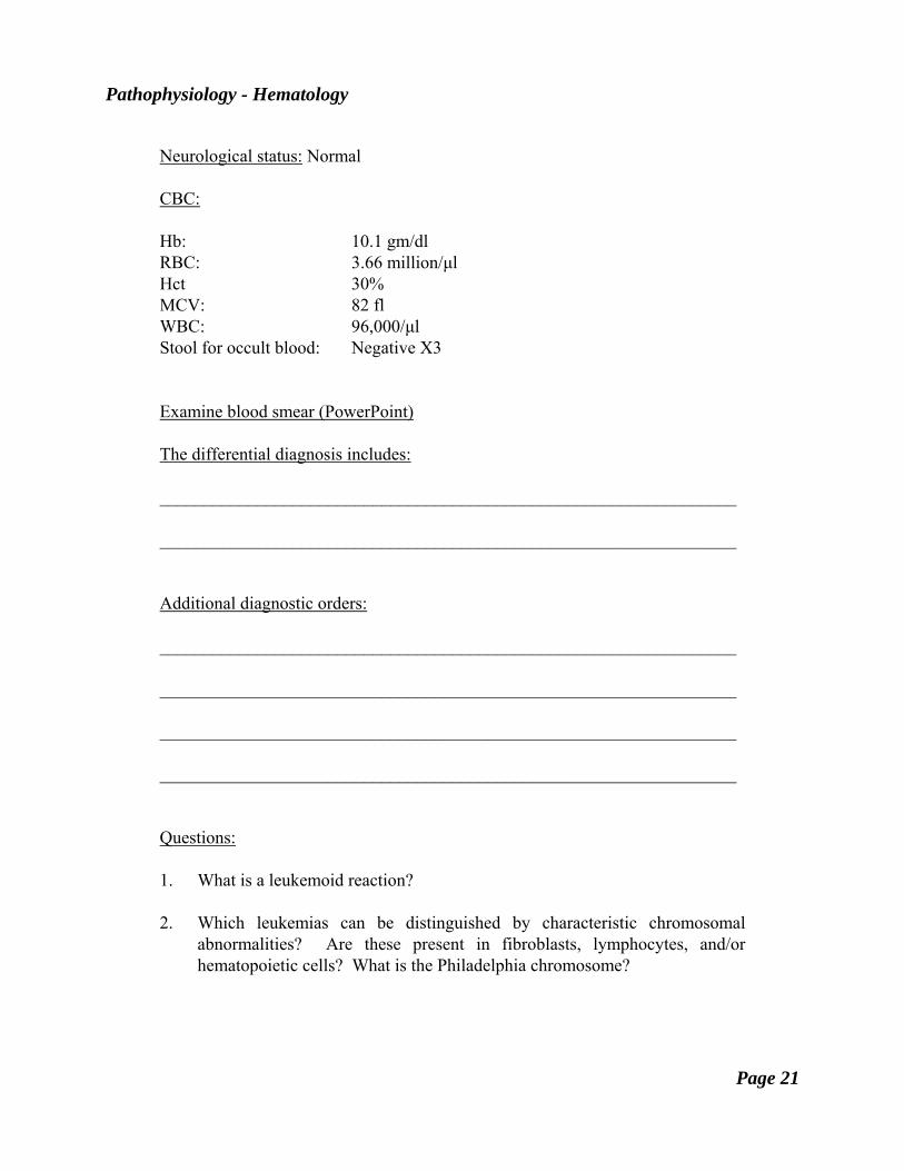
Neurological status: Normal CBC: Hb: 10.1 gm/dl RBC: 3.66 million/μl Hct 30% MCV: 82 fl WBC: 96,000/μl Stool for occult blood: Negative X3 Examine blood smear (PowerPoint) The differential diagnosis includes: _________________________________________________________________ _________________________________________________________________ Additional diagnostic orders: _________________________________________________________________ _________________________________________________________________ _________________________________________________________________ _________________________________________________________________ Questions: 1. What is a leukemoid reaction? 2. Which leukemias can be distinguished by characteristic chromosomal
abnormalities? Are these present in fibroblasts, lymphocytes, and/or hematopoietic cells? What is the Philadelphia chromosome?
Pathophysiology - Hematology
Page 22
3. Did all of this patient's white cells arise from a single precursor cell? What kind of evidence could be helpful to answer this question?
4. What is the usual chromosomal location of the c-ABL gene and what is the
function of the c-ABL protein? How was it discovered? What is the usual chromosomal location of the BCR gene and the function of the BCR protein? How was it discovered? What is the BCR-ABL protein?
5. If this patient's myeloid cells contained the Philadelphia chromosome, would it
be detectable after complete remission was achieved with various forms of chemotherapy?
6. What are some of the clinical and laboratory manifestations indicating
progression of chronic myeloid leukemia to an "accelerated" phase? What could account for such a progression?
7. What are thought to be the common features of the so-called myeloproliferative
syndromes? 8. What is myeloid metaplasia with myelofibrosis, and how is its diagnosis
established?
Pathophysiology - Hematology
Page 23
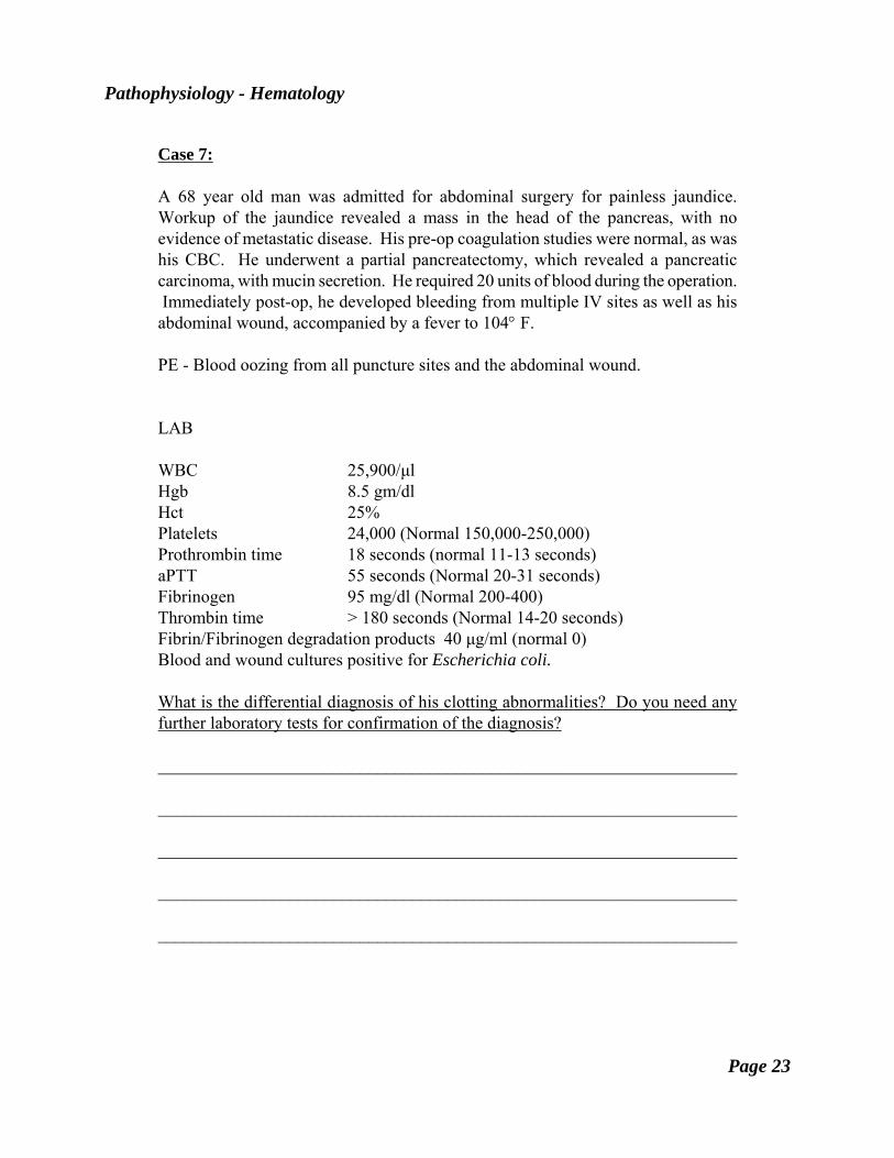
Case 7: A 68 year old man was admitted for abdominal surgery for painless jaundice. Workup of the jaundice revealed a mass in the head of the pancreas, with no evidence of metastatic disease. His pre-op coagulation studies were normal, as was his CBC. He underwent a partial pancreatectomy, which revealed a pancreatic carcinoma, with mucin secretion. He required 20 units of blood during the operation. Immediately post-op, he developed bleeding from multiple IV sites as well as his abdominal wound, accompanied by a fever to 104° F. PE - Blood oozing from all puncture sites and the abdominal wound. LAB WBC 25,900/μl Hgb 8.5 gm/dl Hct 25% Platelets 24,000 (Normal 150,000-250,000) Prothrombin time 18 seconds (normal 11-13 seconds) aPTT 55 seconds (Normal 20-31 seconds) Fibrinogen 95 mg/dl (Normal 200-400) Thrombin time > 180 seconds (Normal 14-20 seconds) Fibrin/Fibrinogen degradation products 40 μg/ml (normal 0) Blood and wound cultures positive for Escherichia coli. What is the differential diagnosis of his clotting abnormalities? Do you need any further laboratory tests for confirmation of the diagnosis? __________________________________________________________________ __________________________________________________________________ __________________________________________________________________ __________________________________________________________________ __________________________________________________________________
Pathophysiology - Hematology
Page 24
What phases of the hemostatic process may be impaired in this patient? __________________________________________________________________ __________________________________________________________________ __________________________________________________________________ __________________________________________________________________ If this patient has DIC, what would be the possible underlying mechanisms for its development? __________________________________________________________________ __________________________________________________________________ __________________________________________________________________ __________________________________________________________________ What therapy would you initiate? __________________________________________________________________ __________________________________________________________________ __________________________________________________________________ 1. What is the mechanism of the bleeding disorder in this patient? 2. Why are there increased levels of FDP's? 3. What might you expect to see on a peripheral blood smear in this patient? 4. What role does plasminogen activation play in his bleeding problems?
Pathophysiology - Hematology
Page 25
5. Is there a role for anticoagulant therapy in this man with increased bleeding? 6. What is the role of antifibrinolytic therapy in this patient? He was treated with antibiotics and recovered from the septicemia, and his coagulation studies and platelet count normalized. However, 14 days later he developed oozing from his gums and his wound site, as well as blood in his stool. Coagulation studies at this time reveal a PT of 18 seconds, an aPTT of 35 seconds, a platelet count of 170,000, a thrombin time of 13 seconds, a fibrinogen level of 585 mg/100 ml, and fibrin degradation products of 0. His appetite has been poor, and he had been started on parenteral nutrition 2 days previously. What is now causing his bleeding difficulty? __________________________________________________________________ __________________________________________________________________ __________________________________________________________________ 7. What is the mechanism for the elevated PT in this patient? 8. Why isn't the thrombin time affected? 9. How would you correct the bleeding abnormality in this patient? Is there more
than one way to do so? 10. If the patient had been treated with warfarin during the 14 days after operation,
could that have accounted for his abnormalities of coagulation? What is the mechanism of action of warfarin?
11. If this patient had instead been treated with heparin, could that have accounted
for the abnormalities seen? What is the mechanism of action of heparin?