pathology of mediastinal tumors - samo-workshop.ch€¦ · hematothorax right side: lobectomy rul...
TRANSCRIPT

Pathology of Mediastinal Tumors
Alex Soltermann
SAMO Meeting Lucerne 2009

Most common lesions (adults)

Clinical presentation
50% of the patients are asymptomatic, lesion discovered incidentally
Symptoms from compression or invasion of adjacent structures, including chest pain, cough, dyspnea
Superior vena cava syndrome usually due to malignancy
• In adults metastatic lung carcinoma and malignant lymphoma
• In children malignant lymphoma and acute leukemia

Metastatic tumors
May mimic primary mediastinal neoplasm, primarily in the middle mediastinum where most lymphnodes are situated
Direct mediastinal extension or nodal metastases:
• Lung carcinoma, e.g. SCLC with huge mediastinal mass but small bronchial lesion
• Tumors of esophagus, pleura, chest wall, vertebra or trachea
• Metastases of breast, thyroid, nasopharynx, larynx, kidney, prostate, testicular germ cell tumors and malignant melanoma
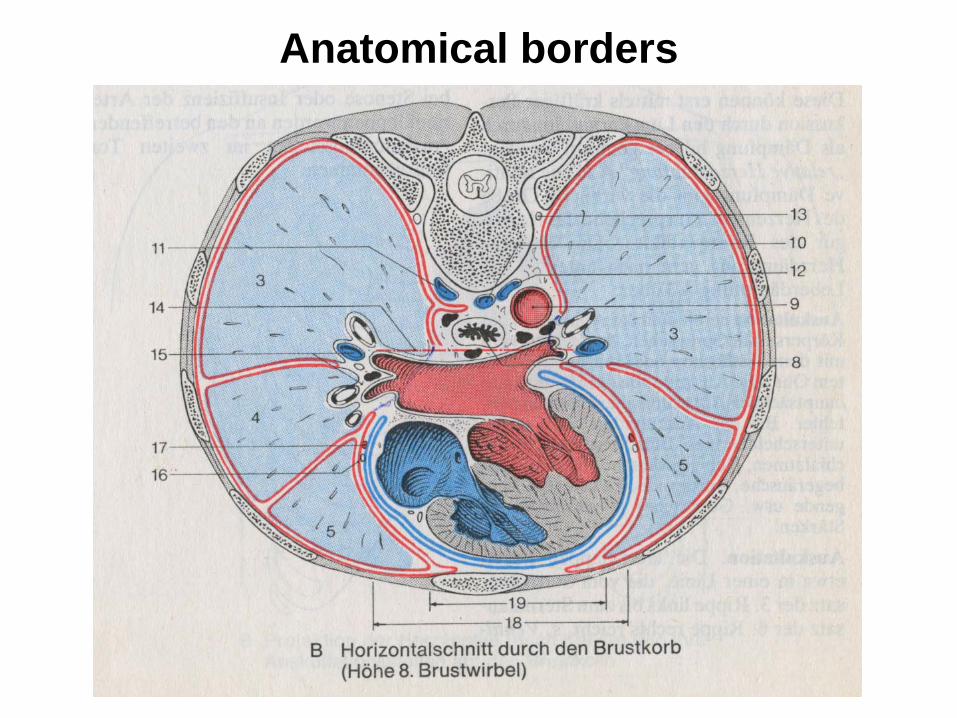
Anatomical borders

Staging problems of NSCLC
Lymph node dissection: additional tissue shell

Inflammatory diseasesAcute mediastinitis
• Predominantly posterior mediastinum
• Perforation of esophagus, descent of infection from within the neck, spread from chest wall infection, after heart surgery
Chronic mediastinitis
• Anterior mediastinum
• Mycotic (histoplasmosis) or tuberculous
Idiopathic fibrosing mediastinitis
• Associated with retroperitoneal fibrosis, inflammatory pseudotumor of the orbit, etc. Cave: DD Hodgkin‘s lymphoma

Cysts
• Pericardial (coelomic cysts)
• Foregut cysts: Bronchial, esophageal, gastric, enteric, pancreatic
Thyroid and parathyroid lesions
• Struma with nodular hyperplasia, pulled down into the anterior prevascular or the retrotracheal compartment (posterior descending goiter)
• 7% of parathyroid adenomas found in superior mediastinum
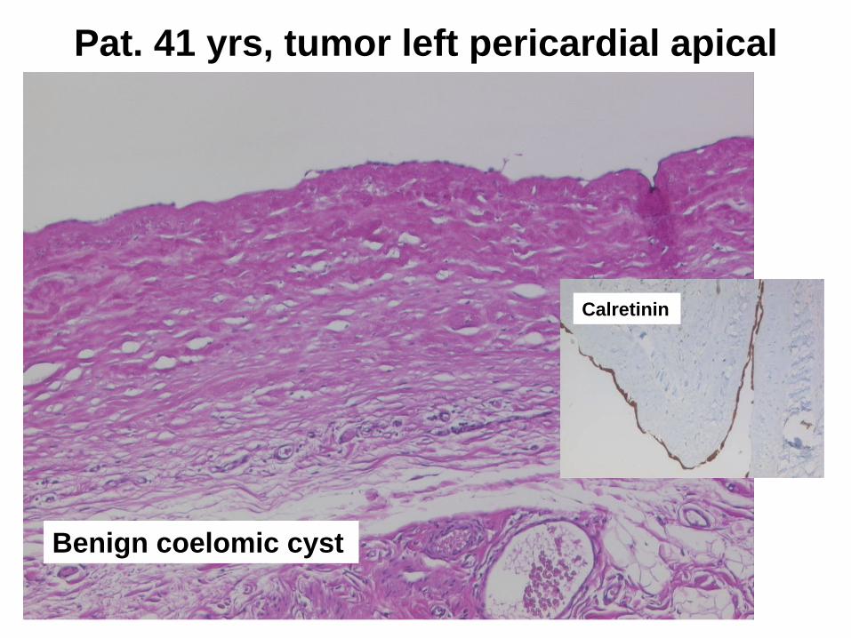
Pat. 41 yrs, tumor left pericardial apical
Benign coelomic cyst
Calretinin

Thymus
•Unilateral (developmental) or multilateral thymic cyst (reactive)
•Acute thymic involution (stress, HIV)
•Thymic follicular hyperplasia of B-lymphocytes (Myasthenia, HIV)
• Myasthenia gravis: Normal, follicular hyperplasia or thymoma

Thymoma
Neoplasm of epithelial cells, admixed with immature thymocytes
Type A: when epithelial cells have spindle/oval shape
Type B: when epithelial cells have dendritic, plump, epithelioid shape
Type C: overt atypia such as carcinoma
ABC rule:
A atrophic (spindle cell thymic cell of adult life)
B bioactive (biologically active organ of the fetus and infant)
C carcinoma
A, AB, B1, B2, B3, C
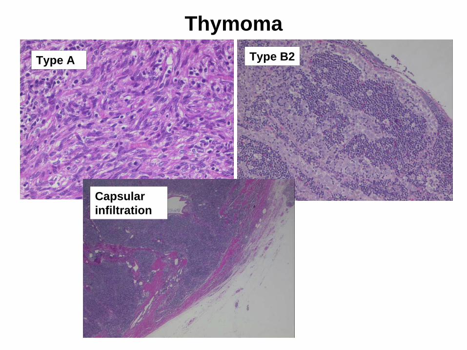
ThymomaType A Type B2
Capsular infiltration

Masaoka and TNM stageI macroscopically completely encapsulated and
microscopically no capsular invasion
II 1 Macroscopic invasion into surrounding fatty tissue or mediastinal pleura or
II 2 Microscopic invasion into capsule
III Macroscopic invasion into neighbouring organ, i. e. lung, pericardium or great vessels
IVa Pleural or pericardial dissemination
IVb Lymphogenous or hematogenous metastasis
Masaoka
stage T N M
I 1 0 0
II 2 0 0
III 3 0 0
IVa 4 0 0
IVb Any
T ≥N1 ≥M1
1. Encapsulated: T1
2. Minimally invasive: T2
3. Widely invasive or pleural or pericardial implants: T3/4
4. Metastatic: N1-2, M1

Thymoma prognosis
• Stage (single most important prognosticator)
• Histologic type: A<AB<B1<B2<B3<C
• Completeness of excision
• Myasthenia gravis

Malignant lymphoma
Anterior, superior or middle mediastinum; thymus or nodes
1. Hodgkin‘s lymphoma: young females, nodular sclerosis, cysts
Cave: multilocular thymic cyst, sclerosing mediastinitis
2. Lymphoblastic lymphoma: Usually immature T-cell type
3. Large cell lymphoma: Large vesicular irregular nuclei
4. Marginal zone B-cell lymphoma: Sjögren‘s disease, IgA type
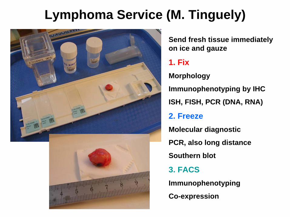
Lymphoma Service (M. Tinguely)
Send fresh tissue immediately on ice and gauze
1. FixMorphology
Immunophenotyping by IHC
ISH, FISH, PCR (DNA, RNA)
2. FreezeMolecular diagnostic
PCR, also long distance
Southern blot
3. FACSImmunophenotyping
Co-expression

Frozen section (Schnellschnitt)
CD15

Germ cell tumors
20% of mediastinal tumors and cysts
Origin from extragonadal germ cells, related to the thymus
• Seminoma (always within thymus)
• Teratoma (mature, immature, with somatic type malignancy)
• Embryonal carcinoma
• Yolk sac tumor
• Choriocarcinoma
• Mixed germ cell tumors
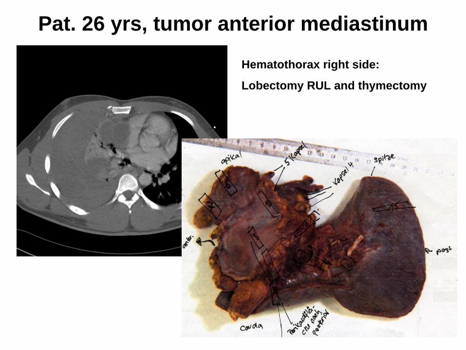
Pat. 26 yrs, tumor anterior mediastinum
Hematothorax right side:
Lobectomy RUL and thymectomy

Mixed malignant seminomatous non seminomatous germ cell tumor with immature teratoma moiety

Mesenchymal tumors
Benign
• Lipoma: above diaphragm, DD thymolipoma or lipomatosis (Cushing, steroid, obesity)
• Lymphangioma and Hemangioma
• Solitary fibrous tumor (SFT)
Malignant
• Liposarcoma
• Synovial sarcoma
• Other sarcoma

Pat. 44 yrs, multilocular thymic cyst
Cystic lymphangioma with dysplastic vessels
D2-40
Neurogenic tumors: PosteriorTumors of sympathetic nervous system (< 10 years)
Neuroblastoma, ganglioneuroblastoma, ganglioneuroma
Tumor of peripheral nerves (> 20 years)
Schwannoma, neurofibroma, malignant peripheral nerve sheath tumor (MPNST), with rhabdomyoblastic features called „Triton tumor“
Typical carcinoid (TC), atypical carcinoid (AC), small cell neuroendocrine carcinoma (SCLC), large cell neuroendocrine carcinoma (LCNEC)
In lung mostly TC and SCLC, in thymus mostly AC
Neuroendocrine tumors (thymus)
Summary
Mediastinal lesions: cysts, benign or malignant tumors
Frequency dependent on location: superior, anterior, middle and posterior mediastinum
Metastases may mimic mediastinal primary
Histology A<AB<B1<B2<B3<C, stage and margins major prognosticators of thymoma

Thank you!
SAMO Meeting Lucerne 2009