pain therapyhepmp.med.bg.ac.rs/wp-content/uploads/2020/04/pain... · 2020-04-22 · their trigger...
TRANSCRIPT
“Strengthening Capacities for Higher Education of Pain Medicine in Western Balkan countries - HEPMP” (Project number: 585927-EPP-1-2017-1-RS-EPPKA2-CBHE-JP (2017 – 3109/001 – 001)
Одрицање од одговорности :Овај пројекат је финансиран уз подршку Европске комисије . Ова публикација одражава само ставове аутора, а Комисија се не може сматрати одговорном за сваку употребу која се може састојати од ин-формација садржаних у њој.
Disclaimer This project has been funded with support from the European Commission. This publication [communication] reflects the views only of the author, and the Commission cannot be held responsible for any use which ma y be made of the information contained therein.
Co-funded by theErasmus+ Programmeof the European Union
PAIN THERAPY
Authors:
Gianluca Villa, Iacopo Lanini and A. Raffaele De Gaudio Department of Heath Science, Section of Anesthesia,Intensive Care and Pain Medicine, University of Florence


1
CONTENTS
PATHOPHYSIOLOGY OF PAIN AND PAIN PATHWAYS ..................................................... 2
NOCICEPTORS .................................................. 2
PERIPHERAL NERVE ......................................... 3
DORSAL HORN OF SPINAL CORD ..................... 4
TRANSMISSION IN THE SPINAL CORD .............8
ASCENDING PATHWAYS....................................8
THALAMIC NUCLEI ..........................................11
CEREBRAL CORTEX ........................................11
CENTRAL MECHANISMS OF PAIN CONTROL . 12
DEFINITION AND CLASSIFICATION OF PAIN .........................................................16
PAIN - IASP DEFINITION: ................................... 16
CLINICAL EVALUATION OF PATIENTS WITH PAIN ....................................................18
PAIN ANAMNESIS ........................................... 18
PSYCHOLOGICAL EVALUATION....................... 18
MEASUREMENT AND QUANTIFICATION OF PAIN ............................. 21
PHYSICAL EXAMINATION ............................... 27
LABORATORY AND INSTRUMENTAL PAIN ASSESSMENT ................................................. 28
NON-INVASIVE TREATMENT OF PAIN AND DRUG THERAPY ........................................... 30
INVASIVE TREATMENT OF PAIN ...................33
CANCER PAIN AND ITS TREATMENT ............ 38
PALLIATIVE CARE .........................................41
PAIN THERAPY Authors:
Gianluca Villa, Iacopo Lanini and A. Raffaele De Gaudio Department of Heath Science, Section of Anesthesia,Intensive Care and Pain Medicine, University of Florence

2
PATHOPHYSIOLOGY OF PAIN AND PAIN PATHWAYS
The perception of pain is mainly based on the migra-tion of pain information from the locus where the initial stimulus is generated toward the frontal cortex, where the information becomes aware. The pain pathways are here analyzed in a centripetal direction, starting from the pe-riphery (nociceptors) up to the central level (the cerebral cortex).
NOCICEPTORS
The nociceptors are the peripheral terminations of pri-mary pseudounipolar sensitive neurons with cell bodies lo-cated into the ganglia of the dorsal roots and in the Gasser’s ganglion of the trigeminal nerve (figure 1).
Painful stimuli can activate different types of nocicep-tors, such as:
�� thermal nociceptors: activated by extreme temperatures (> 45 ° C or <5 ° C). Characterized by Aδ fibers of small diameter (figure 2), equipped with a thin myelin sheath and leading the nervous impulse with a speed of about 5-30 m/s;�� mechanical nociceptors: activated by the pressure of the
high-intensity stimuli applied to the skin. Aδ fibers also characterize them (figure 2);
� polymodal nociceptors: activated by high-intensity me-chanical stimuli, by chemical stimuli or by thermal stimuli (both hot and cold). These are characterized by C fibers (figure 3), with small-diameter, unmyelinated, leading to the generally lower speed impulses at 1 m/s.
� silent nociceptors: found in viscera. They are usually not activated by painful stimuli, but inflammatory process-es and several chemical products significantly influence their trigger point of activation. The silent nociceptors activation may contribute to the onset of secondary hyperalgesia, or central sensitization to pain (see next paragraph “secondary hyperalgesia”).
Figure 1: Somatosensory neurons are located in peripheral gan-glia (trigeminal and dorsal root ganglia) located alongside the spi-nal column and medulla. Afferent neurons project centrally to the brainstem (Vc) and dorsal horn of the spinal cord and peripherally to the skin and other organs. Vc, trigeminal brainstem sensory subnucleus caudalis (1).
Figure 2: A-fiber nociceptors are myelinated and usually have conduction velocities in the Aδ range (red). A-fiber nociceptors project to superficial laminae I and V. (1)

3
These three classes of nociceptors are distributed ex-tensively both to the skin and to the deep tissues, and they often operate together. As an example, if we press our thumb with a hammer, immediately there is a “first” pain of punc-ture type, followed by a feeling of tenderness that some-times becomes a real “second” pain of burning type. The first puncture pain, rapid, is “transmitted” by the Aδ fibers, while the dull pain, slow, is “transmitted” by the C fibers of the polymodal nociceptors.
Viscera are almost exclusively characterized by poly-modal nociceptors, whose afferent fibers run along with sympathetic fibers. In particular, visceral nociceptors, rely on and produce nociception from the visceral wall and in the capsular surface. No nociceptors are presented in the in-ner part (parenchymal) of organs, such as the liver, spleen, or pancreas. In visceral pain, the summation phenomena, both spatial and temporal, are particularly evident. The role of visceral nociceptors is not completely understood; in particular, if the visceral pain receptors are also involved in the regulation of the visceral functions, it is still not well defined. Visceral receptors, indeed, would provide different information, depending on the type of stimulation.
Unlike the somatosensory receptors, the majority of nociceptors are uncovered, unmyelinated, nerve termina-tion. They are silent in the absence of noxious stimuli and are only activated by them. The exact mechanisms that al-low injurious stimuli to depolarize nociceptors and produce potential spikes (transduction) are not completely known. Nociceptor membrane might contain specific proteins that convert thermal energy, mechanical or chemical stimuli into electrical potentials. The capsaicin receptor, as an example, has been found exclusively in the endings of nociceptors; this receptor responds to noxious stimuli, as intense heat, suggesting that the receptor of capsaicin acts as a transduc-er for pain stimuli of thermal nature.
PERIPHERAL NERVE
The peripheral axons of pseudounipolar neurons, lo-cated into the ganglia of the dorsal root and into the Gas-ser’s ganglion, join to form the peripheral nerves. Usually containing motor afferent fibers too, peripheral nerve are coated by three sheaths (endoneurium, perineurium, and epineurium), which protect against neurotropic and stain agents.
The peripheral nerves have their own vascularization (vasa nervorum) (figure 4) and their own innervation (ner-va nervorum), whose integrity is essential for the mainte-nance of physiologic nerve function. Vasoconstriction and ischemia neuritis are well-known complications of anatom-ical or functional alterations of these structures.
The nerve impulses run through the peripheral branches of pseudounipolar neurons, cross the cell bodies, and then run through the central branches that form the dorsal spinal roots. These pain afferencies reach the spinal cord with a metameric distribution; quite precise concern-ing the sensitivity of the teguments, much less precise con-cerning skeletal sensitivity or visceral sensitivity. Pain-re-lated to visceral sensitivity, in particular, runs along with the autonomic nerve fibers, cross through the sympathetic
Figure 3: Most nociceptors are unmyelinated with small diame-ter axons (C-fibers, red). Their peripheral afferent innervates the skin (dermis and/or epidermis) and central process projects to superficial laminae I and II of the dorsal horn. (1)
4
ganglia without interruption and, through the white com-municating branches, reach their cell body located in the spinal ganglion.
DORSAL HORN OF SPINAL CORD
The nociceptive afferent fibers terminate mainly in the dorsal horn of the spinal cord (figura 5).
Basing on the cytological characteristics of neurons observed, the dorsal horn can be divided into six layers (or foils) (figure 6). Different typologies of primary afferent neurons (and thus, different nociceptive information) cease in different laminae of the spinal cord. A close correspon-
dence between functional and anatomical organizations of neurons in the dorsal horn of the spinal cord can thus be recognized.
Specific nociceptive neurons are present in the super-ficial part of the dorsal horn, which includes the marginal zone (layer I) and substantia gelatinosa (layer II); the ma-jority of these receive input directly from Aδ fibers and C. The laminae involved in the transmission of painful stimuli to the higher centers are:�� lamina I: most of the neurons of this layer only respond
to noxious stimuli, and therefore called specific nocicep-tive neurons. Some neurons of this layer named broad
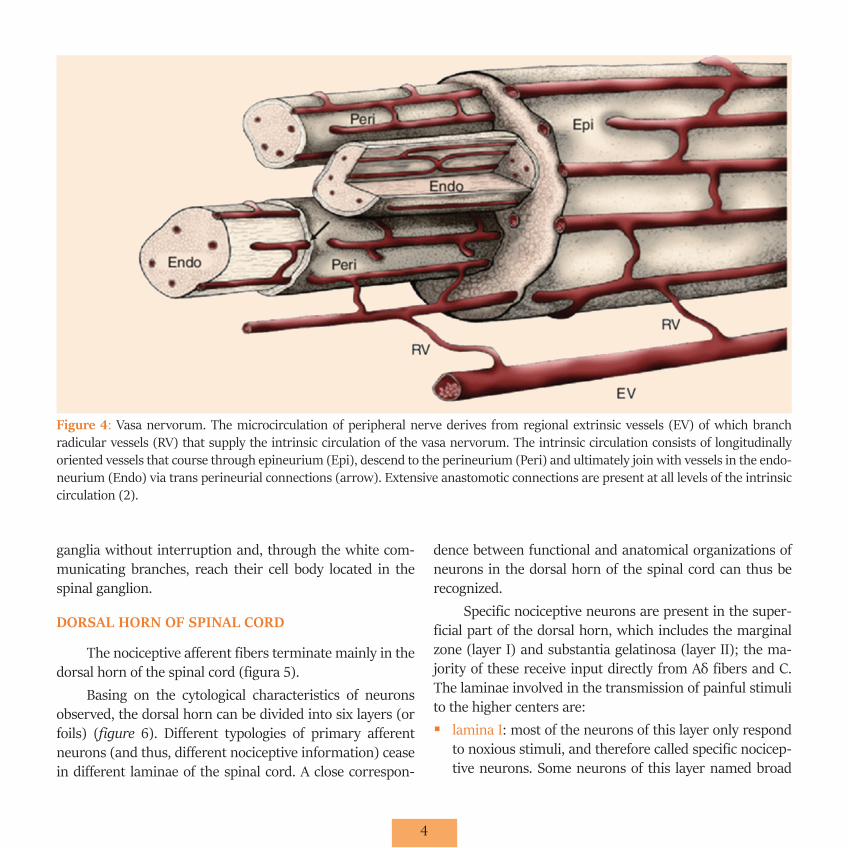
Figure 4: Vasa nervorum. The microcirculation of peripheral nerve derives from regional extrinsic vessels (EV) of which branch radicular vessels (RV) that supply the intrinsic circulation of the vasa nervorum. The intrinsic circulation consists of longitudinally oriented vessels that course through epineurium (Epi), descend to the perineurium (Peri) and ultimately join with vessels in the endo-neurium (Endo) via trans perineurial connections (arrow). Extensive anastomotic connections are present at all levels of the intrinsic circulation (2).

5
Figure 5: Relationship of the meningeal coverings of the spinal cord to spinal root and peripheral nerve connective tissue ensheath-ments. The outermost meningeal covering, the dura mater (DM), is continuous with the outermost connective tissue of peripheral nerve, the epineurium (Epi), while the arachnoid layer (A) merges with the outer perineurial lamellae at the subarachnoid angle (SA). The inner layers of the perineurium (Peri) derive from the inner layers of the root sheath (RS). Inset at upper right shows high-power view of the transition of connective tissues at the subarachnoid angle. As the dorsal and ventral spinal roots pass through subarachnoid space (SS), some of the arachnoid layer is reflected onto the root sheath at the subarachnoid angle, becoming the outermost layers of this connective tissue ensheathment. At the root attachment zone of dorsal and ventral roots, the pia mater (PM) of the spinal cord is reflected onto the spinal root and merges with the outer layers of the root sheath, while the glia limitans (GL) continues across the attachment zone to form the interface between the central and peripheral nervous systems. The innermost layers of the root sheath terminate on the spinal root side of the glia limitans. At the root attachment zones, continuity between the subarachnoid space and the endoneurium (Endo) has been demonstrated ultrastructurally (arrows). Inset at upper left illustrates a high power view of the dorsal root attachment zone, associated spinal cord white matter (WM) and underlying gray matter (GM).(2)
6
spectrum dynamic neurons, respond in a graduated manner to mechanical stimuli and noxious ones;�� lamina II: made almost exclusively of interneurons (ei-
ther excitatory and inhibitory); some of them respond only to noxious stimuli, while other to other non-nox-ious stimuli also;
�� lamina V: it predominantly contains wide dynamic spectrum neurons, which project to the brainstem and thalamus. These neurons receive afferents from mono-synaptic Aß and Aδ fibers and also from C fibers. Many neurons of this lamina also receive afferents from mono-synaptic visceral structures.
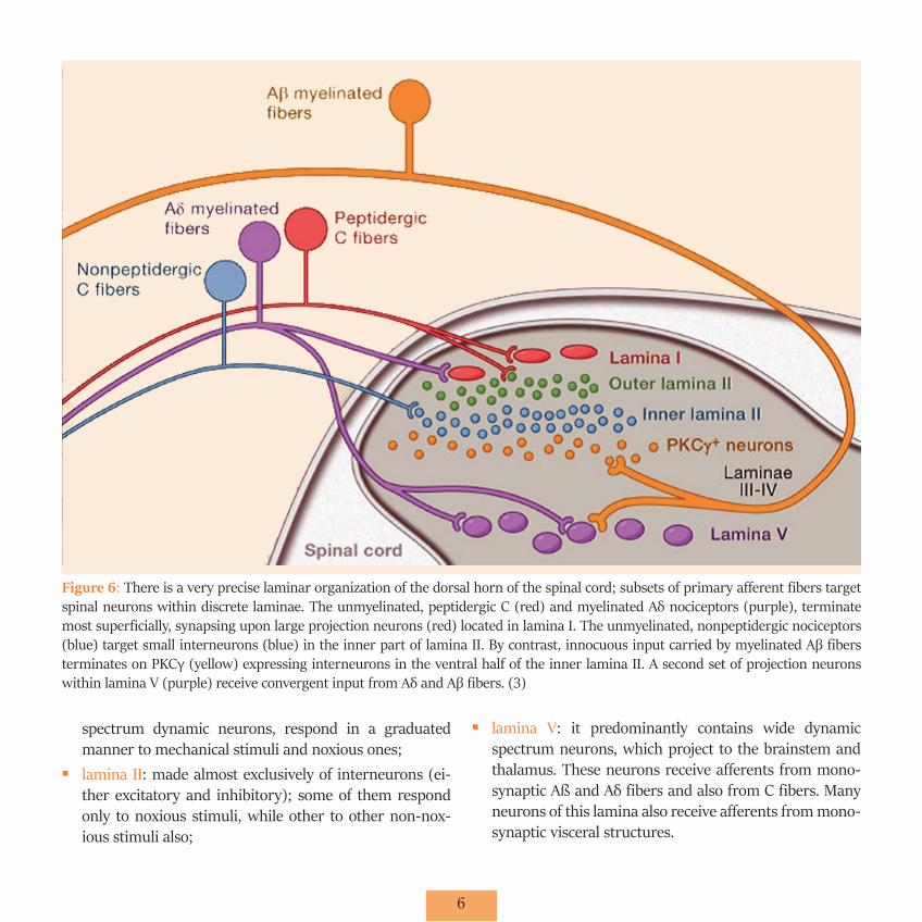
Figure 6: There is a very precise laminar organization of the dorsal horn of the spinal cord; subsets of primary afferent fibers target spinal neurons within discrete laminae. The unmyelinated, peptidergic C (red) and myelinated Aδ nociceptors (purple), terminate most superficially, synapsing upon large projection neurons (red) located in lamina I. The unmyelinated, nonpeptidergic nociceptors (blue) target small interneurons (blue) in the inner part of lamina II. By contrast, innocuous input carried by myelinated Aβ fibers terminates on PKCγ (yellow) expressing interneurons in the ventral half of the inner lamina II. A second set of projection neurons within lamina V (purple) receive convergent input from Aδ and Aβ fibers. (3)
7
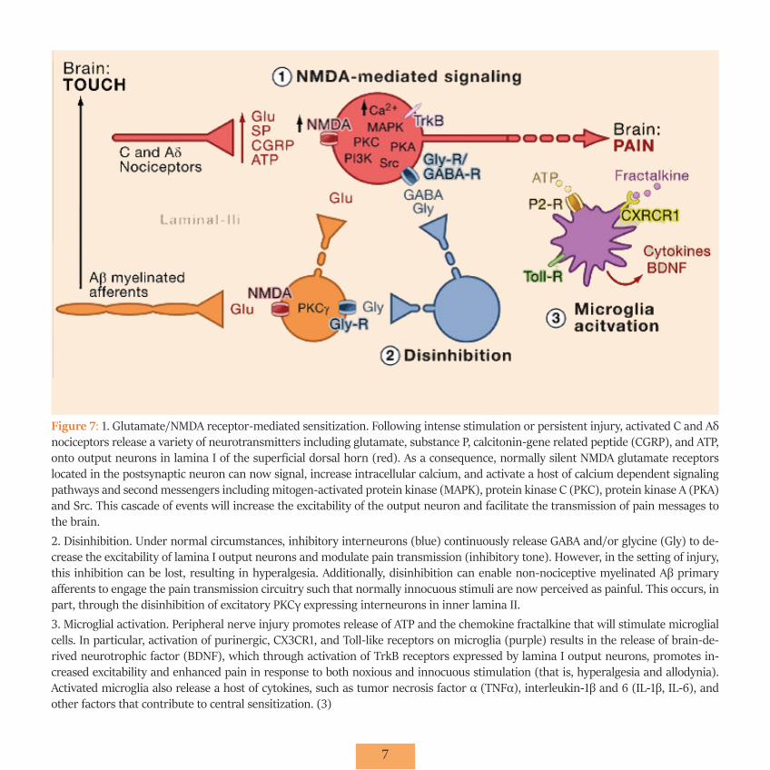
Figure 7: 1. Glutamate/NMDA receptor-mediated sensitization. Following intense stimulation or persistent injury, activated C and Aδ nociceptors release a variety of neurotransmitters including glutamate, substance P, calcitonin-gene related peptide (CGRP), and ATP, onto output neurons in lamina I of the superficial dorsal horn (red). As a consequence, normally silent NMDA glutamate receptors located in the postsynaptic neuron can now signal, increase intracellular calcium, and activate a host of calcium dependent signaling pathways and second messengers including mitogen-activated protein kinase (MAPK), protein kinase C (PKC), protein kinase A (PKA) and Src. This cascade of events will increase the excitability of the output neuron and facilitate the transmission of pain messages to the brain.2. Disinhibition. Under normal circumstances, inhibitory interneurons (blue) continuously release GABA and/or glycine (Gly) to de-crease the excitability of lamina I output neurons and modulate pain transmission (inhibitory tone). However, in the setting of injury, this inhibition can be lost, resulting in hyperalgesia. Additionally, disinhibition can enable non-nociceptive myelinated Aβ primary afferents to engage the pain transmission circuitry such that normally innocuous stimuli are now perceived as painful. This occurs, in part, through the disinhibition of excitatory PKCγ expressing interneurons in inner lamina II.3. Microglial activation. Peripheral nerve injury promotes release of ATP and the chemokine fractalkine that will stimulate microglial cells. In particular, activation of purinergic, CX3CR1, and Toll-like receptors on microglia (purple) results in the release of brain-de-rived neurotrophic factor (BDNF), which through activation of TrkB receptors expressed by lamina I output neurons, promotes in-creased excitability and enhanced pain in response to both noxious and innocuous stimulation (that is, hyperalgesia and allodynia). Activated microglia also release a host of cytokines, such as tumor necrosis factor α (TNFα), interleukin-1β and 6 (IL-1β, IL-6), and other factors that contribute to central sensitization. (3)
8
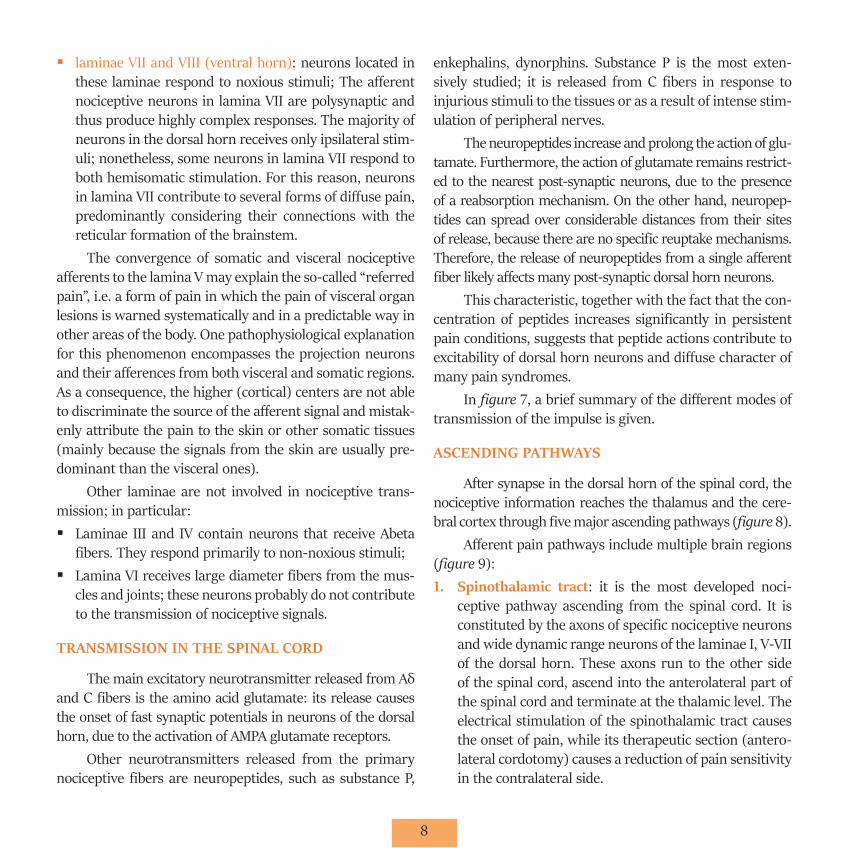
�� laminae VII and VIII (ventral horn): neurons located in these laminae respond to noxious stimuli; The afferent nociceptive neurons in lamina VII are polysynaptic and thus produce highly complex responses. The majority of neurons in the dorsal horn receives only ipsilateral stim-uli; nonetheless, some neurons in lamina VII respond to both hemisomatic stimulation. For this reason, neurons in lamina VII contribute to several forms of diffuse pain, predominantly considering their connections with the reticular formation of the brainstem.
The convergence of somatic and visceral nociceptive afferents to the lamina V may explain the so-called “referred pain”, i.e. a form of pain in which the pain of visceral organ lesions is warned systematically and in a predictable way in other areas of the body. One pathophysiological explanation for this phenomenon encompasses the projection neurons and their afferences from both visceral and somatic regions. As a consequence, the higher (cortical) centers are not able to discriminate the source of the afferent signal and mistak-enly attribute the pain to the skin or other somatic tissues (mainly because the signals from the skin are usually pre-dominant than the visceral ones).
Other laminae are not involved in nociceptive trans-mission; in particular: �� Laminae III and IV contain neurons that receive Abeta
fibers. They respond primarily to non-noxious stimuli;�� Lamina VI receives large diameter fibers from the mus-
cles and joints; these neurons probably do not contribute to the transmission of nociceptive signals.
TRANSMISSION IN THE SPINAL CORD
The main excitatory neurotransmitter released from Aδ and C fibers is the amino acid glutamate: its release causes the onset of fast synaptic potentials in neurons of the dorsal horn, due to the activation of AMPA glutamate receptors.
Other neurotransmitters released from the primary nociceptive fibers are neuropeptides, such as substance P,
enkephalins, dynorphins. Substance P is the most exten-sively studied; it is released from C fibers in response to injurious stimuli to the tissues or as a result of intense stim-ulation of peripheral nerves.
The neuropeptides increase and prolong the action of glu-tamate. Furthermore, the action of glutamate remains restrict-ed to the nearest post-synaptic neurons, due to the presence of a reabsorption mechanism. On the other hand, neuropep-tides can spread over considerable distances from their sites of release, because there are no specific reuptake mechanisms. Therefore, the release of neuropeptides from a single afferent fiber likely affects many post-synaptic dorsal horn neurons.
This characteristic, together with the fact that the con-centration of peptides increases significantly in persistent pain conditions, suggests that peptide actions contribute to excitability of dorsal horn neurons and diffuse character of many pain syndromes.
In figure 7, a brief summary of the different modes of transmission of the impulse is given.
ASCENDING PATHWAYS
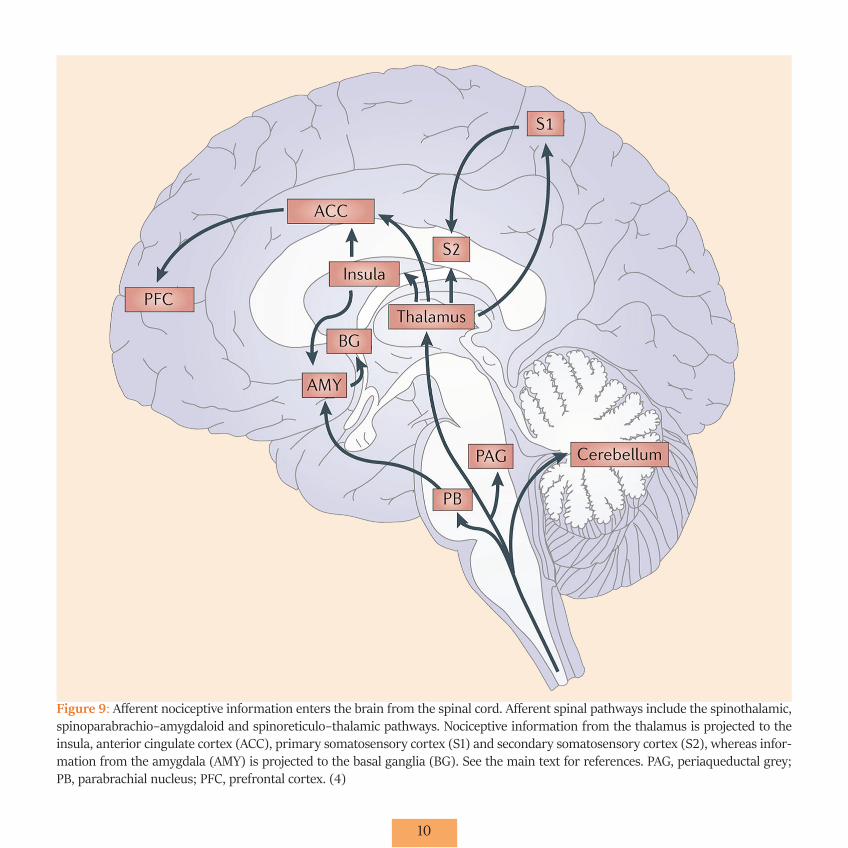
After synapse in the dorsal horn of the spinal cord, the nociceptive information reaches the thalamus and the cere-bral cortex through five major ascending pathways (figure 8).
Afferent pain pathways include multiple brain regions (figure 9):1. Spinothalamic tract: it is the most developed noci-
ceptive pathway ascending from the spinal cord. It is constituted by the axons of specific nociceptive neurons and wide dynamic range neurons of the laminae I, V-VII of the dorsal horn. These axons run to the other side of the spinal cord, ascend into the anterolateral part of the spinal cord and terminate at the thalamic level. The electrical stimulation of the spinothalamic tract causes the onset of pain, while its therapeutic section (antero-lateral cordotomy) causes a reduction of pain sensitivity in the contralateral side.

9
Rana defibrilacija
Figure 8: Primary afferent nociceptors convey noxious information to projection neurons within the dorsal horn of the spinal cord. A subset of these projection neurons transmits information to the somatosensory cortex via the thalamus, providing information about the location and intensity of the painful stimulus. Other projection neurons engage the cingulate and insular cortices via connections in the brainstem (parabrachial nucleus) and amygdala, contributing to the affective component of the pain experience. This ascending information also accesses neurons of the rostral ventral medulla and midbrain periaqueductal gray to engage descending feedback systems that regulate the output from the spinal cord. (3)
10
Figure 9: Afferent nociceptive information enters the brain from the spinal cord. Afferent spinal pathways include the spinothalamic, spinoparabrachio–amygdaloid and spinoreticulo–thalamic pathways. Nociceptive information from the thalamus is projected to the insula, anterior cingulate cortex (ACC), primary somatosensory cortex (S1) and secondary somatosensory cortex (S2), whereas infor-mation from the amygdala (AMY) is projected to the basal ganglia (BG). See the main text for references. PAG, periaqueductal grey; PB, parabrachial nucleus; PFC, prefrontal cortex. (4)
11
2. Spinoreticular tract: it is composed of the axons of neurons of laminae VII and VIII. It also ascends into the anterolateral quadrant of the spinal cord and terminates both to reticular formation and to thalamus. The major-ity of spino-reticular fibers don’t cross the median line.
3. Spinomesencephalic tract: it is composed of the ax-ons of neurons of laminae I and V, which rise in the anterolateral quadrant of the spinal cord and project to the reticular formation, the peri-aqueductal grey and, through the spino-parabrachial tract, to the parabrachi-al nuclei. From the parabrachial nuclei fibers project to the amygdala, which is the main formation of the lim-bic system, the system that is involved in the processing of emotions. Therefore, the section of the spinomesen-cephalic tract might modulate the affective component of pain. Many axons of this tract ascend in the dorsal part of the lateral cord, rather than in the anterolateral quadrant; therefore, if these fibers are not cut during the antero-lateral cordotomy, the sensation of pain may remain or come back later on.
4. Cervicothalamic tract: it originates from neurons of the lateral cervical nucleus, located in the lateral white matter of the two upper cervical segments. The lateral cervical nucleus receives projections from neurons of laminae III and IV of the dorsal horn. The majority of axons in the cervicotalamic tract cross the medial line and ascend in the medial lemniscus of the brainstem, and it reaches some midbrain nuclei and postero-later-al and postero-medial nuclei of ventral thalamus. Some axons of laminae III and IV runs in the dorsal columns of the spinal cord and terminate in the gracile and cu-neate nuclei of the bulb.
5. Spinohypothalamic tract: it is composed of the axons of neurons of laminae I, V, and VIII and projects direct-ly to the supraspinal control centers of the autonomic nervous system. This tract might activate complex en-docrine and cardiovascular responses to pain.
THALAMIC NUCLEI
Different thalamic nuclei process the nociceptive infor-mations. The most important, however, are the lateral and medial nuclear groups.
Lateral Nuclear Group includes: �� ventral posterior nucleus �� ventral postero-lateral nucleus �� posterior nucleus
Through the spino-thalamic tract, this group receives impulses from specific nociceptive neurons and wide dynamic spectrum neurons of laminae I and V. The lateral thalamus is mainly implicated in the localization of the noxious stimulus, which usually reaches the level of consciousness as acute pain.
Lesions of the spinothalamic tract and its terminations cause the onset of an intense pain condition said central pain: for example, the ischemia of a small region of ventral postero-lateral nucleus of the thalamus may cause the tha-lamic syndrome known as Dejerine-Roussy syndrome.
Medial nuclear group includes: �� central lateral nucleus �� intra-laminar complex
It receives afferents mainly from neurons of laminae VII and VIII. Many neurons in the medial tract have wide-spread projections to the basal ganglia and several cortical areas. These neurons, therefore, are not only involved in processing nociceptive information, but also provide infor-mation related to stimuli that trigger a non-specific system that governs the surveillance state.
CEREBRAL CORTEX
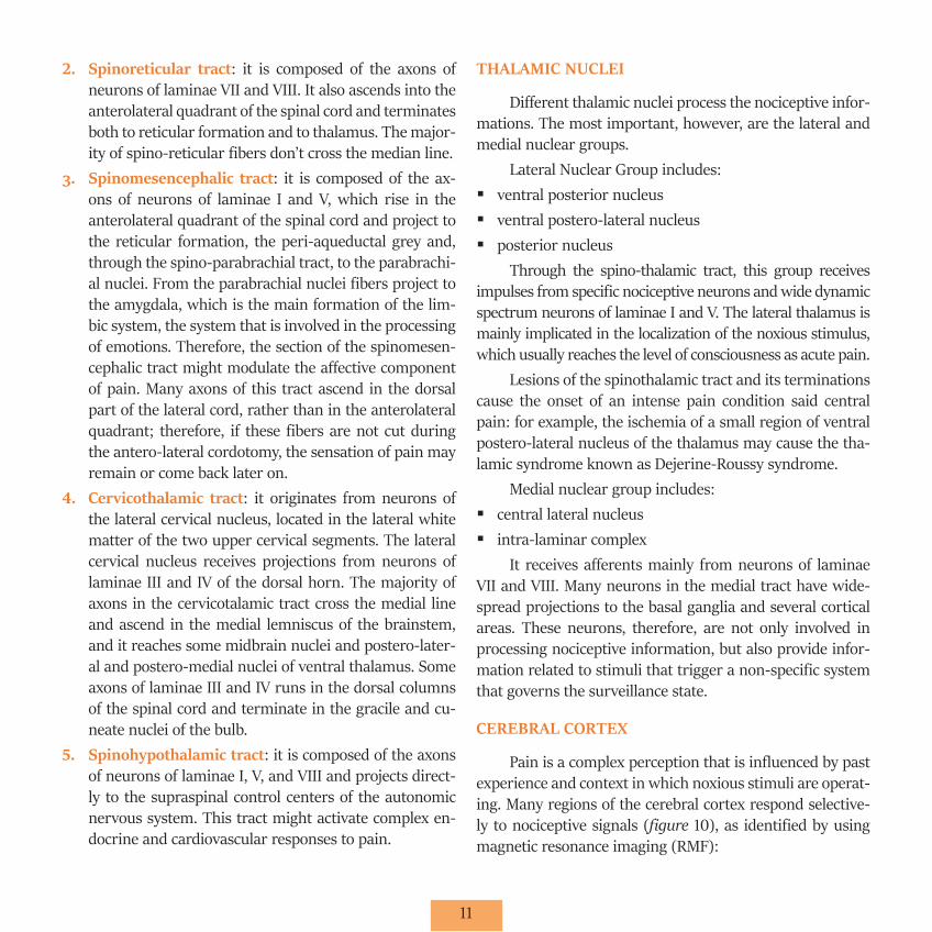
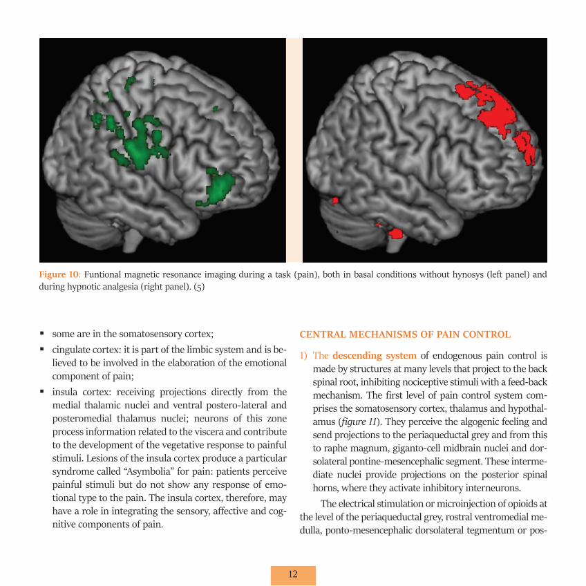
Pain is a complex perception that is influenced by past experience and context in which noxious stimuli are operat-ing. Many regions of the cerebral cortex respond selective-ly to nociceptive signals (figure 10), as identified by using magnetic resonance imaging (RMF):
12
�� some are in the somatosensory cortex; �� cingulate cortex: it is part of the limbic system and is be-
lieved to be involved in the elaboration of the emotional component of pain; �� insula cortex: receiving projections directly from the
medial thalamic nuclei and ventral postero-lateral and posteromedial thalamus nuclei; neurons of this zone process information related to the viscera and contribute to the development of the vegetative response to painful stimuli. Lesions of the insula cortex produce a particular syndrome called “Asymbolia” for pain: patients perceive painful stimuli but do not show any response of emo-tional type to the pain. The insula cortex, therefore, may have a role in integrating the sensory, affective and cog-nitive components of pain.
CENTRAL MECHANISMS OF PAIN CONTROL
1) The descending system of endogenous pain control is made by structures at many levels that project to the back spinal root, inhibiting nociceptive stimuli with a feed-back mechanism. The first level of pain control system com-prises the somatosensory cortex, thalamus and hypothal-amus (figure 11). They perceive the algogenic feeling and send projections to the periaqueductal grey and from this to raphe magnum, giganto-cell midbrain nuclei and dor-solateral pontine-mesencephalic segment. These interme-diate nuclei provide projections on the posterior spinal horns, where they activate inhibitory interneurons.
The electrical stimulation or microinjection of opioids at the level of the periaqueductal grey, rostral ventromedial me-dulla, ponto-mesencephalic dorsolateral tegmentum or pos-
Figure 10: Funtional magnetic resonance imaging during a task (pain), both in basal conditions without hynosys (left panel) and during hypnotic analgesia (right panel). (5)
13
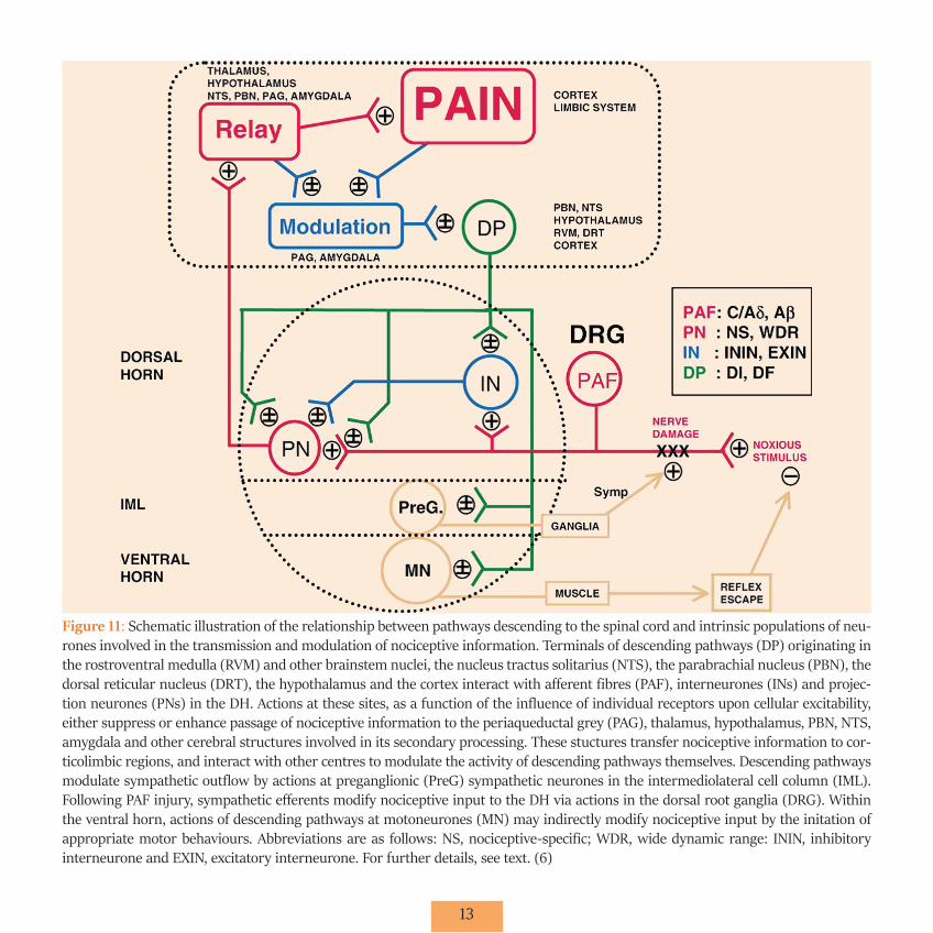
Figure 11: Schematic illustration of the relationship between pathways descending to the spinal cord and intrinsic populations of neu-rones involved in the transmission and modulation of nociceptive information. Terminals of descending pathways (DP) originating in the rostroventral medulla (RVM) and other brainstem nuclei, the nucleus tractus solitarius (NTS), the parabrachial nucleus (PBN), the dorsal reticular nucleus (DRT), the hypothalamus and the cortex interact with afferent fibres (PAF), interneurones (INs) and projec-tion neurones (PNs) in the DH. Actions at these sites, as a function of the influence of individual receptors upon cellular excitability, either suppress or enhance passage of nociceptive information to the periaqueductal grey (PAG), thalamus, hypothalamus, PBN, NTS, amygdala and other cerebral structures involved in its secondary processing. These stuctures transfer nociceptive information to cor-ticolimbic regions, and interact with other centres to modulate the activity of descending pathways themselves. Descending pathways modulate sympathetic outflow by actions at preganglionic (PreG) sympathetic neurones in the intermediolateral cell column (IML). Following PAF injury, sympathetic efferents modify nociceptive input to the DH via actions in the dorsal root ganglia (DRG). Within the ventral horn, actions of descending pathways at motoneurones (MN) may indirectly modify nociceptive input by the initation of appropriate motor behaviours. Abbreviations are as follows: NS, nociceptive-specific; WDR, wide dynamic range: ININ, inhibitory interneurone and EXIN, excitatory interneurone. For further details, see text. (6)

14
terior spinal horns cause analgesia. The pathways involved in endogenous pain control pathways are serotonergic and ad-renergic. It explains the analgesic effect and the potentiation of opioid effect recognized to tricyclic antidepressants, some serotonergic drugs (that act on HT2 receptors and HT5), and the alpha-2 adrenergic receptor blockers (clonidine). The polypeptides of the endogenous opioid system are essential for the activity of the descendants’ circuits of pain control.
The endogenous opioids have received this name be-cause they bind the same receptors bonded by these analge-sics. The endogenous opioids derive by enzymatic hydroly-sis from a large molecule, such as ACTH and menotropins. The main polypeptides opioids are dynorphins, endorphins, and enkephalins; these bind specific opioid receptors and are rapidly hydrolyzed by protease enzymes.
The opioid receptors reside in the areas involved in the perception of pain (periaqueductal gray, raphe magnum, spi-nal trigeminal nucleus, posterior horns of the spinal cord), in the areas related with the endocrine regulation (median eminence) and, finally, in some visceral areas (nerve plexus of the stomach, intestines and bladder). These localizations explain some of the side effects of opioids.
Opioid polypeptides act by activating a specific re-ceptor that inhibits the release of substance P and/or oth-er neurotransmitters from which the passage of the spike from the first to the second neuron of the pain pathway mainly depends.
The discovery of the role of opioid receptors in pain control has been a key step in algological practice. In par-ticular, this has led to the search of drugs active on more specific receptors, trying to eliminate as much as possible the effects of stimulation of the receptors that mediate side effects. Furthermore, this discovery has clarified that mor-phine determines analgesia not only when systemically ad-ministered but also, more selectively and effectively, when applied directly on the posterior spinal horns or when de-livered in microdoses to the ponto-mesencephalic tegment or the periaqueductal grey.
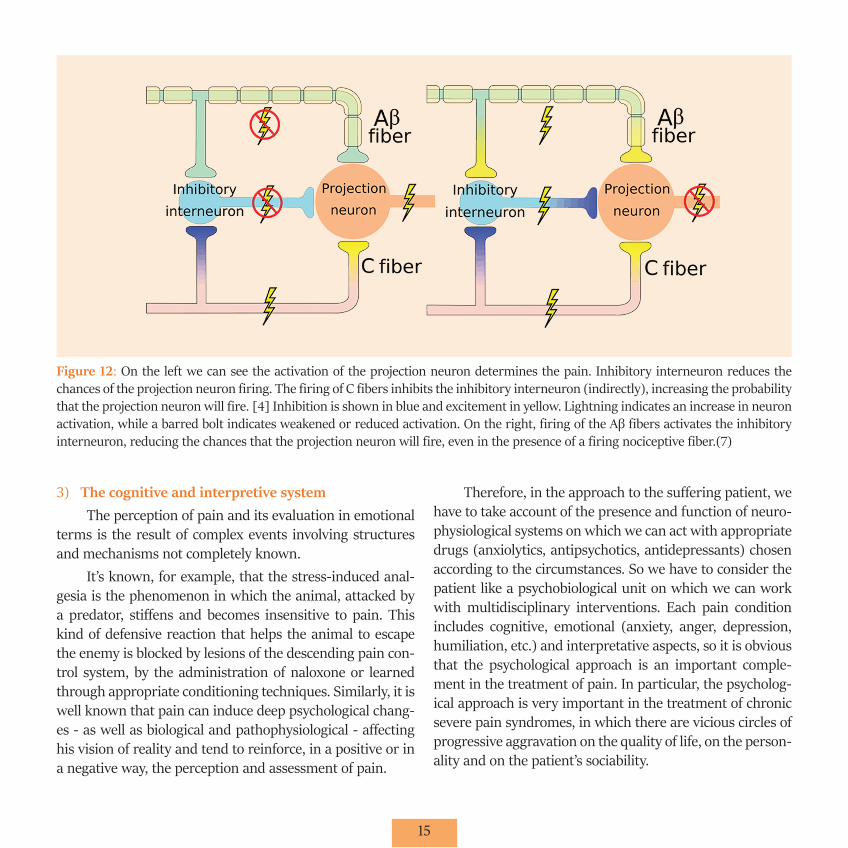
2) Gate Control: In the spinal cord, the transmission of nociceptive information to higher brain centers can be controlled through the interconnections between affer-ent nociceptive and non-nociceptive afferent pathways. The hypothesis that the pain is not simply the product of the activity of nociceptive afferences, but is also regu-lated by other myelinated fibers, not directly involved in the transmission of painful information, was advanced in the 60s and was called gate control theory.
This theory takes into account some important exper-imental observations (figure 12): �� the neurons of lamina V receive excitatory afferents from
Aß fibers (of large diameter, myelinated and not noci-ceptive), Aδ fibers and C fibers �� the Aß fibers inhibit the discharge of the lamina V neu-
rons via the activation of inhibitory interneurons of lam-ina II �� the Aδ and C fibers excite neurons of lamina V, but also
inhibit the discharge of these inhibitory interneurons of lamina II that are activated by the Abeta fibers.
In other words, non-nociceptive fibers close the gate through which occurs the central transmission of pain in-formation, while the nociceptive afferents open it.
This theory provides an explanation of the fact that a vibratory stimulus, which selectively activates afferent large-diameter fibers, can reduce pain. It is the rationale underlying the use of transcutaneous electrical stimulation techniques (SETC) and electrical stimulation of the dorsal columns for pain relief:�� in the case of SETC, the large-diameter afferent fibers,
that innervate the damaged area where the pain is felt, are activated by electrodes of stimulation; �� similarly, the stimulation of the dorsal columns via sur-
face electrodes alleviates pain because presumably it actives in a synchronous manner a large number of Aß fibers.
15
3) The cognitive and interpretive systemThe perception of pain and its evaluation in emotional
terms is the result of complex events involving structures and mechanisms not completely known.
It’s known, for example, that the stress-induced anal-gesia is the phenomenon in which the animal, attacked by a predator, stiffens and becomes insensitive to pain. This kind of defensive reaction that helps the animal to escape the enemy is blocked by lesions of the descending pain con-trol system, by the administration of naloxone or learned through appropriate conditioning techniques. Similarly, it is well known that pain can induce deep psychological chang-es - as well as biological and pathophysiological - affecting his vision of reality and tend to reinforce, in a positive or in a negative way, the perception and assessment of pain.
Therefore, in the approach to the suffering patient, we have to take account of the presence and function of neuro-physiological systems on which we can act with appropriate drugs (anxiolytics, antipsychotics, antidepressants) chosen according to the circumstances. So we have to consider the patient like a psychobiological unit on which we can work with multidisciplinary interventions. Each pain condition includes cognitive, emotional (anxiety, anger, depression, humiliation, etc.) and interpretative aspects, so it is obvious that the psychological approach is an important comple-ment in the treatment of pain. In particular, the psycholog-ical approach is very important in the treatment of chronic severe pain syndromes, in which there are vicious circles of progressive aggravation on the quality of life, on the person-ality and on the patient’s sociability.
Figure 12: On the left we can see the activation of the projection neuron determines the pain. Inhibitory interneuron reduces the chances of the projection neuron firing. The firing of C fibers inhibits the inhibitory interneuron (indirectly), increasing the probability that the projection neuron will fire. [4] Inhibition is shown in blue and excitement in yellow. Lightning indicates an increase in neuron activation, while a barred bolt indicates weakened or reduced activation. On the right, firing of the Aβ fibers activates the inhibitory interneuron, reducing the chances that the projection neuron will fire, even in the presence of a firing nociceptive fiber.(7)
16
DEFINITION AND CLASSIFICATION OF PAIN
A first official list of terms concerning pain dates back to 1979. From there, considering the need for an expansion of the terminology, especially in the field of chronic pain, the IASP (International Association for the Study of Pain) in-troduced the first taxonomic classifications of Pain in 1986, followed by two reviews in 1994 and 2011.
The IASP is an international academic association that promotes research, education and all policies to understand, prevent, and treat pain.
PAIN - IASP DEFINITION:
Unpleasant sensory and emotional experience, associ-ated with actual or potential tissue damage, or otherwise described with terms related to such damage.
The IASP definition is now a milestone in the pain liter-ature, and it is even referenced in several World Health Orga-nization (WHO) publications. WHO has completed the IASP definition recognizing pain as a multidimensional phenome-non with sensory, psychological, cognitive, emotional, behav-ioral and spiritual components. The emotions (the affective component), behavioral responses (the behavioral compo-nent), beliefs, spiritual and cultural attitudes about pain and pain control (the cognitive component) alter the pain percep-tion (the sensory component), changing the transmission of noxious stimuli to the brain (the physiological component).
The most important distinction in pain classification defines acute and chronic pain, which are different in characteristics, in patients’ problems, diagnostic, and therapeutic processes.
�� Acute pain: it is the consequence of acute illness, and is associated with neuroendocrine changes due to stress, to emotional and behavioral changes, which prepare the body to the defense reaction.�� Chronic pain: it lasts more than a month, persists be-
yond illness and causes severe behavioral disorders. Its
pathogenesis is characterized by peripheral and central mechanisms and vicious circles. It is more difficult to view from the diagnostic point of view and is often an important therapeutic challenge.
HyperalgesiaDefined by the IASP as increased pain response to one
nociceptive stimulus of normal intensity (typically thermal or tactile).
Following repeated application of noxious mechanical stimuli, nociceptors nearby the stimulated area, formerly insensitive to mechanical stimuli, begin to respond to these stimuli. Primary hyperalgesia can be observed and then changes in the sensitivity of nociceptors. This phenomenon has been called peripheral sensitization and is thought to be mediated by an axon reflex.
The sensitization of nociceptors as a result of an injury or inflammation may be due to the release of various chem-ical substances by the damaged cells and tissues surround-ing the site of the lesion: among these bradykinin, hista-mine, prostaglandins, leukotrienes, acetylcholine, serotonin and substance P. Each of them has its origin in different cell populations.
Mechanisms of Action are: �� sensitization: causing a lowering of the threshold for ac-
tivation of nociceptors. �� activation: some substances can even directly activate
the nociceptors - such as histamine, released from dam-aged mast cells excites the polymodal nociceptors.
They can also act reinforcing each other: for example, ATP, acetylcholine and serotonin, released by endothelial cells and platelets during injury, act by sensitizing nocicep-tors to other chemical agents, such as prostaglandins and bradykinin. Among the above substances, bradykinin is one of the most algogenic. It directly activates both nociceptors
17
Aδ and C, and increases the synthesis and release of prosta-glandins by neighboring cells.
Behind the non-neuronal cells of damaged tissues, even the primary nociceptive neurons may contribute hy-peralgesia. In particular, tissue injury causes the release of two neuroactive peptides by nociceptive endings: substance P and calcitonin gene-related peptide. These two peptides contribute to the spread of edema by acting directly at the level of venules and inducing vasodilation. These also cause histamine release by mast cells, with the effect of decreas-ing the activation threshold of nociceptors. Because nerves mediate this type of inflammation, it was called “neurogenic inflammation”.
The protracted and repeated nociceptive stimulation can produce long-term changes in the excitability of dor-sal horn neurons. The excitability of dorsal horn neurons is at the base of the central hyperalgesia. In conditions of severe and persistent tissue damage, C fibers spike continu-ously and the response of the dorsal horn neurons increas-es progressively. This phenomenon, known as “loading” or “central sensitization”, derives from the release of glutamate from part of the fibers C and the consequent opening of postsynaptic ion channels regulated by glutamate receptors of NDMA type.
The blockage of the activity of NMDA receptors can, therefore, blocks the “loaded”.
These changes in the long term excitability of the dor-sal horn neurons constitute a kind of memory of the affer-ent C fibers signals. In response to noxious stimuli, periph-eral neurons of the dorsal horn have an induction of very early genes that encode transcription factors such as c-fos. It also induces an increased expression of neuropeptides, neurotransmitters, and their receptors, which probably changes the physiologic properties of these neurons.
The alterations of the biochemical properties and ex-citability of dorsal horn neurons may cause the appearance of spontaneous pain and may also result in a decrease of the
threshold for the onset of pain. This fact is evident in the case of the unexpected phenomenon of phantom limb pain, i.e. that form of persistent pain that seems to take origin from a limb amputee.
Allodynia
Several researchers consider allodynia as a dependent phenomenon, occurring as a result of central sensitization. For example, the dynamic mechanical allodynia (caused by a slight tactile stimulus in motion, such as the contact with the clothes in patients with post-herpetic neuralgia or con-tact with the sheets in patients with diabetic neuropathy) develops as a lesion of pain pathways due to changes in the reactivity of the central nociceptive neurons, so that they respond to low threshold afferent fibers Aß. According to other authors, peripheral sensitization may cause allodynia, due to an abnormal activity of C nociceptors in response to slight mechanical stimulation.

18
CLINICAL EVALUATION OF PATIENTS WITH PAIN
Clinical assessment includes:
1) Pain Anamnesis: it is necessary to evaluate how it oc-curred, potential causes, how long it is present, what its characteristics are, what its impact is on the patient’s cur-rent life (for example, if it precludes sleep or working).
2) Psychological evaluation: especially in chronic pain;3) Measurement and quantification of pain4) Physical examination5) Laboratory and instrumental examinations.
PAIN ANAMNESIS
We can differentiate pain in different types depending on several characteristics:
�� DURATION - ACUTE PAIN- CHRONIC PAIN
�� LOCALIZATION - SUPERFICIAL SOMATIC PAIN, coming from the in-
teguments, very well localized in space and time;- DEEP SOMATIC PAIN, from muscles, tendons, and
joints, not with well-defined spatial location;- VISCERAL PAIN, from the walls of the visceral cavity,
deep, poorly localized, derived from C fibers distributed in relatively low number, and often associated with im-portant autonomic reflexes (sweating, vasomotor reac-tions, nausea etc.). It is due to inflammation, chemical irritation, necrosis, or even distension, contraction or stretching of the viscera (e.g., Biliary colic),
- CENTRAL PAIN from lesions into the central nervous system (thalamic and midbrain).
�� DISTRIBUTION (neuromeres, metamers, dermatomes, glove and sock pain etc.);
�� DESCRIPTION: In some pathological conditions, pain presents distinctive characteristics, so the patient can use different adjectives like stabbing, lancinating, con-stricting, burning etc.
�� TIME FEATURES, evolving over time during the day, in relation to physiological events (meals, emotions, move-ments etc);
�� INTENSITY: strong, moderate, mild, etc.
�� INTERFERENCE with every day’s activities, working life, sleep etc;
�� SIDE IMPACTS, such as autonomic responses (nausea, vomiting, sweating or functional disorders).
PSYCHOLOGICAL EVALUATION
Pain is not always proportionated to the extension of the damaged tissue: a considerable number of psychological variables have been demonstrated as important factors in influencing the perception of pain, by changing the adapta-tion of patients and his responses to treatment.
Psychological factors are divided into three categories:�� Emotional variables (anxiety, fear, depression)�� Cognitive variables (relating to personality, to their con-
victions, imagination)�� Behavioral variables (how pain is expressed, interactions
with family).The ignorance or the misunderstanding of these as-
pects can lead to incorrect diagnosis and therefore uneffec-tive treatment.
1. Emotional variables: Different between acute and chronic pain.Acute pain, particularly when it is severe, is compli-
cated by a large activation of emotions that can interact

19
with mechanisms of sensitivity and that can exacerbate the painful state.
Anxiety is the most common feature observed in this condition. It is a transient feeling of apprehension and fear, which is a reaction to stress and occurs in conjunction with the activation of the autonomic nervous system. Anxiety is typically “anticipatory”, because it is the fear of permanent disability, of the limitation of social activities and of visible physical abnormalities and death. The aggravation of antic-ipation of pain can lead to disorganized and hysterical be-haviors so that the patients can implement inappropriate strategies to deal with future events.
The emotional activation can enhance nociception and involves many systems:
- the sympathetic activity that causes the norepineph-rine and epinephrine release that sensitizes or direct-ly activates the peripheral nociceptors;
- the increase of skeletal muscle tone directly activates muscle and tendons nociceptors enhancing pain;
- the activation of hypothalamic-pituitary system can promote musculoskeletal and visceral pain;
- the inhibition of thalamus and brainstem descending inhibitory systems of pain.
In clinical practice, therefore, we proceed by exclusion: When we can’t identify a somatic cause of pain or when the symptoms are disproportionate be to the possible cause, we have to consider that psychological factors are the predom-inant cause or induce the disorder.
These patients are poorly responsive to analgesic ther-apies and seem to have a better response to psychothera-peutic support.
The emotional consequences of chronic pain appear in the late stages and are more complex and severe. More intense the pain is, continuous and long-standing, more pronounced the emotional aspects are (figure 13).
They consist of:- Depression: It is characterized by increased irritabil-
ity, more than sadness. The patients can argue with
Figure 13: Feedback loops between pain, emotions and cognitionPain can have a negative effect on emotions and on cognitive function. Conversely, a negative emotional state can lead to increased pain, whereas a positive state can reduce pain. Similarly, cognitive states such as attention and memory can either increase or decrease pain. Of course, emotions and cognition can also reciprocally interact. The minus sign refers to a negative effect and the plus sign refers to a positive effect. (4)

20
family and friends even for futile reasons and this can lead to self-isolation. Others develop a compulsive at-titude towards drug abuse. At some point, if the pa-tient loses all hope of improvement and depression becomes more pronounced, desperate behavior or even suicide may occur.
- Sleep disorders: Patients can have difficulties in fall-ing asleep because they can’t find a comfortable po-sition, and because the pain becomes a fixed idea at night when they have no distractions. In the end, they fall asleep exhausted, but it is a restless sleep with constant awakenings, great sense of anxiety towards their suffering, so patients gradually lose energy and wear out. These factors, together with the chronic depletion of endorphins and serotonin (endogenous mechanisms of inhibition of pain), can reduce the pain threshold so that even a trivial lesion can evoke impressive reactions;
- Changing in eating habits: They can consist of the loss of appetite and weight loss or bulimia to obesity facilitated by inactivity;
- Decreased libido and sexual activity;
2. Cognitive variables
The human being does not respond passively to the physical sensations but rather seeks to interpret and make sense of their experience. The psychoanalytic doctrine in-terprets this behavior as a defense reaction. It has more weight in the patient with chronic pain.
The main mental factors are: the interpretations, expec-tations, beliefs or opinions about their health, the concerns, the symbolic meaning that the patient gives to his condition, imagination, attention, how the patient adapts, and for this reason, the patient draws from his previous experiences.
The onset of pain automatically brings the patient to INTERPRETER it:
If the pain is fleeting, the interpretation is dismissive, but if it recurs or persists, the patient links it with a serious illness because of widespread fears and erroneous cognitions (eg. pain = chest pain/ heart, or connection to a tumor).
EXPECTATIONS follow interpretations: The worst expec-tation, which reinforces the pain symptom, is to have a termi-nal illness, to develop a disability or to depend on others and to have a deterioration in the quality of social life or at work.
These expectations lead to CONCERNS that make in-correct interpretations worse.
The cognitive complex ( interpretations, expectations, and concerns) exacerbates anxiety and tension, aggravating pain, leading to resentment toward the people around and the doctors who can not relieve the symptoms. As a result, this leads to contract the patient’s borders around the world of his suffering, generating closure and isolation.
The SYMBOLIC MEANING is the cognitive process by which a recurring pain is associated with an event that you can not or do not want to forget (ie. Healed painful wounds suffered in a concentration camp).
IMAGINATION is the capacity of the individual to as-sociate his pain to some enjoyable event or rewarding ones, making it more acceptable (e.g., a heroic act).
ATTENTION to the physical problem is another factor, so if it is high or low it results in a high or low perception of pain (eg. Hypnosis and psychological techniques trying to manipulate the attention).
In conclusion, cognitive analyses are strategies carried out by the patient, spontaneously or with the help of the therapist, as a defense to minimize the pain and to help him living with his own suffering, improving the quality of life. If poorly targeted (patient left to himself and his own imag-ination) chronic pain may increase and persist.

21
3. Behavioral variablesPain is also expressed in a non-verbal form through
behaviors that have a defensive purpose.It is very important during the interview with the pa-
tient to observe his behavior, which provides further evi-dence of assessment, for example the intensity of pain. Both the facial expressions and the whole body attitude are ex-pression of the intensity of pain.
- FACIAL EXPRESSIONS are more primitive and less influenced by cultural and environmental factors. In acute pain, in particular, it is a typical lowering of eyebrows arches, tight closing of the eyes, horizontal stretching of the open mouth.
- BODY LANGUAGE is intended as posture, gestures, rubbing, intermittent breathing, grimaces, vocaliza-tions, complaints (also as strategies used by the pa-tient to draw attention to his condition).
In the early years of life, given the lack of linguistic competence, it is a priority the use of behavioral assess-ment, as the reflection of removal, facial expression, etc., as well as a thorough interview with parents.
In chronic pain, behavior can become pathological, losing its defensive purpose and therefore reinforcing the painful sensations. From the point of view of the behavior, there are two types of pain:
- RESPONDENT, when the patient’s behavior is appro-priate to the stimulus that causes it;
- OPERATING, when the behavior is altered or excessive and acts as reinforcement for the pain.
The appropriate behaviors are largely innate (e.g., Withdrawal reflex) or acquired; inadequate ones are ac-quired and depend on the personality of the subject, family, social and cultural context, as religious conditioning etc.
Some types of behavior can have comfortable purposes because they can serve to attract attention to themself when this is not obtained as desired. The family experiences pain
with the patient, and ways of life revolve around the pain it-self. The family judges the patient a capricious and helpless individual but forgives all actions by calling into question the patient’s suffering. This concern for the patient and his pain-related behavior reinforce the patient’s chronic pain be-havior and the disability and dependence on family members, lowering the patient’s level of self-esteem and social position.
Some patients are not really able to work; others are encouraged not to do so because they are looking for so-cial benefits. The rest and inactivity may initially be because they produce pain relief. Once the etiological cause of the pain has been eliminated, most patients resume their daily activities; for others, the behavior becomes operative and aims to avoid family and social responsibilities and is due to a sense of the inadequacy of the patient.
Recognizing at an early stage and correcting the op-erating behavioral response with various therapeutic strat-egies suppress or attenuate the reinforcements by acting positively on pain. The behavioral analysis must also ad-dress the surrounding family, social, and medical environ-ment, which can be instrumentalized, often unconsciously by the patient.
MEASUREMENT AND QUANTIFICATION OF PAIN
The QUANTIFICATION of pain is important not only for assessing its intensity but also to evaluate the validity of the therapeutic intervention.
There are several methods of pain measurement, which are carried out through questionnaires or questions proposed to the patient. Some aspects should be taken into consideration:�� there are no satisfactory objective indices for quantifying
pain, which must be considered a subjective experience anyway.�� pain is a multidimensional experience, involving differ-
ent aspects of sensory, emotional, cognitive and behav-ioral nature;
22
�� must be well understood by the patient (attention to the degree of culture);�� must involve minimal work for the patients;�� these methods are more reliable if they measure current
pain or a previous pain up to the previous week; current pain produces distortions in the assessment of previous pain.�� Although the same measurement techniques can be ap-
plied, there are differences in the assessment of acute and chronic pain.
Methods of measurement:
A) One-dimensional: they evaluate only one parameter of the pain experience the intensity;
B) Multidimensional: designed to capture the multiple aspects of subjective experience, typical of chronic pain.
C) Disability rating scale
D) Pain relief scales
A. One-dimensional:- VERBAL DESCRIPTIVE SCALES
Classification scales that consist of a series of adjectives proposed to the patient who will choose the one that best fits the description of his pain (figure 14).Advantages: it is easy to understand, even by subjects of modest culture. Disadvantages: the possible range of responses is lim-ited to those on the chosen list and patients generally choose the central responses, distorting judgment. An example of scale is: mild, uncomfortable, painful, horrible, atrocious.
- NUMERIC RATING SCALES NRS: patients are asked to indicate how severe their pain is on a scale from 0 to 10 (or 0-100), where 0
represents “no pain” and 10 “the worst imaginable pain”(figure 14). Advantage: you also appreciate modest variations in pain intensity
- VISUAL ANALOG SCALE VAS consists of a 10 cm line (figure 14), at one end ‘it says “no pain” and at the other “the worst pain imag-inable.” The score is obtained by inviting the patient to mark a point on the line corresponding to the intensity of his pain.
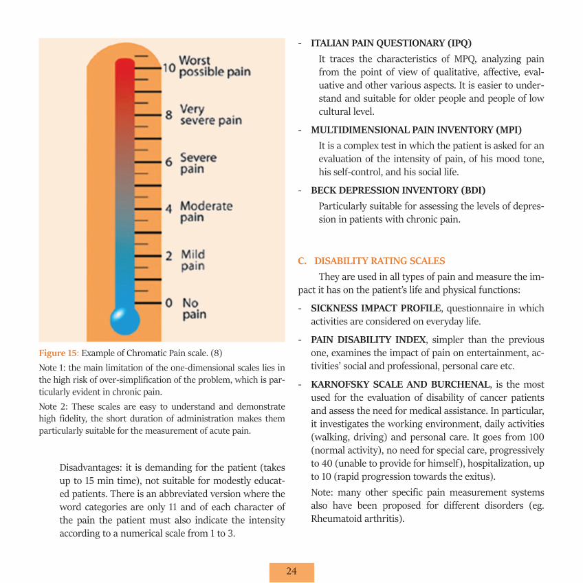
- CHROMATIC SCALE The progressive increase in pain intensity is indicated by the progressive increase in color intensity along a seg-ment whose extremities are represented by “no pain” and “unbearable pain”. The presence of color is a guide of greater understanding for the patient (figure 15).
B. Multidimensional:- McGILL QUESTIONARY (MPQ)
It is the best known and most complete questionnaire. It allows the evaluation of pain in its various character-istics. It consists of 20 groups of words, of which the first 10 represent the sensory characteristics of pain, i.e. the qualitative aspect (e.g. pulsating, stabbing, deaf etc.); the next 5 are affective, i.e. the emotional reac-tions (e.g. tiring, unpleasant etc.); the 16th expresses an evaluation of pain (e.g. unbearable, worrying etc.); the last 4 are the various characteristics (compressing, piercing etc.). The patient must select the groups of words that best suit his pain and mark the words that best describe them. You can assign a score to each di-mension and then a total score. Advantage: more multi-faceted analysis of pain in its various components, ideal for chronic pain.

23
Figure 14: The most common used one-dimensional pain intensity scales. The visual analog scale (VAS) consists of a line, usually 100 mm long, whose ends are labeled as the extremes (no pain and worst pain imaginable); the rest of the line is blank. The patient is asked to put a mark on the line indicating their pain intensity (at the pres-ent time, over the past week, or over the past 2 weeks, etc.). The distance between that mark and the origin is measured to obtain the patient’s score. The addition of markers to the traditional pain VAS form a graphic rating scale (GRS). This scale includes a horizontal line with vertical bars of increasing height and anchors at both ends (no pain and worst possible pain). The line is graded from 0 to 10 (or from 0 to 100). The numerical rating scale (NRS) involves asking patients to rate their pain intensity by selecting a number on a scale from 0-10 (11-point scale), 0-20 (21-point scale), or 0-100 by filling in a questionnaire or stating verbally a numerical level (please indicate on the line below the number between 0 and 10 that best describes your pain. A 0 would mean no pain and a 10 would mean worst pain immaginable). Sometimes descriptive terms, such as none, mild, moderate and severe, are provided along the scale (this forms a ver-bal rating scale, VRS) for guidance, as shown, and the scale is then referred to as a GRS. (8)emotional and social functions require multidimensional qualitative tools and health-related quality of life instruments. The recommendations concerning outcome measurements for pain trials are useful for making routine assessments that should include an evaluation of pain, fatigue, disturbed sleep, physical functioning, emotional functioning, patient global ratings of satisfaction, and quality of life. Despite the growing availability of instruments and theoretical publications related to measuring the various aspects of chronic pain, there is still little agreement and no unified approach has been devised. There is, therefore, still a considerable need for the development of a core set of measurement tools and response criteria, as well as for the development and refinement of the related instruments, standardized assessor training, the cross-cultural adaptation of health status questionnaires, electronic data capture, and the introduction of valid, reliable and responsive standardized quanti-tative measurement procedures into routine clinical care. This article reviews a selection of the instruments used to assess chronic musculoskeletal pain, including validated newly developed and well-established screening instruments, and discusses their advantag-es and limitations.”,”author”:[{“dropping-particle”:””,”family”:”Salaffi”,”given”:”F.”,”non-dropping-particle”:””,”parse-names”:false,”-suffix”:””},{“dropping-particle”:””,”family”:”Ciapetti”,”given”:”A.”,”non-dropping-particle”:””,”parse-names”:false,”suffix”:””},{“drop-ping-particle”:””,”family”:”Carotti”,”given”:”M.”,”non-dropping-particle”:””,”parse-names”:false,”suffix”:””}],”container-title”:”Reuma-tismo”,”id”:”ITEM-1”,”issue”:”4”,”issued”:{“date-parts”:[[“2012”]]},”page”:”216-229”,”title”:”Pain assessment strategies in patients with musculoskeletal conditions”,”type”:”article-journal”,”volume”:”64”},”uris”:[“http://www.mendeley.com/documents/?uuid=a82e87ba-ad6a-4749-94ae-42b8806680c4”]}],”mendeley”:{“formattedCitation”:”(8)
24
Disadvantages: it is demanding for the patient (takes up to 15 min time), not suitable for modestly educat-ed patients. There is an abbreviated version where the word categories are only 11 and of each character of the pain the patient must also indicate the intensity according to a numerical scale from 1 to 3.
- ITALIAN PAIN QUESTIONARY (IPQ)It traces the characteristics of MPQ, analyzing pain from the point of view of qualitative, affective, eval-uative and other various aspects. It is easier to under-stand and suitable for older people and people of low cultural level.
- MULTIDIMENSIONAL PAIN INVENTORY (MPI)It is a complex test in which the patient is asked for an evaluation of the intensity of pain, of his mood tone, his self-control, and his social life.
- BECK DEPRESSION INVENTORY (BDI)Particularly suitable for assessing the levels of depres-sion in patients with chronic pain.
C. DISABILITY RATING SCALES They are used in all types of pain and measure the im-
pact it has on the patient’s life and physical functions:
- SICKNESS IMPACT PROFILE, questionnaire in which activities are considered on everyday life.
- PAIN DISABILITY INDEX, simpler than the previous one, examines the impact of pain on entertainment, ac-tivities’ social and professional, personal care etc.
- KARNOFSKY SCALE AND BURCHENAL, is the most used for the evaluation of disability of cancer patients and assess the need for medical assistance. In particular, it investigates the working environment, daily activities (walking, driving) and personal care. It goes from 100 (normal activity), no need for special care, progressively to 40 (unable to provide for himself), hospitalization, up to 10 (rapid progression towards the exitus).Note: many other specific pain measurement systems also have been proposed for different disorders (eg. Rheumatoid arthritis).
Figure 15: Example of Chromatic Pain scale. (8)Note 1: the main limitation of the one-dimensional scales lies in the high risk of over-simplification of the problem, which is par-ticularly evident in chronic pain.Note 2: These scales are easy to understand and demonstrate high fidelity, the short duration of administration makes them particularly suitable for the measurement of acute pain.

25
D. CLASSIFICATION OF PAIN RELIEFPatients are asked to quantify in percentage values
the relief experienced with the therapies. They can report it orally or on a SEA scale, which presents two extremities: “no relief” and “total relief”. We can also propose graduated scales: 0=no relief,1=light, 2=moderate, 3=evident, 4=total.
IN CONCLUSION, which methods should be used for correct pain measurement depending on the patient’s type of pain?
�� In ACUTE PAIN, that lasts for a short time and in which an appropriate level of medication, behavioral analysis, neuro-vegetative reactions (tachycardia, sweating, etc.) has been determined, a one-dimensional numerical or analog scale before and after therapy may be sufficient.
�� In CHRONIC PAIN, it is useful to evaluate the intensity with a one-dimensional test (e.g. VAS), the quality with McGill’s questionnaire and we can associate, if appro-priate, a disability questionnaire. Always evaluate the personality of the subject and the environment that sur-rounds him also to design the appropriate treatment.
�� In PROGRESSIVE DISEASE-RELATED PAIN (e.g., CANCER PAIN), there may be acute episodes related to nociceptive events, such as painful diagnostic proce-dures, but in any case, we must consider that this is a GLOBAL and then multidimensional pain, with signif-icant behavioral alterations and affective, and a degree progressively higher disability. We can use a multidi-mensional scale and a disability one.
SPECIAL CLASSES OF PATIENTS:Pediatric age Pediatric pain assessment poses a number of problems
that have contributed to increasing the delay in knowledge about how to assess and measure pain in infants and chil-dren (i.e. the deep-rooted conviction that the newborn is
unable to perceive pain due to the immaturity of the ner-vous system, the child’s lack of means of expression and the fear of drugs overdose). It is currently thought instead that pain is perceived by the fetus “in utero” and it has been hy-pothesized that the mnemonic patterns of pain are learned as he passes through the birth canal. The central nervous system at birth is very immature, it will only mature com-pletely in 3-4 years. It was so assumed that newborns would not feel the pain that they could endure surgery without an-esthesia. In fact, infants show no apparent signs of pain, but biological indicators show neuroendocrine alterations that could leave mnemonic traces and general disorders. It can, therefore, be considered that at the moment of birth the transmission of pain is immature but not to be neglected.
For children under one year of age (preverbal age) we will rely on behavioral indexes (crying, coarse movements not aimed at indicating the cause of the pain) and/or physi-ological indexes (heart rate, respiratory rate, oxygen satura-tion, disappearance, insomnia, etc.).
From 12 months onwards (and up to 7 years), behav-iors can be analyzed by the children’s hospital of eastern Ontario scale, which evaluates various indexes such as crying (not crying, moaning, screaming), facial expression (smiling, neutral, wrinkled), body movements, limb’s move-ments, which are given a score.
Starting from 18 months, children are able to verbal-ly express their pain using simple expressions. The lesions that are seen (eg. the presence of blood) are considered the most painful, the child’s conviction to be considered good if he doesn’t complain can reduce his perception of pain.
From the 3rd year children can localize the site of pain.
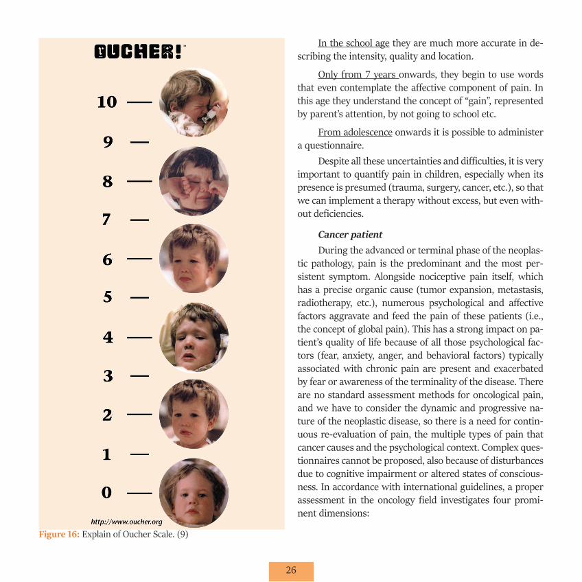
From the 4th year they are capable of expressing the intensity of the pain (much/little) so the measurement of the subjective component can be carried out. They use sim-ple scales with only 4 or 5 options of choice (eg. four colors gradations, the faces of OUCHER scale, figure 16).
26
Figure 16: Explain of Oucher Scale. (9)
In the school age they are much more accurate in de-scribing the intensity, quality and location.
Only from 7 years onwards, they begin to use words that even contemplate the affective component of pain. In this age they understand the concept of “gain”, represented by parent’s attention, by not going to school etc.
From adolescence onwards it is possible to administer a questionnaire.
Despite all these uncertainties and difficulties, it is very important to quantify pain in children, especially when its presence is presumed (trauma, surgery, cancer, etc.), so that we can implement a therapy without excess, but even with-out deficiencies.
Cancer patientDuring the advanced or terminal phase of the neoplas-
tic pathology, pain is the predominant and the most per-sistent symptom. Alongside nociceptive pain itself, which has a precise organic cause (tumor expansion, metastasis, radiotherapy, etc.), numerous psychological and affective factors aggravate and feed the pain of these patients (i.e., the concept of global pain). This has a strong impact on pa-tient’s quality of life because of all those psychological fac-tors (fear, anxiety, anger, and behavioral factors) typically associated with chronic pain are present and exacerbated by fear or awareness of the terminality of the disease. There are no standard assessment methods for oncological pain, and we have to consider the dynamic and progressive na-ture of the neoplastic disease, so there is a need for contin-uous re-evaluation of pain, the multiple types of pain that cancer causes and the psychological context. Complex ques-tionnaires cannot be proposed, also because of disturbances due to cognitive impairment or altered states of conscious-ness. In accordance with international guidelines, a proper assessment in the oncology field investigates four promi-nent dimensions:

27
- the functional state, referring to the ability to satisfy personal needs, ambitions, social role;
- the physical state, referring to their perception of the disease, due to the combination of symptoms of cancer, treatment side effects, and general physical condition;
- psychological state, the positive direction of well-be-ing or negative direction of discomfort, the experience related to one’s own body image and its modifications, the level of anxiety, the depressive component etc.;
- the welfare state, referring to the maintenance of one’s social image, the role in the field of work or family, the maintenance of interests.
Useful in these patients is the SHORT QUESTION-NAIRE FOR PAIN ASSESSMENT that considers the location of the pain, its variability during the day, its intensity with scales from 0 to 10, the pain relief in percent, the interfer-ence of pain on mood tone, walking, other physical activi-ties, work, relationships with others and sleep.
PHYSICAL EXAMINATION
An appropriate diagnosis strictly requires a general assessment of the patient’s physical, neurological, musculo-skeletal and mental condition. The general physical exam-ination should include facial expression, sweating, muscle tension, gait and all those physical characteristics that may highlight a condition of generalized suffering caused by chronic pain. �� INSPECTION of the painful area is fundamental for
the presence of trophic alterations, color changes, alter-ations of the skin annexes that can lead to one or the other diagnosis.�� PALPATION of the region provides additional information
on the pain for the presence of superficial or deep pain that can guide diagnosis; the delimitation of the painful area is fundamental. From the patient’s reaction to palpa-tion, we can understand the patient’s mood and character.
Special objective examination:Examination of the musculoskeletal system
�� Inspection: signs of hypo or muscle atrophies (usually monolateral), hypertrophies (prolonged contractures), fasciculations (neurogenic suffering)
�� Palpation: consistency (presence of fibrous bundles), contractures, trigger points (they are the most frequent cause of musculoskeletal pain but the less recognized and treated. They represent the peculiar element of the so-called myofascial syndromes, which include a very large group of painful diseases characterized by muscle pain, contracture, and functional limitation, sometimes asso-ciated with neuro-vegetative disorders. The pressure of trigger point causes pain radiated to a distant area called the target area (target). Trigger point infiltration can re-solve myofascial syndrome.
�� Percussion: it is performed with the hammer and in normal conditions a fleeting contraction is observed. Re-ductions of this contraction may be caused by inflamma-tory or degenerative myopathies, while it increases after muscle denervation or myotonic syndromes.
�� Examination of the muscular strength: this is a com-plex evaluation because there is often no correspondence between the objective finding and what the patient re-ports. The examination is carried out in two stages: first a segmental study and then a more general study.
UPPER ARTS: evidence of Mingazzini, evidence of the prayer position.
LOWER ARTS: evidence of Mingazzini, evidence of Barrè.
�� Examination of muscle tone: consistency (with palpa-tion), extensibility (passive stretching), passivity (resis-tance opposed by the muscle to passive mobilization).

28
Neurological examination During a neurological examination, it should be tak-
en into account that neurological deficits rarely end on the midline, but tend to follow the peripheral nerve territories; hysterical patients often report high sensory loss without apparent loss of function; patients with significant drug or alcohol intake often give bizarre neurological responses that return to normal when the effects of the drug disappear; this is particularly true in patients with chronic pain in which many narcotics are administered. It is also necessary to distinguish between simulators and hysterics and this is possible by using maneuvers that distract the patient from the possible simulation.�� Cranial nerve examination: The Trigeminal nerve is rap-
idly explored by corneal reflexes and looking for changes in sensitivity on the skin of the face. The facial nerve is examined by evaluating the efficiency of the facial muscles and the facial tone and symmetry of grimaces and winks. The accessory nerve is examined by assessing the tone of the trapezius and sternocleidomastoid.�� Gait: the complexity and the variety of the nervous
structures responsible for walking correspond to vari-ously altered types of gait. The most interesting gaits for the study of pain syndromes are: 1) the mowing gait of the hemiparetic condition in which there is the hyperto-nia of the extensor muscle of the lower limb; 2) the step-ping gait in which we can observe an excessive elevation of the knee with the foot crawling on the ground, char-acteristic of the paralysis of the anterolateral muscles of the leg (polyneuritis, lumbar root compression, lesions of the external sciatic-popliteal); 3) claudicating gait; 4) hysterical gait (e.g. the patient drags a limb as if it were flaccid, but it is intact). �� Reflexes: In neurological semeiotics, the study of reflex-
es is of fundamental importance because, being indepen-dent of the patient’s will and ability to collaborate, they can provide objective data. Moreover, their great sensi-
tivity often allows diagnosing very slight alterations not otherwise diagnosable.
Deep reflexes: Bicipital reflex (no. muscolocutaneous C5,C6) Tricipital reflex (radial no. C6,C7) Styloradial reflex (median no. C6,C7,C8)Styloulnar reflex ( median no. C8,D1) Patellar reflex ( femoral no. L3,L4) Achille’s reflex (post tibial no. S1,S2)
Three different events can occur in relation to the current pathology:
1) areflexia 2) hyporeflexia 3) hyperreflexia 4) Babinski’s reflection.
Sensitivity test: Tactile sensitivity Thermal sensitivity Pain sensitivity Deep sensitivity (deep painful and tactile sensitivity, dis-criminatory sensitivity)
LABORATORY AND INSTRUMENTAL PAIN ASSESSMENT
Electromyography: It is useful in the diagnosis of several neuromuscular
pathologies such as neurogenic muscle atrophies, primitive muscle atrophies and in particular, as far as pain is con-cerned, in polyradicolonevritis.
Somatosensory evoked potentials (SSEP): The examination consists of the recording of potentials
evoked in the sensory nerves by direct nerve stimulation.

29
The evaluation of the conduction velocity of the sensitive fibers allows documenting the existence of functional im-pairments of the afferent fibers in painful syndromes due to neuropathies of various kinds.
Imaging:
1. Traditional radiology: Low resolution, useful to diag-nose severe osteo-articular alterations or reductions in bone mass of more than 30%.
2. TAC
3. MRI very definite image of soft tissue and spongy bone.
4. Scintigraphy: High sensitivity, low definition
5. Ultrasound scanning useful for the study of soft tissue. Images less defined but much less expensive than MRI.
30
NON-INVASIVE TREATMENT OF PAIN AND DRUG THERAPY
Nowadays, we have several analgesics that are effective in the treatment of all types of pain.
A first classification of analgesics distinguishes be-tween real analgesics, such as NSAIDs and opioids, and drugs called “adjuvants”. The latter generally act by enhanc-ing the effect of real analgesics and belong to these cate-gories: antiepileptic drugs, antidepressants, antidepres-sants, corticosteroids, muscle relaxants, sedatives, local anesthetics, etc..
1. NSAIDS They are drugs with peripheral action (inhibition of
nociceptive pain mediators by inhibition of peripheral cyc-lo-oxygenases) and central action (inhibition of central cyc-lo-oxygenase, particularly at the hypothalamus and periaq-ueductal gray matter level).
They are particularly effective in inflammatory and metastatic bone pain (indomethacin) and in some forms of incident pain, where they seem to be even more effective than morphine itself.
Very useful when administered in combination with weak or strong opiates, the effectiveness of the combination in bone pain has been demonstrated.
It’s useful to combine gastric protection. They cause numerous adverse effects, and the most
frequent are: - Reduction in platelet aggregation - Reduction of renal blood flow - Ulcerative lesions of the gastric mucosa - Asthmatic manifestations - Risk of allergic reactions - Ceiling effect, i.e. the existence of a maximum achievable lev-
el of analgesia (increasing the dosage above a certain limit does not increase the analgesia but increases the side effects).
They have an action duration ranging from 3 to 12 hours. Paracetamol is a different drug, it has only analgesic
and not anti-inflammatory effect and does not damage the gastric mucosa or alter platelet aggregation.
2. OPIOIDS According to the WHO, the analgesic opioids indicated
for the treatment of:
mild to moderate pain (weak opiates): - Codeine:
Mechanism of action: it binds mu receptors predom-inantly, and it acts largely after being metabolized to morphine in the liver. Adverse effects: Rare, low addiction and craving Advantages: poor hepatic first passage effect very good enteral efficacy (main advantage). It is usually administered in combination with NSAIDs and parac-etamol. Duration of action: 4/6 hours Power: Codeina vs. Morphine 1/10 Maximum daily dosage: 300 mg
- Tramadol: Mechanism of action: it binds opioid receptors and stim-ulates the neuronal release of serotonin and inhibition of presynaptic reuptake of serotonin and noradrenaline Useful in neuropathic pain Adverse Effects: Not frequent and modest, not addiction and dependence, not respiratory depression, not ceiling effect. Mostly nausea Administration = p.o., i.m., e.v. Duration of action: 6 hours Power: Tramadol vs. Morphine 1/5
31
- Dextropropoxyphene: A synthetic derivative of methadone: mu agonist, weak antagonist of NMDA receptor. It is absorbed by the gastrointestinal tract, long half-life (15 h). Less nausea and vomiting than other opiates.
For moderate to severe pain (strong opiates):The main advantage is the lack of a “roof” effect, so the
dosage can be progressively increased without limits until the total pain control is achieved.
- Morphine: Mechanism of action: agonist of all opioid receptors (mu, ki, delta). Administration = p.o. (bioavailability per os 35-75%. Slow-release forms like MS CONTIN, SKENAN 10-30-60-100 mg; Quick Release Shapes like ORAMORPH); i.m.; e.v.; s.c.; spinal; Metabolism: hepatic metabolism two main metabo-lites: 6-glucuronide morphine (stronger than morphine) and 3-glucuronide morphine (no effect). All metabolites are removed from the kidney. Morphine clearance is re-duced in patients older than 50 years.Duration of action: quick release forms ∼ 4 h; slow-re-lease forms 8-24h; Adverse effects = nausea and vomiting; constipation; sphincters constriction; respiratory depression; toler-ance and addiction.
- Oxycodone:Mechanism of action: Semi-synthetic opioid, mu, and K agonist. Administration: os (OXYCONTIN), e.v., i.m., s.c.Duration of action: e.v. 4-5h. Advantages: according to recent studies, it is more effec-tive on visceral pain.
Adverse effects: fewer side effects than morphine (so fewer nausea, vomiting, respiratory depression).Power: Oxycodone e.v. vs. Morphine e.v. 1/1; Oxycodone o.s. vs. Morphine o.s. 2/1
- Fentanyl: Mechanism of action: mu-opioid receptor agonist. Administration: ev, td (FENPATCH), transmucosal (PECFENT), sl (EFFENTORA), im, spinal and epidural. Duration of action: ev 30 min-1h.Adverse effects = nausea and vomiting; constipation; sphincter constriction; respiratory depression; tolerance and addiction.Power: 75-100 times more than morphine.
- Methadone: Mechanism of action: synthetic opioid, mu receptor ago-nist, NMDA receptor antagonist.Administration: o.s.Metabolism: High bioavailability by oral route - 90% (good absorption, low hepatic first passage effect) Fast effect, long half-life 50 hours (tendency to accumulate in tissues, requires careful monitoring for chronic ther-apies).Duration of action: 4-6 hours after 15-30 mg o.s., usually administered every 8-12 hours to avoid accumulation.Adverse effects: Some studies have shown a lower inci-dence of constipation than morphine.
- Buprenorphine:Mechanism of action: mu-opioid receptor partial ago-nist, k receptor antagonist. Adverse effects: Hallucinations Administration = sublingual administration, i.m. e.v., TDDuration of action = 6 hours sublingually Power: buprenorphine (sl) vs. Morphine 60/1

32
3. ADJUVANTS- Antidepressants acting on mood tone and activate at
low doses descendant pain control system; very useful in neuropathic pain.
- Antispasmodics- Anticonvulsants carbamazepine and gabapentin, anal-
gesic action in neuropathic pain;- Ansiolitics, - Antiemetics, - Steroids, - Muscle relaxants, - Anticholinergics, - Bisphosphonates- Local anesthetics for external use.
Side effects are often attributed to opioids; neverthe-less, many symptoms may be related to the disease itself. There are several therapeutic strategies that allow preven-tion or treatment of opioids side effects, or enhancement of their effects when required: �� General measures (e.g., reduce the dose, adequately hy-
drate the patient, reduce the number of drug combina-tions) �� Use adjuvant drugs �� Chane the route of opioid administration (transcutane-
ous, subcutaneous, e.v.) �� Change the type of opioid (opioid rotation) based on
the principle of “incomplete cross-tolerance”, i.e. a reduc-tion of side effects by changing the opioid (e.g. switching from morphine to methadone).

33
INVASIVE TREATMENT OF PAIN
Invasive therapy has had a good development in the past years because there were no effective drugs with few side effects; today it is limited to 3-5% of the total amount of therapy practiced.
�� Epidural Injection: it is practiced particularly when chronic pain affects the cervical and lumbar spine tract; usually, we use local anesthetics and slow-release cor-ticosteroids, directly in contact with the dura mater
(figure 17). The technique is generally associated with a modest risk of general and local side events. However, it may be associated with peridural bleeding and spinal cord compression in patients with unrecognized clotting defects. Infection is another potential side effect; how-ever, nowadays, the disposable sets and the respect of the asepsis rules have almost canceled this risk out. Sub-dural or intravenous injection are other potential risks, particularly associated with inexperienced operators or anatomic abnormalities.
�� Pharmacological neuromodulation is a method that uses a subarachnoid permanence catheter connected to a reservoir that is filled with drugs. A high-precision pump or resistive system control the release of drugs. It is a generally effective and low-risk method burdened by a high cost of establishment and maintenance.
�� Surgical or physical neuron lesion is nowadays a rare-ly used technique, confined to the cases of otherwise in-tractable pain.
�� Electrical spinal cord stimulation (SCS) consists of inserting a stimulator catheter at the epidural level. This catheter is connected to a subcutaneous stimula-tor. TENS is the principle on which the method is based. This is a technique used for the treatment of chronic pain conditions such as sciatica (LB), failure of surgical interventions in the back (FBSS), post-herpetic neural-gia (PN), regional pain syndrome complex (CRPS) and other painful syndromes in which more conservative treatments have failed and other surgeries are not in-dicated (Vallejo et al., 2007). The guidelines, developed by the Brussels Task Force, set out the main indications for the implantation of SCS, the criteria for patient selec-tion, the outcome measures and the techniques to use. Implantation of SCS is contraindicated in the presence of psychiatric disorders, in the presence of poor compliance Figure 17: Rappresentation of epidural injection. (10)

34
to common treatments, in the absence of adequate social support and in the presence of alcohol or drug abuse. The selection of candidates for the plant carried out by a multidisciplinary team is considered the best solution to ensure the effectiveness of the intervention. A systematic review of the literature on the efficacy of SCS in relieving pain was made by Turner and colleagues (2004) in re-spect of FBSS and CRPS. The authors point out the lack of randomized controlled trials. Indeed, only a study of articles published after 1990 has been rated Class I by the Committee of the American Academy of Neurology Quality Standards (Chang and Lowenstein, 2003). For these reasons, the authors argue that no conclusions can be drawn at this stage as to the effects of treatment with SCS in FBSS and CRPS. The results presented in the re-view show, however, a low to moderate improvement on average perceived pain, although a study concerning the evaluation of the efficacy of a surgical technique should also include:
�– an assessment of non-specific effects such as the pla-cebo effect,
�– an evaluation of disability and activity at work and in leisure before and after treatment,
�– an evaluation of the diagnostic variables relevant to the baseline,
�– an explicit description of the inclusion and exclusion criteria adopted in the selection of candidates for the implant;
the follow-up should be conducted for a period of not less than 6 months. As described in this review, some un-controlled studies show the effectiveness of the implant in chronic pain. An improvement in pain and associated disability after treatment with SCS was reported in FBSS by North and Guarino (1999). In another study, which followed patients for 5 years after implantation, it is re-ported that 25% of subjects returned to work during this
time (North et al., 1991). Finally, in the study by Burchiel and colleagues (1996), it is reported that at the one-year follow-up the pain and quality of life of the patients had improved statistically significantly. In 2005 the British Pain Society, in collaboration with the Society of British Neurological Surgeon published a consensus document fwww.britishpainsociety.org) recommending the use of SCS in the following clinical conditions:
– Strong indication in: FBSS/FNSS, CRPS, neuropathic pain secondary to peripheral nerve damage, refrac-tory angina, post-traumatic brachial plexopathy or post-irradiation.
– Indication of intermediate level: amputation pain, axial pain after spinal surgery, intercostal neuralgia, pain associated with spinal damage, others from pe-ripheral neuropathic pain.
The most recent guidelines are those of the American Society of Interventional Pain Physicians (M.V. Boswell et al, Pain Physician, 2007). They recognize that spinal stimulation for the treatment of pain associated with FBSS and CRPS has strong evidence for short-term out-comes and moderate evidence for long-term outcomes. This, in a context where the majority of therapies are judged to have moderate or limited evidence of efficacy (e.g. epidural steroid injections, intrarticular injections, intradisc electrothermal therapy, anuloplasty, percutane-ous disc decompression), underlines the importance of the use of neurostimulation.
Indications
- Chronic back or leg pain associated with Failed Back Surgery Syndrome (FBSS), especially when the pain is predominantly neuropathic. Several patients devel-op pain initially from discopathy, but the chronic pain that follows is often the result of surgery that causes the accumulation of scar tissue and/or neural injury. FBSS
35
of mainly neuropathic origin is the best indication for neurostimulation. FBSS pain,i.e. nociceptive mainly, re-sponds to intrathecal drug infusion. However, since most FBSS pain includes nociceptive and neuropathic compo-nents, it can be effectively treated with neurostimulation or intrathecal infusion.
- Complex regional pain type I (also referred to as sym-pathetic reflex dystrophy). Indicates several pain condi-tions that may occur following injury. CRPS Type I devel-ops in response to a harmful event (a lesion not related to nerves), such as muscle sprain, bone fracture. Possi-ble causes include repeated trauma, stroke, myocardial infarction and excessive strain. The pain symptoms are complex, diffuse, disproportionate than the original in-jury and worsen in response to pressure on the limbs.
- Complex Regional Pain type II (also called causalgia) The CRPS type II develops in response to a nerve injury, typ-ically to large nerves such as the median nerve or sciatic. The pain is similar to CRPS Type I, but Type II patients may also experience pain as a result of a stimulus that was previously not painful or may be hypersensitive to a stimulus.
- Refractory angina. Angina is a common symptom of cor-onary artery disease, which occurs when blood vessels narrow or clog as a result of atherosclerosis. Angina that can no longer be treated by medical or surgical interven-tion is known as refractory angina
- Post-traumatic brachial plexopathy (brachial plexus avulsion) Brachial plexus avulsion indicates a tearing of the medullary nerve roots going towards the upper limb. It typically occurs due to cervical trauma from a motor-cycle accident. The most compromised roots are those of C5 and C6 (C stands for “cervical”) with complete or only limited loss of motility of the arm, forearm and hand, depending on the number of compromised fibers. The involvement of C7 and C8 inevitably leads to paralysis of the hand. The patient often presents himself with the
pendulous limb, insensitive, but paradoxically painful (neuropathic pain). Pain is one of the most difficult to treat, so much so that the patient himself asks to ampu-tate his limb, which he feels is a foreign body and causes extreme suffering.
- Post-irradiation brachial plexopathy. Currently, plexus suffering is rare due to technical and dosimetric imple-mentation. If it arises, it’s more a resurgence of disease than toxicity. The first symptoms of post-actinic plexop-athy are sensory disorders (disturbing paresthesias such as tingling and hypoesthesia in the root distribution ter-ritories C5-C6-C7) followed by motor impairment (sense of heaviness, hypoesthenia, presence of cramp-like con-tractures) mainly affecting the arm with frequent con-comitant lymphedema.
- Post-herpetic neuralgia (Postherpetic neuralgia or PHN). A serious and painful complication of shingles, an in-fectious disease. The pain is characterized by constant burning and dazzling episodes of stinging pain
- Phantom limb pain. A painful sensation in an amputat-ed limb, as if the limb were still present. This syndrome is often linked to a painful sensation, such as burning, stinging, tickling or tingling.
- Spinal stenosis. A narrowing of the spaces in the spine which gives rise to pressure on the spinal cord and / or nerve roots. The pressure on the bottom of spinal cord or on the nerve roots that branch out from that area may result in pain or numbness in the legs. The pressure on the upper region of the medulla spine (neck) produces similar symptoms in the shoulders.
- Peripheral vascular disease. Caused by atherosclerotic plaque that causes the thickening of the inner lining of the arteries
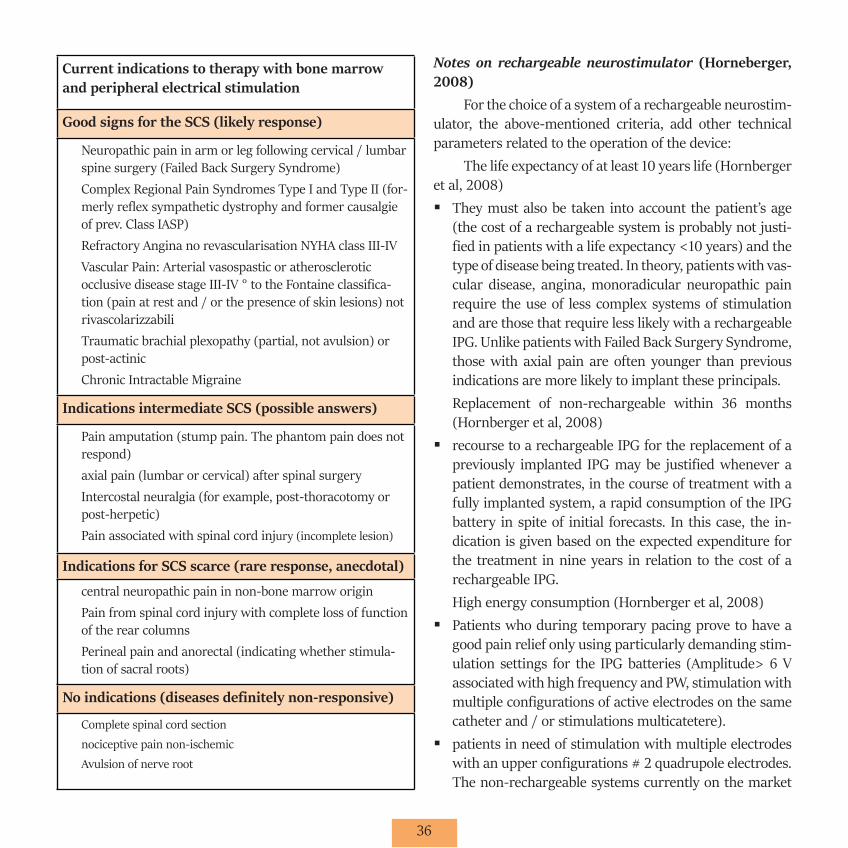
36
Current indications to therapy with bone marrow and peripheral electrical stimulation
Good signs for the SCS (likely response)
Neuropathic pain in arm or leg following cervical / lumbar spine surgery (Failed Back Surgery Syndrome)Complex Regional Pain Syndromes Type I and Type II (for-merly reflex sympathetic dystrophy and former causalgie of prev. Class IASP)Refractory Angina no revascularisation NYHA class III-IVVascular Pain: Arterial vasospastic or atherosclerotic occlusive disease stage III-IV ° to the Fontaine classifica-tion (pain at rest and / or the presence of skin lesions) not rivascolarizzabiliTraumatic brachial plexopathy (partial, not avulsion) or post-actinicChronic Intractable Migraine
Indications intermediate SCS (possible answers)
Pain amputation (stump pain. The phantom pain does not respond)axial pain (lumbar or cervical) after spinal surgeryIntercostal neuralgia (for example, post-thoracotomy or post-herpetic)Pain associated with spinal cord injury (incomplete lesion)
Indications for SCS scarce (rare response, anecdotal)
central neuropathic pain in non-bone marrow originPain from spinal cord injury with complete loss of function of the rear columnsPerineal pain and anorectal (indicating whether stimula-tion of sacral roots)
No indications (diseases definitely non-responsive)
Complete spinal cord section nociceptive pain non-ischemic Avulsion of nerve root
Notes on rechargeable neurostimulator (Horneberger, 2008)
For the choice of a system of a rechargeable neurostim-ulator, the above-mentioned criteria, add other technical parameters related to the operation of the device:
The life expectancy of at least 10 years life (Hornberger et al, 2008) �� They must also be taken into account the patient’s age
(the cost of a rechargeable system is probably not justi-fied in patients with a life expectancy <10 years) and the type of disease being treated. In theory, patients with vas-cular disease, angina, monoradicular neuropathic pain require the use of less complex systems of stimulation and are those that require less likely with a rechargeable IPG. Unlike patients with Failed Back Surgery Syndrome, those with axial pain are often younger than previous indications are more likely to implant these principals.Replacement of non-rechargeable within 36 months (Hornberger et al, 2008)�� recourse to a rechargeable IPG for the replacement of a
previously implanted IPG may be justified whenever a patient demonstrates, in the course of treatment with a fully implanted system, a rapid consumption of the IPG battery in spite of initial forecasts. In this case, the in-dication is given based on the expected expenditure for the treatment in nine years in relation to the cost of a rechargeable IPG.High energy consumption (Hornberger et al, 2008)�� Patients who during temporary pacing prove to have a
good pain relief only using particularly demanding stim-ulation settings for the IPG batteries (Amplitude> 6 V associated with high frequency and PW, stimulation with multiple configurations of active electrodes on the same catheter and / or stimulations multicatetere).�� patients in need of stimulation with multiple electrodes
with an upper configurations # 2 quadrupole electrodes. The non-rechargeable systems currently on the market
37
do not handle more than one or two quadrupole octo-polar, (with the exception IPG Prime Medtronic which nevertheless has a cost which is very close to that of a rechargeable system). From assess and define the direc-tions to stimulation with more electrodes configuration.�� Using subcutaneous stimulation techniques or periner-
vosa device. Such techniques involve departing the need for high intensity stimulations. This indication can be brought into (a)
Preliminary studies demonstrate a wider spread of paresthesia and then a better pain relief using PW higher than those conventionally used and only permitted by re-chargeable systems (indication yet not definable)
In Addition, according to the International Neuromod-ulation Society Note, you need the fulfillment of at least two of the following conditions so that the user is deemed con-sumer to high energy levels, as previously indicated:
a. Daily use ≥ 18 hoursb. stimulation intensity> 4 mAmpc. Pulse duration> 450μs: d. Frequency ≥ 40 Hzis. Positioning 2 leads
Patient Selection The selection of patients in the SOD Palliative Care and
Pain Therapy occurs in outpatient regimen. The doctor who is performing the first visit collects the history and, once as-certained the indication to spinal neuromodulation, shows the facility to the patient. The patient is then directed to a subsequent medical and biopsychosocial evaluation before surgery.
38
CANCER PAIN AND ITS TREATMENT
Pain is one of the main symptoms of cancer.To give an idea of the size of the problem we will give
you some numbers. Worldwide, each year 5 million people die of cancer and 7 million new cases are diagnosed. Accor-ding to the World Health Organization, at least 4 million pe-ople suffer from cancer pain, especially those patients with cancer at an advanced stage: it is estimated that 70-90% of them present painful syndrome of medium-severe intensity.
The patient with terminal cancer is in a clinical con-dition in which the neoplasia is not more curable in direct way. The only things on which we can affect are pain and the suffering.
According to rule 37 of the CODE OF MEDICAL DE-ONTOLOGY: “in the event of prognosis which is likely to be fatal or has reached the end-stage, the physician must limit his work to moral assistance and treatment to avoid un-necessary suffering, providing the patient with appropriate treatment to protect, as far as possible, his quality of life….”.
Alleviating suffering and improving the patient’s qua-lity of life, therefore, is a doctor’s duty as important as pre-serving life. This duty is the only task of the physician who assists a patient considered incurable: reducing the suffe-ring of the assisted person and restoring dignity to the final stages of his life.
The cause of cancer pain might be multifactorial. In particular, pain can result:�� directly from the growth of tumour’s mass with:�� compression of nerve trunks�� infiltration of tissue’s nociceptors (e.g. pleural invasion,
invasion of joints)�� distension, flogosis, obstruction of hollow viscera (e.g.
intestine)�� vascular impairment (ischaemic malignant pain)�� invasion of lymphatic vessels (lymphedema).
�� from the remote repetition of the tumor (metastasis). This can affect all organs and tissues, but the most fre-quently and intensely algogenous sites are bone struc-tures and, in particular, the vertebrae, the pelvis, the long bones, the scapular cingulum. The compression or stretching of the nervous structures, which often occurs, worsens with walking and active and passive move-ments, producing additional suffering to the patient. This is known as “incident pain”, i.e. a pain difficult to treat.�� from the therapeutic treatments, such as chemotherapy
(neurites, aseptic necrosis of the long bones, pharyngi-tis etc.), radiotherapy (fibrosis of the nervous structures, myositis etc.), or surgery.�� from prolonged immobility (eg. Arthritis).�� from the immune deficiency condition, that characteriz-
es the terminal condition, due to the disease’s therapies, with frequent appearance of herpes zoster and post-her-petic neuralgia, or other infectious diseases.
The situation of these patients is even more complex, because, if it is true that 70-90% suffer from pain for the reasons mentioned above, it is certain that in 100% of these patients there is a pain that we can define as “global” or “to-tal”. It is caused by psychogenic causes that support and ag-gravate the pre-existing pain. These psychogenic factors are:�� Anxiety (Concern for the family, fear of death, uncertain-
ty about the future, loss of their dignity and their bodies, financial problems, etc..);�� Depression (Loss of their social position, loss of a role
in the family, chronic fatigue and insomnia, changes in physical appearance, feelings of abandonment, etc..);�� Anger (Bureaucratic difficulties, delayed diagnosis, miss-
ing doctors, lack of visits from friends, treatment failure, etc.).
39
We can also distinguish between two types of oncolo-gical pain:
- Background pain: Pain continuously present in the 24 hours;
- Breakthrough pain (BTcP): temporary exacerbation of pain that occurs in a situation of persistent pain. Possible subtypes: �2 Incident pain, triggered by voluntary acts (e.g.
pain in feeding attempts in case of severe mucosi-tis; pain in movement due to the presence of bone metastasis) or by involuntary acts, foreseeable or not (e.g. distension of viscera, ischaemic events, coughing, bladder spasms or rectal tenesmus) ;�2 Spontaneous or idiopathic pain (by some authors
considered the only true BTcP), not linked to de-tectable stimuli and therefore not preventable; �2 End-of-dose pain, related to the resolution of the
effect of the analgesic drugs.
The BTcP is characterized by: high intensity (VNR: 7-10), rapid onset with quick height (averaging 5 minutes, in a range of 10 seconds to 180 minutes), limited duration (15- 30’, range from 1’’ to 24h) and occasional occurrence (0-5 episodes in the 24h).
A correct clinical approach to the patient suffering from neoplastic pain must include:
1. An accurate gathering of medical history
2. The definition of the stage of the disease
3. The assessment of the recurrence (background and BTcP) and of the intensity of the pain (using appropriate scales). This assessment also should be carried out fre-quently during the treatment period to evaluate the re-sults regarding analgesia, possible side effects and their impact on patient’s quality of life.
4. The assessment of the interaction between sensory and psychological components of pain
5. The assessment of the so-called “performance status” (sleep quality, power, autonomy...)
6. The assessment of the response to analgesic and antineo-plastic therapies previously made.
7. The evaluation of the length of the remaining life (it is a judgment related to clinical experience, however ap-proximate, which is still needed before making thera-peutic decisions with potential inherent risks).
Principles of cancer pain therapyFrom a technical point of view, effective analgesic ther-
apy should follow these general principles:
1 Preventing the onset of pain by the administration of analgesics at regular times for the background pain, and reserving an additional administration to control BTcP. We must also maintain analgesia throughout 24 hours, taking into account the sleep-wake rhythm, keeping in mind that sometimes the night analgesia should be more intense than daytime because the darkness and silence magnify fears and awaken anxieties, fears and nightmares.
2 The route should be the simplest and the least invasive: It is important to suppress all the algogenic and unnecessary stimulations, considering that even the small pain of an intramuscular injection can be poorly tolerated and becomes of high intensity when the suffering is already huge. For this reason, it’s recommended to prescribe oral medications. If this is not possible, we must use the intravenous route, with a stopper or with an internalized access. There is indeed great favor in the EARLY application of a central venous catheter with subcutaneous access that, if well managed, can prevent the suffering from repeated acupunctures (Avoid the intramuscular route!)

40
3 The therapy should be individualized, based also on “performance status”.
4 We must also take into account the possible side effects of the drugs, which can be dull (es. constipation), but they can strongly disturb the patients if not pre-advised.
5 Observe the maximum dosage of drugs with “ceiling ef-fect”
6 Do not combine drugs in the same category
Setting proper cancer pain therapy should be based on the so-called “WHO scale.”
It provides a therapeutic sequence characterized by the transition from the less powerful drugs (NSAIDs) up to the stronger ones (strong opioids: morphine, fentanyl,) passing by weak opioids (codeine, tramadol), going for degrees and depending on the persistence of pain and its intensity.
At all stages, we can, therefore, associate adjuvant drugs (anxiolytics, antidepressants, muscle relaxants, an-ti-emetics, etc.) to meet the specific needs of each patient.
This “therapeutic scale” has been criticized by some authors, who consider it incomplete and too rigid. Howev-er, the worldwide diffusion of these guidelines had a great educational effect on both doctors and population: it has created more attention to the pain of patients with cancer and helped re-evaluate morphine as the most appropriate medication for the control of severe pain.
The dosages of the drugs may reach extremely high levels, due to the development of tolerance and also for the worsening of tumor pathology. However, patients de-velop tolerance also for side effects. They develop also an addiction to morphine or other stronger opioids, but it has no meaning in patients with limited life expectancy and a stronger need to get rid of the pain.
The treatment of pain is still the main aspect of the needs in patients in terminal conditions, but it is not the only one, because we have to consider a whole range of oth-
er therapeutic and social supports, including the so-called palliative care, which must meet the general needs of ter-minally ill patients. These include various general distur-bances, intercurrent disease, medications, nursing, psycho-logical assistance to patients and their families, injections, feeding, personal hygiene, etc., which must be guaranteed in the whole 24 hours, through joint efforts between the hospital, family physicians and institutions operating in the area.
41
PALLIATIVE CARE
The chronic and oncological diseases in the final stages of life are often handled in a hospital centered perspective, characterized by a high-level of invasiveness treatments.
It is well known that approximately 35% of patients who die in the hospital undergo diagnostic and therapeutic procedures in the last 48 hours of life, while the possibility of an imminent exitus is registered in the medical records only in 44% of cases. This often results in a lack of consid-eration of the quality of life in the terminal phase of chronic diseases and in an enormous inappropriate use of health care resources not aimed at the patient’s best interest.
Literature data indicate that, at the end of life, the wishes of patients and their families are generally in the direction of least possible suffering and the opportunity to stay close to the kin.
Various laws enacted over the years affirm the free-dom of choice and the right to health protection in respect of the dignity of the person and the quality of life, identify-ing among the instruments: the Advanced Care Planning and Shared Treatments Planning.
It is clear the need to use in some patients in advanced stages of the disease a different approach, a “palliative” approach that aims to protect the patient and his family from suffering.
The definition of palliative cares has been given by Law 38/2010, in which they are defined as “all the therapeutic, diagnostic and healthcare, interventions addressed at both the sick person and to his family, aimed at the active and total care of patients, whose disease, characterized by an unstoppable evolution and a poor prognosis, does not re-spond to specific treatments”.
The goal of palliative care is to maintain a good quality of life of the patient as long as possible, giving him the abil-ity to maintain contacts with their relationships and their social context world. Succeeding in this intent is one of the aims of medicine and the global care of the patient, with an
approach characterized by continuity and homogeneity of care at all stages of the disease, is the best way to achieve the purpose.
It is necessary to ensure the best etiological treatment and the early recognition of any additional needs (physi-cal, functional, psychological, spiritual, social and rehabil-itation) of the patient. This model of care has, as further objective, the improvement of the humanization of care, also achieved through a deep collaboration between profes-sionals from different disciplines and with the participation in treatment decisions of the patient and family members.
To implement an efficient system, the Palliative Cares must be part of the cultural baggage of all physicians, and it is necessary a specific training for doctors of general medi-cine and for all specialists.
Defining the advanced stage of an evolutionary and ir-reversible disease is not easy but it is necessary. The follow-ing criteria can be very helpful:
�� Therapeutic criteria: absence, exhaustion, lack of oppor-tunities for specific curative treatments.
�� Symptomatic criteria: presence of disabling symptoms that affect the reduction of the performance below 50% of the Karnofsky scale.
�� Temporal evolution criteria: to be determined during the specific assessment of the temporal evolution of the dis-ease.
It has been demonstrated that appropriate use of avail-able resources in both the patient’s home and residential facilities, significantly reduces the length of stay and the number of unnecessary hospitalizations.
The route is divided into different settings: outpatient care, inpatient basis, territory (Home and Hospice). The Hospice (residential structure of Palliative Care) operates in accordance with the principles of unity and continuity with

42
home care and it’s an alternative to the home care when it is not, temporarily or permanently, suitable to accommodate the sick person.
The already mentioned Law 38/2010 provides a net-work of support to terminally ill patients, which must con-sist of a functional and integrated set of district, hospital, health and social services, which are divided into separate organizational lines and specifically dedicated facilities.
The simultaneous care model The National Oncology plan, as in the previous edi-
tions, considers the model of “simultaneous care” (figure 18) the most reliable to ensure the best therapeutic result in terms of life expectancy and quality of life.
Figure 18: Rappresentation of simultaneous care model. (11)
In oncology, the transition from Active Cure to Palli-ative ones is of great difficulty, both for the patient-family unit and for health professionals. In fact, in this passage, different emerging needs often find a response in differ-ent teams, with the consequence of increasing the sense of abandonment of the patient.
The World Health Organization wrote: “The approach of palliative care can be applied at an early stage of the dis-ease, in connection with other therapies designed to pro-
long life, such as chemotherapy and radiotherapy, includ-ing even those investigations that aim to understand and to avoid clinical complications. “
A study published in 2010 in the New England Jour-nal of Medicine shows that a model defined “early palliative care”, applied to patients with lung cancer, leads to a signifi-cant improvement in quality of life and mood, to a reduction of aggression of the treatments in the terminal phase, with a lengthening of the duration of life.
In several cancer centers, this model of care is carried out with the help of a palliative doctor which supports other specialists in taking choices targeted to the welfare of the patient, which improves optimization of resources and facil-itates the transition, sometimes necessary, by “curing” the patient to “taking care” of him.
The end of life in the hospital The anesthesiologists daily treat patients at the end of
life in hospitals, but the greatest risk, as already said, is to exceed on the use of disproportionate therapies, often very invasive and also very expensive, not taking into consider-ation that the patient has a very limited life perspective.
In some situations, characterized by the evident irre-versibility of the disease and the high probability of rapid evolution to the patient’s death, it is right to think of a “re-modeling” of the therapy, aimed at ensuring the best possi-ble quality of remaining life, rather than healing.
A good method to optimize the not simple therapeutic approach in these situations is to create a Diagnostic Assis-tance Treatment Plan (DATP) that guides and harmonizes the patient care pathway at the end of life.
In the AOUC Careggi, DAPT has been realized by creating a multidisciplinary and multiprofessional clin-ical group, coordinated by doctors belonging to the De-partment of “Anesthesia and Intensive Care”. It has the purpose of supporting doctors in the management, in the choices and in the communication with the patient and his
43
family when it is detected that the clinical condition is not predictable, despite the best possible therapies, a signifi-cant survival time.
The activities of the group take place at the depart-ments where the patient is hospitalized and is composed of experts in palliative care, oncologists, radiation therapists, psychologists, nurses and physiotherapists.
All actions are recorded on medical records.From an operational point of view, the protocol is di-
vided into several phases. Medical personnel applying DAPT identifies patients eligible for this path using a prognostic classification system. This system is the “Palliative Prog-nostic Score” (PAP score), it evaluates clinical, biochemical and symptom factors, identifying three groups of patients with different life expectancies. If the score obtained is > 5.5 points (70% chance of survival less than 30 days), the doc-tors who are treating the patient activate the support group.
The group operates in advisory arrangements at the department. At the time of the first evaluation it is neces-sary the presence of the department doctor with the aim to have a correct diagnosis and to share pathway care.
The first patient assessment is carried out by the palli-ativist and it can have the following outcomes:�� a single counseling is performed to support the decisions
of department physician;�� the patient does not take any specific pathway, but the
palliative group remains available in the event of evolu-tion of the symptoms of the patient;�� a therapeutic-care plan is set and managed by the group
in close collaboration with the treatment team.
The main functions of the palliative doctor breaks are:
a) Evaluate the history and the clinical condition of the pa-tient in order to identify the best care pathway;
b) Inform the patient and family, collect their will and shares with them the pathway;
c) Require if necessary the evaluation/intervention of oth-er professionals (oncologist-radiotherapist, psychologist, nurse, physical therapist); if their involvement provides more action and puts specific targets (eg. physical ther-apy cycle), it must be prepared an “ISP” (Individual Support Plan) that will be recorded in the computerized medical record.
d) Verify that any symptomatology is well controlled and, otherwise, suggests changes/therapeutic integrations;
e) Manage the continuity of care process, representing the interface with Territorial Palliative Care (home and/or the Hospice). In particular, he makes contact with these centers and oversees the outcome of the route taken.
The ward doctor who dismisses the patient must re-port in the patient record the main assessments, the inter-vention of the team of palliative care and the type of shared care pathway.
Image bibliography
1. Dubin AE, Patapoutian A. Nociceptors : the sensors of the pain pathway Find the latest version : Review series Nociceptors : the sensors of the pain pathway. Journal of Clinical Investigation. 2010;120(11):3760–72.
2. Mizisin AP, Weerasuriya A. Homeostatic regulation of the endo-neurial microenvironment during development, aging and in re-sponse to trauma, disease and toxic insult. Acta Neuropathologica. 2011;121(3):291–312.
3. Basbaum AL, Bautista DM, Scherrer G, Julius D. Cellular and Molecular Mechanisms of Pain. Cell. 2009;2(October):267–84.
4. Bushnell MC, Ceko M, Low LA. Cognitive and emotional con-trol of pain and its disruption in chronic pain. Physiology & behavior. 2013;14(7):502–11.
5. Casiglia E, Finatti F, Tikhonoff V, Stabile MR, Mitolo M, Gasparotti F, et al. Granone’s Plastic Monoideism Demon-strated by Functional Magnetic Resonance Imaging (fMRI). Psychology. 2019;10(04):434–48.
6. Millan MJ. Descending control of pain. Progress in Neurobiol-ogy. 2002;66(6):355–474.
44
7. Kandel ER, Schwartz JH, Jessell TM, Siegelbaum SA, Huds-peth AJ. Principles of Neural Science. 2000. 482–486 p.
8. Salaffi F, Ciapetti A, Carotti M. Pain assessment strategies in patients with musculoskeletal conditions. Reumatismo. 2012;64(4):216–29.
9. Oucher TM. Oucher Scale (Internet). Available from: http://www.oucher.org/index.html
10. Chen J, Han J, Zhao Z, Wei F, Hseieh J, Bao L, et al. Pain. Neu-roscience in the 21st Century: From Basic to Clinical. 2013. 1–3111 p.
11. Meyers FJ, Linder J. Simultaneous care: Disease treatment and palliative care throughout illness. Journal of Clinical Oncology. 2003;21(7):1412–5.