the general anesthetic propofol excites nociceptors … · the general anesthetic propofol excites...
TRANSCRIPT

THE GENERAL ANESTHETIC PROPOFOL EXCITES NOCICEPTORS BY ACTIVATING TRPV1 AND TRPA1 RATHER THAN GABAA RECEPTORS
Michael JM Fischer1,3*, Andreas Leffler2*, Florian Niedermirtl2*, Katrin Kistner1, Mirjam Eberhardt1, Peter W Reeh1, Carla Nau2 *these authors contributed equally and appear in alphabetical order 1Institute of Physiology and Pathophysiology and 2Department of Anesthesiology, University
of Erlangen-Nuremberg. 3Department of Pharmacology, University of Cambridge, United Kingdom.
Running head: Propofol activates TRPA1, TRPV1 and GABAA Address correspondence to: Andreas Leffler, Dept. of Anesthesiology, Friedrich-Alexander-University Erlangen-Nuremberg, Krankenhausstrasse 12, 91054 Erlangen, Germany, Phone: +49-9131-85-33680, Fax: +49-9131-85-39161, Email: [email protected]
Anesthetic agents can induce a paradox activation and sensitization of nociceptive sensory neurons and thus potentially facilitate pain processing. Here we identify distinct molecular mechanisms which mediate an activation of sensory neurons by 2,6-diisopropylphenol (propofol), a commonly used intravenous anesthetic known to elicit intense pain upon injection. Clinically relevant concentrations of propofol activated the recombinant TRP receptors TRPA1 and TRPV1 heterologously expressed in HEK293t cells. In dorsal root ganglion (DRG) neurons, propofol-induced activation correlated better to expression of TRPA1 than of TRPV1. However, pre-treatment with the protein kinase C activator 4β-phorbol 12-myristate 13-acetate (PMA) resulted in a significantly sensitized propofol-induced activation of TRPV1 in DRG neurons as well as in HEK293t cells. Pharmacological and genetic silencing of both TRPA1 and TRPV1 only partially abrogated propofol-induced responses in DRG neurons. The remaining propofol-induced activation was abolished by the selective GABAA receptor antagonist picrotoxin. Propofol but not GABA evoked a release of calcitonin gene-related peptide (CGRP), a key component of neurogenic inflammation, from isolated peripheral nerves of wild type but not TRPV1 and TRPA1-deficient mice. Moreover, propofol but not GABA induced an intense pain upon intracutaneous injection. As both the release of CGRP and injection pain by propofol seem to be independent of GABAA receptors, our data identify TRPV1 and TRPA1 as key molecules for propofol-induced excitation of sensory neurons. This study warrants further investigations into the role of anesthetics to
induce nociceptor sensitization and to foster postoperative pain.
The intravenous general anesthetic
propofol (2,6-diisopropylphenol) has become one of the most widely used anesthetics. Due to a short context-sensitive half time, propofol is used for sedations as well as for total intravenous anesthesia during surgery. A drawback of propofol however, is its high potential to induce intense burning pain upon injection. Depending on the concentration (0.5 – 2%), the galenic formulation and co-medication, 24 − 90% of all patients receiving propofol experience injection pain (1). It was hypothesized that propofol might indirectly or directly interact with sensory nerve fibers located in the venous adventitia (2-4). A recent study claims that the irritant receptor TRPA1, which is expressed in nociceptive sensory neurons, is the predominant molecular entity mediating activation of peripheral nerve endings by general anesthetics (5). Also, TRPA1 was found to mediate propofol-induced pain behavior induced by intranasal propofol and flexor reflex response upon intra-arterial propofol (5). TRPA1 is an excitatory cation channel which is activated by pungent or irritating substances such as acrolein, mustard oil and formalin (6). TRPA1 has also been demonstrated to be involved in the development of increased pain sensitivity following inflammation and nerve injury (7-10). Another excitatory ion channel of the transient receptor potential family is the capsaicin receptor TRVP1. TRPV1 is activated by noxious stimuli such as capsaicin, protons and noxious heat (11), and a substantial fraction of TRPV1-expressing neurons also express TRPA1 (6). TRPV1 is required for the development of thermal
http://www.jbc.org/cgi/doi/10.1074/jbc.M110.143958The latest version is at JBC Papers in Press. Published on September 7, 2010 as Manuscript M110.143958
Copyright 2010 by The American Society for Biochemistry and Molecular Biology, Inc.
by guest on September 6, 2018
http://ww
w.jbc.org/
Dow
nloaded from

hyperalgesia and acts in concert with TRPA1 to produce bradykinin-induced hyperalgesia (9;12-14). We have previously demonstrated that both TRPV1 and TRPA1 are activated by local anesthetics and a recent report showed sensitizing effects of volatile anesthetics on TRPV1 (15;16). However, there is conflicting evidence for an activation of TRPV1 by propofol (5;17). It is conceivable that activation and sensitization of both TRPA1 and TRPV1 by anesthetics might promote the development of postoperative inflammation and pain. As both pain upon injection and postoperative pain constitute relevant side effects of even minor surgical procedures (18), the present study aims to gain a more complete understanding of molecular mechanisms underlying activation of sensory neurons by propofol.
Experimental procedures Animals- Animal care and treatment were in accordance with the guidelines of the International Association for the Study of Pain (19). Adult Wistar rats (150-200g) and adult C57BL/6, TRPV1−/− and TRPA1−/− as well as TRPV1−/−TRPA1−/− double knockout mice were used. Breeding pairs of TRPV1 and TRPA1 knockout mice were obtained from Dr. John Davis (13) and Dr. David Corey (20) and continuously backcrossed to C57BL/6. Double knockout animals were generated in our animal facility by cross-mating knockouts of both strains. All animals were genotyped by previously reported primers. Cell culture- Animals were killed in pure CO2 atmosphere. Dorsal root ganglia (DRGs) of all lumbar and the first two thoracic segments of the spinal column were excised and transferred into Dulbecco's modified Eagle's medium (DMEM, Invitrogen, Carlsbad, USA) solution containing 50 µg/ml gentamicin (Sigma, St. Louis, USA). The ganglia were treated with 1 mg/ml collagenase and 0.1 mg/ml protease for 30 minutes (both from Sigma) and subsequently dissociated using a fire-polished silicone-coated Pasteur pipette. The cells were plated on poly-L-lysine-coated (200 µg/ml, Sigma) cover slips and cultured in TNB 100 cell culture medium supplemented with TNB 100 lipid-protein complex, 100 µg/ml streptomycin and penicillin (all from Biochrom, Berlin, Germany), and 200 µg/ml glutamine (Invitrogen, Carlsbad, USA) at 37°C and 5% CO2. For patch clamp experiments the medium was supplemented with nerve
growth factor (mouse NGF 2.5S, 100 ng/ml; Alomone Labs, Tel Aviv, Israel). Electrophysiological recordings or Ca2+-imaging were performed within 20-30 hours of dissociation. Mutagenesis and heterologous expression- Mutagenesis of human and mouse TRPA1 was performed as described previously (21;22). Human TRPA1 cDNA was obtained from Dr. Paul Heppenstall (EMBL, Monterotondo, Italy), constructs of mouse and human TRPA1 were obtained from Dr. Ardem Patapoutian (The Scripps Research Institute, La Jolla, USA) and all other cDNAs were obtained from Dr. David Julius (UCSF, San Francisco, USA). All constructs were confirmed by DNA sequencing. HEK293t cells were transfected as described previously (23). Briefly, HEK293t cells were transfected with plasmids of rat TRPV1 (1 μg), rat, mouse or human TRPA1 (5 μg), rat TRPV2, TRPV3, TRPV4 (2 µg) or rat TRPM8 (2 µg) along with a reporter plasmid (CD8-pih3m; 1 μg) by the calcium phosphate precipitation method. After incubation for 12–15 h, the cells were replated in 35 mm culture dishes and used for experiments within 2–3 days. Transfection-positive cells were identified by immunobeads (anti-CD8 Dynabeads; Dynal Biotech). Calcium imaging- Cells grown on cover slips were loaded with 5 µM fura-2 AM and 0.02% pluronic F-127 (both from Invitrogen) for 30 min, placed into a custom-made chamber, and mounted on a Zeiss Axiovert inverse microscope with a 40x NeoFluar objective. Cells were continuously superfused with extracellular fluid (in mM: NaCl 145, KCl 5, CaCl2 1.25, MgCl2 1, Glucose 10, Hepes 10) or test solutions, applied through a gravity-driven multi-channel common outlet superfusion system (24). Cells were illuminated with a 75W xenon arc lamp and a monochromator alternating between 340 and 380 nm of wavelength (Photon Technology International, New Jersey, USA). Images were acquired at 1/s with 200 ms exposure time using a CCD camera, controlled by Image Master software (PTI, Birmingham, USA). Fluorescence background was continuously recorded and subtracted before calculation of fluorescence ratios. All experimental protocols were pre-programmed. Stimulation responses were quantified as area under the curve (AUC) of the fluorescence ratio during the application period, ten seconds before were used as reference period. Absolute increases in calcium concentration were calculated based on Rmin and
by guest on September 6, 2018
http://ww
w.jbc.org/
Dow
nloaded from

Rmax (25). An increase of the intracellular calcium concentration of at least 50 nM during the application period was considered as activation. For chemicals tested by coapplication at the second of three repetitive stimuli, the mean of the first and third response was used as reference. At the end of all protocols a 10 s stimulus of KCl 60 mM was applied as control and normalization reference for comparison between different genotypes. Patch Clamp Recordings- Whole cell voltage clamp was performed on small diameter DRG neurons and transfected HEK293t cells. Membrane currents were acquired with an Axopatch 200B amplifier (Axon Instruments/Molecular Devices, Sunnyvale, CA), lowpassed at 1 kHz and sampled at 2 kHz. Electrodes were pulled from borosilicate glass tubes (TW150F-3; World Precision Instruments, Berlin, Germany) and heat-polished to give a resistance of 1.5 - 2.0 MΩ. The standard external solution contained (in mM): 140 NaCl, 5 KCl, 2 CaCl2, 2 MgCl2, 10 HEPES, 10 glucose (pH 7.4 adjusted with tetramethylammonium hydroxide). In Ca2+-free solutions, CaCl2 was replaced by 5 mM EGTA. The internal solution contained (in mM) 140 KCl, 2 MgCl2, 5 EGTA and 10 HEPES (pH 7.4 adjusted with KOH). If not otherwise noted, cells were held at -60 mV. All experiments were performed at room temperature. Solutions were applied with a gravity-driven polytetrafluorethylen-glass multiple-barrel perfusion system. The pCLAMP 8.1 software (Axon Instruments) was used for acquisition and off-line analysis. Release of CGRP- C57BL/6 mice of either sex with an average weight of about 20 g were used. The sciatic nerve or the skin of both hind paws distal to the knee was excised. The preparations were fixed to acrylic rods by surgical threads and placed for 30 min in a thermostatic shaking bath at 32°C, filled with carbogen-gassed (95% O2, 5% CO2) synthetic interstitial fluid (SIF), containing (in mM) 108 NaCl, 3.5 KCl, 26 NaHCO3, 1.7 NaH2PO4, 9.6 Na-gluconate, 7.6 sucrose, 5.6 glucose, 1.5 CaCl2, and 0.7 MgSO4 (26). In each experiment, the preparations were first incubated in test tubes containing control SIF for two consecutive five-min periods to determine basal CGRP release. During the third five-min incubation period, the preparations were chemically stimulated in test solutions, the fourth and fifth five-min periods were for post-stimulation control. CGRP content of the incubation fluid (120 µl for sciatic nerves, 500
µl for hindpaw skin) was measured using a commercial EIA (bertin pharma, Montigny, France) as described in detail (27). CGRP concentrations were determined photometrically using a microplate reader (Dynatech, USA). Blank samples of SIF and all stimulation solutions were measured, only the propofol vehicle, a triglyceride emulsion interfered with the CGRP assay. A similar 70% recovery rate was measured in samples spiked with 50, 100 and 200 pg/ml CGRP, which allowed correction for this error. Psychophysics- GABA 10 mM was diluted in isotonic saline, the GABA solution and isotonic saline were titrated to pH 7.4 and sterile filtered before injection. All subjects were intracutaneously injected with GABA 10 mM and isotonic saline (50 µl each) with a 27-gauge needle into separate spots in the center of the volar forearm. Both injections were performed in a double-blinded fashion at an interval of 15 min between injections. In a similar design, all subjects were injected with Propofol 1%® and its carrier Lipofundin®, again double-blinded at 15 min interval. Pain was assessed on a numerical rating scale (0–10) at 1-min intervals for a period of 10 min. Chemicals- Propofol (Sigma-Aldrich, Taufkirchen, Germany) was dissolved in dimethyl-sulfoxide to a stock solution of 50 mM or 1 M and diluted in physiological buffer upon use. Clinical colloidal solutions of propofol were obtained from Braun (1% and 2% Propofol-Lipuro®, Melsungen, Germany) and from AstraZeneca (Disoprivan®, 1% propofol, Wedel, Germany); they could only be used diluted (1/1000- 1/10) in physiological buffers, because the original solutions do not contain any essential salts or sugars. Capsaicin, GABA, capsazepine, acrolein, carvacrol, picrotoxin, muscimol and baclofen were obtained from Sigma-Aldrich, PMA from Calbiochem-Novabiochem, and HC-030031 from Enamine (Kiev, Ukraine) and BCTC from Enzo Life Sciences (Lörrach, Germany). Capsaicin 10 mM and PMA 1 mM were dissolved in ethanol, capsazepine 10 mM in dimethyl-sulfoxide. Data analysis- Two data groups containing at least 10 samples were compared with a dependent or independent-samples t-test, repeated measurements and multiple groups were compared by ANOVA and LSD post-hoc test. CGRP release experiments were performed using both hindpaw skin flaps of the same
by guest on September 6, 2018
http://ww
w.jbc.org/
Dow
nloaded from

animal and compared using the Wilcoxon matched pairs test. Association of variables was tested with the product-momentum correlation. Statistical analysis was performed using Statistica 7 (Statsoft, Tulsa, OK, USA), dose-response curves were fitted by the Hill equation in Origin 7.5 (OriginLab, Northhampton, MA, USA). Data are presented as mean ± SEM; p < 0.05 was considered significant; * denominates p < 0.05, ** p < 0.01, *** p < 0.001 and n.s. denominates a non-significant finding.
RESULTS
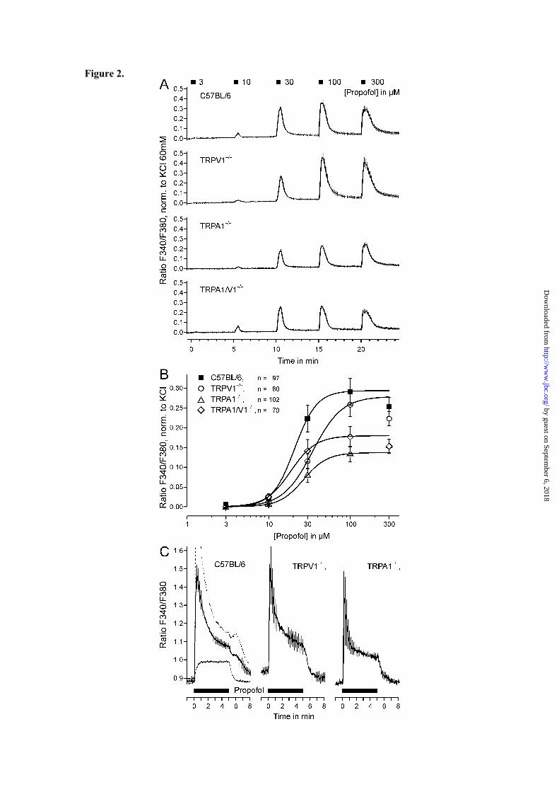
Profofol induces an increase in [Ca2+]i in cultured DRG neurons. To investigate excitatory effects of propofol on sensory neurons, we first applied clinically used formulations of propofol to cultured DRG neurons from adult C57BL/6 mice. A 1000-fold diluted solution of 2% Propofol-Lipuro® (112 µM) evoked a reversible increase in intracellular calcium [Ca2+]i in 79 of 137 neurons when applied for 3 min (Fig. 1A). Notably, this response showed a transient component of less than 60 s and a sustained one which persisted as long as propofol was applied. Lipofundin® (1000-fold diluted), the carrier for propofol in Propofol-Lipuro® induced no increase in [Ca2+]i. In contrast, a robust increase in [Ca2+]i was also induced by 100 µM propofol dissolved in external solution containing 0.2% dimethyl-sulfoxide (Fig. 1B). In absence of extracellular calcium, this increase in [Ca2+]i was completely abrogated (< 4% of control, n = 30, p < 0.001, t-test, Fig. 1B). A third application of propofol in the presence of extracellular calcium however elicited a reduced response, 57 ± 14% in size of the first. Thus, propofol apparently evokes an influx of extracellular calcium in a large subpopulation of DRG neurons. As demonstrated in figures 2A and B, 30 s application of propofol in concentrations of 10 µM or greater repeatedly induced an increase in [Ca2+]i in a concentration-dependent manner (p < 0.001 each, n = 89, t-test dependent samples, Fig. 2A). The corresponding EC50 value for wild-type DRG neurons was 21 ± 1 µM (Fig. 2B). As TRPV1 and TRPA1 were considered to be the main candidates mediating this propofol-induced increase in [Ca2+]i,, concentration-dependent activation by propofol was also investigated in DRG neurons of mutant mice lacking TRPV1, TRPA1 or both receptors
(Fig. 2A). Surprisingly, similar concentration-dependent responses were observed in these DRG neurons (p < 0.001 for ≥ 10 µM propofol, n = 80, 102 and 70 for the respective genotypes, t-test dependent samples). However, the efficacy of propofol was significantly reduced in neurons from knockout mice as compared to wild-type (ANOVA F(3,298)= 6.7, TRPV1−/− p = 0.036 , n = 80; TRPA1−/− p < 0.001, n = 102; TRPV1/A1−/− p = 0.003, n = 70; LSD post-hoc tests, Fig. 2B). While TRPA1−/− neurons generated significantly smaller propofol-induced responses as compared to TRPV1−/− neurons (p = 0.039, LSD post-hoc test), no significant differences were observed between neurons derived from double-knockout animals and the individual knockout animals. We also explored whether the functional properties of propofol-induced responses in DRG neurons are altered in TRPV1−/− and TRPA1−/− neurons. The activation and acute adaptation or desensitization were studied by the application of 100 µM propofol for 300 s. As demonstrated in figure 2C, the increase in calcium showed transient and sustained components in wild-type neurons as well as in neurons lacking TRPV1 or TRPA1. Notably, such a sustained component was not observed with other TRPV1 and TRPA1 agonists (data not shown), suggesting the involvement of another mechanism for propofol-induced activation of DRG neurons. Therefore, the excitatory effect of propofol was further examined by a combined genetic and pharmacological approach.
TRPV1, TRPA1, and GABAA receptors mediate the propofol-induced [Ca2+]i rise in DRG neurons. As displayed in figure 3A-D, three repetitive applications of 30 µM propofol were applied to wild-type C57BL/6, TRPV1−/−, TRPA1−/− and TRPV1/A1−/− neurons. In neurons from TRPA1−/− animals, the second response to propofol in presence of the TRPV1 antagonist BCTC (10 µM) was largely reduced (p < 0.001, n = 68, t-test, Fig. 3A). In neurons from TRPV1−/− animals, the TRPA1 antagonist HC-030031 (50 µM) strongly reduced the response to propofol (p < 0.001, n = 76, t-test. Fig. 3B). Both BCTC and HC-030031 did not increase calcium per se, and the propofol responses after washout of the antagonists were not different from the first one (p = 0.54 and p = 0.82, t-test). HC-030031 was recently reported to be a selective blocker of TRPA1 (8). However, we
by guest on September 6, 2018
http://ww
w.jbc.org/
Dow
nloaded from

found that HC-030031 (50 µM) moderately inhibited calcium increases induced by high external potassium (ANOVA F(1,139)=11.4, p < 0.001). In wild-type neurons, we observed an inhibition by 28% at 15 µM and by 42% at 50 µM HC-030031 (both p < 0.001, n = 23, LSD post-hoc tests, Suppl. 1a). Importantly, HC-030031 also inhibited potassium-induced calcium increases in neurons from TRPA1 knockout animals (inhibition by 10% at 15 µM and by 11% at 50 µM, p = 0.009 and p = 0.020, n = 26, LSD post-hoc tests, Suppl. 1b), although the inhibition was less concentration-dependent and pronounced as compared to neurons from wild-type animals (p < 0.001, LSD post-hoc test). Therefore, the TRPA1-related inhibition by HC-030031 of the propofol response could be overestimated. BCTC, in contrast to HC030031, did not reduce calcium influx evoked by high external potassium (data not shown).
Activation of GABAA receptors by propofol in the CNS is well known and putatively contributes to the hypnotic effects of propofol (28;29). As GABAA receptors are expressed in primary sensory neurons, their activation was considered a possible mechanism mediating the residual, TRPA1/TRPV1-independent action of propofol in DRG neurons. Using the same protocol as for the TRP channels, the contribution of GABAA receptors was investigated using the non-competitive GABAA receptor antagonist picrotoxin (PTX). In neurons from wild-type C57BL/6 mice, 100 µM PTX reduced the response to propofol by 48% (p = 0.019, n = 46, t-test, Fig. 3C). In neurons from TRPV1/A1 double knockout mice, the response was reduced by 83% (p < 0.001, n = 82, t-test, Fig. 3D) and the remaining calcium increase in presence of propofol and PTX was minimal (3.3 ± 0.6 nM). Thus, GABAA receptors indeed appear to mediate propofol-induced activation of sensory neurons.
We next aimed at analyzing the relative contributions of TRPV1, TRPA1 and GABAA receptors to the effect of propofol on DRG neurons. To this end, the sensitivity of wild-type DRG neurons to propofol (30 µM), GABA (10 µM), capsaicin (100 nM), and the TRPA1 agonist acrolein (10 µM) was consecutively investigated (Fig. 4A). The responsiveness to propofol was strongly correlated to the magnitude of responses evoked by GABA (r = 0.67, p < 0.001, n = 83). A similar correlation with propofol was found for responses evoked by acrolein (r = 0.82, p < 0.001), but not by
capsaicin (r = –0.16, p = 0.51, Fig 4B). However, there was also a significant negative correlation between GABA and capsaicin responses in wild-type neurons (r = –0.23, p = 0.04), which may mask the propofol-capsaicin correlation. In fact, a significant positive correlation between propofol and capsaicin (100 nM) resulted if both TRPA1 and GABAA receptors were genetically and pharmacologically silenced (r = 0.62, p < 0.001, n = 502, Fig 4D). In presence of picrotoxin (100 µM), still 18% of all investigated TRPA1−/−
neurons (n = 502) responded to propofol (50 µM). This fraction increased to 38% after conditioning treatment with the protein kinase C (PKC) activator PMA (100 nM for 60 s). Furthermore, the fraction of propofol-sensitive neurons which also responded to capsaicin increased from 45% to 93% following treatment with PMA. Synchronously, the propofol-induced calcium increase was augmented to 306% of the original response (p < 0.001, t-test, Fig. 4C). GABA (10 µM) was completely ineffective under these experimental conditions (Fig. 4D).
Propofol evokes inward currents in
mouse DRG neurons that are mediated by TRPV1, TRPA1, and GABAA receptors. To corroborate the data obtained by calcium imaging and to study the effects of propofol in more detail, whole-cell voltage-clamp was performed on DRG neurons from wild-type C57BL/6 and TRPV1/A1−/− mice. DRG neurons were characterized due to their sensitivity to propofol (300 µM), GABA (50 µM), capsaicin (1 µM) and acrolein (50 µM). The relatively high concentration of propofol was applied to obtain comparable responses. As demonstrated in figure 5A and B, DRG neurons generated propofol-induced inward currents with various amplitudes and heterogeneous kinetic properties. In wild-type neurons, 300 µM propofol evoked inward currents in 75% (44/59) of all neurons tested with a mean current amplitude of 10.6 ± 1.1 pA/pF. Strikingly, both the prevalence (82%, 22/27) and the mean amplitude (14.3 ± 3.5 pA/pF) of propofol-induced currents were not different in TRPV1/A1−/− neurons as compared to wild-type (p = 0.49 and p = 0.23, t-test, Fig. 5C, D). Only 7 (16%) of all propofol-sensitive wild-type neurons were also capsaicin-sensitive, these neurons generated small propofol-induced currents (6.8 ± 1.9 pA/pF). Capsaicin-induced currents were observed in 32% (19/59) of all
by guest on September 6, 2018
http://ww
w.jbc.org/
Dow
nloaded from

wild-type neurons tested. Thus, the majority (12, 63%) of these neurons did not respond to propofol. Similarly, 36.4% (16/44) of all propofol-sensitive wild-type neurons were acrolein-sensitive and generated small propofol-induced currents (7.4 ± 1.3 pA/pF, p = 0.74, t-test, Fig. 5E). In contrast to capsaicin-sensitive neurons, the majority of the acrolein-sensitive neurons (31%, 18/59) also generated propofol-induced currents (89%, 16/18). As demonstrated in figure 5A, acrolein-sensitive neurons typically generated propofol-induced currents with a resurging current following the application of propofol. Furthermore, 55% (24/44) of all propofol-sensitive wild-type neurons did not display sensitivity to capsaicin or acrolein but generated significantly larger propofol-induced currents than neurons that were sensitive to capsaicin or acrolein (13.4 ± 1.6 pA/pF, p = 0.002, t-test, Fig. 5E), suggesting a negative correlation between propofol/GABA responses and the effects of the irritants. GABA 50 µM induced inward currents in 81.5% of wild-type (44/54) as well as TRPV1/A1−/− neurons (22/27). All propofol-sensitive neurons also responded to GABA (wild-type 40/40, TRPV1/A1−/− 22/22). Furthermore, a strong correlation was found for peak current amplitudes evoked by propofol and GABA in neurons from both wild-type (r = 0.86, p < 0.001, Fig. 5F) and TRPV1/A1−/− animals (r = 0.98, p < 0.001, Fig. 5G). Propofol-induced inward currents in neurons derived from TRPV1/A1−/− animals were completely blocked by the GABAA receptor blocker picrotoxin (100 µM, n = 7, Fig. 5H).
Propofol activates and blocks TRPA1. It
was recently suggested that TRPA1 is the only TRP receptor in DRG neurons that is activated by propofol (5). We found that rat TRPA1 expressed in HEK293t cells is indeed activated by propofol in a concentration-dependent and reversible manner (Fig. 6A). Propofol induced inward-currents in TRPA1-HEK293t cells at 0.3µM and higher concentrations, thus displaying a comparable potency on TRPA1 as has been described for GABAA receptors (30). At concentrations above 10 µM, propofol consistently activated currents that appeared to inactivate during stimulation (Fig. 6B). These currents were associated with resurging currents following termination of propofol (see 100 µM propofol in Fig. 6A). This phenomenon has been reported previously (5), and a possible
mechanism might be a propofol-induced pore-block of TRPA1 in the open state. To test this hypothesis, propofol was co-applied when an acrolein-induced inward current had reached steady-state (Fig. 6C). Indeed, 30 µM propofol blocked acrolein-induced currents by 89.2 ± 2.8%, n = 7 (p = 0.018 Wilcoxon, Fig. 6C, D). Figure 6 further demonstrates that propofol-induced currents in TRPA1-HEK293t cells were completely blocked by the TRPA1 antagonist HC-030031 (100 µM, n = 5, Fig. 6E). We also examined the effect of propofol on other TRP receptors supposed to be functionally expressed in sensory neurons or to have an impact on peripheral nociception. As demonstrated by representative current traces in figure 6F, 300 µM propofol activated robust inward currents in HEK293t transiently expressing rat TRPV1 and TRPV3. While TRPM8-HEK293t cells produced minimal propofol-induced currents (< 50 pA, n= 8), no response was observed in TRPV2, TRPV4 or non-transfected HEK293t cells (n = 9 - 14 for each receptor type). In case of TRPV3, current amplitudes regularly increased upon repeated application of propofol (Suppl. 2). This sensitization upon repeated stimulation is a typical feature of TRPV3 (31).
Propofol activates and desensitizes TRPV1. The action of propofol on TRPV1 was further investigated on the recombinant rat TRPV1 heterologously expressed in HEK293t cells. As demonstrated in figure 7A, propofol activated TRPV1 in a concentration-dependent manner with an EC50 of 90 ± 20 µM (n = 6 - 10 for each concentration, Fig. 7B). Propofol-induced inward currents were completely blocked by the competitive TRPV1 antagonist capsazepine (10 µM, 99 ± 3%, n = 10, Fig. 7C). Unlike most TRPV1 agonists, propofol did not sensitize TRPV1 when co-applied in half-maximal effective concentrations together with another TRPV1-agonist. As demonstrated in figure 7D and E, co-application of capsaicin (5 nM) and propofol (100 µM) resulted in even smaller currents than application of capsaicin alone (58.4 ± 13.7% reduction, p = 0.003, n = 8, t-test). The propofol sensitivity of TRPV1 was strongly enhanced by the activation of PKC by PMA, an effect that had previously proven very effective in calcium imaging experiments on DRG neurons (Fig. 4C). When the initial application of 100 µM propofol was followed by a 3 min lasting superfusion of the PKC activator PMA (1 µM), the inward currents activated by a
by guest on September 6, 2018
http://ww
w.jbc.org/
Dow
nloaded from

second application of propofol displayed a 28 ± 6.8-fold increase in peak amplitudes (p = 0.001, t-test, n = 8, Fig. 7F). This PMA-induced sensitization of propofol-induced inward currents was abrogated in the TRPV1 double-mutant S502A/S800A, the putative PKC-phosphorylation sites of TRPV1 (1.6 ± 0.7-fold increase, p = 0.42, t-test, n = 6, Fig. 7E).We also explored the ability of propofol to desensitize TRPV1. A pronounced desensitization and tachyphylaxis is a typical feature of TRPV1 when activated repeatedly. This process is induced by a rise in [Ca2+]i and can thus be significantly reduced by the removal of extracellular Ca2+ ions (21). When applied repeatedly, 300 µM propofol induced inward currents displaying a strong desensitization (ANOVA, F(1, 32)=12.392, p = 0.001, Fig. 7G). The responses to subsequent propofol applications were reduced to 72.3 ± 14.2% and 48.8 ± 10.2% in the presence of extracellular calcium and not significantly different in the absence of extracellular calcium (53.7 ± 20.1%, n = 5 and 37.0 ± 24.0%, n = 7, p = 0.66, LSD post-hoc test, Fig. 7G, H). In contrast, repeated applications of capsaicin (100 nM) in the absence of extracellular Ca2+ produced stable inward currents without any sign of desensitization (2nd application to 99.9 ± 5.5% and 3rd application to 103.4 ± 10.9%, n = 5). Intriguingly, continuous application of propofol (30 µM) during the intervals (3 min) of the capsaicin-applications resulted in a cross-desensitization of the capsaicin-induced currents even in absence of extracellular Ca2+ (2nd application to 63.8 ± 6.2% and 3rd application to 48.6 ± 6.7%, p = 0.001 and 0.012, Wilcoxon, n = 8).
Propofol modulates TRPA1 by mechanisms other than covalent modification of reactive cysteine residues or interacting with residues within TM5. Considering our data and previous reports, propofol obviously activates several TRP channels with the greatest potency for TRPA1 followed by TRPV1. The relatively slow kinetics of propofol-induced TRPA1-currents resemble the properties of currents activated by substances considered to activate TRPA1 by a covalent modification of reactive cysteine residues, i.e. acrolein and mustard oil (32;33). Notably, the same mechanism (i.e. a covalent modification of cystein residues) was demonstrated also to mediate a sensitization or activation of TRPV1 (37, 38). We therefore asked if propofol modulates TRP channels by
this mechanism and first studied mutant constructs of human TRPA1 (hTRPA1) in which the responsible cysteine residues were replaced by serine (hTRPA1-C621S/C641S/C665S). As previously demonstrated, this triple-mutant was completely insensitive to 100 µM acrolein (Fig. 8B). However, it still generated large inward currents following application of 300 µM propofol (697.5 ± 263.3 pA, n= 8) and the previously established TRPA1-agonist carvacrol (250 µM) (Fig. 8B). Thus, modification of cysteine residues is unlikely to be the common mechanism for propofol-induced activation of TRP channels. While performing the experiments on human TRPA1, we observed a prominent species difference between wild type rat and human TRPA1 with regard to propofol-induced inward currents. The above-mentioned resurging currents, which were observed for rTRPA1 following termination of propofol in concentrations above 10 µM (Fig. 6A) were missing for hTRPA1 even with 300 µM propofol (Fig. 8A, n= 7 for wild type hTRPA1). A similar species difference between rodent and human TRPA1 was recently demonstrated for activation by menthol; mouse TRPA1 (mTRPA1) is both activated and blocked by menthol while hTRPA1 is only activated (22). This differential effect might be determined by the pore region including the transmembrane domain 5 (TM5). Based on comparative studies on mammalian TRPA1 and the menthol-insensitive drosophila TRPA1, Xiao and co-workers also identified specific residues within TM5 being crucial for both menthol and thymol sensitivity of TRPA1 (22). Notably, menthol, thymol and its isomer carvacrol all display close structural similarities to propofol (Fig. 8C). Furthermore, menthol and propofol share common interaction sites for activation of GABAA receptors (39, 40). We therefore pursued the possibility that propofol employs the same mechanisms as menthol and thymol to activate TRPA1 and next explored mutant constructs of mouse and human TRPA1. Wild type mTRPA1 behaved similar to rTRPA1 and displayed propofol-induced inward currents at 100 µM and inhibition at 300 µM, followed by resurging currents (Fig. 8D, n = 6). The chimera mTRPA1-hTM5-6 however, in which the region TM5 through TM6 from hTRPA1 was introduced, behaved like wild type hTRPA1 and displayed large propofol-induced currents without a resurging current following application (Fig. 8E, n = 5). Accordingly, the reverse chimera hTRPA1-mTM5-6 behaved like
by guest on September 6, 2018
http://ww
w.jbc.org/
Dow
nloaded from

wild type mTRPA1 and displayed propofol-induced currents at 100 µM and inhibition at 300 µM, followed by prominent resurging currents (Fig. 8F, n = 6). We next examined if residues within TM5, which were demonstrated to be required for activation by menthol and thymol (22) are also required for propofol sensitivity of TRPA1. Surprisingly, 300 µM propofol induced large inward currents in Hek293t cells expressing the menthol-insensitive mutants mTRPA1-S876V/T877L (Fig. 8G, 1909.6 ± 808.6 pA, n= 5) and hTRPA1-S873V/T874L (Fig. 8H, 493.4 ± 162.0 pA, n= 5) (Fig.8 H). Notably, mTRPA1-S876V/T877L generated propofol-induced current lacking a resurging current following application. In contrast to propofol, we found that both mutants were insensitive to carvacrol (data not shown). Thus despite similarities in chemical structure, propofol obviously employs other or additional mechanisms to gate TRPA1 as compared to carvacrol, thymol and menthol.
Propofol induces a TRPV1 and TRPA1 but not GABAA-dependent release of CGRP from isolated peripheral nerves. The experiments performed on cultured cells suggest that the excitatory effect of propofol is largely carried by TRPA1 and GABAA receptors , both masking a substantial contribution of TRPV1 that may at least become relevant under conditions of inflammatory sensitization. However, DRG neurons in culture are just a model of their nociceptive nerve endings that express functional TRPA1 and TRPV1 channels to mediate pain upon activation. In contrast, it is quite uncertain whether GABA is able to excite peripheral nerve fibers to induce pain as well. To address this question, we now aimed at investigating whether propofol is able to activate peripheral nerve endings and induce a release of the proinflammatory neuropeptide calcitonin gene–related peptide (CGRP). Activation of nociceptive neurons leads to a Ca2+-dependent release of CGRP which contributes to neurogenic inflammation, to peripheral sensitization of nociceptive afferents and to central sensitization in the spinal cord (34;35). Isolated mouse sciatic nerves were stimulated with clinically used emulsions of propofol or with Lipofundin®. Propofol-Lipuro® 1% or Disoprivan® 1%, tenfold diluted in SIF (= 5.6 mM propofol), evoked a significant release of CGRP (both p = 0.012 and n = 8, Wilcoxon) with similar efficacies (p = 0.40, U-test). In
contrast, tenfold diluted Lipofundin® did not stimulate CGRP release (Fig. 9A). In TRPV1/A1 double knockout animals, no CGRP release was observed upon stimulation with Propofol-Lipuro® 1% (p = 0.27, n = 8, Wilcoxon). However, high external KCl (60 mM) evoked similar responses to those observed in wildtypes (data not shown). The diluted clinical emulsions are known to contain “free” propofol in the aqueous phase at concentrations of 5 µM and 6.7 µM, respectively (36). These concentrations and 100-fold higher ones of propofol in aqueous solution (using DMSO as a solubilizer) did not evoke a significant CGRP release from the isolated nerve preparation (data not shown). Propofol at 5.6 mM, the same concentration as nominally contained in both tenfold diluted clinical emulsions, induced a significant response of about the same magnitude as diluted Propofol-Lipuro® and Disoprivan® (Fig. 9A). This response was clearly concentration-dependent as the ten-fold higher propofol concentration 56 mM (as in the undiluted original clinical emulsions) caused a five-fold greater CGRP release (Fig. 9B). Surprisingly, the application of 100 µM GABA did not evoke any release of CGRP from sciatic nerves (p = 0.73, n = 7, Wilcoxon). The exposure of hindpaw skin, another established model for activation of peripheral nerve endings, to 10 mM GABA also did not elicit any CGRP release (p = 0.26, n = 6, Wilcoxon, lower concentrations for GABA not shown, Fig. 9C). , Similarly, the selective GABAA receptor agonist muscimol did not elicit any CGRP release (1 µM and 10 µM, n = 4 each, Fig. 9D). Finally, GABA (100 µM) did not amplify the release of CGRP induced by high potassium (60 mM). Correspondingly, the selective GABAB-receptor agonist baclofen (100 µM) did not inhibit high potassium-induced release of CGRP, suggesting that GABAB receptor stimulation is unlikely to mask GABAA–evoked CGRP release (Suppl. 3).
Propofol but not GABA induces an intense pain upon intracutaneous injection. Notably, stimulated CGRP release only covers the peptidergic subpopulation of peripheral nociceptors and propofol-induced injection pain is a phenomenon observed in humans. Therefore, we finally employed psychophysics on volunteers (the authors of the study, n = 5) with the intention to further narrow down the roles of TRP channels and GABAA receptors in
by guest on September 6, 2018
http://ww
w.jbc.org/
Dow
nloaded from

mediating propofol-induced injection pain. 1% Propofol-Lipuro®, Lipofundin®, buffered GABA 10 mM or isotonic saline were injected intracutaneously at separate marked sites of the volar forearm (50 µl). Drugs were applied in a double-blinded fashion and in random order at an interval of 15 min. While propofol injection was reported more or less painful for several minutes by all subjects (on a numerical scale), no painful sensation was reported by anyone of the subjects after injection of Lipofundin®, GABA or isotonic saline.
DISCUSSION
Propofol is one of the most widely used general anesthetics in clinical practice. We show that propofol activates the TRP receptors TRPA1 and TRPV1 in DRG neurons and in HEK293t cells. In addition, GABAA receptors substantially contribute to the propofol-induced response in DRG neurons as measured by both calcium imaging and whole-cell patch clamp. Propofol, but not GABA also evoke release of CGRP from isolated peripheral nerve of wild type but not TRPV1/A1-deficient mice. Finally, intracutaneous injection of propofol induced an intense pain in humans, an effect that could not be mimicked by GABA.
Activation of TRP channels by propofol.
TRPA1 has been reported to be responsible for the activation of nociceptors by general anesthetics, including propofol (5). TRPA1 was identified as the principal molecular determinant of propofol-induced pain in two acute animal models of pain, nocifensive behavior following nasal epithelial application and vascular pain. We confirm with the following findings that TRPA1 significantly contributes to propofol-induced activation of nociceptors: 1. propofol activated recombinant TRPA1, 2. in DRG neurons, the propofol-induced activation correlated well with activation by the TRPA1 agonist acrolein, and it could be antagonized by the TRPA1-inhibitor HC-030031 and 3. deletion of TRPA1 reduced the activation by propofol in TRPA1−/− as compared to both WT and TRPV1−/− neurons. Matta and colleagues reported that DRG neurons from TRPA1 knockout animals failed to respond to propofol 100 µM (5). In contrast, we performed several experiments indicating that propofol induces a significant activation of TRPV1: 1. propofol
activated inward currents in TRPV1-transfected HEK293t cells, 2. the TRPV1-antagonists BCTC and capsazepine inhibited propofol induced responses in HEK293t cells and DRG neurons, 3. propofol activated DRG neurons in the absence of TRPA1 and GABAA receptor contributions and 4. propofol-induced responses were sensitized by activation of PKC. In DRG neurons, the activation of TRPV1 by propofol appeared to be masked by the more obvious TRPA1 and GABAA-mediated effects. These results are reminiscent of a recent report showing that volatile anesthetics activate TRPV1 following phosphorylation through PKC (16). Thus, activation of TRPV1 by general anesthetics may be more important under conditions of inflammation due to a PKC-mediated phosphorylation of TRPV1 triggered by inflammatory mediators such as bradykinin and prostaglandins. The activation of TRPA1 and TRPV1 by propofol might be indicative for a conserved mechanism of chemical activation. A covalent modification of cysteine residues was previously identified as a common mechanism for activation of TRPA1 and TRPV1 (32;33;37;38). We found that the acrolein-insensitive mutant hTRPA1-C621S/C641S/C665S is sensitive to propofol and thus TRP channels most likely are not gated by propofol via covalent modification. The retained carvacrol sensitivity of hTRPA1-C621S/C641S/C665S is not surprising regarding the similar chemical structures of propofol and carvacrol. Differential effects of these substances on hTRPA1 and mTRPA1 indeed appear to be encoded by the same molecular determinants within TM5, i.e. a bimodal action on mTRPA1 with activation and block as compared to only an activation of hTRPA1. However, the previously demonstrated insensitivity of mTRPA1-S876/T877L and hTRPA1-S873V/T874L to menthol and thymol (22), did not apply for propofol which induced large inward currents on both mutants. In contrast, we found that both mTRPA1-S876/T877L and hTRPA1-S873V/T874L were also insensitive to carvacrol. Notably, menthol has general anesthetic properties and activates GABAA receptors via interaction sites which are also known to be required for activation by propofol (39;40). Taking into account the striking similarities between propofol and menthol in chemical structures and mode of action on GABAA receptors, it is rather
by guest on September 6, 2018
http://ww
w.jbc.org/
Dow
nloaded from

surprising that the mechanisms for activation of TRPA1 by propofol are apparently distinct from those required for activation by menthol, thymol and carvacrol. While the mechanism for propofol-induced activation of TRPA1 thus remains to be identified, our data suggest that propofol seems to be an interesting substance for further studies into the molecular pharmacology of TRPA1 and other TRP channels.
Activation of GABAA receptors by propofol. Prolonged application of propofol produced a biphasic response in DRG neurons with a non-adapting component distinct from the desensitization reported for both TRPV1 and TRPA1 activation by agonists in the presence of calcium (41;42). About half of the activation by propofol was concentration-dependently retained in TRPV1/A1 double knockout animals, clearly in contrast with the previously reported results (5). This remaining action of propofol was highly correlated with the current or calcium influx elicited by GABA and was abrogated by the GABAA receptor antagonist picrotoxin. Furthermore, the lack of a major rightward shift of the propofol concentration-response curve in TRPA1−/− or TRPV1−/− neurons suggests a similar potency of propofol on GABAA receptors and TRP channels. Thus, our cellular data clearly suggest that GABAA receptors account for a significant proportion of the propofol sensitivity of sensory neurons. However, we also show that propofol but not GABA or the selective GABAA receptor agonist muscimol evoke a release of CGRP from sciatic nerves in a previously established model (43;44). Similarly, GABA failed to amplify, and the selective GABAB-receptor agonist baclofen failed to inhibit high potassium-induced CGRP release from hindpaw skin. Additionally, intracutaneous injection of GABA did not elicit a painful sensation in humans. Considering the existing literature on the action of GABA on peripheral sensory neurons, these results are in part controversial. In contrast to the inhibitory profile of GABA in the central nervous system, GABA is known to induce a depolarization and an increased excitability in sensory neurons from frogs (45), cats (46), rats (46-48) and humans (49). Accordingly, GABAA receptors are expressed in primary afferent neurons with a significant co-expression with nociceptive markers such as TRPV1 (50-53). Moreover, peripheral injection of GABA or the GABAA receptor agonist muscimol was reported to
induce or increase pain-like behaviour in rodents (54;55). The depolarizing effect of GABA on sensory neurons is due to a high intracellular chloride concentration resulting from expression of the NKCC1 co-transporter (56). While GABAA receptors appear to be expressed in the majority of DRG neurons, it was suggested that only those neurons expressing the Cav3.2/α1H T-type calcium channel are able to generate GABA-induced action potentials (57). Accordingly, a recent report from Carr and co-workers demonstrated a GABAA receptor-mediated increase in excitability in only ~40% of C-fibers in human peripheral nerves. Notably, the same study only reported an increased electrical excitability and not an activation of C-fibers by GABA (49). This notion is in good agreement with the lack of injection pain following intracutaneous injection of GABA in our study, collectively suggesting that GABA itself does not activate action potentials in human C-fibers. In addition, our data add to a rather conflicting literature on the GABA effects on neuropeptide release from C-fibers comprising GABAA mediated inhibition (59), facilitation (60;61) and no effect (62;63). Taken together, our data and a considerable number of previous reports do not support the possibility that GABAA receptors mediate a substantial activation of peripheral sensory neurons or a release of neuropeptides. Additionally, our data do not support the possibility that GABAB receptor stimulation masks any potential GABAA receptor evoked pronociceptive effect. However, given the complexity of GABAergic signaling in sensory neurons, a pro-nociceptive action of propofol mediated by GABAA receptors cannot be totally excluded.
Clinical relevance and conclusions. Our
data reveal TRPA1 and TRPV1 as main mediators of propofol-induced pain and release of neuropeptides. The release of neuropeptides from peripheral and central terminals of sensory neurons induces vascular leakage and dilatation and is thought to contribute to neurogenic inflammation in the periphery and to central sensitization in the spinal dorsal horn. Although corresponding data in a clinical setting is currently lacking, it seems conceivable that propofol can cause a clinically significant sensitization of nociceptors by directly activating or sensitizing TRPA1 and TRPV1. As this condition might persist beyond the intra-operative period, it might prove relevant in the
by guest on September 6, 2018
http://ww
w.jbc.org/
Dow
nloaded from

etiology of post-operative and persistent pain. If this concept was supported by clinical data, it might alter the understanding of the consequences of the intra-operative use of propofol and other general anesthetics with a
similar profile. In this regard, TRPV1 and/or TRPA1 antagonists might prove to be helpful analgesic adjuncts for the prevention and the treatment of post-operative pain.
REFERENCES 1. Picard, P. and Tramer, M. R. (2000) Anesth.Analg. 90, 963-969
2. Stokes, D. N. , Robson, N. , and Hutton, P. (1989) Br.J Anaesth. 62, 202-203
3. Klement, W. and Arndt, J. O. (1991) Br.J Anaesth. 67, 281-284
4. Doenicke, A. W. , Roizen, M. F. , Rau, J. , Kellermann, W. , and Babl, J. (1996) Anesth.Analg. 82, 472-474
5. Matta, J. A. , Cornett, P. M. , Miyares, R. L. , Abe, K. , Sahibzada, N. , and Ahern, G. P. (2008) Proc.Natl.Acad.Sci.U.S.A 105, 8784-8789
6. Story, G. M. , Peier, A. M. , Reeve, A. J. , Eid, S. R. , Mosbacher, J. , Hricik, T. R. , Earley, T. J. , Hergarden, A. C. , Andersson, D. A. , Hwang, S. W. , McIntyre, P. , Jegla, T. , Bevan, S. , and Patapoutian, A. (2003) Cell 112, 819-829
7. Jordt, S. E. , Bautista, D. M. , Chuang, H. H. , McKemy, D. D. , Zygmunt, P. M. , Hogestatt, E. D. , Meng, I. D. , and Julius, D. (2004) Nature 427, 260-265
8. McNamara, C. R. , Mandel-Brehm, J. , Bautista, D. M. , Siemens, J. , Deranian, K. L. , Zhao, M. , Hayward, N. J. , Chong, J. A. , Julius, D. , Moran, M. M. , and Fanger, C. M. (2007) Proc.Natl.Acad.Sci.U.S.A 104, 13525-13530
9. Bautista, D. M. , Jordt, S. E. , Nikai, T. , Tsuruda, P. R. , Read, A. J. , Poblete, J. , Yamoah, E. N. , Basbaum, A. I. , and Julius, D. (2006) Cell 124, 1269-1282
10. Obata, K. , Katsura, H. , Mizushima, T. , Yamanaka, H. , Kobayashi, K. , Dai, Y. , Fukuoka, T. , Tokunaga, A. , Tominaga, M. , and Noguchi, K. (2005) J Clin.Invest 115, 2393-2401
11. Caterina, M. J. , Schumacher, M. A. , Tominaga, M. , Rosen, T. A. , Levine, J. D. , and Julius, D. (1997) Nature 389, 816-824
12. Caterina, M. J. , Leffler, A. , Malmberg, A. B. , Martin, W. J. , Trafton, J. , Petersen-Zeitz, K. R. , Koltzenburg, M. , Basbaum, A. I. , and Julius, D. (2000) Science 288, 306-313
13. Davis, J. B. , Gray, J. , Gunthorpe, M. J. , Hatcher, J. P. , Davey, P. T. , Overend, P. , Harries, M. H. , Latcham, J. , Clapham, C. , Atkinson, K. , Hughes, S. A. , Rance, K. , Grau, E. , Harper, A. J. , Pugh, P. L. , Rogers, D. C. , Bingham, S. , Randall, A. , and Sheardown, S. A. (2000) Nature 405, 183-187
14. Bandell, M. , Story, G. M. , Hwang, S. W. , Viswanath, V. , Eid, S. R. , Petrus, M. J. , Earley, T. J. , and Patapoutian, A. (2004) Neuron 41, 849-857
by guest on September 6, 2018
http://ww
w.jbc.org/
Dow
nloaded from

15. Leffler, A. , Fischer, M. J. , Rehner, D. , Kienel, S. , Kistner, K. , Sauer, S. K. , Gavva, N. R. , Reeh, P. W. , and Nau, C. (2008) J Clin.Invest 118, 763-776
16. Cornett, P. M. , Matta, J. A. , and Ahern, G. P. (2008) Mol.Pharmacol. 74, 1261-1268
17. Tsutsumi, S. , Tomioka, A. , Sudo, M. , Nakamura, A. , Shirakura, K. , Takagishi, K. , and Kohama, K. (2001) Neurosci.Lett. 312, 45-49
18. Perkins, F. M. and Kehlet, H. (2000) Anesthesiology 93, 1123-1133
19. Zimmermann, M. (1983) Pain 16, 109-110
20. Kwan, K. Y. , Allchorne, A. J. , Vollrath, M. A. , Christensen, A. P. , Zhang, D. S. , Woolf, C. J. , and Corey, D. P. (2006) Neuron 50, 277-289
21. Mohapatra, D. P. and Nau, C. (2003) J Biol.Chem. 278, 50080-50090
22. Xiao, B. , Dubin, A. E. , Bursulaya, B. , Viswanath, V. , Jegla, T. J. , and Patapoutian, A. (2008) J Neurosci 28, 9640-9651
23. Mohapatra, D. P. , Wang, S. Y. , Wang, G. K. , and Nau, C. (2003) Mol.Cell Neurosci 23, 314-324
24. Dittert, I. , Vlachova, V. , Knotkova, H. , Vitaskova, Z. , Vyklicky, L. , Kress, M. , and Reeh, P. W. (1998) J.Neurosci.Methods 82, 195-201
25. Poenie, M. and Tsien, R. (1986) Prog.Clin.Biol.Res. 210, 53-56
26. Bretag, A. H. (1969) Life Sci. 8, 319-329
27. Averbeck, B. and Reeh, P. W. (2001) Neuropharmacology 40, 416-423
28. Collins, G. G. (1988) Br.J Pharmacol. 95, 939-949
29. Concas, A. , Santoro, G. , Serra, M. , Sanna, E. , and Biggio, G. (1991) Brain Res. 542, 225-232
30. Rudolph, U. and Antkowiak, B. (2004) Nat.Rev.Neurosci. 5, 709-720
31. Benham, C. D. , Gunthorpe, M. J. , and Davis, J. B. (2003) Cell Calcium 33, 479-487
32. Hinman, A. , Chuang, H. H. , Bautista, D. M. , and Julius, D. (2006) Proc.Natl.Acad.Sci.U.S.A 103, 19564-19568
33. Macpherson, L. J. , Dubin, A. E. , Evans, M. J. , Marr, F. , Schultz, P. G. , Cravatt, B. F. , and Patapoutian, A. (2007) Nature 445, 541-545
34. Planells-Cases, R. , Garcia-Sanz, N. , Morenilla-Palao, C. , and Ferrer-Montiel, A. (2005) Pflugers Arch. 451, 151-159
35. Mogil, J. S. , Miermeister, F. , Seifert, F. , Strasburg, K. , Zimmermann, K. , Reinold, H. , Austin, J. S. , Bernardini, N. , Chesler, E. J. , Hofmann, H. A. , Hordo, C. , Messlinger, K. , Nemmani, K. V. , Rankin, A. L. , Ritchie, J. , Siegling, A. , Smith, S. B. , Sotocinal, S. , Vater, A. , Lehto, S. G. , Klussmann, S. , Quirion, R. , Michaelis, M. , Devor, M. , and Reeh, P. W. (2005) Proc.Natl.Acad.Sci.U.S.A 102, 12938-12943
by guest on September 6, 2018
http://ww
w.jbc.org/
Dow
nloaded from

36. Yamakage, M. , Iwasaki, S. , Satoh, J. , and Namiki, A. (2005) Anesth.Analg. 101, 385-8, table
37. Salazar, H. , Llorente, I. , Jara-Oseguera, A. , Garcia-Villegas, R. , Munari, M. , Gordon, S. E. , Islas, L. D. , and Rosenbaum, T. (2008) Nat.Neurosci 11, 255-261
38. Chuang, H. H. and Lin, S. (2009) Proc.Natl.Acad.Sci.U.S.A 106, 20097-20102
39. Watt, E. E. , Betts, B. A. , Kotey, F. O. , Humbert, D. J. , Griffith, T. N. , Kelly, E. W. , Veneskey, K. C. , Gill, N. , Rowan, K. C. , Jenkins, A. , and Hall, A. C. (2008) Eur.J Pharmacol. 590, 120-126
40. Zhang, X. B. , Jiang, P. , Gong, N. , Hu, X. L. , Fei, D. , Xiong, Z. Q. , Xu, L. , and Xu, T. L. (2008) PLoS.One. 3, e3386
41. Koplas, P. A. , Rosenberg, R. L. , and Oxford, G. S. (1997) J Neurosci. 17, 3525-3537
42. Wang, Y. Y. , Chang, R. B. , Waters, H. N. , McKemy, D. D. , and Liman, E. R. (2008) J Biol.Chem. 283, 32691-32703
43. Sauer, S. K. , Reeh, P. W. , and Bove, G. M. (2001) Eur.J Neurosci. 14, 1203-1208
44. Fischer, M. J. and Reeh, P. W. (2007) Eur.J Neurosci. 25, 3570-3575
45. Barker, J. L. , Nicoll, R. A. , and Padjen, A. (1975) J Physiol 245, 521-536
46. Gallagher, J. P. , Higashi, H. , and Nishi, S. (1978) J Physiol 275, 263-282
47. Desarmenien, M. , Santangelo, F. , Loeffler, J. P. , and Feltz, P. (1984) Exp.Brain Res. 54, 521-528
48. Deschenes, M. , Feltz, P. , and Lamour, Y. (1976) Brain Res. 118, 486-493
49. Carr, R. W. , Sittl, R. , Fleckenstein, J. , and Grafe, P. (2010) PLoS.One. 5, e8780
50. Furuyama, T. , Sato, M. , Sato, K. , Araki, T. , Inagaki, S. , Takagi, H. , and Tohyama, M. (1992) Brain Res.Mol.Brain Res. 12, 335-338
51. Ma, W. , Saunders, P. A. , Somogyi, R. , Poulter, M. O. , and Barker, J. L. (1993) J Comp Neurol. 338, 337-359
52. Persohn, E. , Malherbe, P. , and Richards, J. G. (1991) Neuroscience 42, 497-507
53. Peeters, P. J. , Aerssens, J. , de Hoogt, R. , Stanisz, A. , Gohlmann, H. W. , Hillsley, K. , Meulemans, A. , Grundy, D. , Stead, R. H. , and Coulie, B. (2006) Physiol Genomics 24, 252-263
54. Ault, B. and Hildebrand, L. M. (1994) Neuropharmacology 33, 109-114
55. Carlton, S. M. , Zhou, S. , and Coggeshall, R. E. (1999) Neuroscience 93, 713-722
56. Sung, K. W. , Kirby, M. , McDonald, M. P. , Lovinger, D. M. , and Delpire, E. (2000) J Neurosci 20, 7531-7538
57. Aptel, H. , Hilaire, C. , Pieraut, S. , Boukhaddaoui, H. , Mallie, S. , Valmier, J. , and Scamps, F. (2007) Mol.Cell Neurosci 36, 293-303
by guest on September 6, 2018
http://ww
w.jbc.org/
Dow
nloaded from

58. Spitzer, M. J. , Reeh, P. W. , and Sauer, S. K. (2008) Neuroscience 151, 836-842
59. Go, V. L. and Yaksh, T. L. (1987) J Physiol 391, 141-167
60. Santicioli, P. , Tramontana, M. , Del Bianco, E. , Maggi, C. A. , and Geppetti, P. (1991) Life Sci. 48, L69-L72
61. Lao, L. and Marvizon, J. C. (2005) Neuroscience 130, 1013-1027
62. Malcangio, M. and Bowery, N. G. (1993) J Pharmacol.Exp.Ther. 266, 1490-1496
63. Riley, R. C. , Trafton, J. A. , Chi, S. I. , and Basbaum, A. I. (2001) Neuroscience 103, 725-737
FOOTNOTES
We like to thank Iwona Izydorczyk, Annette Kühn, Kerstin Fischer and Rebecca Günther for excellent technical assistance. This work was supported by grants from the Deutsche Forschungsgemeinschaft (NA-350/3-3 and LU 728/3-1, KFO 130) to A. Leffler, C. Nau and P.W. Reeh. The authors declare no conflict of interest. Abbreviations: 2,6-diisopropylphenol, propofol; CGRP, calcitonin gene-related peptide; DRG, dorsal root ganglion; GABA, gamma-aminobutyric acid; PKC, protein kinase C; SIF, synthetic interstitial fluid; TRPV1, transient receptor potential cation channel, subfamily V, member 1; TRPA1, transient receptor potential cation channel, subfamily A, member 1
by guest on September 6, 2018
http://ww
w.jbc.org/
Dow
nloaded from

FIGURE LEGENDS Figure 1. Propofol evokes an increase in [Ca2+]i in cultured DRG neurons from C57BL/6 mice. (A) A 1000-fold diluted (112 µM) clinical propofol solution (Propofol-Lipuro® 2%) evoked an increase in [Ca2+]i in more than half of all neurons investigated. Note the persistent elevation of [Ca2+]i throughout the 3 min application of propofol. No effect was observed of the 1000-fold diluted carrier solution (Lipofundin®) applied prior to propofol. (B) Propofol-evoked increase in [Ca2+]i is due to an influx of extracellular calcium. Propofol (100 µM) in aqueous solution was applied for 30 s in intervals of 5 minutes. The second application of propfol was performed in calcium-free extracellular solution. The final 60 mM potassium application is not shown in this and further panels. Data are presented as mean ± SEM of all neurons tested. Figure 2. TRPA1 and TRPV1 are not required for propofol-evoked calcium increase in DRG neurons. (A) Concentration-dependent activation by propofol of DRG neurons from wild-type C57BL/6 mice and mutants deficient of TRPV1, TRPA1, or both receptors. Applications lasted for 30 s and were applied at intervals of 5 minutes. Fluorescence ratios of all tested neurons were normalized to the individual response to 60 mM external potassium and averaged. (B) Concentration-response curves of propofol-evoked responses in DRG neurons derived from wild-type C57BL/6, TRPV1−/−, TRPA1−/− and TRPV1/A1−/− mice. Curves were fitted to the Hill equation. Note the retained propofol effect even in the double mutants. (C) Propofol-evoked responses display similar kinetics in neurons from C57BL/6, TRPV1−/− and TRPA1−/− animals. Propofol (100 µM) was applied for 300 s and data are presented as mean ± SEM of the fluorescence ratio. For C57BL/6 neurons two separate means are shown for neurons with a calcium increase above (n = 61, stippled line) and below (n = 51, continuous line) 50 nM in the first minute. Note the different activation time constants of 11 s and 23 s of the calcium increase. In neurons from both TRPV1−/− and TRPA1−/− mice the calcium increase was also sustained over the application period but afterwards returned to baseline. Data are presented as mean ± SEM of all neurons tested. Figure 3. Selective blockers of TRPA1, TRPV1 and GABAA receptors reduce propofol-evoked responses in DRG neurons. (A-D) Propofol 30 µM was applied for 30 s at intervals of 5 min. As indicated in the figures, the respective blocker was coapplied with the second propofol application. (A) In TRPA1−/− neurons, the TRPV1-blocker BCTC reversibly reduced the response to propofol. (B) In TRPV1−/−, neurons the TRPA1-blocker HC-030031 reduced the response to propofol (however, see Suppl. 1). (C, D) The GABAA receptor blocker picrotoxin (100 µM) reduced responses to propofol in C57BL/6 neurons (C) and completely blocked responses in neurons from TRPV1/A1−/− mice (D). Data are presented as mean ± SEM of all neurons tested. Figure 4. DRG neurons of C57BL/6 mice respond to GABA and TRP agonists (94 neurons, 13 dishes, 2 experiments). (A) Mean calcium levels of neurons above (black) and below (grey) 50 nM calcium increase upon stimulation with propofol. (B) The calcium increase evoked by propofol (30 µM) is correlated with the responses to GABA (10 µM) and acrolein (10 µM) but not to capsaicin (100 nM). The panels provide the product-momentum correlation coefficient r. (C) Propofol (50 µM) activated TRPA1−/− neurons in the presence of picrotoxin (100 µM). PMA (100 nM) sensitized the response to subsequent propofol stimulations and (D) the sensitized propofol response is correlated to the response evoked by capsaicin (100 nM). The diameter of propofol-responsive neurons was 22.6 ± 0.7 µm, similar to all tested neurons in this protocol (23.5 ± 0.7 µm) and neurons responsive to GABA (24.2 ± 0.8 µm) or acrolein (23.6 ± 0.8 µm); only capsaicin-sensitive neurons were smaller compared to the other groups (19.7 ± 0.7 µm, p ≤ 0.02 for all comparisons, t-tests). Data are presented as mean ± SEM of all neurons tested.
by guest on September 6, 2018
http://ww
w.jbc.org/
Dow
nloaded from

Figure 5. Propofol evokes inward currents in mouse DRG neurons. (A, B) Representative current traces of DRG neurons treated with 300 µM propofol, 50 µM GABA, 1 µM capsaicin and 50 µM acrolein. Cells were held at –60 mV and each substance was applied for 10 - 30 s at intervals of 2 minutes. (A) Small and transient propofol-evoked currents with a resurgent current following application of propofol were commonly observed in neurons generating small GABA-evoked currents but large capsaicin and acrolein-evoked currents. (B) Large propofol-evoked currents were observed in neurons generating large GABA-evoked currents but no capsaicin- and acrolein-evoked currents. (C, D) The percentage (C) of DRG neurons generating propofol-evoked (> 30 pA) currents and their current amplitudes (D) were not significantly different in neurons derived from wild-type C57BL/6 and TRPV1/A1 double-knockout mice. (E) In wild-type DRG neurons, amplitudes of propofol-evoked currents were larger in capsaicin and acrolein-insensitive neurons compared to capsaicin or acrolein-sensitive neurons. (F, G) In both wild-type C57BL/6 (F) and TRPV1/A1 knockout (G) neurons, a strong correlation was found for peak amplitudes of currents evoked by GABA and propofol. Peak current amplitudes evoked by propofol and GABA were plotted for each cell. The panels show a linear least squares fit and the product-momentum correlation coefficient. (H) The propofol-evoked (300 µM) inward currents in neurons derived from a TRPV1/A1 double-knockout mouse were completely and reversibly blocked by the GABA antagonist picrotoxin (100 µM). Figure 6. Propofol activates and blocks TRPA1. (A) Representative current traces of propofol-evoked inward currents. Increasing concentrations of propofol were applied for 10 - 15 s on different HEK293t cells expressing TRPA1 (Vm –60 mV). To prevent desensitization, only one concentration was tested on each cell. (B) Dose-response curve for propofol-evoked activation of TRPA1. Each concentration was tested on 6 - 10 cells. (C, D) Propofol blocks acrolein-evoked currents in HEK293t-TRPA1 cells. (C) Propofol (30 µM) was co-applied with 50 µM acrolein after the acrolein-evoked current had reached a steady-state. (D) Block by propofol was calculated on normalized currents activated by 50 µM acrolein alone and in combination with 30 µM propofol. (E) The TRPA1-antagonist HC-030031 (100 µM) completely blocked propofol-evoked currents in HEK293t-TRPA1 cells. HC-030031 was co-applied with 10 µM propofol after the propofol-evoked current had reached a steady-state. (F) Propofol activates TRPV1 and TRPV3, but not TRPV2, TRPV4 and TRPM8. Representative current traces of each TRP-subunit treated with propofol. The effect of 300 µM propofol was examined on HEK293t cells expressing TRPV1, TRPV2, TRPV3, TRPV4 and TRPM8. Cells were held at –60 mV. Note the small (< 20 pA) current observed in some TRPM8-expressing cells, probably reflecting a weak activation by propofol. Figure 7. Propofol activates and desensitizes TRPV1. (A) Traces of propofol-evoked inward currents. Increasing concentrations of propofol were applied for 10- 15 s on HEK293t-TRPV1 cells (Vm –60 mV). To prevent desensitization, only one concentration was tested on each cell. (B) Concentration-response curve for propofol-evoked activation of TRPV1. The curve corresponds to a fit with the Hill equation. (C) The TRPV1-antagonist capsazepine (CPZ, 10 µM) blocked propofol-evoked TRPV1-currents. CPZ was co-applied with propofol (100 µm) after the propofol-evoked current had reached a steady-state. (D, E) Propofol does not potentiate capsaicin (Cap)-evoked TRPV1-currents. (D) Capsaicin (5 nM) was applied alone and then co-applied with 100 µM propofol. (E) When normalized to the current activated by capsaicin alone, co-application with propofol resulted in a reduction of the current amplitude. (F) Activation of PKC sensitized propofol-evoked currents in HEK293t-TRPV1 cells. After the first application of propofol (1 µM), cells were treated with the PKC-activator PMA (100 nM). While wild-type TRPV1 was sensitized by PMA, no sensitization was observed on the TRPV1-mutant S502A/S800A. (G, H) Calcium-independent desensitization of propofol-evoked TRPV1-currents. (G) Representative traces displaying desensitization of currents activated by repeated applications of 300 µM propofol. Propofol was applied for 5- 10 s at intervals of 2 min (Vm –60 mV). (H) When normalized to the first application, a calcium-independent desensitization was revealed. (I) Propofol desensitizes capsaicin-evoked currents
by guest on September 6, 2018
http://ww
w.jbc.org/
Dow
nloaded from

in HEK293t-TRPV1 cells. Capsaicin (100 nM) was applied every 2 minutes in a calcium-free extracellular solution. While no significant desensitization of capsaicin-evoked currents was observed in cells treated with control solution, a continuous application of 30 µM propofol resulted in desensitization. Figure 8. Potential interaction sites of propofol on TRPA1. (A, B) Representative current traces of propofol-evoked inward currents on wild type hTRPA1 (A) or hTRPA1-C621S/C641S/C665S (B). Propofol (300 µM), carvacrol (250 µM) and acrolein (100 µM) were applied for 10 - 30 s on HEK293t cells held at Vm –60 mV. (C) Chemical structures of menthol (2-isopropyl-5-methylcyclohexanol), thymol (2-isopropyl-5-methylphenol), carvacrol (5-isopropyl-2-methylphenol) and propofol (2,6-diisopropylphenol). Transmembrane domain 5 is a determinant for species different activation of TRPA1 by propofol. (D- F) Representative current traces of inward currents evoked by propofol (100 and 300 µM) on wild type mTRPA1 (D), the chimera mTRPA1-hTM5/6 (E) and the chimera hTRPA1-mTM5/6 (F). Propofol was applied for 10 - 15 s on HEK293t cells held at Vm –60 mV. Propofol and menthol require distinct interactions sites to activate TRPA1. (G- H) Representative current traces of inward currents evoked by propofol (300 µM), menthol (300 µM) and acrolein (100 µM), on the mutant constructs mTRPA1-S876V/T877L (G) and hTRPA1-S873V/T874L (H). Substances were applied for 10 - 30 s on HEK293t cells held at Vm –60 mV. Figure 9. Propofol-stimulated release of CGRP from isolated sciatic nerves. (A). Lipofundin® is the MCT/LCT carrier solution of Propofol-Lipuro®. Compared to diluted Lipofundin®, tenfold diluted triglyceride emulsions of 1% propofol (5.6 mM in Propofol-Lipuro® and the LCT emulsion Disoprivan®) evoked a significant and reversible increase in CGRP release in C57BL/6 mice. This response was abrogated in nerves from TRPV1/A1−/− double knockout mice (n = 8 sciatic nerves per group). (B) Propofol in aqueous solution (5.6 mM from 1 M stock in DMSO) induced about as much CGRP release as the diluted clinical emulsions of the same overall concentration, and five-fold more CGRP was released at a ten-fold higher propofol concentration which equals the original emulsions (n = 8 sciatic nerves for both concentrations). (C) GABA did not stimulate CGRP release, neither from sciatic nerves (n = 7) nor, at 100-fold higher concentration, from hindpaw skin of C57BL/6 mice (n = 6). (D) Activation of GABAA receptors by muscimol did also not increase CGRP release from hindpaw skin (n = 4 each).
by guest on September 6, 2018
http://ww
w.jbc.org/
Dow
nloaded from


Eberhardt, Peter W. Reeh and Carla NauMichael J. M. Fischer, Andreas Leffler, Florian Niedermirtl, Katrin Kistner, MirjamACTIVATING TRPV1 AND TRPA1 RATHER THAN GABAA RECEPTORSTHE GENERAL ANESTHETIC PROPOFOL EXCITES NOCICEPTORS BY
published online September 7, 2010J. Biol. Chem.
10.1074/jbc.M110.143958Access the most updated version of this article at doi:
Alerts:
When a correction for this article is posted•
When this article is cited•
to choose from all of JBC's e-mail alertsClick here
Supplemental material:
http://www.jbc.org/content/suppl/2010/09/07/M110.143958.DC1
by guest on September 6, 2018
http://ww
w.jbc.org/
Dow
nloaded from