padl-22c phagemid - abdesignlabs.com manual.pdf · 6 padl-22c phagemid instruction manual cloning...
TRANSCRIPT
pADL-22c Phagemid
INSTRUCTION MANUAL
pADL™-22c Phagemid Vector for Phage Display
Catalog #: PD0110
Version: A1.9 – January 2020

2 pADL-22c Phagemid Instruction Manual
pADL-22c Phagemid Instruction Manual 3
Table of Contents
Description 4
Introduction 4
Content, Shipping & Storage 4
Limited Product Warranty 4
Vector Map 5
Feature Table 5
Cloning Site 6
Restriction Site Summary 6
Experimental Procedures 8
General Molecular Biology Techniques 8
Working with Filamentous Phage 8
Bacterial Strains and Helper Phage 8
Plasmid Maintenance 8
Cloning into pADL-22c 9
Sequencing of Inserts 10
Phagemid Virion Production 10
Appendix 12
MSDS Information Error! Bookmark not defined.
Quality Control Error! Bookmark not defined.
Technical Support Error! Bookmark not defined.
References 12
Legal and Disclaimers Antibody Design Labs grants to the buyer with the sale of its phage and/or phagemid vectors (the “Product”) a non-
exclusive, non-transferable, royalty-free, commercial license to use Product in research conducted by the buyer (whether
the buyer is an academic or a for-profit entity). The buyer is NOT granted a license to (a) use Product for human or animal
therapeutic, diagnostic, or prophylactic purposes, (b) act as reseller or distributor of Product, or (c) resell, distribute, or
transfer Product without modification under any name. Antibody Design Labs does not warrant that the use or sale of
Product, the use thereof in combination with other products, or the use of Product in the operation of any process will not
infringe the claims of any United States or other patent(s). If the buyer is not willing to accept the limitations of this license,
without modification, buyer may refuse this license by returning Product unopened and unused. By keeping or using
Product, buyer agrees to be bound by the terms of this license.
4 pADL-22c Phagemid Instruction Manual
Description
Introduction
The pADL™-22c phagemid is a type 3+3 phage display vector with a cloning site for display on the N-terminal side of the full-
length gene III protein. Secretion in the periplasm of the fusion protein is driven by the PelB leader peptide. A HIS tag and a
HA tag followed by an amber codon are conveniently located before the gene III protein. Phage display will be done using
bacterial strains that suppress the amber codon while growth on non-suppressive strains will result in the expression of free
scFvs or Fab fragments in the periplasm space; a classical application of this vector is the production of free scFv and
detection of its binding through the HA tag.
The pADL™-2x phagemid vector series offers optimal characteristics for phage display with optimized expression of the
fusion protein for strong display, suppressible amber codon to direct the fusion either as a gene III fusion or as a secreted
free entity, cloning site amenable to multiple cloning strategies and varied linker and display options. The fusion protein is
under the control of the lac promoter, allowing metabolic repression by glucose and induction by IPTG. A copy of the
lambda t1 terminator located downstream gene III prevents leakiness of the transcription during induction, in particular
preventing excessive expression of the beta-lactamase and rapid consumption of ampicillin.
The vector contains two origins of replication, the f1 origin, which packages the single-stranded phagemid DNA into nascent
virions, and the pMB1 origin of replication derived from pBR322, which results in a high-copy-number phagemid. The pMB1
sequence lacks the rop gene and carries a point mutation in the RNAII transcript (G 2975 in pBR322 to T 1304 on the reverse
complement strand responsible for a temperature-sensitive very high copy number phenotype (Lin-Chao 1992).
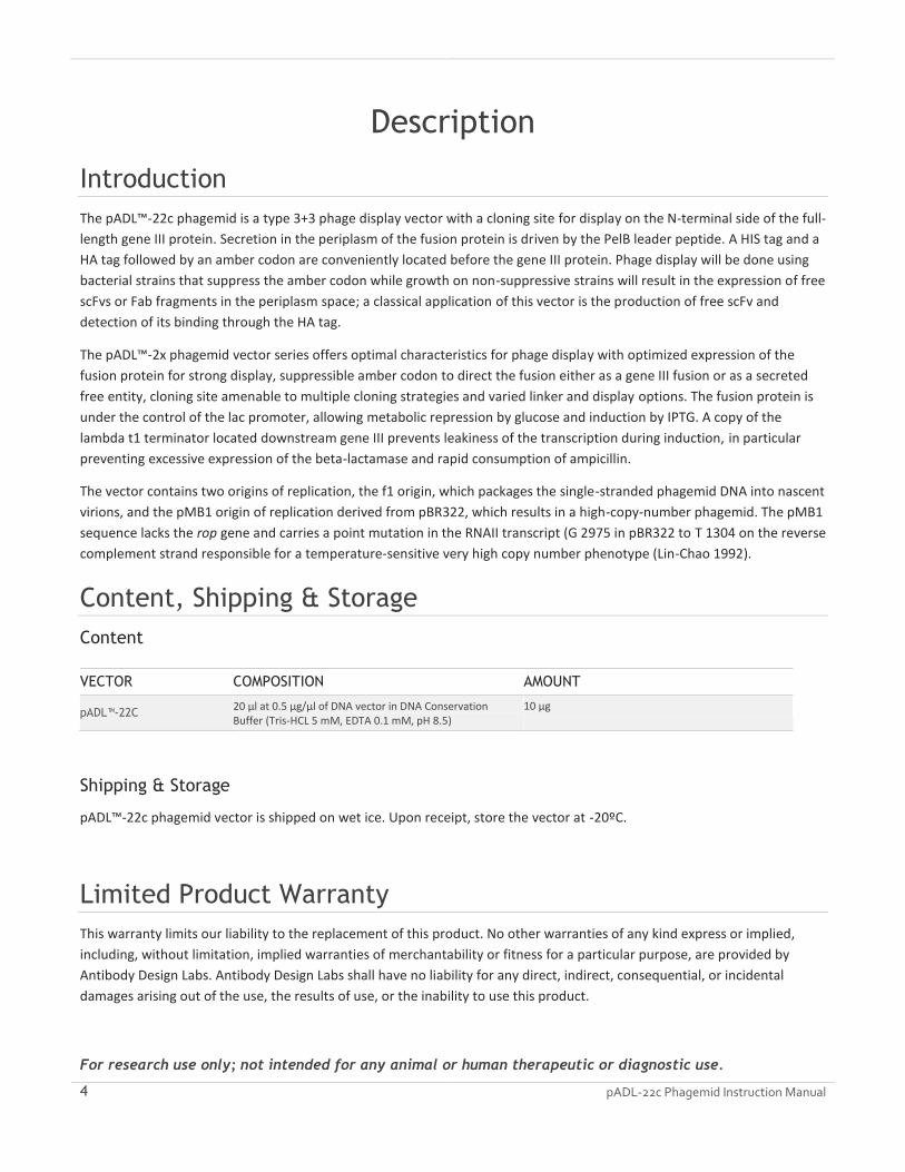
Content, Shipping & Storage
Content
VECTOR COMPOSITION AMOUNT
pADL™-22C 20 µl at 0.5 µg/µl of DNA vector in DNA Conservation Buffer (Tris-HCL 5 mM, EDTA 0.1 mM, pH 8.5)
10 µg
Shipping & Storage
pADL™-22c phagemid vector is shipped on wet ice. Upon receipt, store the vector at -20ºC.
Limited Product Warranty
This warranty limits our liability to the replacement of this product. No other warranties of any kind express or implied,
including, without limitation, implied warranties of merchantability or fitness for a particular purpose, are provided by
Antibody Design Labs. Antibody Design Labs shall have no liability for any direct, indirect, consequential, or incidental
damages arising out of the use, the results of use, or the inability to use this product.
For research use only; not intended for any animal or human therapeutic or diagnostic use.

pADL-22c Phagemid Instruction Manual 5
Vector Map
The figure below illustrates the main features of pADL™-22c phagemid vector. The full vector sequence is available online
for download in varied formats on the product web page; the total length of the vector is 3957 bp.
Feature Table
The features of pADL™-22c phagemid vector are highlighted in the following table.
FEATURE LOCATION DESCRIPTION
TEM1 beta-lactamase 126-986 Ampicillin resistance for selection in E. coli.
pMB1 origin 1141-1760 pBR322 origin for replication in E. coli with a high copy-number.
CAP binding site 1911-1931 Mediate the catabolite repression of the lac operator in the presence of glucose >1% w/v.
-35 signal 1946-1951 Lac promoter -35 signal
-10 signal 1970-1975 Lac promoter -10 signal
PelB leader sequence 2025-2087 PelB leader sequence for export in the periplasm of the host bacteria. The missing terminal methionine and alanine will have to be added during the cloning to obtain a complete leader peptide (MKYLLPTAAAGLLLLAAQPAMA) necessary for proper removal of the leader during the export process.
HIS tag 2108-2125 Peptide H H H H H H
HA tag 2132-2158 Peptide Y P Y D V P D Y A
Amber Stop codon 2162-2164
g3p fusion coding sequence 2165-3385 Full-length gene III fusion protein coding sequence; the M13 g3p protein is fused on its N-terminal side to the linker GPGGQHHHHHHGAYPYDVPDYAS[E]; the exact final sequence of the fusion depends on the cloning strategy (see cloning site).
lt1 3395-3507 Lambda t1 terminator
oriF1 3628-3934 Origin of replication for phage f1
6 pADL-22c Phagemid Instruction Manual
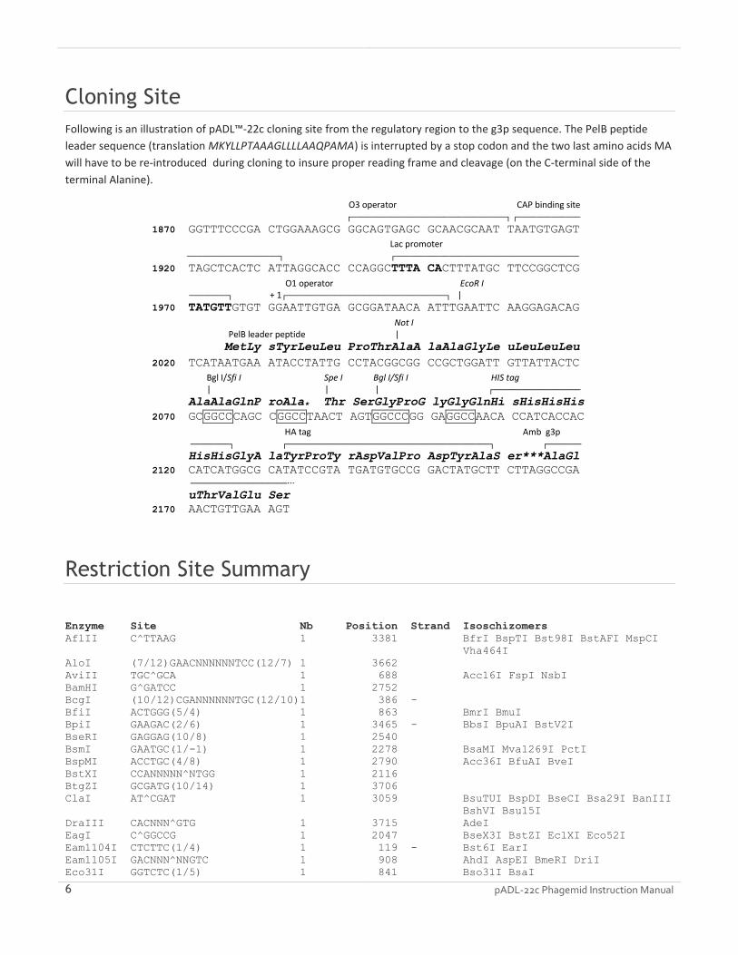
Cloning Site
Following is an illustration of pADL™-22c cloning site from the regulatory region to the g3p sequence. The PelB peptide
leader sequence (translation MKYLLPTAAAGLLLLAAQPAMA) is interrupted by a stop codon and the two last amino acids MA
will have to be re-introduced during cloning to insure proper reading frame and cleavage (on the C-terminal side of the
terminal Alanine).
O3 operator CAP binding site ┌────────────────────────────────┐ ┌─────────────
1870 GGTTTCCCGA CTGGAAAGCG GGCAGTGAGC GCAACGCAAT TAATGTGAGT
Lac promoter ───────────────────┐ ┌──────────────────────────────────────
1920 TAGCTCACTC ATTAGGCACC CCAGGCTTTA CACTTTATGC TTCCGGCTCG
O1 operator EcoR I ────────┐ + 1┌─────────────────────────────────┐ |
1970 TATGTTGTGT GGAATTGTGA GCGGATAACA ATTTGAATTC AAGGAGACAG
Not I PelB leader peptide |
MetLy sTyrLeuLeu ProThrAlaA laAlaGlyLe uLeuLeuLeu
2020 TCATAATGAA ATACCTATTG CCTACGGCGG CCGCTGGATT GTTATTACTC
Bgl I/Sfi I Spe I Bgl I/Sfi I HIS tag | | | ┌──────────────────
AlaAlaGlnP roAla* Thr SerGlyProG lyGlyGlnHi sHisHisHis
2070 GCGGCCCAGC CGGCCTAACT AGTGGCCCGG GAGGCCAACA CCATCACCAC
HA tag Amb g3p ────────┐ ┌──────────────────────────────────────────┐ ┌───────
HisHisGlyA laTyrProTy rAspValPro AspTyrAlaS er***AlaGl
2120 CATCATGGCG CATATCCGTA TGATGTGCCG GACTATGCTT CTTAGGCCGA
────────────────────∙∙∙
uThrValGlu Ser
2170 AACTGTTGAA AGT
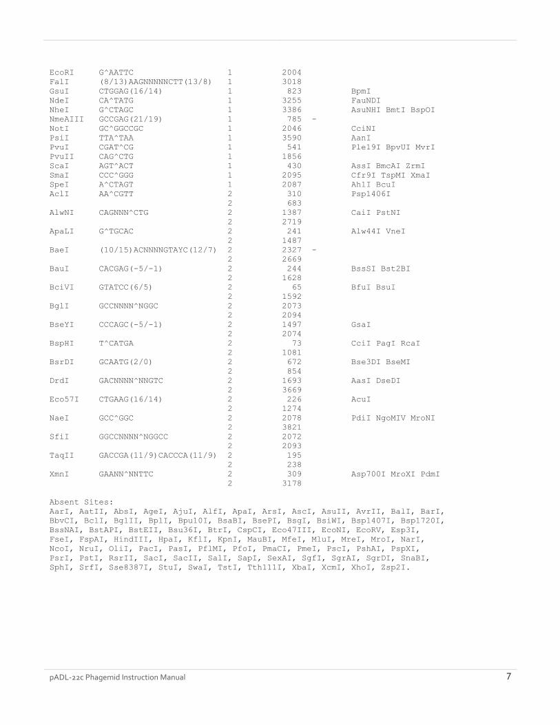
Restriction Site Summary
Enzyme Site Nb Position Strand Isoschizomers
AflII C^TTAAG 1 3381 BfrI BspTI Bst98I BstAFI MspCI
Vha464I
AloI (7/12)GAACNNNNNNTCC(12/7) 1 3662
AviII TGC^GCA 1 688 Acc16I FspI NsbI
BamHI G^GATCC 1 2752
BcgI (10/12)CGANNNNNNTGC(12/10)1 386 -
BfiI ACTGGG(5/4) 1 863 BmrI BmuI
BpiI GAAGAC(2/6) 1 3465 - BbsI BpuAI BstV2I
BseRI GAGGAG(10/8) 1 2540
BsmI GAATGC(1/-1) 1 2278 BsaMI Mva1269I PctI
BspMI ACCTGC(4/8) 1 2790 Acc36I BfuAI BveI
BstXI CCANNNNN^NTGG 1 2116
BtgZI GCGATG(10/14) 1 3706
ClaI AT^CGAT 1 3059 BsuTUI BspDI BseCI Bsa29I BanIII
BshVI Bsu15I
DraIII CACNNN^GTG 1 3715 AdeI
EagI C^GGCCG 1 2047 BseX3I BstZI EclXI Eco52I
Eam1104I CTCTTC(1/4) 1 119 - Bst6I EarI
Eam1105I GACNNN^NNGTC 1 908 AhdI AspEI BmeRI DriI
Eco31I GGTCTC(1/5) 1 841 Bso31I BsaI
pADL-22c Phagemid Instruction Manual 7
EcoRI G^AATTC 1 2004
FalI (8/13)AAGNNNNNCTT(13/8) 1 3018
GsuI CTGGAG(16/14) 1 823 BpmI
NdeI CA^TATG 1 3255 FauNDI
NheI G^CTAGC 1 3386 AsuNHI BmtI BspOI
NmeAIII GCCGAG(21/19) 1 785 -
NotI GC^GGCCGC 1 2046 CciNI
PsiI TTA^TAA 1 3590 AanI
PvuI CGAT^CG 1 541 Ple19I BpvUI MvrI
PvuII CAG^CTG 1 1856
ScaI AGT^ACT 1 430 AssI BmcAI ZrmI
SmaI CCC^GGG 1 2095 Cfr9I TspMI XmaI
SpeI A^CTAGT 1 2087 AhlI BcuI
AclI AA^CGTT 2 310 Psp1406I
2 683
AlwNI CAGNNN^CTG 2 1387 CaiI PstNI
2 2719
ApaLI G^TGCAC 2 241 Alw44I VneI
2 1487
BaeI (10/15)ACNNNNGTAYC(12/7) 2 2327 -
2 2669
BauI CACGAG(-5/-1) 2 244 BssSI Bst2BI
2 1628
BciVI GTATCC(6/5) 2 65 BfuI BsuI
2 1592
BglI GCCNNNN^NGGC 2 2073
2 2094
BseYI CCCAGC(-5/-1) 2 1497 GsaI
2 2074
BspHI T^CATGA 2 73 CciI PagI RcaI
2 1081
BsrDI GCAATG(2/0) 2 672 Bse3DI BseMI
2 854
DrdI GACNNNN^NNGTC 2 1693 AasI DseDI
2 3669
Eco57I CTGAAG(16/14) 2 226 AcuI
2 1274
NaeI GCC^GGC 2 2078 PdiI NgoMIV MroNI
2 3821
SfiI GGCCNNNN^NGGCC 2 2072
2 2093
TaqII GACCGA(11/9)CACCCA(11/9) 2 195
2 238
XmnI GAANN^NNTTC 2 309 Asp700I MroXI PdmI
2 3178
Absent Sites:
AarI, AatII, AbsI, AgeI, AjuI, AlfI, ApaI, ArsI, AscI, AsuII, AvrII, BalI, BarI,
BbvCI, BclI, BglII, BplI, Bpu10I, BsaBI, BsePI, BsgI, BsiWI, Bsp1407I, Bsp1720I,
BssNAI, BstAPI, BstEII, Bsu36I, BtrI, CspCI, Eco47III, EcoNI, EcoRV, Esp3I,
FseI, FspAI, HindIII, HpaI, KflI, KpnI, MauBI, MfeI, MluI, MreI, MroI, NarI,
NcoI, NruI, OliI, PacI, PasI, PflMI, PfoI, PmaCI, PmeI, PscI, PshAI, PspXI,
PsrI, PstI, RsrII, SacI, SacII, SalI, SapI, SexAI, SgfI, SgrAI, SgrDI, SnaBI,
SphI, SrfI, Sse8387I, StuI, SwaI, TstI, Tth111I, XbaI, XcmI, XhoI, Zsp2I.
8 pADL-22c Phagemid Instruction Manual
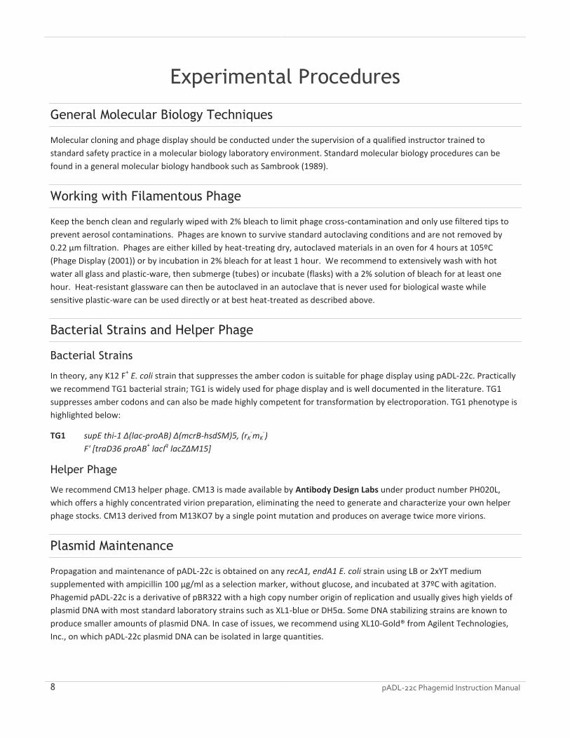
Experimental Procedures
General Molecular Biology Techniques
Molecular cloning and phage display should be conducted under the supervision of a qualified instructor trained to
standard safety practice in a molecular biology laboratory environment. Standard molecular biology procedures can be
found in a general molecular biology handbook such as Sambrook (1989).
Working with Filamentous Phage
Keep the bench clean and regularly wiped with 2% bleach to limit phage cross-contamination and only use filtered tips to
prevent aerosol contaminations. Phages are known to survive standard autoclaving conditions and are not removed by
0.22 µm filtration. Phages are either killed by heat-treating dry, autoclaved materials in an oven for 4 hours at 105ºC
(Phage Display (2001)) or by incubation in 2% bleach for at least 1 hour. We recommend to extensively wash with hot
water all glass and plastic-ware, then submerge (tubes) or incubate (flasks) with a 2% solution of bleach for at least one
hour. Heat-resistant glassware can then be autoclaved in an autoclave that is never used for biological waste while
sensitive plastic-ware can be used directly or at best heat-treated as described above.
Bacterial Strains and Helper Phage
Bacterial Strains
In theory, any K12 F+ E. coli strain that suppresses the amber codon is suitable for phage display using pADL-22c. Practically
we recommend TG1 bacterial strain; TG1 is widely used for phage display and is well documented in the literature. TG1
suppresses amber codons and can also be made highly competent for transformation by electroporation. TG1 phenotype is
highlighted below:
TG1 supE thi-1 Δ(lac-proAB) Δ(mcrB-hsdSM)5, (rK-mK
-)
F' [traD36 proAB+ lacI
q lacZΔM15]
Helper Phage
We recommend CM13 helper phage. CM13 is made available by Antibody Design Labs under product number PH020L,
which offers a highly concentrated virion preparation, eliminating the need to generate and characterize your own helper
phage stocks. CM13 derived from M13KO7 by a single point mutation and produces on average twice more virions.
Plasmid Maintenance
Propagation and maintenance of pADL-22c is obtained on any recA1, endA1 E. coli strain using LB or 2xYT medium
supplemented with ampicillin 100 µg/ml as a selection marker, without glucose, and incubated at 37ºC with agitation.
Phagemid pADL-22c is a derivative of pBR322 with a high copy number origin of replication and usually gives high yields of
plasmid DNA with most standard laboratory strains such as XL1-blue or DH5α. Some DNA stabilizing strains are known to
produce smaller amounts of plasmid DNA. In case of issues, we recommend using XL10-Gold® from Agilent Technologies,
Inc., on which pADL-22c plasmid DNA can be isolated in large quantities.

pADL-22c Phagemid Instruction Manual 9
Cloning into pADL-22c
Primer Design and PelB Leader Sequence
A complete PelB leader sequence MKYLLPTAAAGLLLLAAQPAMA is necessary for export in the periplasm and proper
removal of the leader peptide by host proteases. In the following schema, where [NNN] represents the insert sequence and
[Xxx] the translated amino acid sequence, the short hexanucleotide ATGGCN must be appended immediately to the first
BglI /SfiI site to obtain a complete PelB leader encoding sequence; cleavage will occur on the C-terminal side of the terminal
alanine (codon GCN).
BglI/ Sfi I Spe I BglI/ Sfi I | | |
yLeuLeuLeu LeuAlaAlaG lnProAlaMe tAla [Xxx] ThrSerGlyP roGlyGlyGl
2057 ATTGTTATTA CTCGCGGCCC AGCCGGCCAT GGCN [NNN] ACTAGTGGCC CGGGAGGCCA
Retention of the SpeI site is optional during cloning and the encoded dipeptide ThrSer is not known to interfere with
display.
Cloning in pADL-22c Using BglI/SfiI Sites
Large libraries in the 1 x 10⁹ range and above can easily be constructed using the double BglI/SfiI cloning site.
WORKING WITH BGL I/SFI I SITES
The SfiI restriction enzyme recognizes rare 8-base-long interrupted palindromes GGCCNNNN/NGGCC and leaves 3-
nucleotide-long overhangs after digestion. The pADL-22c cloning site contains one SfiI site close to the end of the PelB
leader sequence and a second SfiI site 8 nucleotides apart from the first site. The PelB sequence of the empty vector has an
early termination by an ochre stop codon and no gene III protein is produced by the vector alone.
The SfiI restriction enzyme requires two copies of its recognition sequence for cleavage to occur; cleavage of the two sites
happens simultaneously through interaction of two SfiI tetramers (Wertzell 1995). Vectors bearing two sites very close to
each other are cut in trans and digestion might not complete. Therefore we strongly recommend opening pADL-22c with
the alternative BglI restriction enzyme, which cuts the shorter 6-base-long interrupted palindromes GCCNNNN/NGGC and
generates identical overhangs.
Sites open with BglI will re-ligate with sites open with SfiI as long as overhangs are complementary. Practically, the
pentanucleotide NNNNN must be identical to the original vector sequence to handle both ligation of the complementary
overhangs and conservation of the amino acid sequence (PelB sequence and linker to protein III). Since the overhang of the
two BglI/SfiI sites are non-palidromic and different, a cut empty vector cannot ligate onto itself; it is therefore possible to
follow a ligation reaction by minigel analysis since remaining unligated vector or unligated insert will migrate unchanged at
their expected size.
PREPARATION OF VECTOR DNA FOR CLONING
1. On ice add successively water, BglI buffer (1x final), pADL-22c vector and BglI enzyme 5 units/µg DNA; make sure the
enzyme volume does not to exceed 1/10 of the total reaction volume.
2. Incubate overnight at 37ºC.
3. Inactivate for 20 min at 70ºC.
4. Confirm the digestion by DNA analysis on a minigel.
5. Purify the cut vector.

10 pADL-22c Phagemid Instruction Manual
For routine cloning, a standard DNA purification kit can be used directly after the digestion to remove the excess of buffer,
the small DNA stuffer and leftover restriction enzyme. For library construction, phenol/chloroform extraction and/or gel
purification may be required.
PREPARATION OF INSERTS
SfiI digestion should be rapid and complete in 4 hours especially for fragments longer or equal to 200 bp where sites are cut
in cis. BglI may be used when the insert sequence is known to be free of BglI site and therefore is not recommended for
building antibody libraries.
Cloning using NotI-SpeI sites
The NotI site located in the first half of the PelB leader encoding sequence may be used in conjunction with the SpeI site to
clone inserts. This strategy has been applied in some early phage display vectors. Consult your restriction enzyme
distributor resources to identify a buffer compatible with both enzymes and follow the concentration schema given above
to conduct the digestion. NotI and SpeI can be inactivated by heat before DNA purification.
Sequencing of Inserts
The following primers give both strong PCR amplification and sequencing traces. Primer locations can be found in the
corresponding GenBank sequence file.
Forward or Sense Primers
phiS2 5’-ATGAAATACCTATTGCCTACGG
phiS4 5’-GCGGATAACAATTTGAATTCAAGGAGACAG
Backward, Antisense or Reverse Primers
psiR2 5’-CGTTAGTAAATGAATTTTCTGTATGAGG
psiR3 5’-GCGTAACGATCTAAAGTTTTGTCG
Nested Sequencing Often it is easier to sequence an insert by PCR on the bacterial culture supernatant or directly from a colony rather than on
tediously isolated plasmids. Use the outward primers phiS2 and psiR3 together with a DNA polymerase not inhibited by
bacterial cultures such as TAQ polymerase for the PCR and sequence the insert with the nested reverse primer psiR2. Use
less than 1 µl of bacterial culture supernatant per 50 µl-PCR reaction or the touch of a toothpick on a colony as DNA
template.
Phagemid Virion Production
A superinfection by a helper phage is necessary for phagemid pADL-22c-containing bacteria to produce virions. Please,
consult the M13KO7 or CM13 helper phage manual for optimal conditions of superinfection. We recommend a rich medium
such as 2xYT medium supplemented with ampicillin 100 µg/ml, kanamycin 50 µg/ml (when M13KO7 or CM13 helper phage
are used), no glucose or less than 0.1% w/v, and incubation from 8 h to overnight at 30ºC and 250 rpm. Supplementation
with IPTG is not necessary to achieve display on the phage with pADL-22c. We recommend adding the helper phage when
the bacterial culture reaches an optical density at 600 nm between 0.4 OD and 0.5 OD; large amounts of non-superinfected
cells due to immunity to superinfection will decrease virion production above 0.5 OD while disparities caused by differences
pADL-22c Phagemid Instruction Manual 11
in phage growth rates will be amplified at a lower OD. Immunity to superinfection refers to the difficulty to transduce
bacteria when protein III is expressed, as it is the case when with phagemids expressing a full-length pIII fusion protein.
Notes
Supplementation with IPTG is not necessary during virion production but is recommended for the expression of free
antibodies in the periplasm with the non-amber suppressive SS320 bacterial strain.
Shorter incubation times 6 to 8 h long will produce less virions; we have not seen improvement of display on shorter
incubation times; inversely, we have not seen sign of proteolysis of the linker after overnight incubation. Always use
freshly prepared buffers from commercial concentrates during virion preparations to limit sources of proteolysis.
Proteolysis usually occurs on concentrated virions; always prepare virions quickly and on ice.
Kanamycin 50 µg/ml is enough to ensure selection with derivatives of M13KO7. Higher concentrations may be
needed on very rich culture media or if your culture medium contains phosphate salts.

12 pADL-22c Phagemid Instruction Manual
Appendix
MSDS Information
MSDSs (Material Safety Data Sheets) are available on the Antibody Design Labs website at the corresponding product page.
Quality Control
Specifications and quality control are detailed on the online product page. Antibody Design Labs certifies that the product
will perform according to these specifications.
Technical Support
Visit Antibody Design Labs’ website at www.abdesignlabs.com for technical resources, including manuals, vector maps and
sequences, application notes, FAQs, etc.
FOR MORE INFORMATION OR TECHNICAL ASSISTANCE, CALL, WRITE, FAX, OR EMAIL US AT:
Antibody Design Labs Email: [email protected]
4901 Morena Blvd, Suite 203 Phone: 1-877-223-3104 (TOLL-FREE)
San Diego, CA 92117 Fax: 1-858-272-6007 (24 hour)
(Monday – Friday 9:00 AM – 5:00 PM PST
References
1. LIN-CHAO S, CHEN WT, WONG TT (1992). HIGH COPY NUMBER OF THE PUC PLASMID RESULTS FROM A ROM/ROP-SUPPRESSIBLE POINT MUTATION IN RNA II. MOL MICROBIOL. 22:3385-93.
2. PHAGE DISPLAY: A LABORATORY MANUAL (2001). EDITED BY C. F. BARBAS III, D. R. BURTON, J. K. SCOTT, AND G. J. SILVERMAN. COLD SPRING HARBOR, LABORATORY PRESS, COLD SPRING HARBOR, NY.
3. SAMBROOK, J., FRITSCH, E.F., AND MANIATIS, T. (1989). IN MOLECULAR CLONING: A LABORATORY MANUAL. COLD SPRING HARBOR LABORATORY PRESS, NY, VOL. 1, 2, 3.
4. SCOTT JK & BARBAS CF (2001). PHAGE-DISPLAY VECTORS (2.1-2.19) IN PHAGE DISPLAY: A LABORATORY MANUAL. EDITED BY C. F. BARBAS III, D. R. BURTON, J. K. SCOTT, AND G. J. SILVERMAN. COLD SPRING HARBOR, LABORATORY PRESS, COLD SPRING HARBOR, NY.
5. WERTZELL L.M. ET AL., (1995). THE SFII RESTRICTION ENDONUCLEASE MAKES A FOUR-STRAND DNA BREAK AT TWO COPIES OF ITS RECOGNITION SEQUENCE. J. MOL. BIOL. 248:581-595
This product is subject to Antibody Design Labs Terms & Conditions of Sales available online at http://www.abdesignlabs.com/terms/.
© 2017 Antibody Design Labs. All rights reserved.