p450 3a and morphine synthesis - jbc. · pdf filep450 3a and morphine synthesis 3 oripavine)...
TRANSCRIPT
P450 3A and morphine synthesis
1
Cytochrome P450 3A Enzymes Catalyze the O6-Demethylation of Thebaine, a Key Step in Endogenous Mammalian Morphine Biosynthesis
Valerie M. Kramlinger, Mónica Alvarado Rojas, Tatsuyuki Kanamori, and F. Peter Guengerich
From the Department of Biochemistry, School of Medicine, Vanderbilt University School of Medicine,
Nashville, Tennessee 37232-0146
Running title: P450 3A and morphine synthesis To whom correspondence should be addressed: Prof. F. Peter Guengerich, Department of Biochemistry, Vanderbilt University School of Medicine, 638B Robinson Research Building, 2200 Pierce Avenue, Nashville, Tennessee 37232-0146, Telephone: (615) 322-2261; FAX: (615) 343-0704; E-mail: [email protected]
Keywords: Cytochrome P450, biosynthesis, natural product, oxygenase, morphine, alkaloids
ABSTRACT Morphine, first characterized in opium from the poppy Papaver somniferum, is one of the strongest known analgesics. Endogenous morphine has been identified in several mammalian cells and tissues. The synthetic pathway of morphine in the opium poppy has been elucidated, and the presence of common intermediates in plants and mammals suggests that biosynthesis occurs through similar pathways (beginning with the amino acid L-tyrosine), and the pathway has been completely delineated in plants. Some of the enzymes in the mammalian pathway have been identified and characterized. Two of the latter steps in the morphine biosynthesis pathway are demethylation of thebaine at the O3- and the O6-positions, the latter of which has been difficult to demonstrate. The plant enzymes responsible for both the O3- and the O6-demethylation are members of the FeII/α-ketoglutarate-dependent dioxygenase family. Previous studies showed that human cytochrome P450 (P450) 2D6 can catalyze thebaine O3-demethylation. We report that demethylation of thebaine at the O6-position is selectively catalyzed by human P450s 3A4 and 3A5, with the latter being more efficient, and rat P450 3A2. Our results do not support O6-demethylation of thebaine by an FeII/α-ketoglutarate-dependent dioxygenase. In rat brain microsomes, O6-demethylation was inhibited by ketoconazole, but not sulfaphenazole, suggesting that P450 3A enzymes are responsible for this activity in the brain. An alternate pathway to
morphine, oripavine O6-demethylation, was not detected. The major enzymatic steps in mammalian morphine synthesis have now been identified.
Morphine is the principal
benzylisoquinoline alkaloid produced in the opium poppy, Papaver somniferum. Its mechanism of action is as an agonist for the μ-opioid receptors that are distributed throughout the brain. Activation of these receptors is associated with analgesia, sedation, euphoria, physical dependence, and respiratory depression. The μ-binding sites are discretely distributed in the human brain, with high densities in the posterior amygdala, hypothalamus, thalamus, nucleus caudatus, putamen, and certain cortical areas (1).
A considerable body of evidence exists that morphine is present in the tissues of various animals that have not been medicated with morphine or other related opioids. In mammals, morphine has been detected in skin, lung, spinal cord, and, most notably, in the brain (2-4). The presence of morphine in brain is of particular interest due to the presence of the μ-opioid receptors. In the rat, morphine levels in structures of the brain have been quantified and range from 26 fmol/g tissue (found globally in the brain) to 7.2 pmol/g tissue measured in the hypothalamus (3,5-7). One study quantified morphine in the temporal lobe from one human brain tissue at 3.4 ng/g tissue (8). The presence of morphine in brain tissue in many mammals and the

P450 3A and morphine synthesis
2
evidence from a human sample suggest that endogenous morphine is present in human brain. Endogenous formation of morphine has been demonstrated in human cells in culture. In a seminal study, human neuroblastoma cells incubated in the presence of 18O2 produced [18O]-labeled morphine (9). Another report demonstrated morphine synthesis by human white blood cells and polymorphonuclear cells in a precursor-dependent manner (10,11). Taken together, these studies suggest that morphine biosynthesis can occur in the human brain. Humans have also been reported to excrete both codeine and morphine in urine, in the absence of treatment with these compounds (12).
The physiological role of endogenous morphine is currently unknown. The endogenous levels that have been quantified are below the plasma concentrations (μM range) achieved during therapeutic use (13). Many hypotheses have been presented regarding the purpose of endogenous morphine that suggest a role in infection, sepsis, or inflammation, as well as neurological pathologies (Parkinson’s disease and schizophrenia) (14-17).
Mammalian morphine biosynthesis appears to be highly similar to the pathway in the opium poppy, with at least some common intermediates (18-20). The multi-step biosynthetic pathway begins with the amino acid L-tyrosine, and the final steps in this pathway include the conversion of thebaine to morphine via two critical demethylation steps at the O3- and O6- positions. Two parallel pathways exist for conversion of thebaine to morphine, in which O3- and O6-demethylation reactions occur, potentially in either order (Fig. 1) (21). Both of these pathways are plausible, in that thebaine, codeine, and oripavine are present in both mammals as well as the opium poppy (5,22-24).
Although the intermediates are known and there is evidence that some of the reactions occur in mammals (9,25,26), the enzymes that catalyze these demethylation steps have only recently been identified. The enzymes that catalyze the O3- and O6-demethylation steps in the plant pathway are members of the FeII/α-ketoglutarate-dependent dioxygenase enzyme family (27). Specifically, in plants thebaine O6-demethylase catalyzes O6-demethylation of both thebaine and oripavine; codeine O-demethylase catalyzes O3-demethylation of both thebaine and codeine (Fig. 1). Independent of plant studies, the discovery that
O3-demethylation of thebaine and codeine can be catalyzed by the human enzyme cytochrome P450 (P450) 2D6 (28) has focused our search for the O6-demethylase to P450s, but this enzyme(s) has remained unknown, and the FeII/α-ketoglutarate dioxygenases can be considered.
Both the P450 and FeII/α-ketoglutarate-dependent dioxygenase families are known to catalyze diverse sets of reactions and are candidates for mediating the O6-demethylation of thebaine and oripavine. The P450 superfamily catalyzes the oxidation of most organic substances, generally using NADPH as an electron donor (29). The general reaction catalyzed by P450s is mixed-function oxidation, manifestations of which include carbon hydroxylation, dealkylation of heteroatomic substrates, heteroatom oxygenation, and the oxidation of unsaturated compounds to epoxides and phenols (30). FeII/α-ketoglutarate-dependent dioxygenases constitute a large family of soluble, non-heme iron-containing oxidases that couple the decarboxylation of α-ketoglutarate to activation of dioxygen to catalyze a variety of oxidation reactions. This family of enzymes most frequently catalyzes carbon hydroxylation, but other formal two-electron oxidations such as desaturation, cyclization, and halogenation are also known (31,32). Except for the two dioxygenases involved in the plant morphine biosynthesis (27,33), O-demethylation by the FeII/α-ketoglutarate-dependent dioxygenases has not been previously reported, to our knowledge.
The present study was designed to identify the enzyme(s) responsible for O6-demethylation of thebaine in mammals, humans in particular. Previous attempts to characterize the enzymes that catalyze O6-demethylation of thebaine and oripavine have been unsuccessful, in large part due to instability of codeinone and morphinone in aqueous solution (34,35). In the present work we devised and validated a coupled enzyme assay to circumvent this issue. We established that human P450s 3A4 and P450 3A5 and rat P450 3A2 catalyze O6-demethylation of thebaine. Using rat brain microsomes, we also provide evidence that P450 3A enzymes are also responsible for thebaine O6-demethylation in mammalian brain tissue. RESULTS
Assays for O6-Demethylation—The O6-demethylation products morphinone (from

P450 3A and morphine synthesis
3
oripavine) and neopinone/codeinone (from thebaine) are Michael acceptors and are inherently unstable, and preliminary attempts to demonstrate their formation in liver homogenates, microsomes, or purified P450s using LC-UV assays were unsuccessful. We also tried LC-MS assays, including ones in which we added NaBH4 or NaBD4 to trap the products, without success, probably because the approach only stabilized the product in solution at the completion of the incubation.
The use of coupled assays is well established in biochemistry. A bacterial morphinone reductase has been extensively studied as a model flavoprotein (34,37,50) and is known to rapidly reduce both morphinone and codeinone. The products, hydromorphone and hydrocodone, are stable and can easily be assayed (e.g. positive-ion ESI LC-MS). In preliminary assays, the concentration of morphinone reductase we established (0.5 µM) was found to be saturating in detecting product formation (vide infra) (Fig. 2B).
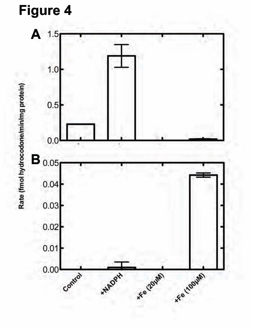
P450-Dependent O6-Demethylation of Thebaine—Incubations of thebaine and human liver microsomes in the presence of an NADPH-regenerating system and the morphinone reductase-NADH trapping system resulted in the formation of three products that were not seen in control reactions with heat-inactivated enzymes. These metabolites were subjected to MS after separation by UPLC. Comparison of two of the compounds with standards of alkaloids that were expected to be formed allowed the identification of the unknown compounds as oripavine (via O3-demethylation, Fig 1) (21) and hydrocodone (via O6-demethylation followed by morphinone reductase-catalyzed reduction, Figs. 1 and 2) (37) (Fig 3). The third compound had an m/z of 298, suggesting the loss of a methyl group; however, the retention did not match that of any standards and was further investigated using [N-C2H3]-thebaine (vide infra). The identification of oripavine as a P450-dependent product is consistent with the previous findings that incubation of P450 2D6 with thebaine resulted in oripivane formation (51). Hydrocodone was also identified in samples that contained human liver homogenates, thebaine, α-ketoglutarate, ascorbate, and 100 μM FeII but not in a similar samples that contained only 20 μM FeII. The amounts of hydrocodone formed are compared in Fig. 4.
Iron-Dependent O6-Demethylation of Thebaine—To further understand the iron concentration dependence of the �-ketoglutarate dioxygenase reaction observed with liver homogenates, thebaine was incubated in the absence of any enzyme but in the presence of α-ketoglutarate, L-ascorbate, and varying concentrations of FeII (Fig 5). The amount of hydrocodone produced appeared to be linearly dependent on the FeII concentration at or above 100 μM, suggesting that the hydrocodone seen in the initial screens was generated non-enzymatically and only due to the presence of the iron.
P450 3A-Dependent O6-Demethylation of Thebaine—A panel of selective inhibitors was used to determine which P450s were responsible for the O3- and O6-demethylation of thebaine in human liver microsomes. Most notably, quercetin and ketoconazole decreased the production of hydrocodone by 40% and 73%, respectively, and quinidine decreased the production of oripavine by 91% (data not presented). Quinidine is known to be a selective inhibitor of P450 2D6, and that result is consistent with our previous findings (51). Concentration-dependence inhibition experiments were done with quercetin and ketoconazole. Quercetin (P450 2C8 inhibitor) did not show concentration dependence (results not shown) and therefore was not further investigated further.
Ketoconazole showed concentration dependent inhibition, with an IC50 of 0.16 μM (Fig. 6), typical for P450 Subfamily 3A reactions (52). Because ketoconazole appears to inhibit all human P450 3A enzymes (49,53), CYP3cide (an inhibitor selective for P450 3A4 ((53,54)), was used to estimate the contribution of the human P450 3A enzymes involved in the O6-demethylation of thebaine. CYP3cide also showed concentration dependence, with an IC50 of 2.1 μM (Fig. 6), typical for P450 3A4 reactions (53,54).
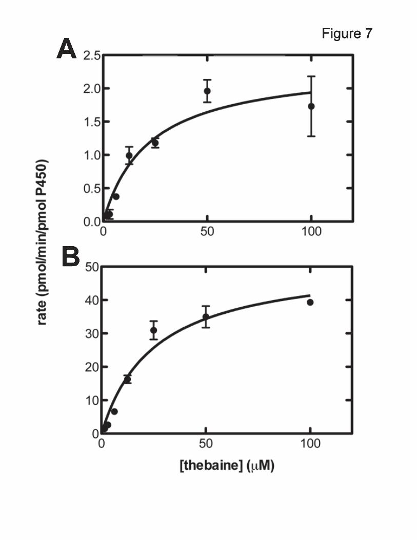
Thebaine O6-Demethylation Catalyzed by Recombinant P450 3A4 and 3A5—Steady-state kinetic parameters for recombinant P450 3A4 and P450 3A5-metiated thebaine O6-demethylation were determined based on the resulting hydrocodone. The results displayed Michaelis-Menten kinetics and the catalytic parameters were estimated (Fig. 7). P450 3A4 and 3A5 had similar apparent Km values of (22 and 26 μM, respectively); however, the maximum rate (kcat) for the conversion of thebaine to codeinone by P450 3A5 (52 min-1)

P450 3A and morphine synthesis
4
was 23-fold higher than that of P450 3A4 (2.3 min-1) (Fig. 7). For reference, the previously determined parameters for P450 2D6 conversion of thebaine to oripavine were kcat 4.6 min-1 and Km 48 µM (38) (kcat/Km 0.1 µM-1 min-1).
N-Demethylation of d3-Thebaine by Human Liver Microsomes, P450 3A4, and P450 3A5—To confirm the identity of the unidentified analyte with m/z 298 (vide supra), d3-thebaine was used as substrate in incubations. An analyte loss of 14 amu indicates a loss of a methyl group. There are three methyl groups on thebaine; an O-methyl at both the 3 and 6 positions and an N-methyl. The products of O3-demethylation (oripavine) and the O6-demethylation (hydrocodone after reduction by coupled enzyme system) were already identified, leaving the N-demethyltion product, northebaine, as the likely third product. By using thebaine and d3-thebaine as substrates in parallel experiments and comparing the products, we were able to determine that northebaine was indeed the third analyte.
When thebaine and d3-thebaine were included as substrates in parallel experiments, with pooled human liver microsomes, three products were observed. The first product (tR 1.9 min), which had an m/z of 298 when starting with thebaine and an m/z of 301 when starting with d3-thebaine, was oripavine. The second (tR 2.05 min) had m/z 300 when starting with thebaine and m/z 303 when starting with d3-thebaine, consistent with hydrocodone. The third, tR 2.4 min, had an m/z of 298 when starting with thebaine or with d3-thebaine, consistent with loss of the N-methyl group, giving northebaine. The northebaine gave fragment ions distinct from hydrocodone (m/z of 298281, 266). Northebaine was also identified as a product when thebaine or d3-thebaine was included as substrates with P450s 3A4 or 3A5. Based on one time point and a standard curve of thebaine, rate estimates were calculated for the conversion of thebaine to northebaine by P450s 3A4 and 3A5 (88 and 27 pmol/pmol P450/minute, respectively.)
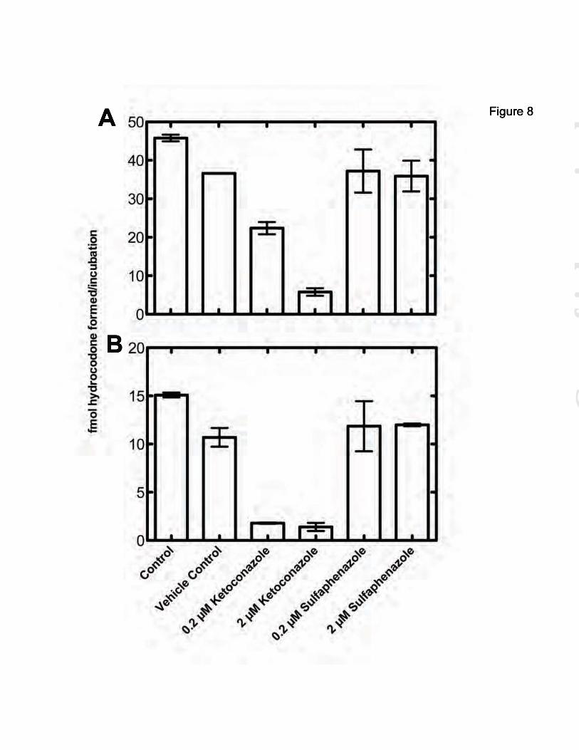
Thebaine O6-Demethylation by P450 3A2 and Rat Brain Microsomes—We have been unsuccessful in demonstrating any P450-dependent activity in frozen brain samples obtained from cattle or rats. Stored human brain samples (Vanderbilt tissue facility) or those provided by the Cooperative Human Tissue Network (Birmingham,
AL; Nashville, TN) were also negative for known P450 reactions, e.g. 7-ethoxycoumarin O-deethylation. We were able to detect activities in microsomes prepared from freshly obtained rat brains, as previously reported (55). Thebaine O6-demethylation was detected with recombinant rat P450 3A2, a prominent rat Subfamily 3A P450 (Fig. 8A) or rat brain microsomes (Fig. 8B). Rates were estimated to be 0.75 fmol/minute/pmol P450 and 1.3 fmol/min/mg total protein for P450 3A2 and rat brain microsomes, respectively (based on a single time point, 15 minutes). Additionally, O6-demethylation of thebaine was inhibited in both P450 3A2 and rat brain microsomes in the presence of either ketoconazole (Subfamily 3A-selective). Sulfaphenazole (P450 Subfamily 2C-selective (48)) did not inhibit O6-demethylation by either the P450 3A2 or rat brain microsomes. DISCUSSION A wealth of complementary biochemical, molecular, and physiological studies from independent laboratories have provided evidence supporting the biosynthesis of morphine in a variety of animal cells and organs (see (21) for comprehensive review). These studies have characterized multiple enzyme-catalyzed reactions and chemically defined intermediate precursor molecules that share marked similarities to the plant biosynthetic scheme previously established in the poppy P. somniferum (3-5,9,10,22,24-26,51,56-58). The final steps of the pathway convert thebaine to morphine, and thebaine must undergo demethylation at both the O3- and O6-positions (Fig. 1). Thus, two parallel pathways are possible for the conversion of thebaine to morphine: one in which O3-demethylation precedes O6-demethylation and the other in which the order is reversed. In plants, both pathways occur and the O3- and O6-demethylation steps are catalyzed by codeine O-demethylase and thebaine O6-demethylase, respectively, with the balance differing among species and strains (27). Because both of these enzymes are in the FeII/α-ketoglutarate-dependent dioxygenase family (33), our initial hypothesis was that an enzyme from this family would also be responsible for these demethylation steps in mammalian systems. None of our results support the initial hypothesis; instead, it was shown that both human and rat P450 3A enzymes catalyze the O6-demethylation of

P450 3A and morphine synthesis
5
thebaine. Additionally, the same reaction is catalyzed by a P450 in rat brain microsomes, apparently a member of the P450 3A Subfamily. These findings highlight a key difference in the biosynthesis of morphine in plants versus mammals. Oripavine and northebaine appear to be final products. The thebaine and oripavine O6-demethylation reactions are somewhat unusual in that they both involve cleavage of an enol ether, as opposed to a aliphatic or phenolic ether. This cleavage is apparently unprecedented in P450 chemistry (30) aside from aryl ethers. As mentioned earlier, initial efforts to directly demonstrate this reaction were unsuccessful, before implementation of the morphinone reductase-coupled assay. Efforts were made to determine if mammalian FeII/α-ketoglutarate-dependent dioxygenases catalyze O3- and O6-demethylation of thebaine, based on the precedence in plants. Our preliminary experiments were designed using typical conditions from the literature, which included exogenous iron concentrations ranging from 50-500 μM (33,59,60). Although thebaine O6-demethylation was observed, we noted that the generation of the product was neither time nor enzyme concentration dependent. Experiments done in the absence of any enzyme showed that O6-demethylation of thebaine was dependent on the iron concentration (Fig. 5). At concentrations ≤ 20 μM iron, no (non-enzymatic) O6-thebaine demethylation was detected. Although total mammalian cellular iron concentrations are estimated between 50-100 μM, iron homeostasis is highly regulated and free iron represents only a minor fraction (<5%); even 20 μM iron is much higher than the biologically relevant concentration and would be highly deleterious (61). When experiments with brain homogenates were repeated with 20 μM iron, no hydrocodone formation was detected. Although no further studies on the mechanism of the iron-catalyzed demethylation were done, it is well known that FeII catalyzes non-enzymatic hydroxyl radical formation from H2O2 via the Fenton reaction (62-64), and thus it is probably not surprising that iron-catalyzed (non-enzymatic) demethylation was observed (see also ref. (65) for iron-catalyzed N-demethylation of oripavine). Because the O-demethylation that was initially observed was an artifact resulting from the high iron concentration, it calls in to question the
use of high iron concentrations that are not biologically relevant. We cannot speculate on the relevance of our results in the general field of FeII/α-ketoglutarate-dependent dioxygenases. Many dioxygenases in this field have been extensively characterized (32,59,60,66-68). However, our results do suggest caution in the assignment of activities to enzymes in this family in the absence of more extensive characterization. We report here that thebaine O6-demethylation is catalyzed by P450 Subfamily 3A enzymes. The use of P450-selective inhibitors, in combination with human liver microsomes, implicated human P450s 3A4 and 3A5 in catalysis of O6-demethylation, confirmed by in vitro experiments with recombinant enzymes. Although both of these enzymes generate the same product, the catalytic efficiency of P450 3A5 was 20-fold higher than that of P450 3A4. When considering the relevance of P450s 3A4 and 3A5 in common-substrate metabolism, the much higher liver concentrations and typical superior catalytic efficiency usually focus responsibility toward P450 3A4 (52,69-71). Here, though, we are considering P450 activity in the brain and P450 3A5 is expressed in the brain with mRNA levels comparable to that of P450 3A4 (72). The higher catalytic efficiency and its measured expression in the brain lead to the proposal that P450 3A5 plays an important role in human morphine biosynthesis.
Numerous searches for thebaine O6-demethylation using microsomes derived from human brain tissue were attempted but were not presented here because no enzymatic activity was observed for any P450-dependent reaction. This was likely due to the difficulty in obtaining human brain samples that undergo careful treatment necessary to retain enzymatic activity. Thus, microsomes were prepared from freshly-acquired rat brains. In the presence of an NADPH-generating system, O6-demethylation was observed when thebaine was incubated with these microsomes (Fig. 8B). Additionally, the inhibition of this activity in the presence of ketoconazole (a P450 3A inhibitor that also inhibits rat P450 2C9) and the retention of the activity in the presence sulfaphenazole (a selective P450 2C inhibitor) (49) indicates that thebaine O6-demethylation in the brain microsomes is due to one or more of the rat P450 3A enzymes. (P450 activity was also lost in our rat brain tissues upon frozen storage—see also
P450 3A and morphine synthesis
6
(73)—consistent with the lack of activity in all frozen human brain samples we obtained.) Thus, in addition to showing that the human P450 3A enzymes can catalyze thebaine O6-demethylation, we report that this demethylation reaction occurs in rat brain, catalyzed by one or more Subfamily 3A P450 enzymes. The results presented here are consistent with a study that measured NADPH-dependent thebaine metabolism in rat brain microsomes (26). In that study codeine was generated at a rate of 0.20 ± 0.04 pmol/h/mg protein (as estimated by a radioimmune assay instead of LC-MS), compared to the present study in which hydrocodone was generated at a rate of 0.075 pmol/h/mg protein in the coupled LC-MS assay.
As described, two parallel pathways lead from thebaine to morphine (Fig 1). The first, in which thebaine is demethylated at the 3-position, yields oripavine, not thebaine, as the substrate for O6-demethylation. When oripavine was incubated with either human liver microsomes or recombinant P450 3A4 or P450 3A5, no O6-demethylation was observed (data not presented). Thus, the results presented in this paper support the second pathway, in which thebaine is first demethylated at the O6-position, as the only pathway that contributes to morphine synthesis, at least in human liver. No clear function has been attributed to endogenous morphine but suggestions include infection, sepsis, and inflammation, as well as major neurological pathologies (Parkinson’s disease, schizophrenia) (reviewed in (21)). Our findings indicate that P450 3A5 is most active in the O6-demethylation of thebaine in human biosynthesis of morphine (Fig. 7). P450 3A5 is highly polymorphic and the wild-type allele, CYP3A5*1, is expressed with a frequency of only 5 to 15% in Caucasian populations, 25 to 40% in various Asian ethnic groups, and approximately 40 to 60% in African and African-American populations (69,74,75). Thus, individuals with P450 3A5 polymorphisms that affect morphine biosynthesis might be expected to have different physiological outcomes related to this. Mikus et. al. (12) measured urinary excretion of codeine and morphine in 40 people who had been phenotyped for P450 2D6 status, utilizing an HPLC separation/radioimmune assay for detection. They reported no difference in morphine levels due to P450 2D6 status and concluded that other P450s might be involved in the synthesis of endogenous
morphine. The relevance of genetic variation of P450s 3A4 and 3A5 to the P450 2D6 work is unknown. In the work of Mikus et. al. (12), considerable inter-individual variability was reported (103-fold) but 10-fold day-to-day intra-individual variabilities where also seen. Whether these variations are real or the result of analytical deficiencies is not clear. Our results can be placed in context of the overall scheme of mammalian synthesis of morphine (Fig. 9). Administration of (R,S)-tetrahydropapaveroline to mice yielded reticuline, salutaridinol, thebaine, and oripavine in urine (24). All of the oxidation steps following the formation of tetrahydropapaveroline have now been characterized in humans in vitro. P450 2D6 or the two Subfamily 3A P450s (3A4, 3A5) have roles in all oxidation steps examined, including the early step of oxidation of tyramine to dopamine (7). A caveat is that many of the demonstrations of catalytic activity have been done with individual enzymes and the possibility exists that some other P450s may also be found to contribute. Although many of the studies involved in elucidating the steps in Fig. 9 have been done in liver systems (26,51), they do not speak to the localization near the neural targets which may play an important role in endogenous morphine biosynthesis in the brain. It is possible that an endocrine mechanism is more relevant to the function of endogenous morphine. In conclusion, our results now implicate several major human P450s in the scheme of Fig. 9, and these are found in a number of sites in the body (36). EXPERIMENTAL PROCEDURES
Materials—Thebaine, hydrocodone, and hydromorphone were purchased from Sigma-Aldrich (St. Louis, MO). Thebaine was recrystallized from hot acetone. Oripavine and [N-C2H3]-thebaine were provided by the late M. H. Zenk (Donald Danforth Plant Science Center, St. Louis, MO). CYP3cide (1-methyl-3-[1-methyl-5-(4-methylphenyl)-1H-pyrazol-4-yl]-4-[(3S)-3-piperidin-1-ylpyrrolidin-1-yl]-1H-pyrazolo[3,4-d]pyrimidine) was a gift from K. D. Hardy (Lipscomb University, Nashville, TN). All other inhibitors were purchased from Sigma-Aldrich. Rat brains were a gift from C. K. Jones (Dept. Pharmacology, Vanderbilt University). Liver microsomes from human liver tissue (set of 10
P450 3A and morphine synthesis
7
individual donors) were prepared in this laboratory according to published protocols (36).
Enzymes—Human P450 3A4- and 3A5-containing Baculosomes (microsomes isolated from insect cells infected with baculovirus) were purchased from Invitrogen/Life Technologies and rat P450 3A2-containing Baculosomes were purchased from BD Biosciences (Woburn, MA). A Pseudomonas putida morphinone reductase cDNA vector was provided by N. C. Bruce (University of York, York, UK) and the enzyme was heterologously expressed and purified according to French and Bruce (37); when subjected to SDS-PAGE on a 10% (w/v) acrylamide gel it showed a single band at 41 kDa.
Preparation of Rat Brain Homogenate and Microsomes—Male Sprague-Dawley rats (350-450 g, Harlan, Indianapolis, IN) used in the present studies were housed under a 12 hour light/12 hour dark cycle and given ad libitum access to food and water. All animal experiments were approved by the Vanderbilt University Animal Care and Use Committee, and experimental procedures conformed to guidelines established by the National Research Council Guide for the Care and Use of Laboratory Animals. Rats were briefly anesthetized with isoflurane and decapitated. Whole brains were removed and immediately washed in ice-cold 150 mM NaCl to remove blood clots. The brain tissue was homogenized in four volumes of ice-cold 0.10 M potassium phosphate buffer (7.6) containing 0.15 M KCl using seven to ten up-and-down strokes in a manually operated Teflon-glass Potter-Elvejhem homogenizer. All steps used to obtain microsomal fractions were carried out at 4 °C. Homogenates were centrifuged at 14,000 × g for 15 minutes at 4 °C (to remove debris, nuclei, and mitochondria) and the resulting supernatant was centrifuged at 100,000 × g for 1 hour at 4 °C. The microsomal pellet was resuspended in 10 mM Tris-acetate (pH 7.4) buffer containing 1.0 mM EDTA and 20% glycerol (v/v) (microsomes from 1 g of brain in ~0.2 ml buffer).
Enzymatic Assays—In general, incubations were conducted in a total volume of 300 µl in 50 mM potassium phosphate buffer (pH 7.4) containing 10 pmol of P450 enzyme (‘Baculosomes’, microsomal preparation from baculovirus-infected insect cells co-expressing human NADPH-P450 reductase, Life Technologies or BD Biosciences) or 0.5 mg protein/ml tissue
homogenate or microsomal preparation. Unless otherwise noted, negative control samples for each experiment contained the same components but were heated in a boiling water bath (5 min) before initiating the reactions.
Additionally, in order to trap the primary reaction products, morphinone reductase (0.5 µM) and NADH (1 mM) were included to convert codeinone and morphinone to the stable products hydrocodone and hydromorphone, respectively (37) (Fig. 2A). Because the substrates codeinone and morphinone are unstable and we did not have a standard, the conditions were validated by two preliminary experiments. A reported substrate of morphinone reductase is 2-cyclohexen-1-one (38). Therefore, in order to validate the activity of morphinone reductase, we repeated the experiment described by Barna et al. and measured the rate of 2-cyclohexen-1-one reduction (38). Our results give a rate of 1.3 s-1 under anaerobic conditions, which is similar to that reported (kcat 0.8 s-1). Anaerobic assays were done using the basic apparatus design of Burleigh et al. (39) as subsequently modified in this laboratory (40,41). Additionally, morphinone reductase was titrated in an incubation with P450 3A5 (described below) and saturation was seen at 0.2 μM morphinone reductase (Fig. 2B).
Following temperature equilibration to 37 °C for 5 min, the reactions were initiated by the addition of an NADPH-regenerating system (0.5 mM NADP+, 10 mM glucose 6-phosphate, and 1 IU ml−1 yeast glucose 6-phosphate dehydrogenase (36)) to analyze P450-dependent activity (42). Reactions generally proceeded at 37 °C for 20 min and were terminated by the addition of 300 μl of ice-cold CH3OH. Quinidine (as internal standard, 0.5 μmol) and pH 9.5 sodium borate buffer (300 μl of 0.5 M solution) were added. The samples were mixed using a vortex device and extracted with 1.0 ml of CH2Cl2, and the layers were separated by centrifugation (103 × g for 10 min). A 0.8 ml aliquot of the CH2Cl2 layer (lower phase) was transferred to a clean tube. An additional 1.0 ml of CH2Cl2 was added to the residual layer to extract the products, followed by mixing and another centrifugation step at 103 × g. The organic layers were combined, and the solvent was removed under an N2 stream. All samples were analyzed by LC/MS/MS (vide infra).
Initial Activity Screens—Initial screening for O6-demethylation activity was performed as
P450 3A and morphine synthesis
8
described above using human brain homogenate, human liver microsomes, or no enzyme source. In these experiments, P450-dependent activity was initiated by the addition of the NADPH-regenerating system and dioxygenase-dependent activity was initiated by the addition of α-ketoglutarate (1 mM), Fe(NH4)2(SO4)2 (100 or 20 µM), and L-ascorbate (1.8 mM). In all cases, morphinone reductase and NADH were also present. Control samples contained the same components that were boiled before initiating the reactions. Control samples contained human liver homogenate, but no NADPH-regenerating system or α-ketoglutarate, Fe(NH4)2(SO4)2, or L-ascorbate.
Steady-State Kinetic Assays—Kinetic assays were performed as described above using Baculosomes containing P450 3A4 or 3A5. Additionally, thebaine concentrations of 1.6, 3.1, 6.3, 12.5, 25, 50, and 100 μM were used. Results were fit to hyperbolic plots in GraphPad Prism (GraphPad Software, La Jolla, CA) to estimate kcat and Km values ± SE.
Morphinone Reductase Titration- To determine a saturating concentration of morphinone reductase, a titration was performed as described above using Baculosomes containing P450 3A5; however, the concentration of morphinone reductase varied (0.006, 0.03, 0.06, 0.18, 0.3, 0.6, 1.2 μM) (Fig. 2B).
Oxidation of d3-Thebaine—Assays were performed as described above using pooled human liver microsomes or Baculosomes containing P450 3A4 or 3A5. In addition to using thebaine as a substrate, separate incubations were done with d3-thebaine ([N-2H3]-thebaine) (100 μM) as substrate.
Inhibition Assays: Human Liver Microsomes—Initial P450 inhibition screens were performed as described above, with inhibitors added. Inhibitor concentrations were as follows: 1 μM -napthoflavone (for P450 1A1, 1A2), 2 μM quinidine (for P450 2D6), 100 μM 4-methylpyrazole (for P450 2E1), 2 μM methoxsalen (for P450 2A6), 50 μM quercetin (for P450 2C8), 5 μM sulfaphenazole (for P450 2C9), 2 μM ketoconazole (for P450 3A4, 3A5), 10 μM fluconazole (for P450 51A1), or 5 μM ticlopidine (for P450 2B6). The concentrations were chosen based upon values from literature that showed efficacy against and selectivity for each P450 (43-48). The half-maximal inhibitory concentration
(IC50) values were determined using conditions described above; however, these incubations contained either ketoconazole (0, 0.2, 0.4, 2.0, or 10 μM) or CYP3cide (0, 0.4, 2.0, 10, or 20 μM). IC50 values were calculated by non-linear regression using the GraphPad Prism equation for Inhibitor Concentration Response (Eq. 1) where X is log [inhibitor] and Y is the percentage of the uninhibited activity. All inhibitors were dissolved in C2H5OH and the volume added was 0.1% (v/v). Control incubations contained no inhibitor but did include 0.1% C2H5OH (v/v).
Equation 1
Inhibition Assays: P450 3A2 and Rat Brain
Microsomes—Ketoconazole or sulfaphenazole (0, 0.2, or 2 μM) was included in incubations containing P450 3A2 (4 pmol) or rat brain homogenate (800 μg protein) as described above. Control samples were incubated with and without vehicle present (0.5% CH3OH, v/v). Concentrations used were chosen based based on published selectivity of inhibitors for rat P450s (49).
Iron Titration Incubations—Thebaine (100 μM), potassium phosphate buffer (50 mM, pH 7.4), morphinone reductase (0.5 μM), NADH (1 mM), and Fe(NH4)2(SO4)2 (0, 20, 100, 225, or 500 μM), in a total volume of 300 μl, were incubated at 37 °C for 20 min. Reactions were terminated, extracted, and analyzed as described in the general incubation section (vide supra).
LC-MS/MS Analysis—Components of the dried extracts, dissolved in 10 mM NH4HCO3:CH3CN:HCO2H (94.9:5:0.1, v/v), were separated on a Waters Acquity UPLC system. Separation of (10 μl) samples was achieved using an ACQUITY® UPLC BEH C18 octadecylsilane (1.7 μm, 2.1 mm × 100 mm) column (Waters) equipped with a C18 guard column (Phenomenex, 2 mm × 4 mm). The mobile phase flow rate was 0.3 ml/min, with binary gradient elution using solvents A (10 mM aqueous NH4HCO3 with 0.1% (v/v) HCO2H) and B (CH3CN with 0.1% (v/v) HCO2H). The gradient started with 95% A (v/v) and was increased to 70% B (v/v) over 6 min, and then held at 70% B (v/v) for 2 min. Compounds were analyzed with a Thermo LTQ mass spectrometer using the positive-ion electrospray ionization (ESI) mode. ESI conditions

P450 3A and morphine synthesis
9
were: N2 sheath gas 40 psi; N2 auxiliary gas 6 psi; spray voltage 20 V; capillary temperature 350 °C; capillary offset 35 V; tube lens offset 80 V. MS-MS conditions were: normalized collision energy 35%; activation Q setting 0.250; activation time 30 ms.
Identification of thebaine product analytes was based on retention times (tR), the ratios of two different MRM transitions, and comparison with the expected values for standards. MRM transitions used were thebaine (m/z 312351, m/z 312381), oripavine (m/z 298218, m/z 298249), hydrocodone (m/z 300199, m/z 300215), and hydromorphone (m/z 286201, m/z 286242). Quantitation was done by constructing standard curves for each analyte (using the internal standard
quinidine).and integrating the peak areas of one MRM transition with Xcalaber software (Thermo Scientific). The limit of quantitation for hydrocodone was 5 pmol and the limit of detection was 1 pmol.
Identification of products of d3-thebaine was based on the tR of the non-deuterated standard and the expected m/z. The relevant masses of the compounds of interest were: for d3-thebaine m/z 315, for oripavine m/z 301, for hydrocodone m/z 303, and for northebaine m/z 298. Northebaine was not available as a standard, and therefore quantitation was based using a standard curve prepared using thebaine. The limit of detection for thebaine was 500 fmol and the limit of quantitation was 2 pmol.
Acknowledgments: We thank the late Prof. M. H. Zenk for the gifts of the oripavine and d3-thebaine and for encouragement in the early phases of this work. We also acknowledge Drs. M. Dostalek and G. Chowdhury for exploratory work on this project and K. Trisler for assistance in the preparation of the manuscript. Conflict of interest: The authors declare that they have no conflicts of interest with the contents of this article. Author contributions: VMK conducted most of the experiments, analyzed the results, and wrote most of the paper. MAR conducted experiments on the roles of P450s 3A4 and 3A5. TK conducted experiments searching for dioxygenase function. FPG conceived the idea for the project, conducted the anaerobic assay of morphinone reductase activity, and wrote the paper with VMK.
P450 3A and morphine synthesis
10
REFERENCES 1. Fowler, C. J., and Fraser, G. L. (1994) µ-, -, -Opioid receptors and their subtypes. A critical
review with emphasis on radioligand binding experiments. Neurochem. Int. 24, 401-426 2. Perea-Sasiain, J. (2008) Biosynthesis of morphine: Its importance in Parkinson's disease. Rev. Fac.
Med. Unal. 56, 161-189 3. Lee, C. S., and Spector, S. (1991) Changes of endogenous morphine and codeine contents in the
fasting rat. J. Pharmacol. Exp. Ther. 257, 647-650 4. Goldstein, A., Barrett, R. W., James, I. F., Lowney, L. I., Weitz, C. J., Knipmeyer, L. L., and
Rapoport, H. (1985) Morphine and other opiates from beef brain and adrenal. Proc. Natl. Acad. Sci. U.S.A. 82, 5203-5207
5. Donnerer, J., Cardinale, G., Coffey, J., Lisek, C. A., Jardine, I., and Spector, S. (1987) Chemical characterization and regulation of endogenous morphine and codeine in the rat. J. Pharmacol. Exp. Ther. 242, 583-587
6. Meijerink, W. J. H. J., Molina, P. E., and Abumrad, N. N. (1999) Mammalian opiate alkaloid synthesis: Lessons derived from plant biochemistry. Shock 12, 165-173
7. Hiroi, T., Imaoka, S., and Funae, Y. (1998) Dopamine formation from tyramine by CYP2D6. Biochem. Biophys. Res. Commun. 249, 838-843
8. Fricchione, G., Zhu, W., Cadet, P., Mantione, K. J., Bromfield, E., Madsen, J., DeGirolami, U., Dworetzky, B., Vaccaro, B., Black, P., and Stefano, G. B. (2008) Identification of endogenous morphine and a mu3-like opiate alkaloid receptor in human brain tissue taken from a patient with intractable complex partial epilepsy. Med. Sci. Monit. 14, Cs45-Cs49
9. Poeaknapo, C., Schmidt, J., Brandsch, M., Drager, B., and Zenk, M. H. (2004) Endogenous formation of morphine in human cells. Proc. Natl. Acad. Sci. U.S.A. 101, 14091-14096
10. Boettcher, C., Fellermeier, M., Boettcher, C., Drager, B., and Zenk, M. H. (2005) How human neuroblastoma cells make morphine. Proc. Natl. Acad. Sci. U.S.A. 102, 8495-8500
11. Zhu, W., Cadet, P., Baggerman, G., Mantione, K. J., and Stefano, G. B. (2005) Human white blood cells synthesize morphine: CYP2D6 modulation. J. Immunol. 175, 7357-7362
12. Mikus, G., Bochner, F., Eichelbaum, M., Horak, P., Somogyi, A. A., and Spector, S. (1994) Endogenous codeine and morphine in poor and extensive metabolisers of the CYP2D6 (debrisoquine/sparteine) polymorphism. J. Pharmacol. Exp. Ther. 268, 546-551
13. Findlay, J. W., Jones, E. C., Butz, R. F., and Welch, R. M. (1978) Plasma codeine and morphine concentrations after therapeutic oral doses of codeine-containing analgesics. Clin. Pharmacol. Ther. 24, 60-68
14. Laux-Biehlmann, A., Grafe, N., Mouheiche, J., Stuber, D., Welters, I. D., Delalande, F., Poisbeau, P., Garnero, P., Metz-Boutigue, M. H., Schneider, F., and Goumon, Y. (2012) Comparison of serum and lithium-heparinate plasma for the accurate measurements of endogenous and exogenous morphine concentrations. Br. J. Clin. Pharmacol. 74, 381-383
15. Charron, G., Doudnikoff, E., Laux, A., Berthet, A., Porras, G., Canron, M. H., Barroso-Chinea, P., Li, Q., Qin, C., Nosten-Bertrand, M., Giros, B., Delalande, F., Van Dorsselaer, A., Vital, A., Goumon, Y., and Bezard, E. (2011) Endogenous morphine-like compound immunoreactivity increases in Parkinsonism. Brain 134, 2321-2338
16. Glattard, E., Welters, I. D., Lavaux, T., Muller, A. H., Laux, A., Zhang, D., Schmidt, A. R., Delalande, F., Laventie, B. J., Dirrig-Grosch, S., Colin, D. A., Van Dorsselaer, A., Aunis, D., Metz-Boutigue, M. H., Schneider, F., and Goumon, Y. (2010) Endogenous morphine levels are increased in sepsis: A partial implication of neutrophils. Plos. One 5, e8791
17. Cousins, D. A., Butts, K., and Young, A. H. (2009) The role of dopamine in bipolar disorder. Bipolar Disord. 11, 787-806
18. Kirby, G. W. (1967) Biosynthesis of the morphine alkaloids. Science 155, 170-173 19. Kutchan, T. M. (1998) Molecular genetics of plant alkaloid biosynthesis, in The Alkaloids:
Chemistry and Biology, Cordell, G.A., ed; Academic Press, San Diego; Vol. 50, pp 257-316
P450 3A and morphine synthesis
11
20. Onoyovwe, A., Hagel, J. M., Chen, X., Khan, M. F., Schriemer, D. C., and Facchini, P. J. (2013) Morphine biosynthesis in opium poppy involves two cell types: sieve elements and laticifers. Plant Cell 25, 4110-4122
21. Laux-Biehlmann, A., Mouheiche, J., Veriepe, J., and Goumon, Y. (2013) Endogenous morphine and its metabolites in mammals: History, synthesis, localization and perspectives. Neuroscience 233, 95-117
22. Kodaira, H., Lisek, C. A., Jardine, I., Arimura, A., and Spector, S. (1989) Identification of the convulsant opiate thebaine in mammalian brain. Proc. Natl. Acad. Sci. U.S.A. 86, 716-719
23. Weitz, C. J., Lowney, L. I., Faull, K. F., Feistner, G., and Goldstein, A. (1986) Morphine and codeine from mammalian brain. Proc. Natl. Acad. Sci. U.S.A. 83, 9784-9788
24. Grobe, N., Lamshoft, M., Orth, R. G., Drager, B., Kutchan, T. M., Zenk, M. H., and Spiteller, M. (2010) Urinary excretion of morphine and biosynthetic precursors in mice. Proc. Natl. Acad. Sci. U.S.A. 107, 8147-8152
25. Weitz, C. J., Faull, K. F., and Goldstein, A. (1987) Synthesis of the skeleton of the morphine molecule by mammalian liver. Nature 330, 674-677
26. Kodaira, H., and Spector, S. (1988) Transformation of thebaine to oripavine, codeine, and morphine by rat liver, kidney, and brain microsomes. Proc. Natl. Acad. Sci. U.S.A. 85, 1267-1271
27. Hagel, J. M., and Facchini, P. J. (2010) Dioxygenases catalyze the O-demethylation steps of morphine biosynthesis in opium poppy. Nat. Chem. Biol. 6, 273-275
28. Dayer, P., Desmeules, J., Leemann, T., and Striberni, R. (1988) Bioactivation of the narcotic drug codeine in human liver is mediated by the polymorphic monooxygenase catalyzing debrisoquine 4-hydroxylation (cytochrome P-450 dbl/bufI). Biochem. Biophys. Res. Commun. 152, 411-416
29. Ortiz de Montellano, P. R. (2015) Substrate oxidation by cytochrome P450 enzymes, in Cytochrome P450: Structure, Mechanism, and Biochemistry (Ortiz de Montellano, P. R., ed.), 4th Ed., Springer, New York. pp 111-176
30. Guengerich, F. P. (2001) Common and uncommon cytochrome P450 reactions related to metabolism and chemical toxicity. Chem. Res. Tox. 14, 611-650
31. Knauer, S. H., Hartl-Spiegelhauer, O., Schwarzinger, S., Hanzelmann, P., and Dobbek, H. (2012) The Fe(II)/alpha-ketoglutarate-dependent taurine dioxygenases from Pseudomonas putida and Escherichia coli are tetramers. FEBS J. 279, 816-831
32. Proshlyakov, D. A., Henshaw, T. F., Monterosso, G. R., Ryle, M. J., and Hausinger, R. P. (2004) Direct detection of oxygen intermediates in the non-heme Fe enzyme taurine/-ketoglutarate dioxygenase. J. Am. Chem. Soc. 126, 1022-1023
33. Farrow, S. C., and Facchini, P. J. (2013) Dioxygenases catalyze O-demethylation and O,O-demethylenation with widespread roles in benzylisoquinoline alkaloid metabolism in opium poppy. J. Biol. Chem. 288, 28997-29012
34. Craig, D. H., Moody, P. C., Bruce, N. C., and Scrutton, N. S. (1998) Reductive and oxidative half-reactions of morphinone reductase from Pseudomonas putida M10: A kinetic and thermodynamic analysis. Biochemistry 37, 7598-7607
35. Lister, D. L., Kanungo, G., Rathbone, D. A., and Bruce, N. C. (1999) Transformations of codeine to important semisynthetic opiate derivatives by Pseudomonas putida m10. FEMS Microbiol. Lett. 181, 137-144
36. Guengerich, F. P. (2014) Analysis and characterization of enzymes and nucleic acids relevant to toxicology. in Hayes' Principles and Methods of Toxicology (Hayes, A. W., and Kruger, C. L., eds.), 6th Ed., CRC Press-Taylor & Francis, Boca Raton, FL. pp 1905-1964
37. French, C. E., and Bruce, N. C. (1994) Purification and characterization of morphinone reductase from Pseudomonas-putida M10. Biochem. J. 301, 97-103
38. Barna, T., Messiha, H. L., Petosa, C., Bruce, N. C., Scrutton, N. S., and Moody, P. C. (2002) Crystal structure of bacterial morphinone reductase and properties of the C191A mutant enzyme. J. Biol. Chem. 277, 30976-30983
P450 3A and morphine synthesis
12
39. Burleigh, B. D., Jr., Foust, G.P., and Williams, C.H., Jr. (1969) A method for titrating oxygen-sensetive organic redox systems with reducing agents in solution. Anal. Biochem. 27, 536-544
40. Guengerich, F. P. (1983) Oxidation-reduction properties of rat liver cytochromes P-450 and NADPH-cytochrome P-450 reductase related to catalysis in reconstituted systems. Biochemistry 22, 2811-2820
41. Guengerich, F. P., Krauser, J. A., and Johnson, W. W. (2004) Rate-limiting steps in oxidations catalyzed by rabbit cytochrome P450 1A2. Biochemistry 43, 10775-10788
42. Sohl, C. D., Cheng, Q., and Guengerich, F. P. (2009) Chromatographic assays of drug oxidation by human cytochrome P450 3A4. Nat. Protoc. 4, 1252-1257
43. Turpeinen, M., Uusitalo, J., Jalonen, J., and Pelkonen, O. (2005) Multiple P450 substrates in a single run: Rapid and comprehensive in vitro interaction assay. Eur. J. Pharm. Sci. 24, 123-132
44. Bu, H. Z., Knuth, K., Magis, L., and Teitelbaum, P. (2001) High-throughput cytochrome P450 (CYP) inhibition screening via a cassette probe-dosing strategy. V. Validation of a direct injection/on-line guard cartridge extraction—tandem mass spectrometry method for CYP1A2 inhibition assessment. Eur. J. Pharm. Sci. 12, 447-452
45. Yin, H., Racha, J., Li, S. Y., Olejnik, N., Satoh, H., and Moore, D. (2000) Automated high throughput human CYP isoform activity assay using SPE-LC/MS method: Application in CYP inhibition evaluation. Xenobiotica. 30, 141-154
46. Zhang, W., Kilicarslan, T., Tyndale, R. F., and Sellers, E. M. (2001) Evaluation of methoxsalen, tranylcypromine, and tryptamine as specific and selective CYP2A6 inhibitors in vitro. Drug. Metab. Dispos. 29, 897-902
47. Richter, T., Murdter, T. E., Heinkele, G., Pleiss, J., Tatzel, S., Schwab, M., Eichelbaum, M., and Zanger, U. M. (2004) Potent mechanism-based inhibition of human CYP2B6 by clopidogrel and ticlopidine. J. Pharmacol. Exp. Ther. 308, 189-197
48. Correia, M. A. (2005) Inhibition of cytochrome P450 enzymes. in Cytochrome P450: Structure, Mechanism, and Biochemistry (Ortiz de Montellano, P. R. ed.), 3rd Ed., Kluwer Academic/Plenum Press, New York. pp 247-322
49. Kobayashi, K., Urashima, K., Shimada, N., and Chiba, K. (2003) Selectivities of human cytochrome P450 inhibitors toward rat P450 isoforms: Study with cDNA-expressed systems of the rat. Drug. Metab. Dispos. 31, 833-836
50. Messiha, H. L., Munro, A. W., Bruce, N. C., Barsukov, I., and Scrutton, N. S. (2005) Reaction of morphinone reductase with 2-cyclohexen-1-one and 1-nitrocyclohexene: proton donation, ligand binding, and the role of residues histidine 186 and asparagine 189. J. Biol. Chem. 280, 10695-10709
51. Grobe, N., Zhang, B., Fisinger, U., Kutchan, T. M., Zenk, M. H., and Guengerich, F. P. (2009) Mammalian cytochrome P450 enzymes catalyze the phenol-coupling step in endogenous morphine biosynthesis. J. Biol. Chem. 284, 24425-24431
52. Niwa, T., Murayama, N., Emoto, C., and Yamazaki, H. (2008) Comparison of kinetic parameters for drug oxidation rates and substrate inhibition potential mediated by cytochrome P450 3A4 and 3A5. Curr. Drug. Metab. 9, 20-33
53. Tseng, E., Walsky, R. L., Luzietti, R. A., Jr., Harris, J. J., Kosa, R. E., Goosen, T. C., Zientek, M. A., and Obach, R. S. (2014) Relative contributions of cytochrome CYP3A4 versus CYP3A5 for CYP3A-cleared drugs assessed in vitro using a CYP3A4-selective inactivator (CYP3cide). Drug. Metab. Dispos. 42, 1163-1173
54. Walsky, R. L., Obach, R. S., Hyland, R., Kang, P., Zhou, S., West, M., Geoghegan, K. F., Helal, C. J., Walker, G. S., Goosen, T. C., and Zientek, M. A. (2012) Selective mechanism-based inactivation of CYP3A4 by CYP3cide (PF-04981517) and its utility as an in vitro tool for delineating the relative roles of CYP3A4 versus CYP3A5 in the metabolism of drugs. Drug. Metab. Dispos.40, 1686-1697
55. Guengerich, F. P. Mason, P. S. (1978) Immunological comparison of hepatic and extrahepatic cytochromes P-450. Mol. Pharmacol. 15, 154-164
P450 3A and morphine synthesis
13
56. Cardinale, G. J., Donnerer, J., Finck, A. D., Kantrowitz, J. D., Oka, K., and Spector, S. (1987) Morphine and codeine are endogenous components of human cerebrospinal-fluid. Life Sci. 40, 301-306
57. Donnerer, J., Oka, K., Brossi, A., Rice, K. C., and Spector, S. (1986) Presence and formation of codeine and morphine in the rat. Proc. Natl. Acad. Sci. U.S.A. 83, 4566-4567
58. Grobe, N., Ren, X., Kutchan, T. M., and Zenk, M. H. (2011) An (R)-specific N-methyltransferase involved in human morphine biosynthesis. Arch. Biochem. Biophys. 506, 42-47
59. Li, D., Fedeles, B. I., Shrivastav, N., Delaney, J. C., Yang, X., Wong, C., Drennan, C. L., and Essigmann, J. M. (2013) Removal of N-alkyl modifications from N2-alkylguanine and N4-alkylcytosine in DNA by the adaptive response protein AlkB. Chem. Res. Toxicol. 26, 1182-1187
60. Fukumori, F., and Hausinger, R. P. (1993) Alcaligenes eutrophus JMP134 "2,4-dichlorophenoxyacetate monooxygenase" is an -ketoglutarate-dependent dioxygenase. J. Bacteriol. 175, 2083-2086
61. Kakhlon, O., and Cabantchik, Z. I. (2002) The labile iron pool: characterization, measurement, and participation in cellular processes. Free Radic. Biol. Med. 33, 1037-1046
62. Winterbourn, C. C. (1995) Toxicity of iron and hydrogen peroxide: The Fenton reaction. Toxicol. Lett. 82-83, 969-974
63. Goldstein, S., and Meyerstein, D. (1999) Comments on the mechansim of "Fenton-Like" reaction. Acc. Chem. Res. 32, 547-550
64. MacFaul, P. A., Wayner, D.D.M, and Ingold, K.U. (1998) A radical account of "Oxygenated Fenton Chemistry." Acc. Chem. Res. 31, 159-162
65. Kok, G. B., and Scammells, P. J. (2011) Further investigations into the N-demethylation of oripavine using iron and stainless steel. Org. Biomol. Chem. 9, 1008-1011
66. Fukumori, F., and Hausinger, R. P. (1993) Purification and characterization of 2,4-dichlorophenoxyacetate/-ketoglutarate dioxygenase. J. Biol. Chem. 268, 24311-24317
67. Hausinger, R. P. (2004) FeII/-ketoglutarate-dependent hydroxylases and related enzymes. Crit. Rev. Biochem. Mol. Biol. 39, 21-68
68. Hausinger, R. S., C. (2015) 2-Oxoglutarate-Dependent Oxygenases, 1st ed., Royal Soc. Chem, Abingdon, Oxfordshire, UK
69. Daly, A. K. (2006) Significance of the minor cytochrome P450 3A isoforms. Clin. Pharmacokinet. 45, 13-31
70. Stevens, J. C. (2006) New perspectives on the impact of cytochrome P450 3A expression for pediatric pharmacology. Drug Discov. Today 11, 440-445
71. Rendic, S. P., and Guengerich, F. P. (2014) Survey of human oxidoreductases and cytochrome P450 enzymes involved in the metabolism of chemicals. Chem. Res. Toxicol. 28, 39-42
72. Agrawal, V., Huang, N., and Miller, W. L. (2008) Pharmacogenetics of P450 oxidoreductase: Effect of sequence variants on activities of CYP1A2 and CYP2C19. Pharmacogenet. Genomics 18, 569-576
73. Tyndale, R. F., Li, Y., Li, N. Y., Messina, E., Miksys, S., and Sellers, E. M. (1999) Characterization of cytochrome P-450 2D1 activity in rat brain: High-affinity kinetics for dextromethorphan. Drug Metab. Dispos. 27, 924-930
74. Kuehl, P., Zhang, J., Lin, Y., Lamba, J., Assem, M., Schuetz, J., Watkins, P. B., Daly, A., Wrighton, S. A., Hall, S. D., Maurel, P., Relling, M., Brimer, C., Yasuda, K., Venkataramanan, R., Strom, S., Thummel, K., Boguski, M. S., and Schuetz, E. (2001) Sequence diversity in CYP3A promoters and characterization of the genetic basis of polymorphic CYP3A5 expression. Nat. Genet. 27, 383-391
75. Daly, A. K., King, B. P., and Leathart, J. B. (2006) Genotyping for cytochrome P450 polymorphisms. Methods Mol. Biol. 320, 193-207

P450 3A and morphine synthesis
14
FOOTNOTES This work was supported in whole or part by National Institute of Health Grants R37 CA090426 and T32 E5007028. The abbreviations used are: P450, cytochrome P450; ESI, electrospray ionization; MRM, multiple reaction monitoring; MS, mass spectrometry. FIGURE LEGENDS FIGURE 1. Latter steps of morphine biosynthesis (21). FIGURE 2. Reduction of codeinone to hydrocodone and morphinone to hydromorphone by morphinone reductase (37). A, reactions utilized; B, dependence of hydrocodone formation in a P450 3A5-supported reaction upon the concentration of morphinone reductase. FIGURE 3. LC-MS/MS traces of thebaine products compared to authentic standards. Thebaine was incubated in the presence of pooled human liver microsomes and the NADPH-generating system (A, B) or human liver microsomes that were boiled to inactivate enzymes and NADPH-generating system (G, H) and compared to authentic standards of oripavine (C, D) and hydrocodone (E, F). Traces shown in black are oripavine (m/z 298218, 249); traces shown in red are for hydrocodone (m/z 300199, 215). FIGURE 4. O6-Demethylation of thebaine in the presence of or absence of human liver microsomes with and without iron. Thebaine was incubated in the presence of pooled human liver homogenate or microsomes (A) or no enzyme (B) for 20 minutes. Control samples contained liver homogenate but did not contain NADPH-generating system. “+NADPH” samples contained NADPH-generating system and liver microsomes, “+Fe” samples contained 20 μM or 100 μM iron, 1 mM α-ketoglutarate, 1.8 mM L-ascorbate and liver homogenate. Results are presented as means ± SD of duplicate (n=2) experiments. FIGURE 5. FeII-dependent O6-demethylation of thebaine. Fe(NH4)2(SO4)2 (0, 20, 100, 250, and 500 μM) was incubated with 50 μM thebaine for 20 minutes under typical assay conditions (absence of enzymes). Results are presented as means ± SD of triplicate experiments. FIGURE 6. Inhibition of hydrocodone formation in human liver microsomes. Thebaine was incubated in the presence of pooled human liver microsomes and the NADPH-generating system with increasing amounts of ketoconazole () or CYP3cide (). Inhibition constants were derived from non-linear fitting to a single-site inhibition model in Prism software (Graphpad, San Diego, CA). Results are presented as means ± SD of triplicate experiments. FIGURE 7. Oxidation of thebaine to codeinone by recombinant P450 3A4 and 3A5. A, P450 3A4. kcat 2.3 ± 0.3 pmol/min/pmol P450; Km 22 ± 7 µM; kcat/Km 0.10 µM-1 min-1. B, P450 3A5. kcat 52 ± 4 pmol/min/pmol P450; Km 26 ± 6 µM; kcat/Km 2.0 µM-1 min-1. The results (±SE) were calculated using GraphPad Prism. FIGURE 8. Inhibition of P450 3A2- or rat brain homogenate-mediated hydrocodone formation. Thebaine was incubated in the presence of P450 3A2 (A) or rat brain homogenate (B). Control samples contain enzyme and the NADPH-generating system. “Vehicle control” samples contain 0.05% (v/v) CH3OH. The inhibitor-containing samples contain 0.2 or 2 μM ketoconazole or sulfaphenazole in the same volume of CH3OH as the vehicle control. Results are presented as means ± SD of duplicate experiments. FIGURE 9. Current scheme of enzymatic mammalian morphine biosynthesis.
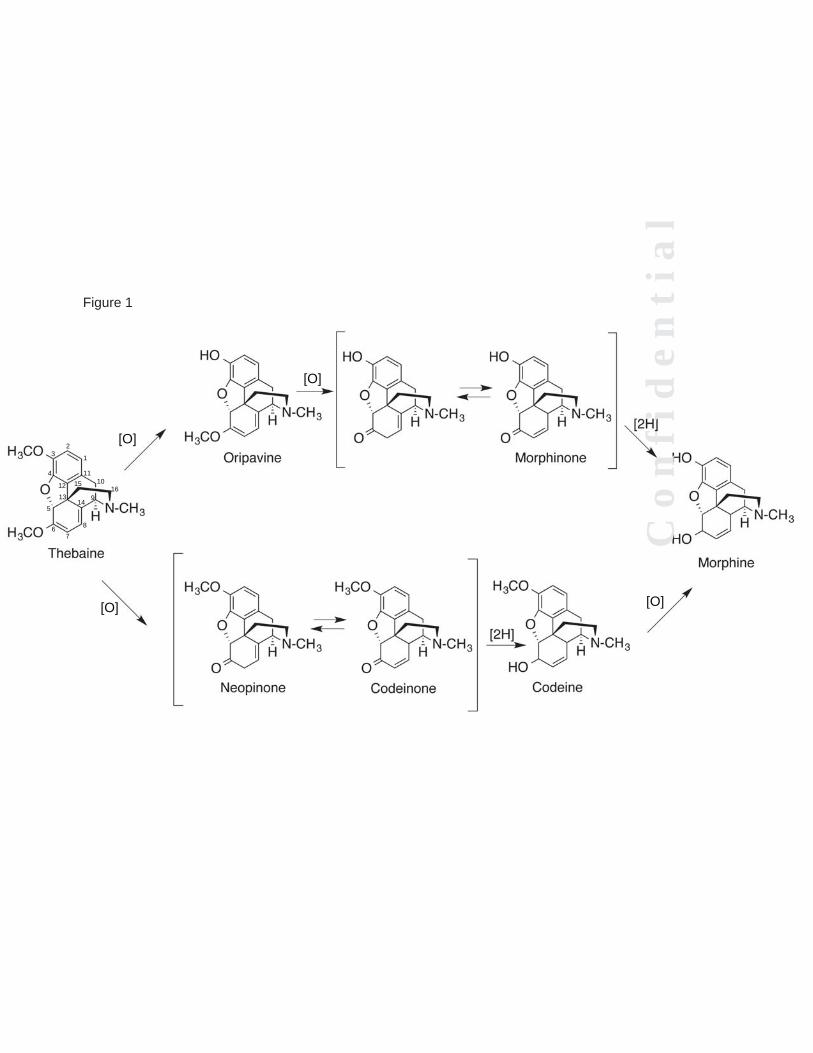
Figure 1
1
23
4
5
67
8
914
13
1011
12 1516
[O]
[O]
[O]
[O]
C o
n f
i d
e n
t i
a l

Figure 2


0 100 200 300 400 500 6000
300
600
900
1200
[Fe2+], μMHyd
roco
done
form
ed (n
mol
/incu
batio
n)
Figure 5

C o
n f
i d
e n
t i
a l
Cf
idt
il

O
NCH3
HO
HO
H
O
NCH3
H3CO
HO
H
P450
2D6
MorphineCodeine
3α,17β-OHsteroid
dehydro-genase
O
NCH3
H3CO
O
H
Codeinone
O
NCH3
H3CO
O
H
Neopinone
O
NCH3
H3CO
H3CO
H
Thebaine
P450
3A5,3A4
P4502D6
O
NCH3
HO
H3CO
H
Oripavine
NCH3
H3CO
H3CO
H
Salutaridine
O
HO
Redution of ketoneAcetyl transferaseNon-enzymatic rearrangement
NCH3
H3CO
H3CO
H
OH
HOP450
2D6,3A4
(R)-Reticuline
H3CO
H3CO
HONCH3
HO
Tetrahydropapaveroline
HO
HO
HONH
HHO
HO
HONH2 HO
+O
Dopamine 4-OH Phenylacetaldehyde(from tyrosine)
HONH2
Tyrosine
CO2H
HONH2
Tyramine
P450
2D6
O
NH
H3CO
H3CO
H
Northebaine
(N-methylasesPNMT, NMT)
P4503A4,3A5
Figure 9
H
C o
n f
i d
e n
t i
a