overview of hematology dr. gamal badr phd in immunology (paris sud university, france) associate...
TRANSCRIPT

Overview of Hematology
Dr. Gamal BadrPhD in Immunology (Paris Sud University, France)Associate Professor of ImmunologyAssiut University, EgyptTel: +2 01110900710Fax: +2 0882344642
E-mails: [email protected] or [email protected] Websites: http://www.aun.edu.eg/membercv.php?M_ID=393 https://www.researchgate.net/profil/Gamal_Badr/ http://scholar.google.com.eg/citations?hl=en&user=dz13dkQAAAAJ orcid.org/0000-0002-6157-7319

Hematology

Topics

BLOOD IS A TYPE OF CONNECTIVE TISSUE
WHAT DOES BLOOD DO?
Transportation Oxygen Nutrients Hormones Waste Products
Regulation Fluid, electrolyte Acid-Base balance Body temperature
Protection Coagulation Fight Infections

Components of Blood
Suspension of cells in a solute of water, proteins, and electrolytes
Average volume is 5 liters 70mL per kg body weight
Blood components are:A. Liquid component -Plasma (55%)
Plasma is a yellow colored solution containing a mixture of water, amino acids, proteins, clotting factors, carbohydrates, lipids, vitamins, hormones, electrolytes, and cellular wastes.

B. Cellular component (45%)
Thrombocytes/Platelets
Leukocytes/ white blood cells (WBCs)
Erythrocytes/ red blood cells (RBCs)

Complete Blood Count (CBC)
White blood cells (WBCs) Normal 4,000 -11,000 /µℓ Differential cells: Neutrophils, lymphocytes, monocytes, eosinophils,
basophils, Dendritic cells.
• Red blood cells (RBCs) ♂ 4.5 – 5.5 x 10⁶/µℓ ♀ 4.0 – 5.0 x 10⁶/µℓ
Hemoglobin (Hgb), Hematocrit (Hct) The hematocrit is the percent of whole blood that is composed of
red blood cells. The hematocrit is a measure of both the number of RBCs and the size of red blood cells.
Mean corpuscular volume (MCV) is a measure of the average volume of a red blood corpuscle
Platelets (PLT) Normal 15,000 -400,000 /µℓ

Hematopoiesis
Development of all blood cells and other formed elements
Sites vary throughout development Before birth: liver, spleen Adult: bone marrow (BM) of axial skeleton.
Stem cells Primitive; self-replicate and differentiate to become
increasingly specialized progenitor cells which form mature cells Process regulated by growth factors (interleukins,
erythropoietin, thrombopoietin, G-CSF) Early lineage division between progenitors for
lymphoid and myeloid cells

Hematopoiesis

Thrombocytes/Platelets
Must be present for clotting to occurInvolved in hemostasis

Leukocytes/White Blood Cells (WBCs)
o GranulocytesContain granules in their
cytoplasm
Basophils Eosinophils Neutrophils AgranulocytesContain no granules in their
cytoplasm
Monocytes Lymphocytes
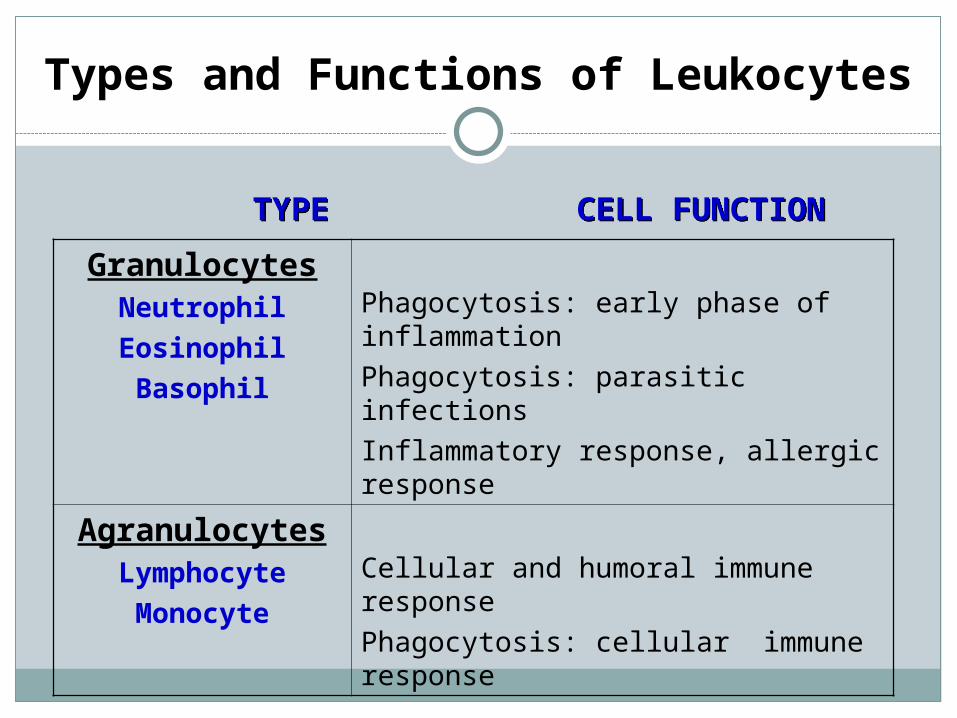
Types and Functions of Leukocytes
GranulocytesNeutrophilEosinophilBasophil
Phagocytosis: early phase of inflammationPhagocytosis: parasitic infectionsInflammatory response, allergic response
AgranulocytesLymphocyteMonocyte
Cellular and humoral immune responsePhagocytosis: cellular immune response
TYPETYPE CELL FUNCTIONCELL FUNCTION

Erythrocytes/Red Blood Cells (RBCs)
Immature RBC (circle and nucleated), while mature RBC is circle and non nucleated
Composed of hemoglobin (Hb or Hgb)Erythropoiesis is the production of RBCs
Stimulated by hypoxia (decrease of O2 in the blood) Controlled by erythropoietin (hormone synthesized in
kidney)
Hemolysis is the destruction of RBCs Releases bilirubin into blood stream Normal lifespan of RBC = 120 days

Structures of the Hematologic System
Bone Marrow a primary lymphoid organ (Hematopoiesis) • Soft connective tissue in core of bones• The production of all types of blood cells (Hematopoiesis)
generated by a remarkable self-regulated system that is responsive to the demands put upon it.
LiverReceives 24% of the cardiac output (1500 ml of blood each minute) Liver has many functions. The hematologic functions: Liver synthesis plasma proteins including clotting factors and
albumin Liver clears damaged and non-functioning RBCs/erythrocytes from
circulationSpleen a secondary lymphoid organ exerts many
functions: Hematopoietic function: Produces fetal RBCs Filter function: Filter and reuse certain cells Immune function: Lymphocytes, monocytes Storage function: 30% platelets stored in spleen

RBCs (erythrocytes)
Mature RBCs are biconcave disks that contain oxygen-carrying hemoglobin, discard their nuclei during development and so cannot reproduce or produce proteins.
In the embryo and fetus, RBCs production occurs in the liver and spleen; but after birth, it occurs in the bone marrow. Normal lifespan = 120 days
Reticulocytes (Immature red blood cells)
oCalculating proportion within circulation assists
in determining cause of anemia Normal % is 1-2% Low % (< 0.4%) suggests decreased production
(i.e. nutritional or marrow problem) High % (< 3%) suggests bleeding or premature
destruction of red blood cells (i.e. hemolysis)

Red Blood Cells
Bone marrow
Blood
RBCs
1 – 2%

RBC Production and Its Control
The total number of red blood cells remains relatively constant due to a negative feedback mechanism utilizing he hormone erythropoietin, which is released from the kidneys and liver in response to the detection of low oxygen levels.

Factors Affecting RBCs Production
Dietary Vitamins B12 and folic acid are needed for DNA synthesis, so
they are necessary for the reproduction of all body cells, especially in hematopoietic tissue.
Dietary Iron is needed for hemoglobin synthesis. Destruction of RBCs with age, RBCs become increasingly fragile and
are damaged by passing through narrow capillaries. Macrophages in the liver and spleen phagocytize damaged red blood
cells. Hemoglobin from the decomposed RBC is converted into heme and
globin. Heme is decomposed into iron which is stored or recycled and
biliverdin and bilirubin which are excreted in bile.


RBCS DISORDERS

RBCs DisordersAnemia
Anemia is defined as a reduction in one or more of the major RBC
measurements: Hgb, Hct, or RBC count
Normal Hgb concentration– 15 in males ; 14 in females
Patients are “anemic” when Hgb is < 12 g/dL
Determining reticulocyte count and MCV are first steps in determining etiology
MCV = 10 x HCT(percent) ÷ RBC numbers (millions/µL)Almost 1/3 of the world population is anemic!

RBCs Disorders - Anemia
Two main approaches that are not mutually exclusive:
1.1. Morphological approach.Morphological approach.
2.2. Biologic or kinetic approach.Biologic or kinetic approach.
Manifestations related to duration and severity of anemia:
May provide important clues as to etiology
Body has physiologic responses to chronic anemia such that many patients are asymptomatic until Hgb < 8 g/dL
Fatigue, pallor, dyspnea, dizziness, ischemic pain, cognitive abnormalities

Anemia - (Morphological approach)
By calculation from an independently-measured RBCs count and hematocrit:
MCV (femtoliters) = 10 x HCT(percent) ÷ RBC (millions/µL)
The normal MCV (76 -100 fL)Microcytosis – small cells (MCV <80) – microcytic
anemiaMacrocytosis – large cells (MCV >100) –
macrocytic anemiaNormocytic anemia
is defined as an anemia with a MCV of 80-100 which is the normal range, but the HCT and Hbg is decreased

Microcytic Anemia (MCV <76 fL)
The normal MCV (76 -100 fL)Microcytosis – small cells (MCV <80)Most common type of anemia encountered in
primary careDifferential diagnosis
Hemoglobinopathy (inherited) Iron deficiency Chronic disease (may also be normocytic) Inflammation Lead poisoning
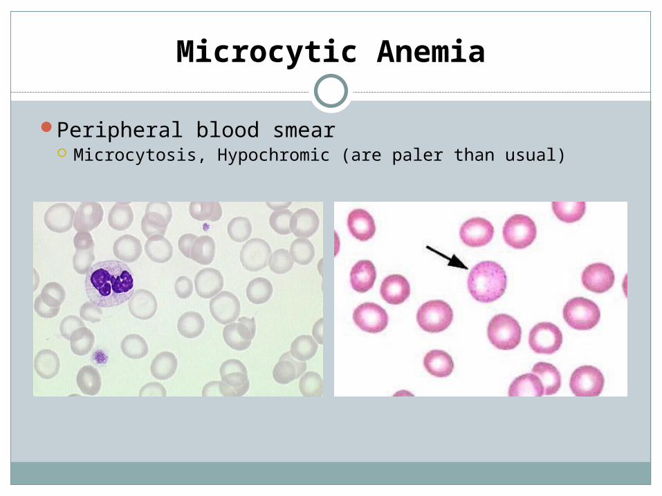
Microcytic Anemia
Peripheral blood smear Microcytosis, Hypochromic (are paler than usual)

Macrocytic Anemia
Macrocytosis – large cells (MCV >100)Differential diagnosis
B12 deficiency Pernicious anemia: (Biermer's anemia, Addison's anemia) is
caused by loss of gastric parietal cells, which are responsible for the secretion of factors that are responsible for absorption of vitamin B12 in the ileum.
Folate deficiency
Check vitamin B12, RBC folate, fasting homocysteine (HC), and methylmalonic acid (MMA) HC and MMA are elevated in subclinical B12 and
folate deficiency

Anemia - (kinetic approach)
Anemia
Production of RBCs(hypoproliferation)
Survival/Destruction of RBCs
Abnormality?
The key test is the reticulocyte count
Mechanisms of Anemia
Decreased erythrocyte production Erythrocyte loss
Decreased erythropoietin production Hemorrhage
Inadequate marrow response to erythropoietin Hemolysis

Anemia - (kinetic approach)
MechanismsShort survival/Destruction of RBCs/ loss of RBCs
Blood loss / hemorrhage Accidents Ulcer or surgery in Gastrointestinal tract (GI), menstruation
Hemolysis Shortened RBC survival time.
• Decreased production (hypoproliferative) Nutritional deficiency (iron, B12, and folate) Systemic illness (chronic kidney disease (CKD), cancer,
rheumatologic disease, etc.) Bone marrow disorders

RBCs Disorders - Anemia
Marrow productionThalassemiasMyelodysplasiaMyelophthisicAplastic anemiaNutritional
deficiencies
Red cell destruction (hemolytic)
HemoglobinopathiesEnzymopathiesMembrane disordersAutoimmune

RBCs Disorders - Red cell destruction
Causes:
Elevated reticulocyte countMechanicalAutoimmuneDrugCongenital

Red cell destruction -Hemolytic Anemia
History and physical findingsHemolytic anemias are either acquired or congenital. The laboratory
signs of hemolytic anemias include: Increased LDH (Lactate dehydrogenase). Increased bilirubin. Increased reticulocyte count. Decreased haptoglobin. Urine hemosiderin Jaundice is common Occasional pain in the left upper abdominal region. (splenomegaly)

Red cell destruction -Hemolytic Anemia
Congenital Membrane defects
Hereditary spherocytosis : auto-hemolytic anemia characterized by the production (RBCs) that are sphere-shaped, rather than bi-concave disk shaped
Hereditary elliptocytosis: also known as ovalocytosis, is an inherited blood disorder in which an abnormally large number of the patient's RBCs are elliptical rather than the typical biconcave disc shape.
Enzyme defects Glucose-6-phosphate dehydrogenase (G6PD) deficiency: X-
linked recessive hereditary disease. Pyruvate kinase deficiency: is an inherited metabolic disorder
of the enzyme Pyruvate kinase which affects the survival of RBCs and causes them to deform into echinocytes on peripheral blood smears.

Red cell destruction -Hemolytic Anemia

Red cell destruction -Hemolytic Anemia
Congenital
Hemoglobin defects: Sickle cell disease
Single base pair mutation results in a single amino acid change.
Under low oxygen, Hgb becomes insoluble forming long polymers
This leads to membrane changes (“sickling”) and vasoocclusion
OXY-STATE DEOXY-STATE

Red cell destruction -Hemolytic Anemia
Aquired Classified according to site of RBC destruction and
whether mediated by immune system Intravascular Extravascular Autoimmune Non-immune
Many causes… be aware of these – Transfusion of incompatible blood Prosthetic valves Cancer Drugs

RBCs Disorders - Anemia
Marrow productionThalassemiasMyelodysplasiaMyelophthisicAplastic anemiaNutritional
deficiencies
Red cell destruction (hemolytic)
HemoglobinopathiesEnzymopathiesMembrane disordersAutoimmune

Marrow Production - Aplastic Anemia
Aplastic anemia is a disease in which the bone marrow, and the blood stem cells that reside there, are damaged. This causes a deficiency of all three blood cell types: red blood cells (anemia), white blood cells (leukopenia), and platelets (thrombocytopenia).
Acquired
ImmunologicalToxins – BenzeneDrugs – methotrexate, chloramphenicolViruses – EBV, hepatitis
HereditaryFanconi anemia (FA) is the result of a genetic defect in a cluster of
proteins responsible for DNA repair

Marrow Production - Aplastic Anemia
All lineages affected.Most patients require red cell transfusions.Transplant when possible.Transfusions should be used selectively to
avoid sensitization (no family donors!).

Polycythemia / Erythrocytosis
Polycythemia is increased total RBC mass - Hct > 65%Above 65% blood viscosity rises exponentially
Complications: Polycythemic hyperviscosity is increased viscosity of the blood resulting from increased numbers of RBCs
Polycythemia occurs in 2-4% of newborns, half of them are symptomatic
Clinical signs result from regional effects of hyperviscosity and from the formation of microthrombi
Tissue hypoxia, Acidosis, HypoglycemiaOrgans affected: CNS, kidneys, adrenals, cardiopulmonary system, GI tractTreatment
Phlebotomy (to cut a vein) is the process of making an incision in a vein.Myelosupressive agents: new therapeutic agents such as: interferon alfa-2b (Intron A) therapy, agents that target platelet number (e.g., anagrelide [Agrylin]), and platelet function (e.g., aspirin).

Benign WBCs Disorders

Leukopenia (Leukocytopenia) -
Leukopenia: is a decrease in the number of WBCsNeutropenia is most common cause
Absolute neutrophil count (ANC) < 1.5 x 10⁹ cells/L Many causes
Benign racial neutropenia common African Americans and Yemenite Jews may have ANC as low as 1.0
Viral infections Epstein-Barr, Hepatitis B, HIV
Drugs Careful review of medications ; be suspicious of any medication
recently started in patient with acute onset neutropenia Splenomegaly Autoimmune disorders
SLE (lupus), Rheumatoid Arthritis, etc. Bone marrow disorders

Leukocytosis
Leukocytosis: is an increase in the number of WBCsWBC count > 11,000Determine which type of WBC is leading to the
leukocytosis Neutrophilia = most common
Causes: Infection Connective tissue disorders Medications (especially steroids, growth factors) Cancer Myeloproliferative disorders Cigarette smoking Stress (physiologic)
Pain, trauma Idiopathic (unknown cause)

Leukocytosis
Patients with acute bacterial infection often present with neutrophilia and band formation Bands = young neutrophils
Viral infections are usually associated with low WBCs ; leukocytosis may suggest complications Ex: bacterial pneumonia with underlying influenza
infection

Leukocytosis
Lymphocytosis: is an increase in the number or
proportion of lymphocytes in the blood Causes:
Viral infections: HBV, HCV, EBV, CMV Tuberculosis Pertussis Drug Reaction Stress (physiologic): Trauma, cardiac arrest, etc Malignancy: ALL, CLL, lymphoma

Malignant WBCs Disorders
Types of Hematopoietic Malignancies
Leukemias • Acute leukemias • Acute myeloid leukemia • Acute lymphoblastic leukemia • Chronic leukemias • Chronic myeloproliferative disorders • Chronic lymphoproliferative disorders
Lymphomas • Non-Hodgkin's lymphoma • Hodgkin's disease
Plasma cell disorders • Myeloma
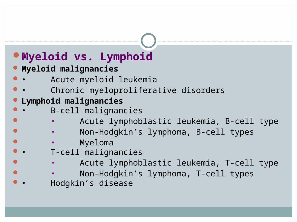
Myeloid vs. Lymphoid Myeloid malignancies • Acute myeloid leukemia • Chronic myeloproliferative disorders
Lymphoid malignancies • B-cell malignancies • Acute lymphoblastic leukemia, B-cell type • Non-Hodgkin’s lymphoma, B-cell types • Myeloma • T-cell malignancies • Acute lymphoblastic leukemia, T-cell type • Non-Hodgkin’s lymphoma, T-cell types • Hodgkin’s disease

Leukemia
Leukemia is a type of cancer of the blood or bone marrow characterized by an abnormal increase of immature WBCs called “blasts".
Leukemia is a broad term covering a spectrum of diseases. In turn, it is part of the even broader group of diseases affecting the blood, bone marrow, and lymphoid system.

Chronic Leukemia
Chronic myelogenous leukemia (CML)
Translocation between long arms of chromosomes 9
and 22 ; “Philadelphia Chromosome” ; bcr/abl protein

Chronic Leukemia
Chronic lymphocytic leukemia (CLL) Clonal malignancy of B-lymphocytes Course is usually indolent ; affects older patients, average age at
diagnosis is 70 years Often found incidentally Fatigue, lymphadenopathy common Hepatosplenomegaly
Immunodeficiency is major clinical concern Lymphocytes are defective ; do not make antibodies in response to
antigens Treatment
Observation Indications for therapy include progressive fatigue, symptomatic
lymphadenopathy, anemia, or thrombocytopenia Gamma globulin (IVIG) used in patients with recurrent or severe
bacterial infections Allogeneic BMT is potentially curative but reserved for select
patients Prognosis improving ; survival is 10-15 years with early disease

Acute Leukemia
Acute Myelogenous Leukemia (AML) Most common in adults Usually no apparent cause
Exposure to radiation, benzene, and certain chemotherapy drugs (alkylators) associated with leukemia
Underlying myelodysplastic syndrome (MDS) is risk factor Symptoms and signs
Related to replacement of marrow space by malignant WBCs Patients often very ill for period of just days or weeks Skeletal pain Bleeding Gingival hyperplasia Infection Pancytopenia with circulating blasts is hallmark ; bone
marrow biopsy required• Auer rods on peripheral smear are pathognomonic

Acute Leukemia

Lymphoma
Hodgkin’s disease Malignancy of B-lymphocytes
Reed-Sternberg cells Various subtypes ; “nodular
sclerosing” is most commonNon-Hodgkin’s Lymphoma (NHL)
Heterogeneous group of cancers affecting lymphocytes Usually classified by histologic grade (low to
high) Follicular lymphoma Small lymphocytic lymphoma Diffuse large B-cell lymphoma Burkitt’s lymphoma Many others

Myeloma
Malignancy of plasma cells Abnormal paraproteins are created leading to
systemic problems IgG – 60% IgM – 20%
Primarily disease of elderly (median age 65 years) Most common hematologic malignancy among African
Americans ; #2 among Caucasians