outcomes after implantation of superflexible nitinol ... · introdução: as intervenções...
TRANSCRIPT

Rev Bras Cardiol Invasiva. 2015;23(3):220-225
0104-1843/© 2015 Sociedade Brasileira de Hemodinâmica e Cardiologia Intervencionista. Published by Elsevier Editora Ltda. This is an open access article under the CC
BY-NC-ND license (http://creativecommons.org/licenses/by-nc-nd/4.0/).
Original Article
Outcomes after implantation of superflexible nitinol stents in the superficial
femoral artery
Patrick Bastos Metzger*, Marilia G. Volpato, Maria Claudia Folino, Fabio Henrique Rossi, Ana Claudia Gomes Petisco, Mohamed Hassan Saleh, Nilo Mitsuru Izukawa, Antonio Massamitsu Kambara
Instituto Dante Pazzanese de Cardiologia, São Paulo, SP, Brazil
DOI of original article: http://dx.doi.org/10.1016/j.rbci.2016.06.012
* Corresponding author: Avenida Dr. Dante Pazzanese, 500, Vila Mariana, CEP: 04012-180, São Paulo, SP, Brazil.
E-mail: [email protected] (P.B. Metzger).
Peer review under the responsibility of Sociedade Brasileira de Hemodinâmica e Cardiologia Intervencionista.
A B S T R A C T
Background: Endovascular interventions in the superficial femoral artery for the treatment of peripheral arterial
occlusive disease have increased over the last decades. The first- and second-generation stents in the superficial
femoral artery have failed to demonstrate improved patency of the treated vessel due to high fracture rates.
The aim of this study was to evaluate the clinical, short-term outcomes of using third-generation superflexible
nitinol stents in the treatment of atherosclerotic lesions in the superficial femoral artery.
Methods: This was a retrospective study carried out in a single center, from June 2013 to May 2014. A total
of 27 patients underwent angioplasty with third-generation, superflexible nitinol stents in atherosclerotic
lesions of the superficial femoral artery.
Results: The mean age was 68 ± 12 years, 55.6% were females, and 74.1% were diabetics. Patients were
classified as TASC B and C in 77.7% of cases. Technical success was 100%. There was an increase in the ankle-
brachial index from 0.35 ± 0.1 before the intervention to 0.75 ± 0.2 at hospital discharge. The mean follow-
up of patients was 6.7 ± 2.3 months. The primary patency rate was 96.3%. The limb salvage rate was 100%.
There were no stent fractures documented by X-rays.
Conclusions: Angioplasty with third-generation superflexible nitinol stent placement was shown to be
effective in the treatment of atherosclerotic lesions of the superficial femoral artery.
© 2015 Sociedade Brasileira de Hemodinâmica e Cardiologia Intervencionista. Published by Elsevier Editora Ltda.
This is an open access article under the CC BY-NC-ND license (http://creativecommons.org/licenses/by-nc-nd/4.0/).
Resultados do uso de stents de nitinol superflexíveis na artéria femoral superficial
R E S U M O
Introdução: As intervenções endovasculares na artéria femoral superficial para o tratamento da doença arterial
oclusiva periférica têm crescido nas últimas décadas. A primeira e a segunda geração de stents na artéria
femoral superficial falharam em demonstrar a melhora da perviedade do vaso tratado, devido às altas taxas de
fratura. O objetivo deste estudo foi avaliar os desfechos clínicos no curto prazo com o uso de stents de nitinol
superflexíveis de terceira geração no tratamento de lesões ateroscleróticas na artéria femoral superficial.
Métodos: Trata-se de um estudo retrospectivo, realizado em único centro, no período de junho de 2013 a
maio de 2014. Um total de 27 pacientes foi submetido à angioplastia com stents de nitinol superflexíveis de
terceira geração em lesões ateroscleróticas da arterial femoral superficial.
Resultados: A média de idades foi de 68 ± 12 anos, 55,6% eram do sexo feminino e 74,1%, diabéticos. Os
pacientes foram classificados em TASC B e C em 77,7% dos casos. O sucesso técnico foi de 100%. Houve
aumento do índice tornozelo-braquial de 0,35 ± 0,1 pré-intervenção para 0,75 ± 0,2 na alta hospitalar. O
seguimento médio dos pacientes foi de 6,7 ± 2,3 meses. A taxa de patência primária foi de 96,3%. A taxa de
salvamento de membro foi de 100%. Não ocorreram fraturas de stent documentadas por raios X.
Conclusões: A angioplastia com uso de stent de nitinol superflexível de terceira geração demonstrou ser
efetiva no tratamento das lesões ateroscleróricas da artéria femoral superficial.
© 2015 Sociedade Brasileira de Hemodinâmica e Cardiologia Intervencionista. Publicado por Elsevier Editora Ltda.
Este é um artigo Open Access sob a licença de CC BY-NC-ND (http://creativecommons.org/licenses/by-nc-nd/4.0/).
A R T I C L E I N F O
Article history:
Received 14 May 2015
Accepted 1 August 2015
Keywords:
Atherosclerosis
Angioplasty
Peripheral arterial disease
Femoral artery
Palavras-chave:
Aterosclerose
Angioplastia
Doença arterial periférica
Artéria femoral
2214-1235
P. Metzger et al. / Rev Bras Cardiol Invasiva. 2015;23(3):220-225 221
Table 1Classification of lesions according to the Trans-Atlantic Inter-Society Consensus II
(TASCII).
Classification Criterion
A Lesions that produce the best results and should be treated via
the endovascular route
B Lesions that produce sufficiently good results with endovascular
methods, and thus these are still the preferred approach, unless
surgical revascularization is required to treat other lesions in the
same anatomic area
C Lesions that exhibit superior long-term results with surgery, and
thus endovascular methods should be used only in patients at
high surgical risk
D Lesions that do not produce sufficiently good results with
endovascular methods to justify them as primary treatment
Introduction
Endovascular interventions for the treatment of peripheral arte-
rial occlusive disease (PAOD) have grown exponentially over the
past few decades.1 Around 40% of these procedures are performed in
the femoral segment.2 However, despite the large number, these in-
terventions still remain a challenge for the interventionists due to
the biomechanical forces exerted by the muscle compartments on
the vessel wall, promoting metal fatigue and stent fracture, in addi-
tion to restenosis.3
The first- and second-generation stents in the superficial femoral
artery failed to demonstrate improved patency of the treated vessel
when compared to conventional surgery, as even with the second-
-generation nitinol stents, fracture rates reach up to 20%.3,4
This study aimed to evaluate the short-term clinical outcomes of
using superflexible third-generation nitinol stents in the treatment
of atherosclerotic lesions in the superficial femoral artery.
Methods
Type of study and population
This was a retrospective, longitudinal, observational study car-
ried out at a referral center for cardiovascular diseases from June
2013 to May 2014. The study included patients of both genders, with
limiting intermittent claudication, pain at rest, or ulceration in the
affected limb, with lesions limited to the superficial femoral arteries
and with at least one leg artery left for distal run-off. Patients who
had a history of severe allergy to iodinated contrast, significant
atherosclerotic disease in aortoiliac territories, and with creatinine
clearance < 30 mL/kg/minute were excluded from the procedure.
Preoperative arteriography was used to classify lesions according
to: (1) the Trans-Atlantic Inter-Society Consensus II (TASC- II) A, B, C,
and D criteria5 (Table 1); (2) the type of lesion (stenosis, occlusion,
dissection, or restenosis); and (3) the location of the lesion in relation
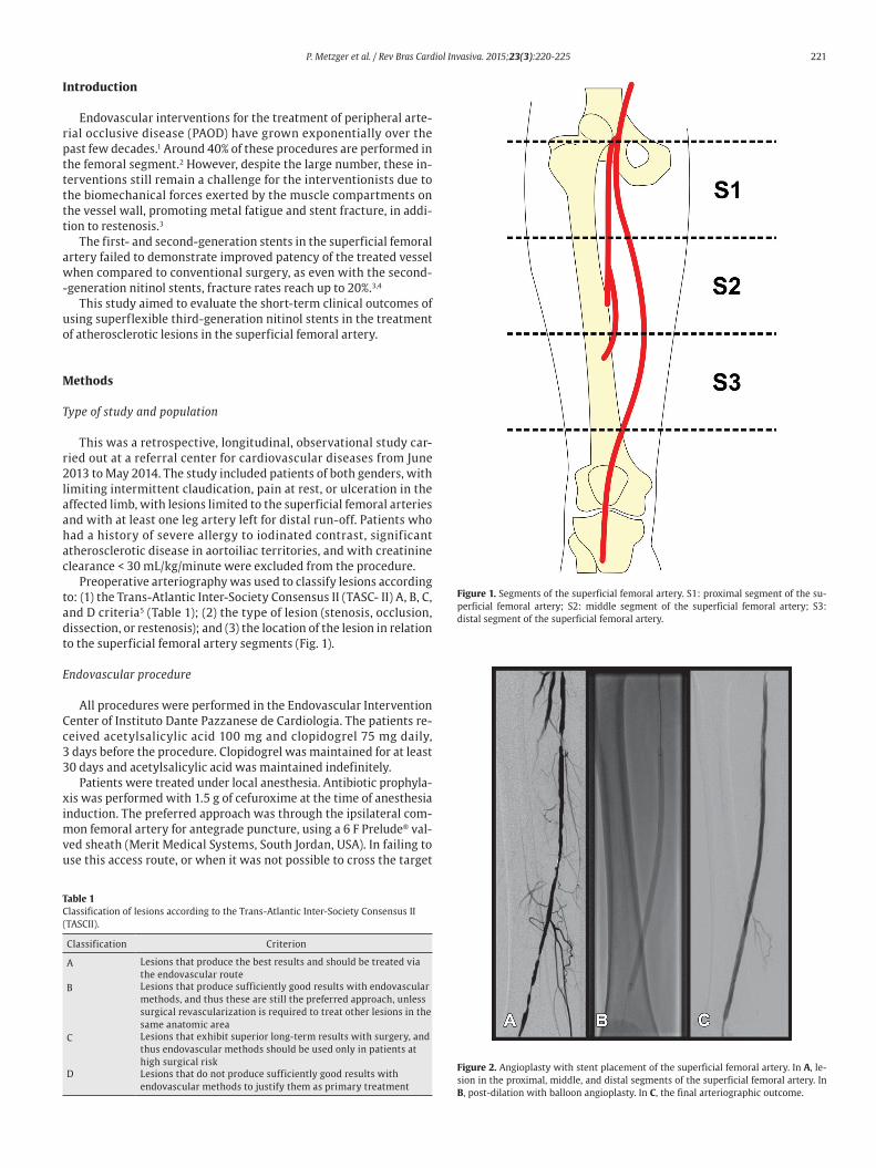
to the superficial femoral artery segments (Fig. 1).
Endovascular procedure
All procedures were performed in the Endovascular Intervention
Center of Instituto Dante Pazzanese de Cardiologia. The patients re-
ceived acetylsalicylic acid 100 mg and clopidogrel 75 mg daily,
3 days before the procedure. Clopidogrel was maintained for at least
30 days and acetylsalicylic acid was maintained indefinitely.
Patients were treated under local anesthesia. Antibiotic prophyla-
xis was performed with 1.5 g of cefuroxime at the time of anesthesia
induction. The preferred approach was through the ipsilateral com-
mon femoral artery for antegrade puncture, using a 6 F Prelude® val-
ved sheath (Merit Medical Systems, South Jordan, USA). In failing to
use this access route, or when it was not possible to cross the target
Figure 1. Segments of the superficial femoral artery. S1: proximal segment of the su-
perficial femoral artery; S2: middle segment of the superficial femoral artery; S3:
distal segment of the superficial femoral artery.
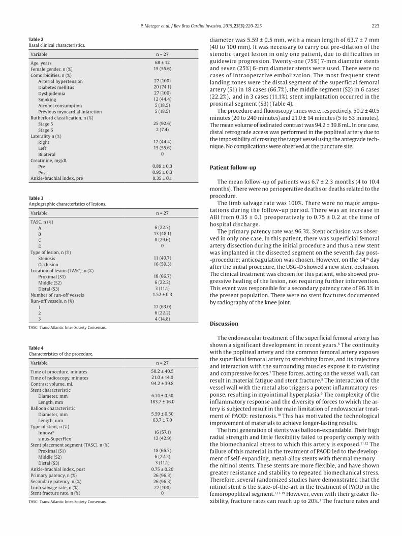
Figure 2. Angioplasty with stent placement of the superficial femoral artery. In A, le-
sion in the proximal, middle, and distal segments of the superficial femoral artery. In
B, post-dilation with balloon angioplasty. In C, the final arteriographic outcome.

222 P. Metzger et al. / Rev Bras Cardiol Invasiva. 2015;23(3):220-225
Figure 3. Doppler vascular ultrasonography. In A, stent assessment in B-mode. In B, color flow assessment. In C, spectral analysis of in-stent flow.
Figure 4. Thigh X-ray. In A, X-ray in the anteroposterior view without stent fracture
signs. In B, X-ray in the right lateral view, without stent fracture signs.
lesion, the retrograde access through popliteal artery puncture was
chosen, using a valved 5 F Prelude® sheath. Target lesions were cros-
sed through luminal or subintimal route using a 0.035' × 150 cm Ra-
diofocus® hydrophilic guidewire (Terumo Interventional Systems,
Somerset, USA) together with an MPA-1 5 F and/or STR 4 F diagnostic
catheter (Cordis Corporation, Warren, USA). Pre-dilation was perfor-
med in cases of occlusion or when adequate stent positioning was not
possible. The third-generation, superflexible sinus-SuperFlex (Opti-
med, Ettlingen, Germany) nitinol stent or Innova® self-expanding sys-
tem (Boston Scientific®, Maple Grove, USA) were used in all cases.
Radiographic control (Fig. 2) were conducted with a Siemens® Ar-
tis Flat Panel device or in the hybrid room, with a Siemens® Artis
zeego Hybrid device.
The immediate postoperative period was carried out in the infir-
mary in all cases, and local hemostasis was performed by manual
compression for 40 minutes.
Postoperative follow-up
Patients were followed through outpatient assessment with phy-
sical examination and ankle-brachial index (ABI) measurement at
15, 30, 90, and 180 days after the procedure. Control with Doppler
ultrasound (USG-D) was performed at 30, 90, and 180 days after sur-
gery, aiming to identify restenosis (Fig. 3). Radiographs of the knee
joint in the posteroanterior and lateral views were performed at 30
and 180 days, aiming to identify stent fractures (Fig. 4).
Outcomes and definitions
The analyzed outcomes were: (1) immediate technical success,
when the target lesion was treated as previously planned, with resi-
dual lesion < 30% in the angiographic control; (2) primary patency,
which indicates uninterrupted permeability after the revasculariza-
tion procedure; (3) secondary patency, which expresses the cases in
which a new intervention is performed to open the occluded vessel
after the primary procedure; (4) perioperative morbimortality for
deaths and complications recorded up to 30 days postoperatively;
(5) major amputations: transfemoral and transtibial amputations;
(6) restenosis, for in-stent lesions > 50% at USG-D, with peak systolic
velocity > 200 cm/s or pre and post-stenosis velocity ratio ≥ 2; (7)
fractures, for disconnection or twisting of stent meshes; and (8) limb
salvage rate.
Continuous variables were expressed as means ± standard devia-
tions and qualitative variables, as absolute values and percentages.
Results
A total of 81 patients underwent angioplasty and stenting in the
femoral artery segment, of whom 27 received third-generation su-
perflexible nitinol stents and constituted the study population. In 16
cases (59.3%), the Innova® self-expanding stent system was used.
The demographic characteristics, comorbidities, and treatment
indications are described in Table 2. The mean age was 68 ± 12 years;
55.6% were females, and 74.1% were diabetics. The left leg was the
most frequently treated (15 cases, 55.6%) and most had ulceration,
with little tissue loss (92.6%) (Rutherford 5). There were no cases of
treatment for intermittent claudication.
Characteristics of lesions and the procedure
Patients were classified as TASC B and C in 77.7% of cases. No pa-
tient was classified as TASC D. Regarding the location, two-thirds of
the lesions were in the distal segment of the superficial femoral ar-
tery (S3). In over half of the cases (59.3%), stents were implanted in
previously occluded vessels and pre-dilation was carried out in all
these lesions (Table 3).
When evaluating the run-off bed, most patients had only one
patent artery (59.3%), and the fibular artery was the most fre-
quently found. A mean of 1.52 ± 0.3 pervious artery per treated
limb was obtained.
Target lesion revascularization was achieved using only one
stent in 26 cases, with a technical success rate of 100%. The use of
two stents was necessary in one case. The mean length of lesion
coverage was 183.7 ± 16 mm (120 to 200 mm). The mean balloon
P. Metzger et al. / Rev Bras Cardiol Invasiva. 2015;23(3):220-225 223
diameter was 5.59 ± 0.5 mm, with a mean length of 63.7 ± 7 mm
(40 to 100 mm). It was necessary to carry out pre-dilation of the
stenotic target lesion in only one patient, due to difficulties in
guidewire progression. Twenty-one (75%) 7-mm diameter stents
and seven (25%) 6-mm diameter stents were used. There were no
cases of intraoperative embolization. The most frequent stent
landing zones were the distal segment of the superficial femoral
artery (S1) in 18 cases (66.7%), the middle segment (S2) in 6 cases
(22.2%), and in 3 cases (11.1%), stent implantation occurred in the
proximal segment (S3) (Table 4).
The procedure and fluoroscopy times were, respectively, 50.2 ± 40.5
minutes (20 to 240 minutes) and 21.0 ± 14 minutes (5 to 53 minutes).
The mean volume of iodinated contrast was 94.2 ± 39.8 mL. In one case,
distal retrograde access was performed in the popliteal artery due to
the impossibility of crossing the target vessel using the antegrade tech-
nique. No complications were observed at the puncture site.
Patient follow-up
The mean follow-up of patients was 6.7 ± 2.3 months (4 to 10.4
months). There were no perioperative deaths or deaths related to the
procedure.
The limb salvage rate was 100%. There were no major ampu-
tations during the follow-up period. There was an increase in
ABI from 0.35 ± 0.1 preoperatively to 0.75 ± 0.2 at the time of
hospital discharge.
The primary patency rate was 96.3%. Stent occlusion was obser-
ved in only one case. In this patient, there was superficial femoral
artery dissection during the initial procedure and thus a new stent
was implanted in the dissected segment on the seventh day post-
-procedure; anticoagulation was chosen. However, on the 14th day
after the initial procedure, the USG-D showed a new stent occlusion.
The clinical treatment was chosen for this patient, who showed pro-
gressive healing of the lesion, not requiring further intervention.
This event was responsible for a secondary patency rate of 96.3% in
the present population. There were no stent fractures documented
by radiography of the knee joint.
Discussion
The endovascular treatment of the superficial femoral artery has
shown a significant development in recent years.6 The continuity
with the popliteal artery and the common femoral artery exposes
the superficial femoral artery to stretching forces, and its trajectory
and interaction with the surrounding muscles expose it to twisting
and compressive forces.7 These forces, acting on the vessel wall, can
result in material fatigue and stent fracture.8 The interaction of the
vessel wall with the metal also triggers a potent inflammatory res-
ponse, resulting in myointimal hyperplasia.9 The complexity of the
inflammatory response and the diversity of forces to which the ar-
tery is subjected result in the main limitation of endovascular treat-
ment of PAOD: restenosis.10 This has motivated the technological
improvement of materials to achieve longer-lasting results.
The first generation of stents was balloon-expandable. Their high
radial strength and little flexibility failed to properly comply with
the biomechanical stress to which this artery is exposed.11,12 The
failure of this material in the treatment of PAOD led to the develop-
ment of self-expanding, metal-alloy stents with thermal memory –
the nitinol stents. These stents are more flexible, and have shown
greater resistance and stability to repeated biomechanical stress.
Therefore, several randomized studies have demonstrated that the
nitinol stent is the state-of-the-art in the treatment of PAOD in the
femoropopliteal segment.1,13-19 However, even with their greater fle-
xibility, fracture rates can reach up to 20%.3 The fracture rates and
Table 2Basal clinical characteristics.
Variable n = 27
Age, years 68 ± 12
Female gender, n (%) 15 (55.6)
Comorbidities, n (%)
Arterial hypertension 27 (100)
Diabetes mellitus 20 (74.1)
Dyslipidemia 27 (100)
Smoking 12 (44.4)
Alcohol consumption 5 (18.5)
Previous myocardial infarction 5 (18.5)
Rutherford classification, n (%)
Stage 5 25 (92.6)
Stage 6 2 (7.4)
Laterality n (%)
Right 12 (44.4)
Left 15 (55.6)
Bilateral 0
Creatinine, mg/dL
Pre 0.89 ± 0.3
Post 0.95 ± 0.3
Ankle-brachial index, pre 0.35 ± 0.1
Table 3Angiographic characteristics of lesions.
Variable n = 27
TASC, n (%)
A 6 (22.3)
B 13 (48.1)
C 8 (29.6)
D 0
Type of lesion, n (%)
Stenosis 11 (40.7)
Occlusion 16 (59.3)
Location of lesion (TASC), n (%)
Proximal (S1) 18 (66.7)
Middle (S2) 6 (22.2)
Distal (S3) 3 (11.1)
Number of run-off vessels 1.52 ± 0.3
Run-off vessels, n (%)
1 17 (63.0)
2 6 (22.2)
3 4 (14.8)
TASC: Trans-Atlantic Inter-Society Consensus.
Table 4Characteristics of the procedure.
Variable n = 27
Time of procedure, minutes 50.2 ± 40.5
Time of radioscopy, minutes 21.0 ± 14.0
Contrast volume, mL 94.2 ± 39.8
Stent characteristic
Diameter, mm 6.74 ± 0.50
Length, mm 183.7 ± 16.0
Balloon characteristic
Diameter, mm 5.59 ± 0.50
Length, mm 63.7 ± 7.0
Type of stent, n (%)
Innova® 16 (57.1)
sinus-SuperFlex 12 (42.9)
Stent placement segment (TASC), n (%)
Proximal (S1) 18 (66.7)
Middle (S2) 6 (22.2)
Distal (S3) 3 (11.1)
Ankle-brachial index, post 0.75 ± 0.20
Primary patency, n (%) 26 (96.3)
Secondary patency, n (%) 26 (96.3)
Limb salvage rate, n (%) 27 (100)
Stent fracture rate, n (%) 0
TASC: Trans-Atlantic Inter-Society Consensus.

224 P. Metzger et al. / Rev Bras Cardiol Invasiva. 2015;23(3):220-225
Table 5Fracture rates in recent studies of percutaneous intervention with second- and third-generation nitinol stent in the superficial femoral artery.
Studies Year Stent n Lesion length
(cm)
Primary patency
in 12 months
Fracture rate
(%)
SIROCCO3 2006 SMART 46 8.1 68.1 (2 years) 20
SUPERA 5006* 2013 Supera 490 12.6 83.3 0
RESILIENT14 2010 Life stent 134 7.1 81.3 3.1
FAST15 2007 Luminexx 3 101 4.5 68.3 12
SUMMIT20* 2013 Epic 100 7 85.1 0
COMPLETE SE21* 2014 Complete SE 196 6.1 72.6 0
MISAGO 222 2012 Misago 744 6.4 87.6 3.1
DURABILITY 20024 2011 Everflex 100 24.2 64.8 6
* Studies with third-generation nitinol stents.
the consequent restenoses led to the development of the third-gene-
ration stents, which had the stent cell interconnection structures
modified, resulting in greater flexibility and lower fracture rates
(Table 5).6,20-22
Three of the most recent studies have shown better results
with the newest generation of stents. The SUMMIT20 study was a
prospective, multicenter trial of the self-expanding, laser-cut ni-
tinol stent EPIC® (Boston Scientific, Maple Grove, USA), which
showed a 15.7% restenosis rate, with primary patency of 92% and
no fractures in the radiographic follow-up in the one-year period.
The COMPLETE SE21 multicentric study, using the Complete SE
stent (Medtronic, Minneapolis, USA), showed primary patency
rate of 72.6%, with a restenosis rate of 8.4% and no evidence of
fractures in the 1-year follow up. The SUPERA 5006 study assessed
490 patients using the SUPERA® stent (IDEV Technologies Inc, We-
bster, USA), resulting in a primary patency of 83.3% and no fractu-
res in the 1-year follow-up period of 304 stents with radiographic
follow-up. In the present study, the use of two types of third-ge-
neration, superf lexible nitinol stents led to a primary patency
rate of 96.3%, with an occlusion rate of 3.7% due to the dissection
of the femoral artery in the initial procedure, and no signs of sig-
nificant restenosis or fracture in the 6 month follow-up observed
with the USG-D and X-rays, respectively.
Currently, the only available data is from the TASC II5 consensus
recommending the femoropopliteal angioplasty as the first choice in
the categories TASC A and B; as a second option for the TASC C cate-
gory; and not recommended for category D. However, this consen-
sus already shows a significant gap due to the remarkable evolution
of techniques and materials in recent years. It is a growing opinion
that the TASC II needs to be revised, especially regarding angioplasty
indications of AFS, a view shared by the present authors. In this stu-
dy, 18 cases (66.7%) were classified as TASC A and B, but in 9 cases
(33.3%), angioplasty with third-generation stent was performed in
TASC C arteriographic lesions as the first option. Goltz et al.23 obser-
ved that TASC A and B patients obtained 79% of primary patency and
those with TASC C and D obtained 68% of patency at 1 year. Of this
group, 87% of patients had previous occlusion, and the rate of proce-
dure-related complications was 7.5%. In the DURABILITY-200 stu-
dy,24 100 patients were assessed, of whom 71% were treated for
intermittent claudication and 29% for critical ischemia, with lesions
classified as TASC C and D, in the femoropopliteal segment, obtai-
ning a primary patency rate of 85.4% at 6 months and of 64.8% at 12
months. In the present study, most stents were implanted in the dis-
tal segment (S3) of the superficial femoral artery. We observed pri-
mary and secondary patency of 96.3% at six months of follow-up in a
population consisting of patients with critical ischemia Rutherford
stages 4 and 5. In the subgroup of patients TASC C, there were no
occlusion events or restenosis during the 6 month follow-up, and no
TASC D patient was treated.
During the clinical follow-up of patients, there was an improve-
ment in the ABI from 0.35 ± 0.1 preoperatively to 0.75 ± 0.2 at the
time of hospital discharge, in addition to a limb salvage rate of 100%.
ABI measurement may have been overestimated due to the high pre-
valence of diabetes in the present population, because the prevalen-
ce of arterial calcification in the distal segments of the leg arteries of
diabetic patients overestimates the measurement of this index.25
Study limitations
The small number of cases, the heterogeneous group of treated
segments in the superficial femoral artery and the short clinical
follow-up, may have compromised the results of the present study.
Additionally, the accuracy of the results may have been affected by
the retrospective analysis of data.
Conclusions
In the present study, angioplasty using a third-generation super-
flexible nitinol stent was shown to be effective in the treatment of
atherosclerotic lesions of the superficial femoral artery. The patency
rates in the treated artery demonstrate the need for stringent clini-
cal follow-up of these patients in the medium and long terms.
Funding source
None declared.
Conflicts of interest
The authors declare no conflicts of interest.
References
1. Anderson PL, Gelijns A, Moskowitz A, Arons R, Gupta L, Weinberg A, et al. Understanding trends in inpatient surgical volume: vascular interventions, 1980-2000. J Vasc Surg. 2004;39(6):1200-8.
2. Kandarpa K, Becker GJ, Hunink MG, McNamara TO, Rundback JH, Trost DW, et al. Transcatheter interventions for the treatment of peripheral atherosclerotic lesions: part I. J Vasc Interv Radiol. 2001;12(6):683-95.
3. Duda SH, Bosiers M, Lammer J, Scheinert D, Zeller T, Oliva V, et al. Drug-eluting and bare nitinol stents for the treatment of atherosclerotic lesions in the superficial femoral artery: long-term results from the SIROCCO trial. J Endovasc Ther. 2006;13(6):701-10.
4. Davaine JM, Azéma L, Guyomarch B, Chaillou P, Costargent A, Patra P, et al. One-year clinical outcome after primary stenting for Trans-Atlantic Inter-Society Consensus (TASC) C and D femoropopliteal lesions (the STELLA “STEnting Long de L'Artère fémorale superf icielle” cohort). Eur J Vasc Endovasc Surg. 2012;44(4):432-41.
5. Norgren L, Hiatt WR, Dormandy JA, Nehler MR, Harris KA, Fowkes FG, et al. Inter-society consensus for the management of peripheral arterial disease (TASC II). Eur J Vasc Endovasc Surg. 2007;33 Suppl 1:S1-75
6. Werner M, Paetzold A, Banning-Eichenseer U, Scheinert S, Piorkowski M, Ulrich M, et al. Treatment of complex atherosclerotic femoropopliteal artery disease with a self-expanding interwoven nitinol stent: midterm results from the Leipzig SUPERA 500 registry. EuroIntervention. 2014;10(7):861-8.

P. Metzger et al. / Rev Bras Cardiol Invasiva. 2015;23(3):220-225 225
7. Scheinert D, Scheinert S, Sax J, Piorkowski C, Bräunlich S, Ulrich M, et al. Prevalence and clinical impact of stent fractures after femoropopliteal stenting. J Am Coll Cardiol. 2005;45(2):312-5.
8. Davaine JM, Quérat J, Guyomarch B, Brennan MÁ, Costargent A, Chaillou P, et al. Incidence and the clinical impact of stent fractures after primary stenting for TASC C and D femoropopliteal lesions at 1 year. Eur J Vasc Endovasc Surg. 2013;46(2):201-12.
9. Schillinger M, Exner M, Mlekusch W, Haumer M, Ahmadi R, Rumpold H, et al. Balloon angioplasty and stent implantation induce a vascular inflammatory reaction. J Endovasc Ther. 2002;9(1):59-66.
10. Schillinger M, Sabeti S, Loewe C, Dick P, Amighi J, Mlekusch W, et al. Balloon angioplasty versus implantation of nitinol stents in the superficial femoral artery. N Engl J Med. 2006;354(18):1879-88.
11. Spoelstra H, Casselman F, Lesceu O. Balloon-expandable endobypass for femoropopliteal atherosclerotic occlusive disease. A preliminary evaluation of fifty-five patients. J Vasc Surg. 1996;24(4):647-54.
12. E Y, He N, Wang Y, Fan H. Percutaneous transluminal angioplasty (PTA) alone versus PTA with balloon-expandable stent placement for short-segment femoropopliteal artery disease: a metaanalysis of randomized trials. J Vasc Interv Radiol. 2008;19(4):499-503.
13. Zeller T, Tiefenbacher C, Steinkamp HJ, Langhoff R, Wittenberg G, Schlüter M, et al. Nitinol stent implantation in TASC A and B superficial femoral artery lesions: the Femoral Artery Conformexx Trial (FACT). J Endovasc Ther. 2008;15(4):390-8.
14. Laird JR, Katzen BT, Scheinert D, Lammer J, Carpenter J, Buchbinder M, et al.; RESILIENT Investigators. Nitinol stent implantation versus balloon angioplasty for lesions in the superficial femoral artery and proximal popliteal artery: twelve-month results from the RESILIENT randomized trial. Circ Cardiovasc Interv. 2010;3(3):267-76.
15. Krankenberg H, Schlüter M, Steinkamp HJ, Bürgelin K, Scheinert D, Schulte KL, et al. Nitinol stent implantation versus percutaneous transluminal angioplasty in superficial femoral artery lesions up to 10 cm in length: the femoral artery stenting trial (FAST). Circulation. 2007;116(3):285-92.
16. Bosiers M, Torsello G, Gissler HM, Ruef J, Müller-Hülsbeck S, Jahnke T, et al. Nitinol stent implantation in long superficial femoral artery lesions: 12-month results of the DURABILITY I study. J Endovasc Ther. 2009;16(3):261-9.
17. Dick P, Wallner H, Sabeti S, Loewe C, Mlekusch W, Lammer J, et al. Balloon angioplasty versus stenting with nitinol stents in intermediate length superficial femoral artery lesions. Catheter Cardiovasc Interv. 2009;74(7):1090-5.
18. Katsanos K, Spiliopoulos S, Karunanithy N, Krokidis M, Sabharwal T, Taylor P. Bayesian network meta-analysis of nitinol stents, covered stents, drug-eluting stents, and drug-coated balloons in the femoropopliteal artery. J Vasc Surg. 2014;59(4):1123-8.
19. Laird JR, Katzen BT, Scheinert D, Lammer J, Carpenter J, Buchbinder M, et al.; RESILIENT Investigators. Nitinol stent implantation vs. balloon angioplasty for lesions in the superficial femoral and proximal popliteal arteries of patients with claudication: three-year follow-up from the RESILIENT randomized trial. J Endovasc Ther. 2012;19(1):1-9.
20. Werner M, Piorkowski M, Thieme M, Nanning T, Beschorner U, Rastan A, et al. SUMMIT registry: one-year outcomes after implantation of the EPIC self-expanding nitinol stent in the femoropopliteal segment. J Endovasc Ther. 2013;20(6):759-66.
21. Laird JR, Jain A, Zeller T, Feldman R, Scheinert D, Popma JJ, et al. Complete SE Investigators. Nitinol stent implantation in the superficial femoral artery and proximal popliteal artery: twelve-month results from the complete SE multicenter trial. J Endovasc Ther. 2014;21(2):202-12.
22. Schulte KL, Kralj I, Gissler HM, Bagnaschino LA, Buschmann I, Pernès JM, et al. MISAGO 2: one-year outcomes after implantation of the Misago self-expanding nitinol stent in the superficial femoral and popliteal arteries of 744 patients. J Endovasc Ther. 2012;19(6):774-84.
23. Goltz JP, Ritter CO, Petritsch B, Kellersmann R, Hahn D, Kickuth R. Endovascular treatment of acute limb ischemia secondary to fracture of a popliteal artery stent. J Vasc Interv Radiol. 2010;21(11):1739-45.
24. Bosiers M, Deloose K, Callaert J, Moreels N, Keirse K, Verbist J, et al. Results of the P rotégé EverF lex 20 0 -mm-long n it inol stent (ev3) in TA SC C and D femoropopliteal lesions. J Vasc Surg. 2011;54(4):1042-50.
25. Rodrigues TO, Metzger PB, Rossi FH. Outcomes of the use of a superflexible nitinol stent in the popliteal artery. Rev Bras Cardiol Invas. 2014;22(2):161-7.