original article vitamin d deficiency …thorax.bmj.com/content/thoraxjnl/70/7/617.full.pdforiginal...
TRANSCRIPT

ORIGINAL ARTICLE
Vitamin D deficiency contributes directly to theacute respiratory distress syndrome (ARDS)Rachel C A Dancer,1 Dhruv Parekh,1 Sian Lax,1 Vijay D’Souza,1 Shengxing Zheng,1
Chris R Bassford,1 Daniel Park,1 D G Bartis,1 Rahul Mahida,1 Alice M Turner,1
Elizabeth Sapey,1 Wenbin Wei,2 Babu Naidu,1 Paul M Stewart,3 William D Fraser,4
Kenneth B Christopher,5 Mark S Cooper,6 Fang Gao,1 David M Sansom,7
Adrian R Martineau,8 Gavin D Perkins,9 David R Thickett1
▸ Additional material ispublished online only. To viewplease visit the journal online(http://dx.doi.org/10.1136/thoraxjnl-2014-206680).
For numbered affiliations seeend of article.
Correspondence toProfessor David Thickett,Centre for TranslationalInflammation and FibrosisResearch, Queen ElizabethHospital, Universityof Birmingham, BirminghamB15 2TH, UK;[email protected]
RCAD and DP are joint firstauthor of this paper.
Received 10 December 2014Revised 10 March 2015Accepted 2 April 2015Published Online First1 May 2015
To cite: Dancer RCA,Parekh D, Lax S, et al.Thorax 2015;70:617–624.
ABSTRACTRationale Vitamin D deficiency has been implicated asa pathogenic factor in sepsis and intensive therapy unitmortality but has not been assessed as a risk factor foracute respiratory distress syndrome (ARDS). Causality ofthese associations has never been demonstrated.Objectives To determine if ARDS is associated withvitamin D deficiency in a clinical setting and todetermine if vitamin D deficiency in experimental modelsof ARDS influences its severity.Methods Human, murine and in vitro primary alveolarepithelial cell work were included in this study.Findings Vitamin D deficiency (plasma 25(OH)D levels<50 nmol/L) was ubiquitous in patients with ARDS andpresent in the vast majority of patients at risk ofdeveloping ARDS following oesophagectomy. In amurine model of intratracheal lipopolysaccharidechallenge, dietary-induced vitamin D deficiency resultedin exaggerated alveolar inflammation, epithelial damageand hypoxia. In vitro, vitamin D has trophic effects onprimary human alveolar epithelial cells affecting >600genes. In a clinical setting, pharmacological repletion ofvitamin D prior to oesophagectomy reduced theobserved changes of in vivo measurements of alveolarcapillary damage seen in deficient patients.Conclusions Vitamin D deficiency is common inpeople who develop ARDS. This deficiency of vitamin Dappears to contribute to the development of thecondition, and approaches to correct vitamin Ddeficiency in patients at risk of ARDS should bedeveloped.Trial registration UKCRN ID 11994.
INTRODUCTIONAcute respiratory distress syndrome (ARDS) occursdue to either direct or indirect proinflammatoryinsults. However, only a proportion of at-riskpatients develop ARDS, with research suggestingthat genetic, age, social and other factors play arole in determining who develops ARDS.1
More than 1 billion people worldwide arebelieved to have vitamin D deficiency.2 Vitamin Dhas important functions besides bone and calciumhomeostasis3 with cells of the innate and adaptiveimmune system responding to vitamin D. Vitamin
D deficiency may therefore increase the risk of bac-terial and viral infection.Vitamin D deficiency is associated with an
increased risk of intensive care admission and mor-tality in patients with pneumonia.4 Deficiency iscommon in critically ill patients and associatedwith adverse outcome.3 Gram-positive bacteria,invasive pneumococcal disease and meningococcaldisease are more common when 25(OH)D3 levelsare low.5 Recent data from an Austrian study incritically ill deficient patients suggests that whentreatment with vitamin D is successful in raisinglevels >75 nmol/L there is a mortality benefit.6
Vitamin D may improve outcomes by reducingboth local and systemic inflammatory responses as aresult of modulating cytokine responses.7 In a mousemodel of lethal endotoxaemia, survival post intraven-ous lipopolysaccharide (LPS) was significantly poorerin the vitamin D receptor knockout mice.8
Our aim was to define the prevalence and sever-ity of vitamin D deficiency in patients with ARDSand to establish whether vitamin D deficiency is arisk factor for and/or a driver of the exaggeratedand persistent inflammation that is a hallmark ofARDS. To achieve these aims, we employed transla-tional clinical studies and in vitro primary cellwork and murine models.
Open AccessScan to access more
free content
Key messages
What is the key question?▸ Is vitamin D deficiency a risk factor for the
development of acute respiratory distresssyndrome (ARDS)?
What is the bottom line?▸ Patients with and at risk of ARDS are highly
likely to be deficient, and severity of vitamin Ddeficiency relates to increased epithelialdamage, the development of ARDS andsurvival.
Why read on?▸ We present evidence that an easily treatable
vitamin deficiency may increase the risk ofARDS in patients at risk.
Dancer RCA, et al. Thorax 2015;70:617–624. doi:10.1136/thoraxjnl-2014-206680 617
Critical care on 31 M
ay 2018 by guest. Protected by copyright.
http://thorax.bmj.com
/T
horax: first published as 10.1136/thoraxjnl-2014-206680 on 22 April 2015. D
ownloaded from
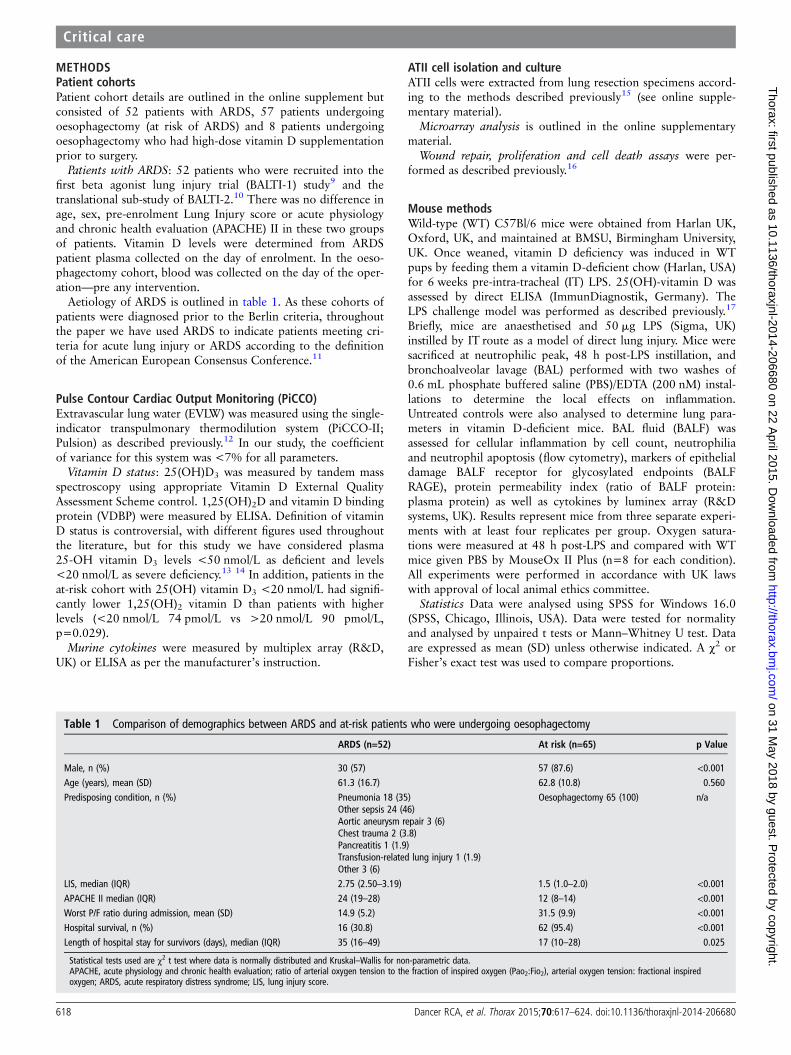
METHODSPatient cohortsPatient cohort details are outlined in the online supplement butconsisted of 52 patients with ARDS, 57 patients undergoingoesophagectomy (at risk of ARDS) and 8 patients undergoingoesophagectomy who had high-dose vitamin D supplementationprior to surgery.
Patients with ARDS: 52 patients who were recruited into thefirst beta agonist lung injury trial (BALTI-1) study9 and thetranslational sub-study of BALTI-2.10 There was no difference inage, sex, pre-enrolment Lung Injury score or acute physiologyand chronic health evaluation (APACHE) II in these two groupsof patients. Vitamin D levels were determined from ARDSpatient plasma collected on the day of enrolment. In the oeso-phagectomy cohort, blood was collected on the day of the oper-ation—pre any intervention.
Aetiology of ARDS is outlined in table 1. As these cohorts ofpatients were diagnosed prior to the Berlin criteria, throughoutthe paper we have used ARDS to indicate patients meeting cri-teria for acute lung injury or ARDS according to the definitionof the American European Consensus Conference.11
Pulse Contour Cardiac Output Monitoring (PiCCO)Extravascular lung water (EVLW) was measured using the single-indicator transpulmonary thermodilution system (PiCCO-II;Pulsion) as described previously.12 In our study, the coefficientof variance for this system was <7% for all parameters.
Vitamin D status: 25(OH)D3 was measured by tandem massspectroscopy using appropriate Vitamin D External QualityAssessment Scheme control. 1,25(OH)2D and vitamin D bindingprotein (VDBP) were measured by ELISA. Definition of vitaminD status is controversial, with different figures used throughoutthe literature, but for this study we have considered plasma25-OH vitamin D3 levels <50 nmol/L as deficient and levels<20 nmol/L as severe deficiency.13 14 In addition, patients in theat-risk cohort with 25(OH) vitamin D3 <20 nmol/L had signifi-cantly lower 1,25(OH)2 vitamin D than patients with higherlevels (<20 nmol/L 74 pmol/L vs >20 nmol/L 90 pmol/L,p=0.029).
Murine cytokines were measured by multiplex array (R&D,UK) or ELISA as per the manufacturer’s instruction.
ATII cell isolation and cultureATII cells were extracted from lung resection specimens accord-ing to the methods described previously15 (see online supple-mentary material).
Microarray analysis is outlined in the online supplementarymaterial.
Wound repair, proliferation and cell death assays were per-formed as described previously.16
Mouse methodsWild-type (WT) C57Bl/6 mice were obtained from Harlan UK,Oxford, UK, and maintained at BMSU, Birmingham University,UK. Once weaned, vitamin D deficiency was induced in WTpups by feeding them a vitamin D-deficient chow (Harlan, USA)for 6 weeks pre-intra-tracheal (IT) LPS. 25(OH)-vitamin D wasassessed by direct ELISA (ImmunDiagnostik, Germany). TheLPS challenge model was performed as described previously.17
Briefly, mice are anaesthetised and 50 mg LPS (Sigma, UK)instilled by IT route as a model of direct lung injury. Mice weresacrificed at neutrophilic peak, 48 h post-LPS instillation, andbronchoalveolar lavage (BAL) performed with two washes of0.6 mL phosphate buffered saline (PBS)/EDTA (200 nM) instal-lations to determine the local effects on inflammation.Untreated controls were also analysed to determine lung para-meters in vitamin D-deficient mice. BAL fluid (BALF) wasassessed for cellular inflammation by cell count, neutrophiliaand neutrophil apoptosis (flow cytometry), markers of epithelialdamage BALF receptor for glycosylated endpoints (BALFRAGE), protein permeability index (ratio of BALF protein:plasma protein) as well as cytokines by luminex array (R&Dsystems, UK). Results represent mice from three separate experi-ments with at least four replicates per group. Oxygen satura-tions were measured at 48 h post-LPS and compared with WTmice given PBS by MouseOx II Plus (n=8 for each condition).All experiments were performed in accordance with UK lawswith approval of local animal ethics committee.
Statistics Data were analysed using SPSS for Windows 16.0(SPSS, Chicago, Illinois, USA). Data were tested for normalityand analysed by unpaired t tests or Mann–Whitney U test. Dataare expressed as mean (SD) unless otherwise indicated. A χ2 orFisher’s exact test was used to compare proportions.
Table 1 Comparison of demographics between ARDS and at-risk patients who were undergoing oesophagectomy
ARDS (n=52) At risk (n=65) p Value
Male, n (%) 30 (57) 57 (87.6) <0.001Age (years), mean (SD) 61.3 (16.7) 62.8 (10.8) 0.560Predisposing condition, n (%) Pneumonia 18 (35)
Other sepsis 24 (46)Aortic aneurysm repair 3 (6)Chest trauma 2 (3.8)Pancreatitis 1 (1.9)Transfusion-related lung injury 1 (1.9)Other 3 (6)
Oesophagectomy 65 (100) n/a
LIS, median (IQR) 2.75 (2.50–3.19) 1.5 (1.0–2.0) <0.001APACHE II median (IQR) 24 (19–28) 12 (8–14) <0.001Worst P/F ratio during admission, mean (SD) 14.9 (5.2) 31.5 (9.9) <0.001Hospital survival, n (%) 16 (30.8) 62 (95.4) <0.001Length of hospital stay for survivors (days), median (IQR) 35 (16–49) 17 (10–28) 0.025
Statistical tests used are χ2 t test where data is normally distributed and Kruskal–Wallis for non-parametric data.APACHE, acute physiology and chronic health evaluation; ratio of arterial oxygen tension to the fraction of inspired oxygen (Pao2:Fio2), arterial oxygen tension: fractional inspiredoxygen; ARDS, acute respiratory distress syndrome; LIS, lung injury score.
618 Dancer RCA, et al. Thorax 2015;70:617–624. doi:10.1136/thoraxjnl-2014-206680
Critical care on 31 M
ay 2018 by guest. Protected by copyright.
http://thorax.bmj.com
/T
horax: first published as 10.1136/thoraxjnl-2014-206680 on 22 April 2015. D
ownloaded from

To test the hypothesis that low 25(OH)D levels are associatedwith the development of ARDS in the at-risk oesophagectomycohort (N=65), we performed multivariable logistic regressionwith the exposure of interest being 25(OH)D3 level <20 nmol/Land ARDS as the outcome. Adjusted ORs were estimated by multi-variable logistic regression models with inclusion of covariateterms chosen, a priori, thought to be plausibly associated withboth 25(OH)D3 level and ARDS in the oesophagectomy patientcohort. We sought to build a parsimonious model that did notunnecessarily adjust for covariates that did not affect bias or thecausal relation between exposure and outcome. Model calibrationwas assessed using the Hosmer–Lemeshow (HL) χ2 goodness-of-fittest and the accompanying p value. Bayesian information criterionand Akaike information criterion were also used to determineglobal model fit. Covariates included in the logistic regressionmodel were age, gender, diagnosis, staging and pack-yearssmoked. The discriminatory ability for ARDS was quantified usingthe c-statistic. In all analyses, p values are two-tailed and valuesbelow 0.05 were considered statistically significant.
RESULTSPlasma vitamin D status in patients with or at risk of ARDSPatients with ARDS (100%) were vitamin D-deficient (plasma25(OH)D3 <50 nmol/L). In total, 55 (96%) out of 57
unsupplemented oesophagectomy patients at risk of ARDS weredeficient preoperatively but levels were higher than in patientswith ARDS. Both patients at risk and patients with ARDS hadsignificantly lower levels of 25(OH)D3 than normal controls(see figure 1). Demographics of patients groups based onvitamin D levels are illustrated in table 2.
In at-risk patients, preoperative median plasma levels of 25(OH)D3 were significantly lower in those patients who were ven-tilated with ARDS postoperatively (ARDS 16.97 nmol/L vs noARDS 25.46 nmol/L, p=0.014). Oesophagectomy patients withsevere vitamin D deficiency (plasma 25(OH)D3 <20 nmol/L)had a 37.5% risk of postoperative lung injury as opposed to a15% risk with vitamin D levels >20 nmol/L (figure 2).
In the at-risk oesophagectomy cohort, preoperative vitamin Dstatus was the only measure to have a significant difference inoesophagectomy patients who develop lung injury postopera-tively (see table 3).
The odds of ARDS in patients with 25(OH)D3 <20 nmol/Lwas 3.5-fold that of patients with 25(OH)D3 ≥20 nmol/L(OR=3.5 (95% CI 1.06 to 11.6; p=0.040)). Following adjust-ment for gender, age, diagnosis, staging data, and pack-years,patients with 25(OH)D3 <20 nmol/L had a 4.2-fold higherodds of ARDS than patients with 25(OH)D <20 nmol/L(OR=4.2 (95% CI 1.13 to 15.9; p=0.032)). The adjustedmodel showed good calibration (HL χ2 11.10, p=0.20) and dis-crimination for ARDS (area under the curve 0.73). When 25(OH)D was analysed with logistic regression as a continuous
Figure 1 Plasma 25(OH)D3 levels in acute respiratory distresssyndrome (ARDS) versus at risk and normal controls. The horizontal barrepresents the median, and the boxes represent IQRs. Vertical linesshow minimum–maximum range. Fifty-two patients with ARDS, 57at-risk patients undergoing oesophagectomy, 18 healthy controls.
Table 2 Comparison of demographics between patients with severe deficiency, moderate deficiency and vitamin D supplemented at-riskpatients undergoing oesophagectomy
Patients with severe25-OH vitamin D3
deficiency (n=25)
Patients with moderate25-OH vitamin D3
deficiency (n=32)
Patients who receivedvitamin D supplementation(n=8) p Value
Male, n (%) 21 (84) 28 (87.5) 8 (100) 0.706Age, yearsmedian (IQR)
60.0 (52.0–68.5) 65.5 (54.5–72.0) 68.0 (63.8–71.3) 0.122
BMI median (IQR) 24.3 (20.6–27.9) 25.4 (21.7–28.3) 24.1 (22.0–26.6) 0.698ASA median (IQR) 2.0 (2.0–2.0) 2.0 (2.0–2.0) 2.0 (2.0–2.75) 0.950Postoperative P/F ratio 41.0 (34.3–53.3) 39.8 (32.0–52.0) 47.8 (39.4–50.8) 0.476Preoperative plasma 25-OH vitamin D3 level (nmol/L)median (IQR)
13.7 (10.9–16.7) 27.6 (22.5–34.9) 66.9 (42.5–92.6) <0.001
All p values shown are Kruskal–Wallis tests from SPSS.ASA, American Society of Anaesthesiologists physical status classification system; BMI, body mass index.
Figure 2 Risk of postoperative acute respiratory distress syndrome insevere 25(OH)D3 deficiency versus less severe deficiency. Severedeficiency (n=25), less severe (n=32).
Dancer RCA, et al. Thorax 2015;70:617–624. doi:10.1136/thoraxjnl-2014-206680 619
Critical care on 31 M
ay 2018 by guest. Protected by copyright.
http://thorax.bmj.com
/T
horax: first published as 10.1136/thoraxjnl-2014-206680 on 22 April 2015. D
ownloaded from

exposure in 1 nmol/L increments, the odds of ARDS decreasesby 17% for every 1 nmol/L decrease in 25(OH)D (OR 0.83(95% CI 0.69 to 0.98; p=0.033)), adjusted for age, gender,diagnosis, staging and pack-years smoked.
Plasma levels of 1,25(OH)2D are lowest in ITU non-survivorsMedian plasma 1,25(OH)2D levels were also significantly lowerin patients with ARDS (35.5 pmol/L) than in at-risk patients(85 pmol/L, p=0.0001). Plasma 1,25(OH)2D was lower atadmission to intensive therapy unit (ITU) in patients who diedthan survivors (figure 3). Plasma levels of 1,25(OH)2D werelower in oesophagectomy patients at risk of ARDS who subse-quently went on to be ventilated for ARDS (68 pmol/L (IQR47–91)) than those who did not get postoperative ARDS(89 pmol/L (IQR 76–109), p=0.007).
Plasma VDBP levels are lower in patients with ARDS.25(OH)D3 circulates tightly bound to the VDBP (also known asGc-actin).18 VDBP levels were 40 mg/dL in normal controls,19 mg/dL in ARDS and 28.7 mg/mL in the at-risk patients at thebeginning of oesophagectomy (figure 4).
Vitamin D levels and perioperative changes in epithelialintegrity in patients at risk of ARDSWe measured perioperative changes in an in vivo measure of theintegrity of the alveolar–capillary barrier, namely EVLW accu-mulation (extravascular lung water index (EVLWI)) and pulmon-ary vascular permeability index (PVPI) using a PiCCO2
catheter19–22 and related this to the patient’s vitamin D status.Severe vitamin D deficiency (25-(OH)D3 <20 nmol/L) was
associated with an increased accumulation of EVLW as assessedby PiCCO EVLWI and evidence of increases of PVPI, a markerof alveolar capillary permeability. Patients supplemented with
Table 3 Univariate analysis of predictors of postoperative ARDS in patients undergoing oesophagectomy
Patients with ARDS (n=15) Patients without ARDS (n=50) p Value
Male, n (%) 14 (93) 43 (86) 0.448Age (years), median (IQR) 61 (53–66) 66 (56–71) 0.304BMI (kg/cm2), median (IQR) 25.2 (23.9–29.1) 24.8 (21.6–28.1) 0.460FEV1 (L), median (IQR) 2.69 (2.28–3.50) 2.85 (2.38–3.3) 0.901FVC (L), median (IQR) 4.3 (3.4–5.2) 4.1 (3.5–4.7) 0.450Tumour type=adenocarcinoma, n (%) 12 (80) 35 (70) 0.511Tumour stage, n (%)*
T2 4 (27) 12 (24) 0.865T3 11 (73) 37 (76)N0 3 (20) 12 (24) 0.719N1–2 12 (80) 37 (76)
Smoker, n (%)Current 6 (40) 11 (22) 0.304Former 7 (47) 34 (68)Never 2 (13) 5 (10)
Pack-years, median (IQR) 30 (20–40) 30 (15–45) 0.740Plasma 25-OH vitamin D3 (nmol/L), Median (IQR) 16.97 (12.98–22.46) 25.46 (17.35–39.77) 0.014Plasma 1,25(OH)2 vitamin D (pmol/L), median (IQR) 68 (47–91) 89 (76–109) 0.007
Only preoperative 25(OH)D3 and 1,25(OH)2D vitamin D were significantly different in univariate analysis.*No lung function available for seven patients, pack-years not available for two patients. One patient (without lung injury) had a benign tumour—not included in staging data.ARDS, acute respiratory distress syndrome; BMI, body mass index.
Figure 3 Plasma 1,25(OH)2D was significantly higher in patients withacute respiratory distress syndrome who survived at least 28 daysfollowing admission than those who died. The horizontal bar representsthe median, and the boxes represent IQRs. Vertical lines showminimum–maximum range. Died (n=32), survived (n=20).
Figure 4 Plasma vitamin D binding protein measured by ELISA inacute respiratory distress syndrome (ARDS) versus at risk and normalcontrols. Fifty-two patients with ARDS, 57 at-risk patients undergoingoesophagectomy, 18 healthy controls.
620 Dancer RCA, et al. Thorax 2015;70:617–624. doi:10.1136/thoraxjnl-2014-206680
Critical care on 31 M
ay 2018 by guest. Protected by copyright.
http://thorax.bmj.com
/T
horax: first published as 10.1136/thoraxjnl-2014-206680 on 22 April 2015. D
ownloaded from

vitamin D prior to oesophagectomy had significantly reducedchanges in PiCCO EVLWI and PVPI than unsupplementedpatients (figures 5 and 6).
Vitamin D deficiency is a determinant of inflammation andepithelial injury in the intratracheal LPS murine model ofALI/ARDSWe studied the response to 50 mg of IT LPS in WT or micemade vitamin D deficient by dietary manipulation. Deficientmice were fed a vitamin D-free diet for 6 weeks and hadmedian plasma vitamin 25(OH)D3 levels of 8 nmol/L (SEM1.15 nmol/L) vs 42 nmol/L (SEM 2.17 nmol/L) in WT(p=0.001). Untreated vitamin D-deficient mice had no observedlung damage or inflammation (figure 7, and data not shown).
Following LPS challenge, vitamin D-deficient mice hadincreased evidence of alveolar epithelial damage as measured byBALF RAGE and BALF permeability index (figure 7A). Cellularinflammation and neutrophil apoptosis in BALF were also ele-vated in vitamin D-deficient mice, along with release of proin-flammatory cytokines tumour necrosis factor-α, CXCL1/KC and
vascular endothelial growth factor (figure 7B and C, respect-ively). These changes resulted in significantly lower oxygen sat-uration as measured by pulse oximetry (figure 7C), which wehave previously demonstrated as a physiological measure ofmurine lung function.15
Vitamin D is trophic for alveolar epithelial cells in vitroATII cells were treated with 100 nmol/L of 25(OH)D3 for 24 h.Microarray analysis revealed that vitamin D treatment caused asustained activation or inhibition of 660 genes that includedpathways involved in vitamin metabolism as well as regulatorsof cell growth, differentiation and response to wounding(GEOSET record GSE46749). The online supplementary tableSA and SB outline the top 25 genes up-regulated and down-regulated by vitamin D and a heat map illustrating up-regulatedand down-regulated genes. Table 4 outlines the biological pro-cesses and molecular functions modified by vitamin D treat-ment. Several of the identified pathways had significantrelevance to proliferation, wound repair and apoptosis, so wetested the functional effects of vitamin D upon these importantrepair/protective processes.
Effect of physiologically relevant doses of 25(OH)D3 uponprimary human alveolar type II cells25(OH)D3 at physiologically relevant concentrations stimulatedscratch wound repair, cell proliferation and attenuated solubleFas ligand (sFasL)-mediated cell death (see figures 8–10).
DISCUSSIONWe have assessed the vitamin D status of a large cohort ofpatients with ARDS and a well-characterised group of patientsat risk of ARDS, namely patients undergoing oesophagectomy.In ARDS cases, vitamin D deficiency was ubiquitous. Survivorsof ARDS had significantly higher levels of vitamin D thannon-survivors.
Our finding of a 30% reduction in VDBP in patients withARDS supports a role for either reduced production orincreased losses as an explanation for some of the degree ofdeficiency seen. Equally the low observed levels of circulating1,25(OH)2D in patients with ARDS suggests a problem withrenal metabolism as this is probably the major source of circulat-ing 1,25(OH)2D.23
Several studies have suggested that vitamin D deficiency maybe a risk factor for adverse outcome in pneumonia24 and lowerrespiratory tract infections in neonates.25 Other studies havesuggested patients with sepsis have significant vitamin D defi-ciency.26 27 Our data suggests in the high-risk oesophagectomygroup that vitamin D status is also a pre-existing risk factor forARDS—especially when deficiency is severe. Patients undergo-ing oesophagectomy with severe preoperative vitamin D defi-ciency had greater risk of postoperative ARDS and increases inPiCCO measures of alveolar permeability than those with lesssevere deficiency.
In our animal model of LPS-induced lung injury, vitamin Ddeficiency was associated with greater BALF cellular inflamma-tion and cytokine release at 48 h. Increased epithelial damageand accumulation of apoptotic neutrophils was also evident.Mice that were vitamin D deficient became more hypoxic, sug-gesting physiologically worse lung injury.
Our animal data is in keeping with recently published data inhamsters treated with LPS.28 In contrast, Klaff et al foundreduced neutrophil chemotactic potential to the chemokine KCex vivo in mice deficient in vitamin D but no differences inLPS-induced BALF neutrophilia. They used a 72 h time point
Figure 5 Changes in extravascular lung water index (EVLWI) at theend of oesophagectomy and on the morning of postoperative day 1.EVLWI was measured using Pulse Contour Cardiac Output Monitoring IIcatheter at the end of the operation and on the morning after theoperation (day 1). Severe deficient (n=25), moderate (n=32) andsupplemented (n=8).
Figure 6 Changes in Pulse Contour Cardiac Output Monitoringpulmonary vascular permeability index (PVPI) at the end ofoesophagectomy and the morning of postoperative day 1. Severedeficient (n=25), moderate (n=32) and supplemented (n=8).
Dancer RCA, et al. Thorax 2015;70:617–624. doi:10.1136/thoraxjnl-2014-206680 621
Critical care on 31 M
ay 2018 by guest. Protected by copyright.
http://thorax.bmj.com
/T
horax: first published as 10.1136/thoraxjnl-2014-206680 on 22 April 2015. D
ownloaded from

and a much lower dose of LPS (2.5 mg), which we suggestaccounts for the differences with our study.
In both our human at-risk patients and the murine model, wehave demonstrated evidence of increased permeability of thealveolar capillary barrier in response to one lung ventilation(EVLWI and PVPI) and LPS challenge respectively (PPI) whensevere deficiency is present, suggesting that vitamin D mighthave protective effects on the alveolar epithelium as well asbeing anti-inflammatory. 1,25(OH)2D has been shown to induceDNA incorporation in human alveolar type II cells,29 but theeffects of physiologically relevant doses of 25(OH)D have notbeen addressed upon type II cells previously. To addresswhether 25(OH)D3 has effects on ATII cells, we demonstratedconsiderable functional activity of a physiological dose of 25(OH)D3 by microarray analysis. Physiologically relevant dosesof 25(OH)D3 stimulated wound repair, cellular proliferationand reduced sFasL-induced cell death. These in vitro experi-ments suggest that 25(OH)D3 may play a trophic role on adulthuman alveolar epithelial cells.
This study has limitations. First, although we obtained bloodfrom patients with ARDS as soon as possible following admis-sion to ITU, we are unable to be sure that levels of 25(OH)D3
were low prior to the development of ARDS in that cohort orwhether levels fall because of the development of ARDS.Second, our at-risk data from oesophagectomy patients has tobe divided into severe deficiency and moderate deficiencybecause of the severity of vitamin D deficiency observed in thatpatient group. Third, our data in oesophagectomy patients’needs validating in an additional significant cohort as well as inother patient groups at risk of ARDS. Finally, our comparison ofEVLWI between our patients in BALTI prevention cohort versusour oesophagectomy patients in the open label vitamin Dreplacement study is a potential limitation. However, the trans-lational protocol of assessments and the two centres in whichthose assessments were performed was the same between thetwo studies. We are currently conducting a randomised placebocontrolled trial to confirm these results due to finish recruitmentin mid-2015, which should further address this question.30
Figure 7 Lung injury and inflammation was significantly higher in vitamin D-deficient mice compared with wild-type (WT) following intra-tracheal(IT)-lipopolysaccharide (LPS). Levels of tumour necrosis factor-α and CXCL1/KC in UTCs were below the detection threshold of the assays performed.UTC, untreated control; N.D., not detected.
622 Dancer RCA, et al. Thorax 2015;70:617–624. doi:10.1136/thoraxjnl-2014-206680
Critical care on 31 M
ay 2018 by guest. Protected by copyright.
http://thorax.bmj.com
/T
horax: first published as 10.1136/thoraxjnl-2014-206680 on 22 April 2015. D
ownloaded from

Taken together, these data suggest that vitamin D deficiency isubiquitous in patients with ARDS and relates to adverseoutcome. In patients undergoing oesophagectomy, severe pre-operative deficiency is associated with evidence of increasedalveolar epithelial damage and EVLWaccumulation as well as anincreased risk of postoperative lung injury. Novel in vitro datafurther suggest a trophic and antiapoptotic role of physiologic-ally relevant doses of 25(OH)D3 upon primary adult humanalveolar epithelial cells. Finally, preoperative restoration ofvitamin D levels in patients with oesophageal cancer who are atrisk of ARDS resulted in significantly less accumulation ofEVLW than in unsupplemented patients postoperatively.
In conclusion, we suggest that clinical strategies should bedeveloped to replete vitamin D levels in patients at risk ofARDS and this approach might also have value as a treatmentfor established ARDS.
Table 4 List of 30 statistically significant gene ontology (GO)terms implicated by differential expression of genes in day 3epithelial (type II like) cells treated with vitamin D3 100 nM relativeto untreated cells
GOAnnotatedgenes Total p Value
587 2230Immune response 48 76 0.00000Immune system process 58 103 0.00000Cytokine activity 25 36 0.00001Extracellular process 45 86 0.00003Signal transducer activity 75 173 0.00008Molecular transducer activity 75 173 0.00008Plasma membrane 133 356 0.00013DNA replication 20 29 0.00015Receptor activity 57 124 0.00015Defence response 42 83 0.00015Monoxygenase activity 12 13 0.00018ATPase activity, coupled to transmembranemovement of substances
6 107 (0.00018)
Primary active transmembrane transporteractivity
6 107 (0.00018)
Hydrolase activity, acting on acidanhydrides, catalysing transmembranemovement of substances
6 107 (0.00018)
P-P-bond-hydrolysis-driven transmembranetransporter activity
6 107 (0.00018)
ATPase activity, coupled to movement ofsubstances
6 107 (0.00018)
Cell surface receptor linked signaltransduction
69 162 0.00025
Chemotaxis 14 17 0.00027Taxis 14 17 0.00027Response to external stimulus 50 108 0.00030Response to wounding 38 76 0.00043
Heme binding 12 14 0.00060Tetrapyrrole binding 12 14 0.00060Cellular biosynthetic process 15 145 (0.00105)Biosynthetic process 29 214 (0.00121)Extracellular region part 58 137 0.00168Nucleotide biosynthetic process 1 61 (0.00168)Cell cycle process 44 97 0.00208Nucleobase-containing small moleculemetabolic process
3 69 (0.00412)
Nucleotide metabolic process 3 68 (0.00466)
p values of underrepresented GO terms are denoted in parentheses.
Figure 8 Scratch wound repair response of primary human alveolartype II cells to 25(OH)D3. Wound area after 24 h was compared withbaseline and expressed as fold change in wound area. Data representsexperiments using cells from six separate lung resection specimens.Analysis of variance p=0.001.
Figure 9 Proliferation of primary human ATII cells in response tophysiological doses of 25(OH)D3 by bromodeoxyuridine incorporation.Experiments were performed using cells from four donors. Analysis ofvariance p=0.001.
Figure 10 Cellular response to soluble Fas ligand (sFasL) 10 ng/mLinduced cell death. Experiments were performed using ATII cells fromfour donors. 100 nmol/L 25(OH)D3 was added at the time of additionof sFasL.
Dancer RCA, et al. Thorax 2015;70:617–624. doi:10.1136/thoraxjnl-2014-206680 623
Critical care on 31 M
ay 2018 by guest. Protected by copyright.
http://thorax.bmj.com
/T
horax: first published as 10.1136/thoraxjnl-2014-206680 on 22 April 2015. D
ownloaded from

Author affiliations1Centre for Translational Inflammation and Fibrosis Research, School of Clinical andExperimental Medicine, University of Birmingham, Birmingham, UK2School of Cancer Sciences, University of Birmingham, Birmingham, UK3Centre for Endocrinology, Diabetes and Metabolism, School of Clinical andExperimental Medicine, University of Birmingham, Birmingham, UK4Norwich Medical School, University of East Anglia, Norwich, UK5Renal Division, Brigham and Women’s Hospital, Harvard Medical School, Boston,Massachusetts, USA6Department of Medicine, Concord Medical School, University of Sydney, Sydney,New South Wales, Australia7Institute of Immunity and Transplantation, University College London, London, UK8Blizard Institute, Queen Mary University of London, London, UK9Warwick Clinical Trials Unit, Warwick Medical School, University of Warwick,Coventry, UK
Correction notice This article has been corrected since it was published Online First.The author’s nameWilliam M Fraser was incorrect and should be William D Fraser.
Acknowledgements We would like to thank the staff and patients of the QueenElizabeth Hospital Birmingham and Heart of England NHS trust for their help inrecruiting to and taking part in these studies. We would like to thank Teresa Melodyand Amy Bradley for trial nurse support for the study.
Contributors RCAD and DP are joint first authors. DRT, FG, ARM, PMS, MSC,WMF and GDP designed the study. SL, VD, SZ, CRB, DP, BN, RM, AMT and ESrecruited the patients and undertook lab analysis/animal work. WW, PMS, WMF,KBC and MSC provided expert advice in their specialist areas. All authorscontributed to the writing of the paper. DT is the guarantor of the data.
Funding These studies were funded by the Wellcome trust (DRT, PMS, MSC, SL),QEHB charities (RCAD, VD, DRT), the Medical Research Council UK (DRT, DP, RCAD)and the NIHR (DP, DRT, GDP). DB was funded by an ERS long-term trainingfellowship and a Marie Curie Intra-European Fellowship.
Competing interests None declared.
Patient consent Obtained
Ethics approval NHS LREC West midlands and all clinical investigations wereconducted according to Declaration of Helsinki principles.
Provenance and peer review Not commissioned; externally peer reviewed.
Data sharing statement The microarray dataset outlined in this paper is freelyavailable to search on PubMed GEOSET.
Open Access This is an Open Access article distributed in accordance with theterms of the Creative Commons Attribution (CC BY 4.0) license, which permitsothers to distribute, remix, adapt and build upon this work, for commercial use,provided the original work is properly cited. See: http://creativecommons.org/licenses/by/4.0/
REFERENCES1 Parekh D, Dancer RC, Thickett DR. Acute lung injury. Clin Med 2011;11:615–18.2 van Schoor NM, Lips P. Worldwide vitamin D status. Best Pract Res Clin Endocrinol
Metab 2011;25:671–80.3 Parekh D, Thickett DR, Turner AM. Vitamin D deficiency and acute lung injury.
Inflamm Allergy Drug Targets 2013;12:253–61.4 Remmelts HH, van de Garde EM, Meijvis SC, et al. Addition of vitamin d status to
prognostic scores improves the prediction of outcome in community-acquiredpneumonia. Clin Infect Dis 2012;55:1488–94.
5 Cannell JJ, Hollis BW. Use of vitamin D in clinical practice. Altern Med Rev2008;13:6–20.
6 Amrein K, Schnedl C, Holl A, et al. Effect of high-dose vitamin D3 on hospitallength of stay in critically ill patients with vitamin D deficiency: the VITdAL-ICUrandomized clinical trial. JAMA 2014;312:1520–30.
7 Kempker JA, Tangpricha V, Ziegler TR, et al. Vitamin D in sepsis: from basic scienceto clinical impact. Crit Care 2012;16:316.
8 Froicu M, Cantorna MT. Vitamin D and the vitamin D receptor are critical for controlof the innate immune response to colonic injury. BMC Immunol 2007;8:5.
9 Perkins GD, McAuley DF, Thickett DR, et al. The beta-agonist lung injury trial(BALTI): a randomized placebo-controlled clinical trial. Am J Respir Crit Care Med2006;173:281–7.
10 Perkins GD, Gates S, Lamb SE, et al. Beta Agonist Lung Injury TrIal-2 (BALTI-2) trialprotocol: a randomised, double-blind, placebo-controlled of intravenous infusion ofsalbutamol in the acute respiratory distress syndrome. Trials 2011;12:113.
11 Bernard GR, Artigas A, Brigham KL, et al. The American-European ConsensusConference on ARDS. Definitions, mechanisms, relevant outcomes, and clinical trialcoordination. Am J Respir Crit Care Med 1994;149(3 Pt 1):818–24.
12 Nathani N, Perkins GD, Tunnicliffe W, et al. Kerbs von Lungren 6 antigen is amarker of alveolar inflammation but not of infection in patients with acuterespiratory distress syndrome. Crit Care 2008;12:R12.
13 Thomas MK, Lloyd-Jones DM, Thadhani RI, et al. Hypovitaminosis D in MedicalInpatients. N Engl J Med 1998;338:777–83.
14 Holick MF. Vitamin D Deficiency. N Engl J Med 2007;357:266–81.15 O’Kane CM, McKeown SW, Perkins GD, et al. Salbutamol up-regulates matrix
metalloproteinase-9 in the alveolar space in the acute respiratory distress syndrome.Crit Care Med 2009;37:2242–9.
16 Perkins GD, Gao F, Thickett DR. In vivo and in vitro effects of salbutamol onalveolar epithelial repair in acute lung injury. Thorax 2008;63:215–20.
17 Lax S, Wilson MR, Takata M, et al. Using a non-invasive assessment of lung injuryin a murine model of acute lung injury. BMJ Open Respir Res 2014;1:e000014.
18 Chishimba L, Thickett DR, Stockley RA, et al. The vitamin D axis in the lung: a keyrole for vitamin D-binding protein. Thorax 2010;65:456–62.
19 Craig TR, Duffy MJ, Shyamsundar M, et al. Extravascular lung water indexed topredicted body weight is a novel predictor of intensive care unit mortality in patientswith acute lung injury. Crit Care Med 2010;38:114–20.
20 Calfee CS, Ware LB, Eisner MD, et al. Plasma receptor for advanced glycation endproducts and clinical outcomes in acute lung injury. Thorax 2008;63:1083–9.
21 Eisner MD, Parsons P, Matthay MA, et al. Plasma surfactant protein levels andclinical outcomes in patients with acute lung injury. Thorax 2003;58:983–8.
22 Ware LB, Koyama T, Billheimer DD, et al. Prognostic and pathogenetic value ofcombining clinical and biochemical indices in patients with acute lung injury. Chest2010;137:288–96.
23 Hewison M. Vitamin D and immune function: an overview. Proc Nutr Soc2012;71:50–61.
24 Leow L, Simpson T, Cursons R, et al. Vitamin D, innate immunity and outcomes incommunity acquired pneumonia. Respirology 2011;16:611–16.
25 Mohamed WA, Al-Shehri MA. Cord blood 25-hydroxyvitamin d levels and the risk ofacute lower respiratory tract infection in early childhood. J Trop Pediatr2013;59:29–35.
26 Grant WB. Low vitamin D status may predict women at risk of sepsis associatedwith delivery. BJOG 2012;119:1018–19; author reply 19–20.
27 Flynn L, Zimmerman LH, McNorton K, et al. Effects of vitamin D deficiency incritically ill surgical patients. Am J Surg 2012;203:379–82; discussion 82.
28 Takano Y, Mitsuhashi H, Ueno K. 1alpha,25-Dihydroxyvitamin D(3) inhibits neutrophilrecruitment in hamster model of acute lung injury. Steroids 2011;76:1305–9.
29 Edelson JD, Chan S, Jassal D, et al. Vitamin D stimulates DNA synthesis in alveolartype-II cells. Biochim Biophys Acta 1994;1221:159–66.
30 Parekh D, Dancer RC, Lax S, et al. Vitamin D to prevent acute lung injury followingoesophagectomy (VINDALOO): study protocol for a randomised placebo controlledtrial. Trials 2013;14:100.
624 Dancer RCA, et al. Thorax 2015;70:617–624. doi:10.1136/thoraxjnl-2014-206680
Critical care on 31 M
ay 2018 by guest. Protected by copyright.
http://thorax.bmj.com
/T
horax: first published as 10.1136/thoraxjnl-2014-206680 on 22 April 2015. D
ownloaded from