original article tobacco nitrosamine nnk increases aldh
TRANSCRIPT

Correspondence: Yasunari Kanda (E-mail: [email protected])*These authors equally contributed to this work.
Tobacco nitrosamine NNK increases ALDH-positive cells via ROS-Wnt signaling pathway in A549 human
lung cancer cellsNaoya Hirata1,2,*, Shigeru Yamada1,2,*, Yuko Sekino1 and Yasunari Kanda1
1Division of Pharmacology, National Institute of Health Sciences, Tokyo, Japan2Pharmacological Evaluation Institute of Japan (PEIJ), Kanagawa, Japan
(Received October 21, 2016; Accepted January 7, 2017)
ABSTRACT — Epidemiological studies suggest that lung cancer, which is a major cause of cancer death, has a critical association with cigarette smoking. Tobacco-specific nitrosamine 4-(methylnitrosamino)-1-(3-pyridyl)-1-butanone (NNK) in cigarette smoke is a major risk factor for carcinogenesis. However, the mechanisms by which NNK promotes cancer development have not been fully elucidated. Growing evi-dence suggests that lung cancer originates from cancer stem cells (CSCs), which are a minor popula-tion of lung cancer cells. In the present study, we investigated the effects of NNK on the CSCs in A549 human lung cancer cells using flow cytometry with aldehyde dehydrogenase (ALDH), a functional mark-er of CSCs. We found that NNK increased the proportion of ALDH-positive cells in a dose-dependent manner. A Wnt inhibitor PNU74654 reduced NNK-induced expression levels of Wnt target gene Dkk1 and increase in ALDH-positive cells. We next examined the signaling pathway that mediates the NNK-induced increase in ALDH-positive cells via Wnt signaling. DCF assay revealed that NNK induced reac-tive oxygen species (ROS) production. The ROS scavenger N-acetylcysteine (NAC) inhibited the NNK-induced Wnt activation and increase in ALDH-positive cells. These data suggest that NNK-induced ROS activate the Wnt signaling pathway in A549 cells. These findings would provide new insights into the role of NNK in the lung CSCs.
Key words: Cancer stem cells, Cigarette smoking, NNK, ROS, Wnt
INTRODUCTION
Epidemiological studies suggest that cigarette smok-ing is related to an increased lung cancer risk (Dela Cruz et al., 2011). Cigarette smoke contains over 4000 differ-ent chemicals (DeMarini, 2004). Among them, tobacco-specific nitrosamine 4-(methylnitrosamino)-1-(3-pyridyl)-1-butanone (NNK) is a risk factor for various diseases, such as lung carcinogenesis and inflammation (Xue et al., 2014). Indeed, NNK has been shown to cause lung car-cinogenesis in female A/J mice (Lu et al., 2010). How-ever, little is known about the relationship between NNK and the increased risk of lung cancer.
NNK is known to exhibit various signaling pathways including, oxidative stress, DNA damage, and phosphor-ylation. For example, NNK induces oxidative stress which causes DNA damage, such as adductions and mutations,
via increased levels of intracellular reactive oxygen spe-cies (ROS) and result in lung cancer progression (Yalcin and de la Monte, 2016). NNK also induces Akt-depend-ent proliferation and NFκB-dependent survival of non-small cell lung carcinoma cells (Tsurutani et al., 2005).
Growing evidence suggests that lung cancer originates from cancer stem cells (CSCs), which constitute a minor population of lung cancer cells and possess the expres-sion of stem cell marker, the property of high drug resist-ance and high tumorigenicity (Visvader and Lindeman, 2008; Rivera et al., 2011). This population gives rise to cells that make up the tumor bulk. Aldehyde dehydroge-nase (ALDH), which is responsible for acetaldehyde oxi-dation, has recently been used as a functional marker for the identification of lung CSCs because ALDH-positive cells exhibited the expression of stem cell marker and the property of high tumorigeneicity (Rodriguez-Torres
Original Article
The Journal of Toxicological Sciences (J. Toxicol. Sci.)Vol.42, No.2, 193-204, 2017
Vol. 42 No. 2
193
and Allan, 2016; Jiang et al., 2009). In addition, ALDH-positive lung cancer cells display resistance to anti-can-cer agents and are associated with reduced survival in patients with stage 1 lung tumors (Jiang et al., 2009).
The self-renewal behavior of CSCs is mediated by sev-eral signaling pathways, such as Wnt, Notch, and Hedge-hog (Liu et al., 2006; Dontu et al., 2004; Smalley and Dale, 1999). Wnt has been reported to be involved in lung CSCs. Teng et al. (2010) demonstrated that Wnt/β-catenin signaling is activated in A549 cells and upregu-lates the stem cell marker Oct4. In addition, Wnt blockers have been shown to decrease sphere formation from PC9 and SPC-A1 lung cancer cell lines, suggesting a possible link between Wnt and lung CSC proliferation (Zhang et al., 2015).
In the present study, we investigated the effects of NNK on the CSCs using ALDH-positive cells in A549 cells as a CSC model. Our results indicate that NNK increases ALDH-positive cells in A549 cells. In addition, we found that Nox-generated ROS mediate an increase in ALDH-positive cells via PI3K/Akt and Wnt signaling in A549 cells, suggesting a novel carcinogenic mechanism by NNK. These findings might explain the development of lung cancer in cigarette smokers.
MATeRIALS AND MeTHODS
Cell cultureA549 cells were obtained from the American Type
Culture Collection (Manassas, VA, USA) and cultured in Dulbecco’s modified Eagle’s medium (DMEM, #D6046; Sigma-Aldrich, St. Louis, MO, USA) supplemented with 10% heat-inactivated fetal bovine serum (Biological Industries, Ashrat, Israel), 100 U/mL penicillin, and 100 µg/mL streptomycin (Gibco BRL, Invitrogen Corp., Carlsbad, CA, USA).
Aldefluor assayThe Aldefluor Kit (Stem Cell Technologies, Dur-
ham, NC, USA) was used to detect the stem cells with high ALDH enzyme activity (Hirata et al., 2010, 2014). Briefly, cells were plated at 1 × 10⁵ cells/60-mm dish. After serum deprivation for 3 days, the cells were sus-pended in Aldefluor assay buffer containing the ALDH substrate BAAA (1 µM) and incubated for 30 min at 37°C. As a negative control, cells were treated with diethylaminobenzaldehyde (15 µM), which is a spe-cific ALDH inhibitor. FACS Aria II Cell Sorter (BD Biosciences, San Diego, CA, USA) was used to measure the ALDH-positive cells.
Sphere-forming assayA sphere-forming assay was performed as previous-
ly described with slight modifications (Hirata et al., 2010, 2014). Briefly, A549 cells were plated on ultra-low attachment 6-well plates (Corning, Acton, MA, USA) at a density of 50,000 cells/mL in serum-free DMEM sup-plemented with N2 supplement (Gibco) and 20 ng/mL basic fibroblast growth factor (R&D Systems, Minneap-olis, MN, USA). The formed spheres were dissociated into single cells and grown as secondary spheres in ultra-low attachment 6-well plates. The number of spheres was microscopically analyzed.
ROS measurementROS were measured using a dye, 5-(and-6)-
chloromethyl-2′,7′-dichlorodihydrofluorescein diacetate (DCFH-DA) (Thermo Fisher Scientific, Waltham, MA, USA). DCFH-DA is oxidized to 2′,7′-dichlorofluorescein (DCF) by hydrogen peroxide and generates a fluorescent signal (Kanda et al., 2011). Briefly, the cells were incu-bated with 10 µM DCFH-DA for 30 min at 37°C. After treatment with 1 µM NNK or 100 µM H2O2 for further 4 hr, the cells were washed with phosphate-buffered saline (PBS) and lysed with 0.1% Triton X-100/PBS. The fluorescence was measured using a Fluoroskan Ascent FL microplate reader with excitation at 488 nm and emission at 515 nm. The fluorescence intensities were normalized to the total protein content.
Real-time PCRTotal RNA was isolated from A549 cells using TRI-
zol reagent (Life Technologies, Carlsbad, CA, USA), and quantitative real-time reverse transcription (RT)-PCR was performed with the QuantiTect SYBR Green RT-PCR Kit (QIAGEN, Valencia, CA, USA) using an ABI PRISM 7900HT sequence detection system (Applied Biosystems, Foster City, CA, USA) as previously described (Hirata et al., 2010, 2014). The relative changes in transcript amounts were normalized to the mRNA levels of glycer-aldehyde-3-phosphate dehydrogenase (GAPDH). The fol-lowing primer sequences were used for the real-time PCR analysis: Hes1, forward, 5′-AGCGGGCGCAGATGAC-3′ and reverse, 5′-CGTTCATGCACTCGCTGAA-3′; Gli1, forward, 5′-GTGCAAGTCAAGCCAGAACA-3′ and reverse, 5′- ATAGGGGCCTGACTGGAGAT-3′; Dkk1, forward, 5′-GGGCGGGAATAAGTACCAG-3′ and reverse, 5′-CATAGCGTGACGCATGCAG-3′; GAPDH, forward, 5′-GTCTCCTCTGACTTCAACAGCG-3′ and reverse, 5′-ACCACCCTGTTGCTGTAGCCAA-3′.
Vol. 42 No. 2
194
N. Hirata et al.
Western blot analysisA western blot analysis was performed as previ-
ously reported (Kanda et al., 2011). Briefly, cells were lysed with RIPA buffer [50 mM Tris pH 7.4, 150 mM NaCl, 1% Triton X-100, 1% deoxycholate, 0.1% sodium dodecyl sulfate, 1 mM dithiothreitol, ethylenediamine-tetraacetic acid-free Complete Mini (Roche, Mannheim, Germany)]. The proteins were then separated by sodium dodecyl sulfate-polyacrylamide gel electrophoresis and electrophoretically transferred to Immobilon-P membranes (Millipore, Billerica, MA, USA). The membranes were probed with anti-β-catenin polyclonal antibodies (1:1,000; Cell Signaling Technology, Danvers, MA, USA), anti-Akt polyclonal antibodies (1:2,000; Cell Signaling Technology), anti-phospho-Akt polyclonal antibodies (1:2,000; Cell Signaling Technology) and anti-β-actin monoclonal antibodies (1:5,000; Sigma-Aldrich). The membranes were then incubated with secondary antibod-ies against rabbit or mouse IgG conjugated to horseradish peroxidase (Cell Signaling Technology). The bands were visualized using the ECL Western Blotting Analysis Sys-tem (GE Healthcare, Buckinghamshire, UK), and imag-es were acquired using a LAS-3000 Imager (FUJIFILM, Tokyo, Japan). The density of each band was quantified using an image analyzer (Multi Gauge, FUJIFILM).
ImmunocytochemistryA549 cells were plated on glass bottom dishes and
stimulated with NNK or Wnt3a for 24 hr. Cells were fixed with 4% paraformaldehyde and permeabilized with 0.2% Triton X-100. Glass bottom dishes were blocked with 5% fetal bovine serum and incubated overnight at 4ºC with anti-β-catenin (1:400). After rinsing with PBS, glass bot-tom dishes were incubated for 1 hr at room tempera-ture with Alexa488-conjugated secondary antibody (Life Technologies). Nuclei were counterstained with DAPI (Nacalai Tesque, Kyoto, Japan). Fluorescence images were obtained using a confocal laser microscope (Nikon A1).
Chemicals and reagentsNNK (> 98%, CAS# 64091-91-4), PNU74654 and
diphenyleneiodonium (DPI) were purchased from Sigma-Aldrich. DAPT and wortmannin were purchased from Enzo Life Sciences (Farmingdale, NY, USA). Nic-otine (> 97%, CAS# 54-11-5) and hydrogen perox-ide was obtained from Wako Pure Chemical Industries (Osaka, Japan). N-Acetylcysteine (NAC) was purchased from Kanto Chemical (Tokyo, Japan). Sonic Hedgehog (Shh) and Wnt3a were purchased from R&D Systems. All other reagents were of analytical grade and obtained from
commercial sources.
Statistical analysisAll data are presented as means ± S.D. ANOVA fol-
lowed by post-hoc Fisher’s tests were used to the analyze data presented in Figs. 1B, 2B, 2C, 2D, 3A, 3B, 3C, 3D, 4A, 4B, 5A, 5B, 5C, 5D and 5E. Student's t-tests were used to analyze the data presented in Figs. 1C and 2A. P-values of less than 0.05 were considered statistically significant.
ReSULTS
NNK increases the proportion of ALDH-positive cells in A549 cells
We and other groups have demonstrated that A549 cells can be used to identify signaling molecules which acquire lung CSCs by an Aldefluor assay (Hirata et al., 2014; Bora-Singhal et al., 2015; Oh et al., 2015). To examine the effect of NNK on the size of the lung CSCs, we performed an Aldefluor assay in human lung cancer cell line A549. Stimulation with 1 µM NNK increased the proportion of ALDH-positive cells (from 0.4% to 1.3%; approximately 3.3-fold; Fig. 1A). The effect of NNK was observed in a dose-dependent manner, with a maximal effect observed at 1 µM (Fig. 1B). Treatment with 10 µM NNK reduced ALDH-positive cells, as compared to 1 µM NNK (0 µM: 2.07 ± 0.26 × 10³ cells, 1 µM: 8.3 ± 0.43 × 10³ cells, 10 µM: 3.96 ± 0.33 × 10³ cells). Since total number of untreated A549 cells was reduced after 10 µM NNK treatment (0 µM: 6.27 ± 0.91 × 10⁵ cells, 1 µM: 6.93 ± 0.29, 10 µM: 5.06 ± 0.53 × 10⁵ cells), we consid-er that 10 µM NNK exhibits non-specific cytotoxicities, such as cell death. To investigate whether the promotion of ALDH-positive cell population is selective for NNK, we examined the effect of nicotine, which is a major com-ponent of tobacco and is known to play a role in the pro-motion and progression of lung carcinogenesis (Catassi et al., 2008; Davis et al., 2009; Iskandar et al., 2013; Chernyavsky et al., 2015). Stimulation with nicotine had little effect on the ALDH-positive cell population at any concentrations (Fig.1C), suggesting that only NNK pro-motes the ALDH-positive cell population. NNK increased both ALDH-positive cells (from 2.07 ± 0.26 × 10³ to 8.3 ± 0.43 × 10³; approximately 4.0-fold) and ALDH-nega-tive cells (from 6.25 ± 0.91 × 10⁵ to 6.85 ± 0.29× 10⁵; approximately 1.1-fold), suggesting that NNK prefer-entially increases ALDH-positive cells. To confirm and extend these observations by Aldefluor assay, we further examined the effect of NNK on CSCs using a sphere-forming assay, which is widely used to assess the self-re-
Vol. 42 No. 2
195
NNK increases ALDH-positive cells in A549 cells

newal capacity of CSCs (Hirata et al., 2014). NNK signif-icantly increased sphere-forming ability by approximately 1.9-fold (Fig. 1C). These data suggest that NNK increases the CSCs in A549 cells.
NNK induces the Wnt pathway in A549 cellsSince CSCs are known to exhibit stem cell properties,
we focused on representative stem cell signaling path-ways, i.e., the Notch, Hedgehog, and Wnt (Takebe et al., 2015). We found that NNK induced Dkk1 (a Wnt tar-get gene) expression in A549 cells (Fig. 2A). To confirm the activation of Wnt pathway, we further examined the expression of other Wnt target gene cyclin D1. We found that NNK also induced cyclin D1 in A549 cells (Fig. 2B). Notch target gene Hes1 expression also increased slightly
in response to NNK (Fig. 2C). In contrast, NNK had lit-tle effect on Hedgehog target gene Gli1 expression, which was upregulated by Shh, a ligand of Hedgehog pathway (Fig. 2D). To investigate whether the Wnt and Notch pathways are involved in the NNK-induced increase in ALDH-positive cells, we studied the effects of pharma-cological signaling inhibitors. We confirmed that the Wnt inhibitor PNU74654, which inhibited interaction between β-catenin and transcriptional factor TCF4 (Trosset et al., 2006), significantly reduced the NNK-induced Dkk1 and cyclin D1 expression (Fig. 2A, B). The Notch inhibitor DAPT also significantly reduced the NNK-induced Hes1 expression (Fig. 2C). PNU74654 significantly reduced the NNK-induced increase in ALDH-positive cells, whereas DAPT had little effect (Fig. 2E). These data suggest that
Fig. 1. NNK increases the lung CSCs in A549 cells. (A) After A549 cells were incubated with 1 µM NNK for 72 hr, the ALDH-positive cells were measured using the Aldefluor assay kit and flow cytometry. (B) Effects of various concentrations of NNK on the ALDH-positive cells. (C) Effects of various concentrations of nicotine on the ALDH-positive cells. (D) After A549 cells were stimulated with 1 µM NNK, a sphere-forming assay was performed using ultra-low attachment plates. Data represent means ± S.D. (n = 3). *P < 0.05.
Vol. 42 No. 2
196
N. Hirata et al.

NNK increases the proportion of ALDH-positive cells via the Wnt pathway in A549 cells.
effect of ROS signaling on the ALDH-positive cells in A549 cells
NNK is known to induce oxidative stress via intracel-
lular ROS generation, which affects lung carcinogenesis (Yalcin and de la Monte, 2016). To investigate whether ROS are involved in the NNK-induced increase in ALDH-positive cells of A549 cells, we used the ROS scavenger NAC. As shown in Fig. 3A, a DCF assay confirmed that NNK generates ROS in A549 cells. NAC inhibited the
Fig. 2. Effect of the Wnt signaling pathway on the ALDH-positive cells in A549 cells. (A) After A549 cells were pretreated with 10 µM PNU74654, the cells were stimulated with 1 µM NNK for 24 hr. Dkk1 expression was measured by real-time RT-PCR. (B) Cyclin D1 expression was measured by real-time RT-PCR. (C) After A549 cells were pretreated with 5 µM DAPT, the cells were stimulated with 1 µM NNK for 24 hr. Hes1 expression was measured by real-time RT-PCR. (D) A549 cells were stimulated with 1 μM NNK or 100 ng/mL Shh for 24 hr. Gli1 expression was measured by real-time RT-PCR. (E) After A549 cells were pretreated with 10 µM PNU74654 and 5 µM DAPT, the cells were stimulated with 1 µM NNK for 72 hr. The ALDH-positive cells were measured using an Aldefluor assay kit and flow cytometry. Data represent means ± S.D. (n = 3). *P < 0.05.
Vol. 42 No. 2
197
NNK increases ALDH-positive cells in A549 cells
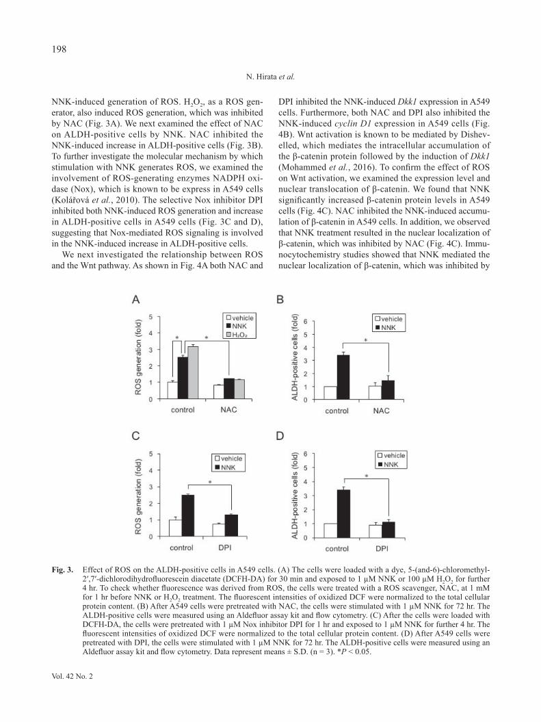
NNK-induced generation of ROS. H2O2, as a ROS gen-erator, also induced ROS generation, which was inhibited by NAC (Fig. 3A). We next examined the effect of NAC on ALDH-positive cells by NNK. NAC inhibited the NNK-induced increase in ALDH-positive cells (Fig. 3B). To further investigate the molecular mechanism by which stimulation with NNK generates ROS, we examined the involvement of ROS-generating enzymes NADPH oxi-dase (Nox), which is known to be express in A549 cells (Kolářová et al., 2010). The selective Nox inhibitor DPI inhibited both NNK-induced ROS generation and increase in ALDH-positive cells in A549 cells (Fig. 3C and D), suggesting that Nox-mediated ROS signaling is involved in the NNK-induced increase in ALDH-positive cells.
We next investigated the relationship between ROS and the Wnt pathway. As shown in Fig. 4A both NAC and
DPI inhibited the NNK-induced Dkk1 expression in A549 cells. Furthermore, both NAC and DPI also inhibited the NNK-induced cyclin D1 expression in A549 cells (Fig. 4B). Wnt activation is known to be mediated by Dishev-elled, which mediates the intracellular accumulation of the β-catenin protein followed by the induction of Dkk1 (Mohammed et al., 2016). To confirm the effect of ROS on Wnt activation, we examined the expression level and nuclear translocation of β-catenin. We found that NNK significantly increased β-catenin protein levels in A549 cells (Fig. 4C). NAC inhibited the NNK-induced accumu-lation of β-catenin in A549 cells. In addition, we observed that NNK treatment resulted in the nuclear localization of β-catenin, which was inhibited by NAC (Fig. 4C). Immu-nocytochemistry studies showed that NNK mediated the nuclear localization of β-catenin, which was inhibited by
Fig. 3. Effect of ROS on the ALDH-positive cells in A549 cells. (A) The cells were loaded with a dye, 5-(and-6)-chloromethyl-2′,7′-dichlorodihydrofluorescein diacetate (DCFH-DA) for 30 min and exposed to 1 µM NNK or 100 µM H2O2 for further 4 hr. To check whether fluorescence was derived from ROS, the cells were treated with a ROS scavenger, NAC, at 1 mM for 1 hr before NNK or H2O2 treatment. The fluorescent intensities of oxidized DCF were normalized to the total cellular protein content. (B) After A549 cells were pretreated with NAC, the cells were stimulated with 1 µM NNK for 72 hr. The ALDH-positive cells were measured using an Aldefluor assay kit and flow cytometry. (C) After the cells were loaded with DCFH-DA, the cells were pretreated with 1 µM Nox inhibitor DPI for 1 hr and exposed to 1 µM NNK for further 4 hr. The fluorescent intensities of oxidized DCF were normalized to the total cellular protein content. (D) After A549 cells were pretreated with DPI, the cells were stimulated with 1 µM NNK for 72 hr. The ALDH-positive cells were measured using an Aldefluor assay kit and flow cytometry. Data represent means ± S.D. (n = 3). *P < 0.05.
Vol. 42 No. 2
198
N. Hirata et al.

Fig. 4. Effect of ROS on NNK-induced Wnt signaling in A549 cells. After pretreatment with NAC (1 mM) or DPI (1 µM) for 30 min, A549 cells were stimulated with 1 µM NNK for 24 hr. (A) Dkk1 expression was measured by real-time RT-PCR. (B) Cyclin D1 expression was measured by real-time RT-PCR. (C) Accumulation of β-catenin protein in the cells was ana-lyzed by western blotting using anti-β-catenin antibodies. The relative densities of bands were quantified with an image ana-lyzer. Relative changes in expression were determined by normalization to β-actin. (D) Intracellular localization of β-catenin was assessed by immunocytochemical staining using anti-β-catenin antibodies. Bar = 20 µm. Data represent means ± S.D. (n = 3). *P < 0.05.
Vol. 42 No. 2
199
NNK increases ALDH-positive cells in A549 cells
NAC (Fig. 4D). Wnt3a caused accumulation and nuclear translocation of β-catenin (Fig. 4C and D). Taken togeth-er, these data suggest that NNK mediates ALDH-positive cells through a ROS-Wnt pathway in A549 cells.
Involvement of PI3K-Akt activation in ROS signaling by NNK
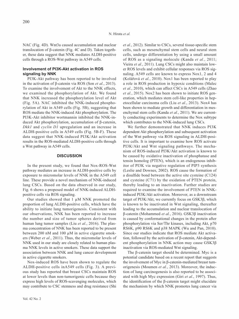
PI3K-Akt pathway has been reported to be involved in the activation of β-catenin via ROS (Son et al., 2013). To examine the involvement of Akt to the NNK effects, we examined the phosphorylation of Akt. We found that NNK increased the phosphorylation level of Akt (Fig. 5A). NAC inhibited the NNK-induced phospho-rylation of Akt in A549 cells (Fig. 5B), suggesting that ROS mediate the NNK-induced Akt phosphorylation. The PI3K-Akt inhibitor wortmannin inhibited the NNK-in-duced Akt phosphorylation, accumulation of β-catenin, Dkk1 and cyclin D1 upregulation and an increase in ALDH-positive cells in A549 cells (Fig. 5B-F). These data suggest that NNK-induced PI3K/Akt activation results in the ROS-mediated ALDH-positive cells through a Wnt pathway in A549 cells.
DISCUSSION
In the present study, we found that Nox-ROS-Wnt pathway mediates an increase in ALDH-positive cells by exposure to micromolar levels of NNK in the A549 cell line. These provide a novel mechanism of NNK-induced lung CSCs. Based on the data observed in our study, Fig. 6 shows a proposed model of NNK-induced ALDH-positive cells via ROS signaling.
Our studies showed that 1 µM NNK promoted the proportion of lung ALDH-positive cells, which have the ability to initiate lung tumorigenesis. Consistent with our observations, NNK has been reported to increase the number and size of tumor spheres derived from human lung tumor samples (Liu et al., 2016). The plas-ma concentration of NNK has been reported to be present between 200 nM and 100 µM in active cigarette smok-ers (Weber et al., 2011). Thus, the micromolar levels of NNK used in our study are closely related to human plas-ma NNK levels in active smokers. These data support the association between NNK and lung cancer development in active cigarette smokers.
Nox-induced ROS have been shown to regulate the ALDH-positive cells inA549 cells (Fig. 3). A previ-ous study has reported that breast CSCs maintain ROS at lower levels than non-tumorigenic cells because they express high levels of ROS-scavenging molecules, which may contribute to CSC stemness and drug resistance (Shi
et al., 2012). Similar to CSCs, several tissue-specific stem cells, such as mesenchymal stem cells and neural stem cells, undergo differentiation by using a small amount of ROS as a signaling molecule (Kanda et al., 2011; Vieira et al., 2011). Lung CSCs might also maintain low-er ROS levels and exhibit cellular responses via ROS sig-naling. A549 cells are known to express Nox1, 2 and 4 (Kolářová et al., 2010). Nox1 has been reported to play a role in ROS production in hypoxic conditions (Malec et al., 2010), which can affect CSCs in A549 cells (Zhao et al., 2015). Nox2 has been shown to initiate ROS gen-eration, which mediates stem cell-like properties in hep-etocellular carcinoma cells (Liu et al., 2013). Nox4 has been shown to mediate growth and differentiation in mes-enchymal stem cells (Kanda et al., 2011). We are current-ly conducting experiments to determine the Nox subtype which contributes to the NNK-induced lung CSCs.
We further demonstrated that NNK induces PI3K dependent Akt phosphorylation and subsequent activation of the Wnt pathway via ROS signaling in ALDH-posi-tive cells. It is important to examine how ROS activate PI3K/Akt and Wnt signaling pathways. The mecha-nism of ROS-induced PI3K/Akt activation is known to be caused by oxidative inactivation of phosphatase and tensin homolog (PTEN), which is an endogenous inhib-itor of PI3K via negative regulation of PIP3 synthesis (Leslie and Downes, 2002). ROS cause the formation of a disulfide bond between the active site cysteine (C124) and cysteine (C71) by the oxidation of PTEN protein, thereby leading to an inactivation. Further studies are required to examine the involvement of PTEN in NNK-induced PI3K/Akt activation. Moreover, as a downstream target of PI3K/Akt, we currently focus on GSK3β, which is known to be inactivated in Wnt signaling, thereafter leading to the accumulation and nuclear translocation of β-catenin (Mohammed et al., 2016). GSK3β inactivation is caused by conformational changes in the protein after phosphorylation via Ser/Thr kinases, including Akt, p70 RS6K, p90 RS6K and p38 MAPK (Wu and Pan, 2010). Since our studies indicate that ROS mediate Akt activa-tion, followed by the activation of β-catenin, Akt-depend-ent phosphorylation in NNK action may cause GSK3β inactivation via ROS-mediated Wnt signaling.
The β-catenin target should be determined. Myc is a potential candidate based on a recent report that suggests the involvement of Myc in β-catenin-mediated breast tum-origenesis (Moumen et al., 2013). Moreover, the induc-tion of lung carcinogenesis is also reported to be associ-ated with high Myc expression (Giri et al., 1997). Thus, the identification of the β-catenin target might elucidate the mechanism by which NNK promotes lung cancer via
Vol. 42 No. 2
200
N. Hirata et al.

Fig. 5. Involvement of PI3K-Akt activation in NNK actions to A549 cells. (A) After A549 cells were stimulated with 1 µM NNK for 1, 2, 4, and 8 hr, phosphorylation level of Akt was analyzed by western blotting using anti-phospho-Akt antibodies. The relative densities of bands were quantified with an image analyzer. Relative changes in expression were determined by normalization to total Akt protein level. (B) After pretreatment with 1 mM NAC or 100 nM PI3K inhibitor wortmannin for 30 min, the cells were stimulated with 1 µM NNK for 4 hr. Akt phosphorylation was analyzed by western blotting using anti-phospho-Akt antibodies. The relative densities of bands were quantified with an image analyzer. Relative changes in expression were determined by normalization to total Akt protein level. (C) After pretreatment with wortmannin, the cells were stimulated with 1 µM NNK for 24 hr. Accumulation of β-catenin protein in the cells was analyzed by western blotting using anti-β-catenin antibodies. The relative densities of bands were quantified with an image analyzer. Relative changes in expression were determined by normalization to β-actin. (D) After pretreatment with wortmannin, the cells were stimulated with 1 µM NNK for 24 hr. Dkk1 expression was measured by real-time RT-PCR. (E) After pretreatment with wortmannin, the cells were stimulated with 1 µM NNK for 24 hr. Cyclin D1 expression was measured by real-time RT-PCR. (F) After pretreatment with wortmannin, the cells were stimulated with 1 µM NNK for 72 hr. The ALDH-positive cells were meas-ured using an Aldefluor assay kit and flow cytometry. Data represent means ± S.D. (n = 3). *P < 0.05.
Vol. 42 No. 2
201
NNK increases ALDH-positive cells in A549 cells

CSCs. Our data suggest that the ROS-mediated Wnt path-
way is important for NNK-induced lung carcinogenesis in vitro. A previous report indicated that NAC treatment decreases the weight of tumors in nude mice xenograft-ed with C87 Lewis lung carcinoma cells (Albini et al., 1995). Moreover, compounds that reduce the expression levels of β-catenin inhibit the growth of xenografted lung tumors in mice (Bi et al., 2014). Since the Wnt signaling inhibitor pyrvinium pamoate is known to inhibit the pro-liferation of lung CSCs derived from lung cell lines (SPC-A1 and PC9) (Jiang et al., 2009), targeting the ROS-me-diated Wnt pathway might provide a promising strategy to treat lung tumorigenesis. It remains to be determined whether Wnt signaling mediates the proliferation of CSCs from clinical samples.
Cigarette smoke contains lung carcinogenic com-pounds, such as nicotine, acetaldehyde, and benzo[a]pyrene (Hecht, 1999). Nicotine is partially involved in a lung CSCs of the A549 cell line (An et al., 2012). Additionally, we have previously reported that nicotine increases the proliferation of breast CSCs via nicotin-ic acetylcholine receptor (nAChR) (Hirata et al., 2010). NNK is derived from nicotine and has the ability to acti-vate nAChR (Singh et al., 2011); accordingly, the effects of NNK might be mediated by nAChR. Further studies of other components of cigarette smoke are required to reveal the mechanism of CSCs.
In conclusion, our results indicate that NNK increas-
es the ALDH-positive cells via the ROS signaling path-way in A549 human lung cancer cells. We also found that ROS mediate ALDH-positive cells via PI3K/Akt and Wnt signaling. The proposed mechanism by which NNK regu-lates ALDH-positive cells might explain the development of lung cancer in cigarette smokers. ROS-PI3K/Akt-Wnt pathway might be a potential therapeutic target for lung cancer treatment.
ACKNOWLeDgMeNTS
This work was supported by a Health and Labour Sciences Research Grant from the Ministry of Health, Labour and Welfare, Japan (#H25-Kagaku-Ippan-002 and #H28-Kagaku-Ippan-003, to Y. Kanda), a Grant-in-Aid for Scientific Research from the Ministry of Edu-cation, Culture, Sports, Science, and Technology, Japan (#26293056, #26670041 to Y. Kanda), the Research on Regulatory Harmonization and Evaluation of Pharmaceu-ticals, Medical Devices, Regenerative and Cellular Ther-apy Products, Gene Therapy Products, and Cosmetics from Japan Agency for Medical Research and develop-ment, AMED (to Y. Sekino), and a grant from the Smok-ing Research Foundation (Y. Kanda).
Conflict of interest---- The authors declare that there is no conflict of interest.
RefeReNCeS
Albini, A., D'Agostini, F., Giunciuglio, D., Paglieri, I., Balansky, R. and De Flora, S. (1995): Inhibition of invasion, gelatinase activ-ity, tumor take and metastasis of malignant cells by N-acetyl-cysteine. Int. J. Cancer, 61, 121-129.
An, Y., Kiang, A., Lopez, J.P., Kuo, S.Z., Yu, M.A., Abhold, E.L., Chen, J.S., Wang-Rodriguez, J. and Ongkeko, W.M. (2012): Cig-arette smoke promotes drug resistance and expansion of cancer stem cell-like side population. PLoS One, 7, e47919.
Bi, X., Xia, X., Mou, T., Jiang, B., Fan, D., Wang, P., Liu, Y., Hou, Y. and Zhao, Y. (2014): Anti-tumor activity of three ginsenoside derivatives in lung cancer is associated with Wnt/β-catenin sign-aling inhibition. Eur. J. Pharmacol., 742, 145-152.
Bora-Singhal, N., Nguyen, J., Schaal, C., Perumal, D., Singh, S., Coppola, D. and Chellappan, S. (2015): YAP1 regulates Oct4 activity and Sox2 expression to facilitate self-renewal and vas-cular mimicry of stem-like cells. Stem Cells, 33, 1705-1718.
Catassi, A., Servent, D., Pareari, I., Cesario, A. and Russo, P., (2008): Multiple roles of nicotine on cell proliferation and inhi-bition of apoptosis: Implications on lung carcinogenesis. Mutat. Res., 659, 221-231.
Chernyavsky, A.I., Shchepotin I.B., Galitovkiy, V. and Grando S.A. (2015): Mechanisms of tumor-promoting activities of nicotine in lung cancer: synergistic effects of cell membrane and mitochon-drial nicotinic acetylcholine receptors. BMC Cancer, 15, 152.
Davis, R., Rizwani, W., Banerjee, S., Kovacs, M., Haura, E.,
Fig. 6. Proposed model of NNK-induced lung ALDH-positive cells via ROS signaling in A549 cells. NNK treatment causes Nox-induced ROS generation, which activates PI3K/Akt signaling, thereafter leading to the cytosol-ic accumulation of the β-catenin protein and nuclear translocation in Wnt pathway. This mediates the ex-pression of Wnt target genes such as Dkk1.
Vol. 42 No. 2
202
N. Hirata et al.
Coppola, D. and Chellappan, S. (2009): Nicotine promotes tumor growth and metastasis in mouse models of lung cancer. PLoS One, 4, e7524.
Dela, Cruz, C.S., Tanoue, L.T. and Matthay R.A. (2011): Lung can-cer: epidemiology, etiology, and prevention. Clin. Chest Med., 32, 605-644.
DeMarini, D.M. (2004): Genotoxicity of tobacco smoke and tobac-co smoke condensate: a Review. Mutat. Res., 567, 447-474.
Dontu, G., Jackson, K.W., McNicholas, E., Kawamura, M.J., Abdallah, W.M. and Wicha, M.S. (2004): Role of Notch signal-ing in cell-fate determination of human mammary stem/progeni-tor cells. Breast Cancer Res., 6, R605-615.
Giri, R.K., Baral, R.N. and Das, B.R. (1997): Induction of lung car-cinogenesis in AKR-mice by N-nitrosodiethylamine/phenobarbi-tone, associated with high expression of c-myc and c-jun onco-proteins. Cancer Lett., 112, 57-63.
Hecht, S.S. (1999): Tobacco smoke carcinogens and lung cancer. J. Natl. Cancer Inst., 91, 1194-1210.
Hirata, N., Sekino, Y. and Kanda, Y. (2010): Nicotine increases can-cer stem cell population in MCF-7 cells. Biochem. Biophys. Res. Commun., 403, 138-143.
Hirata, N., Yamada, S., Shoda, T., Kurihara, M., Sekino, Y. and Kanda, Y. (2014): Sphingosine-1-phosphate promotes expansion of cancer stem cells via S1PR3 by a ligand-independent Notch activation. Nat. Commun., 5, 4806.
Iskandar, A.R., Liu, C., Smith, D.E., Hu, K.Q., Choi, S.W., Ausman, L.M. and Wang, X.D. (2013): β-Cryptoxanthin restores nicotine-reduced lung SIRT1 to normal levels and inhibits nicotine-pro-moted lung tumorigenesis and emphysema in A/J mice. Cancer Prev. Res., 6, 309-320.
Jiang, F., Qiu, Q., Khanna, A., Todd, N.W., Deepak, J., Xing, L., Wang, H., Liu, Z., Su, Y., Stass, S.A. and Katz, R.L. (2009): Aldehyde dehydrogenase 1 is a tumor stem cell-associated mark-er in lung cancer. Mol. Cancer Res., 7, 330-338.
Kanda, Y., Hinara, T., Kang, S.W. and Watanabe, Y. (2011): Reac-tive oxygen species mediate adipocyte differentiation in mesen-chymal stem cells. Life Sci., 89, 250-258.
Kolářová, H., Binó, L., Pejchalová, K. and Kubala, L. (2010): The expression of NADPH oxidases and production of reactive oxy-gen species by human lung adenocarcinoma epithelial cell line A549. Folia Biol (Praha)., 56, 211-217.
Leslie, N.R. and Downes, C.P. (2002): PTEN: The down side of PI 3-kinase signalling. Cell Signal, 14, 285-295.
Liu, L., Yang, Z., Xu, Y., Li, J., Xu, D., Zhang, L., Sun, J., Xia, S., Zou, F. and Liu, Y. (2013): Inhibition of oxidative stress-elicited AKT activation facilitates PPARγ agonist-mediated inhibition of stem cell character and tumor growth of liver cancer cells. PLoS One, 8, e73038.
Liu, S., Dontu, G., Mantle, I.D., Patel, S., Ahn, N.S., Jackson, K.W., Suri, P. and Wicha, M.S. (2006): Hedgehog signaling and Bmi-1 regulate self-renewal of normal and malignant human mammary stem cells. Cancer Res., 66, 6063-6071.
Liu, Y., Yang, S., Li, M.Y., Huang, R., Ng, C.S., Wan, I.Y., Long, X., Wu, J., Wu, B., Du, J., Mok, T.S., Underwood, M.J. and Chen, G.G. (2016): Tumorigenesis of smoking carcinogen 4-(methylnitrosamino)-1-(3-pyridyl)-1-butanone is related to its ability to stimulate thromboxane synthase and enhance stemness of non-small cell lung cancer stem cells. Cancer Lett., 370, 198-206.
Lu, G., Xiao, H., Li, G.X., Picinich, S.C., Chen, Y.K., Liu, A., Lee, M.J., Loy, S. and Yang, C.S. (2010): A gamma-tocopherol-rich mixture of tocopherols inhibits chemically induced lung tumori-
genesis in A/J mice and xenograft tumor growth. Carcinogenesis, 31, 687-694.
Malec, V., Gottschald, O.R., Li, S., Rose, F., Seeger, W. and Hänze, J. (2010): HIF-1 alpha signaling is augmented during intermit-tent hypoxia by induction of the Nrf2 pathway in NOX1-ex-pressing adenocarcinoma A549 cells. Free Radic. Biol. Med., 48, 1626-1635.
Mohammed, M.K., Shao, C., Wang, J., Wei, Q., Wang, X., Collier, Z., Tang, S., Liu, H., Zhang, F., Huang, J., Guo, D., Lu, M., Liu, F., Liu, J., Ma, C., Shi, L.L., Athiviraham, A., He, T.C. and Lee, M.J. (2016): Wnt/β-catenin signaling plays an ever-expanding role in stem cell self-renewal, tumorigenesis and cancer chem-oresistance. Genes Dis., 3, 11-40.
Moumen, M., Chiche, A., Decraene, C., Petit, V., Gandarillas, A., Deugnier, M.A., Glukhova, M.A. and Faraldo, M.M. (2013): Myc is required for β-catenin-mediated mammary stem cell amplification and tumorigenesis. Mol. Cancer, 12, 132.
Oh, S.J., Noh, K.H., Lee, Y.H., Hong, S.O., Song, K.H., Lee, H.J., Kim, S., Kim, T.M., Jeon, J.H., Seo, J.H., Kim, D.W. and Kim, T.W. (2015): Targeting stemness is an effective strategy to con-trol EML4-ALK+ non-small cell lung cancer cells. Oncotarget, 6, 40255-40267.
Rivera, C., Rivera, S., Loriot, Y., Vozenin, M.C. and Deutsch, E. (2011): Lung cancer stem cell: new insights on experimental models and preclinical data. J. Oncol., 2011, 549181.
Rodriguez-Torres, M. and Allan, A.L. (2016): Aldehyde dehydroge-nase as a marker and functional mediator of metastasis in solid tumors. Clin. Exp. Metastasis, 33, 97-113.
Shi, X., Zhang, Y., Zheng, J. and Pan, J. (2012): Reactive oxygen species in cancer stem cells. Antioxid. Redox., Signal, 16, 1215-1228.
Singh, S., Pillai, S. and Chellappan, S. (2011): Nicotinic acetylcho-line receptor signaling in tumor growth and metastasis. J. Oncol., 2011, 456743.
Smalley, M.J. and Dale, T.C. (1999): Wnt signalling in mammalian development and cancer. Cancer Metastasis Rev., 18, 215-230.
Son, Y.O., Pratheeshkumar, P., Wang, L., Wang, X., Fan, J., Kim, D.H., Lee, J.Y., Zhang, Z., Lee, J.C. and Shi, X. (2013): Reac-tive oxygen species mediate Cr(VI)-induced carcinogenesis through PI3K/AKT-dependent activation of GSK-3β/β-catenin signaling. Toxicol. Appl. Pharmacol., 271, 239-248.
Takebe, N., Miele, L., Harris, P.J., Jeong, W., Bando, H., Kahn, M., Yang, S.X. and Ivy, S.P. (2015): Targeting Notch, Hedgehog, and Wnt pathways in cancer stem cells: clinical update. Nat. Rev. Clin. Oncol., 12, 445-464.
Teng, Y., Wang, X., Wang, Y. and Ma, D. (2010): Wnt/beta-catenin signaling regulates cancer stem cells in lung cancer A549 cells. Biochem. Biophys. Res. Commun., 392, 373-379.
Trosset, J.Y. Davit, C., Knapp, S., Fasolini, M., Veronesi, M., Mantegani, S., Gianellini, L.M., Catana, C., Sundström, M., Stouten, P.F. and Moll, J.K. (2006): Inhibition of protein-protein interactions: the discovery of druglike beta-catenin inhibitors by combining virtual and biophysical screening. Pro-teins, 64, 60-67.
Tsurutani, J., Castillo, S.S., Brognard, J., Granville, C.A., Zhang, C., Gills, J.J., Sayyah, J. and Dennis, P.A. (2005): Tobacco components stimulate Akt-dependent proliferation and NFκB-dependent survival in lung cancer cells. Carcinogenesis, 26, 1182-1195.
Vieira, H.L., Alves, P.M. and Vercelli, A. (2011): Modulation of neuronal stem cell differentiation by hypoxia and reactive oxy-gen species. Prog. Neurobiol., 93, 444-455.
Vol. 42 No. 2
203
NNK increases ALDH-positive cells in A549 cells

Visvader, J.E. and Lindeman, G.J. (2008): Cancer stem cells in solid tumours: accumulating evidence and unresolved questions. Nat. Rev. Cancer, 8, 755-768.
Weber, S.M., Bornstein, S., Li, Y., Malkoski, S.P., Wang, D., Rustgi, A.K., Kulesz-Martin, M.F., Wang, X.J. and Lu, S.L. (2011) Tobacco-specific carcinogen nitrosamine 4-(methylnitrosamino)-1-(3-pyridyl)-1-butanone induces AKT activation in head and neck epithelia. Int. J. Oncol., 39, 1193-1198.
Wu, D. and Pan, W. (2010): GSK3: a multifaceted kinase in Wnt signaling. Trends Biochem. Sci., 35, 161-168.
Xue, J., Yang, S. and Seng, S. (2014): Mechanisms of Cancer Induc-tion by Tobacco-Specific NNK and NNN. Cancers (Basel), 6,
1138-1156.Yalcin, E. and de la Monte, S. (2016): Tobacco nitrosamines as cul-
prits in disease: mechanisms reviewed. J. Physiol. Biochem., 72, 107-120.
Zhang, X., Lou, Y., Zheng, X., Wang, H., Sun, J., Dong, Q. and Han, B. (2015): Wnt blockers inhibit the proliferation of lung cancer stem cells. Drug Des. Devel. Ther., 9, 2399-2407.
Zhao, M., Zhang, Y., Zhang, H., Wang, S., Zhang, M., Chen. X., Wang, H., Zeng, G., Chen, X., Liu, G. and Zhou, C. (2015): Hypoxia-induced cell stemness leads to drug resistance and poor prognosis in lung adenocarcinoma. Lung Cancer, 87, 98-106.
Vol. 42 No. 2
204
N. Hirata et al.