original article expression of cancer stem cell markers ... · original article expression of...
TRANSCRIPT

Int J Clin Exp Pathol 2015;8(10):12621-12633www.ijcep.com /ISSN:1936-2625/IJCEP0014801
Original ArticleExpression of cancer stem cell markers and their correlation with pathogenesis in vascular tumors
Jiaojiao Lan1*, Bing Huang2*, Ruixue Liu1, Xinxin Ju1, Yang Zhou1, Jinfang Jiang1, Weihua Liang1, Yaoyuan Shen1, Feng Li1, Lijuan Pang1
1Department of Pathology, Key Laboratory of Xinjiang Endemic and Ethnic Diseases (Ministry of Education), Shihezi University School of Medicine, Shihezi, Xinjiang, China; 2Department of Thoracic and Cardiovascular Surgery, First Affiliated Hospital to Shihezi University School of Medicine, Shihezi 832008, Xinjiang, China. *Co-first authors.
Received August 20, 2015; Accepted September 24, 2015; Epub October 1, 2015; Published October 15, 2015
Abstract: Objective: Vascular tumor, which belongs to a kind of complicated lesion in soft tissue tumor, is derived from mesenchymal tissue. Although many studies have been focused on the pathogenesis of vascular tumors in human, the specific mechanism of the vascular tumors was currently unclear. Previous studies have reported an association of cancer stem cells with the development of tumor in many solid tumors. Thus the purpose of this study was to explore whether different expression level of cancer stem cell markers including CD29, CD44, CD133, nestin and ALDH1 in vascular tumor may help to elucidate the possible pathogenesis of vascular tumor. In present study, tissues of 9 cases of hemangioma, 22 cases of hemangiosarcoma, 3 cases of Kaposi’s sarcoma, and 5 cases of hemangioendothelioma were immunostained for CD29, CD44, CD133, nestin and ALDH1. Of the 39 vascular tumor cases included in the current study, CD29, CD133 and nestin were positive in most vascular tumor cases. Although CD44 and ALDH1 were observed in vascular tumor cases, the percentage of cells staining for the two markers was less than 2% in all cases of vascular tumor. Capillary hemangiomas exhibited significantly higher expression rate of CD29 and nestin compared with malignant vascular tumors and hemangioendotheliomas (P<0.05, Fisher’s exact test), while CD44, CD133 and ALDH1 exhibited no statistically significant difference between these two groups. Pearson correlation analysis exhibited that CD29 expression and nestin expression in vascular tumor were no sta-tistically significant relationship (C=0.288, P=0.063>0.05). Our findings confirmed that the five cancer stem cells markers, including CD29, CD44, CD133, nestin and ALDH1, exhibited different expression levels in vascular tumors and demonstrated that immonhistochemical analysis for cancer stem cells markers may provide useful information for studying the pathogenesis of vascular tumors.
Keywords: Vascular tumor, immunohistochemistry, cancer stem cells, pathogenesis
Introduction
Vascular tumor is derived from mesenchymal tissue tumors, which is classified as soft tissue tumors [1, 2]. According to the 4th WHO classi-fication, it can be divided into benign tumors of blood vessels, malignant vascular tumors, and hemangioendothelimas that are vascular tumor of intermediate malignancy. Hemangiomas are most common benign vascular tumor of child-hood with a unique characteristic process of its rapid growth and slowly spontaneous involu-tion [3-5]. The occurrences of malignant and potentially malignant sarcomas that originated from blood vessels are rarely seen and include hemangiosarcomas [6, 7], Kaposi’s sarcomas [8], hemangioendothelimas [9, 10], usually with
no known cause. The only known reasons of hemangiosarcomas were a few rare specific genetic alterations such as anti-oncogene (p53) mutation, previous irradiation, and exposure to vinyl chloride or thorotrast [11, 12]. Kaposi’s sarcomas often occurred in patients with infec-tion and patients who are immunosuppressed [13, 14]. Although there were several previous studies have paid more attentions on the patho-genesis and origin of vascular tumors, it still remains controversial and was not well under-stood in humans.
Increasing evidence showed that tumors may derive from a small subpopulation of cancer cells possessing stem-like properties, which were defined as cancer stem cells or tumor-ini-

Cancer stem cells and vascular tumors
12622 Int J Clin Exp Pathol 2015;8(10):12621-12633
the phenotype of cancer stem cells in different vascular tumors, which may contribute to our understanding of the pathogenesis and origin of vascular tumor.
Material and methods
Patients and tumor specimens
The study included 9 cases capillary hemangio-ma, 25 cases of malignant vascular tumor (including 3 cases of Kaposi’s sarcoma and 22 cases of hemangiosarcoma). In addition, 5 cases of hemangioendothelioma were identi-fied. A total of 39 cases were obtained from the Department of Pathology, the First Affiliated Hospital, Shihezi University School of Medicine. Tumor tissues were fixed in buffered formalin and subjected to routine processing and paraf-fin embedding. Clinical data including age, sex, and location of tumor were available in all cases (Tables 2, 3). This study was conducted with the approval of the institutional ethics commit-tee at the First Affiliated Hospital of Shihezi University of Medicine.
Immunohistochemical staining
The most representative paraffin blocks were identified by examination of hematoxylin and
tiating cells [15, 16]. The cancer stem cell hypothesis proposes that cancer stem cells may be involved in tumor progression and sev-eral tumor characteristics, such as recurrence and metastasis [17, 18]. These cells with the potential to initiate and maintain tumor devel-opment were first identified in leukemia [19]and subsequently in breast cancer [20], colon cancer [21], sarcoma [22, 23] et al. Specific surface markers can help to identify and iso-late the cancer stem cells [24, 25]. Recent studies have reported that only a small popula-tion of tumor cells can selectively express cer-tain surface markers such as CD29 [26], CD44 [27, 28] and CD133 [29]. Additionally, it was reported that vascular tumors also expressed cancer stem cell markers, indicating an asso-ciation of cancer stem cell with the initiation and pathogenesis of vascular tumors [3, 6, 11]. Thus, the importance of identifying cancer stem cell for understanding of the pathogene-sis and origin of vascular tumors is becoming more evident.
In the present study, we investigated the differ-ent expression of cancer stem cell markers including CD29, CD44, nestin, CD133 and ALDH1 in resected specimens obtained from 39 cases of vascular tumor tissues by immuno-hischemistry. Our results allowed us to define
Table 1. Primary antibodies used in the immunohistochemistry
Antigen Location Antibody species Manufacturer Clone Number
Antigen-retrieval solution Dilution
CD133 C Rabbit polyclonal ARP, Waltham, America 05-PA1021 PCA-CB 1:200CD29 C Rabbit monoclonal Abcam, Cambridge, UK EP1041Y PCA-CB 1:600CD44 M/C Mouse monoclonal DAKO, Glostrup, Denmark DF1485 PCA-CB 1:300Nestin C Rabbit monoclonal Abcam, Cambridge, UK SP103 PCA-CB 1:800ALDH1 C Rabbit monoclonal Abcam, Cambridge, UK EP1933Y PCA-CB 1:300C: cytoplasm, M: membrane, PCA-CB: pressure cooker heating in citrate buffer (0.01 M, pH 6.0).
Table 2. Summary of patients’ clinical information in benign vascular tumorsPatient ID Tumor Age Sex Location1 Capillary hemangioma 7 m F Left shoulder2 Capillary hemangioma 16 m F Lower abdomen and perineum3 Capillary hemangioma 3 y M Back buttocks4 Capillary hemangioma 8 m F Neck5 Capillary hemangioma 2 y F Left upper eyelid6 Capillary hemangioma 8 m M Scalp7 Capillary hemangioma 7 m F Right thigh, right chest subcutaneous8 Capillary hemangioma 7 m F Scalp9 Capillary hemangioma 25 y M NoseF, female; M, male; m, month; y, year.
Cancer stem cells and vascular tumors
12623 Int J Clin Exp Pathol 2015;8(10):12621-12633
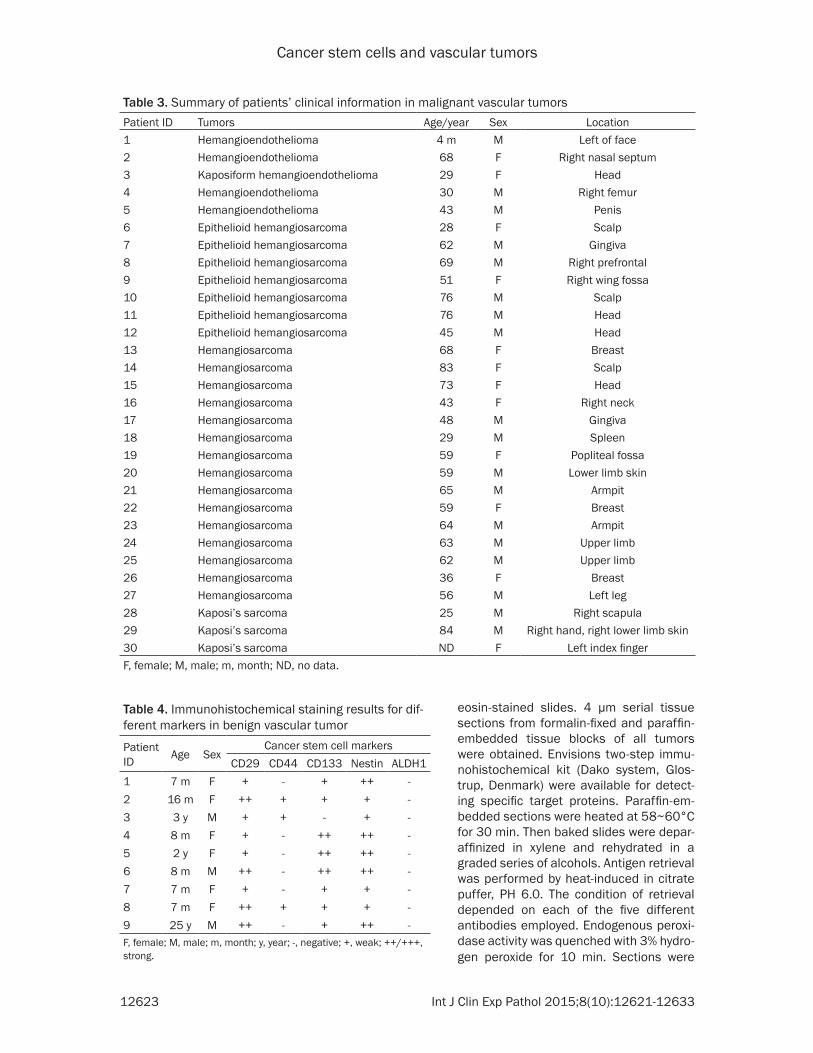
eosin-stained slides. 4 μm serial tissue sections from formalin-fixed and paraffin-embedded tissue blocks of all tumors were obtained. Envisions two-step immu-nohistochemical kit (Dako system, Glos- trup, Denmark) were available for detect-ing specific target proteins. Paraffin-em- bedded sections were heated at 58~60°C for 30 min. Then baked slides were depar-affinized in xylene and rehydrated in a graded series of alcohols. Antigen retrieval was performed by heat-induced in citrate puffer, PH 6.0. The condition of retrieval depended on each of the five different antibodies employed. Endogenous peroxi-dase activity was quenched with 3% hydro-gen peroxide for 10 min. Sections were
Table 3. Summary of patients’ clinical information in malignant vascular tumorsPatient ID Tumors Age/year Sex Location1 Hemangioendothelioma 4 m M Left of face2 Hemangioendothelioma 68 F Right nasal septum3 Kaposiform hemangioendothelioma 29 F Head4 Hemangioendothelioma 30 M Right femur5 Hemangioendothelioma 43 M Penis6 Epithelioid hemangiosarcoma 28 F Scalp7 Epithelioid hemangiosarcoma 62 M Gingiva8 Epithelioid hemangiosarcoma 69 M Right prefrontal9 Epithelioid hemangiosarcoma 51 F Right wing fossa10 Epithelioid hemangiosarcoma 76 M Scalp11 Epithelioid hemangiosarcoma 76 M Head12 Epithelioid hemangiosarcoma 45 M Head13 Hemangiosarcoma 68 F Breast14 Hemangiosarcoma 83 F Scalp15 Hemangiosarcoma 73 F Head16 Hemangiosarcoma 43 F Right neck17 Hemangiosarcoma 48 M Gingiva18 Hemangiosarcoma 29 M Spleen19 Hemangiosarcoma 59 F Popliteal fossa20 Hemangiosarcoma 59 M Lower limb skin21 Hemangiosarcoma 65 M Armpit22 Hemangiosarcoma 59 F Breast23 Hemangiosarcoma 64 M Armpit24 Hemangiosarcoma 63 M Upper limb25 Hemangiosarcoma 62 M Upper limb26 Hemangiosarcoma 36 F Breast27 Hemangiosarcoma 56 M Left leg28 Kaposi’s sarcoma 25 M Right scapula29 Kaposi’s sarcoma 84 M Right hand, right lower limb skin30 Kaposi’s sarcoma ND F Left index fingerF, female; M, male; m, month; ND, no data.
Table 4. Immunohistochemical staining results for dif-ferent markers in benign vascular tumor
Patient ID Age Sex
Cancer stem cell markersCD29 CD44 CD133 Nestin ALDH1
1 7 m F + - + ++ -2 16 m F ++ + + + -3 3 y M + + - + -4 8 m F + - ++ ++ -5 2 y F + - ++ ++ -6 8 m M ++ - ++ ++ -7 7 m F + - + + -8 7 m F ++ + + + -9 25 y M ++ - + ++ -F, female; M, male; m, month; y, year; -, negative; +, weak; ++/+++, strong.
Cancer stem cells and vascular tumors
12624 Int J Clin Exp Pathol 2015;8(10):12621-12633
then incubated with primary antibodies (Table 1) for at least 8 hours at 4°C and PBS was instead of the primary antibodies for negative controls. PBS was used to wash the primary antibodies followed by the appropriate second-ary antibodies (Table 1) for 30 min at 37°C, and reaction was performed using 3.3’- diaminobenzidine peroxidase substrate kit (Dako System, Glostrup, Denmark). Finally, sections were counter-stained with hematoxy-lin, dehydrated and mounted in a neutral mounting medium. Immunohischemical proce-dures including antibodies and primary anti-
bodies used in our study are summarized in Table 1.
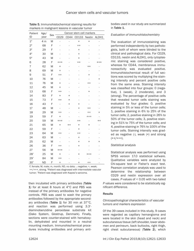
Evaluation of immunohistochemistry
The evaluation of immunostaining was performed independently by two patholo-gists, both of whom were blinded to the clinical and pathological data. For CD29, CD133, nestin and ALDH1, only cytoplas-mic staining was considered positive, whereas for CD44, membranous immu-noreactivity was evaluated positive. Immunohistochemical result of full sec-tions was scored by multiplying the stain-ing intensity and percent positive cells from the same area. Staining intensity was classified into four groups: 0 (nega-tive), 1 (weak), 2 (moderate), and 3 (strong). The percentage of positive cells that revealed tumor cells staining was evaluated by four grades: 0, positive staining in 5% or less of the tumor cells; 1, positive staining in 6% to 25% of the tumor cells; 2, positive staining in 26% to 50% of the tumor cells; 3, positive stain-ing in 51% to 75% of the tumor cells; and 4, positive staining in 76% to 100% of the tumor cells. Staining intensity was grad-ed as negative (-), weak (+) and strong (++/+++).
Statistical analysis
Statistical analysis was performed using SPSS version 17.0 statistical sofware. Qualitative variables were analyzed by Chi-square test or Fisher’s exact test. Pearson correlation analysis was used to determine the relationship between CD29 and nestin expression over all cases. P-values of < 0.05 with two-tailed
Table 5. Immunohistochemical staining results for markers in malignant lesions in vascular tumorPatient ID
Age/year Sex
Cancer stem cell markersCD29 CD44 CD133 Nestin ALDH1
1# 4 m M - - + + -2# 68 F - - ++ - -3# 29 F - - + - -4# 30 M - - + - -5# 43 M - - + - -6 28 F +++ - ++ - +++7 62 M + - - ++ -8 69 M + - - - -9 51 F ++ - - + -10 76 M - - - - +11 76 M + - - ++ -12 45 M + - - - -13 68 F ++ - - + -14 83 F + + - - -15 73 F ++ + - - -16 43 F - + + - -17 48 M - - + ++ -18 29 M ++ - - ++ +19 59 F + - - +++ -20 59 M +++ - ++ - ++21 65 M + ++ + + -22 59 F - - + - -23 64 M ++ - ++ - -24 63 M - - + - -25 62 M - - + + +26 36 F ++ - + - -27 56 M +++ - - - -28* 25 M + - + +++ -29* 84 M ++ - - - -30* ND F + - + ++ -F, female; M, male; m, month; ND, no data; -, negative; +, weak; ++/+++, strong. #Patient was diagnosed with intermediate vascular tumor; *Patient was diagnosed with Kaposi’s sarcoma.
P values were considered to be statistically sig-nificant difference.
Results
Clinicopathological characteristics of vascular tumors and markers expression
Of the 39 cases included in this study, 9 cases were regarded as capillary hemangioma and were located in the skin (head and neck) and subcutaneous tissue (left shoulder, lower abdo-men and perineum, back buttocks, right thigh, right chest subcutaneous) (Table 2), which

Cancer stem cells and vascular tumors
12625 Int J Clin Exp Pathol 2015;8(10):12621-12633
Of 39 vascular tumor samples which included in the present study, only 7 of cases were posi-tive for CD44. Staining for CD44 showed a mixed membranous and cytoplasm pattern of staining in vascular tumors. 3 of 9 Capillary hemangioma cases were positive for CD44 (Figure 2C and 2D). Only 4 of 25 CD44 staining was detected in 4 of 25 malignant vascular tumor including 22 hemangiosarcoma cases (Figure 3C and 3D) and 3 Kaposi’s sarcoma cases (Figure 4C and 4D). CD44 was negative for all 5 hemangoendothelioma cases. Capillary hemangiomas showed no statistically signifi-cant difference compared with malignant vas-cular tumors and hemangioendotheliomas (Table 7, P=0.319, Fisher’s exact test).
20 of 39 vascular tumor cases were positive for nestin (Table 6). Cytoplasmic staining for nes-tin was detected in vascular tumors, while 10 of 25 malignant vascular tumors including 22 hemangiosarcoma cases (Figure 3E and 3F) and 3 Kaposi’s sarcoma cases (Figure 4E and 4F). Nestin-positive tumor cells were presented in all 9 Capillary hemangioma cases (Figure 2E and 2F). Only 1 of 5 hemangioendothelioma cases were positive expression with weak staining. Capillary hemangiomas exhibited sig-nificantly higher expression rate of nestin com-
including 6 women and 3 men with a median age of 8 months (range from 0.6 to 25 years). Immunohistochemical staining results for 5 markers on 9 patients with capillary hemangio-ma were presented in Table 4, respectively. 25 cases were tested with malignant vascular tumor and the median was 60.5 years (range from 25 to 84 years) including 10 women and 15 men, while these malignant tumors occurred in most parts of the body (Table 3). Immunohistochemical staining results for 5 markers on 25 patients with malignant vascu-lar tumor were showed in Table 5, respectively. Additionally, 5 cases were diagnosed with inter-mediate malignancy of vascular tumors which including hemangioendothelioma (n=4) locat-ing in the face, nasal septum, femur and penis, and Kaposiform hemangioendothelioma (n=1) that occurred in the head (Table 3) and the immunohistochemical staining results for 5 markers were presented in Table 5, respective-ly. Distribution of case numbers, age, sex, and location of tumors were listed in Tables 2, 3. Representative hematoxylin and eosin (H&E)-stained histology slides from capillary heman-gioma, hemangiosarcoma, hemangioendothe-lioma and Kaposi’s sarcoma are shown in Figure 1A-D, respectively.
Expression of cancer stem cell markers CD29, CD44 and nestin in vascular tumors
28 of 39 vascular tumor cases were positive for CD29 (Table 6). CD29 was observed in the cytoplasm of vascular tumor tissue. CD29 positive tumor cells were presented in all capillary hemangioma cases (Figure 2A and 2B). In con-trast, CD29 was completely negative in all 5 hemangioen-dothelioma cases. 16 (72.7%) of 22 hemangiosarcoma ca- ses (Figure 3A and 3B) and 3 Kaposi’s sarcoma case (Fig- ure 4A and 4B) were positive expression for CD29. Capillary hemangiomas cases showed significantly higher level of CD29 positive expression compared with malignant vas-cular tumors and hemangio-endotheliomas (Table 7, P= 0.04, Fisher’s exact test).
Figure 1. Vascular tumors stained by hematoxylin and eosin (H&E). Capil-lary hemangioma (A) showing mixture of mature and immature capillary vessels lined by flattened endothelium cells. Hemangiosarcoma (B) com-posed of irregular vascular channels lined by plump epithelioid endothelial cells. Hemangioedothelioma (C) composed of a small amount of round epi-thelioid cells with a hyaline cytoplasm. Kaposi’s sarcoma (D) composed of mixed arrangement of spindle cells with hyperchromatic nuclei. Magnifica-tion, ×200.

Cancer stem cells and vascular tumors
12626 Int J Clin Exp Pathol 2015;8(10):12621-12633
Table 6. CD29, CD44, CD133, Nestin, ALDH1 cancer stem cell markers expression in vascular tumors and staining intensity, respectively CD29-positive (%) CD44-positive (%) CD133-positive (%) Nestin-positive (%) ALDH1-positive (%)
1+ 2+/3+ Total Negative 1+ 2+/3+ Total Negative 1+ 2+/3+ Total Negative 1+ 2+/3+ Total Negative 1+ 2+/3+ Total NegativeHE 0 0 0 (5) 5 (5) 0 0 0 (5) 5 (5) 3 2 5 (5) 0 (5) 1 0 1 (5) 4 (5) 0 0 0 (5) 5 (5)HAS 7 9 16 (22) 6 (22) 3 1 4 (22) 18 (22) 7 3 10 (22) 12 (22) 3 5 8 (22) 14 (22) 4 2 6 (22) 16 (22)KAS 2 1 3 (3) 0 (3) 0 0 0 (3) 3 (3) 2 0 2 (3) 1 (3) 0 2 2 (3) 1 (3) 0 0 0 (3) 3 (3)CH 5 4 9 (9) 0 (9) 3 0 3 (9) 6 (9) 5 3 8 (9) 1 (9) 4 5 9 (9) 0 (9) 0 0 0 (9) 9 (9)Total 14 14 28 (39) 11 (39) 6 1 7 (39) 32 (39) 17 8 25 (39) 14 (39) 8 12 20 (39) 19 (39) 4 2 6 (39) 33 (39)HE, hemangioedothelioma; HAS, hemangiosarcoma; KAS, Kaposi’s sarcoma; CH, capillary hemangioma; ALDH1, aldehyde dehydrogenase 1; -, negative; 1+weak; 2+/3+ strong.
Cancer stem cells and vascular tumors
12627 Int J Clin Exp Pathol 2015;8(10):12621-12633
pared with malignant vascu-lar tumors and hemangioen-dotheliomas (Table 7, P= 0.001, Fisher’s exact test).
Expression of cancer stem cell marker CD133 in vascu-lar tumors
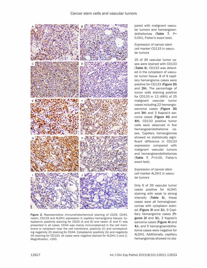
25 of 39 vascular tumor ca- ses were stained with CD133 (Table 6). CD133 was detect-ed in the cytoplasm of vascu-lar tumor tissue. 8 of 9 capil-lary hemangioma cases were positive for CD133 (Figure 2G and 2H). The percentage of tumor cells staining positive for CD133 in 12 (48%) of 25 malignant vascular tumor cases including 22 hemangio-sarcoma cases (Figure 3G and 3H) and 3 Kaposi’s sar-coma cases (Figure 4G and 4H). CD133 positive tumor cells were observed in five hemangioendothelioma ca- ses. Capillary hemangiomas showed no statistically signi- ficant difference in CD133 expression compared with malignant vascular tumors and hemangioendotheliomas (Table 7, P>0.05, Fisher’s exact test).
Expression of cancer stem cell marker ALDH1 in vascu-lar tumors
Only 6 of 39 vascular tumor cases positive for ALDH1 staining with weak to strong intensity (Table 6), these cases were all hemangiosar-comas with cytoplasm stain- ed (Figure 3I and 3J). 9 Capi- llary hemangioma cases (Fi- gures 2I and 3J), 3 Kaposi’s sarcoma cases (Figure 4I and 4J), and 5 hemangioendothe-lioma cases were negative for ALDH1. Additionally, capillary hemangiomas showed no sta-
Figure 2. Representative immunohistochemical staining of CD29, CD44, nestin, CD133 and ALDH1 expression in capillary hemangioma tissues. Cy-toplasmic positivity staining for CD29 (A and B) and nestin (E and F) was presented in all cases. CD44 was mainly immunostained in the cell mem-brane or cytoplasm near the cell membrane, positivity (C) and correspond-ing negativity (D) staining for CD44. Cytoplasmic positivity (G) and negativity (H) staining for CD133, all cases were negative stained for ALDH1 (I and J). Magnification, ×200.
Cancer stem cells and vascular tumors
12628 Int J Clin Exp Pathol 2015;8(10):12621-12633
tistically significant difference compared with malignant vascular tumors and hemangioendo-
thelioma (Table 7, P>0.05, Fisher’s exact test).
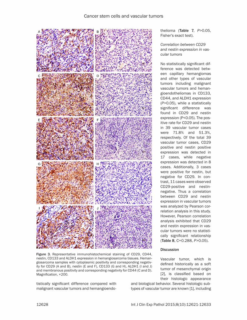
Correlation between CD29 and nestin expression in vas-cular tumors
No statistically significant dif-ference was detected betw- een capillary hemangiomas and other types of vascular tumors including malignant vascular tumors and heman-gioendotheliomas in CD133, CD44, and ALDH1 expression (P>0.05), while a statistically significant difference was found in CD29 and nestin expression (P<0.05). The pos-itive rate for CD29 and nestin in 39 vascular tumor cases were 71.8% and 51.3%, respectively. Of the total 39 vascular tumor cases, CD29 positive and nestin positive expression was detected in 17 cases, while negative expression was detected in 8 cases. Additionally, 3 cases were positive for nestin, but negative for CD29. In con-trast, 11 cases were observed CD29-positive and nestin-negative. Thus a correlation between CD29 and nestin expression in vascular tumors was analyzed by Pearson cor-relation analysis in this study. However, Pearson correlation analysis exhibited that CD29 and nestin expression in vas-cular tumors were no statisti-cally significant relationship (Table 8, C=0.288, P>0.05).
Discussion
Vascular tumor, which is defined historically as a soft tumor of mesenchymal origin [2], is classified based on their histologic appearance
Figure 3. Representative immunohistochemical staining of CD29, CD44, nestin, CD133 and ALDH1 expression in hemangiosarcoma tissues. Heman-giosarcoma samples with cytoplasmic positivity and corresponding negativ-ity for CD29 (A and B), nestin (E and F), CD133 (G and H), ALDH1 (I and J) and membranous positivity and corresponding negativity for CD44 (C and D). Magnification, ×200.
and biological behavior. Several histologic sub-types of vascular tumor are known [1], including
Cancer stem cells and vascular tumors
12629 Int J Clin Exp Pathol 2015;8(10):12621-12633
benign vascular tumors, malignant vascular tumors, and hemangioendothelimas that their
clinical behaviors are between the benign hemangiomas and more malignant angiosarco-mas. Furthermore, the patho-genesis and histogenesis of different types of vascular tumors are complicated and multi-factorial process [30]. Previous investigations sho- wed that multiple genomic alterations or micro-environ-mental differences presum-ably contribute to the devel-opment of vascular tumors [7]. Following recent studies supporting the presence of a highly tumorigenic cells sub-set commonly called cancer stem cells [31-33]. The can-cer stem cells hypothesis holds that cancer stem cells may contribute to the initia-tion, progression and re- currence of cancer [17, 18]. Although the current knowl-edge of the biological proper-ties of cancer stem cells is very limited, cancer stem cells expressing certain spe-cific surface makers have been documented by several reports. CD133 is a common stem cell surface antigens expressing on hematopoietic stem cells and bone marrow-derived endothelial progeni-tor cells [34], while CD29, CD44 and nestin have been describe as the mesenchymal stem cells markers [35].
In vascular tumor, cancer stem cells have been regard-ed as a possible candidate in relation to the origin and pathogenesis of tumors. Khan et al [36] studies firstly reported that the hematopoi-etic stem cells marker CD133 was used to isolate stem cells from hemangioma. Addi- tionally, CD133 positive tu- mor cells were observed in
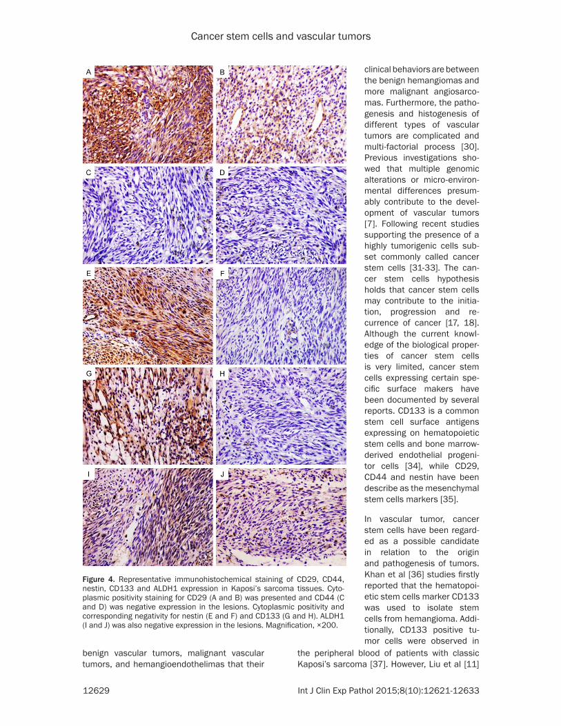
Figure 4. Representative immunohistochemical staining of CD29, CD44, nestin, CD133 and ALDH1 expression in Kaposi’s sarcoma tissues. Cyto-plasmic positivity staining for CD29 (A and B) was presented and CD44 (C and D) was negative expression in the lesions. Cytoplasmic positivity and corresponding negativity for nestin (E and F) and CD133 (G and H). ALDH1 (I and J) was also negative expression in the lesions. Magnification, ×200.
the peripheral blood of patients with classic Kaposi’s sarcoma [37]. However, Liu et al [11]
Cancer stem cells and vascular tumors
12630 Int J Clin Exp Pathol 2015;8(10):12621-12633
confirmed that CD133 detection was negative in almost all cases of hemangiosarcomas and hemangiomas, the hematopoietic stem cells or early endothelial progenitor cells expressing other markers CD34, CD45 and CD117, were participated in tumor formation in vascular tumors. In contrast to previous studies, our study found that most of human hemangiomas and hemangiosarcomas showed strong posi-tive staining for CD133. CD133 is an early marker for hematopoietic stem cells or endo-thelial progenitor cells, the cellular differentia-tion or tumor heterogeneity may be responsible for the CD133 expression level [38-40]. In addi-tion to cancer stem cell marker CD133, mesen-chymal stem cell markers CD29, CD44 and nestin were observed in vascular tumor. Several studies [41-43] have been reported that the isolated tumor cells from proliferating heman-gioma expressed the markers CD29, CD44 and CD105, cell surface marker associated with mesenchymal stem cells. Mesenchymal stem cells are defined by their self-renewal capability and potential for several differentiated cell types [44, 45]. Because of these properties, Yu et al [44] studies demonstrated that mesenchy-mal stem cells were the source of adipocytes in infantile hemangioma during the involuting and involuted phases. According to our results, CD29 and nestin were positive staining in all cases of hemangiomas, and 3 of 9 hemangio-ma cases were positive for CD44. In general, the percentage of CD29 and nestin positive tumor cells in hemangiomas was higher than that in hemangiosarcoams. Intermediate fila-
ment protein nestin was a new expression marker of mesenchymal stem cells. Nestin is well established cancer stem cell marker for several malignant tumors, such as high malig-nant glioma [46] and gastrointestinal stromal tumors [47]. Yang et al [48] studies also report-ed that the expression of nestin was stronger in poorly differentiated hemangiosarcomas com-pared with well differentiated hemangiosarco-mas. These results indicated that vascular tumors were at least partly attribute to the can-cer stem cells themselves and that investiga-tions in cancer stem cells may be especially relevant to understanding the pathogenesis of different type vascular tumors.
To our knowledge, ALDH1 have been consid-ered as a marker to identify cancer stem cells derived from human mammary cancer [49], head and neck squamous cell carcinoma [50]. Overexpression of this marker is associated with poor prognosis in breast [49], bladder [51] and lung cancer [52]. However, the role of ALDH1 in vascular tumor progress has not been described previously. In this study, all heman-giomas, Kaposi’s sarcomas and hemangioen-dothelimas were negative for ALDH1 staining, only 5 of 22 hemangiosarcoma cases was posi-tive for ALDH1 with weak to strong staining. In addition, our other groups found (data unpub-lished) that ALDH1 staining was observed in solitary fibrous tumor (SFT) and perivascular epithelioid cell tumor (PEComa), indicating that the ALDH1 may be a new cancer stem cell marker to explicate the progress of several soft tissue tumors. Thus, further research should be done to identify whether ALDH1 expression may play an important in development and pro-gression of vascular tumors.
In summary, our study showed that five cancer stem cell markers including CD29, CD44, CD133, nestin and ALDH1 exhibited different expression level in different type of vascular tumors. The heterogeneity might be caused by
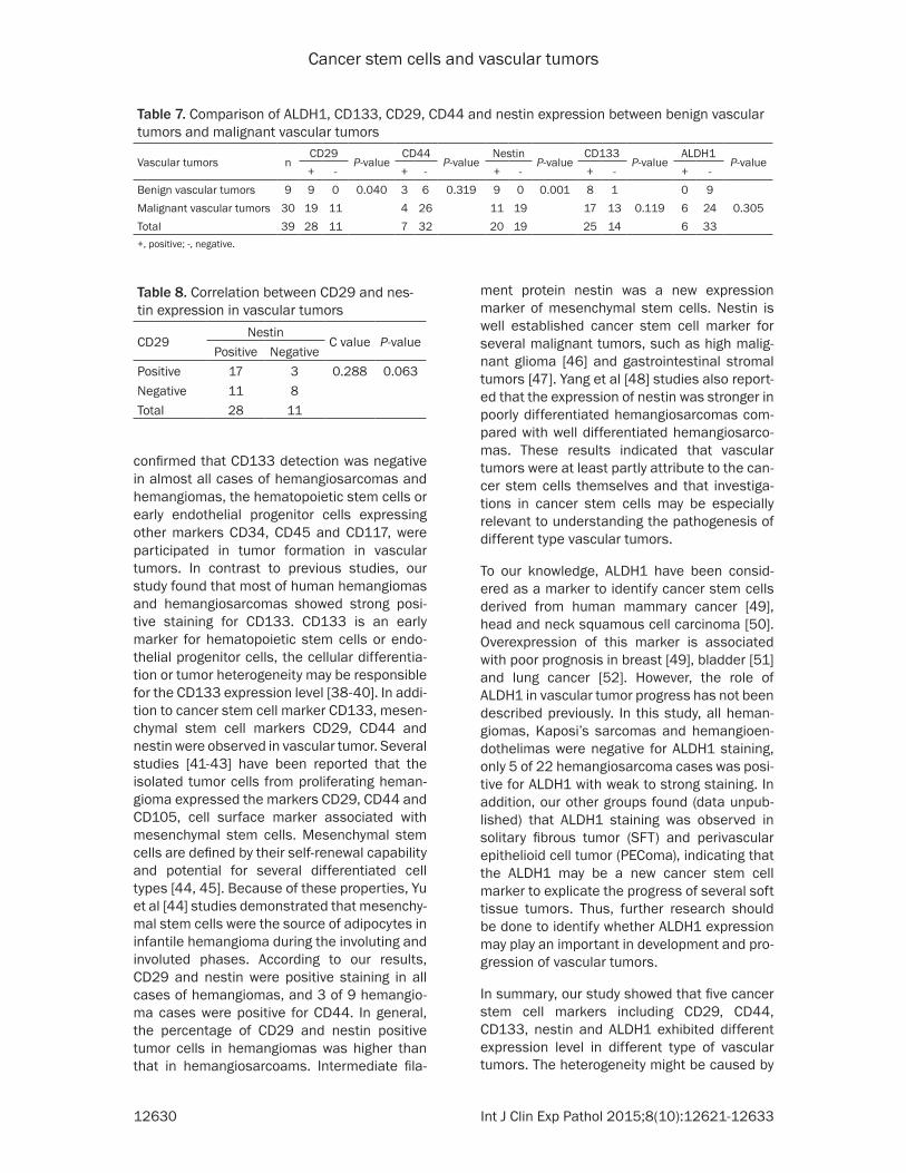
Table 7. Comparison of ALDH1, CD133, CD29, CD44 and nestin expression between benign vascular tumors and malignant vascular tumors
Vascular tumors nCD29
P-valueCD44
P-valueNestin
P-valueCD133
P-valueALDH1
P-value+ - + - + - + - + -
Benign vascular tumors 9 9 0 0.040 3 6 0.319 9 0 0.001 8 1 0 9Malignant vascular tumors 30 19 11 4 26 11 19 17 13 0.119 6 24 0.305Total 39 28 11 7 32 20 19 25 14 6 33+, positive; -, negative.
Table 8. Correlation between CD29 and nes-tin expression in vascular tumors
CD29Nestin
C value P-valuePositive Negative
Positive 17 3 0.288 0.063Negative 11 8Total 28 11

Cancer stem cells and vascular tumors
12631 Int J Clin Exp Pathol 2015;8(10):12621-12633
the different histogenesis and origin of differ-ent vascular tumors. According to our results and other previous investigations, we hypothe-size that mesenchymal stem cells may contrib-ute to vascular tumor formation. This study pro-vided further insight into the cellular origin leading to formation of vascular, suggesting that cancer stem cells may be involved in vas-cular tumor characteristics and progression. Therefore, our observation here may need fur-ther exploration to investigate the function of cancer stem cells in vascular tumors.
Acknowledgements
This work was supported by National Natural Science Foundation of China (No. 81160018, 81560053), the Corps Doctor Foundation (No. 2014BB018), Shihezi University Outstanding Youth Science and Technology Talent Cultivation Plan (2013ZRKXJQ05), One Thousand Youth Talents Plan, the Funders Autonomous Region (Xinjiang graduate student innovation No. XJGRI2014062), the Pairing Program of Shihezi University with Eminent Scholar in Elite University (SDJDZ201508).
Disclosure of conflict of interest
None.
Address correspondence to: Dr. Lijuan Pang, De- partment of Pathology and Key Laboratory of Xinjiang Endemic and Ethnic Diseases (Ministry of Education), Shihezi University School of Medicine, Shihezi, Xinjiang, China. E-mail: [email protected]
References
[1] Katenkamp K and Katenkamp D. Soft tissue tumors: new perspectives on classification and diagnosis. Dtsch Arztebl Int 2009; 106: 632-636.
[2] Ramon F, Degryse H and De Schepper A. Vascular soft tissue tumors: medical imaging. J Belge Radiol 1992; 75: 303-310.
[3] Greenberger S and Bischoff J. Pathogenesis of infantile haemangioma. Br J Dermatol 2013; 169: 12-19.
[4] Khan ZA, Melero-Martin JM, Wu X, Paruchuri S, Boscolo E, Mulliken JB and Bischoff J. Endothelial progenitor cells from infantile hem-angioma and umbilical cord blood display unique cellular responses to endostatin. Blood 2006; 108: 915-921.
[5] Qiu M, Qi X, Dai Y, Wang S, Quan Z, Liu Y and Ou J. Infantile haemangioma: a complicated disease. Front Biosci (Landmark Ed) 2015; 20: 1004-1016.
[6] Kakiuchi-Kiyota S, Crabbs TA, Arnold LL, Pennington KL, Cook JC, Malarkey DE and Cohen SM. Evaluation of expression profiles of hematopoietic stem cell, endothelial cell, and myeloid cell antigens in spontaneous and chemically induced hemangiosarcomas and hemangiomas in mice. Toxicol Pathol 2013; 41: 709-721.
[7] Koch M, Nielsen GP and Yoon SS. Malignant tumors of blood vessels: angiosarcomas, he-mangioendotheliomas, and hemangioperictyo-mas. J Surg Oncol 2008; 97: 321-329.
[8] Chokoeva A and Tchernev G. [Malignant vascu-lar tumors of the vulva]. Akush Ginekol (Sofiia) 2015; 54: 48-52.
[9] Ma JK, Barr J and Vijayakumar S. A multidisci-plinary approach to the management of atypi-cal osseous epithelioid hemangioendothelio-ma. Case Rep Oncol Med 2014; 2014: 917425.
[10] Roy S and Parwani AV. Primary renal epitheli-oid hemangioendothelioma. Case Rep Pathol 2012; 2012: 802515.
[11] Liu L, Kakiuchi-Kiyota S, Arnold LL, Johansson SL, Wert D and Cohen SM. Pathogenesis of hu-man hemangiosarcomas and hemangiomas. Hum Pathol 2013; 44: 2302-2311.
[12] Yonemori K, Tsuta K, Ando M, Hirakawa A, Hatanaka Y, Matsuno Y, Chuman H, Yamazaki N, Fujiwara Y and Hasegawa T. Contrasting prognostic implications of platelet-derived growth factor receptor-beta and vascular endo-thelial growth factor receptor-2 in patients with angiosarcoma. Ann Surg Oncol 2011; 18: 2841-2850.
[13] Gurzu S, Ciortea D, Munteanu T, Kezdi-Zaharia I and Jung I. Mesenchymal-to-endothelial tran-sition in Kaposi sarcoma: a histogenetic hy-pothesis based on a case series and literature review. PLoS One 2013; 8: e71530.
[14] Kandemir NO, Gun BD, Bahadir B, Yurdakan G, Ozdemir N, Karadayi N and Ozdamar SO. c-Kit (CD117) expression in classic Kaposi’s sarco-ma. Clin Exp Dermatol 2010; 35: 525-530.
[15] Liu A, Feng B, Gu W, Cheng X, Tong T, Zhang H and Hu Y. The CD133+ subpopulation of the SW982 human synovial sarcoma cell line ex-hibits cancer stem-like characteristics. Int J Oncol 2013; 42: 1399-1407.
[16] Boman BM and Wicha MS. Cancer stem cells: a step toward the cure. J Clin Oncol 2008; 26: 2795-2799.
[17] Feng JQ, Xu ZY, Shi LJ, Wu L, Liu W and Zhou ZT. Expression of cancer stem cell markers ALDH1 and Bmi1 in oral erythroplakia and the risk of oral cancer. J Oral Pathol Med 2013; 42: 148-153.
Cancer stem cells and vascular tumors
12632 Int J Clin Exp Pathol 2015;8(10):12621-12633
[18] Liu J, Ma L, Xu J, Liu C, Zhang J, Chen R and Zhou Y. Co-expression of CD44 and ABCG2 in spheroid body-forming cells of gastric cancer cell line MKN45. Hepatogastroenterology 2013; 60: 975-980.
[19] Bonnet D and Dick JE. Human acute myeloid leukemia is organized as a hierarchy that origi-nates from a primitive hematopoietic cell. Nat Med 1997; 3: 730-737.
[20] Ponti D, Costa A, Zaffaroni N, Pratesi G, Petrangolini G, Coradini D, Pilotti S, Pierotti MA and Daidone MG. Isolation and in vitro propa-gation of tumorigenic breast cancer cells with stem/progenitor cell properties. Cancer Res 2005; 65: 5506-5511.
[21] Ricci-Vitiani L, Lombardi DG, Pilozzi E, Biffoni M, Todaro M, Peschle C and De Maria R. Identification and expansion of human colon-cancer-initiating cells. Nature 2007; 445: 111-115.
[22] Tolar J, Nauta AJ, Osborn MJ, Panoskaltsis Mortari A, McElmurry RT, Bell S, Xia L, Zhou N, Riddle M, Schroeder TM, Westendorf JJ, McIvor RS, Hogendoorn PC, Szuhai K, Oseth L, Hirsch B, Yant SR, Kay MA, Peister A, Prockop DJ, Fibbe WE and Blazar BR. Sarcoma derived from cultured mesenchymal stem cells. Stem Cells 2007; 25: 371-379.
[23] Suva ML, Riggi N, Stehle JC, Baumer K, Tercier S, Joseph JM, Suva D, Clement V, Provero P, Cironi L, Osterheld MC, Guillou L and Stamenkovic I. Identification of cancer stem cells in Ewing’s sarcoma. Cancer Res 2009; 69: 1776-1781.
[24] Zhou F, Mu YD, Liang J, Liu ZX, Chen HS and Zhang JF. Expression and prognostic value of tumor stem cell markers ALDH1 and CD133 in colorectal carcinoma. Oncol Lett 2014; 7: 507-512.
[25] Calloni R, Cordero EA, Henriques JA and Bonatto D. Reviewing and updating the major molecular markers for stem cells. Stem Cells Dev 2013; 22: 1455-1476.
[26] Vassilopoulos A, Chisholm C, Lahusen T, Zheng H and Deng CX. A critical role of CD29 and CD49f in mediating metastasis for cancer-initi-ating cells isolated from a Brca1-associated mouse model of breast cancer. Oncogene 2014; 33: 5477-5482.
[27] Nishikawa S, Konno M, Hamabe A, Hasegawa S, Kano Y, Fukusumi T, Satoh T, Takiguchi S, Mori M, Doki Y and Ishii H. Surgically resected human tumors reveal the biological signifi-cance of the gastric cancer stem cell markers CD44 and CD26. Oncol Lett 2015; 9: 2361-2367.
[28] Yan Y, Zuo X and Wei D. Concise Review: Emerging Role of CD44 in Cancer Stem Cells: A Promising Biomarker and Therapeutic Target. Stem Cells Transl Med 2015; 4: 1033-43.
[29] Cherciu I, Barbalan A, Pirici D, Margaritescu C and Saftoiu A. Stem cells, colorectal cancer and cancer stem cell markers correlations. Curr Health Sci J 2014; 40: 153-161.
[30] Arbiser JL, Bonner MY and Berrios RL. Hemangiomas, angiosarcomas, and vascular malformations represent the signaling abnor-malities of pathogenic angiogenesis. Curr Mol Med 2009; 9: 929-934.
[31] Jordan CT, Guzman ML and Noble M. Cancer stem cells. N Engl J Med 2006; 355: 1253-1261.
[32] Gibbs CP, Kukekov VG, Reith JD, Tchigrinova O, Suslov ON, Scott EW, Ghivizzani SC, Ignatova TN and Steindler DA. Stem-like cells in bone sarcomas: implications for tumorigenesis. Neoplasia 2005; 7: 967-976.
[33] Yin S, Li J, Hu C, Chen X, Yao M, Yan M, Jiang G, Ge C, Xie H, Wan D, Yang S, Zheng S and Gu J. CD133 positive hepatocellular carcinoma cells possess high capacity for tumorigenicity. Int J Cancer 2007; 120: 1444-1450.
[34] Yin AH, Miraglia S, Zanjani ED, Almeida-Porada G, Ogawa M, Leary AG, Olweus J, Kearney J and Buck DW. AC133, a novel marker for human hematopoietic stem and progenitor cells. Blood 1997; 90: 5002-5012.
[35] P M, S H, R M, M G and W SK. Adult mesenchy-mal stem cells and cell surface characteriza-tion-a systematic review of the literature. Open Orthop J 2011; 5: 253-260.
[36] Khan ZA, Boscolo E, Picard A, Psutka S, Melero-Martin JM, Bartch TC, Mulliken JB and Bischoff J. Multipotential stem cells recapitulate human infantile hemangioma in immunodeficient mice. J Clin Invest 2008; 118: 2592-2599.
[37] Taddeo A, Presicce P, Brambilla L, Bellinvia M, Villa ML and Della Bella S. Circulating endothe-lial progenitor cells are increased in patients with classic Kaposi’s sarcoma. J Invest Dermatol 2008; 128: 2125-2128.
[38] Peichev M, Naiyer AJ, Pereira D, Zhu Z, Lane WJ, Williams M, Oz MC, Hicklin DJ, Witte L, Moore MA and Rafii S. Expression of VEGFR-2 and AC133 by circulating human CD34(+) cells identifies a population of functional endotheli-al precursors. Blood 2000; 95: 952-958.
[39] Beaudry P, Hida Y, Udagawa T, Alwayn IP, Greene AK, Arsenault D, Folkman J, Heymach JV, Ryeom S and Puder M. Endothelial progeni-tor cells contribute to accelerated liver regen-eration. J Pediatr Surg 2007; 42: 1190-1198.
[40] Urbich C and Dimmeler S. Endothelial progeni-tor cells: characterization and role in vascular biology. Circ Res 2004; 95: 343-353.
[41] Yuan SM, Chen RL, Shen WM, Chen HN and Zhou XJ. Mesenchymal stem cells in infantile hemangioma reside in the perivascular region. Pediatr Dev Pathol 2012; 15: 5-12.

Cancer stem cells and vascular tumors
12633 Int J Clin Exp Pathol 2015;8(10):12621-12633
[42] Itinteang T, Davis PF and Tan ST. Infantile hem-angiomas exhibit neural crest and pericyte markers. Ann Plast Surg 2015; 74: 383.
[43] Mai HM, Zheng JW, Wang YA, Yang XJ, Zhou Q, Qin ZP and Li KL. CD133 selected stem cells from proliferating infantile hemangioma and establishment of an in vivo mice model of hemangioma. Chin Med J (Engl) 2013; 126: 88-94.
[44] Yu Y, Fuhr J, Boye E, Gyorffy S, Soker S, Atala A, Mulliken JB and Bischoff J. Mesenchymal stem cells and adipogenesis in hemangioma involu-tion. Stem Cells 2006; 24: 1605-1612.
[45] Itinteang T, Vishvanath A, Day DJ and Tan ST. Mesenchymal stem cells in infantile haeman-gioma. J Clin Pathol 2011; 64: 232-236.
[46] Rani SB, Mahadevan A, Anilkumar SR, Raju TR and Shankar SK. Expression of nestin--a stem cell associated intermediate filament in hu-man CNS tumours. Indian J Med Res 2006; 124: 269-280.
[47] Tsujimura T, Makiishi-Shimobayashi C, Lund- kvist J, Lendahl U, Nakasho K, Sugihara A, Iwasaki T, Mano M, Yamada N, Yamashita K, Toyosaka A and Terada N. Expression of the intermediate filament nestin in gastrointesti-nal stromal tumors and interstitial cells of Cajal. Am J Pathol 2001; 158: 817-823.
[48] Yang XH, Wu QL, Yu XB, Xu CX, Ma BF, Zhang XM, Li SN, Lahn BT and Xiang AP. Nestin ex-pression in different tumours and its relevance to malignant grade. J Clin Pathol 2008; 61: 467-473.
[49] Ginestier C, Hur MH, Charafe-Jauffret E, Monville F, Dutcher J, Brown M, Jacquemier J, Viens P, Kleer CG, Liu S, Schott A, Hayes D, Birnbaum D, Wicha MS and Dontu G. ALDH1 is a marker of normal and malignant human mammary stem cells and a predictor of poor clinical outcome. Cell Stem Cell 2007; 1: 555-567.
[50] Wu J, Mu Q, Thiviyanathan V, Annapragada A and Vigneswaran N. Cancer stem cells are en-riched in Fanconi anemia head and neck squa-mous cell carcinomas. Int J Oncol 2014; 45: 2365-2372.
[51] Su Y, Qiu Q, Zhang X, Jiang Z, Leng Q, Liu Z, Stass SA and Jiang F. Aldehyde dehydrogenase 1 A1-positive cell population is enriched in tu-mor-initiating cells and associated with pro-gression of bladder cancer. Cancer Epidemiol Biomarkers Prev 2010; 19: 327-337.
[52] Huang EH, Hynes MJ, Zhang T, Ginestier C, Dontu G, Appelman H, Fields JZ, Wicha MS and Boman BM. Aldehyde dehydrogenase 1 is a marker for normal and malignant human co-lonic stem cells (SC) and tracks SC overpopula-tion during colon tumorigenesis. Cancer Res 2009; 69: 3382-3389.