original article domination of filamentous anoxygenic
TRANSCRIPT

Folia Biologica (Praha) 66, 24-35 (2020)
Original Article
Domination of Filamentous Anoxygenic Phototrophic Bacteria and Prediction of Metabolic Pathways in Microbial Mats from the Hot Springs of Al Aridhah(microbial mats / hot springs / 16S amplicon / Chloroflexus / Saudi Arabia)
M. YASIR1,5, A. K. QURESHI1, S. SRINIVASAN2, R. ULLAH1, F. BIBI1,5, M. REHAN3,5, S. B. KHAN4, E. I. AZHAR1,5
1Special Infectious Agents Unit, King Fahd Medical Research Center, King Abdulaziz University, Jeddah, Saudi Arabia2Institute of Bioinformatics and Applied Biotechnology, Bangalore, Karnataka-560100, India3King Fahd Medical Research Center, King Abdulaziz University, Jeddah, Saudi Arabia4Department of Chemistry, King Abdulaziz University, Jeddah, Saudi Arabia5Medical Laboratory Technology Department, Faculty of Applied Medical Sciences, King Abdulaziz University, Jeddah, Saudi Arabia
Abstract. Microbial mats in hot springs form a dy-namic ecosystem and support the growth of diverse communities with broad-ranging metabolic capacity. In this study, we used 16S rRNA gene amplicon se-quencing to analyse microbial communities in mat samples from two hot springs in Al Aridhah, Saudi Arabia. Putative metabolic pathways of the micro-bial communities were identified using phylogenetic investigation of communities by reconstruction of unobserved states (PICRUSt). Filamentous anoxy-genic phototrophic bacteria associated with phylum
Received June 17, 2019. Accepted November 8, 2019.
This project work was funded by General Directorate of Research Grants (GDRG), King Abdulaziz City for Science and Technolo-gy (KACST), Saudi Arabia under grant number AT-12-33.
Corresponding author: Muhammad Yasir, Special Infectious Agents Unit, King Fahd Medical Research Center, King Abdulaziz University, Jeddah-21589, Saudi Arabia. E-mail: [email protected]
Abbreviations: AAR4 – Al Aridhah4, AAR5 – Al Aridhah5, AMDR – antimicrobial drug resistance, BID – bacterial infec-tious disease, BOSM – biosynthesis of other secondary metabo-lites, FAP – filamentous anoxygenic phototrophic, FSD – folding, sorting and degradation, GBM – glycan biosynthesis and meta-bolism, KEGG – Kyoto encyclopaedia of genes and genomes, MCV – metabolism of cofactors and vitamins, MTP – metabo-lism of terpenoids and polyketides, OM – organic matter, OTUs – operational taxonomic units, PICRUSt – phylogenetic investi-gation of communities by reconstruction of unobserved states, RR – replication and repair, TK – total potassium, TN – total ni-trogen, TP – total phosphorus.
Chloroflexi were abundant (> 50 %) in both hot springs at 48 °C. Chloroflexi were mainly represent-ed by taxa Chloroflexus followed by Roseiflexus. Cyanobacteria of genus Arthrospira constituted 3.4 % of microbial mats. Heterotrophic microorganisms were mainly represented by Proteobacteria, Actino-bacteria, Bacteroidetes, and Firmicutes. Archaea were detected at a lower relative abundance (< 1 %). Metabolic pathways associated with membrane trans-port, carbon fixation, methane metabolism, amino acid biosynthesis, and degradation of aromatic com-pounds were commonly found in microbial mats of both hot springs. In addition, pathways for produc-tion of secondary metabolites and antimicrobial com-pounds were predicted to be present in microbial mats. In conclusion, microbial communities in the hot springs of Al Aridhah were composed of diverse bacteria, with taxa of Chloroflexus being dominant.
IntroductionHot springs represent an extremophilic environment
and a suitable habitat for studying the thermophilic mi-croorganisms that flourish at the high temperatures found in extreme geochemical conditions (Wilkins et al., 2019). Microbial mats from geothermal ecosystems provide a model for the ecological analysis of microbial commu-nities that thrive in such conditions (Rozanov et al., 2017). Phototrophic microbial mats form as a result of a gradient in sunlight and the physicochemical conditions supported by the activity of microorganisms (Rozanov et al., 2017; Ward et al., 2019). Microbial mats are com-monly observed in hot springs across geographical re-gions (Alcaman-Arias et al., 2018; Ward et al., 2019).
Vol. 66 25
These mats are mainly composed of Cyanobacteria and filamentous anoxygenic phototrophs (FAPs), including Chloroflexi, in neutral and alkaline hot springs (Rozanov et al., 2017; Uribe-Lorio et al., 2019). The relative abun-dance of these taxa mainly depends on the temperature, with Cyanobacteria primarily being found in lower-tem-perature hot springs and Chloroflexi having a higher relative abundance in hot springs that are > 60 °C (Alca-man-Arias et al., 2018; Uribe-Lorio et al., 2019). Chloroflexus spp. and Roseiflexus spp. have been well studied from microbial mats in Mushroom Spring in Yellowstone National Park, United States (Klatt et al., 2013; Thiel et al., 2017). The majority of Cyanobacteria are mesophilic in nature with a few exceptions, including moderate ther-mophiles, such as Fischerella, and the thermophilic Synechococcus, which grows optimally above 55 °C (Alle walt et al., 2006).
Thermophilic microorganisms from hot water springs were initially studied with the use of optical microscopy (Castenholz, 1973), which was later followed by separa-tion and classification of thermophilic strains, using culture-dependent approaches (Pierson and Castenholz, 1974). Studies in the late 1990s revealed that 80–99 % of bacteria present in the environment had not yet been explored by laboratory-based culture-dependent proto-cols and hence remained overlooked in terms of their ecological functions and potential biotechnological ap-plications (Amann et al., 1995). Recent developments in molecular biology and associated technological advances,
such as 16S amplicon sequencing, shotgun sequencing, single-cell genomics, and transcriptomics, have provid-ed a tremendous opportunity for the identification and characterization of microorganisms (Tripathy et al., 2016; Thiel et al., 2017; Wilkins et al., 2019). Such ad-vances have revolutionized research on microbial diver-sity, evolution, and adaptation in specific environments (Uribe-Lorio et al., 2019; Wilkins et al., 2019). For in-stance, distinct bacterial taxa and diverse metabolic ca-pacities were revealed through a metagenomic study of microbial mats from Mushroom Spring (Thiel et al., 2017).
Geological surveys have identified several hot springs in the volcanic region of Jazan and four in the Al-Lith region of Saudi Arabia. These hot springs have been par-tially studied by culture-dependent approaches (Khiya mi et al., 2012; Yasir et al., 2019). In this study, we were the first to use 16S metagenomic analysis to investigate the taxonomic diversity in the microbial mats from the hot springs located at Al Aridhah in the Jazan region.
Material and Methods
Sample collection and physicochemical analysis
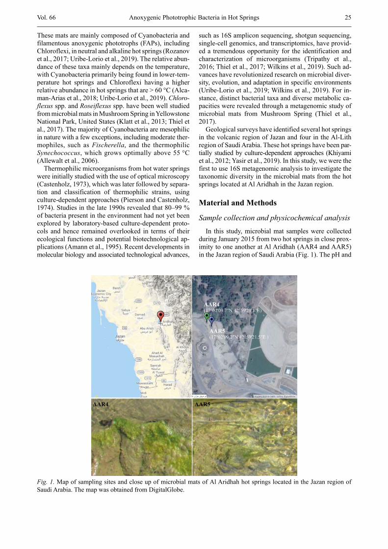
In this study, microbial mat samples were collected during January 2015 from two hot springs in close prox-imity to one another at Al Aridhah (AAR4 and AAR5) in the Jazan region of Saudi Arabia (Fig. 1). The pH and
Anoxygenic Phototrophic Bacteria in Hot Springs
Fig. 1. Map of sampling sites and close up of microbial mats of Al Aridhah hot springs located in the Jazan region of Saudi Arabia. The map was obtained from DigitalGlobe.
26 Vol. 66
temperature of the springs were measured using a Martini portable meter (Martini, Cragieburn, Australia). The mi-crobial mats were 2–3 mm thick, and samples were col-lected with sterilized spatulas and placed in sterilized containers. Temperature and pH at the sampling spots were 48 °C and 7.3, respectively. Samples were stored at 4 °C during transportation and were kept at –20 °C in the laboratory before further processing. Organic matter was determined through the loss of ignition according to the protocol of Dean (1974), and total nitrogen was de-termined using the Kjeldahl method. To determine other elements, 0.5 g of each sample was digested with a nitric acid-perchloric acid (HNO3-HClO4) mixture (Alzubaidy et al., 2016; Ullah et al., 2017). Following digestion, the solution was diluted with 50 ml of distilled water, and the level of total phosphorus was determined calorimet-rically and that of potassium by atomic absorption spec-troscopy. The other metals in the samples were deter-mined by inductively coupled plasma-optical emission spectroscopy, using the instrument according to the ma-nufacturer’s instructions (Ullah et al., 2017).
DNA extraction and 16S amplicon sequencingSamples were vigorously homogenized and metagen-
omic DNA was extracted from the microbial mats with a beads beating protocol using a power soil DNA extrac-tion kit (MO BIO Laboratories, Inc., Carlsbad, CA). Samples were processed for 16S rRNA gene amplicon paired-end sequencing in a MiSeq instrument (Illumina, San Diego, CA) as previously described (Yasir et al., 2015a). Briefly, a PCR-amplified template was gener-ated from genomic DNA using the universal primer set of 515F and 806R targeting the V4 variable region of bacteria and the archaeal 16S rRNA gene. PCR products were purified on AMPure beads, and concentrations were then measured with a dsDNA BR Assay Kit using a Qubit fluorometer (Invitrogen, Carlbad, CA). Sub-sequently, limited-cycle PCR with 1 ng of each PCR product was performed to add adapters and dual-index barcodes to each amplicon. After purification, the librar-ies were normalized according to the Nextera XT proto-col (Illumina). The multiplexed samples were pooled into a single flow cell for sequencing using the MiSeq platform (Illumina) following the manufacturer’s proto-col.
Data analysis and prediction of metabolic functions
Raw FASTQ files were collected from the MiSeq sys-tem (Illumina), and paired-end reads were joined using PANDAseq (Masella et al., 2012). Sequences were fil-tered; they were cleaned of primers and barcodes, all reads with ‘N’ and those with sequences < 200 bp were deleted, and high-quality sequences were de-replicated (Yasir et al., 2015b). The filtered sequence reads were clustered at k = 10 (97 % similarity), and chimeras and singleton reads were deleted. Sequence reads of high quality were grouped into operational taxonomic units
(OTUs) using a sequence similarity threshold of 97 %. The sequences were taxonomically classified using BLAST against the Greengenes database and by an RDP classifier and an RDP training set as described previ-ously (Dowd et al., 2008; Yasir, 2018) using QIIME 1.9.1 (Caporaso et al., 2010).
Putative metabolic functions of microbial communi-ties were identified using the PICRUSt (1.1.3) software, which compares the identified 16S rRNA gene sequences to related known genome sequenced species (Lan gille et al., 2013). OTUs were closed-referenced picked against the Greengenes (v13.5) database at 97 % similarity from each sample sequence file using QIIME (1.9.1) and saved in biome format following the protocol of PICRUSt (1.1.3). Resultant OTUs were used to predict metabolic functions by referencing the Kyoto Encyclopaedia of Genes and Genomes (KEGG) on the web-based Galaxy application. Sequencing data were submitted to the European Nucleotide Archive under accession Nos. SAMEA5203386–SAMEA5203387.
Results
Physicochemical analysis
The temperature of both AAR4 and AAR5 was 48 °C, and their pH was neutral (7.3). Both hot springs were nutritionally poor, and the total organic matter in micro-bial mats was 1.50 ± 0.06 % and 1.70 ± 0.09 %, respec-tively (Table 1). The total nitrogen level in both hot springs was 0.02 %, and the phosphorus level was 0.02 % and 0.01 % in AAR4 and AAR5, respectively. The iron concentration was 139.1 ± 0.5 mg/kg in AAR4 and 133.6 ± 0.8 mg/kg in AAR5. No substantial differences were found in the concentration (mg/kg) of magnesium (AAR4 = 25.90 ± 0.07, AAR5 = 26.1 ± 0.1) and sodium (AAR4 12.13 ± 0.02, AAR5 = 12.05 ± 0.05) between the two hot springs. The total potassium concentration was 0.27 ± 0.01 % in AAR4 and 0.28 ± 0.01 % in AAR5. Both hot springs had a Zn concentration of < 0.5 mg/kg (Table 1).
M. Yasir et al.
Table 1. Physicochemical analysis of the hot springs of Al Aridhah
Variable AAR4 AAR5pH 7.3 7.3Temperature (°C) 48 48OM (%) 1.50 ± 0.06 1.70 ± 0.09TK (%) 0.27 ± 0.01 0.28 ± 0.01TN (%) 0.02 0.02TP (%) 0.02 0.01Iron (mg/Kg) 139.1 ± 0.5 133.6 ± 0.8Magnesium (mg/Kg) 25.90 ± 0.07 26.10 ± 0.10Sodium (mg/Kg) 12.13 ± 0.02 12.05 ± 0.05Zinc (mg/Kg) 0.44 ± 0.03 0.49 ± 0.04

Vol. 66 27Anoxygenic Phototrophic Bacteria in Hot Springs
Microbial diversity analysis
In total, 342,489 trimmed and high-quality (> 200 bp) sequences were obtained from 343,206 raw reads of Al Aridhah hot springs. Sequence reads were classified into 1,074 OTUs at 97 % identity, excluding singletons, and assigned to microbial domains. A total of 36 phyla were detected in the microbial mat samples, including four phyla from archaea. Thirty phyla were commonly present in microbial mats from both hot springs. The dominant six bacterial phyla constituted > 90 % of all the sequence reads. The most dominant phylum was Chloroflexi, with a mean relative abundance value of 55.7 ± 5.6 %, and it was present at a relatively higher abundance in AAR4 (Fig. 2A). Other dominant phyla were Actinobacteria (12.3 ± 2.3 %) and Proteobacteria (10.9 ± 5.1 %), whereas Bacteroidetes, Cyanobacteria, and Firmicutes represented bacterial sequences in the range of 3.3–6.3 % (Fig. 2A). Proteobacteria were pre-dominantly comprised of δ-Proteobacteria (Fig. 2B). Among archaea, Euryarchaeota, Crenarchaeota, Thau-marchaeota, and Korarchaeota were detected in the mi-crobial mat from both hot springs, but were relatively less abundant (≤ 1 %) compared to bacteria.
The sequence reads were classified into 243 bacterial and 24 archaeal families. A relatively high diversity was observed in AAR5 and 232 bacterial families were iden-tified, compared with 193 families in AAR4 (Fig. 3A).
One hundred eighty-two families were commonly de-tected in both hot springs, whereas 49 families were unique to AAR5 and 12 were specifically detected in AAR4 (Fig. 3A). Chloroflexaceae was the dominant family, followed by Roseiflexaceae, Micrococcaceae, and Oscillatoriales in both hot springs (Fig. 3B). Among archaea, 16 families were commonly found in both hot springs, and eight families were unique to AAR5 (Fig. 3C). Methanobacteriaceae, Cenarchaeaceae, Methano-saetaceae, and Thermococcaceae were present at rela-tively higher abundance in both hot springs, whereas Desulfurococcaceae and Thaumarchaeota had higher abundances among archaeal families in the AAR5 mi-crobial mat.
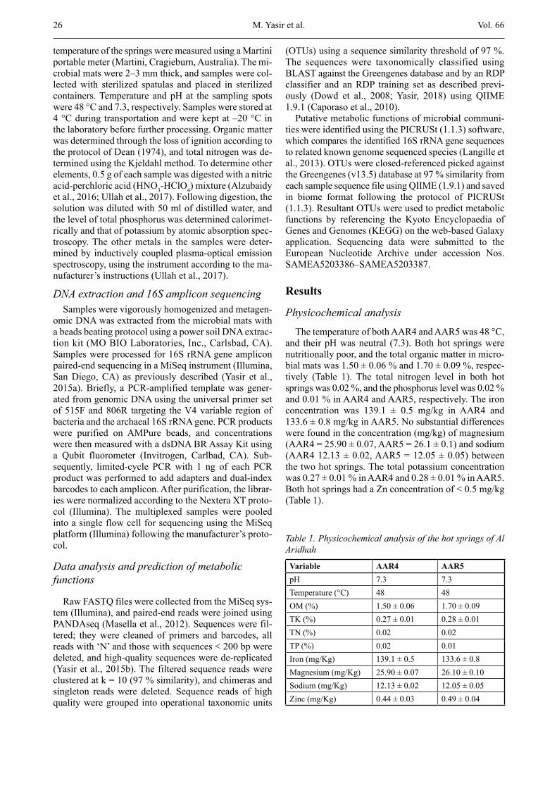
The sequences read were classified into 697 OTUs at the genus level, comprising 657 bacteria and 40 archaea. The highest number of bacterial genera (619) was de-tected in AAR5, whereas 424 genera were found in AAR4 (Fig. 4A). Among the genera detected, 386 were present in both hot springs, 233 were unique to AAR5, and 38 genera were specifically found in AAR4 (Fig. 4A). Moreover, eight genera in AAR4 and 15 genera in AAR5 were present at ≥ 1 % abundance, accounting for 84 % and 79 % of the total sequence reads in the respec-tive samples (Fig. 4B). The genera Chloroflexus (34.6 ± 5.6 %), Roseiflexus (14.2 ± 6.8 %), and Arthrobacter (12.2 ± 2.3 %) were present at a relatively higher abun-dance in both hot springs’ mats (Fig. 4B), whereas
Fig. 2. Percentage of relative abundance of the dominant (A) phyla and (B) classes identified from microbial mats of the hot springs of Al Aridhah

28 Vol. 66
Fig. 3. (A) Network analysis of the OTUs identified at the family taxonomic level of bacteria in microbial mats from the hot springs of Al Aridhah. (B) Percentage of relative abundance of the top 13 families found in the studied samples, and (C) network presentation of identified archaeal families. Nodes connected with two lines are commonly detected in both hot springs, and nodes connected with a single line are unique to the respective hot spring.
M. Yasir et al.
Vol. 66 29
Lewinella, Geobacter, Pelotomaculum, and Meiothermus were present at relatively higher abundance in AAR4 compared with AAR5. Genera Desulfomicrobium, Heliothrix, Anaerolinea, and Leptolyngbya were detected at a higher abundance in AAR5 compared with AAR4. Among archaea, 19 genera were commonly present in both hot springs and 20 genera were unique to AAR5 (Fig. 4C). Candidatus Nitrosocaldus, Methanosaeta,
Methanobrevibacter, Cenarchaeum, and Palaeococcus were present at relatively higher abundance in both hot springs.
Chloroflexi and CyanobacteriaFrom sequence reads, 1,018 bacterial OTUs were
identified at the species level. Five hundred thirty-one OTUs were commonly present in both hot springs from
Fig. 4. (A) Network analysis of OTUs identified at the genus level of bacteria in microbial mats from the hot springs of Al Aridhah. (B) Percentage of relative abundance of the top 25 genera found in the studied samples, and (C) network presentation of identified archaeal genera. Nodes connected with two lines are commonly detected in both hot springs, and nodes connected with a single line are unique to the respective hot spring.
Anoxygenic Phototrophic Bacteria in Hot Springs

30 Vol. 66
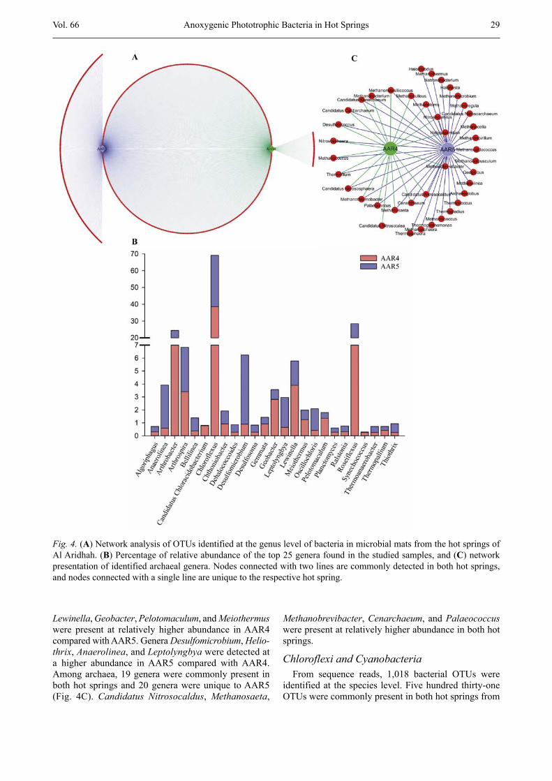
among the 618 OTUs of AAR4 and the 934 OTUs of AAR5. Taxa associated with Chloroflexi were predomi-nantly identified in AAR4 and AAR5 and mainly be-longed to class Chloroflexia followed by Anaerolineae. Forty-two OTUs linked to Chloroflexi at the species level were identified and were predominantly composed of Chloroflexus spp. (36.2 % and 26.5 %) followed by Roseiflexus castenholzii (14.4 % and 7.1 % in AAR4 and AAR5, respectively; Fig. 5A). OTUs of Roseiflexus spp. and Chloroflexus aurantiacus were detected at a relatively higher abundance in both hot springs. Anaerolinea spp. and Oscillochloris trichoides were present at a relatively higher abundance of 3.4 % and 1.7 % in AAR5, respectively, and were detected at < 1 % in AAR4 (Fig. 5A). Other OTUs were detected at < 1 % in the microbial mat from both hot springs. Moreover, only two taxa of Chlorobium spp. and Pelodictyon phaeoclathratiforme from phylum Chlorobi were de-tected and were present at < 1 % abundance.
Fifty-three OTUs from Cyanobacteria were detected from the microbial mats of Al Aridhah hot springs. Twenty-nine OTUs were common in both hot springs. Twenty OTUs were unique to AAR5, and four OTUs were specifically detected in AAR4. Taxa of Arthrospira spp. from Cyanobacteria were predominant, at a relative abundance of 3.4 %, in both hot springs (Fig. 5A). Leptolyngbya spp. were detected at a relatively high abundance of 1.1 % in AAR5 compared with AAR4 (0.5 %). Other OTUs were present at < 1 %, including Synechococcus spp. (Fig. 5A).
Heterotrophic microbial community analysisHeterotrophic bacteria represented < 55 % of the
community of the microbial mats and mainly comprised Actinobacteria, Proteobacteria, Bacteroidetes, and Fir-micutes. Proteobacteria were present at relatively higher abundance in AAR5 compared with AAR4 and were mainly composed of δ-Proteobacteria followed by γ-Proteobacteria and β-Proteobacteria (Fig. 2A, B). A total of 436 OTUs were classified to Proteobacteria. Desulfomicrobium terraneus from δ-Proteobacteria and Pseudoalteromonas spp. from γ-Proteobacteria were identified in AAR5 at a relatively higher abundance of 5.3 % and 1.7 %, respectively (Fig. 5B). Geobacter spp. were found at a relatively higher abundance of 2.8 % in AAR4, and other OTUs were present at < 1 % abun-dance (Fig. 5B).
Fifty-five OTUs were identified from Actinobacteria, and OTUs linked to Arthrobacter spp. were detected at >10 % abundance in both hot springs (Fig. 5B). Of Firmicutes, 188 OTUs were detected and were predomi-nantly composed of taxa from the strictly anaerobic class of Clostridia (Fig. 2B). In total, 97 OTUs were de-tected from Bacteroidetes, and Lewinella spp. were commonly found in both hot springs at > 1 % abundance (Fig. 5B). Other OTUs were detected at < 1 % abun-dance, except for Schleiferia spp. and Anaerophaga spp. (Fig. 5B).
Predicted metabolic functions
The putative metabolic functions of the hot spring mat microbiota were predicted from 16S rDNA ampli-con sequence data using the PICRUSt tool. The estimat-ed KEGG orthologies were found to be involved in the main pathways (level 1) of metabolism, genetic infor-mation processing, environmental information process-ing, cellular processes, and human diseases. Expanding the main pathways at level 2 resulted in 24 sub-path-ways, which were further classified into 203 sub-sub-pathways at level 3. In both hot springs, most of the pre-dicted pathways were associated with the metabolism of amino acids (10.4 ± 0.4 %), carbohydrates (10.3 ± 0.5 %), energy (7.3 ± 0.3 %), lipids (3.7 ± 0.1 %), and glycan biosynthesis and metabolism (2.2 ± 0.1 %). Apart from common amino acid synthesis pathways, different path-ways were predicted for carbohydrate metabolism in-cluding pyruvate, propanoate, glycolysis/gluconeogen-esis, butanoate, and pentose phosphate pathway (Fig. 6).
Notably, xenobiotic biodegradation and metabolism pathways were found at a considerable percentage (3.4 ± 0.3 %) and were mainly linked to the degradation of naphthalene, benzoate, toluene, aminobenzoate, caprol-actam, styrene, xylene, dioxin, and polycyclic aromatic hydrocarbons and the metabolism of xenobiotics by cy-tochrome P450. The predicted energy metabolic path-ways in the bacterial communities involved oxidative phosphorylation and the metabolism of methane and nitrogen. Carbon fixation pathways in prokaryotes were found at > 1 % in both hot springs (Fig. 6), whereas sulphur metabolism was not a major pathway in micro-bial communities of the mat samples. Secondary metabo-lite, terpenoid, and polyketide pathways for the following antibiotics were predicted: streptomycin, tetracycline, novobiocin, ansamycins, penicillin, and cephalosporin (Fig. 6). Pathways involved in infectious diseases, in-cluding the Vibrio cholerae pathogenic cycle and Staphylococcus aureus infection and bacterial toxins, were identified. Moreover, resistance to beta-lactam an-tibiotics was predicted at a relatively lower abundance of < 0.5 %.
Consistent with the harsh environment of hot springs, sequences linked with DNA repair and recombination proteins (2.4 ± 0.1 %) were predominantly identified at level 3 from the sub-pathway of replication and repair within the genetic information processing pathway. In addition, protein folding and associated processing function were predicted at a relative abundance of 0.9 % in both hot springs. Different types of membrane trans-port, mainly the ABC transporter (2.9 ± 0.2 %) and a two-component system (2.1 ± 0.3 %) from the environ-mental information processing pathway, were predicted. Bacterial motility proteins (1.7 ± 0.4 %) were promi-nently found in the cellular processes pathway. Overall, the predicted metabolic pathways and relative abun-dance did not vary markedly in mats from either of the hot springs.
M. Yasir et al.
Vol. 66 31
Fig. 5. (A) Percentage of relative abundance of the dominant taxa associated with Chloroflexi and Cyanobacteria, and (B) heterotrophic bacteria
Anoxygenic Phototrophic Bacteria in Hot Springs

32 Vol. 66
Fig. 6. Relative abundance of putative pathways of microbial communities in microbial mats from the hot springs of Al Aridhah using PICRUSt. The pathways are presented according to KEGG.
Discussion
Physicochemical parameters influence the microbial diversity and community composition in hot springs. The studied hot springs and other geothermal springs in the Jazan region in Saudi Arabia have a neutral pH (Khiyami et al., 2012). The temperature of these hot springs is lower than that of the hot spring at Ain Khulab (71 °C) but in the range of the hot spring of Bani Malik (45 °C) of this region. These hot springs are oligotroph-ic, with a low nutrient level and relatively abundant iron. The poor nutrient level in the Al Aridhah hot springs is in agreement with previous studies from Saudi Arabia, although variation was observed in the chemical analysis, which may affect the microbial diversity and composition of the microbial mats of the AAR4 and
AAR5 hot springs (Al-Dayel, 1988; Khiyami et al., 2012).
Microbial mats in hot springs exhibit spatial and tem-poral heterogeneity and are composed of diverse species capable of a wide range of metabolic processes (Kim et al., 2015; Thiel et al., 2017). Our results demonstrated that the dominant phyla in both of the hot springs were Chloroflexi, Actinobacteria, Proteobacteria, Bacteroide-tes, Cyanobacteria, and Firmicutes, all of which are commonly reported in hot springs around the world (Uribe-Lorio et al., 2019; Wilkins et al., 2019). However, their abundance differed, with members of the Chloro-flexi phylum being highly abundant and Cyanobacteria having low abundance in the Al Aridhah hot springs. Both types grow at 48 °C, but in general, the taxa of Cyanobacteria grow optimally in low-temperature hot
M. Yasir et al.

Vol. 66 33
springs and members of Chloroflexi phylum grow better at higher temperatures (Nubel et al., 2002; Sahoo et al., 2017). Alcaman-Arias et al. (2018) observed Cyano-bacteria and Chloroflexi at a similar relative abundance in the Porcelana hot spring at a temperature of 58 °C. However, several published reports have identified a negative or exclusion relationship between Cyanobac-teria and Chloroflexi, as found in the AAR4 and AAR5 hot springs (Sahoo et al., 2017; Ward et al., 2019). These findings suggest that variation in microbial mats with regard to the taxa abundance across different geographi-cal regions might be influenced by other physicochemi-cal and environmental factors.
In agreement with previous studies, Chloroflexus spp. and Roseiflexus spp. were abundantly found from among FAP bacteria, as found in the extensively studied Mushroom Spring (Allewalt et al., 2006; Bhaya et al., 2007). However, the taxa of Candidatus Thermochlorobacter aerophilum was not detected in the microbial mats of Al Aridhah hot springs, although they were dominantly identified along with other newly identified dominant taxa of Candidatus Chloracidobacterium ther mophilum and class Anaerolineae from phylum Chloroflexi in the metagenomic study from microbial mats of Octopus Spring and Mushroom Spring, both in Yellowstone National Park (Klatt et al., 2011; Garcia Costas et al., 2012).
The relative abundance of heterotrophic bacteria var-ied between the AAR4 and AAR5 hot springs. Acti-nobacteria were present at a relatively higher abundance in AAR4 and Proteobacteria in AAR5, followed by Bacteroidetes and Firmicutes. Actinobacteria, which are commonly found in soil and aquatic environments, are generally involved in geochemical cycles and the turno-ver of organic matter. Proteobacteria were dominated by sulphate-reducing δ-Proteobacteria, and the relative abundance of Desulfomicrobium was markedly different between AAR4 and AAR5. Similarly, bacteria from the phyla Firmicutes, Actinobacteria, Proteobacteria, and Bacteroidetes were found in most of the studied hot springs such as those in Tengchong in China (Pagaling et al., 2012) and Nevada in the United States (Song et al., 2009). In contrast to previous studies, Planctomycetes and Verrucomicrobia were detected at lower abundance in the microbial mats of the hot springs studied (Coman et al., 2013; Chan et al., 2015).
Metabolic pathways in the hot springs of Ain Al Aridhah are probably based on interactions between au-totrophic and heterotrophic microbial communities. An increased abundance of Chloroflexi indicates low pho-tosynthetic activity and the presence of a redox gradi-ent. The presence of anaerobic Clostridia from phylum Firmicutes suggests an anaerobic environment at the lower layer, where organic material is decomposed into low-molecular-weight molecules. Moreover, predicted sulphur pathways in the microbial mat probably exist and may be associated with Proteobacteria, Firmicutes (Urbieta et al., 2015), Chloroflexi, and Chlorobi (Bryant et al., 2012). In line with the methane metabolism pre-
dicted from the taxonomic data, methanotrophs belong-ing to genera Hyphomicrobium, Methyloligella, and Methylobacterium were identified in the Al Aridhah hot springs. In addition, methane-oxidizing Methyloversatilis and Methyloversatilis from β-Proteobacteria were found in both hot springs (Tsubota et al., 2005). Consistent with our KEGG functional analysis from 16S amplicon sequencing data, xenobiotic degradation, aromatic com-pound metabolism, and environmental information pro-cessing pathways have previously been reported in Amazonian and Odisha hot spring microbial communi-ties (Panda et al., 2016; Sahoo et al., 2017). However, variation exists in the percentages of the predicted func-tional categories among this and previous studies, as observed in the taxonomic data (Panda et al., 2016; Sahoo et al., 2017).
In conclusion, 16S metagenomic analysis provided deep insight into the microbial community structure of the microbial mats of the Al Aridhah hot springs in Saudi Arabia. Specific lineages of FAP bacteria, mainly Chloroflexus and Roseiflexus, were dominantly associ-ated with microbial mats of both hot springs. This work and other studies that have observed variation in micro-bial community composition suggest a biogeographic pattern in hot spring microbial mat microbiota. In addi-tion, transcriptomic studies and metagenomic studies based on shotgun sequencing are required to further an-alyse the metabolic potential of the hot spring microbial mats estimated from the taxonomic data in this study.
ReferencesAl-Dayel, M. (1988) Geothermal resources in Saudi Arabia.
Geothermics 17, 465-476.Alcaman-Arias, M. E., Pedros-Alio, C., Tamames, J., Fernan-
dez, C., Perez-Pantoja, D., Vasquez, M., Diez, B. (2018) Diurnal changes in active carbon and nitrogen pathways along the temperature gradient in Porcelana hot spring mi-crobial mat. Front. Microbiol. 9, 2353.
Allewalt, J. P., Bateson, M. M., Revsbech, N. P., Slack, K., Ward, D. M. (2006) Effect of temperature and light on growth of and photosynthesis by Synechococcus isolates typical of those predominating in the octopus spring micro-bial mat community of Yellowstone National Park. Appl. Environ. Microbiol. 72, 544-550.
Alzubaidy, H., Essack, M., Malas, T. B., Bokhari, A., Mot-walli, O., Kamanu, F. K., Jamhor S. A., Mokhtar, N. A., Antunes, A., Simões, M. F., Alam, I., Bougouffa, S., Lafi, F. F., Bajic, V. B., Archer, J. A. (2016) Rhizosphere micro-biome metagenomics of gray mangroves (Avicennia mari-na) in the Red Sea. Gene 576(2 Pt 1), 626-636.
Amann, R. I., Ludwig, W., Schleifer, K. H. (1995) Phyloge-netic identification and in situ detection of individual mi-crobial cells without cultivation. Microbiol. Rev. 59, 143-169.
Bhaya, D., Grossman, A. R., Steunou, A. S., Khuri, N., Cohan, F. M., Hamamura, N., Melendrez, M. C., Bateson, M. M., Ward, D. M., Heidelberg, J. F. (2007) Population level functional diversity in a microbial community revealed by
Anoxygenic Phototrophic Bacteria in Hot Springs

34 Vol. 66
comparative genomic and metagenomic analyses. ISME J. 1, 703-713.
Bryant, D. A., Liu, Z., Li, T., Zhao, F., Costas, A. M. G., Klatt, C. G., Ward D. M, Frigaard N-U, Overmann J. (2012) Comparative and functional genomics of anoxygenic green bacteria from the taxa Chlorobi, Chloroflexi, and Acido-bacteria. In: Advances in Photosynthesis and Respiration: Functional Genomics and Evolution of Photosynthetic Systems. eds. Burnap, R., Vermaas, W., vol 33, pp. 47-102, Springer, Dordrecht, The Nederlands.
Caporaso, J. G., Kuczynski, J., Stombaugh, J., Bittinger, K., Bushman, F. D., Costello, E. K., Fierer, N., Peña, A. G., Goodrich, J. K., Gordon, J. I., Huttley, G. A., Kelley, S. T., Knights, D., Koenig, J. E., Ley, R. E., Lozupone, C. A., McDonald, D., Muegge, B. D., Pirrung, M., Reeder, J., Se-vinsky, J. R., Turnbaugh, P. J., Walters, W. A., Widmann, J., Yatsunenko, T., Zaneveld, J., Knight, R. (2010) QIIME al-lows analysis of high-throughput community sequencing data. Nat. Methods 7, 335-336.
Castenholz, R. W. (1973) The possible photosynthetic use of sulfide by the filamentous phototrophic bacteria of hot springs. Limnol. Oceanogr. 18, 863-876.
Chan, C. S., Chan, K. G., Tay, Y. L., Chua, Y. H., Goh, K. M. (2015) Diversity of thermophiles in a Malaysian hot spring determined using 16S rRNA and shotgun metagenome se-quencing. Front. Microbiol. 6, 177.
Coman, C., Druga, B., Hegedus, A., Sicora, C., Dragos, N. (2013) Archaeal and bacterial diversity in two hot spring microbial mats from a geothermal region in Romania. Extremophiles 17, 523-534.
Dean, W. E. (1974). Determination of carbonate and organic matter in calcareous sediments and sedimentary rocks by loss on ignition: comparison with other methods. J. Sediment. Res. 44, 242-248.
Dowd, S. E., Sun, Y., Wolcott, R. D., Domingo, A., Carroll, J. A. (2008) Bacterial tag-encoded FLX amplicon pyrose-quencing (bTEFAP) for microbiome studies: bacterial di-versity in the ileum of newly weaned Salmonella-infected pigs. Foodborne Pathog. Dis. 5, 459-472.
Garcia Costas, A. M., Liu, Z., Tomsho, L. P., Schuster, S. C., Ward, D. M., Bryant, D. A. (2012) Complete genome of Candidatus Chloracidobacterium thermophilum, a chloro-phyll-based photoheterotroph belonging to the phylum Acidobacteria. Environ. Microbiol. 14, 177-190.
Khiyami, M. A., Serour, E. A., Shehata, M. M., Bahklia, A. H. (2012) Thermo-aerobic bacteria from geothermal springs in Saudi Arabia. Afr. J. Biotechnol. 11, 4053-4062.
Kim, Y. M., Nowack, S., Olsen, M. T., Becraft, E. D., Wood, J. M., Thiel, V., Klapper, I., Kühl, M., Fredrickson, J. K., Bryant, D. A., Ward, D. M., Metz, T. O. (2015) Diel me-tabolomics analysis of a hot spring chlorophototrophic mi-crobial mat leads to new hypotheses of community mem-ber metabolisms. Front. Microbiol. 6, 209.
Klatt, C. G., Wood, J. M., Rusch, D. B., Bateson, M. M., Hamamura, N., Heidelberg, J. F., Grossman, A. R., Bhaya, D., Cohan, F. M., Kühl, M., Bryant, D. A., Ward, D. M. (2011) Community ecology of hot spring cyanobacterial mats: predominant populations and their functional poten-tial. ISME J. 5, 1262-1278.
Klatt, C. G., Liu, Z., Ludwig, M., Kuhl, M., Jensen, S. I., Bry-ant, D. A., Ward, D. M. (2013) Temporal metatranscrip-tomic patterning in phototrophic Chloroflexi inhabiting a microbial mat in a geothermal spring. ISME J. 7, 1775-1789.
Langille, M. G., Zaneveld, J., Caporaso, J. G., McDonald, D., Knights, D., Reyes, J. A., Clemente, J. C., Burkepile, D. E., Vega Thurber, R. L., Knight, R., Beiko, R. G., Huttenhow-er, C. (2013) Predictive functional profiling of microbial communities using 16S rRNA marker gene sequences. Nat. Biotechnol. 31, 814-821.
Masella, A. P., Bartram, A. K., Truszkowski, J. M., Brown, D. G., Neufeld, J. D. (2012) PANDAseq: paired-end assem-bler for illumina sequences. BMC Bioinformatics 13, 31.
Nubel, U., Bateson, M. M., Vandieken, V., Wieland, A., Kuhl, M., Ward, D. M. (2002) Microscopic examination of distri-bution and phenotypic properties of phylogenetically di-verse Chloroflexaceae-related bacteria in hot spring micro-bial mats. Appl. Environ. Microbiol. 68, 4593-4603.
Pagaling, E., Grant, W. D., Cowan, D. A., Jones, B. E., Ma, Y., Ventosa, A., Heaphy, S. (2012) Bacterial and archaeal di-versity in two hot spring microbial mats from the geother-mal region of Tengchong, China. Extremophiles 16, 607-618.
Panda, A. K., Bisht, S. S., De Mandal, S., Kumar, N. S. (2016) Bacterial and archeal community composition in hot springs from Indo-Burma region, North-east India. AMB Express 6, 111.
Pierson, B. K., Castenholz, R. W. (1974) A phototrophic glid-ing filamentous bacterium of hot springs, Chloroflexus au-rantiacus, gen. and sp. nov. Arch. Microbiol. 100, 5-24.
Rozanov, A. S., Bryanskaya, A. V., Ivanisenko, T. V., Malup, T. K., Peltek, S. E. (2017) Biodiversity of the microbial mat of the Garga hot spring. BMC Evol. Biol. 17(Suppl 2), 254.
Sahoo, R. K., Gaur, M., Das, A., Singh, A., Kumar, M., Subudhi, E. (2017) Comparative analysis of 16S rRNA gene Illumi-na sequence for microbial community structure in diverse unexplored Hot Springs of Odisha, India. Geomicrobiol. J. 34, 567-576.
Song, Z., Zhi, X., Li, W., Jiang, H., Zhang, C., Dong, H. (2009) Actinobacterial diversity in hot springs in Tengchong (Chi-na), Kamchatka (Russia), and Nevada (USA). Geomicrobiol. J. 26, 256-263.
Thiel, V., Hugler, M., Ward, D. M., Bryant, D. A. (2017) The dark side of the Mushroom spring microbial mat: life in the shadow of chlorophototrophs. II. metabolic functions of abundant community members predicted from metagen-omic analyses. Front. Microbiol. 8, 943.
Tripathy, S., Padhi, S. K., Mohanty, S., Samanta, M., Maiti, N. K. (2016) Analysis of the metatranscriptome of microbial communities of an alkaline hot sulfur spring revealed dif-ferent gene encoding pathway enzymes associated with energy metabolism. Extremophiles 20, 525-536.
Tsubota, J., Eshinimaev, B., Khmelenina, V. N., Trotsenko, Y. A. (2005) Methylothermus thermalis gen. nov., sp. nov., a novel moderately thermophilic obligate methanotroph from a hot spring in Japan. Int. J. Syst. Evol. Microbiol. 55(Pt 5), 1877-1884.
Ullah, R., Yasir, M., Khan, I., Bibi, F., Sohrab, S. S., Al-Ansa-ri, A., Al-Abbasi, F., Al-Sofyani, A. A., Daur, I., Lee, S. W., Azhar, E. I. (2017) Comparative bacterial community anal-ysis in relatively pristine and anthropogenically influenced
M. Yasir et al.

Vol. 66 35
mangrove ecosystems on the Red Sea. Can. J. Microbiol. 63, 649-660.
Urbieta, M. S., Gonzalez-Toril, E., Bazan, A. A., Giaveno, M. A., Donati, E. (2015) Comparison of the microbial com-munities of hot springs waters and the microbial biofilms in the acidic geothermal area of Copahue (Neuquen, Argenti-na). Extremophiles 19, 437-450.
Uribe-Lorio, L., Brenes-Guillen, L., Hernandez-Ascencio, W., Mora-Amador, R., Gonzalez, G., Ramirez-Umana, C. J., Díez, B., Pedrós-Alió, C. (2019) The influence of tem-perature and pH on bacterial community composition of microbial mats in hot springs from Costa Rica. Microbiologyopen, 8, e893.
Ward, L. M., Idei, A., Nakagawa, M., Ueno, Y., Fischer, W. W., McGlynn, S. E. (2019) Geochemical and metagenomic characterization of Jinata Onsen, a proterozoic-analog hot spring, reveals novel microbial diversity including iron-tolerant phototrophs and thermophilic lithotrophs. Microbes Environ. 34, 278-292.
Wilkins, L. G. E., Ettinger, C. L., Jospin, G., Eisen, J. A. (2019) Metagenome-assembled genomes provide new in-sight into the microbial diversity of two thermal pools in Kamchatka, Russia. Sci. Rep. 9, 3059.
Yasir, M., Angelakis, E., Bibi, F., Azhar, E. I., Bachar, D., Lagier, J. C., (2015a) Comparison of the gut microbiota of people in France and Saudi Arabia. Nutr. Diabetes 5, e153.
Yasir, M., Azhar, E. I., Khan, I., Bibi, F., Baabdullah, R., Al-Zahrani, I. A., Al-Ghamdi, A. K. (2015b) Composition of soil microbiome along elevation gradients in southwestern highlands of Saudi Arabia. BMC Microbiol. 15, 65.
Yasir, M. (2018) Analysis of bacterial communities and char-acterization of antimicrobial strains from cave microbiota. Braz. J. Microbiol. 49, 248-257.
Yasir, M., Qureshi, K. A., Khan, I., Bibi, F., Rehan, M., Khan, S. B., Azhar E. I. (2019) Culturomics-based taxonomic di-versity of bacterial communities in the hot springs of Saudi Arabia. OMICS 23, 17-27.
Anoxygenic Phototrophic Bacteria in Hot Springs