seasonal occurrence of anoxygenic photosynthesis in
TRANSCRIPT
Biogeosciences, 9, 2485–2495, 2012www.biogeosciences.net/9/2485/2012/doi:10.5194/bg-9-2485-2012© Author(s) 2012. CC Attribution 3.0 License.
Biogeosciences
Seasonal occurrence of anoxygenic photosynthesis in Tillari andSelaulim reservoirs, Western India
S. Kurian1, R. Roy1,2, D. J. Repeta2, M. Gauns1, D. M. Shenoy1, T. Suresh1, A. Sarkar1, G. Narvenkar1,C. G. Johnson2, and S. W. A. Naqvi1
1National Institute of Oceanography, Council of Scientific & Industrial Research, Dona Paula, Goa, 403 004, India2Woods Hole Oceanographic Institution, Woods Hole, MA 02543, USA
Correspondence to:S. Kurian ([email protected])
Received: 30 November 2011 – Published in Biogeosciences Discuss.: 16 December 2011Revised: 21 May 2012 – Accepted: 23 May 2012 – Published: 9 July 2012
Abstract. Phytoplankton and bacterial pigment composi-tions were determined by high performance liquid chro-matography (HPLC) and liquid chromatography-mass spec-trometry (LC-MS) in two freshwater reservoirs (Tillari Damand Selaulim Dam), which are located at the foothills ofthe Western Ghats in India. These reservoirs experienceanoxia in the hypolimnion during summer. Water sampleswere collected from both reservoirs during anoxic periodswhile one of them (Tillari Reservoir) was also sampled inwinter, when convective mixing results in well-oxygenatedconditions throughout the water column. During the pe-riod of anoxia (summer), bacteriochlorophyll (BChl)e iso-mers and isorenieratene, characteristic of brown sulfur bac-teria, were dominant in the anoxic (sulfidic) layer of theTillari Reservoir under low light intensities. The winter ob-servations showed the dominance of small cells of Chloro-phyll b-containing green algae and cyanobacteria, with minorpresence of fucoxanthin-containing diatoms and peridinin-containing dinoflagellates. Using total BChle concentrationobserved in June, the standing stock of brown sulfur bacteriacarbon in the anoxic compartment of Tillari Reservoir wasestimated to be 2.27 gC m−2, which is much higher than thesimilar estimate for carbon derived from oxygenic photosyn-thesis (0.82 gC m−2). The Selaulim Reservoir also displayedsimilar characteristics with the presence of BChle isomersand isorenieratene in the anoxic hypolimnion during summer.Although sulfidic conditions prevailed in the water columnbelow the thermocline, the occurrence of photo-autotrophicbacteria was restricted only to mid-depths (maximal concen-tration of BChle isomers was detected at 0.2 % of the sur-face incident light). This shows that the vertical distribution
of photo-autotrophic sulfur bacteria is primarily controlledby light penetration in the water column where the presenceof H2S provides a suitable biogeochemical environment forthem to flourish.
1 Introduction
In stratified lakes having anoxic hypolimnia, photo-autotrophic bacteria have been known to contribute substan-tially to total primary production and biomass (Biebl andPfennig, 1979; Hurley and Watras, 1991). These bacteriause sulfide and other reduced sulfur compounds and some-times also iron sulfide as electron donors and light as en-ergy source for anoxygenic photosynthesis (Garcia-Gil et al.,1990; Imhoff, 1992). These bacteria are divided into the pur-ple sulfur bacteria (PSB) and the green sulfur bacteria (GSB).Purple sulfur bacteria are generally pigmented with bacteri-ochlorophyll (BChl)a or b while green sulfur bacteria con-tain BChlc, d or e along with their characteristic carotenoids.The BChlsc, d ande exist as a series of homologues that dif-fer in the degree of alkylation of the tetrapyrrole macrocycleat positions C-8 and C-12 and in the esterifying alcohol atC-17 (Senge and Smith, 1995). Green sulfur bacteria such asChlorobium (Cb.) phaeobacteroidescontain a large amountof aggregated BChle along with high content of carotenoidsin the chlorosome and are brown colored due to the pres-ence of BChle aggregates. They also contain minor amountsof BChl a in the photosynthetic reaction centre and also as-sociated with the baseplate of the chlorosome (Gerola andOlson, 1986). A few other GSB such asCb. tepidumandCb.
Published by Copernicus Publications on behalf of the European Geosciences Union.

2486 S. Kurian et al.: Seasonal occurrence of anoxygenic photosynthesis
limicola look green due to the presence of large aggregatedBChl c andd, with a low content of carotenoids (Hirabayashiet al., 2004). Recently, the application of HPLC, LC-MS andTandem mass spectrometry has made the analysis of bacteri-ochlorophyll isomers more accurate and specific (Airs et al.,2001a, b; Airs and Keely, 2002; Masse et al., 2004).
The accumulation of phototrophic bacteria close to theoxic-anoxic boundary layer depends strongly on the avail-able light (Pfenning, 1989), which can even act as a limit-ing factor. In comparison with the GSB, the brown-coloredspecies have been shown to live deeper in the water columnwhere the available spectral range is limited to 400–600 nm(Vila and Abella, 1994).
There have been several studies of photo-autotrophic sul-fur bacteria in freshwater lakes (Takahashi and Ichimura,1968; Yacobi et al., 1990; Garcia-Gil and Abella, 1992;Borrego et al., 1997; Vila et al., 1998; Yacobi and Ostro-vski, 2008, 2011). Based on BChle pigments, Yacobi etal. (1990) reported the dominance of brown sulfur photo-synthetic bacteria in the anoxic hypolimnion of Lake Kin-neret, Israel. Bacteriochlorophyll homologues compositionand changes in relation to light intensity were studied by Bor-rego et al. (1997) in several meromictic and holomictic lakesof Europe and USA. Meromictic lakes have been found tosupport dense populations of anaerobic photosynthetic bac-teria as the stagnant bottom waters are rich in nitrogen, phos-phorus and sulfide with the species variability dependent onlight availability and physicochemical characteristics of thelake (Garcia-Gil and Abella, 1992; Borrego et al., 1997).
India has a large number of natural freshwater lakes andman-made reservoirs created by damming rivers, some ofwhich have recently been found to experience anoxic con-ditions including sulphate reduction in the hypolimnia dur-ing summer (Narvenkar et al., 2012). We selected two suchsystems – the reservoirs of Tillari and Selaulim dams – toundertake a study of phytoplankton and bacterial pigmentsin Indian freshwater lakes, the results of which are beingreported here. These reservoirs are located at the foothillsof the Western Ghats in the states of Maharashtra (Tillari)and Goa (Selaulim) (Fig. 1). The former is currently be-ing sampled by us at least once a month since March 2010.This reservoir has been found to be dimictic in that the wa-ter column gets vertically mixed during winter and south-west monsoon periods. In 2010, when sampling for anoxy-genic photosynthetic organisms was carried out, sulfidic con-ditions prevailed from May to July before being terminatedby re-oxygenation of the hypolimnion through groundwa-ter inputs as well as weakening of thermal stratification fol-lowed by wind-induced mixing during the southwest mon-soon. At the end of the southwest monsoon, stratificationwas re-established and the hypolimnion again lost oxygen,but it did not become anoxic. Convective mixing led to thedevelopment of well-oxygenated conditions during the fol-lowing winter (S. W. A. Naqvi, unpublished data). Based onHPLC and LC-MS analyses, we investigate the variability of
22
Fig. 1. Sampling locations (Tillari Reservoir and Selaulim Reser-voir) situated in central India. Tillari Reservoir is located in the Ma-harashtra state with storage capacity of 0.45× 10⁹ m3 and waterdepth of∼ 50 m, while Selaulim Reservoir is located in the Goastate with lower storage capacity (0.23× 10⁹ m3) and shallowerdepth (∼ 20 m). Both these reservoirs were found to turn anoxicduring summer.
phytoplankton and bacterial pigments in relation to the de-velopment of water column anoxia during May–July 2010.We compare these data with one observation during winter(January 2011). In Selaulim Reservoir, which has a smallerstorage capacity (0.23×10⁹ m3 as compared to 0.45×10⁹ m3
for the Tillari Reservoir), observations were made only once– in May 2010 – when intense anoxia prevailed in the hy-polimnion. Finally, an attempt is made to quantify the relativecontribution of oxygenic and anoxygenic photo-autotrophs tomicrobial biomass during summer in the Tillari Reservoir.
2 Methodology
Water sampling in the reservoirs was carried out using 5-lNiskin samplers mounted on a nylon wire and fitted withreversing thermometers to measure temperature at differentsampling depths. Subsamples for dissolved oxygen (DO) andhydrogen sulfide (H2S) were collected carefully avoidingair exchange and taken to the laboratory for analysis. Dis-solved oxygen and H2S were estimated following standardtitrimetric Winkler and colorometric methods, respectively(Grasshoff et al., 1983).
Optical parameters were derived using the radiative trans-fer simulation code Hydrolight Version 5.0 (http://www.sequoia.com). The inputs to the simulations were absorptionand beam attenuation at nine wavelengths measured in situusing the AC-9 spectrophotometer(WET Labs, Inc.) and so-lar irradiance at the surface. The downwelling solar irradi-ance for the day was derived using the solar irradiance model(Gregg and Carder, 1990). Aerosol optical depth at 550 nm,Angstrom exponent, ozone and pressure for the day were
Biogeosciences, 9, 2485–2495, 2012 www.biogeosciences.net/9/2485/2012/
S. Kurian et al.: Seasonal occurrence of anoxygenic photosynthesis 2487
derived from ocean color satellite MODIS Aqua availablefrom Giovanni GES-DISC (Goddard Earth Sciences Dataand Information Services Center) (http://disc.sci.gsfc.nasa.gov/giovanni). Other meteorological parameters such as thewind speed and humidity were derived using climatologicaldata over the region. The volume scattering function for theHydrolight simulations was from Petzold (1972).
For the pigment analysis, water samples were collectedin amber colored bottles and were filtered (∼ 0.5 to 1 l) onWhatman glass fibre filters (GF/F; pore size 0.7 µm) un-der dark conditions and stored at−80◦C until the analy-sis. The frozen filters were extracted within 2–3 weeks ofcollection at 0◦C for 2 min in 3 ml of 100 % methanol us-ing ultrasonic dismembrator model 100 (Fisher Scientific)at 23 kHz. The extracts were filtered using a Teflon syringecartridge (Sartorius Minisart, pore size 0.45 mm, diameter25 mm) to remove any cellular debris, and analyzed usingHPLC (Agilent Technologies) at the National Institute ofOceanography (NIO), Goa using an Eclipse XDB C8 col-umn. Pigments were separated following the procedure asdetailed in Roy et al. (2006) using a binary solvent gradi-ent. Briefly, elution was performed at a rate of 1.1 ml min−1
using a linear gradient program over 45 min as follows:initially 5 % of solvent B (methanol) followed by a lineargradient over 22 min to 95 % B, with an isocratic hold for13 min at 95 % B until 35 min, which returned to initial con-dition of 5 % B prior to the next analysis. Solvent A was70 / 30: methanol / 0.5 M ammonium acetate and the columnwas maintained at 60◦C during the analysis. The eluting pig-ments were detected at 450 and 665 nm (Soret and Qy bands)by the diode array detector. Commercially available stan-dards obtained from DHI Inc. (Denmark) were used for theidentification and quantification of pigments including bothchlorophylls and carotenoids. Identification was based on theretention time and visible spectra matching. Pigment concen-trations were computed from the peak areas as detailed inRoy et al. (2006).
Bacteriochlorophylls were identified and quantified byHPLC and LC-MS at Woods Hole Oceanographic Institution(WHOI), USA. Extraction of samples was done using ace-tone in ultrasonic bath, dried and re-dissolved in methanoland analysed by HPLC (Agilent Technologies) equippedwith C-18 column (15 cm× 4.6 mm, 3 µm, Supelco Inc). Elu-tion was performed at a rate of 1.5 ml min−1 for 60 min us-ing a linear gradient program with the eluent compositionvarying as follows: 100 % solvent A (80 / 20: MeOH / 0.05 Mammonium acetate) to begin with, followed by a linear gra-dient over 20 min to 100 % B (80 / 20: MeOH / acetone); anisocratic hold for 25 min with 100 % B for elution of all themajor pigments then returning to 100 % A for 5 min; finally,10 min hold with 100 % A to equilibrate the column beforenext analysis. Pigments were detected at 410, 442, 470 and660 nm by the diode array detector (DAD). Even though dif-ferent HPLC columns were used in NIO (C-8) and WHOI(C-18), the BChl peaks were well resolved under the above-
mentioned conditions in both cases, appearing a little earlier(by 1.4–1.9 min) when the C18 column was used.
Samples were also analyzed by LC-MS (Agilent Tech-nologies) using an atmospheric pressure chemical ionization(APCI) source operated in the positive ion mode, scanningfrom 300 to 1200m/z. The same column and eluting sol-vents were used and the peaks were confirmed based on massspectra in full scan and also in the selected ion monitoring(SIM) mode. Conditions used for LC-MS analysis were: va-porizer temperature 400◦C, drying gas temperature 300◦Cand discharge current 3 µA. Bacteriochlorophyll standardswere extracted from particulate matter from Salt Pond, USA,in which BChl e isomers were dominant. Bacteriochloro-phyll homologues were separated using fraction collectorand quantified by spectrophotometic measurements assum-ing a molar extinction coefficient 48.9 mM cm−1 (Borrego etal., 1999a). A multi-point calibration table was prepared forBChl e4 isomer and the same calibration was used for BChle2 ande3 isomers as they are different only by CH2 groups.Isorenieratene was quantified based onβ-carotene calibra-tion and was present in the anoxic samples. The standingstock of anoxygenically photosynthesized carbon was esti-mated from the column-integrated BChle using BChle : Cratio of 1 : 20 (Oelze and Golecki, 1988; Yacobi and Os-trovski, 2008). Similarly, the standing stock of oxygenicallyphotosynthesized carbon was calculated using a Chla : C ra-tio of 1 : 40 (Cole et al., 2002).
Sub-samples (1.5 ml) were also collected and preservedwith glutaraldehyde (0.25 % final concentration), frozen inliquid nitrogen and then stored at−80◦C for flow cytomet-ric analysis at NIO. These samples were thawed at room tem-perature and analysed using a FACS Calibur flow cytometerfollowing Marie et al. (1997) to obtain absolute counts. Acombination of the FSC (forward scatter)-SSC (side scatter),the SSC-FL3 (red fluorescence) and the FL2 (orange fluo-rescence) – FL3 plots were used to differentiate populationsin the sample (Casamayor et al., 2007). The data was an-alyzed using CYTOPC software (Vaulot, 1989). A cultureof photosynthetic sulfur bacteria isolated from the seasonalanoxic marine waters off Goa (September, 2006) at a depth of25 m was run to identify this form from other fields. The cul-ture was maintained in Pfenning’s medium (Pfenning, 1989;Pfenning and Truper, 1989, 1992) at a pH of 6.6–6.8 whichwas previously purged with nitrogen and later saturated withCO2 gas. The non-axenic culture medium was periodicallyadded with neutral solution of sodium sulfide with a finalconcentration being 416 µM to replenish the photosyntheticelectron donors and sodium acetate (final conc. 1 µM). HPLCanalysis of the culture (brown in colour) showed the domi-nance of different BChle isomers, confirming the enrichmentof GSB in the culture.
www.biogeosciences.net/9/2485/2012/ Biogeosciences, 9, 2485–2495, 2012

2488 S. Kurian et al.: Seasonal occurrence of anoxygenic photosynthesis
3 Results
3.1 Tillari Reservoir
3.1.1 Hydrographic conditions
During May and June, the water column was strongly strat-ified, with a surface-to-bottom temperature difference of∼ 7◦C (Figs. 2a and 3a), and contained distinct biogeochem-ical regimes, i.e., oxic epilimnion separated from the anoxichypolimnion. In May, DO was close to detection limit atand below 20 m; H2S was present at and below 25 m, withconcentrations reaching up to 8.3 µM close to the bottom(∼ 50 m, Fig. 2a). Similar conditions prevailed in June aswell with the anoxic layer appearing to have become slightlyshallower when compared to the previous month. The high-est H2S concentration observed on this occasion was 9 µM(Fig. 3a). In July, H2S concentration in the anoxic bottomwaters had declined to 3.2 µM (data not shown). Stratificationbroke down during winter (January 2011), with the surfaceto bottom temperature difference being only∼ 1◦C, and thewater column was well oxygenated. In June, computed lightirradiance at the surface was 1523.4 µmol photon m−2 s−1,which decreased to 0.0565 µmol photon m−2 s−1 at the bot-tom of the reservoir.
3.1.2 Phytoplankton pigments
In the Tillari Reservoir, chlorophylla (Chl a) concentra-tion was 2.06 µg l−1 at the surface in May, decreasing to0.04 µg l−1 in bottom waters (50 m; Table 1). Surface Chla
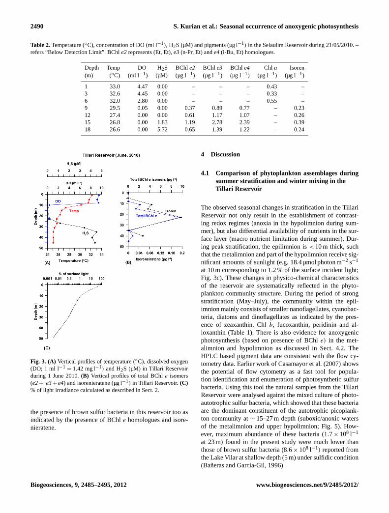
decreased to 1.76 µg l−1 in June and to 0.55 µg l−1 in July(Table 1). During most observations, Chla concentrations re-mained low in the metalimnion; however, when anoxic con-ditions prevailed, this layer became enriched with BChle iso-mers (Table 1) and most other pigments were either absent orbelow detection limits. During summer stratification (May–July, 2010), BChle homologues, characteristic of brown sul-fur bacteria represent the major peaks (a, b and c) in Fig. 4a,which were identified based on UV/Vis and APCI-LC-MSspectra and also by comparing with proper BChleF stan-dards. Based on them/z molecular ions of these isomers(821.5, 835.5 and 849.5) and their respective fragment ions(through the loss of H2O molecule;−18 AMU) in each spec-trum (Fig. 4b, c and d) and published literature, these wereidentified as farnesyl-esterified bacteriochlorophyll (BChleF) homologues such ase2 (Et, Et), e3 (n-Pr, Et) ande4(i-Bu, Et) (Airs et al., 2001b; Glaeser et al., 2002, Masse etal., 2004). BChle2varied from below detection limit (BDL)to 2 µg l−1 with a mean concentration of 0.06 µg l−1. Simi-larly, BChle3ande4concentration ranged between BDL and3.97 µg l−1, and BDL and 3.52 µg l−1, respectively (Table 1).These isomers were present close to the oxic-anoxic interfaceand were detectable even at 11 m depth, where the water wassuboxic (0.17 ml O2 l−1) in June. The highest concentration
23
Fig. 2. (A) Vertical profiles of temperature (◦C), dissolved oxygen(DO; 1 ml l−1
= 1.42 mg l−1) and H2S (µM) in Tillari Reservoirduring 12 May 2010. During summer stratification, water columnturned to be sulfidic below 25 m depth.(B) Vertical profile of totalBChl e isomers (e2+ , e3+ e4,µg l−1) in Tillari Reservoir.
(e2+e3+e4= 9.5 µg l−1) was measured at 23 m where theH2S concentration was 0.42 µM (Fig. 3a and b). The H2Sconcentration increased to 9 µM below 23 m, but the con-centration of BChle decreased. The computed light irradi-ance data show that 15.4 µmol photon m−2 s−1 (∼ 1 % of thesurface incident light) reached 11 m where BChle isomersfirst appeared, and only 0.2 % of the surface incident light(3.1 µmol photon m−2 s−1) reached 23 m where the maximain BChl e isomers were located (Fig. 3c). Isorenieratene, acarotenoid characteristic of brown sulfur bacteria was alsoidentified based on mass spectrum (m/z 529.3) at the depthswhere BChleF homologues were present in June (range BDL−0.19 µg l−1, Fig. 3b).
The highest Chla concentration (2.26 µg l−1) was ob-served in January 2011 (Table 1). Apart from Chla,other chemotaxonomic pigments identified were chlorophyllb, zeaxanthin, fucoxanthin which represent green algae,cyanobacteria and diatoms, respectively. Other minor pig-ments associated with the green algae (Chlorophyceace) suchas neoxanthin, violaxanthin and lutein were also found dur-ing the January observation (Table 1). Chlorophyllb showeda temporal variation similar to Chla, but the concentra-tions were lower by an order of magnitude. The highestconcentration (0.28 µg l−1) was observed in winter. Zeax-anthin, a marker pigment of cyanobacteria was present inhigher concentrations in May (surface) and January andcontributed significantly to the biomass structure in theTillari Reservoir (Table 1). Fucoxanthin also showed simi-lar distribution pattern, but its concentration was lower thanthose of zeaxanthin and Chlb. Peridinin, a marker pig-ment of dinoflagellates was present during May–August.
Biogeosciences, 9, 2485–2495, 2012 www.biogeosciences.net/9/2485/2012/

S. Kurian et al.: Seasonal occurrence of anoxygenic photosynthesis 2489
Table 1. Concentration of pigments (µg l−1) in the Tillari Reservoir during May, June, July, 2010 and January, 2011. Abbreviations are asfollows: Total Chlorophyll (TChl), Bacteriochlorophyll (BChl), Phaeophytin (Phaeo), Isorenieratene (Isoren), Zeaxanthin (Zea), Peridinin(Per), Fucoxanthin (Fuco), Alloxanthin (Allo), Total carotene (Tcar), Neoxanthin (Neo), Violaxanthin (Viola), Lutein (Lut), Diadinoxan-thin (Diad), Diatoxanthin (Diat). – refers “Below Detection Limit”. Chlorophylls, BChls and isoren were measured at WHOI, while othercarotenoids were quantified at NIO. BChle2represents (Et, Et),e3(n-Pr, Et) ande4(i-Bu, Et) homologues.
Field Trip 12/05/2010
Depth (m) TChla Chl b BChl e2 BChl e3 BChl e4 Phaeoa Isoren Zea Per Fuco Allo Tcar Neo Viola Lut Diad Diato
0 2.06 0.13 – – – 0.02 – 0.17 0.03 0.01 0.003 0.023 0.040 0.013 0.030 0.011 0.0025 0.15 – – – – 0.08 – – – – – – – – – – –10 0.61 – – – – 0.01 – – – – – – – – – – –15 0.79 0.06 0.05 0.96 0.07 0.02 – – – – 0.03 0.002 – – – – –20 0.09 0.02 0.08 0.26 0.15 0.01 – – – – – – – – – – –25 0.04 – 0.16 0.2 – – – – – – – – – – – – –30 0.15 – 0.09 0.12 0.11 0.02 – – – – – – – – – – –35 0.13 – – – – 0.02 – – – – – – – – – – –40 0.19 – – – – 0.02 – – – – – – – – – – –50 0.04 – – – – 0.02 – – – – – – – – – – –
Field Trip 01/06/2010
0 1.76 0.11 – – – 0.03 – 0.07 0.03 0.004 – 0.008 0.027 0.009 0.017 0.008 0.0045 1.19 0.07 – – – 0.01 – 0.11 0.01 0.004 – 0.005 0.026 0.002 0.005 0.010 0.00411 1.32 0.07 0.46 0.99 0.72 0.01 0.09 – – – – 0.002 0.016 0.003 0.005 – –15 0.37 – 0.32 0.64 0.46 0.24 0.07 – – – – – – – – – –23 0.09 – 2.0 3.97 3.52 0.10 0.2 – – – – – – – – – –35 0.04 – 0.06 0.07 0.07 – – – – – – – – – – – –40 0.15 0.05 0.19 0.35 0.29 0.04 – 0.04 0.06 0.01 – – 0.018 0.002 0.002 0.013 0.002
Field Trip 22/07/2010
0 0.55 0.05 – – – – – 0.07 0.02 0.008 0.003 0.008 0.013 0.002 0.004 0.008 0.0045 0.29 0.09 – – – – – 0.05 0.01 0.005 0.008 0.004 0.003 0.001 0.004 0.002 0.00110 0.44 0.04 – – – – – 0.09 – – – – 0.005 0.002 0.004 0.003 0.00315 0.10 – – – – – – 0.01 – – – – – – – – –20 0.00 – 0.35 0.64 0.48 – – – – – – – – – – – –30 0.07 – 0.12 0.22 0.18 – – – – – – – – – – – –40 0.19 – – – – – – – – – – – – – – – -
Field Trip 13/01/2011
0 2.27 0.21 – – – 0.03 – 0.2 – 0.017 0.009 0.007 0.04 0.009 0.006 0.010 0.0095 2.82 0.29 – – – – – – – 0.014 0.014 0.007 0.01 0.004 0.01 0.014 0.01210 1.49 0.17 – – – 0.04 – 0.11 – 0.006 0.007 0.007 0.01 0.003 0.008 0.013 0.00320 2.16 0.21 – – – 0.03 – 0.13 0.02 0.008 0.015 0.008 0.02 0.007 0.018 0.009 0.00440 1.27 0.12 – – – 0.002 – 0.10 – – – 0.010 0.004 0.007 0.008 0.007 0.004
Nonmarker photoprotective xanthophylls such as diadinox-anthin and diatoxanthin were also present in these samples;with comparatively higher concentrations in January (0.014and 0.012 µg l−1, respectively; Table 1).
3.1.3 Flow cytometric identification and enumeration ofphotosynthetic sulfur bacteria
Flow cytometric analyses of samples collected from TillariReservoir in June was carried out for identifying the dom-inant groups in the anoxic waters. By comparing flow cy-tograms from a culture of photosynthetic sulfur bacteria(Fig. 5a) with those obtained from the natural population, wecould resolve the identity of the microbial population thriv-ing at the anoxic layer between 11 and 35 m (data for 23 mare shown in Fig. 5c). This group showed clear sub-surfacemaxima with peak abundance (1.7× 10⁸ cells l−1) between15 and 23 m and negligible concentrations at the surface and
near bottom waters (Fig. 5b and d); a trend similar to HPLCbased bacteriocholorophylle distribution.
3.2 Selaulim Reservoir
The Selaulilm Reservoir is shallower (maximum depth∼ 20 m at the time of our sampling) as compared to the TillariReservoir. During our sampling in May 2010, the water col-umn was strongly stratified with the surface-to-bottom tem-perature difference of∼ 6◦C (Fig. 6a). The DO disappearedat 9 m, and H2S was present below 12 m with the highest con-centration (5.7 µM) occurring at 18 m (Table 2). Chlorophylla and zeaxanthin were restricted to the epilimnion, althoughtheir concentrations were much lower than the Tillari Reser-voir. Farnesyl-esterified BChle homologues (e2, e3ande4isomers) were present at 9 m and below all the way downto the deepest sampling depth. The highest concentrations oftotal BChle isomers (6.4 µg l−1) were found at 15 m whereH2S concentration was 1.83 µM (Fig. 6b). Our data showed
www.biogeosciences.net/9/2485/2012/ Biogeosciences, 9, 2485–2495, 2012
2490 S. Kurian et al.: Seasonal occurrence of anoxygenic photosynthesis
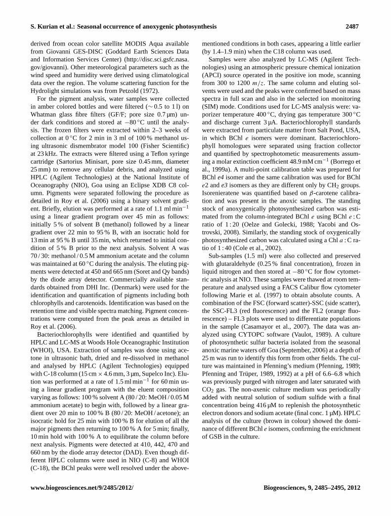
Table 2.Temperature (◦C), concentration of DO (ml l−1), H2S (µM) and pigments (µg l−1) in the Selaulim Reservoir during 21/05/2010. –refers “Below Detection Limit”. BChle2represents (Et, Et),e3(n-Pr, Et) ande4(i-Bu, Et) homologues.
Depth Temp DO H2S BChle2 BChl e3 BChl e4 Chl a Isoren(m) (◦C) (ml l−1) (µM) (µg l−1) (µg l−1) (µg l−1) (µg l−1) (µg l−1)
1 33.0 4.47 0.00 – – – 0.43 –3 32.6 4.45 0.00 – – – 0.33 –6 32.0 2.80 0.00 – – – 0.55 –9 29.5 0.05 0.00 0.37 0.89 0.77 – 0.2312 27.4 0.00 0.00 0.61 1.17 1.07 – 0.2615 26.8 0.00 1.83 1.19 2.78 2.39 – 0.3918 26.6 0.00 5.72 0.65 1.39 1.22 – 0.24
24
Fig. 3. (A) Vertical profiles of temperature (◦C), dissolved oxygen(DO; 1 ml l−1
= 1.42 mg l−1) and H2S (µM) in Tillari Reservoirduring 1 June 2010.(B) Vertical profiles of total BChle isomers(e2+ e3+ e4) and isorenieratene (µg l−1) in Tillari Reservoir.(C)% of light irradiance calculated as described in Sect. 2.
the presence of brown sulfur bacteria in this reservoir too asindicated by the presence of BChle homologues and isore-nieratene.
4 Discussion
4.1 Comparison of phytoplankton assemblages duringsummer stratification and winter mixing in theTillari Reservoir
The observed seasonal changes in stratification in the TillariReservoir not only result in the establishment of contrast-ing redox regimes (anoxia in the hypolimnion during sum-mer), but also differential availability of nutrients in the sur-face layer (macro nutrient limitation during summer). Dur-ing peak stratification, the epilimnion is< 10 m thick, suchthat the metalimnion and part of the hypolimnion receive sig-nificant amounts of sunlight (e.g. 18.4 µmol photon m−2 s−1
at 10 m corresponding to 1.2 % of the surface incident light;Fig. 3c). These changes in physico-chemical characteristicsof the reservoir are systematically reflected in the phyto-plankton community structure. During the period of strongstratification (May–July), the community within the epil-imnion mainly consists of smaller nanoflagellates, cyanobac-teria, diatoms and dinoflagellates as indicated by the pres-ence of zeaxanthin, Chlb, fucoxanthin, peridinin and al-loxanthin (Table 1). There is also evidence for anoxygenicphotosynthesis (based on presence of BChle) in the met-alimnion and hypolimnion as discussed in Sect. 4.2. TheHPLC based pigment data are consistent with the flow cy-tometry data. Earlier work of Casamayor et al. (2007) showsthe potential of flow cytometry as a fast tool for popula-tion identification and enumeration of photosynthetic sulfurbacteria. Using this tool the natural samples from the TillariReservoir were analysed against the mixed culture of photo-autotrophic sulfur bacteria, which showed that these bacteriaare the dominant constituent of the autotrophic picoplank-ton community at∼ 15–27 m depth (suboxic/anoxic watersof the metalimnion and upper hypolimnion; Fig. 5). How-ever, maximum abundance of these bacteria (1.7× 10⁸ l−1
at 23 m) found in the present study were much lower thanthose of brown sulfur bacteria (8.6× 10⁸ l−1) reported fromthe Lake Vilar at shallow depth (5 m) under sulfidic condition(Baneras and Garcia-Gil, 1996).
Biogeosciences, 9, 2485–2495, 2012 www.biogeosciences.net/9/2485/2012/
S. Kurian et al.: Seasonal occurrence of anoxygenic photosynthesis 2491
0
40
80
BChl e2R.T. 22.6
BChl e3R.T. 23.5 BChl e4
R.T. 24.4
Retension Time
803
.4 8
21.4
849
.5
(a) 2
2.6
(b) 2
3.5
(c) 2
4.4
817
.4 8
35.5
(d)
(e )
(B)(A)
(C) (D)
400 600 800 1000400 600 800 1000m/z m/z
400 600 800 1000m/z
20
60
100
0
40
80
20
60
100
0
40
80
20
60
100
20
15
10
5
0
mAU
410
nm
Relat
ive A
bund
ance
Relat
ive A
bund
ance
Relat
ive A
bund
ance
10 20 30 40 50 60 min
831
.5
Fig. 4. (A) Representative chromatogram (LC-MS) obtained for suspended particulate matter having farnesyl-esterified bacteriochlorophylle isomers (Tillari Reservoir, 1 June 2010, 23 m). Major peaks (a), (b) and (c) represent BChle2, e3ande4 respectively. Peaks (d) and (e)correspond to Chlaand isorenieratene respectively.(B) Confirmative mass spectra (LC-MS) for the peak (a) at 22.6 min. representinge2(Et,Et) BChl eF isomer (m/z = 821.4, 803.4 (-H2O)). (C) Peak (b) at 23.5min. representinge3 (n-Pr, Et) BChleF isomer (m/z = 835.5, 817.4(-H2O)). (D) Peak (c) at 24.4 min. representinge4 (i-Bu, Et) BChleF isomer (m/z = 849.5, 831.5 (-H2O)). R.T. denotes retention time.
In January, phytoplankton community structure in theTillari Reservoir was dominated by Chlb-containing greenalgae and cyanobacteria with minor presence of diatoms inthe epilimnion; however, these pigments were also presentin deeper waters (Table 1) presumably due to vertical mix-ing. Although zeaxanthin is present in green algae as wellas cyanobacteria, the presence of other accessory markerpigments such as violaxanthin, lutein and neoxanthin, notfound in cyanobacteria confirms the presence of green al-gae (Jeffrey and Vesk, 1997). Phaeophytina concentrationin bottom waters was high, between May and July (0.11to 0.45 µg l−1), compared to that observed during January(BDL-0.42 µg l−1). This could be attributed to better preser-vation in anoxic waters. Such observations have previouslybeen reported from the Black Sea and other anoxic zones(Sinninghe Damste et al., 1993; Chen et al., 2001; Squieret al., 2002). A recent study of pigment composition in Pe-tit Saut, a tropical reservoir in French Guiana, showed thedominance of Chlorophyceae (Chla, Chl b and lutein) inthe oxic epilimnion, and anoxygenic photo-autotrophic bac-teria below the oxycline as revealed by BChlc and BChld (Junet et al., 2009). An example of how the communitystructure of phytoplankton is dependent on nutrient avail-ability is provided by the results of Zhu et al. (2010) fromTaihu Lake (China) where Chlorophyta were found to be pre-dominant at high concentrations of nitrogen and phosphorus,
whereas cyanobacteria dominated in waters having low con-centrations of these nutrients. Our data suggest co-existenceof both these groups in the Tillari Reservoir.
4.2 Anoxygenic photosynthesis in Tillari and Selaulimreservoirs during summer stratification
As described under Sect. 3.1.2, the highest concentration offarnesyl-esterified BChle homologues was in the upper por-tion of the anoxic layer where the H2S concentration was low(0.42 µM; Fig. 3a and b) corresponding to 0.2 % of the sur-face incident light (Fig. 3c). Garcia-Gil and Abella (1992)studied photo-autotrophic bacterial populations in Spanishlakes and found the amount of light reaching the oxic-anoxicboundary to be the most important factor controlling theirpopulations. However, they reported higher light intensitiesat the oxyclines of these lakes where BChla and e iso-mers were found in high concentrations. In a study carriedout in two meromictic basins of Lake Banyoles, Borrego etal. (1999b) observed high concentration of BChle isomers(up to 200 µg l−1) at ∼ 18 m depth and as summer strati-fication proceeded, the population ofCb. pheobacteroides(bacterial plate) moved to shallower depth (∼ 17 m). Theyascribed the upward migration from 18 m to 16.5 m to theincrease in light intensity at the bacterial plate correspond-ing to a change from 0.004 to 0.05 % of the surface incident
www.biogeosciences.net/9/2485/2012/ Biogeosciences, 9, 2485–2495, 2012

2492 S. Kurian et al.: Seasonal occurrence of anoxygenic photosynthesis
Fig. 5. Flow cytometry (FACS Calibur) analysis of photo-autotrophic sulfur bacteria shown in red colour with an arrow.(A)Represents mixed culture of sulphur bacteria, while natural popula-tion in Tillari Reservoir at 1 m, 23 m and 40 m are shown in dot plots(B), (C) and(D) respectively. FL3-H and FL2-H indicates red andorange fluorescence.
light. These results illustrate the adaptability of the anoxy-genic photosynthetic community to very low incident lightlevels. Although farnesol is the main alcohol esterifying thebacteriochlorophylls at position C-17, earlier studies haveshown that a number of other alcohols are present in natu-ral samples (Caple et al., 1978; Otte et al., 1993) depend-ing on growth conditions, light intensity and stage of devel-opment of the cell (Borrego and Garcia-Gil, 1995; Airs etal., 2001b). Masse et al. (2004) reported that in culturedCb.phaeobacteroides, the isoprenoid esterified side chains dom-inated at low light intensities, while the straight-chain alkylsubstituents dominated at higher light intensities. This phe-nomenon is explained as a result of changing availability ofreducing power, i.e. the highly reduced straight-chain alco-hols have a higher biosynthetic demand for NADPH2 thanthe polyunsaturated isoprenoid with the same number of car-bon atoms (Masse et al., 2004). In the present study, wefound the dominant bacteriochlorophylls to be farnesyl ester-ified (BChl eF) isomers based on their MS spectra, in com-parison with the standards (extracted from Salt Pond, USA),and previous literature.
Although optical measurements were not made in the Se-laulim Reservoir, the vertical distribution patterns of H2S andBChl e isomers described in Sect. 3.2, are very similar tothose observed in the Tillari Reservoir and it is reasonableto conclude that the anoxygenic bacterial population is con-trolled by the combined availability of free sulfide and light.
27
Fig. 6. (A) Vertical profiles of temperature (◦C), dissolved oxygen(DO; 1 ml l−1
= 1.42 mg l−1) and H2S (µM) in Selaulim Reser-voir during 21 May 2010.(B) Vertical profiles of Chla, total BChle isomers (e2+, e3+ e4) and isorenieratene (µg l−1) in SelaulimReservoir.
In both Tillari and Selaulim reservoirs, we found bacteri-ochlorophylle2, e3ande4 isomers typical of brown coloredGSB thriving under low light conditions in natural habitats(Borrego et al., 1997). Among these isomers, the contribu-tions of larger alkyl substituents [(n-Pr, Et) BChleF (42 %)and (i-Bu, Et) BChleF(∼ 34 %)] were higher in both thereservoirs as compared to (Et, Et) BChleF isomer (∼ 24 %).An increase in the amount of homologues with larger alkylsubstituents on the bacteriochlorophylls modifies the aggre-gation and therefore the packing of structures in the chloro-somes, with an impact on the in vivo absorption spectrum(Airs et al., 2001b), which might provide a basis for a cellu-lar response to light limitation (Borrego et al., 1999b). Theisomere1 [(Et, Me) BChl eF] was absent in these systems,as it is clearly traceable only in laboratory cultures grownunder high light intensities (Borrego and Garcia-Gil, 1994,1995). Isorenieratene, a carotenoid characteristic of brownsulfur bacteria was reported earlier in stratified anoxic basinshaving bacteriochlorophylle homologues (Garcia-Gil andAbella, 1992; Borrego et al., 1997, 1999b). We also mea-sured isorenieratene in Tillari Reservoir during our June ob-servation (Fig. 3b), which showed similar vertical profileof BChl e isomers. Isorenieratene was found to be presentin Selaulim Reservoir also with highest concentration of0.39 µg l−1 at 15 m (Fig. 6b) where BChle isomers showedthe highest concentration. This further supports the presenceof anoxygenic photosynthesis in these reservoirs during sum-mer stratification.
Biogeosciences, 9, 2485–2495, 2012 www.biogeosciences.net/9/2485/2012/

S. Kurian et al.: Seasonal occurrence of anoxygenic photosynthesis 2493
4.3 Standing stocks of oxygenically- andanoxygenically-photosynthesized carbon in theTillari Reservoir
Earlier estimates of carbon fixation in lakes by photosyn-thetic sulfur bacteria vary widely depending on sulfide andlight availability (Parkin and Brock, 1980 and referencestherein). In the present study, the total concentration of bac-teriochlorophylle isomers was used as a proxy of biomassof green sulfur bacteria. Yacobi and Ostrovski (2011) re-ported photosynthesis by microalgae and cyanobacteria tobe the major source of organic particles in the epilimnionof Lake Kinneret, while green sulfur bacteria (Chlorobiumphaeobacteroides) was found in the anoxic layer throughoutthe periods of stratification (Bergstein et al., 1979). In theTillari Reservoir too farnesyl-esterified bacteriochlorophylle homologues (e2, e3 and e4 isomers) were the dominantpigments in the anoxic hypolimnion during summer stratifi-cation, while Chla was largely restricted to the epilimnion.We calculated the BChle standing stock (mg m−2) for theJune observation by integrating the total BChle concentra-tion in the anoxic compartment (∼ 11–45 m). The column-integrated BChle (113.8 mg m−2) converted to carbon usingthe published value of the BChle : C ratio (1 : 20) yields astanding stock of 2.27 g m−2 of anoxygenically photosynthe-sized carbon. In comparison, with a Chla of 20.45 mg m−2
(0–45 m water column), the standing stock of oxygenicallyphotosynthesized carbon, calculated using a Chla : C ratioof 1 : 40 comes to 0.82 g C m−2. This is similar to the resultsof Camacho et al. (2000) from Lake Arcas (Spain) who re-ported high population densities of phototrophic micro or-ganisms with high concentration of BChla (up to 381 µg l−1)
at the oxic-anoxic interface. The anoxic layer (9.1–9.8 m)in this lake accounted for 101.8 mg BChla m−2, while thestanding stock of Chla from surface to 9.8 m was lower(72.1 mg m−2).
In an earlier study by Takahashi and Ichimura (1968)carried out in stratified lakes in Japan, organic matter syn-thesized by photosynthetic sulfur bacteria in the reducingzone was reported to contribute 9–25 % of the total annualproduction in lakes rich in H2S and 3–5 % in lakes poorin H2S. In another study by Steenbergen (1982) in LakeVechten (the Netherlands), the photosynthetic sulfur bac-teria accounted for 3.9 to 17.5 % of total daily productiv-ity in the pelagic zone. We do not have data on C-fixationrates to quantify the contribution of GSB to the total pro-duction in the two reservoirs sampled by us. One would ex-pect the oxygenic photosynthetic production to exceed theanoxygenic photosynthetic production simply because it isthe former that supplies the organic matter for the anaerobicmetabolism. This ultimately leads to the production of H2Sthus fuelling the anoxygenic photosynthesis indirectly. Al-gal biomass build-up in the surface layer of the Tillari Reser-voir during summer stratification was limited by low concen-trations of nitrate (NO−3 +NO−
2 = 1.21 µM) and phosphate
(0.08 µM) (Narvenkar et al., 2012), but production duringthe preceding winter/spring periods should have been higher.Although some studies have reported that protozoa predateupon green sulfur bacteria (Sacca et al., 2009), the grazingpressure upon algal populations is presumably higher caus-ing a great impact on their standing stock. Thus, it is mostlikely that the high standing stock of green sulfur bacteria inthe anoxic layer is the result of slow accumulation of biomassdue to low grazing impact. It may be added that while dataon phototrophic sulfur bacteria are presently limited to onlytwo reservoirs, conditions favoring their growth also seemto exist in some other systems (Narvenkar et al., 2012). Thepotential significance of these organisms in biogeochemicalcycling of fresh water systems of South Asia, indicated byour results, needs to be evaluated in detail.
Acknowledgements.The authors wish to thank the Director,NIO and managements of the Tillari and Selaulim reservoirsfor permitting us to carry out the observations. We are gratefulto H. Naik for logistical support and to H. Dalvi, A. Metharand B. R. Thorat for their kind assistance during sampling.S. Patil is acknowledged for his assistance in flow cytometricanalysis. Critical reviews by C. Abella, C. M. Borrego andR. de Wit greatly helped in improving the paper. Financialsupport for this work was provided by the Council of Scientific& Industrial Research (CSIR) and Ministry of Earth Sciences(MoES). S. Kurian acknowledges POGO-SCOR for financialsupport to visit WHOI. R. Roy, G. Narvenkar and A. Sarkarreceived fellowship support from CSIR. D. Repeta acknowledgessupport from US National Science Foundation Center AwardEF0424599 to the Center for Microbial Oceanography: Researchand Education (C-MORE). This is NIO Contribution Number 5181.
Edited by: J. Middelburg
References
Airs, R. L., Atkinson, J. E., and Keely, B. J.: Development and ap-plication of a high resolution liquid chromatographic method forthe analysis of complex pigment distributions, J. Chromatogr., A917, 167–177, 2001a.
Airs, R. L., Borrego, C. M., Garcia-Gil, J., and Keely, B. J.: Iden-tification of the bacteriochlorophyll homologues ofChlorobiumphaeobacteroidesstrain UdG 6053 grown at low light intensity,Photosynth. Res., 70, 221–230, 2001b.
Airs, R. L. and Keely, B. J.: Atmospheric pressure chemical ion-isation liquid chromatography/ mass spectrometry of bacteri-ochlorophyll from Chlorobiaceae, characteristic fragmentation,Rapid Commun. Mass Spectrom., 16, 453–461, 2002.
Baneras, L. and Garcia-Gil, L.J.: Role of photosynthetic micro-bial populations in the phosphorous exchange through the oxic-anoxic boundary in a meromictic eutrophic lake, Arch. Hydro-biol. Spec. Issues Adv. Limnol., 48, 171–181, 1996.
Bergstein, T., Henis, Y., and Cavari, B. Z.: Investigations on the pho-tosynthetic sulfur bacterium Chlorobium phaeobacteroides caus-ing seasonal blooms in Lake Kinneret, Can. J. Microbiol., 25,999–1007, 1979.
www.biogeosciences.net/9/2485/2012/ Biogeosciences, 9, 2485–2495, 2012

2494 S. Kurian et al.: Seasonal occurrence of anoxygenic photosynthesis
Biebl, H. and Pfennig, N.: Anaerobic CO2 uptake by phototrophicbacteria: A review, Arch. Hydrobiol. Beih. Ergeb, Limnol, 12,48–58, 1979.
Borrego, C. M. and Garcia-Gil, J.: Separation of bacteriochloro-phyll homologues from green photosynthetic sulfur bacteria byreversed-phase HPLC, Photosynth. Res., 41, 157–163, 1994.
Borrego, C. M. and Garcia-Gil, L. J.: Rearrangement of light har-vesting bacteriochlorophyll homologues as a response of greensulfur bacteria to low light intensities, Photosynth. Res., 45, 21–30, 1995.
Borrego, C. M., Garcia-Gil, L. J., Vila, X. P., Figueras, J. B., andAbella, C. A.: Distribution of bacteriochlorophyll homologuesin natural populations of brown-colored phototrophic sulfur bac-teria, FEMS Microbiol. Ecol., 24, 301–309, 1997.
Borrego, C. M., Arellano, J. B., Abella, C. A., Gillbro, T., andGarcia-Gil, L. J.: The molar extinction coefficient of bacte-riochlorophyll eand the pigment stoichiometry inChlorobiumphaeobacteroides, Photosynth. Res., 60, 257–264, 1999a.
Borrego, C. M., Baneras, L., and Garcia-Gil, L. J.: Temporal vari-ability of Chlorobium phaeobacteroidesantenna pigments in ameromictic karstic lake, Aquat. Microb. Ecol., 17, 121–129,1999b.
Camacho, A., Vicente, E., and Miracle, M. R.: Spatio-temporal dis-tribution and growth dynamics of phototrophic sulfur bacteriapopulations in the sulfide-rich Lake Arcas, Aquat. Sci., 62, 334–349, 2000.
Caple, M. B., Chow, H., and Strouse, C. E.: Photosynthetic pig-ments of green sulphur bacteria: The esterifying alcohols of bac-teriochlorophyllsc from Chlorobium limicola, J. Biol. Chem.,253, 6730–6737, 1978.
Casamayor, E. O., Ferrera, I., Cristina, X., Borrego, C. M., andGasol, J. M.: Flow cytometric identification and enumeration ofphotosynthetic sulfur bacteria and potential for ecophysiologi-cal studies at the single-cell level, Environ. Microbiol., 9, 1969–1985, 2007.
Chen, C., Bianchi, T. S., McKee, B. A., and Bland, J. M.: Histori-cal trends of hypoxia on the Louisiana shelf: Application of pig-ments as biomarkers, Org. Geochem., 32, 543–561, 2001.
Cole, J. J., Carpenter, S. R., Kitchel, J. F, and Pace, M. L.: Path-ways of Organic Carbon Utilization in Small Lakes: Resultsfrom a Whole-Lake 13C Addition and Coupled Model, Limnol.Oceanogr., 47, 1664–1675, 2002.
Garcia-Gil, L. J. and Abella, C. A.: Population dynamics of pho-totrophic bacteria in three basins of Lake Banyoles (Spain), Hy-drobiologia, 243/244, 87–94, 1992.
Garcia-Gil, L. J., Sala-Genoher, L., Esteva, J. V., and Abella, C. A.:Distribution of iron in Lake Banyoles in relation to the ecology ofpurple and green sulfur bacteria, Hydrobiologia, 192, 259–270,1990.
Glaeser, J., Baneras, L., Rutters, H., and Overmann, J.: Novel bac-teriochlorophylle structures and species-specific variability ofpigment composition in green sulfur bacteria, Arch. Microbiol.,177, 475–485, 2002.
Gerola, P. D. and Olson, J. M.: A new bacteriochlorophylla proteincomplex associated with chlorosomes of green sulfur bacteria,Biochim. Biophys. Acta, 848, 69–76, 1986.
Grasshoff, K., Erhardt, M., and Kremiling, K.: Methods of seawateranalysis, Verlag Chemie, 419 pp., 1983.
Gregg, W. W. and Carder, K. L.: A simple solar irradiance model forcloudless maritime atmospheres, Limnol. Oceanogr., 35, 1657–1675, 1990.
Hirabayashi, H., Ishii, T., Takaichi, S., Inoui, K., and Uejara, K.:The Role of carotenoids in the photoadaptation of the browncolored sulfur bacteriumChlorobium phaeobacteroides, Pho-tochem. Photobiol., 79, 280–285, 2004.
Hurley, J. P. and Watras, C. J.: Identification of bacteriochlorophyllsin lakes via reverse-phase HPLC, Limnol. Oceanogr., 36, 307–315, 1991.
Imhoff, J. F.: Taxonomy, phylogeny and general ecology of anoxy-genic phototrophic bacteria, in: Biotechnology Handbook Photo-synthetic Prokaryotes, edited by: Carr, N. G. and Mann, N. H.,London & New York, Plenum, 53–92, 1992.
Jeffrey, S. W. and Vesk, M.: Introduction to marine phytoplank-ton and their pigment signatures, in: Phytoplankton Pigments inOceanography, edited by: Jeffrey, S. W., Mantoura, F. C., andWright, S. W., UNESCO Publishing, Paris, 85–126, 1997.
Junet, A. de, Abril, G., Guerin, F., Billy, I., and Wit, R. de.: A multi-tracers analysis of sources and transfers of particulate organicmatter in a tropical reservoir (Petit Saut, French Guiana), RiverRes. Appli., 25, 253–271, 2009.
Marie, D., Partensky, F., Jacquet, S., and Vaulot, D.: Enumera-tion and cell cycle analysis of natural populations of marine pi-coplankton by flow cytometry using the nucleic acid stain SYBRGreen I, Appl. Environ. Microbiol., 63, 186–193, 1997.
Masse, A., Airs, R. L., Keely, B. J., and Wit, R. de.: The impact ofdifferent intensities of green light on the bacteriochlorophyll ho-mologue composition of the chlorobiaceaeProsthecochloris aes-tuarii andChlorobium phaeobacteroides, Microbiol., 150, 2555–2564, 2004.
Narvenkar, G., Naqvi, S. W. A., Kurian, S., Shenoy, D. M., Prati-hary, A. K., Naik, H., Patil, S., Sarkar, A., and Gauns, M.: Dis-solved methane in Indian freshwater reservoirs, Environ. Monit.Assess., in review, 2012.
Oelze, J. and Golecki, J. R.: Growth rate and the control of develop-ment of the photosynthetic apparatus inChloroflexus aurantiacusas studied on the basis of cytoplasmatic membrane structure andchlorosome size, in: Green Photosynthetic Bacteria, edited by:Olson, J. M., Ormerod, J. G., Amesz, J. Ormerod, J. G., Amesz,J., Stackebrandt, E., and Truper, H. G., EMBO Workshop, Ny-borg, Denmark, 35–42, 1988.
Otte, S. C. M., Van de Meent, J. E., van Veelen, P. A., Pundsnes, A.S., and Amesz, J.: Identification of the major chlorosomal bac-teriochlorophylls of the green sulfur bacteriaChlorobium vibrio-formeandChlorobium phaeobacteroides; their function in lateralenergy transfer, Photosynth. Res., 35, 159–169, 1993.
Parkin, T. B and Brock, T. D.: Photosynthetic bacterial productionin lakes: The effects of light intensity, Limnol. Oceanogr., 25,711–718, 1980.
Petzold, T. J.: Volume scattering functions for selected ocean wa-ters, Scripps Inst. Oceanogr., Report SI0, 72–78, 1972.
Pfennig, N.: Ecology of phototrophic purple and green sulfur bac-teria, in: Autotrophic bacteria, edited by: Schlegel, H. G. andBowien, B., Springer Verlag, New York, 97–116, 1989.
Pfennig, N. and Truper, H. G.: Anoxygenic phototrophic bacteria,in: Bergey’s manual of systematic bacteriology, edited by: Staley,J. T., Bryant, M. P., Pfenning, N., and Holt, J. G., Baltimore:Williams and Wilkins, 3, 1635–1709, 1989.
Biogeosciences, 9, 2485–2495, 2012 www.biogeosciences.net/9/2485/2012/
S. Kurian et al.: Seasonal occurrence of anoxygenic photosynthesis 2495
Pfennig, N. and Truper HG.: The family Chromatiaceae, in: TheProkaryotes, edited by: Balows, A., Truper, H. G., Dworkin, M.,Harder, W., and Schleifer, K. H., Springer, New York, 2nd edn,3200–3221, 1992.
Roy, R., Prathihary, A., Mangesh, G., and Naqvi, S. W. A.: Spatialvariation of phytoplankton pigments along the south west coastof India, Estuar. Coast. Shelf Sci., 69, 189–195, 2006.
Sacca, A., Borrego, C. M., Renda, R., Triado-Margarit, X., Bruni,V., and Guglielmo, L.: Predation impact of ciliated and flagel-lated protozoa during a summer bloom of brown sulfur bacteriain a meromictic coastal lake, FEMS Microbiol. Ecol., 70, 42–53,2009.
Senge, M. O. and Smith, K. M.: Biosynthesis and structures ofthe bacteriochlorophylls, in: Anoxygenic Photosynthetic Bacte-ria, Advances in Photosynthesis, edited by: Blankenship, R. E.,Madigan, M. T., and Bauer, C. E., Kluwer Academic Publishers,Dordrecht, The Netherlands, Vol II, 137–151, 1995.
Sinninghe Damste, J. S., Wakeham, S. G., Kohnen, M. E. L., Hayes,J. M., and De Leeuw, J. W.: A 6,000-year sedimentary molecularrecord of chemocline excursions in the Black Sea, Nature, 362,827–829, 1993.
Squier, A. H., Hodgson, D. A., and Keely, B.: Sedimentary pig-ments as markers for environmental change in an Antarctic lake,Org. Geochem., 33, 1655–1665, 2002.
Steenbergen, C. L. M.: Contribution of photosynthetic sulfur bac-teria to primary production in Lake Vechten, Hydrobiologia, 95,59–64, 1982.
Takahashi, M. and Ichimura, S.: Vertical distribution and organicmatter production of photosynthetic sulfur bacteria in Japaneselakes, Limnol. Oceanogr., 13, 644–655, 1968.
Vaulot, D.: CYTOPC: Processing software for flow cytometric data,Signal and Noise, 2, 8 pp., 1989.
Vila, X. and Abella, C. A.: Effects of light quality on the physiol-ogy and the ecology of planktonic green sulfur bacteria in lakes,Photosynth. Res., 41, 53–65, 1994.
Vila, X., Abella, C. A., Figueras, J. B., and Hurley, J. P.: Verticalmodels of phototrophic bacterial distribution in the metalimneticmicrobial communities of several freshwater North-Americankettle lakes, FEMS Microbiol. Ecol., 25, 287–299, 1998.
Yacobi, Y. Z. and Ostrovsky, I.: Downward flux of organic matterand pigments in Lake Kinneret (Israel): relationships betweenphytoplankton and the material collected in sediment traps, J.Plankton Res., 30, 1189–1202, 2008.
Yacobi, Y. Z. and Ostrovsky, I.: Sedimentation of photosyntheticpigments during the bloom of the green sulfur bacteriumChloro-bium phaeobacteroidesin Lake Kinneret: spatial patterns, Hy-drobiologia, 660, 117–124, 2011.
Yacobi, Y. Z., Eckert, W., Trueper, H. G., and Berman, T.: High per-formance liquid chromatography detection of phototrophic bac-terial pigments in aquatic environments, Microbial. Ecol., 19,127–136, 1990.
Zhu, W., Wan, L., and Zhao, L.: Effect of the nutrient level on phy-toplankton community structure in different water bodies, J. En-viron. Sci., 22, 32–39, 2010.
www.biogeosciences.net/9/2485/2012/ Biogeosciences, 9, 2485–2495, 2012