organogel–quantum dots hybrid materials displaying fluorescence sensitivity and structural...
TRANSCRIPT
Dynamic Article LinksC<Soft Matter
Cite this: Soft Matter, 2012, 8, 4373
www.rsc.org/softmatter PAPER
Dow
nloa
ded
by S
tanf
ord
Uni
vers
ity o
n 18
Mar
ch 2
013
Publ
ishe
d on
02
Mar
ch 2
012
on h
ttp://
pubs
.rsc
.org
| do
i:10.
1039
/C2S
M07
175D
View Article Online / Journal Homepage / Table of Contents for this issue
Organogel–quantum dots hybrid materials displaying fluorescence sensitivityand structural stability towards nitric oxide†‡
Prashant D. Wadhavane, M. Angeles Izquierdo,* Francisco Galindo, M. Isabel Burguete and Santiago V. Luis*
Received 14th November 2011, Accepted 26th January 2012
DOI: 10.1039/c2sm07175d
Nanoparticle doped hybrid organogels were prepared using the pseudopeptidic macrocycle 1 and CdSe/
ZnS quantum dots (QDs). The new semi-solid materials show the same thermal stability and excellent
optical transparency as compared to the parent organogel in the absence of the nanoparticles, but they
are fluorescent due to the presence of embedded semiconductor nanocrystals. The fluorescence lifetime
of a QD–organogel composite is reported for the first time, and it was found to be similar to that of the
QDs in solution in a related solvent, independent of the concentration of gelator. The chemical sensing
ability of the hybrid organogels towards gaseous nitric oxide was investigated by steady-state
fluorescence spectroscopy. The gels show fluorescence sensitivity towards NO ranging from 0.05 to
0.5 (vol%). The results reported herein constitute a proof of principle of the potential of the hybrid
supramolecular soft materials to develop nitric oxide sensor devices of practical application, especially
because the semi-solid state of the organogel is preserved after interaction with the analyte. This is
remarkable since, for the vast majority of organogels with analytical capabilities reported so far, the
signaling mechanism relies upon the disassembly of the supramolecular structure.
1 Introduction
Low molecular weight gelators are small molecules capable of
self-assembly into ordered supramolecular structures leading to
soft materials with physical properties in between those of solid
and liquid states.1 The resulting fibrillar networks impart a solid-
like macroscopic appearance to the solution and possess
a specific molecular organization with interesting properties for
the development of new supramolecular soft materials. In the
past decade, the development of fluorescent organogels has been
an important field of research, according to their potential
applications for the preparation of optoelectronic devices and
sensors.2 In most of the cases, well established organic fluo-
rophores, such as anthryl,3 pyrenyl,4 BODIPYS5 or linear p-
systems,2b,c are present in the initial structure of the organo-
gelator used for the preparation of those fluorescent organogels.
In other cases, however, photoactive organic molecules have
Universitat Jaume I, Departamento de Qu�ımica Inorg�anica y Org�anica, Av.Sos Baynat, s/n, E-12071 Castell�o de la Plana, Spain. E-mail: [email protected]; [email protected]; Fax: +34964728214; Tel: +34964728236
† Electronic supplementary information (ESI) available: TEMmicrographs of the fibrillar structure of the hybrid organogels(Fig. S1), complementary absorption and emission spectra (Fig. S2, S3and S5), complementary fluorescence decay traces (Fig. S4), evolutionalong the time of the fluorescence intensity in the presence of NO(Fig. S6 and S7), comparative representation of r and t1/2 for differentconcentrations of NO in the QD–organogel medium (Fig. S8), QDssensitivity towards NO (Table S1). See DOI: 10.1039/c2sm07175d
‡ Article dedicated to the memory of Prof. Rafael Suau.
This journal is ª The Royal Society of Chemistry 2012
been entrapped in the fibrillar network of a known organo-
gelator.6 In all these systems, fluorescence techniques have been
shown to be very useful to characterize the resulting supramo-
lecular structures and, as a result, a number of interesting pho-
tophysical properties have been observed.7
An alternative strategy to obtain fluorescent organogels is the
preparation of organic/inorganic hybrid materials. In recent
years, different synthetic methodologies have been developed to
obtain nanoparticle–gel composites based on the interaction of
metal nanoparticles with self-assembled fibrillar networks, and
a great variety of new systems with novel properties have been
reported.8 Many examples involve the use of gold nanoparticles
(GNPs),9 and the resulting materials have optical properties that
can be fine-tuned through the interaction of the GNPs with
organic fluorophores.10 Nevertheless, the gold plasmon reso-
nance band is very broad and can overlap with the absorption of
the organic fluorophores, resulting in the shielding of the
absorbing fluorophore. Moreover, GNPs often quench the
fluorescence of organic chromophores in solution, a phenom-
enon that has also been observed in the organogel media.10,11
A different and less used approach for the preparation of
fluorescent hybrid materials is the use of intrinsically fluorescent
semiconductor quantum dots (QDs). Compared to organic flu-
orophores, these inorganic nanoparticles possess exceptional
features, such as size-dependent emission spectra along with
broad excitation bands, narrow fluorescence spectra and high
photoluminescence efficiencies.12 The synthesis and character-
ization of these nanoparticles have been the object of many
Soft Matter, 2012, 8, 4373–4381 | 4373
Dow
nloa
ded
by S
tanf
ord
Uni
vers
ity o
n 18
Mar
ch 2
013
Publ
ishe
d on
02
Mar
ch 2
012
on h
ttp://
pubs
.rsc
.org
| do
i:10.
1039
/C2S
M07
175D
View Article Online
studies,13 and different biological14 and analytical14b,c,15 applica-
tions have been described for them. One of the most interesting
applications of quantum dots is the development of sensors for
molecular species. Although the studies have been carried out
traditionally in solution,15a the utilisation of QDs supported on
an appropriate material is emerging as a practical alternative for
the implementation of the chemosensors in real apparatus or
devices.16
The incorporation of different quantum dots into self-assem-
bled organogel and hydrogel-based fibrillar networks has been
reported in recent years, although the resulting materials have
been mostly characterized from a qualitative point of view.17
Thus, for instance, Li and co-workers reported the immobiliza-
tion of CdSeS nanoparticles within the dipeptide (diphenylala-
nine)-based organogel network.17e Bardelang and co-workers
described the preparation of photoluminescent dipeptide gels
containing CdSe/ZnS QDs by means of ultrasound17f and qual-
itatively demonstrated that the fluorescence of the QD–gel
composite decreased in the presence of carbon-centered radicals.
These works are pioneering in this field, but the analytical
applicability of the quantum dots embedded within self-assem-
bled soft materials for important analytes of biological or envi-
ronmental interest remains to be explored and systematic studies
are needed to understand the fundamental processes determining
the sensing capability of the hybrid supramolecular soft
materials.
In this work, we report on the use of the pseudopeptidic
macrocycle 1 to prepare organic/inorganic hybrid organogels
and the study of their morphology and photophysical properties
(Fig. 1). Cyclophane 1 is part of a family of synthetic macrocycles
prepared in our group,18 which is able to self-associate to form
fibrils leading to thermoreversible organogels in a number of
solvents.19 It can be easily prepared on a multigram scale and its
modular structure provides excellent motifs for further optimi-
zation. Due to the excellent optical transparency of the organo-
gels created by 1, we have used them in the past to investigate
photoinduced electron transfer processes in organogel media20
and to probe the interaction of fluorescent drugs with the fibrillar
network created by 1.21
Therefore, we selected macrocyclic compound 1 to prepare
soft supramolecular materials containing core–shell CdSe/ZnS
quantum dots. The new hybrid organogels present the same
thermal stability and excellent optical transparency as the parent
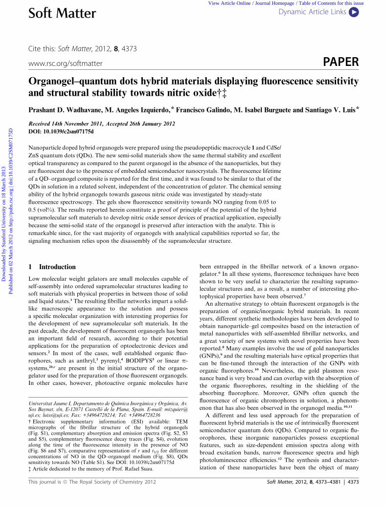
Fig. 1 (A) Chemical structure of organogelator 1. (B) Photograph of
cuvettes showing the fluorescence of CdSe/ZnS QDs in organogel media.
Samples containing: (a) CdSe/ZnS QDs (0.45 mM) in toluene, (b) orga-
nogel obtained frommacrocycle 1 (2.6 mgmL�1) in toluene, (c) CdSe/ZnS
QDs (0.45 mM) in organogel obtained from macrocycle 1 (2.6 mg mL�1)
in toluene.
4374 | Soft Matter, 2012, 8, 4373–4381
organogel, with the advantage of being fluorescent due to the
presence of embedded semiconductor nanocrystals. The photo-
physical properties of the QD–organogel hybrid material were
investigated by means of non-invasive fluorescence techniques.
In particular, time-resolved spectroscopy was used for the first
time to measure the lifetimes of QDs embedded in an organogel
matrix. We have also investigated their chemical sensing ability
towards a species of biomedical and environmental interest, such
as nitric oxide, by steady-state fluorescence spectroscopy. The
results herein reported constitute a proof of principle of the
potential of the developed QD–organogels for sensing nitric
oxide and set the basis for the development of analytical devices
based on this concept. It is worth mentioning that the utilisation
of QDs for NO sensing22 is a quite new field and, to our
knowledge, this application has never been attempted in the gel
state.
2 Experimental section
2.1 Materials
A toluene solution of core–shell CdSe/ZnS quantum dots (QDs),
surface-capped with hexadecylamine, displaying a maximum
emission at 480 nm, was purchased from Aldrich. Toluene
(spectroscopy grade, Scharlab) was used as received. Cyclophane
1 was synthesized according to the described experimental pro-
cedure.18a The spectral characterization of the prepared
compound 1 coincided with the reported data.
2.2 Photophysical characterization
UV-visible absorption measurements were made using a Hew-
lett-Packard 8453 spectrophotometer. Steady-state fluorescence
spectra were recorded in a Spex Fluorog 3-11 equipped with
a 450 W xenon lamp. Fluorescence spectra were recorded in the
front face mode. Time-resolved fluorescence measurements were
done with the technique of time correlated single photon
counting (TCSPC) in an IBH-5000U. Samples were excited with
an IBH 372 nm NanoLED with a FWHM of 1.3 ns and a repe-
tition rate of 100 kHz. Data were fitted to the appropriate
exponential model after deconvolution of the instrument
response function by an iterative deconvolution technique, using
the IBH DAS6 fluorescence decay analysis software. Reduced c2
values (<1.2) and weighted residuals served as parameters for
goodness of fit. All the samples were measured in aerated
conditions, except when otherwise stated.
2.3 Scanning electron microscopy
The scanning electron microscope (SEM) samples were prepared
by slow evaporation of a solution of the macrocycle 1
(2 mg mL�1) in toluene or in toluene containing QDs (0.56 mM)
directly onto the sample holder. The samples were convention-
ally coated prior to the measurements. Images were acquired on
a JEOL 7001F SEM at beam energy 5.0 kV.
2.4 Transmission electron microscopy
The transmission electron microscope (TEM) samples were
prepared by dissolving macrocycle 1 (7 mg mL�1) in toluene or in
This journal is ª The Royal Society of Chemistry 2012
Dow
nloa
ded
by S
tanf
ord
Uni
vers
ity o
n 18
Mar
ch 2
013
Publ
ishe
d on
02
Mar
ch 2
012
on h
ttp://
pubs
.rsc
.org
| do
i:10.
1039
/C2S
M07
175D
View Article Online
toluene containing QDs (0.65 mM). A drop of the solution was
deposited onto a holey carbon coated 300 mesh TEM copper grid
and was dried under air. The dried grid was loaded into a single-
tilt sample holder. The TEM samples were examined on a JEOL
2100 TEM equipped with a high resolution Gatan CCD camera
(11 Mpixels). The TEM was operated at 80 kV.
2.5 Formation of organogels
Gelation of toluene was accomplished inside a 1 � 1 � 4 cm
fluorescence cuvette (quartz) by dissolving the appropriate
amount of 1 in the boiling solvent, and allowing to cool to room
temperature, as previously described.20 For absorption, steady-
state-emission and single-photon counting measurements, the
quantum dots sample was diluted in toluene at the appropriate
concentration for the photophysical measurements (typically
0.45 mM), and such a solution was used to dissolve 1 at high
temperature. After cooling to room temperature inside the
fluorescence cuvette, transparent gels were obtained after 30 min,
which allowed the acquisition of the pertinent spectroscopic
data.
2.6 Nitric oxide sensing
Nitric oxide was synthesized by reacting NaNO2 (0.2 M) with
ascorbic acid (1 M), according to a method previously described
in the literature.22a,23 Different concentrations of nitric oxide
were obtained by diluting freshly prepared pure gaseous NO in
argon under anaerobic conditions. The samples of organogel
obtained from macrocycle 1 (2.6 mg mL�1) containing QDs
(0.45 mM) in toluene were prepared directly in the fluorescence
cuvette (1 � 1 � 4 cm) and they were purged with nitrogen for
30 minutes to accomplish an anaerobic atmosphere. Then, a fixed
volume of a specific concentration of NO (10 mL) was added
through the septum of the cuvette with a syringe. The evolution
of the fluorescence intensity of the QDs was measured immedi-
ately after addition of the nitric oxide. The melting temperatures
of the organogels treated with NO were measured 5 hours after
addition of the gas.
3 Results and discussion
3.1 Preparation of organogels and morphological
characterization
The preparation of the hybrid organogels with optical trans-
parency is not trivial and several practical issues must be
considered when designing the system. Thus, the inorganic
nanoparticles and the organogelator must be dispersed in
a compatible solvent in order to obtain a homogeneous solution
of the system, all the components must be stable under the
experimental conditions used to prepare the organogels and the
resulting soft supramolecular material must exhibit excellent
transparency suitable for the photophysical characterization. In
the course of the experiments carried out in our laboratories, we
have observed that macrocycle 1 is a readily available compound
that yields very transparent gels in toluene and therefore it could
be compatible with the colloidal solutions of the lipophilic CdSe/
ZnS QDs in toluene. Compound 1 has also been observed to
yield very transparent gels in the presence of other organic
This journal is ª The Royal Society of Chemistry 2012
compounds such as aromatic compounds and amines and, thus,
it could be used to investigate a number of photophysical
processes in the organogel media.20,21 In the present study, the
gelation process of toluene by cyclophane 1 in the presence of
CdSe/ZnS QDs capped by hexadecylamine as surface ligand was
investigated. Supramolecular organogels were prepared by dis-
solving a certain amount of 1 in toluene containing quantum
dots. The QDs suspension was prepared in toluene at the
appropriate concentration for the photophysical experiments
and such a solution was used to dissolve 1 at high temperature.
After cooling to room temperature inside the cell, transparent
gels were obtained (Fig. 1B). Formation of the organogel con-
taining quantum dots was investigated at different concentra-
tions of 1 in order to determine the optimal concentration of
cyclophane to form transparent organogels in the presence of the
nanoparticles. The concentrations of 1 used to prepare the
organogels are listed in Table 1.
Concentrations of cyclophane 1 lower than 1.6 mg mL�1 led to
formation of weak organogels, while concentrations higher than
3.2 mg mL�1 led to precipitation of 1 and subsequent collapse of
the fibrillar network. The gel-to-sol transition temperatures of
the different organogels were calculated by the inversion vial
method and are shown in Table 1. The results indicate that the
prepared gels start melting at 44–46 �C, irrespective of the gelatorconcentration and independent of the presence or absence of
QDs. Interestingly, it was possible to decrease the amount
of cyclophane needed to form stable organogels in the presence
of the nanoparticles (from 2.6 mg mL�1 in the absence of QDs to
1.6 mg mL�1 when QDs are present). This trend was confirmed
after several independent measurements. This apparent rein-
forcement of the self-assembled network at low concentrations of
gelator could be due to the presence of weak supramolecular
interactions of the quantum dots with the fibrillar network.8d It
must be noted that, although a physical reinforcement was
previously reported for some hybrid systems,8d,24 this was not
reflected in a significant modification of the gel-to-sol transition
temperatures of the gels in the presence of inorganic nano-
particles,10,25 as is also observed in the present case.
The organic–inorganic soft supramolecular materials main-
tained the same excellent thermal reversibility displayed by the
original organogels. Thus, the hybrid organogels were heated
until solutions were obtained, and they were cooled to room
temperature to reform the gels. The cycle could be repeated four
times in the same cuvette without any sign of fatigue. This result
indicates that the interactions between the nanoparticles and the
organogelator must be relatively weak. Therefore, it is possible to
preserve the reversibility of the self-assembled system in the
presence of the quantum dots.
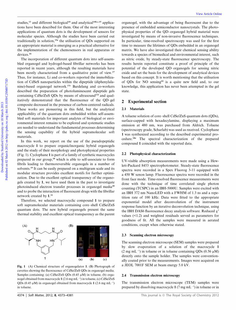
The morphology of the assemblies was examined by scanning
electron microscopy (SEM) and transmission electron micros-
copy (TEM). The SEM images of the xerogel obtained from the
organogel containing QDs show an entangled 3D network con-
sisting of bundles of fibrous aggregates up to ca. 1 mm long
(Fig. 2B). The microscopic structure was similar to that of the
xerogel obtained in the absence of nanoparticles (Fig. 2A),
indicating that the quantum dots do not modify significantly the
microscopic organization of the supramolecular structure. The
TEM images allowed visualising the nanoparticles stacked to the
fibers of 1. Interestingly, the individual quantum dots are isolated
Soft Matter, 2012, 8, 4373–4381 | 4375
Table 1 Melting temperatures of organogels formed by 1 with and without CdSe/ZnS QDs and photophysical properties of the QDs in the differentsamples
Sample Gelator/mg mL�1 Tg/�C labs/nm lem
a/nm s1b/ns a1 (%) s2
b/ns a2 (%) s3b/ns a3 (%) sav/ns
1 + QDsc 1.6 (Gel) 45–47 457 478 10.5 21 36.6 48 137.0 30 103.41d 1.6 (Sol) — — — — — — — — —1 + QDsc 2.6 (Gel) 46–61 457 478 9.3 19 36.5 49 136.3 32 104.51d 2.6 (Gel) 45–54 — — — — — — — —1 + QDsc 3.2 (Gel) 44–57 457 476 10.2 19 35.7 48 137.5 32 106.6QDse 0 (Sol) — 457 477 11.1 24 38.6 52 139.9 24 97.8
a lexc ¼ 366 nm. b lexc ¼ 372 nm, lem ¼ 480 nm. c Hybrid QD–organogel in toluene. d Control organogel in toluene without CdSe/ZnS QDs. e ControlCdSe/ZnS QDs in toluene.
Fig. 2 (A) SEM micrograph of the fibrillar structure of cyclophane 1
xerogel. Scale bar is 100 nm. (B) SEM micrograph of the fibrillar struc-
ture of cyclophane 1 xerogel containing CdSe/ZnS QDs. Scale bar is
100 nm. (C) and (D) TEM micrographs of the fibrillar structure of
cyclophane 1 xerogel containing CdSe/ZnS QDs. Scale bars are: (C)
20 nm, (D) 10 nm.
Dow
nloa
ded
by S
tanf
ord
Uni
vers
ity o
n 18
Mar
ch 2
013
Publ
ishe
d on
02
Mar
ch 2
012
on h
ttp://
pubs
.rsc
.org
| do
i:10.
1039
/C2S
M07
175D
View Article Online
and distributed along the superstructures formed by the orga-
nogelator (Fig. 2C and D and ESI†, Fig. S1). The nanoparticles
could bind the fibrillar network due to weak molecular interac-
tions between the alkyl groups of the hexadecylamine ligand and
the hydrophobic residues of the peptidic fibers, analogously to
related systems.8c,10,11b,17f Although SEM and TEM techniques
are commonly used in the characterization of self-associated
systems,1a,1b,17a,c–f the experiments imply drying of the samples on
a sample holder, and hence the static images acquired under such
experimental conditions need to be handled with care, as they
could not represent the actual situation in the gel state,26 which is
better described by using dynamic and non-invasive techniques
such as steady-state and time-resolved fluorescence, as will be
shown later. In any case, SEM and TEMmeasurements provide,
again, strong evidence of the fact that the presence of the indi-
vidual QDs has little morphological effect on the fibrillar
network.
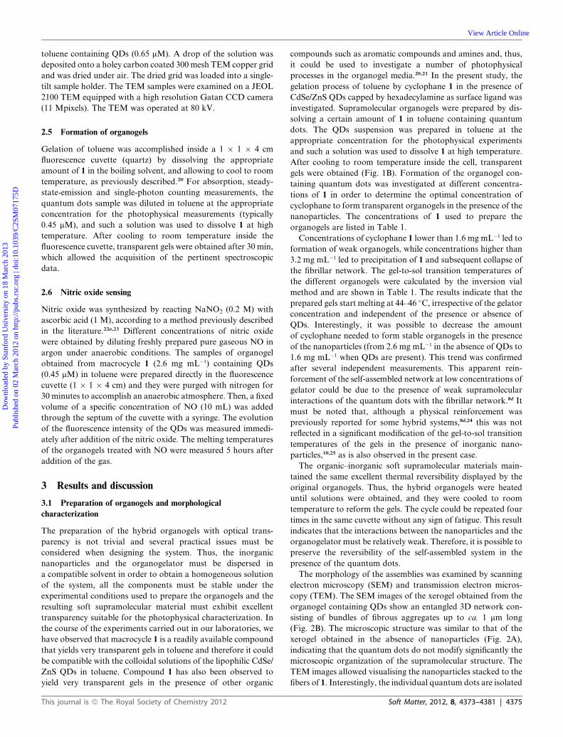
Fig. 3 Absorption spectra of samples prepared in toluene containing
CdSe/ZnS QDs (0.45 mM) and different concentrations of organogelator
1. The absorption spectrum of the control organogel obtained from
macrocycle 1 in toluene without QDs is also shown for comparison
(closed triangles, bottom spectrum).
3.2 Photophysical characterization of organogels containing
quantum dots
The optical properties of the organogels containing CdSe/ZnS
QDs were measured in order to determine the effect of the
organogelator 1 on the photophysical properties of the
4376 | Soft Matter, 2012, 8, 4373–4381
fluorescent nanoparticles. The pertinent spectroscopic data are
summarized in Table 1 and indicate that the absorption and
emission properties of the QDs are not perturbed by their
entrapment in the gel. These results validate the methodology
used to prepare the fluorescent hybrid organogels based on
macrocycle 1 and CdSe/ZnS capped with hexadecylamine, which
involves heating the solution to the boiling point of toluene
under air in the presence of the gelator. Thus, the electronic
absorption spectra of the QDs embedded into the organogel
formed by different concentrations of 1 coincided in shape and
position with those of the QDs in solution at the same concen-
tration of nanoparticles (Fig. 3 and ESI†, Fig. S2). The only
appreciable difference consisted of the increased baseline recor-
ded in the gel state as a consequence of light scattering, a typical
phenomenon observed in gels.21,27
The steady-state fluorescence spectra of the CdSe/ZnS QDs
were measured for samples containing different concentrations
of cyclophane 1 and the data are summarized in Table 1. As can
be seen in Fig. 4A, the emission spectra of the QDs were found to
be similar in toluene and in organogelated toluene at different
concentrations of 1. This indicates that the nanoparticles are
stable and do not aggregate during the self-assembly process.
Noteworthy, the photoluminescence efficiency is preserved in the
organogel state, despite the possible interaction and subsequent
quenching of the QDs with the fibrillar network (Fig. 4A). It is
This journal is ª The Royal Society of Chemistry 2012
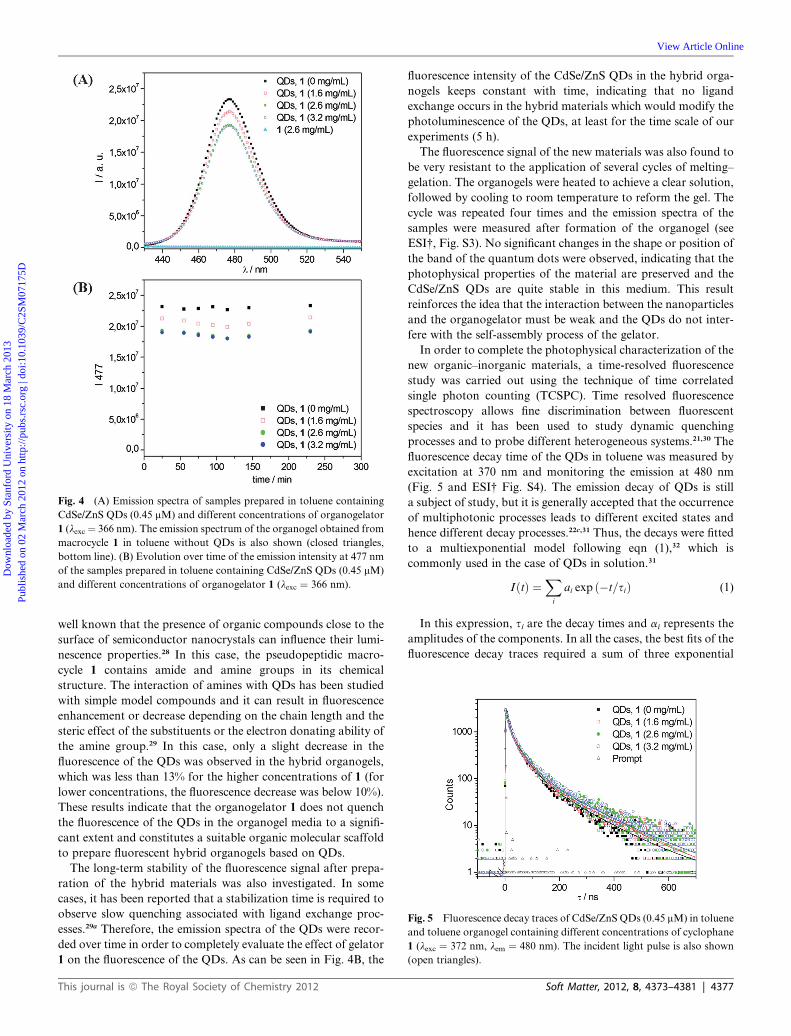
Fig. 4 (A) Emission spectra of samples prepared in toluene containing
CdSe/ZnS QDs (0.45 mM) and different concentrations of organogelator
1 (lexc ¼ 366 nm). The emission spectrum of the organogel obtained from
macrocycle 1 in toluene without QDs is also shown (closed triangles,
bottom line). (B) Evolution over time of the emission intensity at 477 nm
of the samples prepared in toluene containing CdSe/ZnS QDs (0.45 mM)
and different concentrations of organogelator 1 (lexc ¼ 366 nm).
Fig. 5 Fluorescence decay traces of CdSe/ZnS QDs (0.45 mM) in toluene
and toluene organogel containing different concentrations of cyclophane
1 (lexc ¼ 372 nm, lem ¼ 480 nm). The incident light pulse is also shown
(open triangles).
Dow
nloa
ded
by S
tanf
ord
Uni
vers
ity o
n 18
Mar
ch 2
013
Publ
ishe
d on
02
Mar
ch 2
012
on h
ttp://
pubs
.rsc
.org
| do
i:10.
1039
/C2S
M07
175D
View Article Online
well known that the presence of organic compounds close to the
surface of semiconductor nanocrystals can influence their lumi-
nescence properties.28 In this case, the pseudopeptidic macro-
cycle 1 contains amide and amine groups in its chemical
structure. The interaction of amines with QDs has been studied
with simple model compounds and it can result in fluorescence
enhancement or decrease depending on the chain length and the
steric effect of the substituents or the electron donating ability of
the amine group.29 In this case, only a slight decrease in the
fluorescence of the QDs was observed in the hybrid organogels,
which was less than 13% for the higher concentrations of 1 (for
lower concentrations, the fluorescence decrease was below 10%).
These results indicate that the organogelator 1 does not quench
the fluorescence of the QDs in the organogel media to a signifi-
cant extent and constitutes a suitable organic molecular scaffold
to prepare fluorescent hybrid organogels based on QDs.
The long-term stability of the fluorescence signal after prepa-
ration of the hybrid materials was also investigated. In some
cases, it has been reported that a stabilization time is required to
observe slow quenching associated with ligand exchange proc-
esses.29a Therefore, the emission spectra of the QDs were recor-
ded over time in order to completely evaluate the effect of gelator
1 on the fluorescence of the QDs. As can be seen in Fig. 4B, the
This journal is ª The Royal Society of Chemistry 2012
fluorescence intensity of the CdSe/ZnS QDs in the hybrid orga-
nogels keeps constant with time, indicating that no ligand
exchange occurs in the hybrid materials which would modify the
photoluminescence of the QDs, at least for the time scale of our
experiments (5 h).
The fluorescence signal of the new materials was also found to
be very resistant to the application of several cycles of melting–
gelation. The organogels were heated to achieve a clear solution,
followed by cooling to room temperature to reform the gel. The
cycle was repeated four times and the emission spectra of the
samples were measured after formation of the organogel (see
ESI†, Fig. S3). No significant changes in the shape or position of
the band of the quantum dots were observed, indicating that the
photophysical properties of the material are preserved and the
CdSe/ZnS QDs are quite stable in this medium. This result
reinforces the idea that the interaction between the nanoparticles
and the organogelator must be weak and the QDs do not inter-
fere with the self-assembly process of the gelator.
In order to complete the photophysical characterization of the
new organic–inorganic materials, a time-resolved fluorescence
study was carried out using the technique of time correlated
single photon counting (TCSPC). Time resolved fluorescence
spectroscopy allows fine discrimination between fluorescent
species and it has been used to study dynamic quenching
processes and to probe different heterogeneous systems.21,30 The
fluorescence decay time of the QDs in toluene was measured by
excitation at 370 nm and monitoring the emission at 480 nm
(Fig. 5 and ESI† Fig. S4). The emission decay of QDs is still
a subject of study, but it is generally accepted that the occurrence
of multiphotonic processes leads to different excited states and
hence different decay processes.22c,31 Thus, the decays were fitted
to a multiexponential model following eqn (1),32 which is
commonly used in the case of QDs in solution.31
IðtÞ ¼Xi
ai exp ð�t=siÞ (1)
In this expression, si are the decay times and ai represents the
amplitudes of the components. In all the cases, the best fits of the
fluorescence decay traces required a sum of three exponential
Soft Matter, 2012, 8, 4373–4381 | 4377
Dow
nloa
ded
by S
tanf
ord
Uni
vers
ity o
n 18
Mar
ch 2
013
Publ
ishe
d on
02
Mar
ch 2
012
on h
ttp://
pubs
.rsc
.org
| do
i:10.
1039
/C2S
M07
175D
View Article Online
functions to obtain low c2 values as well as random distributions
of the weighted residuals, which are indicators of the goodness of
the fits. The individual fluorescence lifetime components and
their contributions to the total signal are shown in Table 1. A
pattern for the lifetime of the QDs in toluene was established,
which comprised a short lifetime of ca. 11 ns, an intermediate
lifetime of ca. 39 ns and a long lifetime of ca. 139 ns, contributing
24%, 52% and 24%, respectively, to the overall signal. A similar
pattern was observed for the QDs embedded in organogels
containing different concentrations of 1. To our knowledge, this
is the first time that a non-invasive time-resolved fluorescence
technique has been used to investigate the fluorescence lifetimes
of organogel–QD composites.
To better compare the fluorescence lifetimes of all the sam-
ples,31b,c the weighted average lifetime (sav) was calculated by
using (eqn (2))32 and the values are given in Table 1. Although
a slight increase of ca. 6% was observed for the weighted average
lifetime of the QDs in the organogel media, such difference is
relatively small and the weighted average lifetimes of the QDs in
the organogels are thus comparable to that of the QDs in solu-
tion. Most importantly, the weighted average lifetime values
were found to be independent of the concentration of organo-
gelator 1.
sðavÞ ¼X
ais2iX
aisi
!(2)
Hence, the interaction between QDs and the fibrillar entan-
glement of 1must occur to a very low extent. As a matter of fact,
the nanocrystals could be in equilibrium between two states, one
interacting with the self-assembled fibers and the other as free
particles solubilised in the liquid phase (toluene) of the organo-
gel. All the spectroscopic similarities found between QDs in
solution and in the gel state point to the prevalence of the latter
state in the equilibrium, i.e. QDs freely dissolved in the liquid
fraction of the gel. Organogels have been described as soft
materials composed of a series of pools of solvent in which the
mobility of the molecules is identical to that in solution.20,33 This
model could also be compatible with our findings and it is
schematically represented in Fig. 6.
Overall, the optical characterization of the CdSe/ZnS QDs in
the hybrid organogel indicates that the pseudopeptidic macro-
cycle 1 can be used to prepare semi-solid materials with high
Fig. 6 Schematic representation showing the possible location of the
quantum dots (green circles) in the organogel.
4378 | Soft Matter, 2012, 8, 4373–4381
optical transparency and very stable fluorescence. In order to
check the generality of the method, organogels containing CdSe/
ZnS QDs emitting at 530 nm were prepared and the emission
properties of the nanoparticles followed the same behaviour as
observed in the case of the smaller QDs herein reported (data not
shown). Therefore, the excellent photophysical properties of
both CdSe/ZnS QDs emitting at 480 nm and at 530 nm are
preserved in the organogels, making them suitable for investi-
gating their potential use in the development of new formats for
supported optical sensors (see following section).
3.3 Nitric oxide sensing
Gel based systems have been proposed as interesting scaffolds for
the development of optical sensors because photoactive mole-
cules can be entrapped in the gel matrix with retention of the
sensing properties.34 Herein, we investigated the applicability of
the organogels containing quantum dots for the chemical sensing
of nitric oxide (NO), which is a gaseous species known to play
a wide variety of roles in biomedicine35 and environmental
chemistry.36 There are a great number of sensors for NO reported
in the literature, both based on molecular and macromolecular
systems, which highlights the importance of this species.37
Although some groups have developed sensors based on
quantum dots for detecting NO,22a,b as well as nitroxides,38 in
solution, and we have recently reported on a QD–poly-
methacrylate composite for the analysis of NOx,22c to the best of
our knowledge the sensing of nitric oxide in a gel matrix has not
been described.
The organogels containing CdSe/ZnS QDs were prepared as
described in the experimental section. Different concentrations
of gaseous NOwere injected in the cuvettes containing the hybrid
materials under anaerobic conditions. No changes in the
absorption spectra of the QDs were observed after the addition
of NO to the samples (ESI†, Fig. S5). The steady state fluores-
cence spectra were recorded over time after the addition of
variable concentrations of NO. A decrease in the fluorescence
of the CdSe/ZnS QDs was observed in all the cases. An example
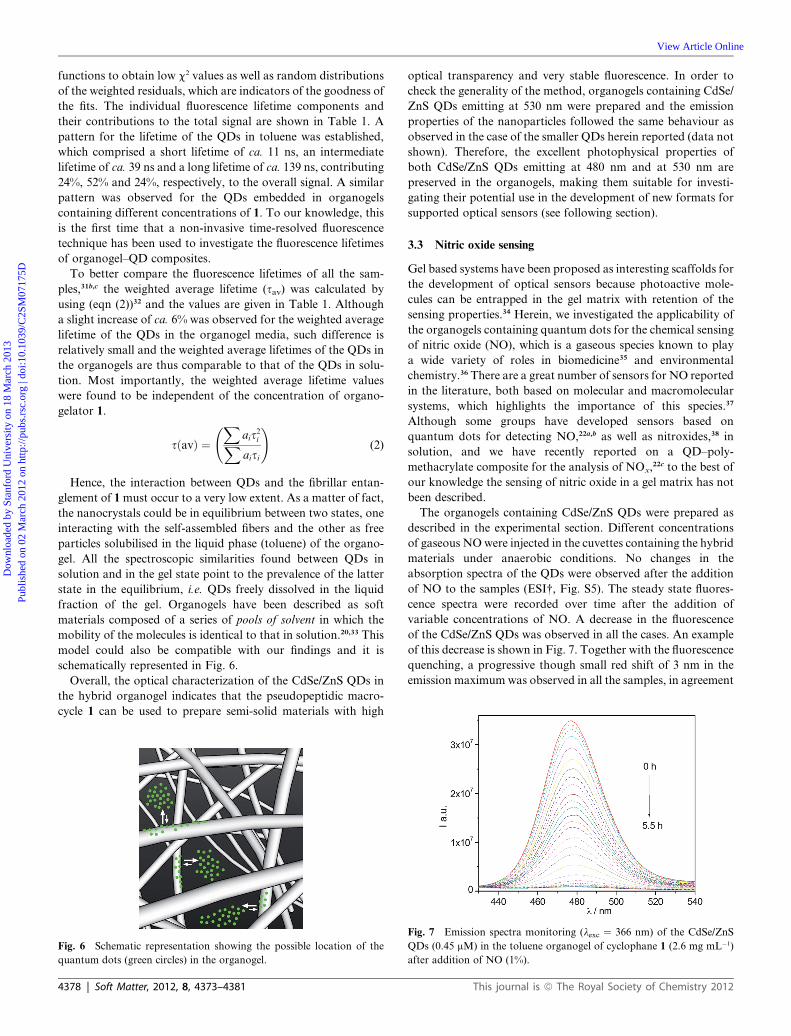
of this decrease is shown in Fig. 7. Together with the fluorescence
quenching, a progressive though small red shift of 3 nm in the
emission maximum was observed in all the samples, in agreement
Fig. 7 Emission spectra monitoring (lexc ¼ 366 nm) of the CdSe/ZnS
QDs (0.45 mM) in the toluene organogel of cyclophane 1 (2.6 mg mL�1)
after addition of NO (1%).
This journal is ª The Royal Society of Chemistry 2012
Table 2 Quantum dots sensitivity towards nitric oxide in organogelmediaa
NO (vol%) t1/2/min �r � 10�4/s�1
0.05 269.5 1.10.15 99.5 1.80.5 30.0 3.31 22.5 4.31.5 26.5 4.02 31.0 3.9
a Samples containing CdSe/ZnS QDs (0.45 mM) in the toluene organogelof cyclophane 1 (2.6 mg mL�1).
Dow
nloa
ded
by S
tanf
ord
Uni
vers
ity o
n 18
Mar
ch 2
013
Publ
ishe
d on
02
Mar
ch 2
012
on h
ttp://
pubs
.rsc
.org
| do
i:10.
1039
/C2S
M07
175D
View Article Online
with previous observations on the interaction of QDs with
a number of analytes.38a This process has been attributed to the
differential quenching of the QDs: upon addition of a quencher,
small particles are quenched more effectively than the large ones,
resulting in an emitting population enriched in larger nano-
particles. This behaviour is observed for all the concentrations of
NO added to the hybrid organogel, indicating that size selective
quenching also occurs in the semi-solid materials as well as in
solution (see ESI†, Fig. S6 and S7).
The plots of the emission maxima of the QDs in the organogel
versus time for different concentrations of NO are shown in
Fig. 8. The fluorescence intensity has been translated into
normalized values to allow a comparative analysis of the effect of
the different concentrations of gas on the quenching process. A
control experiment for the hybrid organogel in the absence of
NO indicates that the fluorescence emission of the QDs was
stable over the time needed to carry out the experiments (black
triangles in Fig. 8, corresponding to 0% NO).
As can be seen in Fig. 8, the response of the hybrid organogels
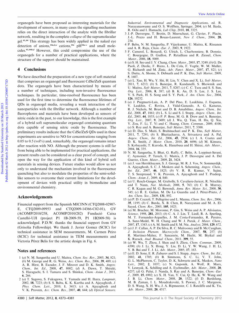
to NO depends on the concentration of the gas. Two parameters
were calculated from these curves in Fig. 8 in order to evaluate
the sensing ability of the supramolecular gels containing
quantum dots. On the one hand, the time at which the emission
of the QDs at 477 nm was reduced 50% relative to the initial
intensity (t1/2) was determined. On the other hand, the slope of
the initial points of every curve (r (s�1) ¼ normalized emission/
time) was calculated in order to estimate the apparent rate of
quenching of the fluorescence of the QDs. The resulting
parameters are gathered in Table 2. For the comparative anal-
ysis, we will only use here the r values (Fig. 9), although similar
conclusions can be drawn using t1/2 (see ESI†, Fig. S8).
The results indicate that upon increasing the concentration of
the gas, the apparent rate of fluorescence quenching increases
until it reaches a plateau at a concentration of 1%. These
preliminary results indicate that the hybrid organogels are
sensitive to NO in concentrations ranging from 0.05 to 1%.
Further increase of the concentration of gas up to 2% did not
affect the apparent rate of quenching, suggesting that the
mechanism is controlled by the diffusion of NO through the
network of the organogel.
Fig. 8 Evolution over time of the fluorescence intensity at 477 nm of the
QDs (0.45 mM) in the toluene organogel of cyclophane 1 (2.6 mg mL�1) in
the presence of different concentrations of NO.
This journal is ª The Royal Society of Chemistry 2012
The quenching of the fluorescence of the CdSe/ZnS QDs by
different concentrations of nitric oxide was also measured in
solution. Although the decrease of the fluorescence of the QDs in
solution was faster (see ESI† Table S1), the fact that the new
hybrid materials display fluorescence sensitivity towards NO in
the same range of concentrations must be considered as a proof
of principle, opening the possibility for developing faster systems
in the future. Current studies are underway in our laboratories to
optimize the response of the hybrid organogels towards NO and
to establish the response of the system in the presence of other
potential quenchers.
It must be noted that the research on new methodologies to
support known chemosensors for gases onto different materials
is a topic of great interest. New materials with sensing capabil-
ities for gases are continuously emerging: polymer gels for the
detection of odors39 and chloride-containing gases,40 organogels
based on carbon tetrachloride for the analysis of nerve gas
stimulant41 or graphene–ionic liquid composites for the detection
of vapors of benzene42 or for the analysis of nitric oxide43 are
selected examples of soft systems useful for the analysis of
gaseous species.
Notably, the structural integrity of the hybrid organogels
herein described is maintained upon addition of NO since they
display identical strength as compared to the original ones before
the interaction with NO. It is important to note that, although
Fig. 9 Comparative representation of the apparent rate (r) of quenching
of the fluorescence of the CdSe/ZnS QDs for different concentrations
of NO in the toluene organogel–QD composite (cyclophane 1
(2.6 mg mL�1), CdSe/ZnS QDs (0.45 mM)).
Soft Matter, 2012, 8, 4373–4381 | 4379
Dow
nloa
ded
by S
tanf
ord
Uni
vers
ity o
n 18
Mar
ch 2
013
Publ
ishe
d on
02
Mar
ch 2
012
on h
ttp://
pubs
.rsc
.org
| do
i:10.
1039
/C2S
M07
175D
View Article Online
organogels have been proposed as interesting materials for the
development of sensors, in many cases the signalling mechanism
relies on the direct interaction of the analyte with the fibrillar
network, resulting in the complete collapse of the supramolecular
gel.41,44 This strategy has been widely applied in the naked eye
detection of anions,44a,b,e cations,44c pH44a–c and small mole-
cules.41,44b,d However, this could compromise the use of the
organogels for a number of practical applications, where the
structure of the support should be maintained.
4 Conclusions
We have described the preparation of a new type of soft material
that comprises an organogel and fluorescent CdSe/ZnS quantum
dots. The organogels have been characterized by means of
a number of techniques, including non-invasive fluorescence
techniques. In particular, time-resolved fluorescence has been
used for the first time to determine the fluorescence lifetimes of
QDs in organogel media, revealing a weak interaction of the
nanoparticles with the fibrillar network. Although a number of
fluorophores and materials have been developed as sensors of
nitric oxide in the past, to our knowledge, this is the first example
of a hybrid soft supramolecular organogel containing quantum
dots capable of sensing nitric oxide by fluorescence. The
preliminary results indicate that the CdSe/ZnS QDs used in these
experiments are sensitive to NO for concentrations ranging from
0.05 to 0.5 (vol%) and, remarkably, the organogel remains intact
after reaction with NO. Although the present systems is still far
from being able to be implemented for practical applications, the
present results can be considered as a clear proof of concept, and
open the way for the application of this kind of hybrid soft
materials in sensing devices. Future studies would allow us not
only to understand the mechanism involved in the fluorescence
quenching but also to modulate the properties of the semi-solid-
like sensors to overcome their current limitations for the devel-
opment of devices with practical utility in biomedicine and
environmental chemistry.
Acknowledgements
Financial support from the Spanish MICINN (CTQ2008-02907-
E, CTQ2009-09953 and CTQ2009-14366-C02-01), GV
(ACOMP/2010/258, ACOMP/2010/282) Fundaci�o Caixa
Castell�o-UJI (project P1 1B-2009-59, P1 1B2009-58) is
acknowledged. P.D.W. thanks the financial support from GV
(Grisolia Fellowship). We thank J. Javier Gomez (SCIC) for
technical assistance in SEM measurements, M. Carmen Peiro
(SCIC) for technical assistance in TEM measurements and
Victoria P�erez Belis for the artistic design in Fig. 6.
Notes and references
1 (a) N. M. Sangeetha and U. Maitra, Chem. Soc. Rev., 2005, 34, 821;(b) M. George and R. G. Weiss, Acc. Chem. Res., 2006, 39, 489; (c)A. R. Hirst, B. Escuder, J. F. Miravet and D. K. Smith, Angew.Chem., Int. Ed., 2008, 47, 8002; (d) A. Dawn, T. Shiraki,S. Haraguchi, S. I. Tamaru and S. Shinkai, Chem.–Asian J., 2011,6, 266.
2 (a) T. Sagawa, S. Fukugawa, T. Yamada and H. Ihara, Langmuir,2002, 18, 7223; (b) S. S. Babu, K. K. Kartha and A. Ajayaghosh, J.Phys. Chem. Lett., 2010, 1, 3413; (c) A. Ajayaghosh andV. K. Praveen, Acc. Chem. Res., 2007, 40, 644; (d) Optical Sensors.
4380 | Soft Matter, 2012, 8, 4373–4381
Industrial, Environmental and Diagnostic Applications, ed. R.Narayanaswamy and O. S. Wolfbeis, Springer, 2004; (e) M. Ikeda,R. Ochi and I. Hamachi, Lab Chip, 2010, 10, 3325.
3 J.-P. Desvergne, T. Brotin, D. Meerschaut, G. Clavier, F. Placin,J.-L. Pozzo and H. Bouas-Laurent, New J. Chem., 2004, 28,234.
4 P. Babu, N. M. Sangeetha, P. Vijaykumar, U. Maitra, K. Rissanenand A. R. Raju, Chem.–Eur. J., 2003, 9, 1922.
5 F. Camerel, L. Bonardi, G. Ulrich, L. Charbonniere, B. Donnio,C. Bourgogne, D. Guillon, P. Retailleau and R. Ziessel, Chem.Mater., 2006, 18, 5009.
6 (a) S. H. Seo and J. Y. Chang,Chem.Mater., 2005, 17, 3249; (b) G. DePaoli, Z. Dzolic, F. Rizzo, L. De Cola, F. Vogtle, W. M. Muller,G. Richardt and M. Zinic, Adv. Funct. Mater., 2007, 17, 821; (c)S. Dutta, A. Shome, S. Debnath and P. K. Das, Soft Matter, 2009,5, 1607.
7 (a) L. Xue, H. Wu, Y. Shi, H. Liu, Y. Chen and X. Li, Soft Matter,2011, 7, 6213; (b) S. Banerjee, R. Kandanelli, S. Bhowmik andU. Maitra, Soft Matter, 2011, 7, 8207; (c) C. C. Tsou and S. S. Sun,Org. Lett., 2006, 8, 387; (d) B. K. An, D. S. Lee, J. S. Lee,Y. S. Park, H. S. Song and S. Y. Park, J. Am. Chem. Soc., 2004,126, 10232.
8 (a) J. Puigmarti-Luis, A. P. Del Pino, E. Laukhina, J. Esquena,V. Laukhin, C. Rovira, J. Vidal-Gancedo, A. G. Kanaras,R. J. Nichols, M. Brust and D. B. Amabilino, Angew. Chem., Int.Ed., 2008, 47, 1861; (b) L. S. Li and S. I. Stupp, Angew. Chem., Int.Ed., 2005, 44, 1833; (c) P. P. Bose, M. G. B. Drew and A. Banerjee,Org. Lett., 2007, 9, 2489; (d) J. Wu, Q. Tian, H. Hu, Q. Xia,Y. Zou, F. Li, T. Yi and C. Huang, Chem. Commun., 2009, 4100;(e) S. Roy and A. Banerjee, Soft Matter, 2011, 7, 5300.
9 (a) D. Das, S. Maiti, S. Brahmachari and P. K. Das, Soft Matter,2011, 7, 7291; (b) S. Bhattacharya, A. Srivastava and A. Pal,Angew. Chem., Int. Ed., 2006, 45, 2934; (c) I. A. Coates andD. K. Smith, J. Mater. Chem., 2010, 20, 6696; (d) M. Kimura,S. Kobayashi, T. Kuroda, K. Hanabusa and H. Shirai, Adv. Mater.,2004, 16, 335.
10 N. M. Sangeetha, S. Bhat, G. Raffy, C. Belin, A. Loppinet-Serani,C. Aymonier, P. Terech, U. Maitra, J. P. Desvergne and A. DelGuerzo, Chem. Mater., 2009, 21, 3424.
11 (a) J. van Herrikhuyzen, S. J. George, M. R. J. Vos, N. Sommerdijk,A. Ajayaghosh, S. C. J. Meskers and A. Schenning, Angew. Chem.,Int. Ed., 2007, 46, 1825; (b) V. R. R. Kumar, V. Sajini,T. S. Sreeprasad, V. K. Praveen, A. Ajayaghosh and T. Pradeep,Chem.–Asian J., 2009, 4, 840.
12 (a) U. Resch-Genger, M. Grabolle, S. Cavaliere-Jaricot, R. Nitschkeand T. Nann, Nat. Methods, 2008, 5, 763; (b) C. B. Murray,C. R. Kagan and M. G. Bawendi, Annu. Rev. Mater. Sci., 2000, 30,545; (c) R. E. Galian, M. De La Guardia and J. P�erez-Prieto, J.Am. Chem. Soc., 2009, 131, 892.
13 (a) P. D. Cozzoli, T. Pellegrino and L. Manna, Chem. Soc. Rev., 2006,35, 1195; (b) C. Burda, X. B. Chen, R. Narayanan and M. A. El-Sayed, Chem. Rev., 2005, 105, 1025.
14 (a) M. Bruchez, M. Moronne, P. Gin, S. Weiss and A. P. Alivisatos,Science, 1998, 281, 2013; (b) C. A. J. Lin, T. Liedl, R. A. Sperling,M. T. Fernandez-Arguelles, J. M. Costa-Fernandez, R. Pereiro,A. Sanz-Medel, W. H. Chang and W. J. Parak, J. Mater. Chem.,2007, 17, 1343; (c) A.M. Smith and S.M. Nie,Analyst, 2004, 129, 672.
15 (a) J. F. Callan, A. P. De Silva, R. C. Mulrooney and B. Mc Caughan,J. Inclusion Phenom. Macrocyclic Chem., 2007, 58, 257; (b)R. Mart�ınez-M�a~nez, F. Sancen�on, M. Hecht, M. Biyikal andK. Rurack, Anal. Bioanal. Chem., 2011, 399, 55.
16 (a) W. Wu, T. Zhou, J. Shen and S. Zhou, Chem. Commun., 2009,4390; (b) J. Li, X. Hong, Y. Liu, D. Li, Y. W. Wang, J. H. Li,Y. B. Bai and T. J. Li, Adv. Mater., 2005, 17, 163.
17 (a) E. D. Sone, E. R. Zubarev and S. I. Stupp, Angew. Chem., Int. Ed.,2002, 41, 1705; (b) B. Simmons, S. C. Li, V. T. John,G. L. McPherson, C. Taylor, D. K. Schwartz and K. Maskos, NanoLett., 2002, 2, 1037; (c) N. Gaponik, A. Wolf, R. Marx,V. Lesnyak, K. Schilling and A. Eychmuller, Adv. Mater., 2008, 20,4257; (d) G. Palui, J. Nanda, S. Ray and A. Banerjee, Chem.–Eur.J., 2009, 15, 6902; (e) X. H. Yan, Y. Cui, Q. He, K. W. Wang andJ. B. Li, Chem. Mater., 2008, 20, 1522; (f) D. Bardelang,M. B. Zaman, I. L. Moudrakovski, S. Pawsey, J. C. Margeson,D. S. Wang, X. H. Wu, J. A. Ripmeester, C. I. Ratcliffe and K. Yu,Adv. Mater., 2008, 20, 4517.
This journal is ª The Royal Society of Chemistry 2012
Dow
nloa
ded
by S
tanf
ord
Uni
vers
ity o
n 18
Mar
ch 2
013
Publ
ishe
d on
02
Mar
ch 2
012
on h
ttp://
pubs
.rsc
.org
| do
i:10.
1039
/C2S
M07
175D
View Article Online
18 (a) J. Becerril, M. Bolte, M. I. Burguete, F. Galindo, E. Garcia-Espana, S. V. Luis and J. F. Miravet, J. Am. Chem. Soc., 2003, 125,6677; (b) I. Alfonso, M. Bolte, M. Bru, M. I. Burguete, S. V. Luisand J. Rubio, J. Am. Chem. Soc., 2008, 130, 6137; (c) M. Bru,I. Alfonso, M. I. Burguete and S. V. Luis, Angew. Chem., Int. Ed.,2006, 45, 6155.
19 J. Becerril, M. I. Burguete, B. Escuder, F. Galindo, R. Gavara,J. F. Miravet, S. V. Luis and G. Peris, Chem.–Eur. J., 2004, 10, 3879.
20 F. Galindo, M. I. Burguete, R. Gavara and S. V. Luis, J. Photochem.Photobiol., A, 2006, 178, 57.
21 M. I. Burguete, M. A. Izquierdo, F. Galindo and S. V. Luis, Chem.Phys. Lett., 2008, 460, 503.
22 (a) S. H.Wang,M. Y. Han andD. J. Huang, J. Am. Chem. Soc., 2009,131, 11692; (b) X. Q. Yan, Z. B. Shang, Z. Zhang, Y. Wang andW. J. Jin, Luminescence, 2009, 24, 255; (c) V. Fabregat,M. A. Izquierdo, M. I. Burguete, F. Galindo and S. V. Luis, Inorg.Chim. Acta, 2012, 381, 212–217.
23 C. A. Bunton, H. Dahn and L. Loewe, Nature, 1959, 183, 163.24 S. Srinivasan, S. S. Babu, V. K. Praveen and A. Ajayaghosh, Angew.
Chem., Int. Ed., 2008, 47, 5746.25 R. K. Das, S. Bhat, S. Banerjee, C. Aymonier, A. Loppinet-Serani,
P. Terech, U. Maitra, G. Raffy, J.-P. Desvergne and A. DelGuerzo, J. Mater. Chem., 2011, 21, 2740.
26 I. Alfonso, M. Bru, M. Isabel Burguete, E. Garc�ıa-Verdugo andS. V. Luis, Chem.–Eur. J., 2010, 16, 1246.
27 K. Murata, M. Aoki, T. Suzuki, T. Harada, H. Kawabata,T. Komori, F. Ohseto, K. Ueda and S. Shinkai, J. Am. Chem. Soc.,1994, 116, 6664.
28 R. E. Galian and M. de la Guardia, TrAC, Trends Anal. Chem., 2009,28, 279.
29 (a) R. E. Galian and J. C. Scaiano, Photochem. Photobiol. Sci., 2009,8, 70; (b) D. V. Talapin, A. L. Rogach, A. Kornowski, M. Haase andH. Weller, Nano Lett., 2001, 1, 207; (c) C. F. Landes, M. Braun andM. A. El-Sayed, J. Phys. Chem. B, 2001, 105, 10554.
30 M. I. Burguete, F. Galindo, R. Gavara, M. A. Izquierdo, J. C. Lima,S. V. Luis, A. J. Parola and F. Pina, Langmuir, 2008, 24, 9795.
31 (a) X. Y. Wang, L. H. Qu, J. Y. Zhang, X. G. Peng and M. Xiao,Nano Lett., 2003, 3, 1103; (b) M. J. Ruedas-Rama, A. Orte,E. A. H. Hall, J. M. Alvarez-Pez and E. M. Talavera,ChemPhysChem, 2011, 12, 919; (c) I.-S. Liu, H.-H. Lo, C.-T. Chien,Y.-Y. Lin, C.-W. Chen, Y.-F. Chen, W.-F. Su and S.-C. Liou,
This journal is ª The Royal Society of Chemistry 2012
J. Mater. Chem., 2008, 18, 675; (d) J. Rubio, M. A. Izquierdo,M. I. Burguete, F. Galindo and S. V. Luis, Nanoscale, 2011, 3, 3613.
32 J. R. Lakowicz, Principles of Fluorescence Spectroscopy, Springer,2006.
33 (a) C. Geiger, M. Stanescu, L. H. Chen and D. G.Whitten, Langmuir,1999, 15, 2241; (b) K. Hanabusa, K. Hiratsuka, M. Kimura andH. Shirai, Chem. Mater., 1999, 11, 649.
34 I. Yoshimura, Y. Miyahara, N. Kasagi, H. Yamane, A. Ojida andI. Hamachi, J. Am. Chem. Soc., 2004, 126, 12204.
35 N. S. Bryan andM.B.Grisham,FreeRadical Biol.Med., 2007, 43, 645.36 D. D. Nelson, J. B. McManus, S. C. Herndon, J. H. Shorter,
M. S. Zahniser, S. Blaser, L. Hvozdara, A. Muller, M. Giovanniniand J. Faist, Opt. Lett., 2006, 31, 2012.
37 (a) T. Nagano and T. Yoshimura, Chem. Rev., 2002, 102, 1235; (b)L. E. McQuade and S. J. Lippard, Curr. Opin. Chem. Biol., 2010,14, 43; (c) Y. Yang, S. K. Seidlits, M. M. Adams, V. M. Lynch,C. E. Schmidt, E. V. Anslyn and J. B. Shear, J. Am. Chem. Soc.,2010, 132, 13114; (d) D. J. Blyth, J. W. Aylott, J. W. B. Moir,D. J. Richardson and D. A. Russell, Analyst, 1999, 124, 129; (e)M. Bru, M. I. Burguete, F. Galindo, S. V. Luis, M. J. Marin andL. Vigara, Tetrahedron Lett., 2006, 47, 1787.
38 (a) M. Laferriere, R. E. Galian, V. Maurel and J. C. Scaiano, Chem.Commun., 2006, 257; (b) C. Tansakul, E. Lilie, E. D. Walter,F. Rivera, A. Wolcott, J. Z. Zhang, G. L. Millhauser andR. Braslau, J. Phys. Chem. C, 2010, 114, 7793.
39 H. Kim and G. Kwak, Macromolecules, 2009, 42, 902.40 H. Lee, S. H. Jung, W. S. Han, J. H. Moon, S. Kang, J. Y. Lee,
J. H. Jung and S. Shinkai, Chem.–Eur. J., 2011, 17, 2823.41 T. H. Kim, D. G. Kim, M. Lee and T. S. Lee, Tetrahedron, 2010, 66,
1667.42 Q. Ji, I. Honma, S. M. Paek, M. Akada, J. P. Hill, A. Vinu and
K. Ariga, Angew. Chem., Int. Ed., 2010, 49, 9737.43 S. R. Ng, C. X. Guo and C. M. Li, Electroanalysis, 2011, 23, 442.44 (a) H. Yang, T. Yi, Z. G. Zhou, Y. F. Zhou, J. C.Wu,M. Xu, F. Y. Li
and C. H. Huang, Langmuir, 2007, 23, 8224; (b) Q. T. Liu,Y. L. Wang, W. Li and L. X. Wu, Langmuir, 2007, 23, 8217; (c)W. Deng and D. H. Thompson, Soft Matter, 2010, 6, 1884; (d)X. Chen, Z. Huang, S. Y. Chen, K. Li, X. Q. Yu and L. Pu, J. Am.Chem. Soc., 2010, 132, 7297; (e) P. Xue, Y. Zhang, J. Jia, D. Xu,X. Zhang, X. Liu, H. Zhou, P. Zhang, R. Lu, M. Takafuji andH. Ihara, Soft Matter, 2011, 7, 8296.
Soft Matter, 2012, 8, 4373–4381 | 4381