organizing principles of human body. hierarchy of structural organization each of these build upon...
TRANSCRIPT

Organizing principles of human body

Hierarchy of Structural Organization
Each of these build upon one another to make up the next level:Chemical level CellularTissueOrganOrgan systemOrganism

Hierarchy of Structural Organization
Chemical level Atoms combine to make molecules 4 macromolecules in the body
Carbohydrates Lipids Proteins Nucleic acids

Hierarchy of Structural Organization
Cellular Made up of cells and cellular
organelles (molecules) Cells can be eukaryotic or prokaryotic Organelles are structures within cells that
perform dedicated functions (“small organs”)
http://cmweb.pvschools.net/~bbecke/newell/Cells.html

Hierarchy of Structural Organization
Tissue Collection of cells that work together
to perform a specialized function 4 basic types of tissue in the human
body: Epithelium Connective tissue Muscle tissue Nervous tissue
www.emc.maricopa.edu

Hierarchy of Structural Organization
Organ Made up of tissue
Heart Brain Liver Pancreas, etc……
Pg 181

Hierarchy of Structural OrganizationOrgan system (11) Made up of a group of related organs
that work together Integumentary Skeletal Muscular Nervous Endocrine Cardiovascular Lymphatic Respiratory Digestive Urinary Reproductive
Circulatory
Pg 341
Urinary System

Levels of Organization:Levels of Organization:
COHN – 99.5%
99.9% =
See figs. 1.3 & 1.4

Hierarchy of Structural Organization
Organism An individual human, animal, plant,
etc…… Made up all of the organ systems Work together to sustain life

The Cell
Cells: structural and functional units of all living
organisms. building blocks of the human body. adult human body contains ~ 75 trillion
cells.
Each cell type performs specific functions. ~200 cell types in humans
subcategories of most

Common Characteristics of Cells
Perform the general functions necessary to sustain life: Obtain nutrients and other materials from
its surrounding fluids. Fuel molecules, O2, building blocks, minerals,etc
Dispose of wastes products Urea (from nitrogen), CO2, metabolic waste
Maintain shape and integrity Size and shape are related to function
Cell division: Mitosis: growth and repair Meiosis: gamete formation

Study of Cells
Cytology: study of cellsMicroscopic anatomy Individual cells observable by light
microscopy Subcellular structures observable by
electron microscopy. TEM SEM
Unit of measure: micrometer (um) RBC: 7-8um

2. Bilateral Symmetry
- left half of the body is a mirror image of the right half.
- structures in the median plane are unpaired, but have identical left and right sides.


Cells
Parts of a cell Cell Membrane (or plasma membrane) Cytoplasm
Cytosol Organelles
Membranous Organelles Non-membranous Organelles
Inclusions Nucleus

Plasma (Cell) Membrane
the outer, limiting barrierseparates the internal contents of the cell from external materials.

Cytoplasm
general term for all cellular contents located between the plasma membrane and the nucleus.

Nucleus
“control center” of the cell controls protein synthesis directs the functional and structural
characteristics of the cell.

Plasma membrane: composition
Lipids Phospholipids
Head: hydrophilic Tail: hydrophobic Form lipid bilayer
Cholesterol Glycolipids
Carbohydrate component Part of glycocalyx


Plasma membrane: composition
Protein Integral membrane proteins Peripheral membrane proteins Some serve as enzymes, ion channels
or receptors Glycoproteins

Fertilization: Four Major Steps
1.Sperm contacts the egg2.Sperm or its nucleus enters the
egg3.Egg becomes activated and
developmentalchanges begin
4.Sperm and egg nuclei fuse

Words to know…Fuse- to physically join together Ovum – egg cell (female gamete)Cleavage – process of cell division during developmentDifferentiation – the process of forming different kinds of cells from similar cells of the early embryoEmbryo – an organism in an early stage of developmentMorula – solid ball of cells formed from cleavageBlastula – hollow ball of cells formed from cleavageGastrula – a hollow ball of cells with an “in pushing” and 3 layers (germ layers)

Fertilization

The Nuclei Fuse Together

Development of the zygote, the study of which is known as embryology or developmental biology. The zygote undergoes a series of mitotic cell divisions called cleavage.The stages of development are: Fertilized ovum (zygote) 2-cell stage 4-cell stage 8-cell stage Morula Blastula Early Gastrula Late Gastrula

Cleavage (divide via mitosis) forms the 2 cell stage
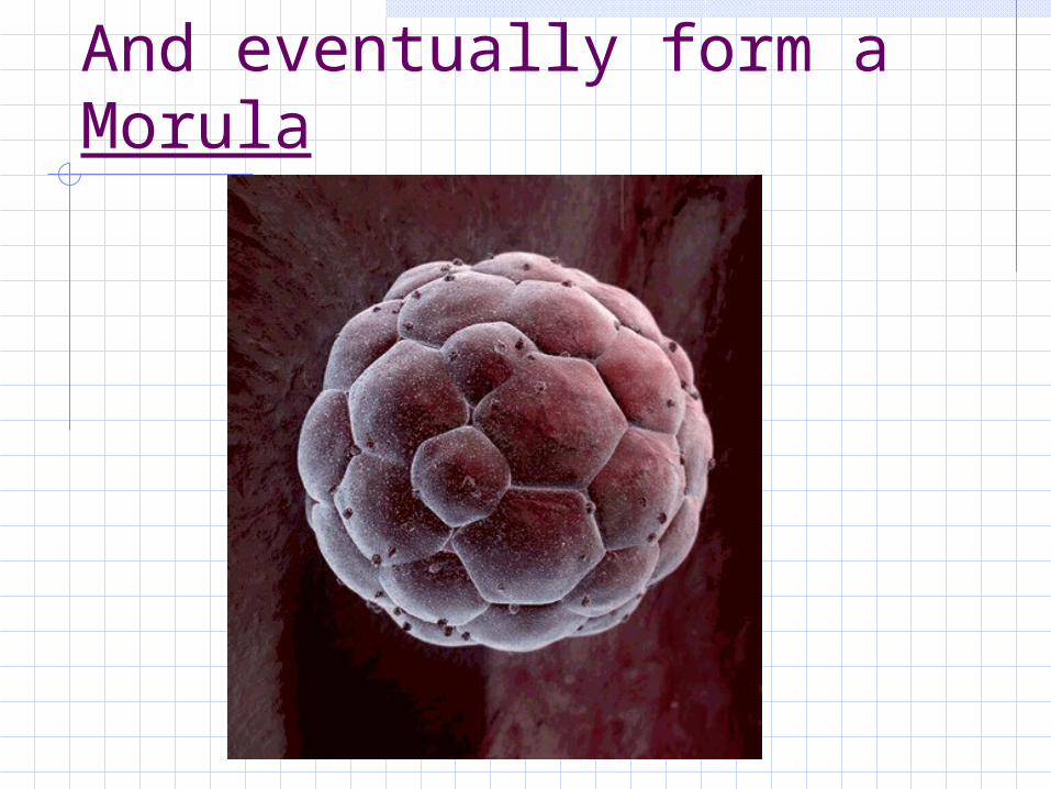
And eventually form a Morula

And next, a gastrula

The Regents Diagram…
Sperm and ovum1. Zygote (fertilized ovum)2. 2-cell stage3. 4-cell stage4. Morula5. Blastula6. Gastrula

Differentiation (Organogenesis)
Organogenesis is the formation of the organs (Organo = organs, genesis = creation)Arises from the layering of cells that occurs during gastrula stageThe layers are germ layers; they have specific fates in the developing embryo: Endoderm
The innermost layer Goes on to form the gut
Mesoderm In the middle Goes on to form the muscles, circulatory system,
blood and many different organs Ectoderm
The outermost Goes on to form the skin and nervous system

Late Gastrula
Mesoderm
Endoderm
Ectoderm

Differentiation of Primary Germ Layers (from the gastrula)
Ectoderm
Mesoderm
Endoderm
Nervous system
Skeleton Digestive tract
Epidermis of skin
Muscles Respiratory system
Circulatory system
Liver, pancreas
Gonads Bladder

Early Human Development Summary
Meiosis makes sperm in males and ovum in femalesSperm and ovum unite nuclei to form a zygoteZygote undergoes cleavage and becomes gastrula with 3 germ layers

11 Organ systems different organs work together to provide specialized functions
Body Coverings
Support & Movement
Integration & coordination
Transport
Absorption / Excretion
Reproduction