optimizing peptide nucleic acid probes for hybridization-based … · 2019-03-03 · antibiotic...
TRANSCRIPT

Analyst
PAPER
Cite this: DOI: 10.1039/c8an02194e
Received 14th November 2018,Accepted 9th January 2019
DOI: 10.1039/c8an02194e
rsc.li/analyst
Optimizing peptide nucleic acid probes forhybridization-based detection and identificationof bacterial pathogens†
Kathleen E. Mach, a,b Aniruddha M. Kaushik,c Kuangwen Hsieh,c Pak Kin Wong,d
Tza-Huei Wangc and Joseph C. Liao*a,b
Point-of-care (POC) diagnostics for infectious diseases have the potential to improve patient care and
antibiotic stewardship. Nucleic acid hybridization is at the core of many amplification-free molecular diag-
nostics and detection probe configuration is key to diagnostic performance. Modified nucleic acids such
as peptide nucleic acid (PNA) offer advantages compared to conventional DNA probes allowing for faster
hybridization, better stability and minimal sample preparation for direct detection of pathogens. Probes
with tethered fluorophore and quencher allow for solution-based assays and eliminate the need for
washing steps thereby facilitating integration into microfluidic devices. Here, we compared the sensitivity
and specificity of double stranded PNA probes (dsPNA) and PNA molecular beacons targeting E. coli and
P. aeruginosa for direct detection of bacterial pathogens. In bulk fluid assays, the dsPNAs had an overall
higher fluorescent signal and better sensitivity and specificity than the PNA beacons for pathogen detec-
tion. We further designed and tested an expanded panel of dsPNA probes for detection of a wide variety
of pathogenic bacteria including probes for universal detection of eubacteria, Enterobacteriaceae family,
and P. mirablis. To confirm that the advantage translated to other assay types we compared the PNA
beacon and dsPNA in a prototype droplet microfluidic device. Beyond the bulk fluid assay and droplet
devices, use of dsPNA probes may be advantageous in a wide variety of assays that employ homogenous
nucleic acid hybridization.
Introduction
Accurate, timely diagnosis is key to efficacious treatment ofinfectious diseases. Particularly for acute bacterial infections,point of care (POC) devices capable of distinguishing whethera patient has an infection, determining the causative patho-genic species, and directing the appropriate treatmentcan improve patient outcomes and promote antibioticstewardship.1–5 Because the current diagnostic standard ofculture and antimicrobial susceptibility testing (AST) requires2–4 days to provide this objective information, there are strong
interests to develop devices to expedite pathogen detectionand characterization.6
Nucleic acid detection is an integral part of many newapproaches to rapid pathogen diagnosis.7–10 PCR and relatedtarget amplification techniques, while highly sensitive, mayrequire complex sample preparation steps to remove amplifica-tion inhibitors and require several hours from sample toanswer. PCR may be a good option for pathogen detection intypically sterile environments such as cerebral spinal fluid andblood where the presence of any bacteria can indicate seriousinfection, however other samples types such as sputum orurine can be prone to false positive results due to contami-nation during sample collection with normal flora.11
Alternatively, amplification-free detection is commonlyachieved through direct hybridization of linear DNA oligo-nucleotide probes labeled with enzymes or fluorescenttags.12,13 The use of oligonucleotide probes with a single labeltypically requires that the target sequence be immobilized on asolid support for hybridization so excess unbound probe canbe washed away.
Dual label probes such as molecular beacons are a goodalternative for homogenous sensors. Molecular beacons are
†Electronic supplementary information (ESI) available. See DOI: 10.1039/c8an02194e
aDepartment of Urology, Stanford University School of Medicine, Stanford, CA, USA.
E-mail: [email protected]; Tel: +1 650-852-3284bVeterans Affairs Palo Alto Health Care System, Palo Alto, CA, USAcDepartments of Mechanical Engineering and Biomedical Engineering,
Johns Hopkins University, Baltimore, MD, USAdDepartment of Biomedical Engineering, The Pennsylvania State University,
University Park, PA, USA
This journal is © The Royal Society of Chemistry 2019 Analyst
Publ
ishe
d on
10
Janu
ary
2019
. Dow
nloa
ded
by S
tanf
ord
Uni
vers
ity o
n 1/
28/2
019
9:12
:29
PM.
View Article OnlineView Journal
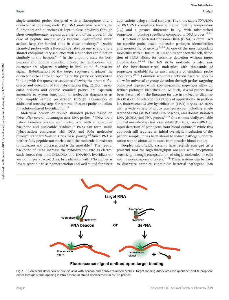
single-stranded probes designed with a fluorophore and aquencher at opposing ends. For DNA molecular beacons thefluorophore and quencher are kept in close proximity throughshort complimentary regions at either end of the probe. In thecase of peptide nucleic acids beacons, hydrophobic inter-actions keep the labeled ends in close proximity.14 Doublestranded probes with a fluorophore label on one strand and ashorter complimentary sequence with a quencher can functionsimilarly to the beacon.15,16 In the unbound state for bothbeacons and double stranded probes, the fluorophore andquencher are adjacent resulting in little to no fluorescentsignal. Hybridization of the target sequence displaces thequencher either through opening of the probe or competitivebinding with the quencher sequence allowing the probe to flu-oresce and detection of the hybridization (Fig. 1). Both mole-cular beacons and double stranded probes are especiallyamenable to system integration in molecular diagnostics asthey simplify sample preparation through elimination ofadditional washing steps for removal of excess probe and allowfor solution-based hybridization.17
Molecular beacon or double stranded probes based onPNAs offer several advantages over DNA probes.18 PNAs are ahybrid between protein and nucleic acid with a polyaminebackbone and nucleotide residues.19 PNAs can form stablehybridization complexes with DNA and RNA moleculesthrough standard Watson–Crick base pairing.20 Since PNA isneither fully peptide nor nucleic acid the molecule is resistantto nucleases and proteases and is thermostable.21 The neutralbackbone of PNAs increase the hybridization rate as electro-static forces that limit DNA/DNA and DNA/RNA hybridizationare no longer a factor. Also, hybridization with PNA probes isless susceptible to salt concentration and well suited for direct
applications using clinical samples. The more stable PNA/DNAor PNA/RNA complexes have a higher melting temperature(Tm) and a greater difference in Tm with mismatchedsequences improving specificity compared to DNA probes.22,23
Detection of bacterial ribosomal RNA (rRNA) is often usedfor specific probe based molecular pathogen identificationand monitoring of growth.24,25 As one of the most abundantmolecules with 15 000 to 70 000 copies per bacterial cell, detec-tion of rRNA allows for sensitive detection without targetamplification.26–28 The 16S rRNA molecule is also oneof the best-characterized molecules with thousands ofsequences available for in silico analysis of candidate probespecificity.29–31 Common sequences between bacterial speciesallow for universal or group detection through probes targetingconserved regions, while species-specific sequences allow forrefined pathogen identification. As such, several probes havebeen described in the literature for use in molecular diagnos-tics that can be adapted to a variety of applications. In particu-lar, fluorescence in situ hybridization (FISH) targets 16S rRNAwith a wide variety of probe configurations including singlestranded DNA (ssDNA) and PNA beacons, and double strandedDNA (dsDNA) and PNA probes.32,33 One commercially availableclinical microbiology test, QuickFISH (OpGen), uses dsPNA forrapid detection of pathogens from blood culture.32 While thisapproach still requires an initial overnight incubation of thepatient sample, it has been shown to reduce pathogen identifi-cation step to about 30 minutes from positive blood culture.
Droplet microfluidic systems have recently emerged as apowerful tool for high-throughput analysis with exceptionalsensitivity through encapsulation of single molecules or cellswithin monodisperse droplets.34–38 These systems can be usedto discretize samples containing bacterial pathogens into
Fig. 1 Fluorescent detection of nucleic acid with beacon and double stranded probes. Target binding dissociates the quencher and fluorophoreeither through strand opening in PNA beacon or strand displacement in dsPNA probes.
Paper Analyst
Analyst This journal is © The Royal Society of Chemistry 2019
Publ
ishe
d on
10
Janu
ary
2019
. Dow
nloa
ded
by S
tanf
ord
Uni
vers
ity o
n 1/
28/2
019
9:12
:29
PM.
View Article Online

thousands to millions of picoliter (pL) droplets, each of whichserves as an isolated reaction chamber containing 1 or 0 bac-terial cells.39–41 Confinement of single cells in pL dropletsresults in a high effective concentration of nucleic acids thatcan increase sensitivity of detection and obviate the need forPCR amplification.41 As such, the single-cell sensitivityafforded by droplet platforms can facilitate drasticallyimproved limits of detection compared to bulk detection.Further, the digital readout from droplet microfluidic systemscan allow for more accurate quantitation of bacteria.
Since a key determinant of sensitive and specific pathogendetection in nucleic hybridization assays is the labeled probe,we directly compared the sensitivity and specificity of PNAbeacons and dsPNA probes for pathogen identification. Herewe report that dsPNAs have better sensitivity and specificitythan PNA beacons targeting the same sequence in our assays.We initially compared the probe configuration in a bulk(100 µl) fluid assay then demonstrated that the advantage ofthe double stranded probe held true when the probes whereused for pathogen detection in a droplet microfluidic system.Our findings may be generalizable for other homogeneoushybridization detection strategies and the probes adaptable toa wide variety of devices.
MethodsProbes and bacterial culture
The pathogen detection probes designed to target all bacteria,Enterobacteriaceae family, E. coli, P. aeruginosa and P. mirabilis16S rRNA and the universal probe were based on previousreports.42–45 Probe sequences are reported in Table 1. All PNAsequences were synthesized by PNA Bio (Newbury Park, CA)and DNA quencher sequences were synthesized by IntegratedDNA Technologies (San Diego, CA).
Uropathogenic E. coli (UPEC) strain (CFT073) was pur-chased from ATCC (Manassa, VA.) All other strains are uro-pathogenic clinical isolates from the Palo Alto VA clinicalmicrobiology laboratory. For bulk pathogen detection experi-ments, bacteria were grown in cation adjusted Muller-Hinton(MH) media at 37 °C in a shaking incubator (Excella E24, New
Brunswick Scientific) to mid-log phase and diluted as appro-priate. For droplet assays, E. coli was cultured and stored at−80 °C until assayed.
Bulk fluid pathogen detection
For pathogen detection, probes were diluted in 2× hybridiz-ation buffer (50 mM Tris pH8, 100 mM NaCl, 1% PVP, 10 mMEGTA) to desired concentration and 25 µl of probe was mixedwith an equal volume of media or culture for a final concen-tration of 25 mM Tris pH8, 50 mM NaCl, 0.5% PVP, 5 mMEGTA, 50% MH media or culture. Final probe and bacteriaconcentrations are indicated in results. For dsPNA probes theDNA quencher sequence was in three-fold molar excess of thelabeled PNA. The probe/bacteria mix was heated to 95 °C for2 min to thermolyse the bacteria followed by incubation at60 °C for 30 min to allow for probe hybridization. To controlincubation time and temperature, 95 °C and 60 °C incubationswere carried out in at thermocycler (Eppendorf Mastercycler).Immediately after hybridization fluorescence was measured ina fluorescence microplate reader (Molecular DynamicsSpectraMax) at excitation wavelength 492 nm and emissionwavelength 518 nm.
Microfluidic device design and fabrication
The microfluidic device design consists of a 10 μm flow-focus-ing nozzle for droplet generation and a 10 μm channel con-striction for droplet detection. The droplet generation anddroplet detection channels of the device are connected by a∼30 cm Tygon tubing (Cole-Parmer) with an inner diameter ofapproximately 500 μm. In order to fabricate the microfluidicdevice, a casting mold was created by spinning a 20 µm layerof SU8-3050 photoresist (MicroChem) onto a 4 inch siliconwafer and patterned using standard photolithography. Themicrofluidic devices were made of polydimethylsiloxane(PDMS) by pouring 50 g of 10 : 1 ratio of Sylgard 184 (DowCorning) base to curing agent onto the SU8 mold. The PDMSdevice was then permanently bonded to cover glass (130 µmthickness, Ted Pella) through oxygen plasma treatment inorder to seal the channels. Prior to device operation, themicrofluidic chips were treated with Aquapel and baked at
Table 1 dsPNA probe sequences
Sequence Length Target
UNI339 GCTGCCTCCCGTAGGA-K-FAM 16 EubacteriaGGAGGGCATCCT-IABk 12
EC467 FAM-O-TCAATGAGCAAAGGT-KK 15 E. coliIABk-AGTTACTCGT 10
PA584 FAM-O-AACTTGCTGAACCAC 15 P. aeruginosaIABk-TTGAACGACT 10
EB1272 FAM-O-TATGAGGTCCGCTTG 15 EnterobacteriaceaeIABk-ATACTCCAGG 10
PM1014 FAM-O-TCCTCTATCTCTAAAGG 17 P. mirabilisIABk-AGGAGATAGAG 11
For each probe sequence, top line is the fluorophore labeled PNA and bottom is DNA quencher. E = glutamate, K = lysine, O = linker sequence,FAM = fluorescein, IABk = Iowa black quencher.
Analyst Paper
This journal is © The Royal Society of Chemistry 2019 Analyst
Publ
ishe
d on
10
Janu
ary
2019
. Dow
nloa
ded
by S
tanf
ord
Uni
vers
ity o
n 1/
28/2
019
9:12
:29
PM.
View Article Online

80 °C for at least 20 minutes to render microfluidic channelsurfaces hydrophobic.
Microfluidic device operation and droplet fluorescence detection
Prior to droplet generation, stock E. coli (ATCC 25922) wasthawed, washed twice, and diluted to 4 × 10⁷ cfu mL−1 in ineither hybridization buffer or MH media. Separately, E. coliprobes at 200 nM PNA beacon or 200 nM dsPNA and 600 nMquencher were diluted in hybridization buffer. Bacterial solu-tion and probes solution were then introduced into separatesections of Tygon tubing. Both sections of Tygon tubing wereindividually connected to Hamilton 1000 glass syringes(Sigma-Aldrich) containing FC-40 oil (Sigma-Aldrich). FC-40 inthe syringe was used to push the aqueous samples from Tygontubing into the device using a syringe pump (HarvardApparatus). BioRad QX200 Droplet Generation Oil was intro-duced into the oil inlet of the device by a separate syringepump. A flow rate of 20 µL h−1 was used for both aqueousphases, while 60 µL h−1 was used for the oil phase. In order toconfirm stable and uniform droplet generation, the device wasimaged using a 4× objective lens and a CCD camera duringdroplet generation and after droplet incubation. Lysis andhybridization were conducted as the droplets flowed throughthe Tygon tube connecting the droplet generation and dropletdetection regions of the microfluidic device. Importantly, sep-arate lengths of the Tygon tube were clamped onto a 95 °CPeltier heater and a 60 °C Peltier heater, such that all dropletswere subject to 2 min at 95 °C followed by 30 min at 60 °C asin the bulk fluid assay. Continuous-flow droplet detection wasconducted using a custom designed optical stage consisting ofa 488 nm laser excitation source (OBIS, Coherent, Inc.) and asilicon avalanche photodiode detector (APD) (SPCM-AQRH13,ThorLabs). The laser was operated at 4 mW power and wasfocused into the detection zone of the device using a 40×objective (Thorlabs RMS40X-PF, NA 0.75, focal depth approxi-mately 0.6 μm). As droplets flowed through the custom laser-induced fluorescence detection zone, fluorescence data wascontinuously acquired and recorded using the APD with0.1 ms sampling time. A custom-built LabVIEW program wasused for fluorescence data acquisition.
Laser induced fluorescence data analysis
A custom MATLAB program was developed for analyzing theraw fluorescence intensity data acquired from the APD. Fromthe fluorescence intensity time trace of each experimental run,the program looks for individual droplets by quantifying peakwidths and peak heights. Once droplet position and fluo-rescence intensity are identified for all droplets in a sample,we plot the intensities as a histogram with 150 bins. Resultinghistograms typically follow a bimodal distribution, and inorder to classify the respective subpopulations, we fit the firstdroplet histogram peak (the empty droplet or “background”subpopulation) with a Gaussian curve. Based on the fittedcurve, we calculate the mean intensity of the empty dropletpopulation as well as the standard deviation. All dropletswhose intensities are 3 standard deviations above the empty
droplet mean intensity are classified as bacteria-containing“positive droplets.” Signal to background ratios are calculatedby dividing the mean intensity of positive droplets by themean intensity of background droplets.
Results
The PNA probe sequences targeting E. coli45 and P. aeruginosa43
used in this study were in previously described FISH assays.However, in the prior assays the probes were labeled with asingle fluorophore and unbound probe removed by washing. Toadapt these probe sequences to solution-based hybridizationassays we compared dual labeled PNA beacons and dsPNAprobes (Fig. 1). For initial comparison of dsPNA and PNAbeacons we used a simple bulk fluid assay of 100 µl volume.Probe solution and bacterial culture were directly mixed and thebacteria thermolysed and incubated to allow probe hybridiz-ation. Immediately after incubation fluorescence was measuredto assess probe binding. This straightforward detection protocoldoes not require nucleic acid purification, washing steps, oramplification and was designed to account for constraintsimposed by integrated diagnostic devices for POC detection.
Comparison of PNA beacon and dsPNA probe specificity andsensitivity
To confirm probe binding and determine the appropriateprobe concentration for pathogen detection, beacon or dsPNAprobes ranging from 25 to 200 nM were tested for fluorescencedetection of E. coli and P. aeruginosa. The highest overall fluo-rescence signal was achieved with the highest concentration ofprobe with both beacon and dsPNA probes targeting eitherE. coli (Fig. 2A) or P. aeruginosa (data not shown). However, thebackground fluorescence without bacteria remained relativelyconstant as the beacon concentration was increased resultingin a modestly higher fluorescence signal (Fig. 2A) and signal-to-background ratio at the highest probe concentration tested(Fig. 2B). Whereas with the dsPNA the background fluo-rescence increased with probe concentration, subsequently thehighest signal over background was achieved at the lowestprobe concentration. This suggests that a lower probe concen-tration could be used with the dsPNA to achieve more sensitivedetection.
In many assays, probe specificity is controlled by optimizinghybridization buffer conditions such as formamide or salt con-centration and by the stringency of wash conditions. However,for integration in devices with solution-based hybridization,we aimed to identify probes that were species specific in uni-versal buffer conditions. The probe sequences used in thiswere found to be species specific when used as single labelPNA probes in FISH assays that included stringent washingsteps. Further, sequence analysis indicates these probes bindto unique sequences in 16S rRNA. Thus, we examined whetherthe probes retained species-specificity in the beacon anddsPNA forms in a solution-based assay without washing steps.We compared the ability of probes designed to E. coli and
Paper Analyst
Analyst This journal is © The Royal Society of Chemistry 2019
Publ
ishe
d on
10
Janu
ary
2019
. Dow
nloa
ded
by S
tanf
ord
Uni
vers
ity o
n 1/
28/2
019
9:12
:29
PM.
View Article Online

P. aeruginosa in PNA beacon and dsPNA configurations to dis-criminate between uropathogenic E. coli (UPEC), P. aeruginosa(Pa127), P. mirabilis (Pm159), and K. pneumoniae (Kp128)strains. The E. coli targeting probe preferentially bound toE. coli in both conformations however the signal differentialbetween target and other species was greatest for the dsPNA atboth 25 and 200 nM probe concentrations (Fig. 3). On theother hand, the dsPNA designed to target P. aeruginosa showedappropriate specificity, but the beacon targeting the samesequence appeared non-specific in our assay with false positivesignal from K. pneumoniae.
To expand our probe library beyond targeting E. coli andP. aeruginosa, we designed additional dsPNA probes for detec-tion and classification a broader panel of pathogens. Weadapted 16S rRNA probe sequences for dsPNA detection foruniversal eubacterial detection (UNI339), classification ofEnterobacteriaceae (EB1272), and Proteus mirabilis (PM) basedon previous reports.42,44 We focused on development of dsPNAprobes because they had better specificity than the beacons.To confirm specificity of all of the dsPNA probes we testedthem against a broad panel of Gram negative human patho-gens. Using the simple heat lysis and direct detection protocol,all of the probes exhibited the expected specificity (Fig. 4,primary data in ESI Fig. 1†). The universal probe was positive
with all bacteria tested, the EB probe detected all bacteriaexcept P. aeruginosa, the only non-Enterobacteriaceae species inthe testing panel, and the EC, PA and PM probes were onlypositive for their appropriate target species.
Fig. 4 Specificity of a dsPNA probes with expanded panel of patho-gens. Green squares indicate positive signals with probes targeting allbacteria (UNI339), Enterobacteriaceae (EB1272), E. coli (EC467),P. aeruginosa (PA584), or P. mirabilis (PM1014) from bacterial cultures ofE. coli (UPEC) P. mirabilis (Pm159), Klebsiella pneumoniae (Kp180),Pseudomonas aeruginosa (Pa127), Klebsiella oxytoca (Ko345),Enterobacter aerogenes (Ea222), Enterobacter cloacae (El185),Providencia stuartii (Ps389), Serratia marcesens (Sm235) or Morganellamorganii (Mm278).
Fig. 3 Comparison of PNA beacon and dsPNA probe specificity.200 nM or 25 nM PNA beacon or dsPNA targeting E. coli (EC467, A) orP. aeruginosa (PA584, B) was assayed for detection of the E. coli (UPEC),P. aeruginosa (Pa127), K. pneumoniae (Kp128), and P. mirabilis (Pm159).
Fig. 2 Effect of probe concentration on uropathogenic E. coli (UPEC)detection. Fluorescent signal (A) and signal to background ratio (B) fordetection of 2 × 10⁸ cfu ml−1 UPEC with varying concentrations ofEC467 PNA beacon or dsPNA with 3-fold excess quencher probe.
Analyst Paper
This journal is © The Royal Society of Chemistry 2019 Analyst
Publ
ishe
d on
10
Janu
ary
2019
. Dow
nloa
ded
by S
tanf
ord
Uni
vers
ity o
n 1/
28/2
019
9:12
:29
PM.
View Article Online

Sensitivity of detection in a relevant concentration range tothe target is essential for pathogen detection. As with thespecificity assay, we compared sensitivity of the PNA beaconand dsPNA a 100 µl bulk assay. Both the E. coli andP. aeruginosa targeting dsPNA probes had a higher signal-to-background ratio at the 25 nM concentration than the corres-ponding PNA beacon (Fig. 5). The sensitivity of detection inthe bulk assay was about 107 cfu ml−1, which is a much higherconcentration than is found in most clinical samples (e.g.infected urine samples).
Detection of bacteria with single-cell sensitivity
To improve sensitivity of detection and assess our PNA probesin a clinically translatable system, we employed a dropletmicrofluidic platform for high-throughput single-cell analysisof pathogens (Fig. 6A). We designed a PDMS-based microflui-dic droplet device that can passively co-encapsulate single bac-terial cells and PNA probes into 7 pL volume droplets (Fig. 6B).A single bacterium confined in a 7 pL droplet drasticallyelevates the local bacterial concentration (equivalent to 1.4 ×10⁸ cells per mL in bulk analysis) and hence the local target16S rRNA concentration. Likewise, the significant reduction involume facilitates an equivalent reduction in assay back-ground, yielding an overall increase in signal to backgroundratio compared to bulk measurements. After single-bacteriaencapsulation during droplet generation, droplets are trans-
ported through 95 °C and 60 °C regions to simulate the appro-priate lysis and hybridization conditions as the bulk assay.Finally, the droplets pass through a flow constriction whereeach droplet can be measured sequentially using a customlaser-induced fluorescence (LIF) system. As droplets individu-ally traverse the detection constriction of our microfluidicdevice, we register a corresponding peak in the fluorescencetime trace. Droplets containing probes hybridized to theircorresponding target produce higher fluorescence signal thanempty droplets or droplets without target.
We used our droplet platform to test the performance ofPNA beacon and dsPNA probes in detecting single pathogeniccells. We co-flowed PNA beacon or dsPNA probes as well asE. coli (ATCC 25922) suspended in Mueller-Hinton broththrough our device and generated droplets containing E. colicells and PNA probes. Following passage through the lysis andhybridization zones, the droplets were sequentially detectedusing the LIF system. Fig. 6Ci shows the time trace of fluo-rescence of droplets with E. coli and PNA beacon. Two popu-lations, droplets containing the hybridized probe–targetcomplex (green in Fig. 6Ci) and empty droplets containing notarget but only the self-quenching probes (orange in Fig. 6Ci),were observed. Notably, the reduction in fluorescence back-ground facilitated by droplets allows us to detect differentiablesignal from droplets containing individual bacterial cells. Thiscapability of our platform to detect single-bacterial cells cantherefore allow for improved limit of detection for bacterialenumeration. From the resulting droplet fluorescence data foreach condition, a histogram of droplet fluorescence intensitywas plotted (Fig. 6Ci and 6Cii). Gaussian curve fitting wasused to define a threshold 3 standard deviations above themean of the empty droplet population (40). After analyzing∼1 000 000 droplets, the average ratio of signal from bacteria-containing droplets to signal from empty droplets (“S/B”) wasdetermined to be ∼2.2. Similarly, when detecting bacteriausing dsPNA probes, not only was the average signal from bac-teria-containing droplets much higher, but the average S/B forbacterial detection was ∼3.4, outperforming the PNA beacon.
Stronger fluorescence signals and higher S/B result in moreaccurate bacterial quantitation when using dsPNA probes. Inour droplet experiments, we aimed to encapsulate single bac-terial cells in ∼10% of droplets (mean occupancy λ = 0.1).When detecting single-bacteria using the PNA beacon, after30 min of hybridization, the rate of observed positive (observedλ ≈ 0.01) droplets is much less than the rate of expected posi-tive droplets (expected λ = 0.1). This implies that 30 min wasinsufficient for all the positive droplets to develop signal. Incontrast, for dsPNA, the rate of observed positive droplets(observed λ ≈ 0.08) more closely matches the rate of expectedbacteria containing droplets. We note that the slight disagree-ment in mean occupancy here may have resulted from an inac-curate estimation of the initial bacterial concentration (deter-mined via plating) or from a misestimation of droplet volume.However, it is clear that due to the high S/B achieved withdsPNA probes, better quantitation of single-bacteria is possibleafter as little as 30 min of hybridization. While these experi-
Fig. 5 Comparison of PNA beacon and dsPNA probe sensitivity. 25 and200 nM PNA EC467 beacon or EC467 dsPNA targeting E. coli was usedto detect varying concentrations of E. coli (A) or P. aeruginosa (B).
Paper Analyst
Analyst This journal is © The Royal Society of Chemistry 2019
Publ
ishe
d on
10
Janu
ary
2019
. Dow
nloa
ded
by S
tanf
ord
Uni
vers
ity o
n 1/
28/2
019
9:12
:29
PM.
View Article Online

ments demonstrate the superiority of the dsPNA probe indetecting single E. coli cells in a fairly complex media(Mueller-Hinton broth), we also observed superior S/B andaccurate quantitation using dsPNA probes for bacteria sus-pended in a simpler salt solution (hybridization buffer, datanot shown).
Discussion
Highly sensitive and specific probe–target hybridization lies atthe core of molecular diagnostics for infectious disease appli-cations where there are considerable unmet needs. Rationaltarget selection and probe design enable direct hybridizationand obviate sample preparation steps including washing andtarget amplification, thereby facilitating translation towardsPOC applications. PNA-based probes offer numerous advan-tages compared to DNA and are well suited for direct appli-cations using clinical samples. In this study, we compareddifferent PNA probe configurations for direct detection of bac-terial pathogens involved in common infections includingurinary tract, blood stream, and surgical site. While both PNA
beacon and dsPNA probes are amenable to solution-basedhybridization, we found that dsPNA probes outperformed thePNA beacon of the same sequence in regard to analytical sensi-tivity and specificity. The performance characteristics of thedsPNA was first demonstrated in bulk fluid assays and thenconfirmed in a pL-droplet microfluidic platform, suggestingthat our results are generalizable to many different systems.
While PNA probes are more costly and more challenging tosynthesize compared to DNA probes, they offer the advantagesof improved stability and hybridizations kinetics over DNAcounterparts. Additionally, previous reports comparing DNAand PNA beacons for detection of E. coli 16S rRNA found thePNA beacon to be significantly more sensitive than the DNAbeacon. PNA beacon at 50 nM bound purified RNA in therange of 0.4 to 25 nM, whereas the DNA beacon could onlydetect the RNA at the highest concentration.46 With approxi-mately 20 000 copies of rRNA per cells, 0.4 nM translates toroughly 107 cfu ml−1, consistent with E. coli detection with thePNA beacon in our current study. We found a further increasein sensitivity from the beacon to the dsPNAs. dsPNA probeshave also been used in some FISH based assays such asQuickFish (OpGen).47–49 Their success with FISH using dsPNA
Fig. 6 Single-bacteria detection sensitivity with PNA probes using a droplet microfluidic device. (A) Single bacteria are encapsulated with eitherPNA beacon or dsPNA probes into 7 pL droplets. Droplets flow through a lysis region for 2 min, followed by a hybridization region for 30 min, andfinally a fluorescence detection region where each droplet is individually interrogated. (B) Our PDMS-based microfluidic device consists of a dropletgeneration region, after which droplets are transported through 95 °C and 60 °C heaters for lysis and hybridization before re-entering the device fordetection. (C) Fluorescence time trace of E. coli detected using (i) PNA beacon show that droplets containing target rRNA have higher fluorescencethan empty droplets containing quenched probes. Here, we observe an average S/B of 2.2 and underquantification after histogram analysis, whereaswhen using (ii) dsPNA probes, we observe a higher S/B of 3.4 and more accurate quantification.
Analyst Paper
This journal is © The Royal Society of Chemistry 2019 Analyst
Publ
ishe
d on
10
Janu
ary
2019
. Dow
nloa
ded
by S
tanf
ord
Uni
vers
ity o
n 1/
28/2
019
9:12
:29
PM.
View Article Online

probes is similar to droplets in that the small encapsulation ofthe cell increases the effective concentration of rRNA. In a10 pL droplet the rRNA concentration from a single bacterialcell is about 5 nM and for FISH assays the single cell rRNAconcentration is even higher. The sensitivity of both FISH anddroplet assays benefits from the high effective local concen-tration, however the current FISH assays require more hands-on steps than the droplet assay. For FISH, the bacteria arefixed and permeabilized on a microscope slide, then incubatedwith probe and examined microscopically, whereas in thedroplet platform all assay steps and detection are automated.
While rRNA is a good target for species identification dueto high cellular concentration and well-characterizedsequences, conserved sequences between species necessitatehigh probe specificity to distinguish pathogens. Probes forsolution-based diagnostics directly using biological sampleideally would retain specificity without the need to adjust saltor wash conditions. PNA probes are less sensitive to salt con-centrations due to the neutral backbone and both beacon anddsPNA probes should eliminate the need for washing steps.18
However, we found poor specificity in the beacon configur-ation compare to the dsPNA. This may be due in part tobeacon hybridization to small stretches of homology resultingin partial opening of the probe (Table 1). Whereas in thedsPNA an excess of the quencher strand allows for competitivebinding where only the full sequence can dislodge thequencher.
Quantitative detection of rRNA can be used in antibioticsusceptibility testing (AST) assays.25 As such the dsPNA probesdeveloped here have the potential to be used in a combinedpathogen identification and AST assay by comparing fluo-rescence intensity with and without antibiotic. Further, theprobe-based pathogen identification and AST could beadapted for use in a droplet microfluidic system. Other reportsof AST in droplet systems have used resazurin as a nonspecificreporter of cell growth,40,50 replacing resazurin with rRNAspecific probes would allow for species identification and mayimprove analysis of polymicrobial samples.
The advantages of the dsPNA probes for bulk fluid anddroplet microfluidic assays may translate well to other in vitrodetection systems, however for some applications beacons maybe more suitable. Beacons have been successfully used fordynamic intracellular nucleic acid localization in live cells.51–54
Initial live cell imaging studies with DNA beacons found thatthe presence of nucleases in some cells resulted in false posi-tive signals, but the use of PNAs and other nuclease resistantDNA modifications solved this problem.51,55,56 In our currentdsPNA design the quencher sequence is a DNA oligonucleotideand thus may require further modification for use in live cells.However, most nucleases require divalent cations as cofactorsfor activity and are inhibited by chelators such as EGTA. Forin vitro diagnostics the addition of EGTA, as we have includedin our assays, should inhibit nucleases and favor use of thedouble stranded probes.
While the hybridization characteristics of the dsPNA probeslikely will remain favorable for use in a wide variety assays,
this study has several limitations. Here we tested detection of asampling of Gram negative bacterial species, yet for use in adiagnostic platform the probe panel would have to beexpanded for detection of more diverse species. Additionally,the simple heat lysis used here for the Gram negative bacteriamay not be robust for Gram positive species or yeast due to thethicker cell walls of these species, therefore the cell lysis wouldlikely need to be modified to allow automated detection. Acritical measure for development of these probes will be directtesting of clinical samples. The background fluorescence frombiological samples may interfere with probe detection and setrequirement for higher overall signal. Thus, increasing theprobe concentration for different devices could allow for abalance between the need for higher signals to overcome thebackground of a biological sample and improved signal tonoise in low background samples. Alternatively, using fluoro-phores with less potential for background from biologicalsamples such as near infrared dyes may allow for robust detec-tion directly from biological samples.
Conclusion
We have demonstrated that dsPNA probes have better speci-ficity and sensitivity than the equivalent PNA beacon in ourhomogenous assays. Through testing in bulk and dropletmicrofluidic assays we conclude that the dsPNA is a superiorchoice for incorporation into a microfluidic device.
Conflicts of interest
There are no conflicts to declare.
Acknowledgements
This work was supported by the National Institutes of HealthR01 1AI117032 to THW, PKK and JCL.
References
1 A. Galar, J. Leiva, M. Espinosa, F. Guillen-Grima,S. Hernaez and J. R. Yuste, J. Infect., 2012, 65, 302–309.
2 A. F. Veesenmeyer, J. A. Olson, A. L. Hersh, C. Stockmann,K. Korgenski, E. A. Thorell, A. T. Pavia and A. J. Blaschke,Infect. Dis. Ther., 2016, 5, 555–570.
3 K. K. Perez, R. J. Olsen, W. L. Musick, P. L. Cernoch,J. R. Davis, L. E. Peterson and J. M. Musser, J. Infect., 2014,69, 216–225.
4 F. P. Maurer, M. Christner, M. Hentschke and H. Rohde,Infect. Dis. Rep., 2017, 9, 6839.
5 E. L. Tsalik, E. Petzold, B. N. Kreiswirth, R. A. Bonomo,R. Banerjee, E. Lautenbach, S. R. Evans, K. E. Hanson,J. D. Klausner, R. Patel and the Diagnostics Devices
Paper Analyst
Analyst This journal is © The Royal Society of Chemistry 2019
Publ
ishe
d on
10
Janu
ary
2019
. Dow
nloa
ded
by S
tanf
ord
Uni
vers
ity o
n 1/
28/2
019
9:12
:29
PM.
View Article Online

Committee of the Antibacterial Resistance LeadershipGroup, Clin. Infect. Dis., 2017, 64, S41–S47.
6 A. M. Caliendo, D. N. Gilbert, C. C. Ginocchio,K. E. Hanson, L. May, T. C. Quinn, F. C. Tenover, D. Alland,A. J. Blaschke, R. A. Bonomo, K. C. Carroll, M. J. Ferraro,L. R. Hirschhorn, W. P. Joseph, T. Karchmer,A. T. MacIntyre, L. B. Reller, A. F. Jackson and A. InfectiousDiseases Society of, Clin. Infect. Dis., 2013, 57(Suppl. 3),S139–S170.
7 D. R. Call, Crit. Rev. Microbiol., 2005, 31, 91–99.8 P. L. Quan, T. Briese, G. Palacios and W. Ian Lipkin,
Antiviral Res., 2008, 79, 1–5.9 M. Bercovici, G. V. Kaigala, K. E. Mach, C. M. Han,
J. C. Liao and J. G. Santiago, Anal. Chem., 2011, 83, 4110–4117.
10 A. C. Yu, G. Vatcher, X. Yue, Y. Dong, M. H. Li, P. H. Tam,P. Y. Tsang, A. K. Wong, M. H. Hui, B. Yang, H. Tang andL. T. Lau, Front. Med., 2012, 6, 173–186.
11 S. Yang and R. E. Rothman, Lancet Infect. Dis., 2004, 4,337–348.
12 K. E. Mach, P. K. Wong and J. C. Liao, Trends Pharmacol.Sci., 2011, 32, 330–336.
13 S. M. Yoo and S. Y. Lee, Trends Biotechnol., 2016, 34, 7–25.
14 S. Tyagi and F. R. Kramer, Nat. Biotechnol., 1996, 14, 303–308.
15 Q. Li, G. Luan, Q. Guo and J. Liang, Nucleic Acids Res.,2002, 30, E5.
16 D. Meserve, Z. Wang, D. D. Zhang and P. K. Wong, Analyst,2008, 133, 1013–1019.
17 J. Zheng, R. Yang, M. Shi, C. Wu, X. Fang, Y. Li, J. Li andW. Tan, Chem. Soc. Rev., 2015, 44, 3036–3055.
18 B. Hyrup and P. E. Nielsen, Bioorg. Med. Chem., 1996, 4, 5–23.19 P. E. Nielsen, M. Egholm, R. H. Berg and O. Buchardt,
Science, 1991, 254, 1497–1500.20 M. Egholm, O. Buchardt, L. Christensen, C. Behrens,
S. M. Freier, D. A. Driver, R. H. Berg, S. K. Kim, B. Nordenand P. E. Nielsen, Nature, 1993, 365, 566–568.
21 V. V. Demidov, V. N. Potaman, M. D. Frank-Kamenetskii,M. Egholm, O. Buchard, S. H. Sonnichsen andP. E. Nielsen, Biochem. Pharmacol., 1994, 48, 1310–1313.
22 U. Giesen, W. Kleider, C. Berding, A. Geiger, H. Orum andP. E. Nielsen, Nucleic Acids Res., 1998, 26, 5004–5006.
23 H. Stender, M. Fiandaca, J. J. Hyldig-Nielsen and J. Coull,J. Microbiol. Methods, 2002, 48, 1–17.
24 E. Altobelli, R. Mohan, K. E. Mach, M. L. Sin, V. Anikst,M. Buscarini, P. K. Wong, V. Gau, N. Banaei and J. C. Liao,Eur. Urol. Focus, 2017, 3, 293–299.
25 K. E. Mach, R. Mohan, E. J. Baron, M. C. Shih, V. Gau,P. K. Wong and J. C. Liao, J. Urol., 2011, 185, 148–153.
26 S. Bakshi, A. Siryaporn, M. Goulian and J. C. Weisshaar,Mol. Microbiol., 2012, 85, 21–38.
27 H. Bremer and P. P. Dennis, EcoSal Plus, 2008, 3, DOI:10.1128/ecosal.5.2.3.
28 P. C. Woo, S. K. Lau, J. L. Teng, H. Tse and K. Y. Yuen, Clin.Microbiol. Infect., 2008, 14, 908–934.
29 F. O. Glockner, P. Yilmaz, C. Quast, J. Gerken, A. Beccati,A. Ciuprina, G. Bruns, P. Yarza, J. Peplies, R. Westram andW. Ludwig, J. Biotechnol., 2017, 261, 169–176.
30 J. R. Cole and J. M. Tiedje, RNA Biol., 2014, 11, 239–243.31 J. R. Cole, Q. Wang, J. A. Fish, B. Chai, D. M. McGarrell,
Y. Sun, C. T. Brown, A. Porras-Alfaro, C. R. Kuske andJ. M. Tiedje, Nucleic Acids Res., 2014, 42, D633–D642.
32 H. Frickmann, A. E. Zautner, A. Moter, J. Kikhney,R. M. Hagen, H. Stender and S. Poppert, Crit. Rev.Microbiol., 2017, 43, 263–293.
33 A. Moter and U. B. Gobel, J. Microbiol. Methods, 2000, 41,85–112.
34 K. Churski, T. S. Kaminski, S. Jakiela, W. Kamysz,W. Baranska-Rybak, D. B. Weibel and P. Garstecki, LabChip, 2012, 12, 1629–1637.
35 T. S. Kaminski, O. Scheler and P. Garstecki, Lab Chip, 2016,16, 2168–2187.
36 A. Kaushik, K. Hsieh and T. Wang, WIREs Nanomed.Nanobiotechnol., 2018, 10, e1522.
37 V. Taly, D. Pekin, A. El Abed and P. Laurent-Puig, TrendsMol. Med., 2012, 18, 405–416.
38 H. Zec, D. J. Shin and T. H. Wang, Expert Rev. Mol. Diagn.,2014, 14, 787–801.
39 D. K. Kang, M. M. Ali, K. Zhang, S. S. Huang, E. Peterson,M. A. Digman, E. Gratton and W. Zhao, Nat. Commun.,2014, 5, 5427.
40 A. M. Kaushik, K. Hsieh, L. Chen, D. J. Shin, J. C. Liao andT. H. Wang, Biosens. Bioelectron., 2017, 97, 260–266.
41 T. D. Rane, H. C. Zec, C. Puleo, A. P. Lee and T. H. Wang,Lab Chip, 2012, 12, 3341–3347.
42 R. I. Amann, B. J. Binder, R. J. Olson, S. W. Chisholm,R. Devereux and D. A. Stahl, Appl. Environ. Microbiol., 1990,56, 1919–1925.
43 S. Malic, K. E. Hill, A. Hayes, S. L. Percival, D. W. Thomasand D. W. Williams, Microbiology, 2009, 155, 2603–2611.
44 R. Mohan, K. E. Mach, M. Bercovici, Y. Pan, L. Dhulipala,P. K. Wong and J. C. Liao, PLoS One, 2011, 6, e26846.
45 H. Perry-O’Keefe, S. Rigby, K. Oliveira, D. Sorensen,H. Stender, J. Coull and J. J. Hyldig-Nielsen, J. Microbiol.Methods, 2001, 47, 281–292.
46 C. Xi, M. Balberg, S. A. Boppart and L. Raskin, Appl.Environ. Microbiol., 2003, 69, 5673–5678.
47 M. K. Deck, E. S. Anderson, R. J. Buckner, G. Colasante,J. M. Coull, B. Crystal, P. Della Latta, M. Fuchs, D. Fuller,W. Harris, K. Hazen, L. L. Klimas, D. Lindao,M. C. Meltzer, M. Morgan, J. Shepard, S. Stevens, F. Wuand M. J. Fiandaca, J. Clin. Microbiol., 2012, 50, 1994–1998.
48 M. K. Deck, E. S. Anderson, R. J. Buckner, G. Colasante,T. E. Davis, J. M. Coull, B. Crystal, P. D. Latta, M. Fuchs,D. Fuller, W. Harris, K. Hazen, L. L. Klimas, D. Lindao,M. C. Meltzer, M. Morgan, J. Shepard, S. Stevens, F. Wuand M. J. Fiandaca, Diagn. Microbiol. Infect. Dis., 2014, 78,338–342.
49 H. Salimnia, M. R. Fairfax, P. Lephart, M. Morgan,J. J. Gilbreath, S. M. Butler-Wu, K. E. Templeton,
Analyst Paper
This journal is © The Royal Society of Chemistry 2019 Analyst
Publ
ishe
d on
10
Janu
ary
2019
. Dow
nloa
ded
by S
tanf
ord
Uni
vers
ity o
n 1/
28/2
019
9:12
:29
PM.
View Article Online

F. J. Hamilton, F. Wu, R. Buckner, D. Fuller, T. E. Davis,A. M. Abdelhamed, M. R. Jacobs, A. Miller, B. Pfrommerand K. C. Carroll, J. Clin. Microbiol., 2014, 52, 3928–3932.
50 L. Jiang, L. Boitard, P. Broyer, A. C. Chareire, P. Bourne-Branchu, P. Mahe, M. Tournoud, C. Franceschi,G. Zambardi, J. Baudry and J. Bibette, Eur. J. Clin.Microbiol. Infect. Dis., 2016, 35, 415–422.
51 T. Kuang, L. Chang, X. Peng, X. Hu and D. Gallego-Perez,Trends Biotechnol., 2017, 35, 347–359.
52 R. Monroy-Contreras and L. Vaca, J Nucleic Acids, 2011,2011, 741723.
53 B. M. Wile, K. Ban, Y. S. Yoon and G. Bao, Nat. Protoc.,2014, 9, 2411–2424.
54 H. Y. Yeh, M. V. Yates, A. Mulchandani and W. Chen, Proc.Natl. Acad. Sci. U. S. A., 2008, 105, 17522–17525.
55 A. K. Chen, M. A. Behlke and A. Tsourkas, Nucleic AcidsRes., 2007, 35, e105.
56 Z. Ma, X. Wu, C. J. Krueger and A. K. Chen, Genomics,Proteomics Bioinf., 2017, 15, 279–286.
Paper Analyst
Analyst This journal is © The Royal Society of Chemistry 2019
Publ
ishe
d on
10
Janu
ary
2019
. Dow
nloa
ded
by S
tanf
ord
Uni
vers
ity o
n 1/
28/2
019
9:12
:29
PM.
View Article Online