molecular hybridization of nucleic acids
TRANSCRIPT

Chapter 4
Molecular Hybridization of Nucleic acids

• Molecular hybridization of nucleic acids is the process in which two single-stranded nucleic acid molecules with complementary base sequences form a double-stranded nucleic acid molecule. Nucleic acid hybridization technology is a fundamental tool in molecular biology, and has been applied in various fields such as detection of gene expression, screening specific clone from cDNA or genomic library, determining the location of a gene in chromosome and diagnosis of diseases.

1.1 Principles Of Nucleic Acid Hybridization

• The technique of nucleic acid hybridization is established and developed on the basis of the denaturation and renaturation of nucleic acids. Hydrogen bonds in double-stranded nucleic acids can be disrupted by some physicochemical elements, and two strands of nucleic acids are separated into single strand.
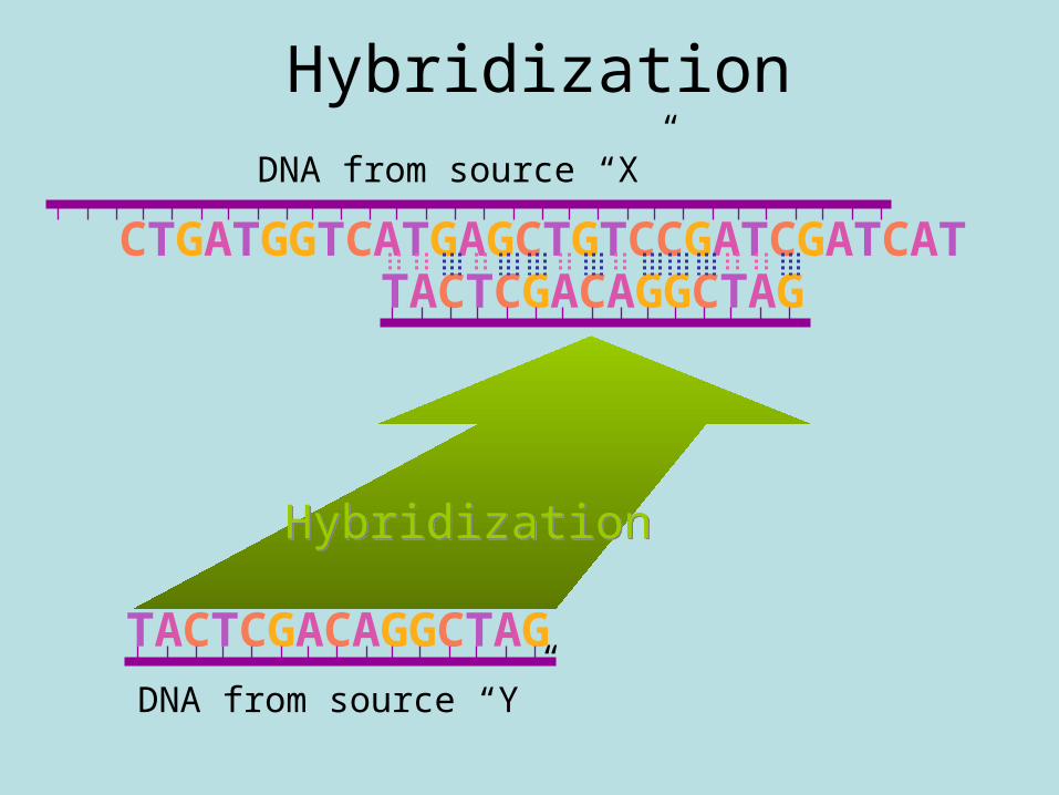
Hybridization
DNA from source “Y”
TACTCGACAGGCTAG
CTGATGGTCATGAGCTGTCCGATCGATCAT
DNA from source “X”
TACTCGACAGGCTAG
HybridizationHybridization


• If different single-stranded DNA molecules, or DNA and RNA molecules, or RNA molecules are mixed together in a solution, and the renaturation is allowed to occur under proper conditions, single-stranded DNA or RNA will bind with each other to form a local or whole molecule of double-stranded structure as long as the single-stranded molecules are complementary, no matter what kind of sources they come from.

• Nucleic acid hybridization as a technique involves using a labeled nucleic acid probe, which is a known DNA or RNA fragment, to bind with the target nucleic acids, which is usually a poorly understood, heterogeneous population of nucleic acids. A probe labeled with detectable tracer is the prerequisite for determining a specific DNA sequence or gene in a sample or genomic DNA by nucleic acid hybridization.

• The target nucleic acids to be analyzed are usually denatured, and then mixed with the labeled probe in the hybridization system. The probe will bind to the segment of nucleic acid with complementary sequence under proper conditions. The hybridization can be identified by the detection of the tracer labeling the probe. Thus the existence or the expression of specific gene can be determined.

2.1 Preparation And Labeling Of Nucleic Acid

2.1.1 Preparation of probes
• Probes may be single-stranded or double-stranded molecules, but the working probe must be single-stranded molecules. The probes used in hybridization of nucleic acids include oligonucleotide(15-50 nucleotides), genomic DNA fragment, cDNA fragment and RNA.

• Oligonucleotide probes are short single-stranded DNA fragments designed with a specific sequence complementary to the given region of the target DNA. They are usually synthesized in vitro.

• Genomic DNA probes can be prepared from the cloned DNA fragment in plasmid.
• cDNA probes can be prepared from the cloned cDNA in plasmid, or amplified directly from mRNA by RT-PCR.
• RNA probes are usually transcribed in vitro from a cloned cDNA in a proper vector. The size of genomic DNA probes, cDNA probes and RNA probes may be 0.1 kb to 1 kb.

2.1.2 Labeling of probes
• Probe is usually labeled with a detectable tracer, which is either isotopic or non-isotopic. The purified oligonucleotide is labeled in vitro by using a suitable enzyme to add the labeled nucleotide to the end of the oligonucleotide.

• For the preparation of the labeled RNA probes, RNA probes are usually synthesized by RNA polymerase in the presence of ATP, GTP, CTP and the labeled UTP, with specific fragment of a gene or cDNA in a proper vector as template. RNA probes can then be generated and be labeled at the same time.

• Genomic DNA probes and cDNA probes are usually labeled in the process of DNA synthesis in vitro. In the reaction of DNA synthesis with a DNA probe as template, if a labeled-dNTP, which can be incorporated into newly-synthesized DNA chain, is added as a substrate, the labeled DNA probe will be formed.

• There are different, sensitive detecting methods for each of the labels used in nucleic acid hybridization. After hybridization, the location and the quantity of the hybrid molecules can be determined. The labels in common use include radioactive (32P and 35S) and nonradioactive (digoxigenin, biotin, fluorescein) substances which are used to label dNTP.

3.1 Hybridization Of Nucleic Acids

3.1.1 Southern blot hybridization
• Southern blot hybridization is an assay for sample DNA by DNA-DNA hybridization which detects target DNA fragments that have been size-fractionated by gel electrophoresis (Figure 4-1). In Southern blot hybridization, the target DNA is digested with restriction endonucleases, size-fractionated by agarose gel electrophoresis, denatured and transferred to a nitrocellulose or nylon membrane for hybridization.

• DNA fragments are negatively charged because of the phosphate groups so to migrate towards the positive electrode, and sieved through the porous gel during the electrophoresis. Shorter DNA fragments move faster than longer ones. For fragments between 0.1 and 20kb in length, the migration speed depends on the length of fragment. Thus, fragments in this size range are fractionated by size in a conventional agarose gel electrophoresis system.

• Following electrophoresis, the sample DNA fragments are denatured in strong alkali, such as NaOH. Then, the denatured DNA fragments are transferred to a nitrocellulose or nylon membrane and become immobilized on the membrane. Subsequently, the immobilized single-stranded target DNA sequences are allowed to interact with labeled single-stranded probe DNA.

• The probe will bind only to complementary DNA sequences in the target DNA to form a target-probe heteroduplex. As the positions of the immobilized single-stranded target DNA fragments on membrane are faithful records of the sieve separation achieved by agarose electrophoresis, they can be related back to the original gel to estimate their size.

Figure 4-1 Southern blot hybridization detects target DNA fragments that have been size-fractionated by gel electrophoresis

• Southern blot hybridization technique is widely applied in researches since its invention. It could be applied for analysis of gene expression, screening of recombinant plasmids, analysis of gene mutation, and identification of the existence of a given DNA such as DNA from pathogenic microorganism. It could also be used to detect deletion of gene by restrictions mapping.

Hybridization• The bases in DNA will only pair in very specific ways:
G with C and A with T• In short DNA sequences, imprecise base pairing will
not be tolerated• Long sequences can tolerate some mispairing only if
hydrogen bonding of the majority of bases in a sequence exceeds the energy required to overcome mispaired bases
• The source of any single strand of DNA is irrelevant, merely the sequence is important, thus complimentary DNA from different sources can form a double helix
• This phenomenon of base pairing of single stranded DNA strands to form a double helix is called hybridization as it may be used to make hybrid DNA composed of strands from different sources

• Because DNA sequences will seek out and hybridize with other sequences with which they base pair in a specific way much information can be gained about unknown DNA using single stranded DNA of known sequence
• Short sequences of single stranded DNA can be used as “probes” to detect the presence of their complementary sequence in any number of applications including:– Southern blots– Northern blots (in which RNA is probed)– In situ hybridization– Dot blots . . .
• In addition, the renaturation, or hybridization, of DNA in solution can tell much about the nature of organism’s genomes

Library Screening• The most common method of library screening
involves hybridization of probes to target DNA• Hybridization refers to the specific way DNA
sequences base pair with their exact complement
• Probes - Single stranded nucleic acids used to hybridize with a target DNA. Generally probes are radioactive or marked in some other way so that they can easily be identified after binding to target DNA
• To design probes for hybridization screening, something must be known in advance about the target sequence

Hybridization Screening• Takes advantage of the fact that
complementary strands of DNA can recognize one another
• By sticking DNA from many colonies or plasmid in a library to a membrane
• Making the DNA single stranded
• Then hybridizing a probe to the DNA on the membrane thus marking target DNA on the membrane, colonies or plasmid containing the target DNA can be identified

Cover with X-ray film
Develop X-ray film
Hybridization ScreeningMembrane
Transfer cells to membrane Lyse cells - DN
A and protein stick to membrane
Locate colony with target clone
Block membrane - Prevents probe from sticking to membrane
Add probe
Wash off excess probe

Southern Blots• Called Southern blots after their inventor
• Involve four steps:
1 Digestion of DNA using restriction enzymes
2 Separation of the DNA fragments by size using gel electrophoresis
3 Transfer of fragments to a nitrocellulose or nylon membrane
4 Hybridization of a probe to the fragment or fragments of interest
5 Probe detection (autorad development)

1 2 3
Making A Southern Blot 1 + 2 Digestion and Electrophoresis
Experimental
3
Marker
1
Control
2

Membrane
Making A Southern Blot 3DNA Transfer To Membrane
DNA
Gel
Buffer
Gel Membrane
Paper Towels

Membrane with bound DNA
Addition of blocking reagent
Parts of the membrane not already covered with DNA now bind blocking reagent
Probe covers the membrane, but only binds to complimentary DNA
Probe addition
After washing
Probe only remains annealed to complimentary DNA
Making A Southern Blot 4Probe Hybridization

Fragments complimentary to the probe appear as bands on the autorad
Making A Southern Blot 5Autorad Development
Membrane with probe bound to complimentary DNA X-ray film is placed
over the membrane and left until radiation from the probe has exposed the film