optical and electron microscopic changes in …
TRANSCRIPT

O P T I C A L A N D E L E C T R O N M I C R O S C O P I C
C H A N G E S I N U L T R A V I O L E T -
I R R A D I A T E D C H R O M O S O M E S E G M E N T S
W I L L I A M B L O O M , M.D. , and R O B E R T J. L E I D E R , M.D.
From the Committee on Biophysics and the Department of Anatomy, The University of Chicago
A B S T R A C T
Chromosome segments of urodele cells lose some substance after irradiation with about l0 -~ ergs/# ~ of heterochromatic ultraviolet light. These segments stain faintly or negatively with the Feulgen and pyronine- methyl-green methods and weakly with the Alfert-Gesch- wind stain for basic protein. In the living cells, Perry found in these chromosome segments a decrease of 50 to 60 per cent in absorption at 2400, 2600, and 2800 A, i.e., in the region of intense chromosomal absorption that is maximal at 2600 A. Apparently the material lost contains DNA (?DNP) and we call the process DNA-steresis. In such cells, fixed in neutral formalin in Tyrode's solution and stained with phosphotungstic acid, electron microscopy shows that the unirradiated parts of the chromosomes consist of (a) a homo- geneous or finely fibrillar material (component-A) filling the meshes of (b) an irregular network with bars 40 to 300 A in diameter, some of which continue into a similar inter- chromosomal network. DNA-steretic portions of the chromosomes consist mainly of this network and only small amounts of component-A, which presumably contains the DNA. We have not been able to demonstrate DNA-steresis with the electron microscope after primary fixation with OsO4 or KMnO4. Structural changes due to DNA-steresis are com- pared with certain nuclear changes in the mitotic cycle.
A small portion of a chromosome or of an inter- kinetic nucleus of a salamander cell in culture, after exposure to ultraviolet light in the Uretz microbeam (Uretz, Bloom, and Zirkle, 1, Uretz and Perry, 2), undergoes a prompt change in refractive index at the irradiated site and appears as a pale area in the living cell by dark phase- contrast microscopy (Bloom, Zirkle, and Uretz, 3). This happens with a flux of ultraviolet within certain limits and with certain lengths of exposure. The affected area appears optically empty in contrast to the sharp black and gray details of the adjacent chromosomal or other nuclear structures (Fig. 1). This change extends gradually beyond the original site of bombardment. The larger the volume of chromosomes irradiated, the less intense
is the local reaction; none is visible after ultraviolet irradiation of all of them.
When phase contrast is replaced by ordinary transillumination there is little or no indication of a difference and the continuity of the chromo- some through the irradiated area appears unin- terrupted. With such optics, the effect of irradi- ation may be indicated by a change in brilliance in the irradiated area. Izutsu (4), using an ultraviolet microbeam of the Uretz type, found that the irradiated portions of chromosomes of grasshopper spermatocytes "became pale with or without phase-contrast microscopy immediately after exposure of fifteen seconds or more." He did not report what the U V flux was.
The change in refractive index suggests a
269
Dow
nloaded from http://rupress.org/jcb/article-pdf/13/2/269/1266270/269.pdf by guest on 18 February 2022

profound upset in structure and a prob~ible loss of substance. One of our first tests of the nature of this localized alteration in the chromosomes was the application of a variety of the fixing and staining methods of light microscopy. Some of our early results were reported briefly by Bloom and Leider (5) who found that the irradiated areas stained only faintly with the Feulgen method or Delafield's hematoxylin and did not stain blue as chromatin does with Mallory's phosphotungstic acid hematoxylin, or green with the pyronine- methyl-green stain. The weak or negative reactions with the Feulgen and pyronine-methyl-green stains suggest that D N A may have been greatly changed or lost at the site of irradiation. This conclusion is bolstered by Perry's observation (6) of a decrease of about 50 per cent in absorption at 2400, 2600, and 2800 A in the irradiated portions of chromosomes. In some preliminary measure- ments by interference microscopy, Haynes and Stodolsky (7) have observed a decrease in optical path length (,'~50 m/~) between the unirradiated and irradiated segments that would suggest a substantial loss of dry mass from the latter.
The chromosomes in the living cells in our cultures are not birefringent and we have been unable to discern any type of birefringence in the irradiated parts of the chromosomes. Some of the changes found with the electron microscope in the irradiated parts of the chromosomes have been reported briefly by Bloom (8, 9).
In several previous communications we have spoken of the visible effect of local ultraviolet irradiation as "pal ing," for with dark phase- contrast microscopy the irradiated area appeared as a pale segment in an otherwise dark chromo- some. Had we used bright phase-contrast micro- scopy we probably would have called the change in these areas "darkening." Although we are reluctant to introduce another term, "steresis," indicating loss of a specific part, expresses our meaning more accurately. Since the accumulating evidence suggests strongly that D N A is part of the substance lost, the process might be called "DNA- steresis" and we will use this term for brevity.
During our study of partial cell irradiation, we found that exposure of parts of chromosomes to very large amounts of ultraviolet light inhibited completion of DNA-steresis and made the irradi- ated parts very dark, instead of light, in the dark phase-contrast microscope. We also found with the electron microscope that such parts did not
show all the changes in structure characteristic of DNA-steresis. A low power micrograph of such overirradiated chromosomes has been published by Bloom (10). Out of this finding grew our experiments using ultraviolet irradiation of large parts of cells as a method of fixation. In the present paper we will include some mention of its usefulness as a primary fixative either alone or followed by formalin or OsO4.
For the purposes of this report, the term "chro- mosome" covers not only the stages in development of typical metaphase chromosomes, but also the less characteristic structures forming the fused chromosome masses of telephase and their changes in early reconstruction. We are unable to visualize and define "chromosomes" in late reconstruction, interkinesis, and exceedingly early prophase.
In the following pages we present illustrations and details of our findings on the changes in the locally irradiated part~ of chromosomes, including those seen with the electron microscope. I t will also be shown how the changes we found in the irradiated parts have offered some clues helpful in interpreting chromosome structure.
M A T E R I A L A N D M E T H O D S
Practically all of the cells used in this work were in the epithelial sheet growing out of the cardiac meso- thelium of Triturus viridescens and Arablystoma tigrinum in tissue culture. This tissue was chosen for several reasons: (a) this epithelium forms an optically favorable material, since the cells usually remain in contact with their neighbors throughout the mitotic cycle and do not round up enough in metaphase to produce the disturbing halos seen with phase con- trast in isolated cells in culture; (b) the relatively large cells have 22 long, conspicuous chromosomes; (c) the mesothelium, like other epithelial tissues in culture, liquefies the fibrin of the medium, with the result that the cells become closely attached to the coverslip as the sheet grows out from the explant; and (d) a room maintained at 70 to 72°F (a favorable temperature for the urodele cells) obviates the need for incubators around the observing and microbeam microscopes.
The simple culture medium consists of 1 part of heparinized or reconstituted lyophilized chicken plasma, 6 parts of Tyrode's solution and 3 parts of distilled water (to approximate the lower tonicity of urodele plasma).
In experiments with ultraviolet light the cultures must be made on quartz coverslips which are then mounted on hollow chambers. These are made by boring a 1/~-inch hole in a 2 by 3 inch slide 1 mm thick; a glass coverslip is then placed under the hole
270 THE JOmCNAL O~ CnLL BIOLOGY • VOLUME 18, 196~
Dow
nloaded from http://rupress.org/jcb/article-pdf/13/2/269/1266270/269.pdf by guest on 18 February 2022

and sealed to the slide with an acetone solution of polyvinyl acetate (Gelva); the acetone is allowed to evapora te before sterilization of the slide by dry heat. For studies of ul traviolet absorpt ion, quar tz cover- slips mus t be used instead of glass to cover the base of the hole. Several drops of the dilute p lasma are placed in the well and most of it aspira ted before the coverslip with cul ture is applied. T h e residual th in layer of p lasma in the well aids in ma i n t a in ing the tonici ty of the cul ture and in prevent ing the deposi- tion of small droplets of condensed water vapor which would dis turb the pa th of l ight for microscopic study.
T h e peak of mitoses in the cul tures is usual ly reached between the 5th and 7th days after explanta- tion. T h e advanc ing mesothel ial sheet of cells lique- ties the fibrin, leaving the cells closely applied to the coverslip. Mitoses f requent ly occur in waves, often in a par t icular par t of a cul ture at a given t ime, a l though scat tered dividing cells are found at a lmost any t ime of the day or night . Four to 8 hours usual ly elapse from the earliest indicat ion of the onset of mitosis unt i l reconstruct ion is well advanced (at 72 °F).
Wi th the Uretz ultraviolet m i c r o b e a m appa ra tu s one aims at the desired spot (demarca ted by cross- hairs in the ocular) and the morphological changes which result can be observed directly. This flexible in s t rumen t permits i r radiat ion of targets with circular spots as small as 1 #, or wi th long na r row beams, or squares, triangles, or any desired shape. In the observat ions repor ted here most of the irradiat ions lasted 5 to 10 seconds; in some instances, especially those involving late me t aphase chromosomes, the irradiat ions were from 12 to 15 seconds. T h e y were m a d e with an 8 /z spot or with a long band about 3 # wide; the energy flux was abou t 10 -2 ergs//~2/ second with he terochromat ic ultraviolet f rom an AH6 mercu ry arc lamp. T h e energy delivered was about the same as in the earlier exper iments in which the irradiat ions were m a d e wi th he terochromat ic ultraviolet l ight f rom a Hanov i a germicidal l amp for exposures of 3 to 7 minutes . A l t hough the spectra f rom these two sources are not identical, in the present studies there were only minor differences in their effects, and these were apparen t ly due to the m u c h longer exposure requi red with the weaker Hanov ia source.
T h e relative effectiveness of the various wave lengths of ultraviolet l ight in p roduc ing the localized change in the ch romosome has been s tudied by Zirkle and Uretz, whose findings are being publ ished separately. Cont ra ry to expectat ions they found tha t 2600 A was not the most effective wave length.
If the i r radiat ion is carr ied out in me taphase and one ch romosome lies above another , the uppe r one can be m a d e DNA-steret ic wi thout m u c h visible
change in the lower one. This result is in par t due to the precise focal p lane of the Uretz m i c r o b e a m and, p robably to a lesser extent, to absorpt ion by the upper chromosome. This makes it difficult to deter- mine, in the living cell, how extensive the change in the ch romosome is. By vary ing the focus du r ing i r radiat ion so as to include port ions of bo th the uppe r and lower chromosomes, bo th will be DNA-steret ic . Owing to the hollow conical shape of the ultraviolet b e a m above the focal point, one result of vary ing the position of the focal point is to b roaden the l ightly irradiated, na r row zone of ch romosome ad jacent to the m a i n i r radia ted port ion of the upper chromosome. After we found tha t unavoidab le i rradiat ion of the " m a r g i n a l zone" was of great help in ana lyz ing the changes seen with the electron microscope, we often b roadened this zone intent ional ly by focussing up and down dur ing i r radiat ion of the target.
Histological Technics
After a cell is selected for s tudy its location is ma rked by d rawing a r ing about 250 # in d iamete r a round it on the coverslip with a d i a m o n d marker . This serves to identify the cell as it passes t h rough the various exper imenta l and histological procedures.
Most of the cells for cytological s tudy were fixed in Zenker-formol (9 parts of Zenker stock solution and 1 par t full s t rength formal in neut ra l ized with an excess of MgCO3) for 12 to 15 minutes , washed in several changes of water unti l the yellow stain with b i ch romate was removed, passed th rough 50 per cent alcohol with iodine, then into 75 per cent alcohol, after which they were s ta ined in various ways. A m o n g the m a n y stains applied after this fixation, the most useful were found to be dilute Delafield's hematoxyl in , Mal lory ' s phosphotungs t ic acid h e m a - toxylin, He idenha in ' s i ron-hematoxyl in , azure I I - eosin, azan, the Feulgen method . T h e cul tures for the Alfer t -Geschwind m e t h o d for basic proteins were fixed in aqueous 10 per cent neut ra l formal in ; those to be s ta ined with the pyronine-methyl -green m e t h o d were fixed in Carnoy ' s mixture . In addit ion, m a n y prepara t ions for the Feulgen and other stains were m a d e after fixation in 1 per cent OsO4 or after 10 per cent neut ra l formalin. In all cases where the Feulgen stain was used, the period of hydrolysis in HC1 was carefully l imited to 12 minu tes at 60°C.
Technics for Electron Microscopy
T h e m e t h o d of identifying, fixing, embedding , and m o u n t i n g the exper imenta l cell for electron mlcrot- o m y has been described by Bloom (10). Here it is necessary to repeat only briefly the principle of the special technic developed for the s tudy of a par t icular cell in a cul ture or smear . As men t ioned above, the posit ion of the selected cell is m a r k e d by d rawing a small r ing a round it on the coverslip wi th a d i a m o n d
W. BLOO~ AND R. J. LEIDEn Changes in Ultraviolet-Irradiated Segments 271
Dow
nloaded from http://rupress.org/jcb/article-pdf/13/2/269/1266270/269.pdf by guest on 18 February 2022
marker . This helps to locate the cell at any stage of subsequen t operat ions such as the part ial cell irradi- ation, fixation, dehydra t ion , e m b e d d i n g in the plastics, and above all in m o u n t i n g it for micro tomy. Careful d i ag rams and, dur ing the last 2 years, Polaroid L a n d pho tomic rographs were m a d e of the exper imenta l cell and its neighbors. These are indispensable aids in the identification of the cell and the por t ion of it which was irradiated.
Fixation by Liquids
T h e p rob lem of fixation of cultures is different in some aspects f rom tha t of a small block of tissue con ta in ing perhaps 10,000 to 20,000 cells or even more. For s tudy of the living cell, we selected the f lat tened ep i the l ium (mesothel ium) adher ing to the coverslip in the migra t ion zone of the cultures. T h e per iphery of these ceils is perhaps 1 or 2 # thick and the greatest thickness of the nuc lear portion, even at the he ight of me taphase , is p robably not more t h a n 7 or 8 ~, usual ly less t h a n 6 #. It is obvious tha t fix- ing, dehydra t ing , and e m b e d d i n g agents come into immedia te contact wi th the cell wi thout being di luted with mater ia ls extracted from in tervening cells, as occurs in the pene t ra t ion of a fixative th rough a block. I n addi t ion to l per cent OsO4 in distilled water or in Veronal buffer at p H 7.4 or p H 7.8 (Palade) a n u m b e r of o ther fixatives were used. For the s tudy of nuc lear changes in the mitot ic cycle and the effects of i r radiat ing segments of urodele chromosomes we found the following fixing solution to give the most informat ive prepara t ions : 1 vo lume of 40 per cent fo rmaldehyde sa tu ra ted with MgCO3, 6 volumes of Tyrode ' s solution a n d 3 of distilled H 2 0 . W e shall refer to this fixative as "neu t ra l f o r m a l i n - T y r o d e . "
Zenker-formol-osmic, Zenker stock wi th or wi thout formalin, Low's solution, Da l ton ' s solution, K M n O 4 as advised by Luft, Carnoy 's , and m a n y other fixa- tives were tried a n d found to be of little value for our purposes because of vacuolizat ion, or ext reme shrinkage, or failure to demons t ra te the i r radia ted area.
For fixation in o s m i u m tetroxide the coverslips, with cul ture side down, were floated on 1 per cent solutions of this reagent for 10 to 20 minu tes and after a rapid rinse in distilled water were passed (culture side up) t h rough a succession of alcohols (5 minu tes each in 25, 50, 75, 95, and 100 per cent). Those fixed in neut ra l f o rma l i n -T y rode were fixed for 30 minutes , r insed in water , and then transferred to 25 per cent alcohol. Ear ly in this work we passed such cul tures rapidly t h rough the alcohols as indicated for the OsO4-fixed cultures. However, for reasons given in the th i rd p a r a g r a p h of the Discussion it is bet ter to leave t h e m in 75 per cent alcohol for several hours. For convenience we have s tandard ized on 5
minutes each in 25 per cent and 50 per cent alcohol, overnight in 75 per cent alcohol, 5 minu tes each in 95 per cent and absolute alcohol, and then t h rough the embedd ing procedures.
Fixation by Ultraviolet Light
T h e largest a rea we can irradiate in the micro- scopic field with our present appara tus , us ing a X 50 objective with the intense A H 6 high pressure mercu ry arc, is a circle 80 # in diameter . This is large enough to i rradiate m u c h more t h a n the nuc lear area bu t not the entire cy toplasm of the greatly ex tended urodele mesothel ial cells,
Wi th a flux of he te rochromat ic ul traviolet l ight of abou t l0 -2 e rgs /#2 /second we have i r radiated the whole nuclear zone of cells for periods of l0 to 70 seconds. No visible changes occur in the first l0 to 20 seconds, bu t after 30 to 40 seconds small blebs some- t imes appear at the edge of the i r radiated area. After the i r radiat ion we exposed the ceils to the usua l liquid fixatives or directly to di lute alcohols and have obta ined results which convince us tha t the i rradia- t ion acts as a good fixing agen t for some structures.
Staining
T h e most informat ive stains have been 0.5 per cent phosphotungs t ic acid (PTA) or u rany l aceta te in the absolute alcohol used for dehydra t ing the cultures. T h e cultures s tayed in the stain for 5 minutes . In a few instances they were left in for m u c h longer times, bu t the resul t ing sections were too dense to show m u c h s t ruc ture in the electron beam. W e found tha t this procedure gave us more regular results t han s ta in ing the sectioned cells on the grids.
Embedding Practically all of our work was done by dissolving
polymerized mixtures of vary ing percentages of butyl and me thy l methacry la tes in e thylene dichloride. Benzol could also be used as the solvent except for osmium-f ixed material . T h e procedure is s imple: the coverslip wi th the cul ture passes f rom absolute alcohol into a mix tu re of equal par ts of absolute alcohol a n d e thylene dichloride, t hen into two changes of e thylene dichloride. T h e excess is removed by touch ing the edge of the coverslip wi th a bit of filter paper (care being taken to prevent any drying of the cells) and then a drop of a viscous solution of the polymers in e thylene dichloride is placed on the culture. After perhaps ]/~ hour it will have lost m u c h of its solvent and ano ther drop is added. W e usual ly allow this to dry overnight a l t hough this is no t necessary. T h e cell unde r observat ion is identified by the d iagrams and Polaroid L a n d pho tographs m a d e at the t ime of fixation. T h e su r round ing u n w a n t e d parts of the cul ture are then cu t away with a clean
272 THE JOURNAL OF CELL BIOLOGY " VOLUME 13, 196~
Dow
nloaded from http://rupress.org/jcb/article-pdf/13/2/269/1266270/269.pdf by guest on 18 February 2022

razor blade. The embedded culture has now to be mounted onto the tip of a Lucite rod which fits the chuck of the microtorne. This is accomplished by covering the tip of the rod with a small drop of the viscous solution of polymerized methacrylates in ethylene dichloride and placing it in position over the selected cell. This had previously been centered by placing the circle circumscribing it on the coverslip over a cross inscribed in the glass (or quartz) plate forming the base of the centering ring and holder devised for this purpose (10).
After a couple of days, or longer if desired, the Lucite rod, now firmly attached to the culture and with its long axis perpendicular to the plane of the coverslip, is removed from the positioning device. Before removing the coverslip it is again necessary to compare the topography of the cell or cells in the inscribed circle with the diagrams and Polaroid Land photographs previously made so as to be certain of identifying the selected cell under study. If the quartz coverslip was carbon coated before the culture was made, it is easily removed by cooling with solid (]02. If it has not been carbon coated the cells often stick to the quartz. We have had no difficulty removing the cells from uncoated glass coverslips.
The culture then is trimmed for cutting. If the plastic is a bit tacky, the free surface is exposed in a closed Stender dish overnight to let the last traces of the ethylene dichloride evaporate. The trimming can be done free hand, with the help of a dissecting microscope, using clean razor blades. For the past year we have been using a device with which it is possible to control the descent of the razor blade in a guillotine-like holder and to rotate the block at successive 90 ° turns. This permits rapid and more accurate trimming of the block to include only the selected cell or cells (and incidentally removes some of the tension on the person trimming the block), but is dispensable.
The universal joint on the chuck in the Porter- Blum microtorne was replaced by a rigid chuck which holds the Lucite rod with the embedded cell on its tip. This ensures sectioning the cell in the plane of the original surface of contact of the cell with the coverslip. Most of the cells studied were in gray or silver gray sections. Slightly thicker sections did not show much structure in the formalin-fixed, phospho- tungstic acid-stained cells. The sections, usually mounted on carbon-coated Formvar films on the grids, were studied with an EMU-3C (RCA) micro- scope.
We have obtained a few good preparations after embedding in Vestopal. We had little success in embedding with heat polymerization of mixtures of butyl and methyl methacrylate or with the water- soluble epoxy resin (11) which Dr. W. St/iubli sent us. The greatly increased viscosity of the latter in the higher concentrations caused difficulty in infiltrating
the cultures thoroughly; in addition, the tough poly- mer of the embedded cultures adhered to the quartz coverslips. A few cultures were embedded by using the methacrylate monomer mixture instead of ethylene dichloride as solvent for the polymerized methacrylates. We found no advantage in using the monomers.
O B S E R V A T I O N S
Living Cells
Unless otherwise noted, all of these observations wcrc made with medium dark phase-contrast microscopy; after an appropriate dose of ultraviolet light the change in refractive index of the irradi- ated chromosome segment starts promptly and
reaches a max imum intensity within 5 minutes, al though longer periods are occasionally required (Fig. 1). With time the stcretic change may extend for some microns beyond the irradiated zone. Indeed, we have seen it extend from an original 8 ]z segment to over 20 ]z in the course of a couple of hours.
The clearest observations are those made when the chromosomes are in a single layer, as in the mcsothelial cells in late prophasc or in prometa-
phase. In such cells, a lightening of the dark gray of the chromosomes appears a minute or less after irradiation with an AH6 source for 6 to l0 seconds, (or 1 or 2 minutes later during the course of a 5
minute irradiation with a Hanovia lamp). Within the next few minutes this change progresses so that the outlines of the irradiated portions of chromo- somes disappear and this area becomes an optically empty zone demarcated by adjacent unirradiated
structures. A micron or so of the chromosome adjoining the irradiated area may appear darker than the rest of the chromosome, particularly if the
irradiation is carried out in prophase. If too little ultraviolet is used, the effects may
not be appreciable or they may be incomplete in which case the irradiated portion appears light gray in contrast to the dark gray of the unir- radiated portions.
With too much ultraviolet, brightening may start but will not progress and the center of such an overirradiated area may become very dark or show a slight brownish tinge in the dark phase- contrast microscope. Depending upon the amount of ultraviolet used there may or may not be a narrow, bright zone at the periphery of the over- irradiated area.
There are other factors which modify the onset
W. BLOOM AND R. J. LEIDER Change8 in Ultraviolet-Irradiated 8egmenss 273
Dow
nloaded from http://rupress.org/jcb/article-pdf/13/2/269/1266270/269.pdf by guest on 18 February 2022

of the change as seen in the living cells. It appears more rapidly and after less irradiation, the earlier the stage in the mitotic cycle. In late anaphase it is elicited only by large doses and in telophase it may be impossible to produce it without causing other severe and obscuring effects of the irradi- ation. Indeed, amounts of energy insufficient to produce visible change in these stages may retard reconstruction of an irradiated daughter nucleus
chromosomes with the phase microscope were fixed and stained in a variety of ways. Some of the technics did not demonstrate these areas as, for example, those for phosphatase tried by Amenta (12). However, a number of the routine histological methods showed the steretic places nicely, as did several of the more specific methods for chromosomes. For brevity's sake, the following description is limited to the results we obtained
FmURE 1
Metaphase of living mesothelial cell before (1 a) and after (1 b) irradiation with a narrow stripe of ultraviolet light. In the almost complete DNA-steretic area (arrows) pale gray traces of chromosomes can still be seen. Medium dark phase contrast, X 1100.
as compared with its unirradiated mate which serves as an excellent control. The changed chromosome may be incorporated within the daughter cell, especially if the kinetochore region has not been irradiated. Such areas may be ob- served for days in the daughter cells; they do not resume the appearance of normal nuclear struc- ture.
Fixed and Stained Preparations
Living cells which showed the typical DNA- steretic change of the irradiated portions of
with the more informative of these procedures applied to cells irradiated with heterochromatic ultraviolet light over a circle 8/z in diameter or a long narrow stripe.
In the whole mount preparations, the DNA- steretic areas are clearly demonstrated after fixation with Zenker-formol and staining with routine hematoxylin methods. The irradiated cells pictured in Figs. 2 to 6 and 8 were fixed with Zenker-formol. After staining with hema- toxylin and eosin (Fig. 2), the portions of chromo- somes in the I)NA-steretic area have lost their
274 THE JOURNAL OF CELL BIOLOGY • VOLUME 13, 1962
Dow
nloaded from http://rupress.org/jcb/article-pdf/13/2/269/1266270/269.pdf by guest on 18 February 2022

Each of the cells in Figs. 2 and 3, in prophase , a n d Figs. 4 and 5, in metaphase , was i r radiated for 5 minu tes with 8 /z spots of he te rochromat ic ultraviolet light f rom a Hanov ia l amp to produce DNA-steresis. These areas are indicated by arrows. Fixat ion in Zenker-formol. X 1640.
FIGURE
T h e steretic ch romosome segments have lost their basophil ia and appear somewha t swollen or fused. Hematoxyl in-eos in stain.
FIGURE
T h e steretic port ions do not stain so deeply with He idenha in ' s i ron-hematoxyl in as do the unirradiated.
FIGURE 4
C h r o m o s o m e s are not seen in the DNA-stere t ic a rea which stains a diffuse pale blue with di lu te Delafield 's hematoxyl in .
FIGUaE 5
Steretic ch romosome segments stain paler t han the un i r rad ia ted parts. He idenha in ' s i ron-hematoxyl in stain.
W. BLOOM AND R. J. LEIDER Changes in Ultra~olet-lrfadiated Segments
Dow
nloaded from http://rupress.org/jcb/article-pdf/13/2/269/1266270/269.pdf by guest on 18 February 2022

D N A steresis, indicated by arrows, was produced in the cells of Figs. 6 to 8, and I0 to 12 by irradiation with 8/~ spots, and in cells of Figs. 9 and 13 with nar row stripes of ultraviolet light. Cells of Figs. 6 to 8 were irradiated for 5 minutes with the Hanovia l amp; cells of Figs. 9 to 13 were irradiated for 10, 15, or 20 seconds with the AH6 arc. The cells were fixed and stained in several ways. X 1640.
FIGtmE 6
Prophase. Fixed in Zenker-formol and stained with the Feulgen method for DNA. There is a faint diffuse staining in the steretic area.
F I G ~ E 7
Metaphase. The DNA steretic chromosome segments do not stain with the methyl- green of the pyronine-methyl-grecn mixture. Carnoy 's fixation.
FIGURE 8
Pr0phase. The continuity of chromosomes in the DNA-steretic area is obvious but the characteristic staining for chromat in is negative. Zenker-formol fixation; stained with Mallory 's phosphotungstic acid hematoxylin.
FIGURE 9
The steretic area stains faintly with the Alfert-Geschwind method for basic proteins. Formalin fixation.
FIGURE 10
Prophase. T h e Feulgen stain is negative in the DNA-steretic area. Fixed 24 minutes after irradiation in 1 per cent osmium tetroxide.
FIGURE 11
Metaphase. I r radia ted in prometaphase with 8 # spot for 10 seconds. The Feulgen reaction is almost negative in the DNA-steretic area. During the 53 minutes between irradiation and fixation, portions of two chromosomes moved over part of the irra- diated area. 1 per cent osmium te t roxide fixation.
FIGURE 1~
Metaphase. I r radia ted in prometaphase with 8 # spot for 15 seconds. The Feulgen reaction is negative in the DNA-steretie area. Fixed in i per cent osmium tetroxide, 54- minutes after irradiation.
FIGURE 13
Metaphase. Partial cell ultraviolet irradiation of chromosome segments for 20 seconds produced DNA-steresis in zone indicated by arrows. Fixation was by total nuclear irradiation followed by 10 per cent neutral formalin. Feulgen staining of the steretic area, a l though greatly diminished, is still definitely positive.
THe: Jot~i~NAt, OF CELL BIOLOGY - VOLUM~ 13, 196g
Dow
nloaded from http://rupress.org/jcb/article-pdf/13/2/269/1266270/269.pdf by guest on 18 February 2022

W. BLOOM AND R. J. L]~IDER Change8 in Ultraviolet-Irradiated Segments
Dow
nloaded from http://rupress.org/jcb/article-pdf/13/2/269/1266270/269.pdf by guest on 18 February 2022

basophilia and stain with eosin; they appear slightly swollen. Actually, this may be an earlier stage of movement of material resulting from the irradiation than that shown in Fig. 4. The latter was stained only with dilute Delafield's hema- toxylin. The irradiated area appears as a pale staining oval in which the continuity of the chromosomes is not visible; presumably, it represents material liberated from them by the irradiation and precipitated either by the long continued irradiation or the fixing agent. We have seen a similar precipitation of chromosome material after severe overirradiation with the AH6 lamp.
Typical appearances of steretic areas after staining with Heidenhain's iron hematoxylin are shown in Figs. 3 and 5. The relatively dark staining of the chromosomes in this portion of the metaphase cell (Fig. 5) may be due to inoufficient differentiation of the stain in the iron alum.
After similar irradiation and fixation, staining with the Feulgen method shows a diffuse and faintly positive staining of the DNA-steretic area (Fig. 6), suggestively like the diffuse staining of a comparable area in the cell of Fig. 4 stained with Delafield's hematoxylin.
An equally striking demonstration of DNA- steresis follows staining with the phosphotungstic acid hematoxylin of Mallory as shown, typically, in a cell in prophase (Fig. 8). The chromosomes stain deep blue except in the irradiated area where the residual chromosomal material stains light brown. In such preparations the chromo- somes are seen to maintain their continuity as they pass into and through the irradiated area and the two zones are sharply separated by their contrasting staining reactions.
Staining with pyronine-methyl-green, after DNA-steresis and fixation in Carnoy's solution, reveals that the irradiated parts of the chromo- somes do not stain with the methyl green and are sharply demarcated from the unirradiated contiguous parts (Fig. 7).
The cell shown in Fig. 9 was fixed in 10 per cent neutral formalin and stained with the Alfert- Geschwind method for basic proteins. The chromosome segments in the DNA-steretic area stain a faint green in contrast to the brilliant deep green of the unirradiated parts of the chromo- somes.
Because of our use of OsO4 solutions in fixing cells for electron microscopic study of the irradi-
ated parts of their chromosomes, we also fixed many ceils in these solutions for Feulgen staining. Three cells shown in Figs. 10 to 12 were from a culture fixed in 1 per cent aqueous OsO4 stained with the Feulgen method after localized irradi- ation with an AH6 lamp. The cell in Fig. 10, in the granular stage of early prophase, showed the steretic change in the irradiated area and some darkening of the chromosomes at its margin within 4 minutes after irradiation. The change occurred more promptly in this cell than in those in early metaphase in the same culture. The DNA-steretic area is prominent and it is questionable whether even a trace of Feulgen-positive material is present. The interval from irradiation to fixation was longer for the other two ceils. The chromosome segments in the irradiated area of the cell shown in Fig. 12 stain tan from the OsO4. If a trace of Feulgen-positive material is present in this area, it is definitely less than the faint pink staining in the comparable area of the cell shown in Fig. 11. It is noteworthy that this cell received two-thirds as much irradiation as that of Fig. 12. In Fig. 11 the ends of several unirradiated chromosomes moved over part of the DNA-steretic area during the interval before fixation.
In addition to the cells which show the reactions of DNA-steretic areas to fixation and staining, such as those described, other cells were exposed similarly to partial nuclear irradiation to produce steresis and were then fixed by irradiating the entire nucleus with ultraviolet light. After this some of these were placed in formalin and subse- quently stained with the Alfert-Geschwind method for basic proteins or the Feulgen method while others were processed for electron microscopy. Those stained with the Alfert-Geschwind technic show the same change as those fixed only in formalin, as in Fig. 9. None of those stained with Feulgen shows a completely negative reaction in the steretic area. An example o f this is seen in Fig. 13, of a cell in metaphase which was irradi- ated for 20 seconds with a narrow band of uhra- violet light. DNA-steresis occurred promptly and completely. Forty minutes after the partial nuclear irradiation, the entire nuclear zone was irradiated with the same flux of ultraviolet light for 10 seconds; the cell was fixed in 10 per cent neutral formalin and stained with the Feulgen method. In the steretic area the Feulgen reaction is much weaker than in the other parts of the chromosomes.
W. BLooM AND R. J. LEIDER Changes in Ultraviolet-Irradiated Segments 275
Dow
nloaded from http://rupress.org/jcb/article-pdf/13/2/269/1266270/269.pdf by guest on 18 February 2022

Electron Microscopic Observations
Neutral Formalin-Tyrode Fixation: When this study was begun, it was anticipated that there would be great difficulties in following the experi- mental cell in the tissue culture through all the fixing, embedding, and sectioning procedures preliminary to electron microscopy, but, as mentioned above under Material and Methods, this proved to be relatively easy. We did encounter an unexpected and disconcerting obstacle of another type: despite the striking changes seen with light microscopy, we could not find the irradiated areas with the electron microscope in ceils fixed with solutions of osmium tetroxide (see Fig. 23) or potassium permanganate. How- ever, after fixation in neutral formalin-Tyrode without heavy atom staining, the irradiated parts of the chromosomes stand out as strikingly in the electron microscope (Figs. 14 and 15) as in the living cell (Fig. l) or after fixation and staining for the light microscope (Figs. 2 to 13).
In such neutral formalin-Tyrode-fixed cells, these urodele chromosomes have a homogeneous appearance (Ch in Figs. 14 and 15). This resembles that produced by several other fixatives, such as absolute alcohol, and the appearance of chromo- somes of Earle's L strain of mouse cells after fi'eeze-drying as found by Dales (13).
Although these chromosomes do not have a distinct surface membrane they are sharply demarcated in late prophase, metaphase, and anaphase from interchromosomal structures. The homogeneous, sharply contoured chromosomes are quite different in appearance from the indis- tinctly outlined masses of rodlets and dots seen with the electron microscope after OsO4 fixation (Fig. 23). Between the chromosomes are scattered
fine, dark granules and somewhat paler, irregular, ill-defined bands or cords. Sometimes there are small granules in these cords but more frequently
they occur at their margins. Some of the inter- chromosomal cords and the less well defined
strands meet and fuse with the edges of the chromo- somes, giving them an irregular outline. Scattered among these structures are apparent open spaces of varying size which probably represent shrinkage or places from which substances have been dis- solved.
Unlike the non-irradiated portion where the edge of the chromosome is definite, the edges of the chromosomes in the heavily irradiated seg- ments are ragged and blurred (Fig. 14, ItCh). Indeed it is impossible in many places in such areas to determine exactly where the chromosome ends. This is suggestively like the indefinite de- limitation of non-irradiated chromosomes fixed in OsO4 (Fig. 23). In the transition zone between the irradiated and non-irradiated parts of the chromosomes there are closely packed small vacu- oles indicated by arrows in Fig. 14. The smallest of them are fairly regular in contrast to the larger ones which may vary considerably. At higher magnifications the vacuoles measure 75 to 250 A and may appear to be oval or ellipsoidal--perhaps as a result of compression during sectioning (Fig. 15, arrows). They are smaller than the irregular vacuoles in the more intensely irradiated central areas. The large interchromosomal vacuoles of Fig. 15 are not a result of the irradiation since they occurred throughout the nuclear part of the cell; they probably arose from damage during preparation for microscopy.
Neutral Formalin- Tyrode Fixation and Phosphotung- stic Acid Staining: Since the DNA-steretic portions were distinctly different from the rest of the chromosomes in neutral formalin-Tyrode-fixed cells, the next step was to stain some of them with heavy elements. Most informative have been staining with phosphotungstic acid or uranyl acetate dissolved in the absolute alcohol prior to embedding. This method gave us uniformly better results than staining the sections on grids. In comparing our findings with those of others, it must be borne in mind that most investigators apply these compounds to cells fixed in OsO4
FmURE 14
Metaphase chromosomes with DNA-steresis resulting from irradiation with a narrow band of ultraviolet. Homogeneous parts of the chromosomes (Ch) merge into the vacuo- lated and paler irradiated segments (IrCh). In places, it is difficult to see limits between DNA-steretic chromosomes and interchromosomal constituents. Arrows on the chro- mosomes indicate fine vacuoles in the zone of marginal irradiation. 10 per cent neutral formalin-Tyrode fixation. No stain. Methacrylate embedding. X 24,300.
276 THE JOURNAL OF CELL BIOLOOY • VOLUME 13, 1962
Dow
nloaded from http://rupress.org/jcb/article-pdf/13/2/269/1266270/269.pdf by guest on 18 February 2022

W. BLooM AND R. J. LEIDER Changes in Ultraviolet-Irradiated Segmenls 277
Dow
nloaded from http://rupress.org/jcb/article-pdf/13/2/269/1266270/269.pdf by guest on 18 February 2022
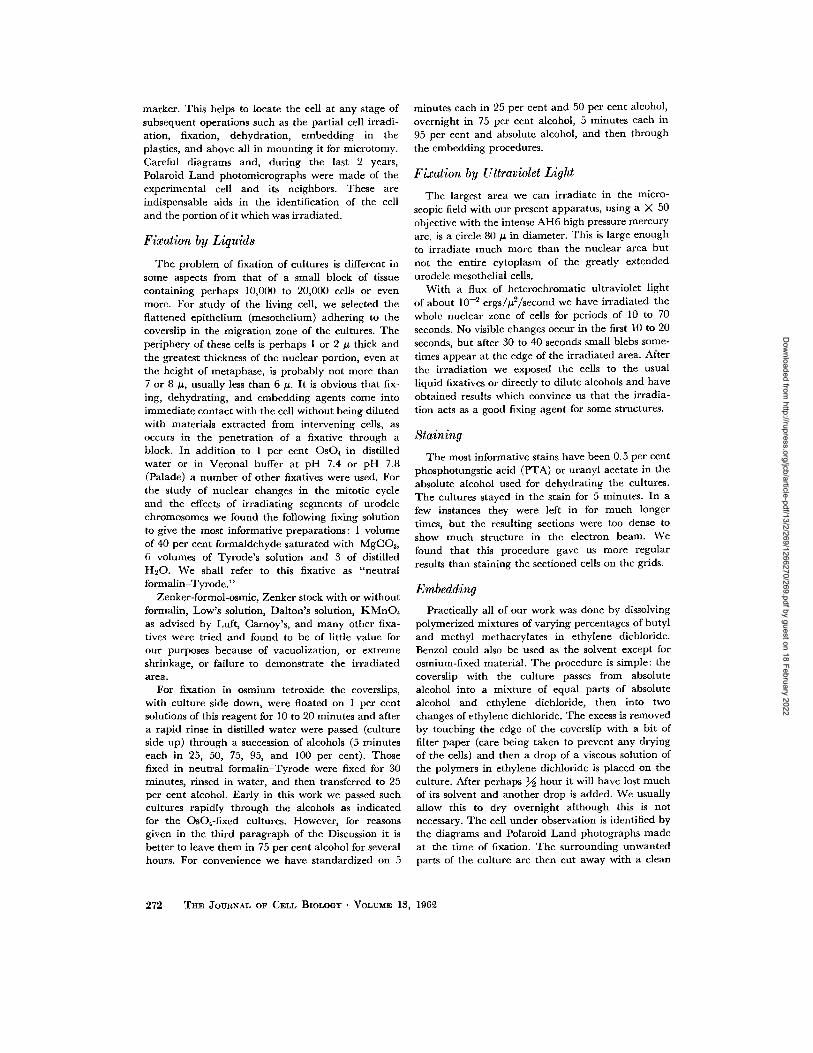
FmURE 15
M c t aphasc chromosomes showing transi t ion from non- i r rad ia tcd ch romosome (Ch) t h r o u g h a marg i - na l zone with small vacuoles (arrows) to DNA-stcrct ic ch romosome scgmcnts (ItCh). In tc rchromo- somal s t ructures (In) are unusua l ly hcavily vacuolatcd, possibly owing to d a m a g c dur ing p repara - tion. I r radia tcd with 8 ~ spot of ultraviolct light. Fixat ion in 10 per cent neut ra l f o rma l in -Tyrodc ; l ightly s ta ined with Dclaficld's hcmatoxyl in . Mcthacry la tc embedding . X 35,700.
278 THE JOURNAL OF CELL BIOLOGY • VOLUME 13, 196~
Dow
nloaded from http://rupress.org/jcb/article-pdf/13/2/269/1266270/269.pdf by guest on 18 February 2022

FIGVRE 16
This micrograph is of metaphase chromosomes at edge of DNA-steretic zone (ItCh) produced by irradiation with a narrow stripe of ultraviolet light. In the lowest, unirradiated chromosome, com- ponent-A (A) and strands of reticulum (R) are indicated. Mz, marginal zone; R' interchromosomal reticulum. Fixed in l0 per cent neutral formalin-Tyrode ; stained with alcoholic phosphotungstic acid. Methacrylate embedding. X 64,000.
solutions. This accounts for some of the differences between our observations on the neutra l fo rmal in- Tyrode-fixed chromosomes and nuclei and those obta ined on preparat ions fixed in OsO4 by others and by ourselves.
In the neut ra l formal in-Tyrode-f ixed cells stained with alcoholic phosphotungst ic acid the in terchromosomal mater ia l is found to consist of an irregularly meshed re t iculum (R t) of elongate, b ranch ing strands usually 75 to 150 A in diameter , a l though occasional ones may be as small as 40 A and others up to 300 A in their short axis (see IR
in Figs. 17 and 18). These strands are often darker at their margins than at thei r centers and are a t tached to and continue, singly or in small bundles, into the chromosomes (R' in Figs. 18, 21).
Similar structures are m u c h more numerous wi th in the chromosomes (R in Figs. 16 to 19, 21). In rare places they may be ar ranged in the long axis of the chromosome, bu t their usual disposition, as seen in the th in sections, is as short segments of wavy, or bent , b ranch ing filaments which are parts of a network with i rregular meshes cut at random. Al though they are frequently close to
W. BLOOM AND R. J. LEIDER Changes in Ultraviolet-Irradiated Segments 279
Dow
nloaded from http://rupress.org/jcb/article-pdf/13/2/269/1266270/269.pdf by guest on 18 February 2022

FZGURE 17
A mon tage of three adjacent micrographs of me taphase chro- mosome, including the ul t ra- violet-irradiated, DNA-steret ic part (Irrad. zone). C o m p o n e n t - A (A) seems to be slightly d imin- ished in the lower marg in of mic rograph a and seems largely gone in most of b. Strands of in terchromosomal re t icu lum (IR) cont inue into the chromosome; (seen better at h igher magnif ica- tion at R ~ in Fig. 18). In the DNA-steret ic portion the s trands (R) are cont inuous with those in the in terchromosomal regions (In). Fixed in neutra l formalin- Tyrode. Stained with alcoholic phosphotungst ic acid before em- bedding and with uranyl acetate on the grids. Methacryla te embedding. X 63,000.
Irrad. z o n e
280 THE JOURNAL OF CELL BIOLOGY " VOLUME 13, 1960,
Dow
nloaded from http://rupress.org/jcb/article-pdf/13/2/269/1266270/269.pdf by guest on 18 February 2022

FIGURE 18
Higher magnification of Fig. 17a. Component-A is prominent (A) in contrast to its paucity in the irradiated part of the chromosome (Fig. 17b and Fig. 19). The chromosomal reticulum (R) is con- tinuous (R ~) with the interchromosomal reticulum (IR). Ch, chromosomes. X 129,000.
one another, they are usually separated, singly or in groups, by a homogeneous or finely granular or fibrillated material. Because it varies in appearance with the phases of mitosis and with different fixatives and stains, we will designate it for the present as "component -A" (A in Figs. 16, 18). When it is homogeneous it looks much like the
chromosomal substance in unstained neutral formalin-Tyrode-fixed preparations. Component- A tends to be suggestively fibrillar in early pro- phase, finely granular or homogeneous in meta- phase, and homogeneous in late anaphase and in telophase. It would seem that the more condensed the developing chromosomes become, the more
W. BLOOM AND R. J. LEIDER Changes in Ultraviolet-Irradiated Segments 281
Dow
nloaded from http://rupress.org/jcb/article-pdf/13/2/269/1266270/269.pdf by guest on 18 February 2022

FIGURE 19
Higher magnification of Fig. 17b, the irradiated, DNA-steretic zone. Owing to the absence of com- ponent-A (except at A), the close-meshed chromosomal reticulum (R) is now obvious. X 129,000.
homogeneous is the appearance of this component. Very often, bars of the reticulum (R) within the chromosomes stain paler after they become sur- rounded by component-A.
With low magnification (5000) in the formalin- phosphotungstic acid preparations, the irradiated areas stand out rather prominently because of some vacuolization of the chromosomes and the much greater irregularity of their margins.
With higher magnification it is obvious that in the steretic areas the chromosomes have undergone a great change. In Fig. 16 the lowest chromosome was not irradiated, while the upper parts of the other two were irradiated and show DNA-steresis. In addition to the vacuolization, which probably varies with the amount of irradiation and may be quite prominent (Fig. 16, IrCh), there is a striking change due to the disappearance of component-A,
282 THE JOURNAL OF CELL BIOLOGY • VOI~UME 13, 196~
Dow
nloaded from http://rupress.org/jcb/article-pdf/13/2/269/1266270/269.pdf by guest on 18 February 2022

with the result tha t the chromosomes here consist largely of a b ranch ing network (R). In the transi- t ion zone (Mz) between the obviously uni r rad i - ated and the i r radiated portions, one can follow the progressive decrease in component-A.
In a montage of three adjacent micrographs in Fig. 17, and in Figs. 18 and 19, which are enlarge- ments of two of them, we can follow the t ransi t ion
(IR). T h e DNA-steret ic area fills most of Fig. 19. Here the re t iculum (R) forms a clearly anasto- mosing network which extends th roughout this port ion of the chromosome and also is cont inuous wi th the interchromosomal re t iculum (at In), so tha t it is difficult to see just where the chromo- some ends (Fig. 19). There is a bi t of component -A at and near (A) in this figure.
~IGURE ~0
1Vietaphase chromosomes irradiated with a narrow stripe of ultraviolet light. Arrows point to the DNA-steretic area which appears as a dark band cutting across the mottled grey chromosomes (Ch). IR, interchromosomal reticulum. The outlined area is shown in Fig. 21 at higher magnification. Fixed in 10 per cent neutral formalin-Tyrode and stained with alcoholic phosphotungstic acid before em- bedding in Vestopal. )< 12,900.
between the un i r rad ia ted and DNA-steret ic portions of the chromosomes. These micrographs are f rom a cell in metaphase fixed in neutra l formal in-Tyrode , stained in toto with alcoholic phosphotungst ic acid and then stained on the grid with uranyl acetate. This combined stain gives more contrast to some of the structures, especially the edges of the chromosomes and the outlines of the re t iculum within them (R). I t also darkens the inner parts of the in terchromosomal re t iculum
T h e results were no different when we substi- tuted a mixture of the methacry la te monomers for ethylene dichloride as the solvent for the polymerized methacrylates in our embedd ing process, or when we used Vestopal instead of the methacrylates for embedding. Figs. 20 and 21 show tha t after neutra l fo rmal in -Tyrode fixation, phosphotungst ic acid staining and Vestopal embedding, we obta ined essentially the same results as wi th methacry la te embedding. T h a t is,
W. BLOOM AND R. J. LEIDER Change,,* in Ul#avlolet-Irradiated Segments 283
Dow
nloaded from http://rupress.org/jcb/article-pdf/13/2/269/1266270/269.pdf by guest on 18 February 2022

the steretic area appears as a dark mass at low magnif icat ion and at h igher magnifications this pa r t of the chromosome is seen to consist of a close meshed network (R) continuous (R') with the looser in terchromosomal one (IR). There is a na r row zone of transit ion (Mz in Fig. 21) at the
sharply delineated in this p repara t ion as in metaphase chromosomes of most of our me tha - cryla te-embedded cultures. Some portions of the interchromosomal re t iculum appear to be s tretched between adjacent chromosomes, perhaps because of shrinkage.
FIGURE ~1
This is higher magnification of the rectangle outlined in Fig. 20. The DNA-steretic area (IrCh) con- sists almost entirely of the intrachromosomal reticulum (R) which is connected (R t) with that between the chromosomes (IR). MZ, margin of irradiated zone. Ch, chromosome. Vestopal embedding. X 44,000.
marg in of the i r radiated area. The uni r rad ia ted chromosomes are sharply demarca ted from the in terchromosomal areas and component -A is a mott led gray, just as in m a n y of the similarly fixed and stained cultures embedded in metha- crylate. The strands of in t rachromosomal ret icu- lum surrounded by component -A are not quite so
Neutral Formalin-Tyrode Fixation and Uranyl Acetate Staining: Somewhat different pictures are obtained with this fixation and subsequent staining wi th uranyl acetate in absolute alcohol before embedding. Wi th this stain, the chromosomes in late prophase are fairly sharply delimited and are composed of a finely granular or suggestively
284 THE JOURNAL OF CELL BIOLOGY • VOLUME 13, 196~
Dow
nloaded from http://rupress.org/jcb/article-pdf/13/2/269/1266270/269.pdf by guest on 18 February 2022

FIGURE ~
Prophase i r radiated with a na r row stripe of u | t rav io |e t light. In the obvious DNA-stere t ic a rea (ItCh), demarca t ed by arrows, residues of the chromosomes are barely detectable. T h e non- i r rad ia ted de- ve |op ing chromosomes (D) are distinct. Fixed in neut ra l fo rmal in -Tyrode . Sta ined wi th 1.2 per cent K M n O 4 in refrigerator for 22 hours. M e t h a c w l a t e embedding . M 7,700.
W. BLOOM AND R. J. LEII)ER Changes in Ultraviolet-Irradlated Segments 285
Dow
nloaded from http://rupress.org/jcb/article-pdf/13/2/269/1266270/269.pdf by guest on 18 February 2022

fibrillar material and an irregular network of open spaces communicat ing with interchromo- somal areas, Component-A fills the meshes between these spaces and is more positively stained by uranyl acetate. I t appears more granular or fibrillar than after phosphotungstic acid staining. In prophase the interchromosomal structures consist of a dark granular network and an ill formed, fuzzy or finely granular, moderately dark gray material much like that of component-A and often connected with it. Such parts of the inter- chromosomal substance will presumably become an integral part of the chromosomes by the time they reach maximum size in metaphase.
As the developing chromosomes condense into the metaphase state they take on a paler, more homogeneous appearance. In the DNA-steretic area they lose their distinctive margins and most if not all of component-A. This part of the chromo- somes now appears as a dark reticulum obviously continuous with the looser interchromosomal reticulum.
Fixation in Potassium Permanganate: We have not seen the steretic area in preparations fixed in potassium permanganate. However, if the cells showing DNA-steresis in the living are fixed in neutral formal in-Tyrode and then stained with potassium permanganate at 0°C for 24 hours or longer, the change is clearly visible in the electron microscope. Fig. 22, from a cell in early prophase, shows the differences between irradiated (ItCh) and unirradiated portions of the developing chromosomes (D). The darkly staining chromo- somes merge into the ill defined structures in the DNA-steretic area.
Os04 Fixation Compared with Exposure to Os04 after Other Types of Fixation." Chromosomes with DNA-steresis show no change by which this area can be recognized after OsO4 fixation when examined with the electron microscope. In such experiments all the chromosomes look alike
(Fig. 23, Ch). In many places it is not possible to tell exactly where they end and interchromosomal material (In) begins. In thin sections the chromo- somes consist of small dots and rods and some longer, double-contoured structures (arrow) 75 to 300 A in diameter. The latter probably correspond to the reticular structures seen in the phospho- tungstic acid-stained preparations after neutral formalin-Tyrode fixation. In some OSO4-fixed preparations, larger black bodies are scattered in the chromosomes; we do not know what they represent.
Although the steretic area cannot be found with the electron microscope after primary OsO4 fixation, it is readily demonstrable if exposure of the cell to OsO4 is preceded by fixation with total nuclear U V irradiation, as in Fig. 24. DNA- steresis was produced by a band of ultraviolet light running through the nuclear zone; the margin of the area exposed to partial nuclear irradiation and then to total nuclear irradiation (P.N.L + T.N.L) is indicated by arrows. In the part exposed only to total nuclear irradiation (T.N.L) the chromosomes (Ch) have sharply demarcated contours and are formed of densely packed granules and rods, while the chromosome segments in the steretic part (IrCh) consist of fewer granules and have vague outlines which gradually merge into interchromosomal areas (In).
Fixation by Total Nuclear Irradiation Followed by Neutral Formalin-Tyrode and Phosphotungstic Acid." Cells so treated (Figs. 25 and 26, P.N.I + T.N.L) show somewhat more of component-A in the steretic parts of the chromosomes than do cells treated in similar fashion except for the omission of total nuclear irradiation (T .N L). In Fig. 26 the right-hand portion of the micrograph shows dense homogeneous component-A in the parts of the chromosomes exposed only to total nuclear irradiation (T.N.L). At the margin of this zone (arrows) component-A becomes darker and faint
17IGURE ~3
This micrograph of a cell after fixation in OsO4 is an instance of the failure of this reagent to demonstrate differences between unirradiated and irradiated chromo- somes. Despite the obvious DNA-steresis seen in the living cell, after fixation all of the metaphase chromosomes (Ch) have the same appearance. They consist of short rods and granules and their margins often merge with interchromosomal structures (In). Occasional double-contoured structures (arrow) are probably the same as the strands of chromosomal reticulum seen in micrographs of cells fixed in neutral formalin-Tyrode and stained with phosphotungstic acid. Mi, mitochondrion. OsO~ fixation. Metha- crylate embedding. X 30,400.
236 THE ~OURNAL OF CELL BIOLOGY • VOLUME 13, 196~
Dow
nloaded from http://rupress.org/jcb/article-pdf/13/2/269/1266270/269.pdf by guest on 18 February 2022

W. BLOOM AND R. J. LEIDER Changes in Ultraviolet-Irradiated Segments 287
Dow
nloaded from http://rupress.org/jcb/article-pdf/13/2/269/1266270/269.pdf by guest on 18 February 2022

outlines of elements of the chromosomal reticulum begin to be visible. Toward the left side of the micrograph more of the reticular structure (R) is revealed, but here less of the dark component-A is seen. In this region the continuity of the inter- chromosomal reticulum (IR) and that within the chromosome (R) can be followed.
Unirradiated Chromosomes in Early and Late Stages of Mitosis: In the course of our experiments, we also studied the nuclear changes in unirradiated cells throughout the mitotic cycle. Here we shall make brief mention of certain changes in prophase and telophase which have helped us to analyze DNA-steresis.
Our practice has been to observe a particular living cell and photograph it before it was fixed for electron microscopic study. In very early stages of mitosis the nuclear substance, except for the nucleoli and chromatin particles, appears as a dark network after neutral formalin-Tyrode fixation and phosphotungstic acid staining (Fig. 27, IR). When portions of this reticulum begin to aggregate (R), component-A becomes visible (A). The developing chromosomes continue to enlarge by accretion of more and more of the condensing portions of the reticulum with attached com- ponent-A and by fusion of the masses already formed (Fig. 27, D1 to D4," Fig. 28, D). From the start of formation of small chromosomal masses until the fully formed chromosomes of metaphase are reached (Figs. 16 to 21), the dark strands of interchromosomal reticulum become paler as they become surrounded by obvious component-A. In late anaphase and especially in early telophase, as the chromosomes fuse, component-A becomes exceedingly dense and appears unstained or nearly so (Figs. 29, 30). Although the continuity of some strands of the reticulum (R p in Figs. 29 to 31) into the chromosomes is maintained, they cannot be traced far into the dense component-A. However, persistent traces of the reticulum remain along
lines of fusion of the chromosomes (Fig. 29, F), often demarcated by irregular rows and groups of vacuoles (Fig. 30, R"). In such places the few strands of reticulum are darkly stained.
As the fused chromosome mass begins to enlarge and reconstruct into a daughter nucleus, com- ponent-A begins to disperse and the reticulum of the nucleus gradually becomes apparent, at first as ghost-like images (near A, Fig. 31) and later as sharply demarcated dark structures (R). At this stage the strands attain their greatest width, some of them measuring 400 A across. Occasional ones seem to be larger, but these may represent branchings.
While the nucleus enlarges progressively, more and more of component-A disappears from view, with the result that most of the nucleus appears to consist of reticulum. The number of strands of the reticulum passing through the nuclear membrane (Figs. 30 and 31, R') seems small as compared with the number which aggregated to form the chromo- somes in the preceding prophase. Component-A disappears last from a thin zone beneath the nuclear membrane (Fig. 31).
Thus, in the course of the mitotic cycle there are dramatic changes in the relative visibility of component-A and the nuclear reticulum, varying from the picture at the height of telophase, when component-A completely obscures the reticulum, to that during interkinesis, when the latter is the main structure visible. However, the unirradiated interkinetic nuclear reticulum has not lost any of its DNA, whereas irradiated, steretic chromo- some segments have.
D I S C U S S I O N
All of our observations on developing and recon- structing chromosomes and on DNA-steresis indicate that DNA is contained in the submicro- scopic material (component-A) attached to the chromosomal reticulum. In the early stages of
FIGURE ~4
DNA-steretic changes in chromosomes, after partial nuclear irradiation (P.N.I.), are demonstrated by OsO4 after fixation of the whole nuclear zone with ultraviolet light (T.N.L). The label P.N.L + T.N.L indicates that this zone was irradiated with ultra- violet light to produce DNA-steresis and again as a part of total nuclear irradiation for fixation. The margin of the area exposed to partial nuclear irradiation and then to total nuclear irradiation is indicated by arrows. Localized postirradiation steresis (IrCh) is apparent. The non-steretic parts of chromosomes (Ch) have sharp outlines in contrast to the steretic ones. In, interchromosomal structure. Compare this figure with Fig. 23. Methacrylate embedding. X 24,000.
288 THE JOURNAL OF CELL BIOLOGY • VOLUME 13, 196~
Dow
nloaded from http://rupress.org/jcb/article-pdf/13/2/269/1266270/269.pdf by guest on 18 February 2022

W. BLOOM AND R. J. LEIDER Changes in Ultraviolet-Irradiated Segments 289
Dow
nloaded from http://rupress.org/jcb/article-pdf/13/2/269/1266270/269.pdf by guest on 18 February 2022

chromosome formation there are occasional places which at first glance suggest that component-A might be within the strands of the reticulum. How- ever, after thorough study we are convinced that these sites arise by condensation of the strands with their attached wisps of component-A. This is apparently the same process responsible for the paler staining of the portions of reticulum within chromosomes. The condensation reaches its greatest extent in late anaphase and early telo- phase when nearly all of the chromosomal reticu- lum is completely obscured.
It is difficult to say how much D N A remains in the steretic segments. The weak or negative Feul- gen staining of these portions indicated that much of the DNA was gone (Figs. 11 and 12), a con- clusion supported by the decreased absorption at 2400, 2600 and 2800 A as measured by Perry. When nuclei were fixed by ultraviolet irradiation followed by neutral formalin-Tyrode, the Feulgen reaction in the steretic parts became weak but was still positive (Fig. 13) and component-A was present in small amounts (Fig-s. 25 and 26). Our conclusion is that a large part of the DNA leaves promptly after irradiation and that some of the residue may be removed from the irradiated parts by the dehydrating or embedding or staining reagents. The complete removal of D N A may possibly be obtained by the delivery of an optimal amount of ultraviolet irradiation delicately bal- anced between over- and underirradiation.
Our attempt to localize the position of com- ponent-A, and implicitly of DNA, in the chromo- somes has been hindered by the complex solubili- ties of chromosomal constituents in the fixing agents and in the various concentrations of alcohol and other solvents used for dehydration and embedding. For instance, when cultures, after neutral formalin-Tyrode fixation, were passed quickly through the alcohols (5 minutes in each concentration) and stained with phosphotungstic acid, the metaphase chromosomes showed com-
ponent-A in the meshes of the reticulum, but the small aggregations of reticular strands in begin- ning prophase did not show any component-A. However, when such cultures were left in 75 per cent alcohol for several hours, component-A was just as obvious in early prophase as in metaphase. It thus appears that in chromosomes, after fixation in neutral formalin-Tyrode, finely divided DNA (?DNP) becomes less soluble in 95 per cent and absolute alcohol after a prolonged stay in 75 per cent alcohol.
Since the sum of our findings is that much of the DNA (?DNP) has left the irradiated portions of the chromosomes, we sought an explanation of the fact that the DNA-steretic regions are not dis- tinguishable from the unirradiated chromosomes in electron micrographs of cells fixed with OsO4 or KMnO4. This failure may mean either that these agents do not precipitate the D N A (?DNP) or do not react with it to produce a material opaque to the electron beam. We incline to the latter in- terpretation since unirradiated interkinetic nuclei and chromosomes fixed in OsO4 give positive Feulgen reactions and the DNA-steretic areas do not (Figs. 10 to 12). This also agrees with Bahr's (14) observation that solutions of D N A and of DNP do not react visibly with OsO4 in vitro.
We have failed to explain, or prevent, some variations in appearance of component-A from preparation to preparation. Sometimes it is light gray or medium gray or almost white; at other times it is irregularly mottled in shades of gray. In the neutral formalin-Tyrode-fixed cultures, espe- cially when stained with phosphotungstic acid, the non-irradiated parts of chromosomes show a greater susceptibility to compression during sec- tioning than the DNA-steretic portions of the same chromosomes or the other cellular structures. We have not noticed this reaction after other types of fixation. We have also encountered differ- ences in staining of the reticular strands, especially in metaphase chromosomes; they may be a dark
FmURE ~5
The nuclear zone of this cell, in prometaphase, received a narrow ribbon of partial nuclear UV irradiation (P.N.I.) to produce DNA-steresis and 12 minutes later was fixed with total nuclear UV irradiation (T.N.L). The culture was then placed in neu- tral formalin. The arrows indicate the margins of DNA-steresis. Note the mottled gray of the chromosomes (Ch) due to sectioning. The sterctic area (P.N.L + T.N.L) is darker and has lost most but not all of its component-A. IR, strands of interchromosomal reticulum. Stained with alcoholic phosphotungstic acid and embcdded in methacrylate. The outlined area is enlarged in Fig. 26. X 33,000.
290 THE JOURNAL OF CELL BmLOGY " VOLUME 13, 196~
Dow
nloaded from http://rupress.org/jcb/article-pdf/13/2/269/1266270/269.pdf by guest on 18 February 2022

W. BLOOM AND R. J. LEIDEn Changes in Ultraviolet-Irradiated Segments 291
Dow
nloaded from http://rupress.org/jcb/article-pdf/13/2/269/1266270/269.pdf by guest on 18 February 2022

or medium gray, or a pale gray difficult to dis- criminate in some instances from that of com- ponent-A.
The neutral formalin-Tyrode-fixed cells when stained with alcoholic phosphotungstic acid seem to show much greater vacuolization of the DNA- steretic parts of chromosomes and of the inter- chromosomal structures, and more shrinkage of the cytological structures in general than do simi- larly fixed cells, with or without subsequent staining with alcoholic uranyl acetate.
Neutral formalin-Tyrode fixation, although of crucial help in this investigation, is defective in some respects, a fallibility shared with the other fixatives used for electron microscopy. For ex- ample, neither it nor OsO4 solutions permit the visualization, in the electron microscope, of the kinetochores which we see in the living cells. We must emphasize, however, that the use of this formalin solution has permitted us to study the portions of chromosomes which we irradiated and in which we saw striking changes in the living cells and with histochemical procedures, and this we were not able to do in our OsO4-fixed cells.
S U M M A R Y A N D C O N C L U S I O N S
Parts of chromosomes of urodele mesothelial cells, in culture, were locally irradiated with ap- proximately l0 -1 ergs/# 2 of heterochromatic ultraviolet light. At the irradiated places the chromosomes showed a marked change in refrac- tive index which reflects a loss of substance.
Preliminary measurements of the living cells with the interference microscope (by Haynes and Stodolsky) suggest that there is a substantial loss of material from the irradiated portions of the chromosomes.
We have concluded that the material lost con* tains D N A (and possibly some basic protein) since we found that the irradiated segments stain faintly or not at all with the Feulgen method or with the methyl green of the pyronine-methyl- green mixture, and that they stain weakly with the Alfert-Geschwind procedure for basic protein. This
conclusion is also supported by Perry's observa- tion that, in living cells, the irradiated parts show a 50 to 60 per cent decrease in absorption at 2400, 2600, and 2800 A, that is, in the region of intense chromosomal absorption that has a peak at or near 2600 A.
Our electron microscopic studies, based largely on cells fixed in 10 per cent neutral formalin- Tyrode and stained with phosphotungstic acid or uranyl acetate, indicate that these urodele somatic chromosomes consist of at least two main constitu- ents: (a) a dense reticulum and (b) a material (component-A) which fills its meshes. The strands of the reticulum (40 to more than 300 A in diame- ter) are continuous in places with a similar network between the chromosomes. The ultraviolet-ir- radiated portions of the chromosomes appear to consist mainly of the reticulum, with little com- ponent-A visible. Since stages of progressive de- crease of component-A are obvious in the marginal zone between the unirradiated and irradiated areas, it would seem that this component contains the D N A (or perhaps DNP) which disappears after irradiation. We have used the term "DNA- steresis" for this process.
The irregular network, visible throughout the nucleus of the interkinetic cell (in preparations fixed in neutral formalin-Tyrode and stained with phosphotungstie acid), is interpreted as forming a
framework--presumably of pro te in- - to parts of which D N A (?DNP) is attached; these two com- ponents condense to form the visible chromosomes of prophase and metaphase. They become espe-
cially dense in telophase and undergo equally striking reverse changes in the reconstructing nuclei of the daughter cells.
As the dispersed chromosomal constituents of the interkinetic nucleus aggregate and condense in foci during prophase, dispersed component-A be- comes readily visible in the electron microscope
as part of the chromosomes. It almost completely obscures the reticular strands in the condensed chromosome mass of late anaphase and telophase.
FIGURE 26
Enlargement of area in rectangle of Fig. 25. The steretic area is to the left (P.N.I. + T.N.I.) and its margin is indicated by arrows. Chromosome (Ch) shows a little mottling, a result of sectioning. IR, strands of intcrchromosomal reticulum continue into chro- mosomes. The chromosomal reticular strands (R) appear more numerous as component- A (A) decreases progressively from the chromosome at the right (Ch) to the one at the left. Irradiated, fixed and stained as described in Fig. 25. >( 99,000.
292 THE JOURNAL OF CELL BIOLOGY • VOLUME 18, 1962
Dow
nloaded from http://rupress.org/jcb/article-pdf/13/2/269/1266270/269.pdf by guest on 18 February 2022

W. BLOOM AND 1~. J. LEIDER Changes in Ultravlolet-Irradiated Segments 293
Dow
nloaded from http://rupress.org/jcb/article-pdf/13/2/269/1266270/269.pdf by guest on 18 February 2022

FIGURE ~7
Early prophase, not irradiated, showing aggregation of strands of interchromosomal reticulum (IR) into progressively larger masses (D1 to D4) and accumulation of component-A between the strands (A). R, chromosomal reticulum. Fixed in neutral formalin-Tyrode. Stained in alcoholic phospho- tungstic acid and embedded in methacrylate. X 85,000.
294 T im JOURNAL OF CELL BIOLOGY • VOLUME ]3, 196~
Dow
nloaded from http://rupress.org/jcb/article-pdf/13/2/269/1266270/269.pdf by guest on 18 February 2022

When it disperses in the reconstructing nucleus the strands become visible again.
A part of the nuclear reticulum persists in meta- phase as interchromosomal material connected with the chromosomal reticulum; some of the reticulum extends through the membrane of the
The failure to differentiate the irradiated from the unirradiated parts of the chromosomes with primary OsO4 or KMnO4 fixation is probably due to a failure of these substances to form an electron- opaque combination with component-A which presumably contains D N A and most of which has
F~GURE 28
Early prophase, not irradiated, with further stages in aggregation of interchrosomal reticulum (IR) and component-A (A) into larger masses (D) than those shown in Fig. 27. Fixed in neutral formalin- Tyrode and stained in alcoholic phosphotungstic acid; methacrylate embedding. X 52,000.
fused telophase chromosome mass into the cyto-
plasm. We have not determined exactly when this
connection disappears.
After primary fixation of the nuclear part of the
cells with ultraviolet light, followed by treatment
with formaldehyde or dilute alcohols, in the DNA-
steretic segments definite but small amounts of
Feulgen-positive mater ia l - -and of component -A--
resist the solvent action of the reagents used to pre-
pare the cells for optical and electron microscopy.
left the irradiated portion. However, if the cell with irradiated chromosome segments is first fixed in neutral formalin-Tyrode solution or is subjected to total cell ultraviolet irradiation and then treated with 1 per cent OsO4, this reagent will demonstrate differences between the irradiated and unirradi- ated parts.
Primary fixation of the whole nuclear portion of the cell by short but intense ultraviolet irradiation has been a great aid in our analysis of chromosome structure.
W. BLOOM AND R. J. LEIDER Changes in Ultra~olet-lrradiated Segments 295
Dow
nloaded from http://rupress.org/jcb/article-pdf/13/2/269/1266270/269.pdf by guest on 18 February 2022

We are indebted to Mr. John Costa for designing and making much of the delicate apparatus used in this work and to Mrs. Esther Bohlman Patterson for her meticulous drawings of the ceils.
This work was aided by grants from the United States Public Health Service, the United States
B I B L I O G R A P H Y
1. URETZ, R. B., BLOOM, W., and ZIRKLE, R. E., Irradiation of parts of individual cells. II. Effects of an ultraviolet microbeam focused on parts of chromosomes, Science, 1954, 120, 197.
2. URETZ, R. B., and PERRY, R. P., Improved ultraviolet microbeam apparatus, Rev. Sc. Instr., 1957, 28,861.
3. BLOOM, W., ZIRKLE, R. E., and URETZ, R. B., Irradiation of parts of individual cells. III. Effects of chromosomal and extrachromosomal irradiation on chromosome movements, Ann. New York Acad. SC., 1955, 59, 503.
4. IZt~TSU, K., Irradiation of parts of single mitotic apparatus in grasshopper spermatocytes with an ultraviolet microbeam, Mie Med. J., 1959, 9, 15.
5. BLOOM, W., and LEIDER, R. J., Change in refrac- tive index of chromosomes by ultraviolet or proton microbeam irradiation, Rad. Research, 1955, 3, 214.
6. PERRY, R. P., Changes in the ultraviolet absorp- tion spectrum of parts of living cells following irradiation with an ultraviolet microbeam, Exp. Cell Research, 1957, 12, 546.
Atomic Energy Commission, the Office of Naval Research, and the Dr. Wallace C. and Clara A. Abbott Memorial Fund, University of Chicago.
Dr. Leider's present address is 100 North Euclid Avenue, St. Louis, Missouri.
Received for publication, August 21, 1961.
7. HAYNES, R. H., and STODOLSKY, M., personal communication.
8. BLOOM, W., Submicroscopic changes in "paling" of irradiated parts of chromosomes, Science, 1960, 131, 1316.
9. BLOOM, W., Changes in fine structure of parts of chromosomes after localized ultraviolet irradi- ation, Ann. New York Acad. Sc., 1960, 90, 353.
10. BLOOM, W., Preparation of a selected cell for electron microscopy, J. Biophysic. and Biochem. Cytol., 1960, 7, 191.
11. STKUBLI, W., Nouvelle mati~re d'inclusion hydrosoluble pour la cytologic 61ectronique. Compt. rend. Acad. sc., 1960, 250, 1137.
12. AMENTA, P. S., Staining of ultraviolet irradiated chromosomes by the Gomori alkaline phos- phatase technique, Stain Techn., 1961, 36, 15.
13. DALES, S., A study of the fine structure of mam- mallan somatic chromosomes, Exp. Cell Re- search, 1960, 19, 577.
14. BAHR, G. F., Osmium tetroxide and ruthenium tetroxide and their reactions with biologically important substances. Electron stains iII , Exp Cell Research, !954, 7,457.
FIGURE ~9
End of anaphase with fusion of the tails of chromosomes. (The kinetoehore regions, the first parts to fuse, are far beyond the right-hand side of the micrograph.) Ch, chromosomes with mottled component-A. Small collections of chromosomal strands are visible at (F) along the lines of chromosome fusion. Strands of interchromosomal reticulum (R ~) are attached to and penetrate the chromosomes but are visible only for short distances before being masked by component-A. Fixed in neutral formalin- Tyrode and stained with alcoholic phosphotungstic acid. Methacrylate embedding, X 42,000.
296 THE $OURlqAL OF CELL BIOIA~Y • VOLUME 13, 196~
Dow
nloaded from http://rupress.org/jcb/article-pdf/13/2/269/1266270/269.pdf by guest on 18 February 2022

W. BLOOM AND R. J. LEIDER Changes in Ultraviolet-Irradiated Segments 297
Dow
nloaded from http://rupress.org/jcb/article-pdf/13/2/269/1266270/269.pdf by guest on 18 February 2022

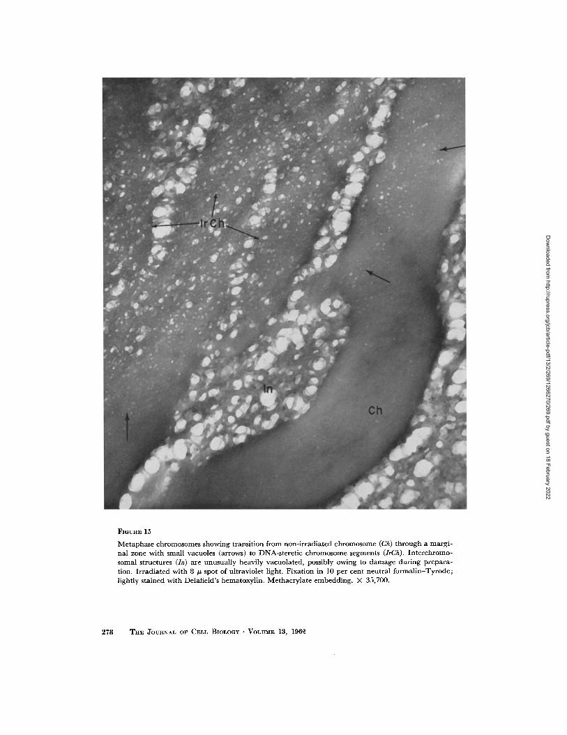
FIGURE 30
The telophase nucleus, slightly later in the cycle than that of Fig. 29, shows scattered, faintly outlined reticular strands in the masses of homogeneous component-A (A) The mottling is apparently a result of compression in sectioning. A dark edge (iV) delimits the nuclear mass from cytoplasm. Attached to this limiting structure are dark strands (R') like strands (R') of Fig. 29. Irregular lines and groups of chromosomal strands (R') probably represent surfaces of fusion of anaphase chromosomes and, perhaps, the earliest stages of reconstruction. Fixed in neutral formalin-Tyrode and stained with phosphotungstic acid. Methacrylate embedding. X 49,500.
298 THE JOURNAL OF CELL BIOLOGY • VOLUME 13, 196°~
Dow
nloaded from http://rupress.org/jcb/article-pdf/13/2/269/1266270/269.pdf by guest on 18 February 2022

W. BLOOM AND 1~. J. LEIDER Change$ in Ultraviolet-Irradiated Segments 299
Dow
nloaded from http://rupress.org/jcb/article-pdf/13/2/269/1266270/269.pdf by guest on 18 February 2022
FIGURE 31
Stage in nuc lear reconstruct ion shortly after tha t shown in Fig. 30. M u e h less compo- nen t -A is visible (A) and m u c h more of the ch romosomal re t i cu lum (R) is seen. Re t icu- lar s t rands (R ~) pass ing t h rough the edge of the nucleus (AT) can still be seen. Fixed in neut ra l f o rma l in -Ty rode and s ta ined with alcoholic phosphotungs t ic acid. M e t h - acrylate embedding . X 49,500.
300 THE JOURNAL OF CELL BIOLOGY • VOLUME 13, 196~
Dow
nloaded from http://rupress.org/jcb/article-pdf/13/2/269/1266270/269.pdf by guest on 18 February 2022

W. BLOOM AND R. J. LEIDER Change~ iX Ultraviolet-Irradiated Segments 301
Dow
nloaded from http://rupress.org/jcb/article-pdf/13/2/269/1266270/269.pdf by guest on 18 February 2022