open the brain is required for normal muscle and nerve ... · spinal cord induces alterations in...
TRANSCRIPT
ARTICLE
The brain is required for normal muscle and nervepatterning during early Xenopus developmentCelia Herrera-Rincon 1, Vaibhav P. Pai 1, Kristine M. Moran1, Joan M. Lemire1 & Michael Levin 1
Possible roles of brain-derived signals in the regulation of embryogenesis are unknown. Here
we use an amputation assay in Xenopus laevis to show that absence of brain alters subsequent
muscle and peripheral nerve patterning during early development. The muscle phenotype can
be rescued by an antagonist of muscarinic acetylcholine receptors. The observed defects
occur at considerable distances from the head, suggesting that the brain provides long-range
cues for other tissue systems during development. The presence of brain also protects
embryos from otherwise-teratogenic agents. Overexpression of a hyperpolarization-activated
cyclic nucleotide-gated ion channel rescues the muscle phenotype and the neural
mispatterning that occur in brainless embryos, even when expressed far from the muscle or
neural cells that mispattern. We identify a previously undescribed developmental role for the
brain and reveal a non-local input into the control of early morphogenesis that is mediated by
neurotransmitters and ion channel activity.
DOI: 10.1038/s41467-017-00597-2 OPEN
1 Biology Department and Allen Discovery Center, Tufts University, 200 Boston Avenue, suite 4600, Medford, MA 02155-4243, USA. Correspondence andrequests for materials should be addressed to M.L. (email: [email protected])
NATURE COMMUNICATIONS |8: 587 |DOI: 10.1038/s41467-017-00597-2 |www.nature.com/naturecommunications 1

The brain and central nervous system (CNS) generateinformation that controls muscle activity to implementbehavior in adult organisms. However, the CNS may also
provide an instructive influence over the behavior of multiplecell types during the establishment, repair and maintenanceof complex anatomical patterns in vivo. For example, thepatterning disorder known as cancer has been linked toneural control1. Tumors are readily induced by denervation incockroach2, and are more readily induced by chemical
carcinogens in denervated rabbit ears compared withcontralateral controls bearing normal innervation3. The same hasbeen observed with sarcomas implanted into normal ordenervated frog limbs4. Even normal tissue can disorganize in theabsence of neural signaling, as occurs in the papillae of themammalian tongue after denervation5. Thus, the presence ofgrowth control signals between the CNS and target cells has led tosuggestions of the CNS as a potential approach to treat tumorprogression in the clinic6.
Ctr
lB
R–
Brain removal Early Late
a
e
c
Con
trol
(C
trl)
f
h
Bra
inle
ss (
BR
– )
90° 135° 180°
Mean angle of myotome fibersat late
Rostral–caudal axis
*
*
*
d g
90° 135° 180°
Mean angle of myotome fibersat early
Ro
stral–c
au
da
laxis
**
**
**
CtrlBR–
b
Early Late
CorrectIncorrect
fb
hb
sm
cg
e
ARTICLE NATURE COMMUNICATIONS | DOI: 10.1038/s41467-017-00597-2
2 NATURE COMMUNICATIONS |8: 587 |DOI: 10.1038/s41467-017-00597-2 |www.nature.com/naturecommunications
Likewise, pattern regulation during regeneration requiresneural signals. Appendage regeneration has long been known tobe dependent on local neural supply7, although this is apparentlyan acquired addiction to developmental presence of innervation8.Recent studies have implicated roles for neural signaling in boneregeneration9 and epidermal de-differentiation10. The role ofCNS in regeneration is instructive, not merely permissive.The polarity and contiguity of the CNS determines head/tailspecification in a number of invertebrate models, includingplanaria11, 12. In salamander, the presence of nerve in a non-native location can induce the formation of ectopic limbs atwound sites13. Moreover, in the frog model, focal damage to thespinal cord induces alterations in the specific pattern of tailregeneration, with distinct morphological changes resulting fromspinal cord interruptions at different locations14. Despite recentadvances15, the molecular connection and reciprocal influencebetween nerves and patterning remains poorly understood16,especially with respect to the ultimate source of neutrally medi-ated patterning signals.
One of the most interesting contexts for neural pattern controlis embryogenesis, when anatomical structures are first established.Parasympathetic innervation controls morphogenesis of thesubmandibular gland, influencing the branching and generalpatterning of the organ17. Muscle cells are also affected byinteractions with nerves18, especially in terms of gene expressionof myogenic regularity factors (MRFs)19. Complementingmolecular and cell-level readouts, regenerative medicineapproaches to birth defects and bioengineered organs requiresaddressing neural inputs into large-scale patterning. However,possible roles of the brain (or brain-derived signals) for earlypatterning, long before behavior begins, remain unknown.
In Xenopus, somitogenesis occurs in an anterior-posterior,bilaterally symmetric manner. In the clock and wavefrontmodel20, 21, the presomitic mesodermal cells dynamically oscillatebetween permissive and nonpermissive conditions for segmen-tation (clock) as the embryonic development proceeds fromanterior to posterior direction (wavefront along with tissueshortening-mediated Doppler effect) resulting in formation ofsegmented somites. Wnt, Shh, BMP, Fgf and Notch pathways areinvolved in regulating the clock with rhythmic activation of thesepathways conserved across vertebrates. The intersection ofantagonistic gradients of retinoic acid (anterior to posterior) andFgf and Wnt (posterior to anterior) determine the wavefront22, 23.Given the tandem physiology of nerves and muscles, mightinnervation help orchestrate the symphony of signals that leads tosomitogenesis? Does the presumptive nervous system releaseinstructive signals that might be involved in somitogenesisand muscle development? Is there any element of this processco-opted into muscle regeneration? Similarly, the peripheralinnervation forms a stereotypic and complex neural network,
which also must be precisely patterned in a way that integratessize and positional information across the whole body24.
Most of the existing knowledge about neural inputs focuses onthe local microenvironment; very few studies have examined thecontribution of the CNS in providing long-range instructive cuesto muscle or peripheral innervation. To probe the role of distalneural structures and to search for tissues that could providecomplex nerve-mediated information to downstream patterningtargets, we focused on the brain. Here, we establish a brainlessXenopus embryonic model to reveal a role for the brain inthe patterning of muscle (somite) and peripheral innervation.Crucially, we show that defects induced by extirpation ofthe brain, or by teratogenic agents in the context of normalembryogenesis, can be largely rescued in the absence of brain bythe dorsal overexpression of an exogenous ion channel orexposure to a muscarinic-receptor antagonist. The characteriza-tion of defects and their repair by global (pharmacological) orspatially targeted (molecular-genetic) reagents reveal the firstmolecular components of the signaling cascade by which thebrain directs complex embryonic patterning processes.
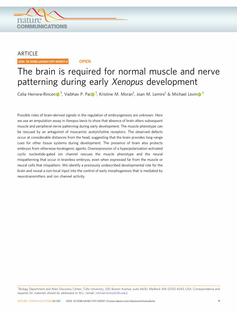
ResultsBrain is required for normal muscle development and pat-terning. To investigate the possible role of brain-derived signalsfor muscle morphogenesis and patterning, we established an assayin Xenopus laevis: brain removal at stage 25 (Fig. 1a, b), perfor-med at a time when its main subdivisions (forebrain, midbrainand hindbrain) and the rostral-caudal and dorsal-ventral axis arealready defined25. By removing the brain at the early tailbud stage(i.e., when somitogenesis is starting), we were able to studymuscle structure development in completely brainless developinganimals (> 85% of the microsurgeries we performed resulted inviable animals). To determine whether the brain is required forthe onset and/or patterning of myotomes, we evaluated themuscle phenotype at two relevant time points, corresponding tothe different myogenic waves (reviewed in ref. 23): early- (stages30–41; first and second waves completed) and late- (stages 42–48;third wave completed) stages after brain removal, respectively.
Soon after brain removal (stages 30–41), animals developingwithout a brain (BR−) began to display a notable decrease(−43± 7%) in the collagen density of the myotomes, compared tothe control animals (Ctrl) (OD mean value of 64± 8 units forBR− group compared to 113± 13 units for Ctrl group; t-testP < 0.01; n= 79) (Fig. 1c, d, turquoise and magenta short arrows).Analysis of the muscle structure revealed that the somiteswere 25% shorter in BR− than those found in control animals(101± 24 μm vs. 148± 13 μm, t-test P< 0.01; n= 75) (Fig. 1c, d,double-headed arrow indicates the length of one myotome).Moreover, the spatial organization of the somatic muscle was also
Fig. 1 The absence of the early brain leads to abnormal muscle development and patterning. a After fertilization, the brain was removed from stage 25embryos to generate BR− animals. Morphological evaluation of muscle phenotype was performed at early- (stages 30–41) and late- (42–48) stages.b Lateral views of stage 25 embryos before (left) and after (right) brain removal. The area occupied by the developing brain is marked with a white-dashedline. (left) rostral is left and dorsal is up. Scale bar, 250 μm. cg, cement gland; e, eye; fb, forebrain, hb, hindbrain, sm, somites. c–h The brain is required fornormal muscle development and patterning, as shown after quantitative evaluation of collagen density (short arrows), length of myotome fibers (double-headed arrows), central body axis and myotome angle (overlaid dashed axis and arrowhead-like lines) at early c, d and late f–h stages. At the onset ofdevelopment, BR− embryos possessed a lower collagen density in myotome fibers (magenta arrow in c compared to turquoise arrow in b), a significantlymore open central angle along the rostra-caudal axis e and shorter somites than control (Ctrl) embryos. During development, defects in the organization ofcentral body axis and muscle patterning were not corrected at any anatomical level in BR− (magenta dashed lines in g compared to turquoise dashed lines in f.The mean angle for BR− is significantly displaced to 180°, compared to those in Ctrl (H). c, d, f, g Photomicrographs taken under polarized light. Rostral isupper right and dorsal is up. Turquoise and magenta arrows indicate correct and incorrect anatomical pattern, respectively. Scale bar, 500 μm. e, h Graphicrepresentation of the mean angle of myotome fibers at rostral, central and caudal levels (blue squares) of Ctrl (white) and BR− (gray) embryos. Datarepresent the mean and s.d. of three independent replicates (n= 75 animals per group). P values after t (equal variances, black labels) or Mann–Whitney(unequal variances, blue labels) tests are indicated as **P< 0.01, *P< 0.05, ns no significant difference
NATURE COMMUNICATIONS | DOI: 10.1038/s41467-017-00597-2 ARTICLE
NATURE COMMUNICATIONS |8: 587 |DOI: 10.1038/s41467-017-00597-2 |www.nature.com/naturecommunications 3
perturbed. The body axis and mean angle of the muscle fibersalong the anteroposterior axis were significantly altered in BR−,with a mean of 16± 2° more-opened angles compared to Ctrlones (t-test P< 0.01 for rostral and caudal levels, Mann–Whitneytest P< 0.01 for central level levels; n= 75) (Fig. 1e and Table 1).
We then asked whether the early defects in muscle patterningwere also present during subsequent development in BR−
embryos. To address this question, we analyzed the number ofsomites (indicator of segmentation) and the fine muscle structure(angle and length of the myotome fibers) at late stages (stages42–48). No significant differences were detected for the meannumber of somites between Ctrl and BR− embryos (33± 3 vs.32± 4; t-test P= 0.74; n= 75), suggesting that the brain was notfunctionally implicated in segmentation per se, and that our assaydoes not generally (nonspecifically) impair embryogenesis of thesomites. In contrast, the analysis of the fine muscle structureconfirmed that the defects in myotome organization werenot repaired during subsequent development. Myotomesin late-staged BR− embryos were 10% shorter (136± 14 μm vs.160± 13 μm, t-test P< 0.01; n= 75) than in control animals.Likewise, the central axis in the late-stage BR− was significantlydisplaced, somites lacked the typical chevron-shape, and adifference of −9± 1° in the mean angle of the myotome fiberswas detected when compared to Ctrl animals (Mann–Whitneytest P< 0.05 for rostral and central levels, t-test P< 0.05 forcaudal level; n= 75) (Fig. 1f–h, overlaid dashed axis andarrowhead-like lines, and Table 1). Control animals subjectedto sham surgeries where either yolk mass or tailbud was resected(Yolk− or Tail− embryos, respectively) displayed normal musclearchitecture, indistinguishable from control myotome fibers, bothin terms of length/definition and angle of the central myotomes(at distance from the initial resection, in the case of Tail− sham-embryos) (Supplementary Fig. 1A–G), demonstrating thatsurgery per se (and subsequent regenerative responses) do notinduce muscle mispatterning. We conclude from these data thatwhile the brain is not required for the somite segmentation(partitioning of the presomitic mesoderm into somites), it has akey role in both the onset and establishment of correct musclepatterning and structure.
Muscle organization was adversely affected both at themicroscopic tissue organization level, as well as macroscopically– at the animal morphological level (incidence of aberrantphenotype within both Ctrl and BR− population; Table 2). Amacroscopic evaluation of embryo morphology revealed a higherpercentage of abnormal embryos in the BR− population, both atearly (66± 6% vs. 13± 1%; z-test, P< 0.01) and at later stages(80± 1% vs. 16± 2%; z-test P< 0.01) compared to Ctrl animals.Muscle organization was affected by brain absence rather thanany indirect detrimental effect of tissue removal surgery,as demonstrated the percent of aberration in Yolk− population
(14± 2% and 15± 5% at the onset and later in development,respectively; z-test compared to Ctrl P > 0.05 for both cases;Supplementary Fig. 1H).
Taken together, these results clearly indicate the importance ofthe early embryonic brain for normal development of musclestructure occurring at considerable distance.
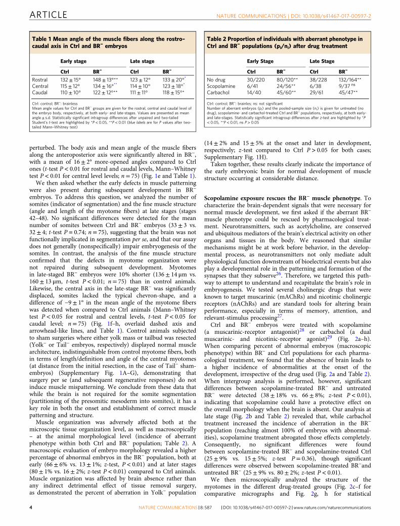
Scopolamine exposure rescues the BR− muscle phenotype. Tocharacterize the brain-dependent signals that were necessary fornormal muscle development, we first asked if the aberrant BR−
muscle phenotype could be rescued by pharmacological treat-ment. Neurotransmitters, such as acetylcholine, are conservedand ubiquitous mediators of the brain’s electrical activity on otherorgans and tissues in the body. We reasoned that similarmechanisms might be at work before behavior, in the develop-mental process, as neurotransmitters not only mediate adultphysiological function downstream of bioelectrical events but alsoplay a developmental role in the patterning and formation of thesynapses that they subserve26. Therefore, we targeted this path-way to attempt to understand and recapitulate the brain’s role inembryogenesis. We tested several cholinergic drugs that wereknown to target muscarinic (mAChRs) and nicotinic cholinergicreceptors (nAChRs) and are standard tools for altering brainperformance, especially in terms of memory, attention, andrelevant-stimulus processing27.
Ctrl and BR− embryos were treated with scopolamine(a muscarinic-receptor antagonist)28 or carbachol (a dualmuscarinic- and nicotinic-receptor agonist)29 (Fig. 2a–h).When comparing percent of abnormal embryos (macroscopicphenotype) within BR− and Ctrl populations for each pharma-cological treatment, we found that the absence of brain leads toa higher incidence of abnormalities at the onset of thedevelopment, irrespective of the drug used (Fig. 2a and Table 2).When intergroup analysis is performed, however, significantdifferences between scopolamine-treated BR− and untreatedBR− were detected (38± 18% vs. 66± 8%; z-test P< 0.01),indicating that scopolamine could have a protective effect onthe overall morphology when the brain is absent. Our analysis atlate stage (Fig. 2b and Table 2) revealed that, while carbacholtreatment increased the incidence of aberration in the BR−
population (reaching almost 100% of embryos with abnormal-ities), scopolamine treatment abrogated those effects completely.Consequently, no significant differences were foundbetween scopolamine-treated BR− and scopolamine-treated Ctrl(25± 9% vs. 15± 5%; z-test P= 0.36), though significantdifferences were observed between scopolamine-treated BR−anduntreated BR− (25± 9% vs. 80± 2%; z-test P< 0.01).
We then microscopically analyzed the structure of themyotomes in the different drug-treated groups (Fig. 2c–f forcomparative micrographs and Fig. 2g, h for statistical
Table 1 Mean angle of the muscle fibers along the rostro–caudal axis in Ctrl and BR− embryos
Early stage Late stage
Ctrl BR− Ctrl BR−
Rostral 132± 15° 148± 13°** 123± 12° 133± 20°*
Central 115± 12° 134± 16°** 114± 10° 123± 18°*
Caudal 110± 10° 122± 12°** 111± 11° 118± 15°*
Ctrl: control; BR−: brainlessMean angle values for Ctrl and BR− groups are given for the rostral, central and caudal level ofthe embryo body, respectively, at both early- and late-stages. Values are presented as meanangle± s.d. Statistically significant intragroup differences after unpaired and two-tailedStudent’s t-test are highlighted by *P< 0.05, **P< 0.01 (blue labels are for P values after two-tailed Mann–Whitney test)
Table 2 Proportion of individuals with aberrant phenotype inCtrl and BR− populations (pi/ni) after drug treatment
Early Stage Late Stage
Ctrl BR− Ctrl BR−
No drug 30/220 80/120** 38/228 132/164**Scopolamine 6/41 24/56** 6/38 9/37 ns
Carbachol 14/40 45/60** 29/61 45/47**
Ctrl: control; BR−: brainles; ns: not significantNumber of aberrant embryos (pi) and the pooled-sample size (ni) is given for untreated (nodrug), scopolamine- and carbachol-treated Ctrl and BR− populations, respectively, at both early-and late-stages. Statistically significant intragroup differences after z-test are highlighted by *P< 0.05, **P< 0.01, ns P> 0.05
ARTICLE NATURE COMMUNICATIONS | DOI: 10.1038/s41467-017-00597-2
4 NATURE COMMUNICATIONS |8: 587 |DOI: 10.1038/s41467-017-00597-2 |www.nature.com/naturecommunications
comparisons). Evaluation of somitogenesis and fine somaticmuscle structure revealed that both the organization and size ofmyotomes in BR− treated with scopolamine resembled the typicalCtrl muscle phenotype (turquoise arrows in Fig. 2c, e), anddiffered clearly from the typical BR− muscle phenotype
(represented in Fig. 2d). Both the mean number of somites(32± 2) and length of muscle fibers at early- (141± 7 μm)and late- (157± 17 μm) staged scopolamine-treated BR− werestatistically similar to the values for the untreated Ctrl population(with mean number of 32± 2 somites and mean lengths of
No dr
ug
First w
eek
Secon
d wee
k
First a
nd
seco
nd wee
k 0%
20%
40%
60%
80%
100%
60
iScopolamine exposure and rescue
No drugFirst week
Second weekFirst and second week
1 w0 2 w
No dr
ug
Scopo
lamine
Carba
chol
0%
20%
40%
60%
80%
100%
Per
cent
abn
orm
alat
late
No dr
ug
Scopo
lamine
Carba
chol
0%
20%
40%
60%
80%
100%P
erce
nt a
bnor
mal
at e
arly
**
**
**
bans
**ns**
**
*** Ctrl
BR–
220 120 41 5640 60 228 164 38 3761 47
Contro
l BR
–
BR– +
scop
olam
ine
BR– +
carb
acho
l0
50
100
150
200
250
Leng
th o
f myo
tom
e fib
ers
(μm
) at
ear
ly
**nsns
75 75 50 50
g
Contro
lBR
–
BR– +
scop
olam
ine
BR– +
carb
acho
l 0
50
100
150
200
250
Leng
th o
f myo
tom
e fib
ers
(μm
) at
late
*ns
**
75 75
c d
fe
Ctrl BR–
BR– + scopolamine
CtrlBR–
h
****
ns
Per
cent
abn
orm
alin
bra
inle
ss
CorrectIncorrectAberrant
BR– + carbachol
65 53 55
47 37
NATURE COMMUNICATIONS | DOI: 10.1038/s41467-017-00597-2 ARTICLE
NATURE COMMUNICATIONS |8: 587 |DOI: 10.1038/s41467-017-00597-2 |www.nature.com/naturecommunications 5

148± 13 μm at early stage and mean lengths of 160± 13 μm atlate stage; Fig. 2g, h). Conversely, carbachol treatment in BR− haddramatic negative consequences for muscle formation andpatterning, which was especially clear at the later stages, deviatingthe organization of somites and myotomes from the typicalBR−-induced aberrant muscle phenotype. At early stages, the sizeof the myotomes (mean length of 137± 11 μm) was similar towhat was measured in the Ctrl embryos. However, later on indevelopment, a significantly lower number of somites (24± 4; P< 0.01 after post-hoc Bonferroni’s test), longer myotome fibers(196± 35 μm; P< 0.01 after post-hoc Dunn’s test), and completeasymmetric and disorganized muscle patterning were observed inthe carbachol-treated BR−(magenta and yellow arrows in Fig. 2f).Taken together, we conclude that the brain may inhibit themuscarinic pathway to achieve correct organization of thesomatic muscle system and that the absence or prevention ofmuscarinic signaling (for example, the pharmacological antagon-ism mediated by scopolamine) is able to rescue the aberrant BR−
muscle phenotype.We next sought to determine when during muscle develop-
ment the rescue effect of scopolamine occurs. BR− embryos wereexposed to scopolamine for different lengths of time (Fig. 2i left,for a schematic representation of drug-exposure timing). Weobserved that scopolamine treatment is able to fix the BR− musclephenotype if the drug exposure starts immediately after the brainremoval (Fig. 2i, right), regardless of whether BR− are treated forone (first-week experimental group) or 2 weeks (first- andsecond-week experimental group). In either case, treatment wasable to significantly decrease the percentage of abnormal tadpoles(51± 13% and 55± 7%, respectively; z-test P< 0.01 for bothgroups) when compared to the BR− population that had not beenexposed to drug (87± 11% of abnormalities). Our results indicatethat scopolamine acts on events during the first week (from stages25 to 37) after brain removal.
Ectopic HCN2 expression rescues BR− muscle phenotype.Considering the many examples of muscle formation andpatterning mediated by bioelectrical signaling in both mammals30
and amphibians31, as well as the ability of some channelsto rescue profound embryonic defects32, we wonderedwhether the exogenous expression of a specific ion channel,hyperpolarization-activated cyclic nucleotide-gated ion channel 2(HCN2), could counter the effects of brain removal. The HCN2channel is known to be an important modulator of functionalbioelectric state33 and can hyperpolarize cells, and it has beenrecently shown to be implied in firing and rhythmic propertiesof the cholinergic neurons in both CNS and gastrointestinaltract34, 35. We found the HCN2 channel to be endogenously
expressed in the developing neural tube along the base and lateralregions and in the perisomitic area (Supplementary Fig. 2). Toaddress the role of bioelectric signaling during muscle develop-ment in organisms incapable of relaying signals from their brainsto other tissue, embryos were injected at two-cell stage, in bothblastomeres, with mRNA encoding wild-type HCN2 (Fig. 3a,turquoise arrows, HCN2-WT group). Uninjected and water-injected embryos served as controls. In addition, in order tounderstand the signaling between HCN2-expressing cells and thestructural muscle outcome (either a local or long-distance effect),we evaluated the muscle phenotype after injection of HCN2-WTmRNA in only one side (left-right) of the embryo (1/2 HCN2-WT group). Co-injection with lacZ mRNA (as a reporter ofinjected cells’ progeny; Fig. 3b, left, blue arrow) and detection ofthe β-galactosidase (β-gal) distribution were used to select andevaluate quantitatively those embryos with strongly unilateralexpression (Fig. 3b, right, blue arrow indicates high β-galexpression and, thus, ipsilateral injected side).
First, we quantified the proportion of embryos with abnormalphenotype within each BR− population (untreated or uninjected,water-injected, HCN2-WT and 1/2 HCN2-WT; Fig. 3c). Ourresults revealed that the expression of a WT HCN2 mRNA inBR− clearly reduced the onset of abnormalities in the macroscopicmorphology and significantly decreased the percent of abnormalembryos within the population (from 85± 7% in non-injectedBR− embryos to 45± 7% in HCN2-WT BR− group; z-testP< 0.01). We observed the same rescue effect of HCN2 channelwhen only one side of the animal was injected. The incidence ofaberrant phenotypes in the 1/2 HCN2-WT BR− group wassignificantly lower than that for the regular (or uninjected)BR− population (65± 3% of aberrant individuals in the 1/2HCN2-WT BR− group; z-test P< 0.01 compared to thenon-injected BR−group).
We then microscopically analyzed the somatic-myotomepatterning in BR− embryos under the different ion channelmisexpression conditions (Fig. 3d–g). Analysis of fine musclestructure revealed that the HCN2-WT BR− mutants exhibitedsomites and myotomes that were perfectly organized, differingclearly from the typical BR− muscle phenotype (Fig. 3d, e forcomparative micrographs). The differences in the number ofsomites (30± 2) and the mean length of myofibers (134± 11 μmat early stage and 165± 15 μm at late stage) were not statisticallysignificant from those measured in uninjected Ctrl embryos (afterpost-hoc Bonferroni’s test). The unilateral HCN2 expression inanimals developing without brain had a protective effect on themuscle organization of both the local side and the uninjectedcontralateral side. Like the results for the both sides HCN2-injections, the size of myotome fibers in the uninjectedcontralateral side were similar to the Ctrl embryos, both at the
Fig. 2 Scopolamine rescues the BR− muscle phenotype. a, b Quantification of the mean percentage of abnormal embryos and statistical comparisonsamong Ctrl and BR− populations under normal conditions and after drug treatment, at early- (a) and late- (b) stages after brain removal. Values are plottedas mean %± s.d. (no-pooled data from, at least, three different replicates). c–f. Typical muscle phenotype for Ctrl (c) and BR− (d), and BR− afterscopolamine (e) or carbachol treatment (f), as seen under polarized light. Rostral is upper right and dorsal is up. Turquoise, magenta and yellow arrowsindicate correct, incorrect and aberrant formation, respectively. Scale bar, 100 μm. g, h Quantification of the mean length of myotome fibers and statisticalcomparisons among untreated Ctrl and untreated BR−, scopolamine-treated BR− and carbachol-treated BR− at early- (g, one-way ANOVA, P< 0.01) andlate- (h, Kruskal–Wallis test, P< 0.01) stages after brain removal. No significant differences after a posteriori analysis were detected among the differentCtrl groups. Data represent the mean and s.d. of three independent replicates. i. Scopolamine exposure and rescue effects on BR− phenotype. (left)Graphical representation of the different exposure times to scopolamine in BR−, after brain removal (t= 0, magenta arrow; white band means no drug andblue bands means drug treatment) and for a 2-week (2w) period. First-week experimental group was exposed to scopolamine immediately after brainremoval and consecutively for the first week. Second-week animals were exposed to the drug 1 week after the brain removal, for 1-week period. First- andsecond-week animals were exposed to scopolamine immediately after the brain removal and for the 2 next consecutive weeks. (right) Quantification of themean percentage of embryos with abnormal phenotype within each BR− group. Values are plotted as mean %± s.d. (no-pooled data from three differentreplicates). For all panels, number in bars indicates n or number of embryos analyzed for each group. P values after z-test a, b, i and post-hoc Bonferroni’sg or Dunn’s test h are indicated as **P< 0.01, *P< 0.05, ns P> 0.05
ARTICLE NATURE COMMUNICATIONS | DOI: 10.1038/s41467-017-00597-2
6 NATURE COMMUNICATIONS |8: 587 |DOI: 10.1038/s41467-017-00597-2 |www.nature.com/naturecommunications
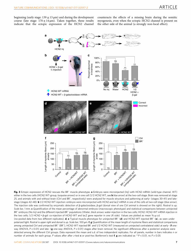
beginning (early stage: 139± 13 μm) and during the developmentcourse (late stage: 170± 14 μm). Taken together, these resultsindicate that the ectopic expression of the HCN2 channel
counteracts the effects of a missing brain during the somiticmyogenesis, even when the ectopic HCN2 channel is present onthe other side of the animal (a strongly non-local effect).
BR– – HCN2 WT
d
a
HCN2 + β-gal½ Injection
J
HCN2-WT mRNA
HCN2-WT + β-galactosidase mRNA
BR– + HCN2 WT
e
0
50
100
150
200
250
Contro
lBR
–
BR– +
1/2H
CN2 W
T
(unin
jecte
d sid
e)
BR– +
HCN2W
T
Contro
lBR
–
BR– +
1/2H
CN2 W
T
(unin
jecte
d sid
e)
BR– +
HCN2W
T 0
50
100
150
200
250
Leng
th o
f myo
tom
e fib
ers
(μm
)at
late
Leng
th o
f myo
tom
e fib
ers
(μm
)at
ear
ly
gf
**ns ns **
ns ns
CtrlBR–
70
cb
No Inj Water HCN2
165 33 5 57 3535
ns
****
1/2HCN2 +
β-gal
Per
cent
abn
orm
alin
bra
inle
ss
CorrectIncorrect
Ctr
lB
R–
HCN2 Inj
0%
20%
40%
60%
80%
100%
70 75 55 3575 30 45
Fig. 3 Ectopic expression of HCN2 rescues the BR− muscle phenotype. a Embryos were microinjected (Inj) with HCN2 mRNA (wild-type channel, WT)either in the two cells (HCN2WT-group, turquoise arrows) or in one cell (1/2 HCN2WT, see b blue arrow) at the two-cell stage. Brain was removed at stage25, and animals with and without brain (Ctrl and BR−, respectively) were analyzed for muscle structure and patterning at early- (stages 30–41) and late-stage (stages 42–48). b 1/2 HCN2-WT injection: embryos were microinjected with HCN2 and lacZ mRNA in one of the cells at two-cell stage (blue arrow).The injection side was confirmed by enzymatic detection of β-galactosidase, β-gal (dorsal view of one Ctrl animal is showed on the right). Rostral is up.Scale bar, 1 mm. c Quantification of the mean percentage of abnormal embryos (macroscopic phenotype) and statistical comparisons between uninjectedBR− embryos (No Inj) and the different injected-BR− populations (Water, black arrows: water-injection in the two cells; HCN2: HCN2-WT mRNA injection inthe two cells; 1/2 HCN2 + β-gal: co-injection of HCN2-WT and lacZ gene reporter in one LR side). Values are plotted as mean %± s.d.(no-pooled data from two different replicates). d, e Typical muscle phenotype for uninjected BR− (d) and HCN2-WT injected BR− (e), as seen underpolarized light. Rostral is upper right and dorsal is up. Scale bar, 100 μm. f, g Quantification of the mean length of myotome fibers and statistical comparisonsamong uninjected Ctrl and uninjected BR− (BR−), HCN2-WT injected BR- and 1/2 HCN2-WT (measured on uninjected contralateral side) at early- (f one-way ANOVA, P< 0.01) and late- (g one-way ANOVA, P< 0.01) stages after brain removal. No significant differences after a posteriori analysis weredetected among the different Ctrl groups. Data represent the mean and s.d. of two independent replicates. For all panels, number in bars indicates n ornumber of animals for each group. P values after after z-test c or post-hoc Bonferroni’s test f, g are indicated as **P< 0.01, ns P> 0.05
NATURE COMMUNICATIONS | DOI: 10.1038/s41467-017-00597-2 ARTICLE
NATURE COMMUNICATIONS |8: 587 |DOI: 10.1038/s41467-017-00597-2 |www.nature.com/naturecommunications 7
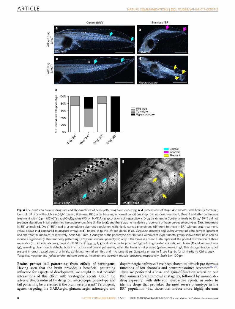
Brains protect tail patterning from effects of teratogens.Having seen that the brain provides a beneficial patterninginfluence for aspects of development, we sought to test possibleinteractions of this effect with teratogenic agents. Could theadverse effects induced by drugs on macroscopic phenotype andtail patterning be prevented if the brain were present? Teratogenicagents targeting the GABAergic, glutamatergic, adrenergic and
dopaminergic pathways have been shown to perturb pre-nervousfunctions of ion channels and neurotransmitter receptors36, 37.Thus, we performed a loss- and gain-of-function screen on ourBR− animals (brain removal at stage 25, followed by immediate-drug exposure) with different neuroactive agents, in order toidentify drugs that provoked the most severe phenotype in theBR− population (i.e., those that induce more highly aberrant
Drug– B
R+
Drug– BR
–
Drug+ B
R+
Drug+ BR
–0%
20%
40%
60%
80%
100%
% o
f ind
ivid
uals
with
phe
noty
pe
Wild typeCurvatureHypercurvature
e
b
Control (BR+)
With
out d
rug
(dru
g– ) W
ith d
rug
(dru
g+)
Wild type
c
Brainless (BR–)
Curvature
Hypercurvature
b
d
CorrectIncorrectAberrant
a
f g
Drug+ + brain Drug+ – brain
Fig. 4 The brain can prevent drug-induced abnormalities of body patterning from occurring. a–d Lateral view of stage-45 tadpoles with brain (left column;Control, BR+) or without brain (right column; Brainless, BR−) after housing in normal conditions (top row; no drug treatment, Drug−) and after continuoustreatment with 10 μm (RS)-(Tetrazol-5-yl)glycine (RS, an NMDA receptor agonist), respectively. Drug treatment in Control animals (c, Drug+ BR+) did notproduce alterations in tail patterning (turquoise arrows in c similar to a), and there was no incidence of aberrant or hypercurved phenotypes. Drug treatmentin BR− animals (d, Drug+ BR−) lead to a completely aberrant population, with highly curved phenotypes (different to those in BR− without drug treatment,yellow arrows in d compared to magenta arrows in b). Rostral is to the left and dorsal is up. Turquoise, magenta and yellow arrows indicate correct, incorrectand aberrant tail modules, respectively. Scale bar, 1 mm. e Analysis of the phenotype distributions within each experimental group showed that RS is able toinduce a significantly aberrant body patterning (a ‘hypercurvature’ phenotype) only if the brain is absent. Data represent the pooled distribution of threereplicates (n= 75 animals per group). P< 0.01 for X2(0.05, 6). f, g Evaluation under polarized light of drug-treated animals, with brain (f) and without brain(g), revealing clear muscle defects, both in structure and overall patterning, when the brain is not present (yellow arrows in g). This disorganization is notpresent in drug-treated control animals, exhibiting normal somites and myotome fibers (turquoise arrows in f, see Fig. 2c for similarity to Ctrl group).Turquoise, magenta and yellow arrows indicate correct, incorrect and aberrant muscle structure, respectively. Scale bar, 100 μm
ARTICLE NATURE COMMUNICATIONS | DOI: 10.1038/s41467-017-00597-2
8 NATURE COMMUNICATIONS |8: 587 |DOI: 10.1038/s41467-017-00597-2 |www.nature.com/naturecommunications
Contro
lBR
–
BR– +
1/2
HCN2
WT
(unin
jecte
d sid
e)
BR– +
HCN2W
T
BR– +
carb
acho
l
BR– +
scop
olam
ine0
20
40
Tubu
lin la
belin
g (O
D)
inte
rnal
neu
ropi
l ***
**
ns
3233354537
ns
23
CtrlBR–
CorrectIncorrectAberrant
ba
e
c
Contralateral uninjected side Injected side
f
g h
d
Fig. 5 The absence of a brain generates an abnormal neural network in the entire animal body. a, b Acetylated-tubulin (Tub) immunoexpression for Ctrl (a)and BR− (b) animals. There three types of nerve fibers: (i) commissural fibers (dorsoventral axis, long arrows); (ii) longitudinal fibers (anteroposterior axis,short arrow); and (iii) internal neuropil (no defined axis, unfilled triangles). Animals developed without a brain show normal commissural and longitudinalnerve fibers (turquoise long arrows in b), with some alterations (magenta long arrow), but a dense internal neuropil (yellow unfilled triangles).c, d Tub-immunoexpression for BR− treated with cholinergic drugs: scopolamine (c) and carbachol (d). Scopolamine treatment was not able to rescue theaberrant internal network (yellow unfilled triangles in c), and carbachol-treated animals exhibited a chaotic nerve patterning (magenta and yellow arrowsin d). e Ectopic HCN2-WT expression (injected in both cells at two-cell stage) fixed the BR−–induced internal nerve branching. f Quantification of the meanOD of internal neuropil and statistical comparisons among untreated/uninjected Ctrl and untreated/uninjecetd BR− (BR−, without drug treatment norion channel misexpression), scopolamine-treated BR− (BR− + scopolamine), carbachol-treated BR− (BR− + carbachol), HCN2-WT both-sides injected BR−
(BR− + HCN2 WT), and HCN2-WT LR side-injected BR− (BR− + 1/2 HCN2 WT) embryos (one-way ANOVA, P< 0.01). No significant differences after aposteriori analysis were detected among the different Ctrl groups. Data represent the mean OD units and s.d. of two independent replicates. Number inbars indicates n or number of animals analyzed for each group. P values after post-hoc Bonferroni’s test are indicated as **P< 0.01, *P< 0.05, ns P> 0.05.g, h. Ectopic HCN2 expression in only one LR side fixes the BR−-induced internal nerve branching. Tub-immunoexpression on β-gal-reacted sections(dark deposits) in a 1/2 HCN2-WT BR−, showing both the contralateral uninjected side (g) and the injected (h) of the same embryo. Aberrant neuralnetwork was completely rescued (turquoise arrows), exhibiting a similar nerve pattern to the Ctrl group in both sides. a-e, h: Rostral is upper right and dorsalis up. g: Rostral is upper left and dorsal is up. Scale bar, 100 μm
NATURE COMMUNICATIONS | DOI: 10.1038/s41467-017-00597-2 ARTICLE
NATURE COMMUNICATIONS |8: 587 |DOI: 10.1038/s41467-017-00597-2 |www.nature.com/naturecommunications 9
phenotypes than the ones induced by brain removal). Weobserved that introduction of (RS)-(Tetrazol-5-yl)glycine (RS), anagonist of the NMDA-glutamate receptor38 significantlyincreased the occurrence of aberrant tail phenotypes within BR−
embryos (P< 0.01 for X2(0.05,6); Fig. 4) after 2 weeks of treatment
(stages 42–48).The most frequent tail phenotype in (untreated) BR− embryos
is characterized by a single lateral bending, starting approximatelyin the one-third posterior of the tail (see the most anterior arrowdrawn on Fig. 4a–d photomicrographs). Continuous treatmentwith RS in BR− caused a 99± 3% of highly bent tails (includingcurvature in notochord and spinal cord and spiraling of the tail;see Fig. 4d, yellow arrows, for a representative profile). Strikingly,these RS-induced effects only occurred in animals developedwithout brain. The RS treatment of Ctrl animals (normaldevelopment with brain, BR+) had no effect on tail patterning,and severe phenotypes were not detected. The analysis of thefrequency of distribution of the different phenotypes within eachpopulation revealed significant differences among the experi-mental conditions (Fig. 4e; X2
(0.05,6)= 370.8; P< 0.01). Themacroscopically identifiable changes in tail patterning, inducedby RS treatment in BR− embryos, were also accompanied by clearqualitative alterations in the fine muscle structure and somiteorganization (Fig. 4f, g). We conclude that presence of the brainhelps embryogenesis resist the disrupting effects of otherwisestrongly teratogenic agents.
To identify possible pathways mediating this brain-protectingeffect, we tested whether scopolamine (which rescues BR−-musclephenotype) could also protect against the effects of teratogens inBR− embryos. We found that in the absence of a brain,scopolamine treatment partially counteracted the teratogeniceffects of RS (Scopo+, Supplementary Fig. 3A–C). Scopolamine-treated RS-BR− embryos (Drug+BR−Scopo+) are much healthierthan BR− embryos, but they do not exhibit an entirely recoveredCtrl-like muscle phenotype (Supplementary Fig. 3D). Macro-scopic tail phenotype and muscle structure analysis revealed thatscopolamine significantly decreases the occurrence of highlyaberrant phenotypes (from 92± 2% in Drug+BR−Scopo− to15± 1% in Drug+BR−Scopo+, X2
(0.05, 4)= 136.3; P< 0.01), leadingto a phenotype distribution similar to what occurs in the regularBR− population (31± 9% in BR− without drug treatment orDrug−BR−Scopo−). We conclude that scopolamine treatmentprevents the severe deformities caused by this teratogen, but itseffect is not sufficient to fully prevent the muscle defects caused inBR− by the presence of the drug.
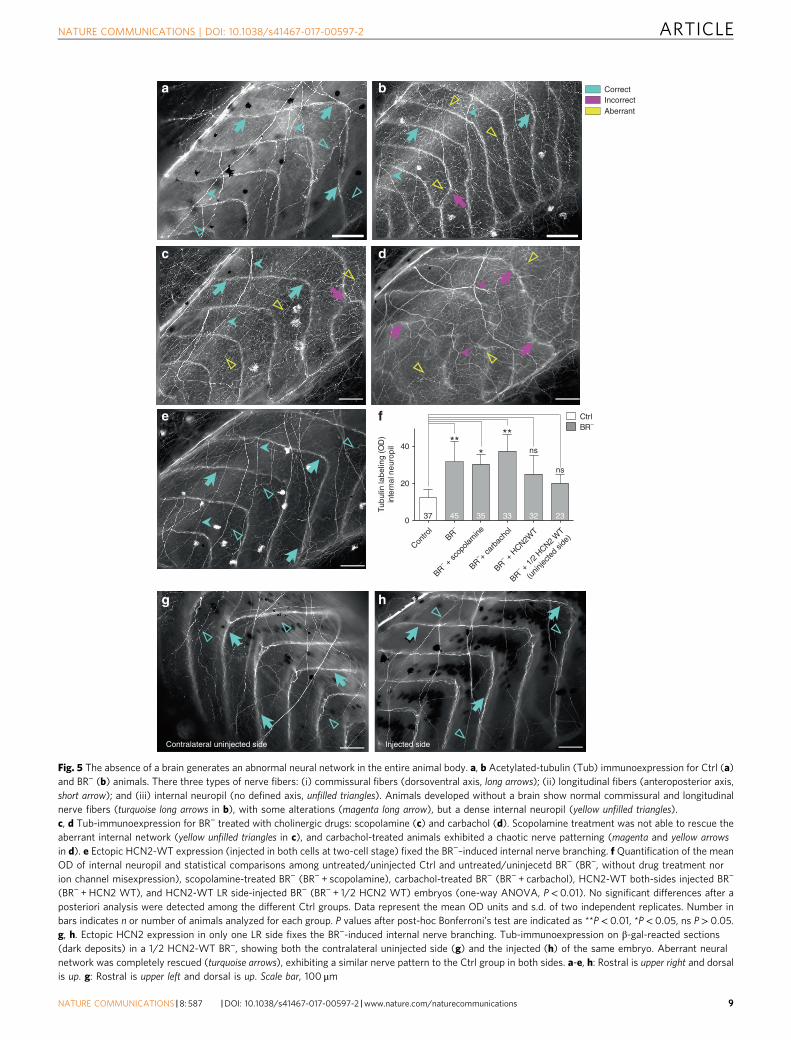
The absence of brain leads to abnormal neural development.Having seen the profound effects of the brain on the developingmusculature, we next asked what type of alterations/reorganiza-tions could have occurred on the remaining nervous tissue afterbrain removal (peripheral innervation).
We visualized the body-wide neural network at late-stageembryos by immunolabeling them with acetylated alpha-Tubulinantibody (Tub) and quantifying the Tub-immunolocalization byOD measurements (ranging from 0 (black, no expression) to 255(white, maximal expression)). This antibody is a widely-acceptedmarker for nerve fibers because stabilized microtubules, such asthose found in neuronal processes, contain important amounts ofacetylated tubulin39. The typical neural pattern in the peripheralnervous system (PNS) of Ctrl embryos revealed that, atlate stages, three types of fibers can be clearly identifiable afterTub-immunostaining (Fig. 5a): (i) commissural nerve fibers,running along dorsoventral axis (long arrows); (ii) longitudinalnerve fibers, along anteroposterior axis (short arrows); and(iii) internal network or neural network underlying the space
between two consecutive segments defined by the commissuralones (unfilled triangles). The internal neuropil in Ctrl animalsconsisted of a thin network, barely detected by OD measurements(OD mean value of 15± 7 units). After brain removal, andsimilarly to errors detected for segmentation, commissural andlongitudinal fibers in BR− were mispatterned (note someincorrect commissural nerve distribution coincident with defectsat the level of the somitic myogenesis, magenta arrow in Fig. 5b).Strikingly, embryos developed without a brain exhibited a robustectopic branching (internal neuropil), with nerve fibers chaoti-cally orientated through the animal body (yellow unfilledtriangles in Fig. 5b, OD mean value of 32± 11 units; P< 0.01compared to Ctrl group, after post-hoc Bonferroni’s test). Resultsfrom sham-surgery embryos (extirpating non-brain regions)confirmed that this nerve misspatterning is specifically due tobrain removal, as demonstrated after Tub immunolocalization(Supplementary Fig. 4). We conclude that the absence of a brainprovokes a dramatic and specific change of the branching of theinternal nerve net in the whole animal body (aberrant neuralnetwork).
To test whether the effect of brain removal was due to lack ofan endogenous pruning phase, we analyzed the motoneuronaxonal patterning in early stage embryos (stages 31–41;Supplementary Fig. 5) using the antibody znp1. This antibodylabels primary motoneuron axons40, and is widely used for manydifferent animal models during embryogenesis26. We found nosignificant difference in the density of motor axonal branches(internal neuropil) among stage 31, 35 and 45 Ctrl embryos(after post-hoc Bonferroni’s test). Conversely, both errorsin axonal establishment (magenta arrows in SupplementaryFig. 5B, D, F showing lack of trajectory compared to turquoisearrows in Supplementary Fig. 5A, C, E) and ectopic/aberrantbranching (yellow unfilled triangles in Supplementary Fig. 5B, D, Fpointing branches projecting off the main axons; compare toturquoise unfilled triangles in Supplementary Fig. 5A, C, E) weredetected from the onset and during the progression of thedevelopment in BR− embryos. Thus we conclude that thebrainless phenotype involved ectopic growth of neural tissue,not a failure of normal pruning.
Having detected that ectopic branching was a BR−-inducedspecific effect on peripheral nerve structure, we asked next if thepharmacological treatment and/or ectopic ion channel expressionused to rescue the muscle phenotype would have similar effectson the BR−-aberrant neural network (Fig. 5c–f). Results derivedfrom cholinergic-drug treatment revealed that the BR−-induceddense internal neuropil was not fixed by scopolamine orcarbachol. Unlike the rescue effects on muscle phenotype,scopolamine was not able to prevent the massive internal neuralbranching (Fig. 5c; OD mean value of 31± 5 units; P< 0.05compared to Ctrl group, after post-hoc Bonferroni’s test).Carbachol-treated BR− embryos showed a completelydisorganized nerve structure, more aberrant than those in drug-untreated BR−, with aberrations in the three different typesof nerve fibers (Fig. 5d; OD mean value for internal neuropil of37± 9 units; P< 0.01 compared to Ctrl group, after post-hocBonferroni’s test). However, analysis of HCN2-injected embryosshowed that the nerve sprouting consequent to developingwithout a brain was efficiently rescued by expression of HCN2WT (Fig. 5e; OD mean value of 25± 11 units; P> 0.05 comparedto Ctrl group, after post-hoc Bonferroni’s test). We conclude thatthe aberrant neural network in BR− can be fixed by ion channelmisexpression providing additional channels, but not by thepharmacological treatment targeting existing ones.
Given that the BR−-induced aberrant neural network wasrescued by HCN2 overexpression, we tested the spatial signalingbetween HCN2-expressing cells and the responding PNS, by
ARTICLE NATURE COMMUNICATIONS | DOI: 10.1038/s41467-017-00597-2
10 NATURE COMMUNICATIONS |8: 587 |DOI: 10.1038/s41467-017-00597-2 |www.nature.com/naturecommunications

means of quantitative evaluation of the nerve patterning in thecontralateral uninjected side of 1/2 HCN2-WT overexpressingembryos (see text above and Fig. 3b for experimental injectiondetails). 65% of uninjected sides (low HCN2 expression) hadinternal neuropil similar to those in both injected side (highHCN2 expression), and Ctrl animals (compare internal neuropilin a typical contralateral uninjected side (Fig. 5g) to that one ininjected side (Fig. 5h) and Ctrl animal (Fig. 5a)), displaying anOD mean value of 20± 5 units (P> 0.05 compared to Ctrl group,after post-hoc Bonferroni’s test; the quantitative evaluation of theintensity of Tub protein signal on injected side was not performedbecause OD values could not be comparable to the other analysis,due to the characteristic dark precipitate in the cells expressingβ-gal). Our results suggest that the rescue effects of HCN2 are not
only mediated by the cells expressing this specific channel(autonomous cell behavior), but that, in absence of brain, thealteration of bioelectrical state could promote accurate nervepatterning via long-distance signals.
Brain regulates muscle and nerve patterning via distinctmodes. Next we investigated the possible pathway by which brainacts on distant tissues: electrical efferent pathway, via spinal cord,vs. alternative or exo-spinal pathway, by severing the spinalconnection between brain and the rest of the body (Fig. 6a andTable 3). We analyzed the muscle and nerve patterning inembryos developed with a brain, but with a cervical fragment ofspinal cord resected at stage 25 (SC−; Fig. 6a top panel). The mean
Spinal cord resection
HCN2 + β-gal ventral injection
HCN2 + β-gal dorsal injection
BR– + HCN2 ventral
BR– + HCN2 ventral BR– + HCN2 dorsal
SC–
SC–
BR– + HCN2 dorsal
CorrectIncorrectAberrant
90°
120°
150°
Ang
le o
f myo
tom
e fib
ers
(cen
tral
leve
l)
***
*
ns
100
120
140
160
180
200
Leng
th o
f myo
tom
e fib
ers
(cen
tral
leve
l, μm
) ns
**** ns
e
CtrlSC
–
BR–
BR– +
HCN2
vent
ral
BR– +
HCN2
dors
alCtrl
SC–
BR–
BR– +
HCN2
vent
ral
BR– +
HCN2
dors
alCtrlSC
–
BR–
BR– +
HCN2
vent
ral
BR– +
HCN2
dors
al
0
20
40
Tubu
lin la
belin
g (O
D)
inte
rnal
neu
ropi
l
*
*
ns
*
fd
Spinal cord resection HCN2 ventral HCN2 dorsalcba
NATURE COMMUNICATIONS | DOI: 10.1038/s41467-017-00597-2 ARTICLE
NATURE COMMUNICATIONS |8: 587 |DOI: 10.1038/s41467-017-00597-2 |www.nature.com/naturecommunications 11

angle of the myotome fibers in SC− (128± 16°) differedsignificantly from the typical chevron-shape angle in Ctrl animals(114± 10°, P < 0.05 after post-hoc Dunn’s test), leading to anoverall altered muscle organization (Fig. 6a middle panel,magenta dashed arrowhead-like line). However, SC− presentedless severe muscle phenotype than BR−: most of the myofiberspresented normal fine structure (with non-significant differencesin the mean length of the myotome fibers compared to Ctrl, afterpost-hoc Bonferroni’s test) with unfrequented structural aberra-tions. SC− neural patterning exhibited some degree of organiza-tion for commissural and longitudinal fibers, but frequent errorswere present (magenta arrows in Fig. 6a bottom panel). Internalneuropil was, nevertheless, profoundly altered (yellow unfilledtriangles), displaying the typical BR− aberrant or ectopic nervebranching. We conclude that while muscles can develop moder-ately well without direct spinal cord-dependent brain signaling,the observed brain effects on nerve patterning require an intactspinal cord.
Having seen the differential effect of brain signaling on muscleand nerves, and considering the HCN2 rescue effects, we nextspecifically targeted the dorsal (neural) regions or ventral (somaticmuscle) regions of brainless animals with HCN2 mRNA. Embryoswere microinjected with HCN2 (wild-type channel) and lacZmRNA either in the two ventral cells (Fig. 6b upper panel) or inthe two dorsal cells (Fig. 6c upper panel) at the four-cell stage.Embryos with HCN2 ventral injections that developed without abrain (BR− +HCN2 ventral) presented profound defects in musclestructure, both in angle and in length/organization of themyotome fibers (Fig. 6b middle panel). Ectopic or aberrantpatterning was also present, as seen in BR−. Conversely, embryoswith HCN2 dorsal injections that developed without a brain(BR− +HCN2 dorsal) presented a perfectly organized myotome,with normal myofiber structure and organization, indistinguish-able from typical Ctrl-muscle patterning (Fig. 6c middle panel).Nerve patterning in BR− +HCN2 ventral animals was markedlyaltered for all the different fiber types, with an extensive andmispatterned intermyotomal nerve branching (Fig. 6b lowerpanel). Conversely, HCN2-mRNA injections in dorsal module ofthe embryo lead to a well-organized nerve phenotype, indis-tinguishable from what occurs in Ctrl embryos (Fig. 6c lowerpanel). Quantification is shown in Fig. 6d–f. We conclude that inorder to rescue the BR−-induced effects, HCN2 needs to beexpressed in dorsal structures (neural tube).
DiscussionHere we show that the early morphogenesis and patterning oftrunk muscle structure and innervation in animals developingwithout a brain are highly abnormal. Brainless (BR−) animals’peripheral neural network is profoundly disorganized, with fiberschaotically oriented through the animal body, while the muscleorganization was adversely affected both at the microscopic tissueorganization level (length/definition and angle of myotomes), aswell as at the animal morphological level (aberrant phenotype).The effect is brain-specific, as removal of other body regions doesnot induce this effect. The brain is not only required for normaldevelopment, but also exerts a protective effect, ameliorating theeffects of teratogenic drugs which are made notably worse inbrainless embryos.
We gained insight into the mechanism of brain-dependent,long-range patterning effects by rescue assays. Ectopic expressionof a hyperpolarization-activated cyclic nucleotide-gated ionchannel (HCN2) was sufficient to prevent muscle andnerve mispatterning in brainless animals. The HCN2 rescueeffect only occurs when CNS-fated blastomeres are targeted,suggesting that bioelectrical signals, when acting within neuraltissues, can mimic the endogenous effects of the brain. Futurework will determine the relative contributions, to the HCN2rescue, of modulating spiking-encoded activity in the nervoussystem41, and alteration of non-neural distributions of restingpotential that have likewise been implicated in developmentalpatterning42.
We also started exploring the potential therapeutic implica-tions of our findings, by recapitulating the protective effects usingpharmacological agents targeting endogenous channels(not requiring exogenous misexpression). Drugs targeting thecholinergic system differentially affected BR−-induced outcomes.A dual nicotinic and muscarinic agonist exacerbated the defectsin muscle structure; in contrast, suppression of muscarinicpathway, by means of scopolamine treatment, rescued it. The factthat scopolamine can partially rescue the defects in muscle, butnot the aberrant nerve phenotype, and that spinal cord-transectedanimals develop a partially normal muscle phenotype, suggestthat the brain could play a direct role in muscle development thatmay not involve spinal pathway and peripheral nerves (Fig. 7a, b).Taken together, these data reveal an essential role for brain-derived signaling during embryogenesis, long before its involve-ment in behavior, and show that the patterning effects of the
Fig. 6 Brain effects on muscle and nerve patterning are partially mediated via spinal cord and mimicked via the dorsal expression of HCN2. a-c Upper row,a Lateral view of a stage-33 embryo following spinal cord resection (SC−) at stage 25. Site of injury is indicated by magenta arrow. b, c Embryos weremicroinjected with HCN2 (wild-type channel) and lacZ mRNA either in the two ventral cells (b, blue arrows) or two dorsal cells (c, blue arrows) at the four-cell stage. Animals were evaluated at stages 42–48. HCN2-ventral embryos were β-galactosidase negative (β-gal−, white arrow) for brain (center image in b,dorsal view) and SC (right image in b, lateral view) and β-gal+ (blue arrow) for ventral myotomes (right image in b). HCN2-dorsal embryos were β-gal+ forbrain (center image in c) and SC (right image in c) and β-gal− for ventral myotomes (right image in c). For lateral views, rostral is left and dorsal is up. Scalebar, 500 μm. Middle row, Typical muscle phenotype for SC− (left panel), HCN2-ventral injected BR− (center panel), and HCN2-dorsal injected BR− (rightpanel), as seen under polarized light. Muscle patterning (angle of the myotomes, magenta dashed arrowhead-like line) in SC− was altered compared to Ctrl.SC− presented a less severe phenotype than BR− displaying myofibers with normal structure (turquoise arrow) and some incorrect patterning (magentaarrow). BR− + HCN2 ventral embryos presented profound defects in muscle structure, both in angle (magenta dashed line) and in length/organization(magenta arrow) of the myotome fibers. Ectopic or aberrant patterning was also present (yellow arrow). BR− + HCN2 dorsal embryos presented anorganized myotome, with normal myofiber structure and organization (turquoise dashed line and arrows). Lower row, typical nerve patterning (commissuralfibers indicated by long turquoise arrow, longitudinal fibers indicated by head arrows, and internal neuropil indicated by unfilled triangles) for SC− (left panel),HCN2-ventral injected BR− (center panel) and HCN2-dorsal injected BR− (right panel), shown on anti-acetylated alpha-tubulin antibody staining. SC−
exhibited some degree of organization for commissural and longitudinal fibers (turquoise arrows), but frequent errors were present (magenta arrows).Internal neuropil was, nevertheless, profoundly altered, displaying the typical BR−aberrant or ectopic nerve branching (yellow). Nerve patterning in BR− +HCN2 ventral was markedly altered for all the different fiber types. Conversely, HCN2-mRNA injections in dorsal cells lead to an entirely well-organizednerve phenotype, indistinguishable from controls. Rostral is upper right and dorsal is up. Scale bar, 100 μm. d-f. Quantification of the mean angle(d Kruskal–Wallis, P< 0.01) and length (e one-way ANOVA, P< 0.01) of central myotome fibers and Tub-positive internal neuropil (f one-way ANOVA,P< 0.01), along with statistical comparisons for each experimental group vs. Ctrl (P values above the bar). Data represent the mean OD units and s.d. oftwo independent replicates (n= 50 animals per group). P values after post-hoc analysis are indicated as **P< 0.01, *P< 0.05, ns P> 0.05
ARTICLE NATURE COMMUNICATIONS | DOI: 10.1038/s41467-017-00597-2
12 NATURE COMMUNICATIONS |8: 587 |DOI: 10.1038/s41467-017-00597-2 |www.nature.com/naturecommunications
brain can be largely mimicked by available reagents targetingcells’ bioelectric state.
X. laevis is uniquely suited for the study of biophysicalmechanisms underlying pattern regulation. Somites in X. laevisare comprised of myotome fibers43 and embryonic myogenesis inXenopus involves intricate interplays between several MRFs:MyoD, Myf5, Myf6 (also called Mrf4), Myogenin and Myf6.Dynamic temporal and spatial expression patterns of these factorsorchestrate the main steps of muscle development: lineagespecification of muscle cells, differentiation of myocytes, fusioninto myofibers, and formation of muscle groups (reviewed inref. 23). Future developments integrating in vivo physiologicalmonitoring with transcriptional reporters will address theinteraction of neurotransmitter and bioelectric signals fromthe brain with the transcriptional control of these and otherimportant factors.
Our results show that brain input is important for patterningand myotome organization (Fig. 1c–h) rather than the earlyevents of myogenesis, such as fate or induction of cell lineages.Somite segmentation is not altered by brain removal, asdemonstrated by the normal number of somites in BR− animals atlate stages, suggesting that the periodicity of somite formationand initiation of myogenesis (mediated mainly by MyoD andMyf5) do not require early brain-derived signaling. Brain inputsmight be acting in later events, when the myotome fibers havebeen already established in the somite. At these differentiationsteps, expression of Mrf4 and Myogenenin might be susceptibleto brain signaling. In Xenopus, Mrf4 has been showed to be themain myogenic factor subject to nerve influence. This evidence is,however, conflicting and some authors postulate that the initialactivation of Mrf4 is nerve-independent in embryonic Xenopus44.However, different studies demonstrate a neural influence onMrf4 expression. In fact, muscle denervation leads to decreaseMrf4 levels, in both development and regeneration19. Eachmyogenic factor might have a distinct role in the regulation ofnerve-regulated genes, such as different subunits of a neuro-muscular junction (NMJ) receptor: the nicotinic acetylcholinereceptor nAChR45.
A variety of studies have demonstrated the relationshipbetween innervation and correct anatomical development ofmuscle structures, in different vertebrates. Denervation of ratskeletal muscles in utero provokes degeneration and myofiberfragmentation, as well as a slowing down of myofiber growth46.Similar findings were seen in frogs, where the denervation of thehind limb leads to a ~ 12% reduction in growth, and for thedevelopment of limb transplants in chick embryos47. Abnorm-alities in overall patterning (size and shape) of the limbs inabsence of nerve influence have been also described in sala-manders, being the muscle the most sensitive to nerve absence48.
Such data are usually thought to be explained by ‘trophic’ orpermissive effect of nerves49. Our results, where brainlessembryos have more innervation than the control ones (Fig. 5a, b),suggest an additional and ‘instructive’ role, mediated by signalsnormally originating in the brain.
Our results reveal a long-distance role of brain-derivedsignaling at both organ level (overall muscle phenotype;Fig. 2a, b) and tissue level (myofibrillar structure; Fig. 2g, h). Thepartially fixed muscle phenotype after the spinal cord resection orafter scopolamine treatment seems to indicate that in addition tothe spinal pathway, muscle is susceptible to a long-distance actionof the brain, perhaps via diffusion of neurotransmitter signals(schematized in Fig. 7).
Some of the most tantalizing data reveal a role for the brainand CNS in patterning of distal structures. Older studies havepostulated a role of the brain-derived signals, which areconducted along the spinal cord, on morphogenesis in Xenopustail regeneration; the subcommissural organ was suggested asthe source of this signaling50. Recent experiments using pointablation in the spinal cord14 showed that patterning of the finalregenerated tail is influenced for both the injury position alongthe AP axis and a non-linear combinatory effect when twodifferent AP injury levels are performed. Hence, shape-instructivelong-distance signaling is not explained by simple presence ofnerve (trophic effects) but appears to generate distinct informa-tion along different positions in the spinal cord. Our ‘hypercur-vature’ phenotype in BR− animals (Fig. 4e) is comparable toMondia et al.’s most severe one, suggesting that similar signalsfrom brain might are acting on development and regeneration.
We report the first steps to identify the mediators of the earlybrain’s effect by showing that the effects on myopatterningderived from the absence of a brain can be completely rescued byectopic alteration in neurotransmitter (Fig. 2) and ion channel(Figs. 3 and 6c middle panel) signaling.
Even in the absence of a brain, muscarinic AChR (mAChR)suppression (via scopolamine treatment) led to close-to-normalmuscle development, while nicotinic AChR (nAChR) andmAChR activation (carbachol treatment) provoked a moreaberrant muscle phenotype. Exogenous application of acet-ylcholine modulates the intrinsic properties of spinal motoneur-ons after SC transection in the juvenile salamander through themAChR51. Likewise, functional nAChR have been reported inspinal motoneurones of X. laevis embryo52. These studies supportthe reorganization in spinal cord circuits in absence of brain asthe main target for actions of scopolamine and carbachol onmuscle cells. The alteration of membrane potential (Vmem), by adirect action of these drugs on receptors in the early muscle cells,could be an important part of the mechanism. We hypothesizethat scopolamine is acting on NMJ, specifically at presynaptic/synaptic level, blocking the Ach actions by mAChRs on slow ionflows52 (If; Fig. 7d). This If disinhibition could counter the excessof excitability induced by the presence of ectopic branching inBR−. Our results suggest a strategy to pharmacologically targetmuscular defects, by means of regulating the balance betweenslow If and action potentials at NMJ level.
Prior work revealed patterning roles of several neuro-transmitters37, 53, and suggested that neurotransmitter drugscould be potent teratogens36. We found that an NMDA agonistprovoked severe phenotypes in BR− animals (RS, Fig. 3). NMDA-glutamate receptors (NMDAR) in muscles are also excitatory anddepolarize the muscle cells leading to their contraction (such ascarbachol does on nAChR). Given recent work on the importanceof steady-state developmental voltage gradients during Xenopusmuscle patterning31, our findings are consistent with thehypothesis that maintaining the balance between developmentalbioelectricity (resting potential gradients) and discrete action
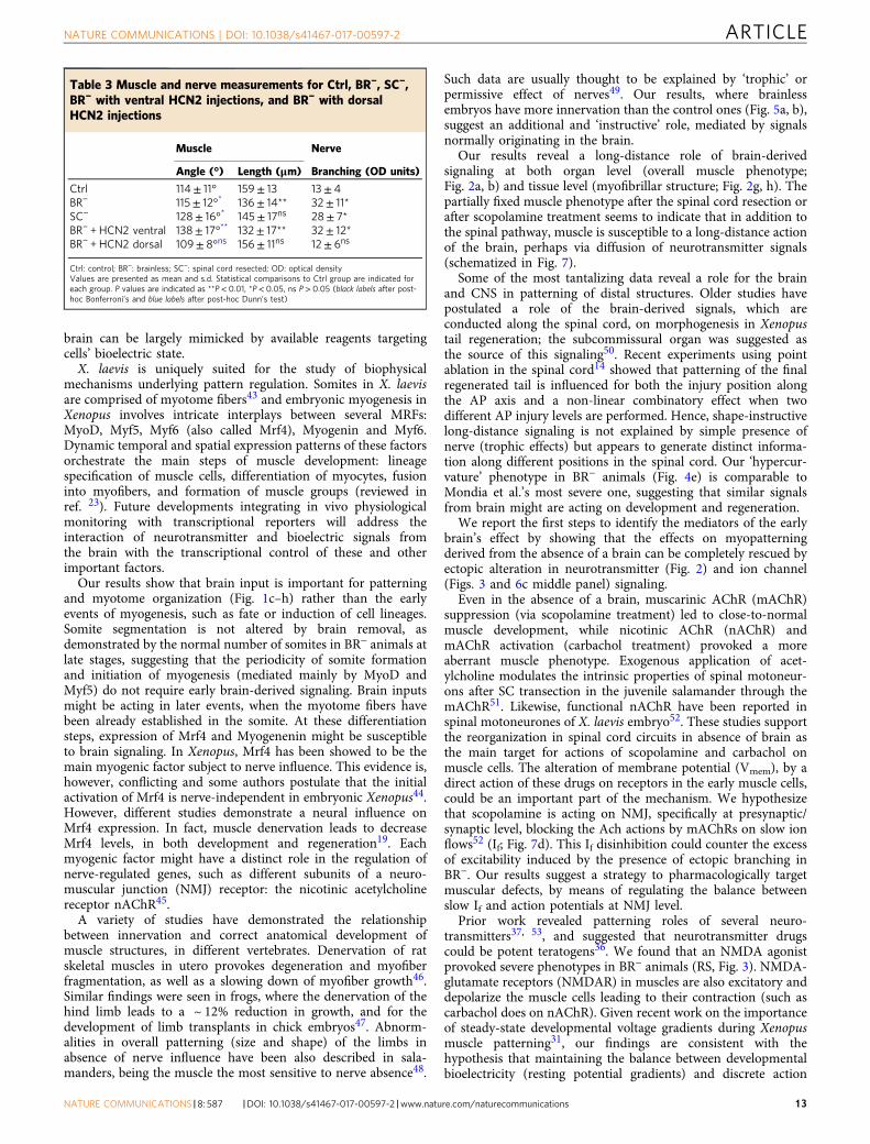
Table 3 Muscle and nerve measurements for Ctrl, BR−, SC−,BR− with ventral HCN2 injections, and BR− with dorsalHCN2 injections
Muscle Nerve
Angle (°) Length (μm) Branching (OD units)
Ctrl 114± 11° 159± 13 13± 4BR− 115± 12°* 136± 14** 32± 11*SC− 128± 16°* 145± 17ns 28± 7*BR− + HCN2 ventral 138± 17°** 132± 17** 32± 12*BR− + HCN2 dorsal 109± 8°ns 156± 11ns 12± 6ns
Ctrl: control; BR−: brainless; SC−: spinal cord resected; OD: optical densityValues are presented as mean and s.d. Statistical comparisons to Ctrl group are indicated foreach group. P values are indicated as **P< 0.01, *P< 0.05, ns P> 0.05 (black labels after post-hoc Bonferroni’s and blue labels after post-hoc Dunn’s test)
NATURE COMMUNICATIONS | DOI: 10.1038/s41467-017-00597-2 ARTICLE
NATURE COMMUNICATIONS |8: 587 |DOI: 10.1038/s41467-017-00597-2 |www.nature.com/naturecommunications 13

2
Brain
Nerve
Muscle
a
With Brain Brainless
RS/Carbachol
HCN2 ventral
Scopolamine
HCN2 dorsal
c d
3a
Bot
tom
-up
Loca
l (P
erip
hery
)
Top
-dow
nLo
ng-r
ange
inst
ruct
ive
cues
b
Rhythmic activity
Correct patterning
Mispatterning
Mispatterning
Tonic activity
HCN2 ventral + BR– Carbachol + BR–
RS +BR–
ACh
IfmAChR
Scopolamine + BR–
Slow ion flowsBioelectricity
Fast ion flowsSpiking
Promote control of slow If on fast firing
HCN2 dorsal
Brain
Viaextra-spinal
Via
spin
al
RS + BR– + Scopo
3b
1
Ectopic branching
Fig. 7 Brain signaling for muscle and nerve development and patterning a. Schematic representation drawing of a Xenopus embryo, showing the maincomponents of our experiments: brain (blue), spinal cord-peripheral nerves (pink) and somites-muscle (brown). Brain effects on nerve patterning couldoccur directly (2), by using efferent spinal pathway. Brain effects on muscle patterning could occur indirectly (3a), by acting on neurons, or directly (3b), byacting on muscle. b A spinal mechanism, for coding the information about patterning and morphogenesis, could occur via direct signaling from the brain tothe neurons in the spinal cord (pink circle). According to our results (different treatments are indicated with purple labels), the effects of the peripheralinnervation on muscle cells can be partially explained in terms of developmental bioelectricity or changes in Vmem excitability. We hypothesize that at thesestages, the brain is in part controlling the bioelectric state of peripheral tissues, and a correct balance (turquoise triangle) of brain activity (long-rangeinstructive cues or top-down perspective) and local signals (bottom-up perspective) is necessary for correct morphogenesis. Both an excess of tonicactivity (induced after carbachol or RS treatment) and an excess of slow If gradients through membrane lead to mispatterning. The extra-spinal pathway bywhich the brain is acting on muscles can be mimicked pharmacologically, with pharmacological agents targeting bioelectricity (i.e., scopolamine). Wehypothesize that scopolamine is acting at presynaptic/synaptic level, blocking the inhibitory ACh actions (via mAChRs) on slow ion flows, and leading theVmem to appropriate values for muscle patterning. c, d. Schematic representation of neuromuscular specificity in normal development (c, with brain) and inabsence of the brain (d, BR−). Our results suggest that ectopic branching detected in the absence of a brain is not due to deficits in early pruning or targetretrograde signaling. Pathfinding behavior at the onset of Xenopus development starts at the spinal cord level, as early patterned electrical gradients in SCcells is required for the correct axon guidance. The different treatments applied in our experiments (purple labels and circles) are placed on the cellular/subcellular domains where they are probably acting
ARTICLE NATURE COMMUNICATIONS | DOI: 10.1038/s41467-017-00597-2
14 NATURE COMMUNICATIONS |8: 587 |DOI: 10.1038/s41467-017-00597-2 |www.nature.com/naturecommunications
potentials at NMJ may be important during muscle developmentand inhibitory signals from the brain (probably via extra-spinalalternative pathways) may be acting to shield the developingmuscle cells from such excitatory stimulus that might causemuscle mispatterning (Fig. 7b, brown rectangle representingmuscle membrane). Recently, it has been showed that activationof NMDAR impairs the myogenic differentiation in C2C12 cellsthrough mTOR/MAPK signaling pathway54. The protective effectof brain detected in the RS-treated control animals (vs. thedevastating effects observed in RS-treated BR−-animals) evidencesthe key role of the brain signaling for the correct morphogenesis.The possibility of exploiting and perhaps strengthening brain-derived protective signals represent an exciting area of researchfor future efforts in the field of birth defects.
In addition to the muscle effects, the absence of a brain duringdevelopment generates an abnormal patterning and organizationof the peripheral innervation or PNS of the animal (Fig. 5;somatic component of the PNS). We used immunohistochemistrywith an antibody to acetylated α-tubulin39 to analyze the somaticneural processes coming from both primary motoneurons andsensory neurons, in order to study the instructive role of braininputs in patterning and global organization of the nervoussystem.
Cell fate and differentiation for primary motoneurons andsensory neurons start early in embryogenesis. Rohon-Beard (RB)neurons cells originate during gastrulation and present electricalexcitability as early as stage 2024. Neural crest (NC) materialsegregates at gastrula stage (around stage 15) and starts migratingaround stage 2025. In our assay, brain removal is done after theonset of the migration, at stage 25, and hence, when the fate of thetrunk NC cells has been already specified55.
In neural morphogenesis, after differentiation and migration,and once the progenitors have reached the final location, newsteps are necessary for axon growth and guidance (pathwayselection), formation of initial connections (target selection), andconnection remodeling and pruning (address selection). The earlyneural morphogenesis, differentiation and migration of X. laevis,both for sensory and motor somatic neurons56, earlier thanthe time point for the brain removal, lead us to suggest thataberrations detected in neural patterning in BR− animals mightbe due, to the later steps on pathfinding and synapse formation.Thus, the peripheral innervation pattern is an epigenetic outcomethat depends on complete development of the functionalphysiology of the brain; in brain removal, not all of the signalsrequired for a correct peripheral pattern are conveyed.
The peripheral innervation formed in brainless animals ismispatterned throughout the whole animal body (Fig. 5). Howneuromuscular specificity arises during embryonic developmenthas been a controversial issue. Are correct connectionspre-established from the outset or do motor axons projectrandomly into the developing muscles followed by extensivepruning of incorrect connections? Our analysis of brainless ani-mals (Supplementary Fig. 5) shows that nerves are altered veryearly in development (as soon as stage 31), with clear errors infinding the correct trajectory (actually in BR− nerve fibers fail inturning to create the correct intermyotome division, see Supple-mentary Fig. 5B, ventral magenta arrow). Our results suggest thatbrain-derived signals are important for correct early patterning,not for maintenance of a default pruning program.
HCN2 targeted to dorsal part (brain and spinal cord) rescuesboth muscle and nerve. Spinal cord-transected embryos showthe same aberrant ectopic branching than BR−, suggest thatpathfinding behavior at the onset of Xenopus development startsat the spinal cord (or CNS) level (Fig. 7c; as previously wasshowed in zebrafish57 or axolotl58). Moreover, in line with recentevidence59, we show that the early patterned electrical in spinal
motoneurons (as HCN2 dorsal is able to rescue the whole phe-notype and brain effects on nerve patterning require an intactspinal cord) is required for the correct axon guidance. Whileprevious work did not explain the mechanisms by which therhythmic activity in spinal neurons affects early nerve develop-ment, our data are consistent with a role for the brain in deter-mining patterned morphogenesis by controlling slow ion flows onprimary-neuron fast firing (Fig. 7b).
Interestingly, we discovered a rescue effect on the neuralphenotype only by means of reagents that target membranepotential (Vmem), induced by the ectopic expression of HCN2(Figs. 5e–h and 6c bottom panel). While targeted misexpressionof ion channels has been demonstrated to rescue patterning of thebrain itself32, we report here that using ion channel expressioncan overcome developmental defects stemming from braindamage. Future work will exploit the emerging advances inoptical imaging of neural activity in vivo60 to characterize thetemporal properties of brain-derived signals and their modifica-tion by HCN2.
One remarkable aspect is that HCN2 expression can rescuepatterning of cells on the other side of the animal – cells thatdo not themselves express HCN2 (Fig. 5g). Such long-rangebioelectrical signaling has been observed in bioelectric tumorsuppression61 and control of apoptosis/proliferation32. Our datareveal that expression of HCN2 at a remote location can inducerepair of peripheral neural structures in a damaged background,suggesting a range of therapies where easily-accessible tissues aretargeted to induce repair in a difficult-to-reach site. Futurework extending closed-loop optogenetic control to neural andnon-neural somatic tissues62 will refine the specific bioelectricstate that facilitates normal network structure and test theseinterventions in adult disease models.
Scopolamine-treated BR− animals display an aberrant neuralnetwork but normal muscle phenotype, suggesting distinctregulatory mechanisms. We do not claim that correction in nervepatterning (by HCN2 injection; Fig. 5e–h) is the only factorresponsible for muscle correction by that treatment (Fig. 3b). Thedirect action of ion channels in muscle cells and their indirectaction on muscle patterning through peripheral nerves are notmutually exclusive possibilities. Future work will be directedtowards understanding their mutual contributions to the repairprocess. The fact that pharmacological modulation of Achtransmission does not affect the neural patterning (Fig. 5c, d) butbioelectrical modulations do (as in vitro studies seems also toindicate63), suggests that both long-distance brain input anddevelopmental bioelectricity are potential targets for futureapplications in the area of muscle-nerve communicationpathologies.
Here, we establish an experimental model for the study of long-range patterning control, and the discovery of pre-behaviorfunctions of the nascent brain. This model is amenable to optical,biophysical, genetic, and chemical approaches, and offers theunique opportunity to target diverse spatial sites (due to theXenopus fate-map) to test non-cell-autonomous mechanisms ofbrain-dependent instructive patterning signals. Future develop-ment of transgenic promoters will allow tissue-specific tests, whiletargeted ablation technology may enable finer dissection of brainregions responsible for various patterning outcomes.
Our data suggest a revision of the view of the brain as quiescentprior to the animal’s independent activity, showing that itssignaling role spans the control of pattern formation andbehavior. This is consistent with recent proposals that theimportant mechanistic and conceptual commonalities existbetween the algorithms of neuroscience and those that guidepattern regulation64. This model system and dataset serves as abase for future studies of local and long-range influences over
NATURE COMMUNICATIONS | DOI: 10.1038/s41467-017-00597-2 ARTICLE
NATURE COMMUNICATIONS |8: 587 |DOI: 10.1038/s41467-017-00597-2 |www.nature.com/naturecommunications 15

large-scale patterning. The relationship of instructive morpho-genetic signals mediated by bioelectric events to the computa-tional capabilities of the brain is an exciting direction for futurework. Moreover, our data point to widely-available and alreadyhuman-approved drugs as potential ‘morphoceuticals’ – agentsthat can be capitalized upon to prevent or perhaps even reversespecific kinds of anatomical defects. More broadly, these resultssuggest a direction for regenerative medicine towards the devel-opment of implanted organoids and hybrid electrochemicalconstructs to provide bioelectric and neurotransmitterstimulation.
MethodsAnimal husbandry. Xenopus laevis embryos were fertilized in vitro accordingto standard protocols25 in 0.1X Marc’s Modified Ringer’s solution (MMR; 10 mMNa+, 0.2 mM K+, 10.5 mM Cl–, 0.2 mM Ca2+, pH 7.8). Xenopus embryos werehoused at 14 °C and staged according to Nieuwkoop and Faber65. All experimentalprocedures involving Xenopus embryos were approved by the Institutional AnimalCare and Use Committees and Tufts University Department of Laboratory AnimalMedicine under protocol M2014-79.
Microsurgery. Stage-25 embryos were randomly grouped in one of the twoexperimental groups: Control (Ctrl) and Brainless (BR−; see Fig. 1a for a schematicrepresentation of the experimental design). Randomized controlled trial conditionswere maintained throughout the experiment. Embryos were anesthetized in a0.02% tricaine solution (pH 7.5) in 0.1X MMR. Brain removal in BR− group wasperformed under dissecting microscope and using a dissecting knife (FST #10055-12). Once movement ceased, a single cut removed the anterodorsal region corre-sponding to the brain (from the cement gland to the most anterior somite; Fig. 1b).After brain removal, animals were allowed to heal in 0.75X MMR for 1 h. Afterwashing, untreated and surgically treated animals were raised at 14 °C andscored and analyzed for phenotype distribution and the different morphologicalparameters, respectively, at stages 30–41 (early stage) and 42–48 (late stage).Following the same experimental and care conditions, additional spinal cord (SC)resection (SC−; Fig. 6a) experiments were performed on stage-25 embryos. Toprevent regeneration66, one segment of the spinal cord, sizing 50–100 μm in length,was completely removed by using two forceps with super-fine tips (Dumont #5ST,FST 11252-00). SC segments were removed at cervical level, immediately posteriorto the hindbrain, and taking special care of minimizing the damage on the mostrostral myotomes.
Two different Sham surgeries were performed by removing pieces of tissue ofcomparable size at different locations of the stage-25 animal body (SupplementaryFig. 1): part of the endodermal yolk mass (Yolk− or yolk resection) or the mostposterior part of the embryo body (Tail− or tailbud resection).
In order to feed the BR− tadpoles, a nutritive medium was modified from ref. 67.The nutritive medium consisted of 9.5% Ham’s nutrient mixture F12 (with 1.0 mML-glutamine, Sigma 51651 C) and 0.5% calf serum (from formula-fed bovine calves,iron supplemented, Sigma C8056) dissolved in 0.1X MMR. From stage 45, Ctrl andBR− tadpoles were fed with this nutritive medium for 2 h every day.
Microinjections. Capped synthetic mRNAs generated using mMessage mMachinekit (Ambion) were dissolved in nuclease-free water and injected into embryosimmersed in 3% Ficoll solution using standard methods25. The mRNA injectionswere made using borosilicate glass needles calibrated to bubble pressures of 55 to60 kPa in water, delivering 100- to 130-ms pulses. Each injection delivered between0.5–1 nl or 0.5–1 ng of mRNA per blastomere into the embryos. Two differentsets of injections were performed. Firstly, at the two-cell stage (Fig. 3a, b), eitherone (1/2 HCN2 Inj) or both (HCN2 Inj) blastomeres were injected. Secondly, at thefour-cell stage (Fig. 6b, c), either the two ventral (HCN2 ventral Inj) or the twodorsal (HCN2 dorsal Inj) blastomeres were injected. Injections were done into thecenter of cells at the animal pole.
Constructs used were HCN2-WT-2A-GFP or HCN2-WT68 and β-galactosidasein PCS2 and both RNAs (HCN2-WT and β-galactosidase) were mixed at 3:5 and1:5 dilutions respectively, for microinjections. HCN2 is a potassium/sodiumhyperpolarization-activated cyclic nucleotide-gated ion channel, type 269. Embryoswere injected in 3% Ficoll solution and after 30 min, were washed and then rearedin 0.1X MMR until desired stages.
Drug exposure. Ctrl and BR− Xenopus embryos were exposed to specificpharmacological agents, dissolved in 0.1X MMR, from stage 25 (immediately afterbrain removal) to stage 48. The drugs were refreshed every three days. Two drugstargeting cholinergic receptors and one directed to NMDA-glutamatergic receptorwere used: 10 μM scopolamine (a muscarinic-receptor antagonist; Tocris 1414),10 μM carbamoylcholine chloride (carbachol, a dual muscarinic- and nicotinic-receptor agonist; Tocris 2810) and 10 μM (RS)-(Tetrazol-5-yl)glycine (RS; anNMDA receptor agonist; Tocris 0312), respectively. All drug treatments were
performed using embryos from mixed batches of fertilizations, using at least threebiological replicates.
Stock solutions of all pharmaceuticals were created by dissolving the compoundin Millipore water (to a final drug concentration of 100 mM for both scopolamineand carbachol, and to 50 mM for RS) and then stored in aliquots at –20 °C. Furtherdilution of all compounds was made in normal media (0.1X MMR). Controlexperiments were performed using embryos in 0.1X MMR, both for Ctrl and BR−
groups. Drug concentrations were determined through toxicity screens and wereapplied at levels that did not result in lethality or observable developmental defects.
Immunofluorescence and histochemistry. Immediately after treatment, at therelevant time points (immediately after brain removal and first 2 weeks and thirdand fourth weeks after brain removal for early- and late-stage studies, respectively),anesthetized tadpoles were fixed in MEMFA overnight at 4 °C for whole-mountimmunofluorescence25. Briefly, embryos were washed twice in 1× phosphatebuffered saline (PBS), and permeabilized in PBS 0.1% Triton X-100 (PBST) for30 min. Animals were then blocked with 10% normal goat serum in PBST for 1 h atroom temperature (RT). Samples were rocked overnight at 4 °C using znp1(Developmental Studies Hybridoma Bank, used at 1:250 dilution) and anti-acetylated alpha-tubulin antibody (Tub; Sigma T7451 used at 1:500 dilution).Following primary exposure, embryos were washed three times in PBST before a60-min RT incubation with AlexaFluor-555 conjugated secondary antibody(Invitrogen) used at 1:500 diluted in PBST. Following secondary incubation,animals were washed three times for 15 min in PBST and imaged on an OlympusBX-61 microscope equipped with a Hamamatsu ORCA AG CCD camera, andcontrolled by Metamorph software. Particular care was taken to ensure thatembryos from all the different groups were processed in the same batch at the sameimmunofluorescence session.
Beta-galactosidase enzymatic detection. Embryos injected with β-galactosidase(β-gal) mRNA were fixed (30 min in MEMFA at RT) at the relevant stages, washedtwice in PBS with 2 mM MgCl2, and stained with X-gal (Roche Applied Sciences,Indianapolis, IN) staining solution at 37 °C for 3 h. Embryos were then rinsed threetimes in PBS and analyzed.
In situ hybridization. Xenopus embryos at stage 35 were collected and fixed inMEMFA25 and in situ hybridization was performed25, 70. The embryos werewashed with phosphate buffered saline 0.1% Tween-20 (PBST) and transferredthrough a series of methanol washes 25%-50%-75%-100%. In situ antisense probewas generated in vitro from linearized template using DIG labeling mix (Roche).Chromogenic reaction times were optimized for signal to background ratio.Antisense RNA probes for Xenopus HCN2 were generated from X. laevis hcn2.LIMAGE clone 5514485 (purchased from Dharmacon): a HindIII fragment wasdeleted, leaving exons 2-4 and part of exon 5 as probe. In situ hybridized embryoswere then agarose embedded and sectioned. Briefly, 4% low melting point agarosewas melted at poured into plastic scaffolds. Gently dried, in situ hybridizedembryos were oriented within the agarose for transverse sectioning. The agaroseblocks were then allowed to solidify, trimmed and sectioned using Leica VT1000Svibratome to obtain transverse sections.
Phenotype scoring and morphological evaluation. Ctrl-, Sham-Yolk−-, andBR−-Xenopus embryos were scored for abnormalities in the macroscopic pheno-type. Average number of animals with clearly identifiable left-right axis bendingand/or tail defects were used to evaluate the percent abnormal within eachpopulation. Embryos were photographed (lateral view) using a Nikon AZ100 withan attached QImaging CD camera controlled by QCapture software.
Mean collagen density, angle and length of myotome fibers, and number ofsomites. All muscle structure studies were performed on 4× or 10× images takenunder polarized light. Birefringence microscopy was performed on an OlympusBX-61 compound microscope with a universal condenser (U-UCD8). Thetransmitted light DIC slider (U-DICTS) was pulled out and the polarizing filterwas rotated such the background appeared darkest. Embryos were positioned at a45° angle for imaging. Analysis of birefringence images was performed in ImageJ(National Institutes of Health, Maryland, US).
Collagen density in early-staged embryos (Fig. 1c, d; short arrows) wasevaluated by means of optical density (OD) using a gray scale gradient of 0-255(white to black), on 4× polarized-light photomicrographs31. Each embryo wassampled using a systematic procedure in order to measure the OD at differentanatomical levels, along the anteroposterior axis. Six OD-myotome measurementswere used to determine mean value for each animal. At least 30 embryos withineach experimental group were analyzed. Possible aberrations originating from theoptical system of the camera were corrected by background image subtraction.
Mean angle and length of myotome fibers (Fig. 1c–g; overlaid dashedarrowhead-like lines and double-headed arrows, respectively) for both early- andlate-staged embryos was performed on 10× polarized-light images, using ImageJsoftware. For each animal, three somites at anterior, central and posterior levels,respectively, were systematically random sampled. To obtain a representative meanvalue of length, nine myotome fibers, from dorsal to ventral axis, were measuredfor each somite at each anatomical level. The number of somites, between end of
ARTICLE NATURE COMMUNICATIONS | DOI: 10.1038/s41467-017-00597-2
16 NATURE COMMUNICATIONS |8: 587 |DOI: 10.1038/s41467-017-00597-2 |www.nature.com/naturecommunications
the gut and tip of the tail, for both early- and late-staged embryos was evaluated on4x birefringence images.
Analysis of the neural network branching (see Fig. 5, unfilled triangles) in early-and late-staged embryos was performed by evaluating the intensity of znp1 andacetylated alpha-tubulin (Tub) immunostaining, respectively, through gray-levelmeasures (OD). In order to obtain one OD-mean value per animal, multiplemeasurements were taken along the anteroposterior axis. Each measurementconsisted of the mean value of the pixels of a fixed-size window, placed betweentwo consecutive somites (intermyotomal or internal neuropil). The size of thewindow remained constant across subjects. All individuals among whomcomparisons are being made were produced in the same batch, treated identicallyfor processing and imaging–conditions were not changed. In addition, possibleaberrations originating from the optical system of the camera were corrected bybackground image subtraction.
Statistics. All statistical analysis was performed using GraphPad Prism (GraphPadSoftware, Inc., CA, US) and Microsoft Excel (Microsoft Corporation, WA, US)software. Each dish of tadpoles was considered a replicate.
When comparing numerical variables (OD, number of somites, angle andlength of the myotome fibers), data from, at least, two replicates (minimal replicatesize: n= 25 embryos) were analyzed per each experimental condition. Firstly, datawere tested for homogeneity of variances by Bartlett’s test. If variances are similar,we applied unpaired and two-tailed Student’s t-test (two independent groups),one-way ANOVA test (multiple independent groups), or two-way ANOVAtest (two independent variables) followed by post-hoc Bonferroni’s test (whenP< 0.05). In case of unequal variances (or non-normal distributions for a samplesize of less than 50), two-tailed Mann–Whitney and Kruskal–Wallis test followedby pot-hoc Dunn’s test (when P< 0.05), respectively, were used.
For differences in sample proportion (percent of aberrant embryos) ordistribution analysis (frequency of phenotypes), data from, at least, two replicatesand sample size equal or greater than 50 embryos were pooled and analyzed bymultiple-sample z-test or Χ2 test, respectively.
The significance level (α) was set to 0.05 in all cases. The statistical values arereported as mean± s.d.
Data availability. The authors declare that all data supporting the findings of thisstudy are available within the article and its Supplementary Information Files orfrom the corresponding author upon reasonable request.
Received: 5 January 2017 Accepted: 10 July 2017
References1. Tsonis, P. A. Embryogenesis and carcinogenesis: order and disorder. Anticancer
Res. 7, 617–623 (1987).2. Scharrer, B. & Lochhead, M. S. Tumors in the invertebrates: a review. Cancer
Res. 10, 403–419 (1950).3. Pawlowski, A. & Weddell, G. Induction of tumours in denervated skin. Nature
213, 1234–1236 (1967).4. Outzen, H. C., Custer, R. P. & Prehn, R. T. Influence of regenerative capacity
and innervation on oncogenesis in the adult frog (Rana pipiens). J. Natl CancerInst. 57, 79–84 (1976).
5. Li, Y. K., Yang, J. M., Huang, Y. B., Ren, D. D. & Chi, F. L. Shrinkage ofipsilateral taste buds and hyperplasia of contralateral taste buds followingchorda tympani nerve transection. Neural Regener. Res. 10, 989–995 (2015).
6. Saloman, J. L., Albers, K. M., Rhim, A. D. & Davis, B. M. Can stopping nerves,stop cancer? Trends Neurosci. 39, 880–889 (2016).
7. Singer, M. The influence of the nerve in regeneration of the amphibianextremity. Q. Rev. Biol. 27, 169–200 (1952).
8. Yntema, C. L. Regeneration in sparsely innervated and aneurogenic forelimbs ofAmblystoma larvae. J. Exp. Zool. 140, 101–123 (1959).
9. Recidoro, A. M. et al. Botulinum toxin induces muscle paralysis and inhibitsbone regeneration in zebrafish. J. Bone Miner. Res. 29, 2346–2356 (2014).
10. Satoh, A., Graham, G. M., Bryant, S. V. & Gardiner, D. M. Neurotrophicregulation of epidermal dedifferentiation during wound healing and limbregeneration in the axolotl (Ambystoma mexicanum). Dev. Biol. 319, 321–335(2008).
11. Best, J. B., Goodman, A. B. & Pigon, A. Fissioning in planarians: control by thebrain. Science 164, 565–566 (1969).
12. Oviedo, N. J. et al. Long-range neural and gap junction protein-mediated cuescontrol polarity during planarian regeneration. Dev. Biol. 339, 188–199 (2010).
13. Satoh, A., Gardiner, D. M., Bryant, S. V. & Endo, T. Nerve-induced ectopiclimb blastemas in the Axolotl are equivalent to amputation-induced blastemas.Dev. Biol. 312, 231–244 (2007).
14. Mondia, J. P. et al. Long-distance signals are required for morphogenesis of theregenerating Xenopus tadpole tail, as shown by femtosecond-laser ablation.PLoS ONE 6, e24953 (2011).
15. Kumar, A. & Brockes, J. P. Nerve dependence in tissue, organ, and appendageregeneration. Trends Neurosci. 35, 691–699 (2012).
16. Sherrard, R. M. & Bower, A. J. Role of afferents in the development and cellsurvival of the vertebrate nervous system. Clin. Exp. Pharmacol. Physiol. 25,487–495 (1998).
17. Knox, S. M. et al. Parasympathetic innervation maintains epithelial progenitorcells during salivary organogenesis. Science 329, 1645–1647 (2010).
18. Crisp, S. J., Evers, J. F. & Bate, M. Endogenous patterns of activity are requiredfor the maturation of a motor network. J. Neurosci. 31, 10445–10450 (2011).
19. Nicolas, N., Mira, J. C., Gallien, C. L. & Chanoine, C. Neural and hormonalcontrol of expression of myogenic regulatory factor genes during regenerationof Xenopus fast muscles: myogenin and MRF4 mRNA accumulation areneurally regulated oppositely. Dev. Dyn. 218, 112–122 (2000).
20. Cooke, J. & Zeeman, E. C. A clock and wavefront model for control of thenumber of repeated structures during animal morphogenesis. J. Theor. Biol. 58,455–476 (1976).
21. Soroldoni, D. et al. Genetic oscillations. A Doppler effect in embryonic patternformation. Science 345, 222–225 (2014).
22. Hubaud, A. & Pourquie, O. Signalling dynamics in vertebrate segmentation.Nat. Rev. Mol. Cell Biol. 15, 709–721 (2014).
23. Sabillo, A., Ramirez, J. & Domingo, C. R. Making muscle: morphogeneticmovements and molecular mechanisms of myogenesis in Xenopus laevis.Semin. Cell Dev. Biol. 51, 80–91 (2016).
24. Lamborghini, J. E. Rohon-beard cells and other large neurons inXenopus embryos originate during gastrulation. J. Comp. Neurol. 189, 323–333(1980).
25. Sive, H., Grainger, R. M. and Harland, R. Early development of Xenopus laevis.(Cold Spring Harbor Laboratory Press, 2000).
26. Behra, M. et al. Acetylcholinesterase is required for neuronal and musculardevelopment in the zebrafish embryo. Nat. Neurosci. 5, 111–118 (2002).
27. Klinkenberg, I. & Blokland, A. The validity of scopolamine as apharmacological model for cognitive impairment: a review of animal behavioralstudies. Neurosci. Biobehav. Rev. 34, 1307–1350 (2010).
28. Lochner, M. & Thompson, A. J. The muscarinic antagonists scopolamine andatropine are competitive antagonists at 5-HT receptors. Neuropharmacology125, 1562–1566 (2016).
29. Akk, G. & Auerbach, A. Activation of muscle nicotinic acetylcholine receptorchannels by nicotinic and muscarinic agonists. Br. J. Pharmacol. 128,1467–1476 (1999).
30. Hinard, V., Belin, D., Konig, S., Bader, C. R. & Bernheim, L. Initiation ofhuman myoblast differentiation via dephosphorylation of Kir2.1 K + channelsat tyrosine 242. Development 135, 859–867 (2008).
31. Lobikin, M., Pare, J. F., Kaplan, D. L. & Levin, M. Selective depolarization oftransmembrane potential alters muscle patterning and muscle cell localizationin Xenopus laevis embryos. Int. J. Dev. Biol. 59, 303–311 (2015).
32. Pai, V. P., Lemire, J. M., Chen, Y., Lin, G. & Levin, M. Local and long-rangeendogenous resting potential gradients antagonistically regulateapoptosis and proliferation in the embryonic CNS. Int. J. Dev. Biol. 59, 327–340(2015).
33. Bruzauskaite, I. et al. Relevance of HCN2-expressing human mesenchymalstem cells for the generation of biological pacemakers. Stem Cell Res. Ther. 7, 67(2016).
34. Yang, S. et al. The distribution of HCN2-positive cells in the gastrointestinaltract of mice. J. Anat. 221, 303–310 (2012).
35. Zhao, Z. et al. Involvement of HCN channel in muscarinic inhibitory action ontonic firing of dorsolateral striatal cholinergic interneurons. Front. CellNeurosci. 10, 71 (2016).
36. Hernandez-Diaz, S. & Levin, M. Alteration of bioelectrically-controlledprocesses in the embryo: a teratogenic mechanism for anticonvulsants. Reprod.Toxicol. 47, 111–114 (2014).
37. Sullivan, K. G. & Levin, M. Neurotransmitter signaling pathways required fornormal development in Xenopus laevis embryos: a pharmacological surveyscreen. J. Anat. 229, 483–502 (2016).
38. Duncan, G. E., Inada, K., Farrington, J. S. & Koller, B. H. Seizure responses andinduction of Fos by the NMDA agonist (tetrazol-5-yl)glycine in a genetic modelof NMDA receptor hypofunction. Brain Res. 1221, 41–48 (2008).
39. Chu, D. T. & Klymkowsky, M. W. The appearance of acetylated alpha-tubulinduring early development and cellular differentiation in Xenopus. Dev. Biol.136, 104–117 (1989).
40. Menelaou, E. et al. Embryonic motor activity and implications for regulatingmotoneuron axonal pathfinding in zebrafish. Eur. J. Neurosci. 28, 1080–1096(2008).
41. Borodinsky, L. N. & Belgacem, Y. H. Crosstalk among electrical activity, trophicfactors and morphogenetic proteins in the regulation of neurotransmitterphenotype specification. J. Chem. Neuroanat. 73, 3–8 (2016).
NATURE COMMUNICATIONS | DOI: 10.1038/s41467-017-00597-2 ARTICLE
NATURE COMMUNICATIONS |8: 587 |DOI: 10.1038/s41467-017-00597-2 |www.nature.com/naturecommunications 17
42. Levin, M. Molecular bioelectricity: how endogenous voltage potentials controlcell behavior and instruct pattern regulation in vivo. Mol. Biol. Cell 25,3835–3850 (2014).
43. Afonin, B., Ho, M., Gustin, J. K., Meloty-Kapella, C. & Domingo, C. R. Cellbehaviors associated with somite segmentation and rotation in Xenopus laevis.Dev. Dyn. 235, 3268–3279 (2006).
44. Ataian, Y., Owens, J. & Hinterberger, T. MRF4 gene expression in Xenopusembryos and aneural myofibers. Dev. Dyn. 226, 551–554 (2003).
45. Launay, T. et al. Expression and neural control of myogenic regulatory factorgenes during regeneration of mouse soleus. J. Histochem. Cytochem. 49,887–899 (2001).
46. Hanzlikova, V. & Schiaffino, S. Studies on the effect of denervation indeveloping muscle. 3. Diversification of myofibrillar structure and origin of theheterogeneity of muscle fiber types. Z. Zellforsch. Mikrosk. Anatomie 147,75–85 (1973).
47. Hamburger, V. & Waugh, M. The primary development of the skeleton innerveless and poorly innervated limb transplants of chick embryos. Physiol.Zool. 13, 367–382 (1940).
48. Popiela, H. In vivo limb tissue development in the absence of nerves: aquantitative study. Exp. Neurol. 53, 214–226 (1976).
49. Singer, M. Trophic functions of the neuron. VI. Other trophic systems.Neurotrophic control of limb regeneration in the newt. Ann. N.Y. Acad. Sci.228, 308–322 (1974).
50. Hauser, R. Morphogenetic action of subcommissural organ on tail regenerationin Xenopus larvae. Roux Arch. Dev. Biol. 169, 170–184 (1972).
51. Chevallier, S., Nagy, F. & Cabelguen, J. M. Muscarinic control of the excitabilityof hindlimb motoneurons in chronic spinal-transected salamanders. Eur. J.Neurosci. 28, 2243–2253 (2008).
52. Perrins, R. & Roberts, A. Cholinergic and electrical synapses betweensynergistic spinal motoneurones in the Xenopus laevis embryo. J. Physiol. 485,135–144 (1995).
53. Fukumoto, T., Kema, I. P. & Levin, M. Serotonin signaling is a very early step inpatterning of the left-right axis in chick and frog embryos. Curr. Biol. 15,794–803 (2005).
54. Auh, Q. S. et al. N-methyl-d-aspartate (NMDA) impairs myogenesis in C2C12cells. Muscle Nerve http://dx.doi.org/10.1002/mus.25511 (2016).
55. Henion, P. D. & Weston, J. A. Timing and pattern of cell fate restrictions in theneural crest lineage. Development 124, 4351–4359 (1997).
56. Edlund, A. F., Davidson, L. A. & Keller, R. E. Cell segregation, mixing, andtissue pattern in the spinal cord of the Xenopus laevis neurula. Dev. Dyn. 242,1134–1146 (2013).
57. Westerfield, M. & Eisen, J. S. Neuromuscular specificity: pathfinding by identifiedmotor growth cones in a vertebrate embryo. Trends Neurosci. 11, 18–22 (1988).
58. Freeman, J. M. & Davey, D. F. The precision of pathway selection by developingperipheral axons in the axolotl. J. Embryol. Exp. Morphol. 91, 117–134 (1986).
59. Hanson, M. G. & Landmesser, L. T. Normal patterns of spontaneous activityare required for correct motor axon guidance and the expression of specificguidance molecules. Neuron 43, 687–701 (2004).
60. Jarvis, S. & Schultz, S. R. Prospects for optogenetic augmentation of brainfunction. Front. Syst. Neurosci. 9, 157 (2015).
61. Chernet, B. T. & Levin, M. Transmembrane voltage potential of somatic cellscontrols oncogene-mediated tumorigenesis at long-range. Oncotarget 5,3287–3306 (2014).
62. Adams, D. S., Lemire, J. M., Kramer, R. H. & Levin, M. Optogenetics inDevelopmental Biology: using light to control ion flux-dependent signals inXenopus embryos. Int. J. Dev. Biol. 58, 851–861 (2014).
63. Lee, M. J. & Chiao, C. C. Short-term alteration of developmental neural activityenhances neurite outgrowth of retinal explants. Invest. Ophthalmol. Vis. Sci. 57,6496–6506 (2016).
64. Pezzulo, G. & Levin, M. Re-membering the body: applications of computationalneuroscience to the top-down control of regeneration of limbs and othercomplex organs. Integr. Biol. (Camb.) 7, 1487–1517 (2015).
65. Nieuwkoop, P. D. & Faber, J. Normal Table of Xenopus laevis (Daudin): ASystematical and Chronological Survey of the Development from the FertilizedEgg Till the End of Metamorphosis. (Garland Pub., 1994).
66. Edwards-Faret, G. et al. Spinal cord regeneration in Xenopus laevis. Nat. Protoc.12, 372–389 (2017).
67. Shaffer, B. M. The isolated Xenopus laevis tail: a preparation for studying thecentral nervous system and metamorphosis in culture. J. Embryol. Exp.Morphol. 11, 77–90 (1963).
68. Ye, B. & Nerbonne, J. M. Proteolytic processing of HCN2 and co-assembly withHCN4 in the generation of cardiac pacemaker channels. J. Biol. Chem. 284,25553–25559 (2009).
69. Biel, M., Wahl-Schott, C., Michalakis, S. & Zong, X. Hyperpolarization-activated cation channels: from genes to function. Physiol. Rev. 89, 847–885(2009).
70. Harland, R. M. In situ hybridization: an improved whole-mount method forXenopus embryos. Methods Cell Biol. 36, 685–695 (1991).
AcknowledgementsWe thank Rakela Colon and Erin Switzer for general laboratory assistance and Xenopushusbandry, Dany S. Adams for help with microscopy, Nian-Qing Shi for the HCN2-WTDNA construct and Jean-Francois Pare for assistance with the sub-cloning of cDNAHCN2 into PCS2. We thank Kelly McLaughlin and Joshua Finkelstein for their helpfulcomments on the manuscript. This paper is dedicated to the memory of Panagiotis‘Takis’ Tsonis, a profound multi-disciplinary scientist who significantly impacted ourunderstanding of biological control systems. This research was supported by the AllenDiscovery Center program through The Paul G. Allen Frontiers Group (12171). We alsogratefully acknowledge support from the W.M. Keck Foundation (5903), the G. Haroldand Leila Y. Mathers Charitable Foundation (TFU141), and the National Institutes ofHealth (AR055993, AR061988).
Author contributionsC.H.-R. performed experiments (microsurgery, developmental analysis, drugexperiments, immunofluorescence and birefringence imaging). M.L. and C.H.-R.designed the experiments and interpreted data. V.P.P. performed mRNA injectionexperiments. K.M.M. assisted with the immunofluorescence experiments and the spinalcord experiments. J.M.L. cloned the HCN2 mRNA construct that was used for the in situexperiments. C.H.-R., M.L. and V.P.P. wrote the paper together.
Additional informationSupplementary Information accompanies this paper at doi:10.1038/s41467-017-00597-2.
Competing interests: : The authors declare no competing financial interests.
Reprints and permission information is available online at http://npg.nature.com/reprintsandpermissions/
Publisher's note: Springer Nature remains neutral with regard to jurisdictional claims inpublished maps and institutional affiliations.
Open Access This article is licensed under a Creative CommonsAttribution 4.0 International License, which permits use, sharing,
adaptation, distribution and reproduction in any medium or format, as long as you giveappropriate credit to the original author(s) and the source, provide a link to the CreativeCommons license, and indicate if changes were made. The images or other third partymaterial in this article are included in the article’s Creative Commons license, unlessindicated otherwise in a credit line to the material. If material is not included in thearticle’s Creative Commons license and your intended use is not permitted by statutoryregulation or exceeds the permitted use, you will need to obtain permission directly fromthe copyright holder. To view a copy of this license, visit http://creativecommons.org/licenses/by/4.0/.
© The Author(s) 2017
ARTICLE NATURE COMMUNICATIONS | DOI: 10.1038/s41467-017-00597-2
18 NATURE COMMUNICATIONS |8: 587 |DOI: 10.1038/s41467-017-00597-2 |www.nature.com/naturecommunications