open access research article sinus-lift by use of hemihydrate … · 2017-11-22 · cronicon open...
TRANSCRIPT
CroniconO P E N A C C E S S EC DENTAL SCIENCE
Research Article
Sinus-lift by Use of Hemihydrate-Calcium Sulphate: A Prospective Clinical, Radiographical, Histological Study of Implant Integration in the Posterior
Maxilla
Hossein Kashani1*, Amir Dasmah2, Reza Mokhtari3 and Christer Dahlin4
1Department of Oral and Maxillofacial Surgery, The Sahlgrenska Academy, University of Gothenburg, Gothenburg, Sweden 2Department of Oral and Maxillofacial Surgery, Mölndal Hospital, Mölndal, Sweden 3Department of ENT and Oral and Maxillofacial Surgery, NU Hospital Organization, Trollhättan, Sweden 4Department of Biomaterials, Institute for Surgical Sciences, The Sahlgrenska Academy, and BIOMATCELL VINN Excellence Center of Bio-materials and Cell Therapy, University of Gothenburg, Göteborg, Sweden
*Corresponding Author: Hossein Kashani, Department of Oral and Maxillofacial Surgery, Institute of Odontology, Sahlgrenska Academy, University of Gothenburg, Gothenburg, Sweden.
Citation: Hossein Kashani., et al. “Sinus-lift by Use of Hemihydrate-Calcium Sulphate: A Prospective Clinical, Radiographical, Histological Study of Implant Integration in the Posterior Maxilla”. EC Dental Science 15.6 (2017): 240-249.
Received: November 01, 2017; Published: November 22, 2017
Abstract
The objective of this study was to evaluate implant survival in reconstructed bone after the use of a synthetic bone substitute material (Calcium Sulphate). Clinical, radiological and histological parameters were studied in 25 patients. Unilateral and bilateral sinus augmentation was performed. An 80% + 20% mixed alpha hemihydrate Calcium Sulphate with autograph bone respectively was applied into the maxillary sinus. After six months, dental implants were installed. Prior to implant installation, all patients un-derwent a CT examination. In total 91 implants were inserted in all 25 subjects, 74 of these in augmented bone. Additionally, two micro-implants were installed bilaterally in 8 patients. After four months of healing, a third operation was performed to insert the healing abutments into the implants. Simultaneously, the micro-implants were retrieved for histological analysis. Implant stability was assessed through resonance frequency analysis at installation and at abutment connection. The pattern of bone formation in the experimental sites was investigated using histomorphometric measurements.
Implant survival rate was 96,8% at abutment connection. The mean ISQ value at implant placement was 65.8 and at abutment connection 66,5. The mean bone to implant contact was 27,0%.
Keywords: Calcium Sulphate; Bone Substitute; Sinus Augmentation; Dental Implants; Prospective; Clinical Study
Introduction
Edentulous jaw in a resorbed maxilla due to pneumatisation of the maxillary sinus often requires bone grafting to enable implant in-sertion [1-3]. To increase the vertical bone height in the posterior maxilla, the different bone substitute materials have been used in sinus lift procedures, but the ideal material is still a matter of debate. It is suggested that the perfect bone substitute should be safe, inexpensive and have the property to completely resorb in the surgical site [4]. It should also provide osteoconduction and osteoinduction properties for promoting new bone formation. Autogenous bone grafts have been long considered to be the preferred bone graft material [5-8]. Lack of immunological rejection and the presence of stem cells and growth factors, which result to both osteoconductive and osteoinductive abilities, give the autogenous bone graft the upper hand [9,10]. However because a second entry to the donate site will be needed to har-vest the bone graft, this additional procedure may increase the risk for future surgical complications and morbidity as described in the

241
Sinus-lift by Use of Hemihydrate-Calcium Sulphate: A Prospective Clinical, Radiographical, Histological Study of Implant Integration in the Posterior Maxilla
Citation: Hossein Kashani., et al. “Sinus-lift by Use of Hemihydrate-Calcium Sulphate: A Prospective Clinical, Radiographical, Histological Study of Implant Integration in the Posterior Maxilla”. EC Dental Science 15.6 (2017): 240-249.
literature [11,12]. Several grafting materials and techniques have been used and described in the literature with varying clinical outcomes [7,13-15]. Although some studies using deproteinized bovine bone together with autogenous bone in the sinus lift procedure have shown promising results [16,17], the disadvantage of this grafting procedure remains the non-existent resorption of the bovine bone graft. In a clinical, histological study of bovine hydroxyapatite in combination with autogenous bone conducted by Hallman., et al. [18], no sign of resorption of the bovine hydroxy-apatite was reported.
Calcium sulphate (CaS) is another grafting material with a long track record in orthopaedic surgery [19]. Due to its ability to be well tolerated, biodegradable and osteoconductive [20], it has also been used in oral surgery [21-24].
Aim of the Study
The aim of the present study was to conduct a clinical, radiographic and histological study, using calcium sulphate alpha-hemihydrate in sinus lift procedure in a broad patient material. Furthermore, to obtain a more exact data on the amount of new bone regeneration, computed tomography was used as the radiographic examination. With regard to data on the osseointegration status, resonance fre-quency analysis was used.
Materials and MethodsPatients
Twenty-five patients (twelve men and thirteen women) with a mean age of 65 years (range 38 - 77) were enrolled in this study. The patients were consecutively chosen from those referred to the department of ENT and Oral and Maxillofacial Surgery in the Norra Älvs-borg County Hospital (NÄL) in Trollhättan, Sweden for dental implants. The inclusion criteria for maxillary sinus floor augmentation were patients depicting alveolar ridge deficiencies with classes III-VI determined by Cawood and Howell [2]. Also, patients should have less than 5 mm residual bone to the maxillary sinus floor in vertical dimension after radiographic examination.
The exclusion criteria were severe systemic diseases such as malignancies, endocrine disorders, known leukocyte dysfunctions and heavy smokers (< 10 cigarettes per day). Also, patients receiving Bisphosphonate treatment were excluded. All participants were subject-ed to preoperative radiographic examination, using computed tomography (Simens Somatom Sensation 4 CT Scanner) using 2 mm axial scans. Patients were informed about the study and were asked to sign a consent form by the current declaration on biomedical research in human subjects. The study was approved by the Regional Ethical Review Board in Gothenburg, Sweden, Dnr: 343-10.
Anaesthesia and surgical procedures
The surgeries were performed under local anaesthesia using lidocaine 2% with epinephrine (1:80.000) (Xylocaine/Adrenalin®, Astra AB, Södertälje, Sweden). All patients were received preoperative medications with 2g penicillin - V (Kåvepenin, Astra AB) or in case of PC-V allergy, 600 mg Clindamycin (Dalacin, Pfizer) as a single dose. And as analgesics, 600 mg Ibuprofen (Ibumetin, Astra AB) were given 1 hour before the surgery. After surgery, penicillin-V 1 g x 3 or Clindamycin 300 mg x 3 was continued during seven days.
An incision was performed on the lateral site of the maxillary sinus, and a full mucoperiostal flap was raised, giving proper access to the edentulous area. Then, using a thin round burr, an osteotomy was performed creating a rectangular window approximately 20 mm wide and 10 mm in height. The bony window was either carefully removed or left attached to the underlying sinus membrane, which was elevated from the sinus floor. Next, calcium sulphate alpha-hemihydrate (CalMatrix binder 0.34, Citagenix Inc, Quebec, Canada) mixed with autogenous bone particles harvested from the surgical site was applied into the maxillary sinus. The intention was to create an 80/20% ration between the calcium sulphate and autogenous bone particles. Eight of the patients had bilateral sinus lift procedures. Finally, a layer of resorbable barrier membrane (CalForma) (Citagenix Inc, Quebec, Canada) was applied over the augmented area. The healing period following the sinus lift procedure was set to 6 months.

242
Sinus-lift by Use of Hemihydrate-Calcium Sulphate: A Prospective Clinical, Radiographical, Histological Study of Implant Integration in the Posterior Maxilla
Citation: Hossein Kashani., et al. “Sinus-lift by Use of Hemihydrate-Calcium Sulphate: A Prospective Clinical, Radiographical, Histological Study of Implant Integration in the Posterior Maxilla”. EC Dental Science 15.6 (2017): 240-249.
Implant placement and biopsy retrieval
Prior to implant installation, all patients underwent a radiographic examination, using computed tomography to evaluate the extent of new bone regeneration in the maxillary sinuses. Different kinds of implants were used due to the requests from the referral prosthodon-tists. A total of 91 implants were inserted in all twenty-five subjects. Furthermore, 25 mini-implants were placed in the grafted areas from the lateral side. A 2 mm wide drill was used so that a specially designed machined screw-shaped mini-implant 2 x 5 mm (Burlington, MA, USA) could be placed into the surgical sites (Figure 1).
Figure 1: Micro-implants in place, one laterally positioned in the reconstructed bone, and one in residual bone.
After four months of healing, abutment connection was performed, and at the same time, the mini-implants and surrounding bone tissue were removed, using a trephine burr with a diameter of 3 mm for histological analysis.
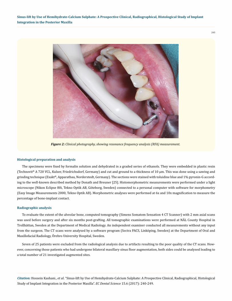
Resonance frequency analysis (Osstell™, Integration Diagnostics AB, Göteborg, Sweden) was performed both at implant installation and at the time of abutment connection four months later (Figure 2).
243
Sinus-lift by Use of Hemihydrate-Calcium Sulphate: A Prospective Clinical, Radiographical, Histological Study of Implant Integration in the Posterior Maxilla
Citation: Hossein Kashani., et al. “Sinus-lift by Use of Hemihydrate-Calcium Sulphate: A Prospective Clinical, Radiographical, Histological Study of Implant Integration in the Posterior Maxilla”. EC Dental Science 15.6 (2017): 240-249.
Figure 2: Clinical photography, showing resonance frequency analysis (RFA) measurement.
Histological preparation and analysis
The specimens were fixed by formalin solution and dehydrated in a graded series of ethanols. They were embedded in plastic resin (Technovit® A 720 VCL, Kulzer, Friedrichsdorf, Germany) and cut and ground to a thickness of 10 µm. This was done using a sawing and grinding technique (Exakt®, Apparatbau, Norderstedt, Germany). The sections were stained with toluidine blue and 1% pyronin-G accord-ing to the well-known described method by Donath and Breuner [25]. Histomorphometric measurements were performed under a light microscope (Nikon Eclipse 80i, Tekno Optik AB, Göteborg, Sweden) connected to a personal computer with software for morphometry (Easy Image Measurements 2000, Tekno Optik AB). Morphometric analyses were performed at 6x and 10x magnification to measure the percentage of bone-implant contact.
Radiographic analysis
To evaluate the extent of the alveolar bone, computed tomography (Simens Somatom Sensation 4 CT Scanner) with 2 mm axial scans was used before surgery and after six months post-grafting. All tomographic examinations were performed at NÄL County Hospital in Trollhättan, Sweden at the Department of Medical Radiology. An independent examiner conducted all measurements without any input from the surgeon. The CT scans were analysed by a software program (Sectra PACS, Linköping, Sweden) at the Department of Oral and Maxillofacial Radiology, Örebro University Hospital, Sweden.
Seven of 25 patients were excluded from the radiological analysis due to artifacts resulting to the poor quality of the CT scans. How-ever, concerning those patients who had undergone bilateral maxillary sinus floor augmentation, both sides could be analyzed leading to a total number of 21 investigated augmented sites.

244
Sinus-lift by Use of Hemihydrate-Calcium Sulphate: A Prospective Clinical, Radiographical, Histological Study of Implant Integration in the Posterior Maxilla
Citation: Hossein Kashani., et al. “Sinus-lift by Use of Hemihydrate-Calcium Sulphate: A Prospective Clinical, Radiographical, Histological Study of Implant Integration in the Posterior Maxilla”. EC Dental Science 15.6 (2017): 240-249.
ResultsClinical evaluation and Resonance Frequency Analysis (RFA)
The healing period for sinus lift procedure was uneventful in all patients. Three implants were recorded lost, thus resulting in an implant survival rate of 96.8% after four months of healing. However, no more implant fail was found at one-year follow-up. At implant installation, the RFA analysis showed a mean value of 65.8%, and at abutment placement, the average mean was 66.5%.
Histological findings
Histological examinations after six months of healing showed new bone formation in the vicinity of the mini-implants (Figure 3). There was no tract of calcium sulphate in 11 of 15 specimens. Bone to implant contact (BIC) assessed by histomorphometry had a mean value of 27.0%. In a region of interest (six consecutive implant threads), bone to implant contact was 27.8% with a remaining calcium sulphate value of 0.80%.
Figure 3: Close up with light micrograph showing bone to implant contact.
Radiological findings
After six months of healing, there was no distinct radiological trace of the grafted material.
In 21 surgical sites, the mean value of bone in length was 5.28 mm before the augmentation and 7.74 mm after six months of healing. The corresponding values in width were 6.13 mm and 6.66 mm before augmentation and post healing respectively. Furthermore, the mean vertical bone gain after augmentation was 2.45 mm.

245
Sinus-lift by Use of Hemihydrate-Calcium Sulphate: A Prospective Clinical, Radiographical, Histological Study of Implant Integration in the Posterior Maxilla
Citation: Hossein Kashani., et al. “Sinus-lift by Use of Hemihydrate-Calcium Sulphate: A Prospective Clinical, Radiographical, Histological Study of Implant Integration in the Posterior Maxilla”. EC Dental Science 15.6 (2017): 240-249.
Discussion
The aim of the present study was to investigate the effect of CaS alpha-hemihydrate as a bone graft inlay in sinus lift procedure before implant installation. Medical grade CaS hemihydrate has previously shown promising results when used in human extraction sockets Guarnieri., et al [26]. Furthermore, De Leonardis and Pecora [21] reported that augmentation procedure with CaS within the sinuses results in new tissue formation. The authors also reported an overall success rate of 98.5% for the placed implants after one-year follow-up. Thus, the high implant survival rate in this study is consistent with previously reported studies involving sinus lift with CaS inlay and implant placement in posterior maxilla when using a two-stage surgical protocol. In the present study, the resonance frequency analysis proved to be almost the same at implant placement and abutment surgery. According to Rasmusson (1998), the resonance frequency is dependent on two factors: the stiffness of the implant-bone system and the height of the transducer above the marginal bone level. Since the transducer height used both at implant placement and at abutment surgery, have been the same, the almost similar results in RFA measurements could be related to good primary stability already at the time of implant placement. Good primary stability is in turn re-lated to adequate marginal bone height at the time of implant placement without exposure of implant threads.
Histological results by De Leonardis and Pecora [27] have shown new bone formation and progressive lamellar maturation. Further-more, it has been stated that CaS resorbs rapidly and leaves an amorphous structure resembling CaS [28]. This amorphous structure has been described by De Leonardis and Pecora [21] to contain calcium phosphate and to function as a scaffold for a new bone generation by Guarnieri., et al [22].
Histological findings in the present study show an extensive amount of graft resorption. Dasmah., et al. [29] reported only 8.8% calci-um sulphate remnants in core biopsies taken from ten consecutive patients. The authors also reported 21.1% new bone formation in core biopsies after four months of healing. In a case report by Scarano., et al. [30] CaS was used to fill a bony defect around the placed implant. After four months of healing, a small portion of the peri-implant bone at the interface with the implant was harvested. Light microscopy reported the presence of trabecular bone with osteoblasts and osteoclasts in close vicinity of the implant. Transmission electron micros-copy revealed an abundant quantity of newly formed bone with two different characteristics: An amorphous layer in close contact with the detached implant followed by an osteoid steam and mature bone. No remnants of CaS were present. The authors also found newly formed blood vessels. The current study shows 27.0% in mean value of bone to implant contact after six months of healing around the mini-implants. Light microscopy could also reveal some areas of amorphous structure around the mini-implant threads in close vicinity to newly formed bone structure (Figure 3). Thus, our results are in accordance with previously conducted studies that report rapid and in some cases total degradation of CaS with new bone formation.
By using computed tomography as the radiographic method of choice, the results of new bone formation could be presented in three dimensions. In an experimental study, Johansson., et al. [31] have recommended the use of CT for volumetric measurements of bone grafts to the maxillofacial region. Moreover, a comprehensive method for measurements of maxillary and frontal sinuses, have been described by Sahlstrand-Johnson., et al. [32] using computed tomography. In the present study, the CT examinations performed in some patients did not regrettably include the total maxillary sinus areas. This resulted in an inability to assess the whole volume of a maxillary sinus to calculate the volume change after the grafting procedure.
Thus, although we could not perform and present a volumetric calculation in this study, our radiographic measurements did show some degree of maxillary floor augmentation (Figure 4 and 5).

246
Sinus-lift by Use of Hemihydrate-Calcium Sulphate: A Prospective Clinical, Radiographical, Histological Study of Implant Integration in the Posterior Maxilla
Citation: Hossein Kashani., et al. “Sinus-lift by Use of Hemihydrate-Calcium Sulphate: A Prospective Clinical, Radiographical, Histological Study of Implant Integration in the Posterior Maxilla”. EC Dental Science 15.6 (2017): 240-249.
Figure 4: Diagram Showing bone length pre and post-grafting. The length can be observed in all 21 subjects.
Figure 5: Diagram showing bone width pre and post-grafting. The width can be found in all 21 subjects.

247
Sinus-lift by Use of Hemihydrate-Calcium Sulphate: A Prospective Clinical, Radiographical, Histological Study of Implant Integration in the Posterior Maxilla
Citation: Hossein Kashani., et al. “Sinus-lift by Use of Hemihydrate-Calcium Sulphate: A Prospective Clinical, Radiographical, Histological Study of Implant Integration in the Posterior Maxilla”. EC Dental Science 15.6 (2017): 240-249.
Conclusion
The results of this study show that CaS is a biocompatible grafting material with an osteoconductive ability that shows some degree of bone regeneration when used as bone graft inlay in maxillary sinuses. This enables successful placement, osseointegration and loading of implants in the posterior maxilla.
Acknowledgements
This study was supported by grants from the NU Hospital Organization, Västra Götalandsregionen, and FOU in Fyrbodal, Sweden.
Conflict of Interest
The authors report no conflicts of interest.
Bibliography
1. Tallgren A. “The continuing reduction of the residual ridges in complete denture wears: a mixed-longitudinal study covering 25 years”. Journal of Prosthetic Dentistry 27.2 (1972): 120-132.
2. Cawood J I and Howell R A. “A classification of the edentulous jaw”. Journal of Oral and Maxillofacial Surgery 17.4 (1988): 232-236.
3. Sennerby L., et al. “A new microtomographic technique for non-invasive evaluation of the bone structure around implants”. Clinical Oral Implants Research 12.1 (2001): 91-94.
4. Orsini G., et al. “Bone-defect healing with calcium-sulphate particles and cement: an experimental study in rabbit”. Journal of Biomedi-cal Materials Research Part B: Applied Biomaterials 68.2 (2004): 199-208.
5. Boyne P J and James R A. “Grafting of the maxillary sinus floor with autogenous marrow and bone”. Journal of Oral Surgery 38.8 (1980): 613-616.
6. Wood R M and Moore D L. “Grafting of the maxillary sinus with intraorally harvested autogenous bone prior to implant placement”. The International Journal of Oral and Maxillofacial Implants 3.3 (1988): 209-214.
7. Moy P K., et al. “Maxillary sinus augmentation: Histomorphometric analysis of graft material for maxillary sinus floor augmentation”. Journal of Oral and Maxillofacial Surgery 51.8 (1993): 857-862.
8. Thor A., et al. “Reconstruction of the severely resorbed maxilla with autogenous bone, platelet-rich plasma, and implants: 1-year results of a controlled prospective 5-year study”. Clinical Implant Dentistry and Related Research 7.4 (2005): 209-220.
9. Burchardt H. “The biology of bone graft repair”. Clinical Orthopaedics and Related Research 174 (1983): 28-42.
10. Boëck-Neto R J., et al. “Histomorphometric evaluation of human sinus floor augmentation healing responses to the placement of calcium phosphate or Ricinus communis polymer associated with autogenous bone”. Clinical Implant Dentistry and Related Research 7.4 (2005): 181-188.
11. Beirne O R. “Comparison of complications after bone removal from the lateral and medial plates of the ileum for mandibular augmen-tation”. Journal of Oral and Maxillofacial Surgery 15.3 (1986): 269 -272.
12. Raghoebar G M., et al. “Morbidity of chin bone harvesting”. Clinical Oral Implant Research 12.5 (2001): 503-507.
13. Hallman M., et al. “Histologic analysis of clinical biopsies taken 6 months and 3 years after maxillary sinus floor augmentation with 80% bovine hydroxyapatite and 20% autogenous bone mixed with fibrin glue”. Clinical Implant Dentistry and Related Research 3.2 (2001): 87-96.
248
Sinus-lift by Use of Hemihydrate-Calcium Sulphate: A Prospective Clinical, Radiographical, Histological Study of Implant Integration in the Posterior Maxilla
Citation: Hossein Kashani., et al. “Sinus-lift by Use of Hemihydrate-Calcium Sulphate: A Prospective Clinical, Radiographical, Histological Study of Implant Integration in the Posterior Maxilla”. EC Dental Science 15.6 (2017): 240-249.
14. Lundgren S., et al. “Sinus membrane elevation and simultaneous insertion of dental implants: a new surgical technique in maxillary sinus floor augmentation”. Periodontology 2000 47 (2008): 193-205.
15. Lindgren C., et al. “A 3-year clinical follow-up of implants placed in two different biomaterials used for sinus augmentation”. The In-ternational Journal of Oral and Maxillofacial Implants 27.5 (2012): 1151-1162.
16. Hallman M., et al. “A 3-year prospective follow-up study of implant-supported fixed prostheses in patients subjected to maxillary sinus floor augmentation with an 80: 20 mixture of deproteinized bovine bone and autogenous bone Clinical, radiographic and reso-nance frequency analysis”. International Journal of Oral and Maxillofacial Surgery 34.3 (2005): 273-280.
17. Mordenfeld A., et al. “A 10-year clinical and radiographic study of implants placed after maxillary sinus floor augmentation with an 80: 20 mixture of deproteinized bovine bone and autogenous bone”. Clinical Implant Dentistry and Related Research 16.3 (2014): 435-446.
18. Hallman M., et al. “A clinical histologic study of bovine hydroxyapatite in combination with autogenous bone and fibrin glue for maxil-lary sinus floor augmentation. Results after 6 to 8 months of healing”. Clinical Oral Implant Research 12.2 (2001): 135-143.
19. Peltier L F. “The use of plaster of Paris to fill defects in bone”. Clinical Orthopaedics 21 (1961): 1-31.
20. Strocchi R., et al. “Bone regeneration with calcium sulphate: evidence for increased angiogenesis in rabbits”. Journal of Oral Implantol-ogy 28.6 (2002): 273-278.
21. De Leonardis D. “Augmentation of the Maxillary Sinus with Calcium Sulphate: One-Year Clinical Report from a Prospective Longitudi-nal Study”. The International Journal of Oral and Maxillofacial Implants 14.6 (1999): 869-878.
22. Guarnieri R., et al. “Maxillary sinus augmentation using granular calcium sulphate (surgiplaster sinus): radiographic and histologic study at 2 years”. The International Journal of Periodontics and Restorative Dentistry 26.1 (2006): 79-85.
23. Pecora G E., et al. “Short-term healing following the use of calcium sulphate as a grafting material for sinus augmentation: A clinical report”. The International Journal of Oral and Maxillofacial Implants 13.6 (1998): 866-873.
24. Yoshikawa G., et al. “Guided bone regeneration (GBR) using membranes and calcium sulphate after apicectomy. A comparative histo-morphometrical study”. International Endodontic Journal 35.3 (2002): 255-263.
25. Donath K and Breuner G A. “A method for the study of decalcified bones and teeth with attached soft tissue”. Journal of Oral Pathology 11 (1982): 318-325.
26. Guarnieri R., et al. “Medical grade calcium sulphate hemihydrate in the healing of human extraction sockets. Clinical and histological observations at 3 months”. Journal of Periodontology 75.6 (2004): 902-908.
27. De Leonardis D and Pecora G E. “Prospective study on the augmentation of the maxillary sinus with calcium sulphate. Histological results”. Journal of Periodontology 71.6 (2000): 940-947.
28. Dasmah A., et al. “Intramembranous bone tissue responses to calcium sulphate: an experimental study in the rabbit maxilla”. Clinical Oral Implants Research 22.12 (2011): 1404-1408.
29. Dasmah A., et al. “A clinical and histological case series study on calcium sulphate for maxillary sinus floor augmentation and delayed placement of dental implants”. Clinical Implant Dentistry and Related Research 14 (2012): 259-265.
30. Scarano A., et al. “Peri-implant bone regeneration with calcium sulphate: A light and transmission electron microscopy case report”. Implant Dentistry 16.2 (2007): 195-203.

249
Sinus-lift by Use of Hemihydrate-Calcium Sulphate: A Prospective Clinical, Radiographical, Histological Study of Implant Integration in the Posterior Maxilla
Citation: Hossein Kashani., et al. “Sinus-lift by Use of Hemihydrate-Calcium Sulphate: A Prospective Clinical, Radiographical, Histological Study of Implant Integration in the Posterior Maxilla”. EC Dental Science 15.6 (2017): 240-249.
31. Johansson B., et al. “Volumetry of simulated bone grafts in the edentulous maxilla by computed tomography: an experimental study”. Dentomaxillofacial Radiology 30.3 (2001): 153-156.
32. Sahlstrand-Johnson P., et al. “Computed tomography measurements of different dimensions of maxillary and frontal sinuses”. BMC Medical Imaging 11 (2011): 8.
Volume 15 Issue 6 November 2017© All rights reserved by Hossein Kashani., et al.