on having no head: cognition throughout biological systems · 2017-04-13 · fpsyg-07-00902 june...
TRANSCRIPT

fpsyg-07-00902 June 17, 2016 Time: 13:17 # 1
REVIEWpublished: 21 June 2016
doi: 10.3389/fpsyg.2016.00902
Edited by:Eddy J. Davelaar,
Birkbeck, University of London, UK
Reviewed by:Vassilis Cutsuridis,
Foundation for Researchand Technology – Hellas, Greece
Jack Adam Tuszynski,University of Alberta, Canada
*Correspondence:Michael Levin
Specialty section:This article was submitted to
Cognitive Science,a section of the journalFrontiers in Psychology
Received: 08 March 2016Accepted: 31 May 2016
Published: 21 June 2016
Citation:Baluška F and Levin M (2016) On
Having No Head: Cognitionthroughout Biological Systems.
Front. Psychol. 7:902.doi: 10.3389/fpsyg.2016.00902
On Having No Head: Cognitionthroughout Biological SystemsFrantišek Baluška1 and Michael Levin2*
1 Department of Plant Cell Biology, IZMB, University of Bonn, Bonn, Germany, 2 Biology Department, Tufts Center forRegenerative and Developmental Biology, Tufts University, Medford, MA, USA
The central nervous system (CNS) underlies memory, perception, decision-making, andbehavior in numerous organisms. However, neural networks have no monopoly on thesignaling functions that implement these remarkable algorithms. It is often forgottenthat neurons optimized cellular signaling modes that existed long before the CNSappeared during evolution, and were used by somatic cellular networks to orchestratephysiology, embryonic development, and behavior. Many of the key dynamics thatenable information processing can, in fact, be implemented by different biologicalhardware. This is widely exploited by organisms throughout the tree of life. Here,we review data on memory, learning, and other aspects of cognition in a range ofmodels, including single celled organisms, plants, and tissues in animal bodies. Wediscuss current knowledge of the molecular mechanisms at work in these systems, andsuggest several hypotheses for future investigation. The study of cognitive processesimplemented in aneural contexts is a fascinating, highly interdisciplinary topic that hasmany implications for evolution, cell biology, regenerative medicine, computer science,and synthetic bioengineering.
Keywords: aneural, cognition, plants, bioelectric signaling, computation, memory, information, learning
INTRODUCTION
Survival in a complex, dynamic, and highly competitive environment requires biological systems tomake numerous decisions with respect to possible activities (Conrad, 1996; Holcombe and Paton,1998). Evolutionary pressure to optimize decision-making has led to the inevitable exploitation ofpast history (memory) and information processing (computation). Importantly however, decisionsare made at every level of biological organization. For example, multicellular organisms, suchas animals and higher plants, exhibit multilayer complex goal-directed behaviors also at theircellular and subcellular levels. Underlying physiological systems must maintain homeostasisand predict future conditions (Freddolino and Tavazoie, 2012) in the face of unpredictablechanges in environmental conditions, while cells must coordinate their activity in an exquisite 3-dimensional ballet of embryogenesis and complex organ regeneration. At the extremes of the scaleof organization, dynamic self-organizing subcellular components like cytoskeleton and molecularnetworks (Albrecht-Buehler, 1985; Craddock et al., 2012; for plant cells see Volkmann and Baluška,1999; Barlow and Baluška, 2000) and colonies of organisms (Shapiro, 1998; Couzin, 2009) performsimilar functions in their own contexts. Here, “cognition” refers to the total set of mechanismsand processes that underlie information acquisition, storage, processing, and use, at any level oforganization (Lyon, 2015).
Frontiers in Psychology | www.frontiersin.org 1 June 2016 | Volume 7 | Article 902
fpsyg-07-00902 June 17, 2016 Time: 13:17 # 2
Baluška and Levin Aneural Memory and Cognition
Memory is an essential component of these processes, at alllevels. For our purposes, memory can be defined as experience-dependent modification of internal structure, in a stimulus-specific manner that alters the way the system will respond tostimuli in the future as a function of its past. This requires a labileyet stable medium, to provide the necessary latency. The processmay or may not involve a degree of intelligence, in the senseof the ability to compress prior stimuli into informationally-compact representations (inference). In essence, sensory memoryis a message to one’s future self – a view reminds us that memoryis thus another instance of biological communication (which, asexchange of signals, is ubiquitous among all levels of biologicalorganization). Put this way, we can see that in principle manybiological mechanisms can be exploited for this purpose. Theupdates in the configuration (or state) of a system, as occurs alsoduring sensory memory formation in all organisms, is formallyknown as “computation” in computer science.
These concepts are quite general. However, outside of theunconventional cognition community (Calvo and Baluška, 2015)or biological computation community (Adamatzky et al., 2008),it is widely assumed that memory is the exclusive province ofbrains, or even complex animals. Older work exploring theseissues in plants (reviewed by Gremiaux et al., 2014), non-neuralsomatic tissues (Mackie, 1970), and even inorganic media (Bose,1926), have been largely forgotten in favor of the remarkableadvances in recent cognitive neurosciences with their focus on thebrain. Nevertheless, plant cells are known to be able to use actionpotentials to control their movements and behavior since timeswhen Charles Darwin and Jagadis Chandra Bose turned theirinterest toward plants (Darwin, 1880; Shepherd, 2005; Baluškaet al., 2009a). Currently, surprisingly, higher plants are emergingas behaviorally active organisms, enjoying bio-communicationand showing plant-specific cognition and intelligence (Trewavas,2005, 2014; Karban et al., 2014a,b; Calvo and Baluška, 2015;Calvo, 2016).
Here, we survey a wide-ranging literature on memoryand sensory systems-based cognition in organisms (biologicalsystems) lacking animal/human-type brains. Our goal is toacquaint readers interested in cognition with numerous aneuralmodel systems in which this subject can be pursued, andto draw the attention of bench biologists working on thosesystems to cognitive, information-focused perspectives on themechanisms they are studying. Importantly, in discussingcognitive performance in the various systems, we do not meanthe full-blown human-like cognitive performance, or human-type of self-awareness and consciousness. We are avoidingissues of the ‘Hard’ problem of cognitive science, and donot claim anything like higher-order symbolic representations.Our definition is purely functional and minimalist (Calvoand Baluška, 2015), drawing attention to the similarities incomputational tasks performed by diverse biological systems,at all levels of complexity, other than animal and humanbrains. Figure 1 illustrates the full spectrum of cognitive levelsand capabilities upon which the various systems we discusscan be placed (Rosenblueth et al., 1943). Our review beginswith a consideration of the familiar substrate of cognition:neural dynamics, and of mechanisms that blur the boundaries
between neural and non-neural cell functions. We thenproceed through progressively more divergent cognitive systems,considering molecular networks, single cell behaviors, networksof cells in various tissues, and organism-wide informationprocessing during regenerative repair. We conclude with somecommon threads of cognition across levels of organization,which suggest a unified perspective on these highly diversesystems.
NEURONS: THEIR POWERS,EVOLUTIONARY HISTORY, ANDBEYOND
Recent work has begun to encompass cognition in ex vivosystems, with studies that have shown training and learning incultured minimal neural networks (DeMarse and Dockendorf,2005; Dranias et al., 2013; Pimashkin et al., 2013). Even in vivo, itis increasingly recognized how much processing happens beforesignals get to the brain of the central nervous system (CNS); arecent example is the discovery that neurons in the skin performedge detection (Pruszynski and Johansson, 2014).
Importantly, CNS neurons do not embody cognition due toany magical, unique property. Their computational powers derivefrom the dynamics of networks of linked elements that propagateand integrate signals, and the ability to alter connectivity amongthose elements (network topology) based on prior activity. Infact, these basic properties are present in biological systemsat many complexity scales (from subcellular protein networksto coupled tissues). Might they too underlie some aspects ofcognitive-like information processing? Indeed, neurons did notinvent their special tricks – they merely optimized them for speedto drive adaptive behavior. These functions, and the molecularmechanisms that implement them – ion channels, electricalsynapses (gap junctions), and neurotransmitter molecules are allancient (Goldsworthy, 1983; Baluška, 2010; Brunet and Arendt,2016; Moroz and Kohn, 2016). Neural networks evolved fromfar older signaling pathways that orchestrated development,physiology, and other cellular functions long before the CNSarrived on the evolutionary scene (Buznikov et al., 1996; Levinet al., 2006; Keijzer et al., 2013). Already simple cells of bacteriaenjoy sensory systems feeding into cognitive-behavioral circuitsand showing many other neural features (Miller and Koshland,1977; Koshland, 1980; Lyon, 2015). Electrical long-distancesignaling and information exchange via spatially propagatingwaves of potassium is synchronizing bacterial biofilms (Beagleand Lockless, 2015; Nunes-Alves, 2015; Prindle et al., 2015).Integrated bacteria within the biofilm community appear toact as some kind of ‘microbial brain’. Obviously, the neuronalcommunication has bacterial origins (Baluška and Mancuso,2009).
The main principles by which neural networks store andprocess information – plasticity, excitability, and experience-dependent change (Daoudal and Debanne, 2003) are readilyapplicable to numerous cell types. Indeed, the computationalpowers of glia and other non-spiking cells in the brain areincreasingly appreciated in their contributions to mammalian
Frontiers in Psychology | www.frontiersin.org 2 June 2016 | Volume 7 | Article 902

fpsyg-07-00902 June 17, 2016 Time: 13:17 # 3
Baluška and Levin Aneural Memory and Cognition
FIGURE 1 | A scale of cognitive levels. There are many types of cognition, from simple reflexive behaviors all the way to systems that can internally modelthemselves and their environment to compute counterfactuals and make complex choices. Various biological systems can be considered cognitive to the extent thatmodeling them at one of these levels provides improved (more accurate or efficient) predictive and control capabilities. Reproduced from Rosenblueth et al. (1943).
cognition and intelligence (Oberheim et al., 2009; Goldman et al.,2015). Astrocyte networks perform computations (Schummerset al., 2008), and models of memory have long been proposedthat rely on non-spiking neurons (Aur, 2012), revealing thatneural-specific, discrete action potentials are not a pre-requisitefor memory dynamics.
At the same time, tissues other than neurons are able toconduct the kind of signaling impulses that are considered thesine qua non of cognition. For example, excitation and impulsepropagation have been shown in skin (Roberts and Stirling, 1971;James and Soffe, 2011). The evolution of neurons from excitableprecursors has been reviewed elsewhere (Mackie, 1970; Baluškaand Mancuso, 2009; Baluška, 2010; Moroz and Kohn, 2016),as have the many similarities between neurons and other celltypes (Bharti and Arnheiter, 2005; Yaar and Park, 2012). In thisoverview, we cast our net even broader, examining examplesof cognition outside of the CNS domain of life (Calvo andBaluška, 2015; Lyon, 2015; Calvo, 2016), with or without spiking,in cellular networks of complex metazoans, or within single-cells. We also review some of the mechanisms that underlie thiscognition which is inherent to cellular life at all levels of biologicalcomplexity, and suggest a few novel experimental directions thatmay exploit the deep lessons suggested by the ubiquitous natureof aneural cognition.
CROSSOVER BETWEEN NON-NEURALAND NEURAL MECHANISMS
The interplay of neural and non-neural signaling has been shownin several regenerative systems. Neural inputs are required foramphibian limb regeneration (Singer, 1952; Kumar and Brockes,2012), although curiously, this is not a hardwired requirementbut must be learned: limbs that grew without the presence ofa nerve later do not require nerve to regenerate, unlike normallimbs. This phenomenon has been termed “nerve addiction”(Yntema, 1959a,b; Filoni et al., 1995), extending the principleof experience-dependent long term change to limb regeneration.Neural inputs are also needed for maintenance of tissue structurein the rodent tongue (Takeda et al., 1996; Sollars et al., 2002),suppression of tumors in rabbits (Pawlowski and Weddell, 1967;Pawlowski, 1970), and regulation of specific pattern in distal tailregeneration in amphibia (Mondia et al., 2011).
Neural and non-neural information systems must cooperateespecially when pattern formation and memory intersect.A unique model system for these studies is planaria, thefree-living flatworm (Gentile et al., 2011); this is a uniquemodel system that regenerates every part of its body (Reddienand Sánchez Alvarado, 2004) and also possesses a truecentralized brain (Sarnat and Netsky, 1985; Pagán, 2014) and
Frontiers in Psychology | www.frontiersin.org 3 June 2016 | Volume 7 | Article 902
fpsyg-07-00902 June 17, 2016 Time: 13:17 # 4
Baluška and Levin Aneural Memory and Cognition
learning capabilities (Wells, 1967; Sheiman and Tiras, 1996;Nicolas et al., 2008). In this model species, the dynamicsof behavioral memory can be studied during complete brainregeneration [in the axolotl, this can also be done, albeitwith only partial brain regeneration (Pietsch and Schneider,1969)]. Classical studies (McConnell et al., 1959; Corning,1966), as well as more recent work performed using automatedanalysis methods (Shomrat and Levin, 2013), showed thatmemories in planaria survive decapitation – tail fragmentstrained on a task regenerate brains and then show evidenceof recall of the original information. This requires thebody to store learned information and imprint it on thenascent brain after it is rebuilt. The mechanisms of thisinteraction are completely unknown, but offer an unprecedentedopportunity to study transfer between neural and somaticmemory systems.
MOLECULAR MECHANISMS OFNON-NEURAL COGNITION
Memory, and often the intermediate processes of computation,requires that “stimuli produce a permanent record written on theirritable substance” (Semon and Simon, 1921). What underlyingmechanisms have been implicated in non-neural memory andrelated processes?
One of the best candidates for mechanisms underlyinginformation processing at the single cell level is the cytoskeleton(Albrecht-Buehler, 1985; Craddock et al., 2010; Sahu et al., 2013),which has all of the necessary properties: it is a large, complexstructure that is readily modified by a variety of molecularpathways (writing data), is interpreted by numerous motorproteins and other machinery (reading data), and implementsa rich set of discrete transition states that could implementcomputational operations (Hameroff and Watt, 1982; Lahoz-Beltra et al., 1993; Volkmann and Baluška, 1999; Craddocket al., 2012). The cytoskeleton has long been a favorite locusof information storage and control in single-celled organisms,where it regulates behavior (Eisenstein, 1967; Hamilton, 1975)and serves as a non-genomic repository for permanent somaticchanges such as cell surface chirality changes (Nelsen et al., 1989).The dynamic actin cytoskeleton behaves as excitable medium(Khan et al., 2012).
Another medium for information processing is withinchemical networks, such as reaction-diffusion (RD) dynamicsthat underlie pattern formation in embryogenesis (Kondo, 2002;Kondo and Miura, 2010; Raspopovic et al., 2014). Recent workhas revealed that RD systems and similar excitable chemicalmedia can be designed so as to execute specific computations, andare being used for the design of minimal cognition controllers(Dale and Husbands, 2010) and other kinds of computationincluding planning (Adamatzky et al., 2003; Adamatzky et al.,2008; Costello et al., 2009). Remarkably, it was shown long ago(Rosen, 1968) that Rashevsky’s 2-factor systems (a model forneuronal excitation) is formally equivalent to Turing’s RD schemefor self-organizing morphogenesis (Turing, 1952). Grossbergthen described extensive parallelism between signal processing
in chemical gradients during development and neural memoryand visual processing (Grossberg, 1978). RD systems are Turing-complete (Scarle, 2009) and support semantical interpretations(Schumann and Adamatzky, 2009), making them an excellentcandidate for complex computations. Recent work used in silicoevolution of chemical networks to show that simple, plausiblereactions can be found which perform associative learning andBayesian behavior which includes memory traces (McGregoret al., 2012). These data are especially exciting in that they implythat associative learning can readily evolve in metabolic, generegulatory, or intracellular signaling networks.
The transcriptional control machinery that guidesembryogenesis has also been modeled as cognitive processes.Gene regulatory networks can be modeled as neural networks(Watson et al., 2010), with genes representing nodes andfunctional links representing inductive or repressive relationshipsamong those genes. That landmark study showed that changes tothe connections in the regulatory net represent a kind of Hebbianplasticity (as genes whose expression is up-regulated in specificenvironments tend to become co-regulated and thus expressedtogether). In part due to this fire-together-wire-together process,a GRN will develop an associative memory of phenotypesselected in the past. This view sheds important light on therelationship between homeostasis and evolvability and showsthat a transcriptional network can develop memory and recallcapabilities often thought to be reserved for classical cognitivesystems. As a consequence of memory, genetic networks canexhibit predictive ability, enabling anticipatory behavior withrespect to physiological stimuli (Tagkopoulos et al., 2008).A similar result was obtained for protein networks, showingthat signaling via the tumor suppressor P53 could be modeledas a neural net (Ling et al., 2013), while MAP kinase pathwaysimplement specific decision-making processes (McClean et al.,2007). Embryos make use of genetically encoded cellularmemory, for example in the case of HOX gene expressionpatterns, which constitute a form of positional memory – “aninternal representation by a cell of where it is located withina multicellular organism” (Chang et al., 2002; Rinn et al.,2006; Wang et al., 2009), and hysteresis in Hedgehog proteinsignaling (Balaskas et al., 2012), all of which are used to guide thesubsequent activity of cells as a function of prior “experience”.
Additional memory media include the extracellular matrix(Becchetti et al., 2010; for plant cell walls see Humphrey et al.,2007; Seifert and Blaukopf, 2010; Hamann, 2015) and chromatincomplex markings (Francis and Kingston, 2001; Maurange andParo, 2002; Ringrose and Paro, 2004), both of which are idealmedia for recording traces representing specific environmentaland/or physiological events. These are examples of internalstigmergy – activity that leaves traces in a labile intracellularor extracellular medium which can be read as memories in thefuture by cells making decisions for migration, differentiation,apoptosis, or signaling (Theraulaz and Bonabeau, 1999; Ricciet al., 2007).
Importantly, many cell types communicate electrically, notjust excitable nerve and muscle (McCaig et al., 2005; Levin,2007a,b, 2012a; Bates, 2015). Recent molecular data show thatdevelopmental bioelectricity is an important modality by which
Frontiers in Psychology | www.frontiersin.org 4 June 2016 | Volume 7 | Article 902

fpsyg-07-00902 June 17, 2016 Time: 13:17 # 5
Baluška and Levin Aneural Memory and Cognition
cell networks process information that instructs patterningduring regeneration, development, and cancer suppression(Levin, 2014a,b,c). Thus, one obvious candidate for cognitionoutside the brain is via the same mechanism used in the brain –bioelectrical networks (Levin and Stevenson, 2012; Mustard andLevin, 2014). Indeed it is likely that the processing in the brainis a direct extension (and speed optimization) of far oldermechanisms used originally for morphogenesis (Buznikov andShmukler, 1981; Levin et al., 2006). Developmental bioelectricityin animal systems features slowly-changing, continuous voltagechanges as opposed to millisecond discrete (binary) spikingusually studied in the brain. However, the brain also includesnon-spiking neurons (Victor, 1999) that have computationalcompartments similar to the membrane voltage domainsobserved in embryonic and other non-neural cells (Levin,2007b; Adams and Levin, 2012). It has recently been proposed(Levin, 2012b, 2013; Mustard and Levin, 2014) that non-neural tissues support the same two types of plasticity as seenin the brain: changes of connectivity via electrical synapses(gap junctions) which corresponds to synaptic plasticity, andchanges of ion channel function which corresponds to intrinsicplasticity (Marder et al., 1996; Turrigiano et al., 1996; Daoudaland Debanne, 2003). In addition to computation via changesin resting potential, which is a primary regulator of patternmemory in embryogenesis and regeneration (Adams, 2008; Funk,2013; Levin, 2014b), as well as of processing in the brain(Sachidhanandam et al., 2013; Yamashita et al., 2013), ion pumpssuch as the ubiquitous sodium-potassium ATPase, have beensuggested as computational elements (Forrest, 2014).
Most of these bioelectrically active systems are based onion dynamics at membranes which modify bioelectric fieldsvia activities of ion channels and transporters (Taylor, 1974;Wayne, 1993, 1994; Hille, 2001). These membrane-associatedelectric fields feed-back on membranes and associated proteins(Jaffe, 1977; Tsong and Astumian, 1986; Westerhoff et al., 1986;Bezanilla, 2002, 2006, 2008). They also control endocytosis andvesicle trafficking (Antov et al., 2005; Baluška and Wan, 2012).Relevantly, even biochemical reactions are under electric control(Aragones et al., 2016; Xiang and Tao, 2016), as is transcription(Pai et al., 2015b) and chromatin modification (Carneiro et al.,2011; Chernet and Levin, 2014).
COGNITIVE CAPABILITIES OF SINGLECELLS
While the dominant model of neural-based cognition relieson the signaling dynamics among networks of neurons, it’sbecoming increasingly appreciated that single neurons canexecute subtraction, addition, low- and band-pass filtering,normalization, gain control, saturation, amplification,multiplication, and thresholding with respect to the input-output relations they implement (Koch and Segev, 2000).Memory and computation is thus not exclusively a multi-cellularphenomenon, and is not restricted to somatic neural cells.Recent computational studies have revealed conditions underwhich cells expressing ion channels can keep a stable memory
with respect to resting potential, and these conditions do notspecifically require neuronal cell identity – they can be fulfilled bynumerous cell types, somatic as well as free-living (Ramanathanand Broach, 2007; Cervera et al., 2014; Law and Levin, 2015).
The amoeba of Dictyostelium discoideum migrate by extendingpseudopods in an alternating pattern. The specific pattern ofthe pseudopods’ zig-zag behavior was recently shown to bepredictable by viewing the cell surface as an excitable medium.In this model, the appearance of a pseudopod makes the localcortex temporarily more excitable (a kind of potentiation),while globally new pseudopods are inhibited. This model thusincludes a memory of previous pseudopod locations, andquantitatively fits data from cell tracking experiments and theknown chemotactic sensitivity of these cells (Cooper et al., 2012).
Budding yeast also keep a history which influences their futurebehavior – a memory of past events. They avoid pheromone-induced cessation of cell cycle after a deceptive mating attempt(failure to reach a putative partner cell within a specific timeperiod). The mechanisms of this are beginning to be unraveled(driven by the dynamics of the maternally segregating G1/Sinhibitor Whi3), and the authors term the macromolecularassemblies that mediate this memory “mnemons”, cellularstructures that encode previous environmental conditions(Caudron and Barral, 2013). With respect to the search for themolecular substrate of specific memories, this yeast work may beahead of similar efforts in the brain (Ungar, 1972, 1974a,b).
The flexible and versatile responses of bacteria to theirenvironment has drawn significant attention of synthetic,molecular, and evolutionary biologists, as well as those interestedin unconventional computational media (Miller and Koshland,1977; Koshland, 1980; Ben-Jacob, 2009; Norris et al., 2011). Singlebacteria are able to migrate toward beneficial targets, and awayfrom noxious stimuli. The control algorithm for this behavior haslong been the subject of investigation, with respect to the short-term memory needed for following gradients (Vladimirov andSourjik, 2009) as well as “infotaxis” policies that do not requiregradient sensing (Vergassola et al., 2007). Especially exciting arethe recent findings that bacterial communities (biofilms) processinformation and make decisions about nutrient distribution andmetabolism as an integrated whole, using ion channels (Prindleet al., 2015) and a kind of volume transmission as occurs inthe brain (Agnati et al., 2006; Fuxe et al., 2013; Zhang et al.,2013). Ciliates (protozoa) exhibit learning and a form of memory,which even survives loss of nuclei and some cytoplasm (Gelber,1962; Applewhite et al., 1969; Hamilton, 1975; Clark, 2013). Themechanism is unknown, but may involve electrical signaling(Applewhite, 1972; Kunita et al., 2014).
In addition to cells that make their living independently, singlesomatic cells from metazoan organisms also exhibit memoryand decision-making (Albrecht-Buehler, 1985) during directedsteering (Albrecht-Buehler, 1982) – a capability that also extendsto cell fragments (Albrecht-Buehler, 1980) and even humansperm that adjust their flagellar beat to reach the egg via calcium-dependent tracking of chemical attractants (Alvarez et al., 2013).
The immune system has long been a paradigm of patternrecognition and classification (Carter, 2000). While themainstream view of immune function is that of an evolutionary
Frontiers in Psychology | www.frontiersin.org 5 June 2016 | Volume 7 | Article 902

fpsyg-07-00902 June 17, 2016 Time: 13:17 # 6
Baluška and Levin Aneural Memory and Cognition
system driven by selection, a cognitive perspective has beenproposed (Cohen, 1992a,b) as an alternative theoreticalframework for understanding the body’s remarkable ability todistinguish self from non-self and adapt via immunologicalmemory. It is interesting that the converse proposal has beenmade as well, to understand brain dynamics via a selectionistmodel (Fernando et al., 2012). Thus, in some sense, evolutionaryand cognitive dynamics could be parallel (isomorphic?) waysto explain complex systems. If true in general, it may havesignificant implications for evolutionary theory.
An interesting kind of cognitive process is revealed bydrug addiction. The increased tolerance with exposure isdesensitization (one kind of basic memory element). Drugaddiction reactions have been shown in somatic mammalian cellsin culture (Corssen and Skora, 1964; Manner et al., 1974; Higginset al., 1978), suggesting that this form of memory is not always abody-level phenomenon that necessarily involves the brain.
SLIME MOLDS: BETWEENUNICELLULAR LIFE AND METAZOANBODIES
Physarum polycephalum is a slime mold that has been extensivelyused in studies of biological information processing (Nakagakiet al., 2004; Saigusa et al., 2008). By computing optimal pathsfor nutrients throughout its syncytial body, the organism canimplement behavior that solves challenging spatial optimizationproblems, such as solving mazes and finding efficient highwaylayouts (Nakagaki et al., 2004; Saigusa et al., 2008; Adamatzkyand Alonso-Sanz, 2011). This organism shows how the internaldynamics of morphogenesis, even at this primitive step towarda multicellular bodyplan, can implement decision-making andcomputation. It is particularly interesting that in this system, akind of variational (least-action, or minimization) principle isexplicitly implemented by a biological medium (Friston, 2010;Friston et al., 2015), providing a much-needed “base case” forstarting to understand the common features of goal-directedactivity across levels of organization from cells to body structuresto organism behavior.
Physarum also shows evidence of memory. In their study ofthe Traveling Salesman Problem (requiring an optimal strategyfor connecting regions in space), Zhu et al. (2013) found thatwhen two individuals were created by dividing one individual,they remained correlated in their exploration even though theywere spatially separated, suggesting the presence of a long-termmemory in the intrinsic dynamics.
COGNITION IN PLANTS
Although plants are still considered generally to be outside ofneuronal and cognitive organisms, due to their lacking of animal-type of neurons and brains, plant cells have many features whichare considered neuronal, including plasma membrane excitabilitysupporting action potentials, acentriolar microtubules, motileTrans Golgi Networks, and synaptic-like actin-enriched cell-cell
adhesion domains (Wayne, 1993, 1994; Barlow and Baluška,2000; Baluška et al., 2003, 2005, 2008, 2009b; Baluška, 2010).Especially cells in root apices are very active in these neuronal-like activities and act as brain-like command centers (Baluškaet al., 2004, 2009a,b, 2010; Baluška and Mancuso, 2009, 2013),navigating growing roots in their search for water and mineralnutrients in soil, and active root avoidance or escape from toxic,stressful and dangerous situations (Burbach et al., 2012; Yokawaet al., 2014; Yokawa and Baluška, 2015, 2016).
The classic studies on plants showing animal-like featuresand activities were accomplished more that 150 years agoby Charles Darwin, assisted with his son Francis Darwin,and Claude Bernard (Darwin, 1880; Bancroft and Richter,1930; Perouansky, 2012). Later, Jagadis Bose accomplished hissophisticated experiments on plants, confirming and extendingthe previous results obtained by Charles Darwin and ClaudeBernard (Shepherd, 2005). Despite the fact that plant actionpotentials are known for more than 150 years now, and these areknown to control many plant processes (Wayne, 1993, 1994; Masiet al., 2009; Volkov et al., 2010; Sukhov et al., 2011; Böhm et al.,2016; Hedrich et al., 2016), plant action potentials are still ignoredby the mainstream. For example, there is no single mention ofplant action potentials in the book Plant Physiology by LincolnTaiz, which represent the most accepted view of plants in biology(Taiz, 2010).
Claude Bernard performed many anesthetic experiments. Heexpanded experimental materials from animals to plants. Heshowed that the Mimosa plant (Mimosa pudica), closing leavesupon touch, was unresponsive when exposed to a diethyl etheratmosphere which immobilized mice. Claude Bernard arrived atthe conclusion that plants and animals share a common biologicalprinciple that is disrupted by anesthetics. He hypothesized thatsimilarly as animals, also plants are able to actively sense theirenvironment. He called this ability plant “sensitivity”. In order totest his ideas, he performed anesthesia on plants and the resultsof these experiments were presented in 1878 in “Leçons sur lesphénomènes de la vie communs aux animaux et aux végétaux”(Bernard, 1878; Bancroft and Richter, 1930). Later, sensitivity ofplants to anesthetics was confirmed not only for Mimosa andDionea, but also for many other plants (Milne and Beamish, 1999;De Luccia, 2012; Gremiaux et al., 2014).
Similarly as neurons, plant cells are excitable and plant-specificaction potentials serve for long-distance communication andintegration of plant bodies. Action potential also control rapidplant organ movements such as closing the Dionea traps ortouch-induced movements of Mimosa leafs (Volkov et al., 2010;Böhm et al., 2016; Hedrich et al., 2016). Our preliminary datawith Dionea traps suggest that anesthetics block action potentials(Yokawa et al., in preparation). Moreover, action potentialscontrol also nutrient transporters in Dionea prey-stimulatedtraps (Böhm et al., 2016; Hedrich et al., 2016). In the root apex,the transition zone is very active not only in electric activities(Masi et al., 2009), and synaptic-like cell-cell communication(Baluška et al., 2003, 2004, 2005, 2009a,b, 2010; Baluška andMancuso, 2013), but also in sensory-based control of root growthnavigation associated with high electric activity. Root apexnavigation is based on complex computations as roots sample
Frontiers in Psychology | www.frontiersin.org 6 June 2016 | Volume 7 | Article 902

fpsyg-07-00902 June 17, 2016 Time: 13:17 # 7
Baluška and Levin Aneural Memory and Cognition
continuously huge amounts of abiotic and biotic informationfrom their environment in order to find water and nutrient richzones in soil; and to avoid dry, toxic and dangerous zones. Ourdata suggest that root navigation is controlled via computationsaccomplished at the root apex synapses and associated withelectric activities (Masi et al., 2009).
Plants are emerging as excellent biological computationalsystems. For example, leaves maintain stable temperature neartheir surfaces despite large fluctuations of temperature inthe atmosphere (Helliker and Richter, 2008; Pincebourde andWoods, 2012). They relay in leaf stomata which acts as plantthermostats tissue, with individual stomata acting as autonomousunits showing collective behavior (Hetherington and Woodward,2003; Peak et al., 2004). In the case of plant leaves, stomataare simultaneously the sensors of external information, theprocessing units that calculate gas exchange rates and sensitivelyregulate their controls. Plants solved the dilemma of optimal gasexchanges via elegant parsimonious computational techniques inwhich input, output, and processing are all accomplished by usingthe same hardware.
Additional nice examples of plant computation include theability of plants to compute starch synthesis and degradationrates (Scialdone et al., 2013; Webb and Satake, 2015), rootapex computation of numerous abiotic and biotic parametersto navigate optimally root growth in complex environment ofpatchy soil (Baluška et al., 2009a,b, 2010; Masi et al., 2009; Baluškaand Mancuso, 2013), as well as computations accomplished viaDionea leaf traps (Volkov et al., 2010; Böhm et al., 2016). Actionpotentials are relevant for most (perhaps all) of plant-specificcomputations (Masi et al., 2009; Volkov et al., 2010; Böhm et al.,2016; Hedrich et al., 2016).
In the root apex transition zone, cells and their membranesoscillate in almost all their activities (Baluška and Mancuso,2013). These root apex transition zones resemble presomiticmesoderm segmentation clocks underlying vertebrate embryosegmentation (Moreno-Risueno et al., 2010; Traas and Vernoux,2010; Moreno-Risueno and Benfey, 2011).
ANIMAL CELL PHYSIOLOGY ASINFORMATION PROCESSING
A number of non-neural cells have been shown to exhibitmemory, with respect to somatic position (Carlson, 1983; Changet al., 2002; McCusker and Gardiner, 2014) or differentiation(Xiong and Ferrell, 2003), implemented via long-term stablechanges in bioelectric state (Marder et al., 1996; Turrigiano et al.,1996; Rosen and Cohen, 2006) and transcriptional profile (Kraglet al., 2009; Wang et al., 2009). These are now beginning to beunderstood via physiological modeling and dynamical systemstheory that views memories as attractors in transcriptional,bioelectric, or epigenetic state space (Huang et al., 2005; Cerveraet al., 2015; Law and Levin, 2015).
Moving up in organization, several tissues have been suggestedto exhibit memory. One is bone, which has many similaritiesto a neural network, both molecularly and functionally (Turneret al., 2002). For example, the neurotransmitter glutamate plays a
role in cell-to-cell communication among bone cells. Glutamateof course is a key neurotransmitter for learning and memoryin the hippocampus. Bone cells exhibit habituation (to repeatedmechanical stimuli) and sensitization (to mechanical loading) –two of the most basic components of memory. Skull bones reactquite differently to mechanical loading and hormones than dolong bones, and it has been speculated that the past history ofweight bearing imparts long-term cellular memory to the bonecell network, manifesting as differential responses to a varietyof stimuli. A model involving long-term potentiation via theNMDA receptor has been proposed to explain memory of paststresses, and its subsequent influence over growth control, hasbeen proposed (Spencer and Genever, 2003; Ho et al., 2005).Muscle comprises of some of the largest cells of animals, andalso process, store and retrieve information via muscle-specificmemory which can last from 15 years up to the entire lifetime inhumans (Bruusgaard et al., 2010; Gundersen, 2016).
A most interesting set of studies have examined thephenomenon of cardiac memory. This is a clinically importantpathway, in which specific changes of heartbeat pattern canpersist stably (Otani and Gilmour, 1997; Goldberger and Kadish,1999; Rosen and Plotnikov, 2002). This phenomenon has beenmodeled as a simple memory-like quantity that determinesthe relationship among the durations and amplitudes of actionpotentials (Otani and Gilmour, 1997). Most importantly,a specific mathematical model has been proposed forcardiac memory, taken after Hebbian plasticity in the brain(Chakravarthy and Ghosh, 1997; Zoghi, 2004).
The most recent addition to this body of work is the studyof pancreas physiology (Goel and Mehta, 2013), which studiedgap junctions (electrical synapses used for ionic communicationin the brain, heart, and other organs), and their role insecretion of insulin from the pancreatic islets of Langerhansin response to glucose stimulation. Gap junctions synchronizeoscillations of resting potentials among beta cells, and thuscontrol insulin secretion. Past measurements of gap junctionalconductance was unable to explain systemic properties, such asdiminished junctional coupling in type-2 diabetes. In contrastto the prevailing tendency to focus on bottom-up views ofthe molecules involved and their interactions, Goel and Mehtaviewed the process top–down, as a learning-like adaptation.Modeling gap junctions as links in a network of beta cells,subjected to homeostatic plasticity, they elucidated the system-level properties of this tissue, explaining why reductions ingap junction-mediated coupling in diabetes is necessary for anincrease in blood insulin levels following hyperglycemia. It is notyet known if these mechanisms also underlie classical studiesby Pavlov and others (Gantt, 1974, 1981; Gantt et al., 1987) onthe classical conditioning of body organs to sugar, adrenaline,histamine, and other physiological stimuli.
We next consider larger-scale multicellular systems, at thelevel of organs or whole bodyplans (Levin, 2012b). Many species’bodies exhibit pattern memory during regeneration (Baddouret al., 2012; Lobo et al., 2014), and even transplanted organsmaintain spatial information, such as transplanted eyes whichsend out optical axons to penetrate the brain on the sidecorresponding to its former location in a donor animal (Koo
Frontiers in Psychology | www.frontiersin.org 7 June 2016 | Volume 7 | Article 902

fpsyg-07-00902 June 17, 2016 Time: 13:17 # 8
Baluška and Levin Aneural Memory and Cognition
and Graziadei, 1995). It should be noted that one challenge tomulticellularity is the ever-present danger of cancer – defectionof somatic cells from the anatomical goals of the organismtoward more primitive “every man for himself ” behavior ofindividual cells and tumors (Johnston et al., 1992; Vincent,2012; Chen and He, 2016). The interplay between the tumorand host has been analyzed using game theory (Dingli et al.,2009; McEvoy, 2009), consistent with each being an autonomoussystem with internal and external information channels, goals,and functional capabilities. Control networks regulating cancerhave been analyzed from the perspective of learning (Gyurkoet al., 2013), which represents an interesting new area for furtherresearch. Interestingly, recent data implicate in carcinogenesis thesame bioelectric mechanisms that orchestrate pattern regulationand keep cells away from tumorigenesis (Chernet B. and Levin,2013; Chernet B.T. and Levin, 2013; Yang and Brackenbury, 2013;Bates, 2015).
SOMATIC PATTERN MEMORIES:NON-NEURAL BIOELECTRICITY
The first task of any animal body is to assemble the progeny ofa fertilized egg cell into a specific 3-dimensional pattern duringembryogenesis. Then comes the need to maintain anatomicalintegrity over the lifespan, despite individual cell senescence,injury, and neoplastic conversion. Thus, long before animalsdeveloped brains to execute adaptive behaviors, cells had tohave ways to coordinate their activity in an exquisite balletthat self-assembles, and then continuously remodels and repairs,a complex anatomical form. Some animals (e.g., salamanders)can regenerate their limbs, eyes, jaws, hearts, and portionsof the brain (Sanchez Alvarado and Tsonis, 2006). Mammalshave reduced powers of regeneration, but deer regenerate antlerbone (adding up to 1 cm per day) every year, while humansregenerate their livers, and children regenerate their fingertips.Tails grafted onto the sides of salamanders slowly remodel intolimbs (a structure more appropriate to their new location), andmammalian embryos can be split in half or combined together,resulting in normal embryos (reviewed in (Mustard and Levin,2014)). All of these capabilities require significant informationstorage and processing, and many take place prior to (or without)the presence of the CNS.
Embryogenesis, regeneration, and metamorphosis stopprecisely when the correct anatomical shape has been produced;this is a process akin to goal-directed behaviors, in the sensethat the system can pursue multiple paths toward the same(anatomical) goal state, can accommodate unpredictable externalperturbations (is not hardwired but flexible), and rests whenit is satisfied (can recognize when its goal is achieved). All ofthese examples show the remarkable information processingthat cells carry out, in order to create and maintain specificshapes (Levin, 2012b). Analogously to how brains implementgoal-seeking behavior via information processing, non-neuralcell networks process information about current and futureanatomical shape. While the brain operates muscles and glandsin service of activity in ecological space, the computational
processes of non-neural somatic networks control cell behaviors(differentiation, migration, proliferation) to optimize the body’smovement through morphospace (Stone, 1997; Rasskin-Gutmanand Izpisua-Belmonte, 2004; Newman and Bhat, 2009).
A primary goal of developmental biology, syntheticbioengineering, and regenerative medicine is to learn tounderstand and control patterning networks, for applicationsin birth defects, organ regeneration, and cancer reprogramming(Ingber and Levin, 2007; Doursat et al., 2013). In particular it iscrucial to tame the endogenous closed-loop pattern regulatorysystems (flexible remodeling and regeneration pathways), as theseoffer the opportunity to exploit modularity to achieve neededchanges in growth and form without micromanaging the details.What mechanisms underlie the ability of tissues to measurelarge-scale shape, detect deviations from a “remembered” correcttarget morphology, implement remodeling toward repairingthat shape, and know when to stop (Levin, 2011)? Recentwork has shown that as in the brain, these control networksmake use of ion channels, gap junctions (electrical synapses),and neurotransmitters (Levin, 2012a; Tseng and Levin, 2013).A parsimonious hypothesis is that this is no coincidence, and thatthe brain learned its prodigious computational tricks from farmore ancient pathways, co-opting developmental bioelectricityand optimizing it for the speed needed for behavior. Whilethe brain operates on millisecond-scale bioelectric spiking,developmental bioelectricity involves steady, slow changes in ionfluxes, resting potentials, and electric fields.
A long history of work implicated bioelectric events inpatterning (Jaffe, 1981; Nuccitelli, 2003; McCaig et al., 2005).Recent advances in molecular physiology have revealed thatgap junctions, ion channels, and neurotransmitter pathwaymolecules – workhorses of cognitive processes in the CNS –are broadly expressed throughout the body, beginning priorto fertilization. Analogously to the brain, non-neural tissuescontinuously regulate resting potential (Vmem) and local fieldpotentials (extracellular electric fields), as well as regulate themovement of neurotransmitters among cells (Pullar, 2011; Bates,2015).
Signaling mediated by bioelectric events plays a crucial,instructive role in pattern formation (Funk, 2013; Levin,2014b). Ion channel-mediated changes in Vmem not only affectsindividual cell behaviors such as proliferation, differentiation,apoptosis, and migration (Sundelacruz et al., 2009), but alsodetermines large-scale parameters such as organ size, shape,and axial patterning of the entire body (Beane et al., 2011;Perathoner et al., 2014). In a range of model systems, Vmemregulates the formation of the brain, eye, wing, and face, andcontrols patterning along the anterior-posterior and left-rightaxes during embryonic development (Levin et al., 2002; Dahalet al., 2012; Pai et al., 2015a). Moreover, experimental controlof bioelectric gradients has enabled induction of regenerativeability in non-regenerative contexts (Tseng et al., 2010; Leppiket al., 2015), induced reprogramming of gut tissue into completeeyes (Pai et al., 2012), and normalized tumors (Chernet B. andLevin, 2013; Chernet B.T. and Levin, 2013). Electrical synapses(gap junctions, GJs) and neurotransmitters like serotonin area key component of several patterning systems, having been
Frontiers in Psychology | www.frontiersin.org 8 June 2016 | Volume 7 | Article 902

fpsyg-07-00902 June 17, 2016 Time: 13:17 # 9
Baluška and Levin Aneural Memory and Cognition
implicated in embryonic left–right asymmetry, bone patterning,tumor suppression, and brain size control (Levin and Mercola,1998; Iovine et al., 2005; Chernet et al., 2015; Pai et al., 2015a).As in the brain, these elements often work together, such as thebioelectrically controlled movement of serotonin through GJsduring left-right patterning and control of nerve growth (Levinet al., 2006; Blackiston et al., 2015). The molecular pieces are nowbeing identified, but the idea of neurotransmitters being ancient“pre-nervous” developmental signaling molecules is an old one(Buznikov and Shmukler, 1981).
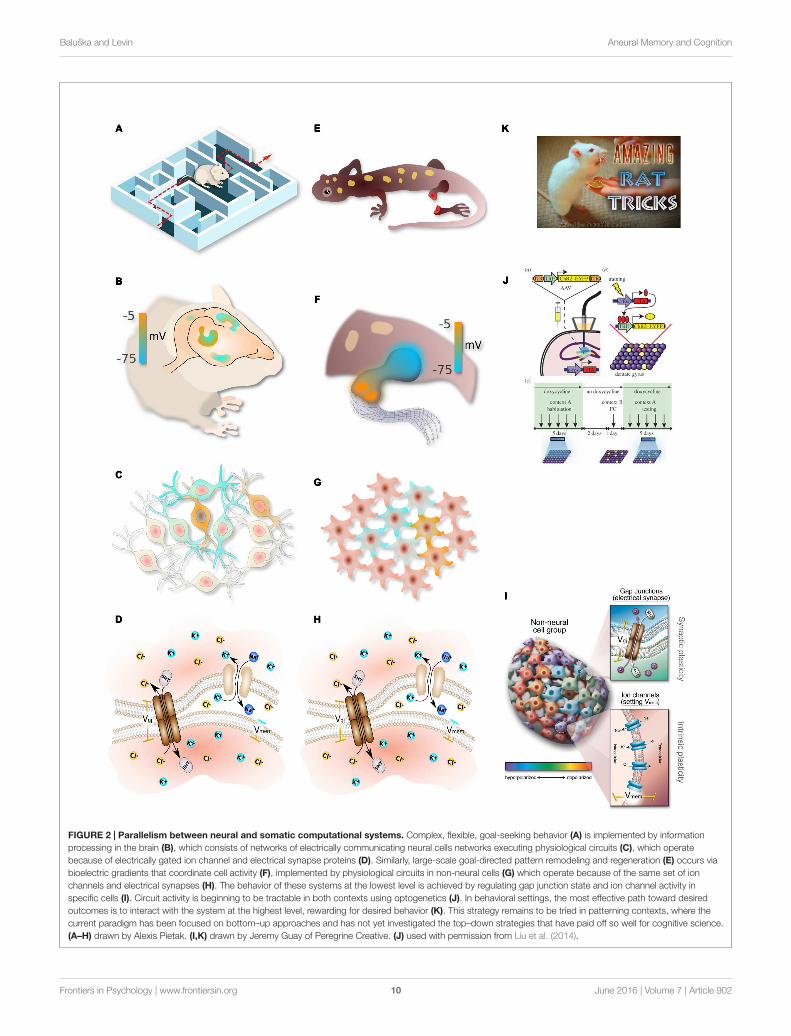
The analogy between the brain and somatic pattern control(Figure 2) makes several specific predictions. One is thation channels, GJs, and neurotransmitters should play a rolein development; this has been amply demonstrated by theidentification of patterning channelopathies (Levin, 2013),functional experiments in regenerative and developmentalbiology (Stewart et al., 2007), and the teratogenic effectsof numerous psychoactive drugs (Hernandez-Diaz and Levin,2014). Another key prediction concerns the encoding ofinstructive information. In the brain, genetics establish thehardware – genes encode the available components and thusdefine the limits of cellular activity. However, the informationcontent of the brain is not directly encoded by the genome,but rather arises dynamically through environmental stimuli(learning) and self-organizing dynamics of the electrochemicalcircuitry (plasticity). Is this the case in pattern formation as well?
Can “long term somatic memory” be edited, in the contextof a wild-type genome, leading to a permanent change? A firstexample of this was shown in a different species of planaria(Nogi and Levin, 2005), where targeting GJs for just 48 hours ina chunk of tissue caused it to regenerate 2 heads – one at theformer anterior end (normal), and one at the posterior-facingend (which would normally grow a tail). Strikingly, these 2-headed worms continue to regenerate as 2-headed when cut insubsequent rounds of regeneration, in plain water, months afterthe GJ blocking reagent is long gone from the tissue (Oviedo et al.,2010). The target morphology – the shape to which this animalregenerates upon damage – has been permanently re-writtenby temporarily editing the physiological network. This findinghas clear similarity to plasticity [well-known to be exhibited byelectrical synapses (Pereda et al., 2013)]: a brief induced change ofGJ connectivity becomes stabilized to a long-term change (Levin,2014a). This interaction between bioelectric activity and voltage-gated GJs makes developmental bioelectrical networks especiallysuitable as a labile yet stable memory medium (Palacios-Pradoand Bukauskas, 2009). Another brain-like property exhibited inthis effect is its holographic nature: in each round of cutting,the ectopic head (perhaps “epigenetically reprogrammed”) isremoved, and a middle fragment of the gut still knows it mustmake 2 heads if cut. The patterning information is distributednon-locally throughout the network.
This field is advancing rapidly in its mechanistic detailsat the cellular level: the genetics of endogenous ion channelscausing the gradients, the transduction mechanisms that controltranscription after Vmem change, and the gene expression changesdownstream of bioelectrical signaling are all becoming clear(Yang and Brackenbury, 2013; Pai et al., 2015b). Techniques,
such as optogenetics (Adams et al., 2013, 2014), are allowingimposition of specific voltage patterns onto tissue in vivo. As inthe brain, where optogenetics is used to insert memories directlyinto brains (Ramirez et al., 2013; Liu et al., 2014), these techniqueswill be crucial to learn to rewrite pattern memories duringregeneration or embryogenesis. However, as in neuroscience,there is more than one level at which progress needs to bemade. A mature understanding of the brain requires synthesisof data from people working on the genetics and biochemistryof specific neurotransmitter receptors and their downstreammolecular signaling, with the insights of workers at the level ofcircuits, behavior, cognitive science, and psychology.
Classic work explored the extensive parallels between chemicalgradients during development and signal processing in thevisual system (Grossberg, 1978), and indeed early quantitativemodels of patterning (explaining self-regulatory features likeproportion regulation) were based on visual system function(Hartline et al., 1956; Gierer and Meinhardt, 1972). Morerecent efforts include the notion of memory for position duringregeneration (Chang et al., 2002; Kragl et al., 2009; Wang et al.,2009) and development (Beloussov, 1997) and for signalinghysteresis during development (Balaskas et al., 2012), excitablecortex memory models of pseudopod dynamics (Cooper et al.,2012), and neural network models of chemical signaling (Linget al., 2013) (which showed formal isomorphisms betweengene regulation networks and Hebbian learning in neural nets)(Watson et al., 2010; Ling et al., 2013). In addition to classicalneuroscience concepts, more exotic group cognition models havebeen applied to patterning (Gunji and Ono, 2012), while afew recent studies investigated the decision-making and formalcomputational capabilities of RD systems – a chemical signalingmodality often used to model morphogenesis (Adamatzky et al.,2003, 2008; Costello et al., 2009; Dale and Husbands, 2010, whichis now known to be Turing-complete (Scarle, 2009) and supportsemantic interpretations (Schumann and Adamatzky, 2009).Despite these fascinating efforts to identify elements of cognitive-like processing in well-known elements of pattern formation,developmental biology is still firmly centered in a mechanisticperspective, seeking explanations in terms of pathways and notinformation (systems that know things and make decisions basedon that understanding). However, it is crucial to note thatattributing true knowledge and memory to biological systemsis not mystical thinking – computational neuroscience showsus a clear proof of concept that information-level, cognitiveapproaches to cellular networks are viable, and in fact necessary,strategy for understanding a system at all of its salient levels.
Thus, neuroscience offers developmental biology morethan just tools and molecular mechanisms: it offers a uniqueparadigm, otherwise unavailable to molecular and cell biologists,of the emergence of higher levels of organization that have bothcausal potency and experimental tractability. The field is inneed of new formalisms and conceptual tools for linking thedynamics of physiological circuits with downstream patterningoutcomes. Developmental biology is currently focused entirelyin a bottom-up mode, with molecules being the preferred level ofexplanation. Neuroscience teaches us that we must look upwardas well as downward, for emergent levels with their own rules
Frontiers in Psychology | www.frontiersin.org 9 June 2016 | Volume 7 | Article 902
fpsyg-07-00902 June 17, 2016 Time: 13:17 # 10
Baluška and Levin Aneural Memory and Cognition
FIGURE 2 | Parallelism between neural and somatic computational systems. Complex, flexible, goal-seeking behavior (A) is implemented by informationprocessing in the brain (B), which consists of networks of electrically communicating neural cells networks executing physiological circuits (C), which operatebecause of electrically gated ion channel and electrical synapse proteins (D). Similarly, large-scale goal-directed pattern remodeling and regeneration (E) occurs viabioelectric gradients that coordinate cell activity (F), implemented by physiological circuits in non-neural cells (G) which operate because of the same set of ionchannels and electrical synapses (H). The behavior of these systems at the lowest level is achieved by regulating gap junction state and ion channel activity inspecific cells (I). Circuit activity is beginning to be tractable in both contexts using optogenetics (J). In behavioral settings, the most effective path toward desiredoutcomes is to interact with the system at the highest level, rewarding for desired behavior (K). This strategy remains to be tried in patterning contexts, where thecurrent paradigm has been focused on bottom–up approaches and has not yet investigated the top–down strategies that have paid off so well for cognitive science.(A–H) drawn by Alexis Pietak. (I,K) drawn by Jeremy Guay of Peregrine Creative. (J) used with permission from Liu et al. (2014).
Frontiers in Psychology | www.frontiersin.org 10 June 2016 | Volume 7 | Article 902
fpsyg-07-00902 June 17, 2016 Time: 13:17 # 11
Baluška and Levin Aneural Memory and Cognition
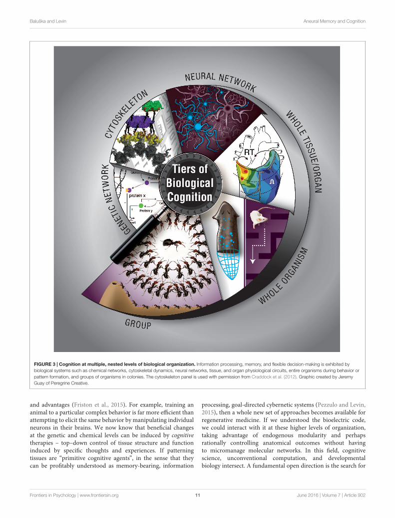
FIGURE 3 | Cognition at multiple, nested levels of biological organization. Information processing, memory, and flexible decision-making is exhibited bybiological systems such as chemical networks, cytoskeletal dynamics, neural networks, tissue, and organ physiological circuits, entire organisms during behavior orpattern formation, and groups of organisms in colonies. The cytoskeleton panel is used with permission from Craddock et al. (2012). Graphic created by JeremyGuay of Peregrine Creative.
and advantages (Friston et al., 2015). For example, training ananimal to a particular complex behavior is far more efficient thanattempting to elicit the same behavior by manipulating individualneurons in their brains. We now know that beneficial changesat the genetic and chemical levels can be induced by cognitivetherapies – top–down control of tissue structure and functioninduced by specific thoughts and experiences. If patterningtissues are “primitive cognitive agents”, in the sense that theycan be profitably understood as memory-bearing, information
processing, goal-directed cybernetic systems (Pezzulo and Levin,2015), then a whole new set of approaches becomes available forregenerative medicine. If we understood the bioelectric code,we could interact with it at these higher levels of organization,taking advantage of endogenous modularity and perhapsrationally controlling anatomical outcomes without havingto micromanage molecular networks. In this field, cognitivescience, unconventional computation, and developmentalbiology intersect. A fundamental open direction is the search for
Frontiers in Psychology | www.frontiersin.org 11 June 2016 | Volume 7 | Article 902

fpsyg-07-00902 June 17, 2016 Time: 13:17 # 12
Baluška and Levin Aneural Memory and Cognition
a computational pipeline to extract goal patterns from bioelectricstate data, parallel to efforts to extract image data from brainmeasurements (Nishimoto et al., 2011). The flow of knowledgewill likely not all be unidirectional: cracking the bioelectric codein patterning tissues is likely to in turn benefit fundamentalneuroscience by showing, in perhaps a simpler context howto extract semantic content from bioelectrical cell states in thebrain.
CONCLUSION
How does biological matter give rise to decision-making,memory, representation, and goal-directed activity?Implementation-independence is a core principle of computerscience: an algorithm does what it does regardless of what kindof medium is implementing the steps. However, in the biologicalsciences, the study of memory and other cognitive functionshas largely been the province of neurobiology, which studiesthe information processing and computational functions ofone type of system: collections of neurons. Instead, we havesurveyed a broad range of systems at various scales, frommolecular to organismal, which have their own distinct ability toprocess information, make decisions, and achieve specific goalstates (Figure 3). Neural-like computation, decision-making,and memory have been reported in sperm (Alvarez et al., 2013),amoebae (Zhu et al., 2013), yeast (Caudron and Barral, 2013), andplants (Gagliano et al., 2014), using ubiquitous mechanisms likecytoskeletal elements which appear to be also involved in neuralinformation processing (Sahu et al., 2013). It is clear that neuralnetworks have no monopoly on such functions. Remarkably, itis not only the positive (adaptive) cognitive functions that arewidely conserved: some of the same illusions to which advancedbrains’ perception and rational reasoning fall prey are beingfound in systems from slime molds to multi-animal colonies(Beekman and Latty, 2015; Sakiyama and Gunji, 2016).
McCulloch said “Why the mind is in the head? Becausethere, and only there, are hosts of possible connections to beperformed as time and circumstance demand it” (McCulloch,1951). Given the facts of protein, cytoskeletal, transcriptional,and bioelectric networks, it appears that many different mediaat various scales have the ability to form and rewire experience-dependent connections. The “dynamical hypothesis” (van Gelder,1998) asks, what if the brain is better understood as adynamical system, than a computational one? We invert thishypothesis, and ask what if some dynamical systems are betterunderstood as cognitive agents? The appearance of memory andcomputation at many levels of biological organization suggestsa fractal organization of cognitive subsystems within systems –molecular, cellular, tissue, and body-wide (Figure 2). This hasbeen suggested in the brain [Smythies’ nested doll hypothesis,(Smythies, 2015)] but may indeed exist throughout the biologicalworld. Whether each successive level of organization is in somesense smarter than the ones below it, or whether structures derivetheir cognitive powers from those of lower levels, remains to bediscovered. It should be noted, however, that even in advancedbrains, the relationship between cognitive capacity and biological
structure is not trivial to pin down, as shown by the occasionalexample of potent function in the presence of severe structuraldeficits (Lorber, 1978, 1981; Nahm et al., 2012).
The hypothesis of nested, widely prevalent cognitive layerssuggests a rich research program, including: (1) the developmentof improved methods for reading/writing bioelectrical stateinformation into somatic tissues and sculpting non-neuralbioelectric circuits (optogenetics beyond excitable cells andin the synthetic biology of gap junction and neurotransmittersignaling; Adams et al., 2013), (2) continued work on crackingthe bioelectric code (bioelectric state information maps ontothe topology of various patterning outcomes in tractablemodel systems such as planaria; Tseng and Levin, 2013), (3)formulation and testing of quantitative, molecular modelsof LTP, habituation, sensitization, plasticity, and higher-order learning applied to protein interaction networks,gene regulatory circuits, cytoskeletal dynamics, and cellbehavior during morphogenesis, (4) use of reagents thatimpact cognition (hallucinogens, anesthetics; Kawamoto et al.,2005), stimulants, nootropics/cognitive enhancers, etc.) incellular, developmental, and regenerative patterning assays toprobe conservation of pathways between neuroscience andmorphogenesis, (5) creation of larger-scale computationalmodels of regeneration and functional experiments inmorphogenesis based on goal-seeking and error minimizationalgorithms with molecularly specified metrics (Slack, 1980;Chao et al., 2008), (6) exploration of molecular models ofcognitive concepts (attention, autism spectrum, sleep, visualillusions/hallucinations, addiction) in specific patterning andmispatterning contexts, (7) bioengineering platforms that rewardand punish in vitro patterning systems for specific changesin growth and morphogenesis (instrumental learning andtop–down control of shape in developmental or regenerativecontexts), and (8) a mechanistic investigation of the mechanismof persistence of memories through massive brain regeneration,which is likely to reveal the interface between somatic and neuralmemories (Blackiston et al., 2008; Shomrat and Levin, 2013).
We have avoided here the thorny issues of philosophy ofmind that arise from trying to define exactly under whatconditions words like “knowledge” are appropriate, in favorof an intentional stance-like pragmatic, engineering approachgrounded in cybernetics. The coverage of cognitive terms acrossbiology can expand to the extent that information-centeredapproaches are shown to be effective in predicting and controllingthe behavior of biological systems. The practical implications forbiotechnology, unconventional computation, and regenerativemedicine are enormous. Equally likely, the lessons we learnfrom unconventional cognitive systems will deeply impact ourmost basic understanding of how mind emerges from thebrain.
AUTHOR CONTRIBUTIONS
ML and FB both contributed sections to the review according totheir specialties. Both provided novel ideas, edited each other’stext, and prepared the finished product.
Frontiers in Psychology | www.frontiersin.org 12 June 2016 | Volume 7 | Article 902

fpsyg-07-00902 June 17, 2016 Time: 13:17 # 13
Baluška and Levin Aneural Memory and Cognition
FUNDING
ML gratefully acknowledges support of the Templeton WorldCharity Foundation (TWCF0089/AB55) and the G. Harold andLeila Y. Mathers Charitable Foundation.
ACKNOWLEDGMENTS
ML thanks members of the Levin lab, as well as Daniel C. Dennett,Giovanni Pezzulo, Francisco Vico, and Guven Guzeldere formany useful discussions on these topics.
REFERENCESAdamatzky, A., and Alonso-Sanz, R. (2011). Rebuilding Iberian motorways with
slime mould. Biosystems 105, 89–100. doi: 10.1016/j.biosystems.2011.03.007Adamatzky, A., Costello, B. D., Melhuish, C., and Ratcliffe, N. (2003). Experimental
reaction-diffusion chemical processors for robot path planning. J. Intell. Robot.Syst. 37, 233–249. doi: 10.1023/A:1025414424756
Adamatzky, A., Costello, B. D. L., and Shirakawa, T. (2008). Universal computationwith limited resources: belousov-zhabotinsky and physarum computers. Int. J.Bifurcat. Chaos 18, 2373–2389. doi: 10.1142/S0218127408021750
Adams, D. S. (2008). A new tool for tissue engineers: ions as regulators ofmorphogenesis during development and regeneration. Tiss. Eng. Part A 14,1461–1468. doi: 10.1089/ten.tea.2008.0080
Adams, D. S., Lemire, J. M., Kramer, R. H., and Levin, M. (2014). Optogeneticsin developmental biology: using light to control ion flux-dependent signals inXenopus embryos. Int. J. Dev. Biol. 58, 851–861. doi: 10.1387/ijdb.140207ml
Adams, D. S., and Levin, M. (2012). General principles for measuring restingmembrane potential and ion concentration using fluorescent bioelectricityreporters. Cold Spring Harb. Prot. 2012, 385–397. doi: 10.1101/pdb.top067710
Adams, D. S., Tseng, A. S., and Levin, M. (2013). Light-activation ofthe Archaerhodopsin H+-pump reverses age-dependent loss of vertebrateregeneration: sparking system-level controls in vivo. Biol. Open 2, 306–313. doi:10.1242/bio.20133665
Agnati, L. F., Leo, G., Zanardi, A., Genedani, S., Rivera, A., Fuxe, K., et al.(2006). Volume transmission and wiring transmission from cellular tomolecular networks: history and perspectives. Acta Physiol. 187, 329–344. doi:10.1111/j.1748-1716.2006.01579.x
Albrecht-Buehler, G. (1980). Autonomous movements of cytoplasmic fragments.Proc. Natl. Acad. Sci. U.S.A. 77, 6639–6643. doi: 10.1073/pnas.77.11.6639
Albrecht-Buehler, G. (1982). Control of tissue cell movement. Natl. Cancer Inst.Monogr. 60, 117–122.
Albrecht-Buehler, G. (1985). Is cytoplasm intelligent too? Cell Muscl Motil. 6, 1–21.doi: 10.1007/978-1-4757-4723-2_1
Alvarez, L., Friedrich, B. M., Gompper, G., and Kaupp, U. B. (2013).The computational sperm cell. Trends Cell Biol. 24, 198–207. doi:10.1016/j.tcb.2013.10.004
Antov, Y., Barbul, A., Mantsur, H., and Korenstein, R. (2005). Electroendocytosis:exposure of cells to pulsed low electric fields enhances adsorption and uptake ofmacromolecules. Biophys. J. 88, 2206–2223. doi: 10.1529/biophysj.104.051268
Applewhite, P. B. (1972). The flow of ions in learning and memory. J. Theor. Biol.36, 419–423. doi: 10.1016/0022-5193(72)90109-9
Applewhite, P. B., Lapan, E. A., and Gardner, F. T. (1969). Protozoan habituationlearning after loss of macronuclei and cytoplasm. Nature 222, 491–492. doi:10.1038/222491a0
Aragones, A. C., Haworth, N. L., Darwish, N., Ciampi, S., Bloomfield, N. J., Wallace,G. G., et al. (2016). Electrostatic catalysis of a Diels–Alder reaction. Nature 531,88–91. doi: 10.1038/nature16989
Aur, D. (2012). From neuroelectrodynamics to thinking machines. Cogn. Comput.4, 4–12. doi: 10.1007/s12559-011-9106-3
Baddour, J. A., Sousounis, K., and Tsonis, P. A. (2012). Organ repair andregeneration: an overview. Birth Def. Res. C Embr. Today 96, 1–29. doi:10.1002/bdrc.21006
Balaskas, N., Ribeiro, A., Panovska, J., Dessaud, E., Sasai, N., Page, K. M., et al.(2012). Gene regulatory logic for reading the Sonic Hedgehog signaling gradientin the vertebrate neural tube. Cell 148, 273–284. doi: 10.1016/j.cell.2011.10.047
Baluška, F. (2010). Recent surprising similarities between plant cells and neurons.Plant Signal. Behav. 5, 87–89. doi: 10.4161/psb.5.2.11237
Baluška, F., and Mancuso, S. (2009). Deep evolutionary origins of neurobiology:turning the essence of ‘neural’ upside-down. Commun. Integr. Biol. 2, 60–65.doi: 10.4161/cib.2.1.7620
Baluška, F., and Mancuso, S. (2013). Root apex transition zone as oscillatory zone.Front. Plant Sci. 4:354. doi: 10.3389/fpls.2013.00354
Baluška, F., Mancuso, S., Volkmann, D., and Barlow, P. W. (2004). Root apices asplant command centres: the unique ‘brain-like’ status of the root apex transitionzone. Biologia 59, 9–17.
Baluška, F., Mancuso, S., Volkmann, D., and Barlow, P. W. (2009a). The‘root-brain’ hypothesis of Charles and Francis Darwin: revival after morethan 125 years. Plant Signal. Behav. 4, 1121–1127. doi: 10.4161/psb.4.12.10574
Baluška, F., Mancuso, S., Volkmann, D., and Barlow, P. W. (2010). Root apextransition zone: a signalling – response nexus in the root. Trends Plant Sci. 15,402–408. doi: 10.1016/j.tplants.2010.04.007
Baluška, F., Šamaj, J., and Menzel, D. (2003). Polar transport of auxin: carrier-mediated flux across the plasma membrane or neurotransmitter-like secretion?Trends Cell Biol. 13, 282–285. doi: 10.1016/S0962-8924(03)00084-9
Baluška, F., Schlicht, M., Volkmann, D., and Mancuso, S. (2008). Vesicularsecretion of auxin: evidences and implications. Plant Signal. Behav. 3, 254–256.doi: 10.4161/psb.3.4.5183
Baluška, F., Schlicht, M., Wan, Y.-L., Burbach, C., and Volkmann, D. (2009b).Intracellular domains and polarity in root apices: from synaptic domains toplant neurobiology. Nova Acta Leopold 96, 103–122.
Baluška, F., Volkmann, D., and Menzel, D. (2005). Plant synapses: actin-basedadhesion domains for cell-to-cell communication. Trends Plant Sci. 10, 106–111. doi: 10.1016/j.tplants.2005.01.002
Baluška, F., and Wan, Y. L. (2012). “Physical control overendocytosis,” inEndocytosis in Plants, ed. J. Šamaj (Berlin: Springer-Verlag), 123–149.
Bancroft, W. D., and Richter, G. H. (1930). Claude Bernard’s theory of narcosis.Proc. Natl. Acad. Sci. U.S.A. 16, 573–577. doi: 10.1073/pnas.16.9.573
Barlow, P. W., and Baluška, F. (2000). Cytoskeletal perspectives on root growthand morphogenesis. Annu. Rev. Plant Physiol. Plant Mol. Biol. 51, 289–322. doi:10.1146/annurev.arplant.51.1.289
Bates, E. (2015). Ion channels in development and cancer. Annu. Rev. Cell. Dev.Biol. 31, 231–247. doi: 10.1146/annurev-cellbio-100814-125338
Beagle, S. D., and Lockless, S. W. (2015). Microbiology: electrical signalling goesbacterial. Nature 527, 44–45. doi: 10.1038/nature15641
Beane, W. S., Morokuma, J., Adams, D. S., and Levin, M. (2011). A chemicalgenetics approach reveals H,K-ATPase-mediated membrane voltage isrequired for planarian head regeneration. Chem. Biol. 18, 77–89. doi:10.1016/j.chembiol.2010.11.012
Becchetti, A., Pillozzi, S., Morini, R., Nesti, E., and Arcangeli, A. (2010). Newinsights into the regulation of ion channels by integrins. Int. Rev. Cell. Mol. Biol.279, 135–190. doi: 10.1016/S1937-6448(10)79005-5
Beekman, M., and Latty, T. (2015). Brainless but multi-headed: decision making bythe acellular slime mould Physarum polycephalum. J. Mol. Biol. 427, 3734–3743.doi: 10.1016/j.jmb.2015.07.007
Beloussov, L. V. (1997). On the active memory in developing systems. Riv. Biol. 90,31–46.
Ben-Jacob, E. (2009). Learning from bacteria about natural information processing.Ann. N. Y. Acad. Sci. 1178, 78–90. doi: 10.1111/j.1749-6632.2009.05022.x
Bernard, C. (1878). Lectures on Phenomena of Life Common to Animals and Plants.Paris: Ballliere, and Son.
Bezanilla, F. (2002). Voltage sensor movements. J. Gen. Physiol. 120, 465–473. doi:10.1085/jgp.20028660
Bezanilla, F. (2006). The action potential: from voltage-gated conductancesto molecular structures. Biol. Res. 39, 425–435. doi: 10.4067/S0716-97602006000300005
Bezanilla, F. (2008). How membrane proteins sense voltage. Nat. Rev. Mol. Cell.Biol. 9, 323–332. doi: 10.1038/nrm2376
Bharti, K., and Arnheiter, H. (2005). When pigment cells turn into neurons.J. Invest. Dermatol. 125, 10–11. doi: 10.1111/j.0022-202X.2005.23876.x
Frontiers in Psychology | www.frontiersin.org 13 June 2016 | Volume 7 | Article 902
fpsyg-07-00902 June 17, 2016 Time: 13:17 # 14
Baluška and Levin Aneural Memory and Cognition
Blackiston, D. J., Anderson, G. M., Rahman, N., Bieck, C., and Levin, M. (2015).A novel method for inducing nerve growth via modulation of host restingpotential: gap junction-mediated and serotonergic signaling mechanisms.Neurotherapeutics 12, 170–184. doi: 10.1007/s13311-014-0317-7
Blackiston, D. J., Silva Casey, E., and Weiss, M. R. (2008). Retention of memorythrough metamorphosis: can a moth remember what it learned as a caterpillar?PLoS ONE 3:e1736. doi: 10.1371/journal.pone.0001736
Böhm, J., Scherzer, S., Krol, E., Kreuzer, I., von Meyer, K., Lorey, C., et al. (2016).The Venus flytrap Dionaea muscipula counts prey-induced action potentials toinduce sodium uptake. Curr. Biol. 26, 286–295. doi: 10.1016/j.cub.2015.11.057
Bose, J. (1926). Response in the Living and the Non-Living. London: Longmans,Green & Co.
Brunet, T., and Arendt, D. (2016). From damage response to action potentials: earlyevolution of neural and contractile modules in stem eukaryotes. Philos. Trans.R. Soc. Lond. B. Biol. Sci. 371, 1685. doi: 10.1098/rstb.2015.0043
Bruusgaard, J. C., Johansen, I. B., Egner, I. M., Rana, Z. A., and Gundersen, K.(2010). Myonuclei acquired by overload exercise precede hypertrophy and arenot lost on detraining. Proc. Natl. Acad. Sci. U.S.A. 107, 15111–15116. doi:10.1073/pnas.0913935107
Burbach, C., Markus, K., Yin, Z., Schlicht, M., and Baluška, F. (2012).Photophobic behaviour of maize roots. Plant Signal. Behav. 7, 876–880. doi:10.4161/psb.21012
Buznikov, G., Shmukler, Y., and Lauder, J. (1996). From oocyte to neuron: doneurotransmitters function in the same way throughout development? Cell Mol.Neurobiol. 16, 537–559. doi: 10.1007/BF02152056
Buznikov, G. A., and Shmukler, Y. B. (1981). Possible role of “prenervous”neurotransmitters in cellular interactions of early embryogenesis: a hypothesis.Neurochem. Res. 6, 55–68. doi: 10.1007/BF00963906
Calvo, P. (2016). The philosophy of plant neurobiology: a manifesto. Synthese 193,1323–1343. doi: 10.1007/s11229-016-1040-1
Calvo, P., and Baluška, F. (2015). Conditions for minimal intelligence acrosseukaryota: a cognitive science perspective. Front. Psychol. 6:1329. doi:10.3389/fpsyg.2015.01329
Carlson, B. M. (1983). Positional memory in vertebrate limb development andregeneration. Prog. Clin. Biol. Res. 110, 433–443.
Carneiro, K., Donnet, C., Rejtar, T., Karger, B. L., Barisone, G. A., Diaz, E., et al.(2011). Histone deacetylase activity is necessary for left-right patterning duringvertebrate development. BMC Dev. Biol. 11:29. doi: 10.1186/1471-213X-11-29
Carter, J. H. (2000). The immune system as a model for patternrecognition and classification. J. Am. Med. Inform. Assoc. 7, 28–41. doi:10.1136/jamia.2000.0070028
Caudron, F., and Barral, Y. (2013). A super-assembly of Whi3 encodes memory ofdeceptive encounters by single cells during yeast courtship. Cell 155, 1244–1257.doi: 10.1016/j.cell.2013.10.046
Cervera, J., Alcaraz, A., and Mafe, S. (2014). Membrane potential bistabilityin nonexcitable cells as described by inward and outward voltage-gated ionchannels. J. Phys. Chem. B 118, 12444–12450. doi: 10.1021/jp508304h
Cervera, J., Manzanares, J. A., and Mafe, S. (2015). Electrical coupling in ensemblesof nonexcitable cells: modeling the spatial map of single cell potentials. J. Phys.Chem. B 119, 2968–2978. doi: 10.1021/jp512900x
Chakravarthy, S. V., and Ghosh, J. (1997). On Hebbian-like adaptation in heartmuscle: a proposal for ‘cardiac memory’. Biol. Cybern. 76, 207–215. doi:10.1007/s004220050333
Chang, H. Y., Chi, J. T., Dudoit, S., Bondre, C., van de Rijn, M., Botstein, D.,et al. (2002). Diversity, topographic differentiation, and positional memoryin human fibroblasts. Proc. Natl. Acad. Sci. U.S.A. 99, 12877–12882. doi:10.1073/pnas.162488599
Chao, Z. C., Bakkum, D. J., and Potter, S. M. (2008). Shaping embodied neuralnetworks for adaptive goal-directed behavior. PLoS Comp. Biol. 4:e1000042. doi:10.1371/journal.pcbi.1000042
Chen, H., and He, X. (2016). The convergent cancer evolution toward a singlecellular destination. Mol. Biol. Evol. 33, 4–12. doi: 10.1093/molbev/msv212
Chernet, B., and Levin, M. (2013). Endogenous voltage potentials and themicroenvironment: bioelectric signals that reveal, induce and normalize cancer.J. Clin. Exp. Oncol. Suppl. 1, S1–S2.
Chernet, B. T., Fields, C., and Levin, M. (2015). Long-range gap junctionalsignaling controls oncogene-mediated tumorigenesis in Xenopus laevisembryos. Front. Physiol. 5:519. doi: 10.3389/fphys.2014.00519
Chernet, B. T., and Levin, M. (2013). Transmembrane voltage potential is anessential cellular parameter for the detection and control of tumor developmentin a Xenopus model. Dis. Mod. Mech. 6, 595–607. doi: 10.1242/dmm.010835
Chernet, B. T., and Levin, M. (2014). Transmembrane voltage potential of somaticcells controls oncogene-mediated tumorigenesis at long-range. Oncotarget 5,3287–3306. doi: 10.18632/oncotarget.1935
Clark, K. B. (2013). Ciliates learn to diagnose and correct classical error syndromesin mating strategies. Front. Microbiol. 4:229. doi: 10.3389/fmicb.2013.00229
Cohen, I. R. (1992a). The cognitive paradigm and the immunological homunculus.Immunol. Today 13, 490–494. doi: 10.1016/0167-5699(92)90024-2
Cohen, I. R. (1992b). The cognitive principle challenges clonal selection. Immunol.Today 13, 441–444. doi: 10.1016/0167-5699(92)90071-E
Conrad, M. (1996). Cross-scale information processing in evolution, developmentand intelligence. BioSystems 38, 97–109. doi: 10.1016/0303-2647(95)01579-5
Cooper, R. M., Wingreen, N. S., and Cox, E. C. (2012). An excitable cortex andmemory model successfully predicts new pseudopod dynamics. PLoS ONE7:e33528. doi: 10.1371/journal.pone.0033528
Corning, W. C. (1966). Retention of a position discrimination after regeneration inplanarians. Psychol. Sci. 5, 17–18. doi: 10.3758/BF03328256
Corssen, G., and Skora, I. A. (1964). “Addiction” reactions in cultured Hhumancells. JAMA 187, 328–332. doi: 10.1001/jama.1964.03060180014003
Costello, B. D., Toth, R., Stone, C., Adamatzky, A., and Bull, L. (2009).Implementation of glider guns in the light-sensitive Belousov-Zhabotinskymedium. Phys. Rev. Stat. Nonlin Soft. Matter Phys. 79(Pt 2), 026114. doi:10.1103/PhysRevE.79.026114
Couzin, I. D. (2009). Collective cognition in animal groups. Trends Cogn. Sci. 13,36–43. doi: 10.1016/j.tics.2008.10.002
Craddock, T. J., Tuszynski, J. A., and Hameroff, S. (2012). Cytoskeletal signaling:is memory encoded in microtubule lattices by CaMKII phosphorylation? PLoSComp. Biol. 8:e1002421. doi: 10.1371/journal.pcbi.1002421
Craddock, T. J., Tuszynski, J. A., Priel, A., and Freedman, H. (2010). Microtubuleionic conduction and its implications for higher cognitive functions. J. Integr.Neurosci. 9, 103–122. doi: 10.1142/S0219635210002421
Dahal, G. R., Rawson, J., Gassaway, B., Kwok, B., Tong, Y., Ptacek, L. J., et al. (2012).An inwardly rectifying K+ channel is required for patterning. Development 139,3653–3664. doi: 10.1242/dev.078592
Dale, K., and Husbands, P. (2010). The evolution of reaction-diffusioncontrollers for minimally cognitive agents. Artif. Life 16, 1–19. doi:10.1162/artl.2009.16.1.16100
Daoudal, G., and Debanne, D. (2003). Long-term plasticity of intrinsicexcitability: learning rules and mechanisms. Learn. Memory 10, 456–465. doi:10.1101/lm.64103
Darwin, C. (1880). The Power of Movement in Plants. London: John Murray.De Luccia, T. P. (2012). Mimosa pudica, Dionaea muscipula and anesthetics. Plant
Signal. Behav. 7, 1163–1167. doi: 10.4161/psb.21000DeMarse, T. B., and Dockendorf, K. P. (2005). Adaptive flight control with living
neuronal networks on microelectrode arrays. Proc. IEEE Explor. IJCNN 3,1548–1551.
Dingli, D., Chalub, F. A., Santos, F. C., Van Segbroeck, S., and Pacheco, J. M.(2009). Cancer phenotype as the outcome of an evolutionary game betweennormal and malignant cells. Br. J. Cancer. 101, 1130–1136. doi: 10.1038/sj.bjc.6605288
Doursat, R., Sayama, H., and Michel, O. (2013). A review of morphogeneticengineering. Nat. Comput. 12, 517–535. doi: 10.1007/s11047-013-9398-1
Dranias, M. R., Ju, H., Rajaram, E., and Van Dongen, A. M. (2013). Short-termmemory in networks of dissociated cortical neurons. J. Neurosci. 33, 1940–1953.doi: 10.1523/JNEUROSCI.2718-12.2013
Eisenstein, E. M. (1967). “The use of invertebrate systems for studies on thebases of learning and memory,” in The Neurosciences—A Study Program, edsG. C. Quarton, T. Melnechuk, and F. U. Schmitt (New York, NY: RockefellerUniversity Press), 653–665.
Fernando, C., Szathmary, E., and Husbands, P. (2012). Selectionist andevolutionary approaches to brain function: a critical appraisal. Front. Comput.Neurosci. 6:24. doi: 10.3389/fncom.2012.00024
Filoni, S., Velloso, C. P., Bernardini, S., and Cannata, S. M. (1995). Acquisition ofnerve dependence for the formation of a regeneration blastema in amputated
Frontiers in Psychology | www.frontiersin.org 14 June 2016 | Volume 7 | Article 902
fpsyg-07-00902 June 17, 2016 Time: 13:17 # 15
Baluška and Levin Aneural Memory and Cognition
hindlimbs of larval Xenopus laevis: the role of limb innervation and that of limbdifferentiation. J. Exp. Zool. 273, 327–341. doi: 10.1002/jez.1402730407
Forrest, M. D. (2014). The sodium-potassium pump is an informationprocessing element in brain computation. Front. Physiol. 5:472. doi:10.3389/fphys.2014.00472
Francis, N. J., and Kingston, R. E. (2001). Mechanisms of transcriptional memory.Nat. Rev. Mol. Cell. Biol. 2, 409–421. doi: 10.1038/35073039
Freddolino, P. L., and Tavazoie, S. (2012). Beyond homeostasis: a predictive-dynamic framework for understanding cellular behavior. Annu. Rev. Cell. Dev.Biol. 28, 363–384. doi: 10.1146/annurev-cellbio-092910-154129
Friston, K. (2010). The free-energy principle: a unified brain theory? Nat. Rev.Neurosci. 11, 127–138. doi: 10.1038/nrn2787
Friston, K., Levin, M., Sengupta, B., and Pezzulo, G. (2015). Knowing one’splace: a free-energy approach to pattern regulation. J. R. Soc. Interface 12. doi:10.1098/rsif.2014.1383
Funk, R. (2013). Ion gradients in tissue and organ biology. Biol. Syst. 2:1000105.Fuxe, K., Borroto-Escuela, D. O., Romero-Fernandez, W., Zhang, W. B., and
Agnati, L. F. (2013). Volume transmission and its different forms in the centralnervous system. Chin. J. Integr. Med. 19, 323–329. doi: 10.1007/s11655-013-1455-1
Gagliano, M., Renton, M., Depczynski, M., and Mancuso, S. (2014). Experienceteaches plants to learn faster and forget slower in environments where itmatters. Oecologia 175, 63–72. doi: 10.1007/s00442-013-2873-7
Gantt, W. H. (1974). Autokinesis, schizokinesis, centrokinesis and organ-systemresponsibility: concepts and definition. Pavlov J. Biol. Sci. 9, 187–191.
Gantt, W. H. (1981). Organ-system responsibility, schizokinesis, and autokinesisin behavior. Pavlov J. Biol. Sci. 16, 64–66.
Gantt, W. H., McGuigan, F. J., and Ban, T. A. (1987). Critical issues in Psychology,Psychiatry, and Physiology. Memorial to W. Horsley Gantt. New York, NY:Gordon and Breach Science Publishers.
Gelber, B. (1962). Acquisition in Paramecium aurelia during spaced training.Psychol. Rec. 12, 165–177.
Gentile, L., Cebria, F., and Bartscherer, K. (2011). The planarian flatworm: anin vivo model for stem cell biology and nervous system regeneration. Dis.Model. Mech. 4, 12–19. doi: 10.1242/dmm.006692
Gierer, A., and Meinhardt, H. (1972). A theory of biological pattern formation.Kybernetik 12, 30–39. doi: 10.1007/BF00289234
Goel, P., and Mehta, A. (2013). Learning theories reveal loss of pancreatic electricalconnectivity in diabetes as an adaptive response. PLoS ONE 8:e70366. doi:10.1371/journal.pone.0070366
Goldberger, J. J., and Kadish, A. H. (1999). Cardiac memory. Pacing Clin.Electrophysiol. 22, 1672–1679. doi: 10.1111/j.1540-8159.1999.tb00388.x
Goldman, S. A., Nedergaard, M., and Windrem, M. S. (2015). Modeling cognitionand disease using human glial chimeric mice. Glia 63, 1483–1493. doi:10.1002/glia.22862
Goldsworthy, A. (1983). The evolution of plant action potentials. J. Theor. Biol. 103,645–648. doi: 10.1016/0022-5193(83)90287-4
Gremiaux, A., Yokawa, K., Mancuso, S., and Baluška, F. (2014). Plant anesthesiasupports similarities between animals and plants: claude Bernard’s forgottenstudies. Plant Signal. Behav. 9:e27886. doi: 10.4161/psb.27886
Grossberg, S. (1978). “Communication, memory, and development,” in Progress inTheoretical Biology, eds R. Rosen and F. Snell (Amsterdam: Elsevier).
Gundersen, K. (2016). Muscle memory and a new cellular model for muscleatrophy and hypertrophy. J. Exp. Biol. 219, 235–242. doi: 10.1242/jeb.124495
Gunji, Y. P., and Ono, R. (2012). Sociality of an agent during morphogeneticcanalization: asynchronous updating with potential resonance. BioSystems 109,420–429. doi: 10.1016/j.biosystems.2012.05.005
Gyurko, D. M., Veres, D. V., Modos, D., Lenti, K., Korcsmaros, T., and Csermely, P.(2013). Adaptation and learning of molecular networks as a descriptionof cancer development at the systems-level: potential use in anti-cancertherapies. Semin. Cancer Biol. 23, 262–269. doi: 10.1016/j.semcancer.2013.06.005
Hamann, T. (2015). The plant cell wall integrity maintenance mechanism – a casestudy of a cell wall plasma membrane signaling network. Phytochemistry 112,100–109. doi: 10.1016/j.phytochem.2014.09.019
Hameroff, S. R., and Watt, R. C. (1982). Information processing in microtubules.J. Theor. Biol. 98, 549–561. doi: 10.1016/0022-5193(82)90137-0
Hamilton, T. C. (1975). “Behavioral plasticity in protozoans,” in Aneural Organismsin Neurobiology, ed. E. M. Eisenstein (New York: Plenum Press), 111–130.
Hartline, H. K., Wagner, H. G., and Ratliff, F. (1956). Inhibition in the eye ofLimulus. J. Gen. Physiol. 39, 651–673. doi: 10.1085/jgp.39.5.651
Hedrich, R., Salvador-Recatalà, V., and Dreyer, I. (2016). Electrical wiringand long-distance plant communication. Trends Plant Sci 21, 376–387. doi:10.1016/j.tplants.2016.01.016
Helliker, B. R., and Richter, S. L. (2008). Subtropical to boreal convergence oftree-leaf temperatures. Nature 454, 511–515. doi: 10.1038/nature07031
Hernandez-Diaz, S., and Levin, M. (2014). Alteration of bioelectrically-controlledprocesses in the embryo: a teratogenic mechanism for anticonvulsants. Reprod.Toxicol. 47, 111–114. doi: 10.1016/j.reprotox.2014.04.008
Hetherington, A. M., and Woodward, F. I. (2003). The role of stomatain sensing and driving environmental change. Nature 424, 901–908. doi:10.1038/nature01843
Higgins, M. L., Stadnicki, S. S., Shaw, T. J., and Leach, F. R. (1978). Effects ofaddicting and nonaddicting drugs on HeLa cells. Tex. Rep. Biol. Med. 36,95–110.
Hille, B. (2001). Ion Channels of Excitable Membranes. Sunderland, MA: SinauerAssociates Inc.
Ho, M.-L., Tsai, T. N., Chang, J. K., Shao, T. S., Jeng, Y. R., and Hsu, C.(2005). Down-regulation of N-methyl D-aspartate receptor in rat-modeleddisuse osteopenia. Osteop. Int. 16, 1780–1788. doi: 10.1007/s00198-005-1928-y
Holcombe, M., and Paton, R. (1998). Information Processing in Cells and Tissues.New York, NY: Plenum Press.
Huang, S., Eichler, G., Bar-Yam, Y., and Ingber, D. E. (2005). Cell fates as high-dimensional attractor states of a complex gene regulatory network. Phys. Rev.Lett. 94:128701. doi: 10.1103/PhysRevLett.94.128701
Humphrey, T. V., Bonetta, D. T., and Goring, D. R. (2007). Sentinels at the wall:cell wall receptors and sensors. New Phytol. 176, 7–21. doi: 10.1111/j.1469-8137.2007.02192.x
Ingber, D. E., and Levin, M. (2007). What lies at the interface of regenerativemedicine and developmental biology? Development 134, 2541–2547. doi:10.1242/dev.003707
Iovine, M. K., Higgins, E. P., Hindes, A., Coblitz, B., and Johnson, S. L. (2005).Mutations in connexin43 (GJA1) perturb bone growth in zebrafish fins. Dev.Biol. 278, 208–219. doi: 10.1016/j.ydbio.2004.11.005
Jaffe, L. F. (1977). Electrophoresis along cell membranes. Nature 265, 600–602. doi:10.1038/265600a0
Jaffe, L. F. (1981). The role of ionic currents in establishing developmental pattern.Philos. Trans. R. Soc. Lond. B 295, 553–566. doi: 10.1098/rstb.1981.0160
James, L. J., and Soffe, S. R. (2011). Skin impulse excitation of spinal sensoryneurons in developing Xenopus laevis (Daudin) tadpoles. J. Exp. Biol. 214,3341–3350. doi: 10.1242/jeb.058446
Johnston, R. N., Pai, S. B., and Pai, R. B. (1992). The origin of the cancercell: oncogeny reverses phylogeny. Biochem. Cell Biol. 70, 831–834. doi:10.1139/o92-130
Karban, R., Wetzel, W. C., Shiojiri, K., Ishizaki, S., Ramirez, S. R., and Blande,J. D. (2014a). Deciphering the language of plant communication: volatilechemotypes of sagebrush. New Phytol. 204, 380–385. doi: 10.1111/nph.12887
Karban, R., Yang, L. H., and Edwards, K. F. (2014b). Volatile communicationbetween plants that affects herbivory: a meta-analysis. Ecol. Lett. 17, 44–52. doi:10.1111/ele.12205
Kawamoto, S., Yoshida-Noro, C., and Tochinai, S. (2005). Bipolar headregeneration induced by artificial amputation in Enchytraeus japonensis(Annelida, Oligochaeta). J. Exp. Zoolog. A Comp. Exp. Biol. 303, 615–627. doi:10.1002/jez.a.205
Keijzer, F., van Duijn, M., and Lyon, P. (2013). What nervous systems do: earlyevolution, input-output, and the skin brain thesis. Adapt. Behav. 21, 67–85. doi:10.1177/1059712312465330
Khan, S. M., Ali, R., Asi, N., and Molloy, J. E. (2012). Active actin gels. Commun.Integr. Biol. 5, 39–42. doi: 10.4161/cib.18286
Koch, C., and Segev, I. (2000). The role of single neurons in information processing.Nat. Neurosci. 3, 1171–1177. doi: 10.1038/81444
Kondo, S. (2002). The reaction-diffusion system: a mechanism for autonomouspattern formation in the animal skin. Genes Cells 7, 535–541. doi:10.1046/j.1365-2443.2002.00543.x
Frontiers in Psychology | www.frontiersin.org 15 June 2016 | Volume 7 | Article 902

fpsyg-07-00902 June 17, 2016 Time: 13:17 # 16
Baluška and Levin Aneural Memory and Cognition
Kondo, S., and Miura, T. (2010). Reaction-diffusion model as a frameworkfor understanding biological pattern formation. Science 329, 1616–1620. doi:10.1126/science.1179047
Koo, H., and Graziadei, P. P. (1995). Eye primordium transplantation in Xenopusembryo. Anat. Embryol. 191, 155–170. doi: 10.1007/BF00186787
Koshland, D. E. Jr. (1980). Bacterial chemotaxis in relation to neurobiology.Annu. Rev. Neurosci. 3, 43–75. doi: 10.1146/annurev.ne.03.030180.000355
Kragl, M., Knapp, D., Nacu, E., Khattak, S., Maden, M., Epperlein, H. H.,et al. (2009). Cells keep a memory of their tissue origin during axolotl limbregeneration. Nature 460, 60–65. doi: 10.1038/nature08152
Kumar, A., and Brockes, J. P. (2012). Nerve dependence in tissue, organ,and appendage regeneration. Trends Neurosci. 35, 691–699. doi:10.1016/j.tins.2012.08.003
Kunita, I., Kuroda, S., Ohki, K., and Nakagaki, T. (2014). Attempts to retreat froma dead-ended long capillary by backward swimming in Paramecium. Front.Microbiol. 5:270. doi: 10.3389/fmicb.2014.00270
Lahoz-Beltra, R., Hameroff, S. R., and Dayhoff, J. E. (1993). Cytoskeletal logic:a model for molecular computation via Boolean operations in microtubulesand microtubule-associated proteins. Biosystems 29, 1–23. doi: 10.1016/0303-2647(93)90078-Q
Law, R., and Levin, M. (2015). Bioelectric memory: modeling resting potentialbistability in amphibian embryos and mammalian cells. Theor. Biol. Med.Model. 12:22. doi: 10.1186/s12976-015-0019-9
Leppik, L. P., Froemel, D., Slavici, A., Ovadia, Z. N., Hudak, L., Henrich, D., et al.(2015). Effects of electrical stimulation on rat limb regeneration, a new look atan old model. Sci. Rep. 5:18353. doi: 10.1038/srep18353
Levin, M. (2007a). Gap junctional communication in morphogenesis.Prog. Biophys. Mol. Biol. 94, 186–206. doi: 10.1016/j.pbiomolbio.2007.03.005
Levin, M. (2007b). Large-scale biophysics: ion flows and regeneration. Trends CellBiol. 17, 262–271. doi: 10.1016/j.tcb.2007.04.007
Levin, M. (2011). The wisdom of the body: future techniques and approachesto morphogenetic fields in regenerative medicine, developmental biology andcancer. Reg. Med. 6, 667–673. doi: 10.2217/rme.11.69
Levin, M. (2012a). Molecular bioelectricity in developmental biology: new toolsand recent discoveries: control of cell behavior and pattern formationby transmembrane potential gradients. Bioessays 34, 205–217. doi:10.1002/bies.201100136
Levin, M. (2012b). Morphogenetic fields in embryogenesis, regeneration, andcancer: non-local control of complex patterning. Biosystems 109, 243–261. doi:10.1016/j.biosystems.2012.04.005
Levin, M. (2013). Reprogramming cells and tissue patterning via bioelectricalpathways: molecular mechanisms and biomedical opportunities. Syst. Biol.Med. 5, 657–676. doi: 10.1002/wsbm.1236
Levin, M. (2014a). Endogenous bioelectrical networks store non-genetic patterninginformation during development and regeneration. J. Physiol. 592, 2295–2305.doi: 10.1113/jphysiol.2014.271940
Levin, M. (2014b). Molecular bioelectricity: how endogenous voltage potentialscontrol cell behavior and instruct pattern regulation in vivo. Mol. Biol. Cell 25,3835–3850. doi: 10.1091/mbc.E13-12-0708
Levin, M. (2014c). Transmembrane voltage potential of somatic cells controlsoncogene-mediated tumorigenesis at long-range. Oncotarget 5, 3287–3306. doi:10.18632/oncotarget.1935
Levin, M., Buznikov, G. A., and Lauder, J. M. (2006). Of minds and embryos: left-right asymmetry and the serotonergic controls of pre-neural morphogenesis.Dev. Neurosci. 28, 171–185. doi: 10.1159/000091915
Levin, M., and Mercola, M. (1998). Gap junctions are involved in theearly generation of left-right asymmetry. Dev. Biol. 203, 90–105. doi:10.1006/dbio.1998.9024
Levin, M., and Stevenson, C. G. (2012). Regulation of cell behavior and tissuepatterning by bioelectrical signals: challenges and opportunities for biomedicalengineering. Annu. Rev. Biomed. Eng. 14, 295–323. doi: 10.1146/annurev-bioeng-071811-150114
Levin, M., Thorlin, T., Robinson, K. R., Nogi, T., and Mercola, M. (2002).Asymmetries in H+/K+-ATPase and cell membrane potentials comprise avery early step in left-right patterning. Cell 111, 77–89. doi: 10.1016/S0092-8674(02)00939-X
Ling, H., Samarasinghe, S., and Kulasiri, D. (2013). Novel recurrent neural networkfor modelling biological networks: oscillatory p53 interaction dynamics.Biosystems 114, 191–205. doi: 10.1016/j.biosystems.2013.08.004
Liu, X., Ramirez, S., and Tonegawa, S. (2014). Inception of a false memory byoptogenetic manipulation of a hippocampal memory engram. Philos. Trans. R.Soc. Lond. B Biol. Sci. 369, 20130142. doi: 10.1098/rstb.2013.0142
Lobo, D., Solano, M., Bubenik, G. A., and Levin, M. (2014). A linear-encodingmodel explains the variability of the target morphology in regeneration. J. R.Soc. Interf. 11:20130918. doi: 10.1098/rsif.2013.0918
Lorber, J. (1978). Is your brain really necessary. Arch. Dis. Child. 53, 834–834.Lorber, J. (1981). Is your brain really necessary? Nurs. Mirror 152, 29–30.Lyon, P. (2015). The cognitive cell: bacterial behavior reconsidered. Front.
Microbiol. 6:264. doi: 10.3389/fmicb.2015.00264Mackie, G. O. (1970). Neuroid conduction and the evolution of conducting tissues.
Quart. Rev. Biol. 45, 319–332. doi: 10.1086/406645Manner, G., Foldes, F. F., Kuleba, M., and Deery, A. M. (1974). Morphine tolerance
in a human neuroblastoma line: changes in choline acetylase and cholinesteraseactivities. Experientia 30, 137–138. doi: 10.1007/BF01927692
Marder, E., Abbott, L. F., Turrigiano, G. G., Liu, Z., and Golowasch, J. (1996).Memory from the dynamics of intrinsic membrane currents. Proc. Natl. Acad.Sci. U.S.A. 93, 13481–13486. doi: 10.1073/pnas.93.24.13481
Masi, E., Ciszak, M., Stefano, G., Renna, L., Azzarello, E., Pandolfi, C., et al. (2009).Spatiotemporal dynamics of the electrical network activity in the root apex.Proc. Natl. Acad. Sci. U.S.A. 106, 4048–4053. doi: 10.1073/pnas.0804640106
Maurange, C., and Paro, R. (2002). A cellular memory module conveys epigeneticinheritance of hedgehog expression during Drosophila wing imaginal discdevelopment. Genes Dev. 16, 2672–2683. doi: 10.1101/gad.242702
McCaig, C. D., Rajnicek, A. M., Song, B., and Zhao, M. (2005). Controllingcell behavior electrically: current views and future potential. Physiol. Rev. 85,943–978. doi: 10.1152/physrev.00020.2004
McClean, M. N., Mody, A., Broach, J. R., and Ramanathan, S. (2007). Cross-talkand decision making in MAP kinase pathways. Nat. Genet. 39, 409–414. doi:10.1038/ng0407-567b
McConnell, J. V., Jacobson, A. L., and Kimble, D. P. (1959). The effects ofregeneration upon retention of a conditioned response in the planarian.J. Comp. Physiol. Psychol. 52, 1–5. doi: 10.1037/h0048028
McCulloch, W. S. (1951). “Why the mind is in the head?,” in Cerebral Mechanismsin Behavior: The Hixon Symposium, ed. L. A. Jeffress (New York: John Wiley &Sons), 42–81.
McCusker, C. D., and Gardiner, D. M. (2014). Understanding positional cuesin salamander limb regeneration: implications for optimizing cell-basedregenerative therapies. Dis. Mod. Mech. 7, 593–599. doi: 10.1242/dmm.013359
McEvoy, J. W. (2009). Evolutionary game theory: lessons and limitations, a cancerperspective. Br. J. Cancer 101, 2060–2061. doi: 10.1038/sj.bjc.6605444
McGregor, S., Vasas, V., Husbands, P., and Fernando, C. (2012). Evolution ofassociative learning in chemical networks. PLoS Comp. Biol. 8:e1002739. doi:10.1371/journal.pcbi.1002739
Miller, J. B., and Koshland, D. E. Jr. (1977). Sensory electrophysiology ofbacteria: relationship of the membrane potential to motility and chemotaxisin Bacillus subtilis. Proc. Natl. Acad. Sci. U.S.A. 74, 4752–4756. doi:10.1073/pnas.74.11.4752
Milne, A., and Beamish, T. (1999). Inhalational and local anesthetics reduce tactileand thermal responses in Mimosa pudica. Can. J. Anaesth. 46, 287–289. doi:10.1007/BF03012612
Mondia, J. P., Levin, M., Omenetto, F. G., Orendorff, R. D., Branch, M. R., andAdams, D. S. (2011). Long-distance signals are required for morphogenesis ofthe regenerating Xenopus tadpole tail, as shown by femtosecond-laser ablation.PLoS ONE 6:e24953. doi: 10.1371/journal.pone.0024953
Moreno-Risueno, M. A., and Benfey, P. N. (2011). Time-based patterning indevelopment: the role of oscillating gene expression. Transcription 2, 124–129.doi: 10.4161/trns.2.3.15637
Moreno-Risueno, M. A., Van Norman, J. M., Moreno, A., Zhang, J., Ahnert,S. E., and Benfey, P. N. (2010). Oscillating gene expression determinescompetence for periodic Arabidopsis root branching. Science 329, 1306–1311.doi: 10.1126/science.1191937
Moroz, L. L., and Kohn, A. B. (2016). Independent origins of neurons and synapses:insights from ctenophores. Philos. Trans. R. Soc. Lond. B Biol. Sci. 371:20150041.doi: 10.1098/rstb.2015.0041
Frontiers in Psychology | www.frontiersin.org 16 June 2016 | Volume 7 | Article 902

fpsyg-07-00902 June 17, 2016 Time: 13:17 # 17
Baluška and Levin Aneural Memory and Cognition
Mustard, J., and Levin, M. (2014). Bioelectrical mechanisms for programminggrowth and form: taming physiological networks for soft body robotics. SoftRobotics 1, 169–191. doi: 10.1089/soro.2014.0011
Nahm, M., Greyson, B., Kelly, E. W., and Haraldsson, E. (2012). Terminal lucidity:a review and a case collection. Arch. Gerontol. Geriatr. 55, 138–142. doi:10.1016/j.archger.2011.06.031
Nakagaki, T., Kobayashi, R., Nishiura, Y., and Ueda, T. (2004). Obtaining multipleseparate food sources: behavioural intelligence in the Physarum plasmodium.Proc. Biol. Sci. R. Soc. 271, 2305–2310. doi: 10.1098/rspb.2004.2856
Nelsen, E. M., Frankel, J., and Jenkins, L. M. (1989). Non-genic inheritance ofcellular handedness. Development 105, 447–456.
Newman, S. A., and Bhat, R. (2009). Dynamical patterning modules: a "patternlanguage" for development and evolution of multicellular form. Int. J. Dev. Biol.53, 693–705. doi: 10.1387/ijdb.072481sn
Nicolas, C., Abramson, C., and Levin, M. (2008). “Analysis of behavior in theplanarian model,” in Planaria: A Model for Drug Action and Abuse, eds R. Raffaand S. Rawls (Austin: RG Landes Co), 83–94.
Nishimoto, S., Vu, A. T., Naselaris, T., Benjamini, Y., Yu, B., and Gallant, J. L.(2011). Reconstructing visual experiences from brain activity evoked by naturalmovies. Curr. Biol. 21, 1641–1646. doi: 10.1016/j.cub.2011.08.031
Nogi, T., and Levin, M. (2005). Characterization of innexin gene expression andfunctional roles of gap-junctional communication in planarian regeneration.Dev. Biol. 287, 314–335. doi: 10.1016/j.ydbio.2005.09.002
Norris, V., Zemirline, A., Amar, P., Audinot, J. N., Ballet, P., Ben-Jacob, E., et al.(2011). Computing with bacterial constituents, cells and populations: frombioputing to bactoputing. Theor. Biosci. 130, 211–228. doi: 10.1007/s12064-010-0118-4
Nuccitelli, R. (2003). A role for endogenous electric fields in wound healing. Curr.Top. Dev. Biol. 58, 1–26. doi: 10.1016/S0070-2153(03)58001-2
Nunes-Alves, C. (2015). Biofilms: electrifying long-range signalling. Nat. Rev.Microbiol. 13:737. doi: 10.1038/nrmicro3588
Oberheim, N. A., Takano, T., Han, X., He, W., Lin, J. H., Wang, F., et al.(2009). Uniquely hominid features of adult human astrocytes. J. Neurosci. 29,3276–3287. doi: 10.1523/JNEUROSCI.4707-08.2009
Otani, N. F., and Gilmour, R. F. Jr. (1997). Memory models for the electricalproperties of local cardiac systems. J. Theor. Biol. 187, 409–436. doi:10.1006/jtbi.1997.0447
Oviedo, N. J., Morokuma, J., Walentek, P., Kema, I. P., Gu, M. B., Ahn, J. M.,et al. (2010). Long-range neural and gap junction protein-mediated cuescontrol polarity during planarian regeneration. Dev. Biol. 339, 188–199. doi:10.1016/j.ydbio.2009.12.012
Pagán, O. R. (2014). The First Brain: The Neuroscience of Planarians. New York:Oxford University Press.
Pai, V. P., Aw, S., Shomrat, T., Lemire, J. M., and Levin, M. (2012). Transmembranevoltage potential controls embryonic eye patterning in Xenopus laevis.Development 139, 313–323. doi: 10.1242/dev.073759
Pai, V. P., Lemire, J. M., Pare, J. F., Lin, G., Chen, Y., and Levin, M. (2015a).Endogenous gradients of resting potential instructively pattern embryonicneural tissue via Notch signaling and regulation of proliferation. J. Neurosci.35, 4366–4385. doi: 10.1523/JNEUROSCI.1877-14.2015
Pai, V. P., Martyniuk, C. J., Echeverri, K., Sundelacruz, S., Kaplan, D. L., andLevin, M. (2015b). Genome-wide analysis reveals conserved transcriptionalresponses downstream of resting potential change in Xenopus embryos, axolotlregeneration, and human mesenchymal cell differentiation. Regeneration 3,3–25. doi: 10.1002/reg2.48
Palacios-Prado, N., and Bukauskas, F. F. (2009). Heterotypic gap junction channelsas voltage-sensitive valves for intercellular signaling. Proc. Natl. Acad. Sci. U.S.A.106, 14855–14860. doi: 10.1073/pnas.0901923106
Pawlowski, A. (1970). [The role of nerve elements of the skin in the course ofexperimental carcinogenesis and of basal cell epithelioma and squamous cellcarcinoma in man]. Neuropatol. Pol. 8, 205–239.
Pawlowski, A., and Weddell, G. (1967). Induction of tumours in denervated skin.Nature 213, 1234–1236. doi: 10.1038/2131234a0
Peak, D., West, J. D., Messinger, S. M., and Mott, K. A. (2004). Evidencefor complex, collective dynamics and emergent, distributed computation inplants. Proc. Natl. Acad. Sci. U.S.A. 101, 918–922. doi: 10.1073/pnas.0307811100
Perathoner, S., Daane, J. M., Henrion, U., Seebohm, G., Higdon, C. W., Johnson,S. L., et al. (2014). Bioelectric signaling regulates size in zebrafish fins. PLoSGenet. 10:e1004080. doi: 10.1371/journal.pgen.1004080
Pereda, A. E., Curti, S., Hoge, G., Cachope, R., Flores, C. E., and Rash,J. E. (2013). Gap junction-mediated electrical transmission: regulatorymechanisms and plasticity. Biochim. Biophys. Acta 1828, 134–146. doi:10.1016/j.bbamem.2012.05.026
Perouansky, M. (2012). The quest for a unified model of anesthetic action.A century in Claude Bernard’s shadow. Anesthesiology 117, 465–474. doi:10.1097/ALN.0b013e318264492e
Pezzulo, G., and Levin, M. (2015). Re-membering the body: applications ofcomputational neuroscience to the top-down control of regeneration oflimbs and other complex organs. Integr. Biol. (Camb.) 7, 1487–1517. doi:10.1039/c5ib00221d
Pietsch, P., and Schneider, C. W. (1969). Brain transplantation in Salamanders -an approach to memory transfer. Brain Res. 14, 707–715. doi: 10.1016/0006-8993(69)90210-8
Pimashkin, A., Gladkov, A., Mukhina, I., and Kazantsev, V. (2013). Adaptiveenhancement of learning protocol in hippocampal cultured networks grownon multielectrode arrays. Front. Neural. Circ. 7:87. doi: 10.3389/fncir.2013.00087
Pincebourde, S., and Woods, H. A. (2012). Climate uncertainty on leaf surfaces:the biophysics of leaf microclimates and their consequences for leaf-dwellingorganisms. Funct. Ecol. 26, 844–853. doi: 10.1111/j.1365-2435.2012.02013.x
Prindle, A., Liu, J., Asally, M., Ly, S., Garcia-Ojalvo, J., and Suel, G. M. (2015).Ion channels enable electrical communication in bacterial communities. Nature527, 59–63. doi: 10.1038/nature15709
Pruszynski, J. A., and Johansson, R. S. (2014). Edge-orientation processing infirst-order tactile neurons. Nat. Neurosci. 17, 1404–1409. doi: 10.1038/nn.3804
Pullar, C. E. (2011). The Physiology of Bioelectricity in Development, TissueRegeneration, and Cancer. Boca Raton, FL: CRC Press.
Ramanathan, S., and Broach, J. R. (2007). Do cells think? Cell. Mol. Life Sci. 64,1801–1804. doi: 10.1007/s00018-007-7001-6
Ramirez, S., Liu, X., Lin, P. A., Suh, J., Pignatelli, M., Redondo, R. L., et al.(2013). Creating a false memory in the hippocampus. Science 341, 387–391. doi:10.1126/science.1239073
Raspopovic, J., Marcon, L., Russo, L., and Sharpe, J. (2014). Modeling digits. Digitpatterning is controlled by a Bmp-Sox9-Wnt Turing network modulated bymorphogen gradients. Science 345, 566–570. doi: 10.1126/science.1252960
Rasskin-Gutman, D., and Izpisua-Belmonte, J. C. (2004). Theoretical morphologyof developmental asymmetries. Bioessays 26, 405–412. doi: 10.1002/bies.10410
Reddien, P. W., and Sánchez Alvarado, A. (2004). Fundamentals ofplanarian regeneration. Annu. Rev. Cell Dev. Biol. 20, 725–757. doi:10.1146/annurev.cellbio.20.010403.095114
Ricci, A., Micini, A., Viroli, M., Gardelli, L., and Oliva, E. (2007). “Cognitivestigmergy: towards a framework based on agents and artifacts,” in E4MAS, edsD. Weyns, H. V. D. Parunak, and F. Michel (Berlin: Springer-Verlag), 124–140.
Ringrose, L., and Paro, R. (2004). Epigenetic regulation of cellular memory by thePolycomb and Trithorax group proteins. Annu. Rev. Genet. 38, 413–443. doi:10.1146/annurev.genet.38.072902.091907
Rinn, J. L., Bondre, C., Gladstone, H. B., Brown, P. O., and Chang, H. Y. (2006).Anatomic demarcation by positional variation in fibroblast gene expressionprograms. PLoS Genet. 2:e119. doi: 10.1371/journal.pgen.0020119
Roberts, A., and Stirling, C. A. (1971). Properties and propagation of a cardiac-like impulse in skin of young tadpoles. Zeitschr. Vergl. Physiol. 71, 295–310. doi:10.1007/BF00298141
Rosen, M., and Plotnikov, A. (2002). The pharmacology of cardiac memory.Pharmacol. Ther. 94, 63–75. doi: 10.1016/S0163-7258(02)00172-9
Rosen, M. R., and Cohen, I. S. (2006). Cardiac memory. new insights into molecularmechanisms. J. Physiol. 570, 209–218. doi: 10.1113/jphysiol.2005.097873
Rosen, R. (1968). Turing’s morphogens, two-factor systems and active transport.Bull. Math. Biophys. 30, 493–499. doi: 10.1007/BF02476609
Rosenblueth, A., Wiener, N., and Bigelow, J. (1943). Behavior, purpose, andteleology. Philos. Sci. 10, 18–24. doi: 10.1086/286788
Sachidhanandam, S., Sreenivasan, V., Kyriakatos, A., Kremer, Y., and Petersen,C. C. (2013). Membrane potential correlates of sensory perception in mousebarrel cortex. Nat. Neurosci. 16, 1671–1677. doi: 10.1038/nn.3532
Frontiers in Psychology | www.frontiersin.org 17 June 2016 | Volume 7 | Article 902
fpsyg-07-00902 June 17, 2016 Time: 13:17 # 18
Baluška and Levin Aneural Memory and Cognition
Sahu, S., Ghosh, S., Hirata, K., Fujita, D., and Bandyopadhyay, A. (2013). Multi-level memory-switching properties of a single brain microtubule. Appl. Phys.Lett. 102:123701. doi: 10.1063/1.4793995
Saigusa, T., Tero, A., Nakagaki, T., and Kuramoto, Y. (2008).Amoebae anticipate periodic events. Phys. Rev. Lett. 100:018101. doi:10.1103/PhysRevLett.100.018101
Sakiyama, T., and Gunji, Y. P. (2016). The Kanizsa triangle illusion in foraging ants.Biosystems 14, 9–14. doi: 10.1016/j.biosystems.2016.02.003
Sanchez Alvarado, A., and Tsonis, P. A. (2006). Bridging the regeneration gap:genetic insights from diverse animal models. Nat. Rev. Genet. 7, 873–884. doi:10.1038/nrg1923
Sarnat, H. B., and Netsky, M. G. (1985). The brain of the planarian as the ancestorof the human brain. Can. J. Neurol. Sci. 12, 296–302.
Scarle, S. (2009). Implications of the Turing completeness of reaction-diffusionmodels, informed by GPGPU simulations on an XBox 360: Cardiacarrhythmias, re-entry and the Halting problem. Comput. Biol. Chem. 33, 253–260.
Schumann, A., and Adamatzky, A. (2009). Toward semantical modelof reaction-diffusion computing. Kybernetes 38, 1518–1531. doi:10.1108/03684920910991504
Schummers, J., Yu, H., and Sur, M. (2008). Tuned responses of astrocytes and theirinfluence on hemodynamic signals in the visual cortex. Science 320, 1638–1643.doi: 10.1126/science.1156120
Scialdone, A., Mugford, S. T., Feike, D., Skeffington, A., Borrill, P., Graf, A., et al.(2013). Arabidopsis plants perform arithmetic division to prevent starvation atnight. eLife 2:e00669. doi: 10.7554/eLife.00669
Seifert, G. J., and Blaukopf, C. (2010). Irritable walls: the plant extracellular matrixand signaling. Plant Physiol. 153, 467–478. doi: 10.1104/pp.110.153940
Semon, R. W., and Simon, L. (1921). The MNEME. London: The Macmillancompany.
Shapiro, J. A. (1998). Thinking about bacterial populations asmulticellular organisms. Annu. Rev. Microbiol. 52, 81–104. doi:10.1146/annurev.micro.52.1.81
Sheiman, I. M., and Tiras, K. L. (1996). “Memory and morphogenesis in planariaand beetle,” in Russian Contributions to Invertebrate Behavior, eds C. I.Abramson, Z. P. Shuranova, and Y. M. Burmistrov (Westport, CT: Praeger),43–76.
Shepherd, V. A. (2005). From semi-conductors to the rhythms of sensitive plants:the research of J.C. Bose. Cell. Mol. Biol. 51, 607–619.
Shomrat, T., and Levin, M. (2013). An automated training paradigm reveals long-term memory in planarians and its persistence through head regeneration.J. Exp. Biol. 216, 3799–3810. doi: 10.1242/jeb.087809
Singer, M. (1952). The influence of the nerve in regeneration of the amphibianextremity. Q. Rev. Biol. 27, 169–200. doi: 10.1086/398873
Slack, J. M. (1980). A serial threshold theory of regeneration. J. Theor. Biol. 82,105–140. doi: 10.1016/0022-5193(80)90092-2
Smythies, J. (2015). On the possible role of protein vibrations in informationprocessing in the brain: three Russian dolls. Front. Mol. Neurosci. 8:38. doi:10.3389/fnmol.2015.00038
Sollars, S. I., Smith, P. C., and Hill, D. L. (2002). Time course of morphologicalalterations of fungiform papillae and taste buds following chorda tympanitransection in neonatal rats. J. Neurobiol. 51, 223–236. doi: 10.1002/neu.10055
Spencer, G. J., and Genever, P. G. (2003). Long-term potentiation in bone–a rolefor glutamate in strain-induced cellular memory? BMC Cell Biol. 4:9. doi:10.1186/1471-2121-4-9
Stewart, S., Rojas-Munoz, A., and Izpisua Belmonte, J. C. (2007). Bioelectricity andepimorphic regeneration. Bioessays 29, 1133–1137. doi: 10.1002/bies.20656
Stone, J. R. (1997). The spirit of D’arcy Thompson dwells in empiricalmorphospace. Math. Biosci. 142, 13–30. doi: 10.1016/S0025-5564(96)00186-1
Sukhov, V., Nerush, V., Orlova, L., and Vodeneev, V. (2011). Simulation ofaction potential propagation in plants. J. Theor. Biol. 291C, 47–55. doi:10.1016/j.jtbi.2011.09.019
Sundelacruz, S., Levin, M., and Kaplan, D. L. (2009). Role of membrane potentialin the regulation of cell proliferation and differentiation. Stem Cell Rev. Rep. 5,231–246. doi: 10.1007/s12015-009-9080-2
Tagkopoulos, I., Liu, Y. C., and Tavazoie, S. (2008). Predictive behaviorwithin microbial genetic networks. Science 320, 1313–1317. doi:10.1126/science.1154456
Taiz, L. (2010). Plant Physiology, 5th Edn. Sunderland, MA: Sinauer Associates Inc.Takeda, M., Suzuki, Y., Obara, N., and Nagai, Y. (1996). Apoptosis in mouse
taste buds after denervation. Cell Tissue Res. 286, 55–62. doi: 10.1007/s004410050674
Taylor, R. E. (1974). Excitable membranes. Annu. Rev. Phys. Chem. 25, 387–405.doi: 10.1146/annurev.pc.25.100174.002131
Theraulaz, G., and Bonabeau, E. (1999). A brief history of stigmergy. Artif. Life 5,97–116. doi: 10.1162/106454699568700
Traas, J., and Vernoux, T. (2010). Oscillating roots. Science 329, 1290–1291. doi:10.1126/science.1195572
Trewavas, A. (2005). Plant intelligence. Naturwissenschaften 92, 401–413. doi:10.1007/s00114-005-0014-9
Trewavas, A. (2014). Plant Behaviour and Intelligence. Oxford: Oxford UniversityPress.
Tseng, A., and Levin, M. (2013). Cracking the bioelectric code: probing endogenousionic controls of pattern formation. Commun. Integr. Biol. 6, 1–8. doi:10.4161/cib.22595
Tseng, A. S., Beane, W. S., Lemire, J. M., Masi, A., and Levin, M. (2010). Inductionof vertebrate regeneration by a transient sodium current. J. Neurosci. 30,13192–13200. doi: 10.1523/JNEUROSCI.3315-10.2010
Tsong, T. Y., and Astumian, R. D. (1986). Absorption and conversion of electricfield energy by membrane bound ATPases. Bioelectrochem. Bioenerg. 15, 457–476. doi: 10.1016/0302-4598(86)85034-6
Turing, A. M. (1952). The chemical basis of morphogenesis. Philos. Trans. Roy Soc.B. 237, 37–72. doi: 10.1098/rstb.1952.0012
Turner, C. H., Robling, A. G., Duncan, R. L., and Burr, D. B. (2002). Do bonecells behave like a neuronal network? Calcif. Tissue Int. 70, 435–442. doi:10.1007/s00223-001-1024-z
Turrigiano, G. G., Marder, E., and Abbott, L. F. (1996). Cellular short-term memoryfrom a slow potassium conductance. J. Neurophysiol. 75, 963–966.
Ungar, G. (1972). Molecular coding of information in nervous system.Naturwissenschaften 59, 85–91. doi: 10.1007/BF00591779
Ungar, G. (1974a). Molecular coding of memory. Life Sci. 14, 595–604. doi:10.1016/0024-3205(74)90394-4
Ungar, G. (1974b). Peptides and memory. Biochem. Pharmacol. 23, 1553–1558. doi:10.1016/0006-2952(74)90366-9
van Gelder, T. (1998). The dynamical hypothesis in cognitive science. Behav. BrainSci. 21, 615–628; discussion 629–665. doi: 10.1017/S0140525X98001733
Vergassola, M., Villermaux, E., and Shraiman, B. I. (2007). ‘Infotaxis’ as a strategyfor searching without gradients. Nature 445, 406–409. doi: 10.1038/nature05464
Victor, J. D. (1999). Temporal aspects of neural coding in the retina and lateralgeniculate. Network 10, R1–R66. doi: 10.1088/0954-898X_10_4_201
Vincent, M. (2012). Cancer: a de-repression of a default survival program commonto all cells? A life-history perspective on the nature of cancer. Bioessays 34,72–82. doi: 10.1002/bies.201100049
Vladimirov, N., and Sourjik, V. (2009). Chemotaxis: how bacteria use memory.Biol. Chem. 390, 1097–1104. doi: 10.1515/BC.2009.130
Volkmann, D., and Baluška, F. (1999). The actin cytoskeleton in plants: fromtransport networks to signaling networks. Microsc. Res. Technol. 47, 135–154.doi: 10.1002/(SICI)1097-0029(19991015)47:2<135::AID-JEMT6>3.0.CO;2-1
Volkov, A. G., Foster, J. C., Ashby, T. A., Walker, R. K., Johnson, J. A., andMarkin, V. S. (2010). Mimosa pudica: electrical and mechanical stimulationof plant movements. Plant Cell Environ. 33, 163–173. doi: 10.1111/j.1365-3040.2009.02066.x
Wang, K. C., Helms, J. A., and Chang, H. Y. (2009). Regeneration, repair andremembering identity: the three Rs of Hox gene expression. Trends Cell Biol.19, 268–275. doi: 10.1016/j.tcb.2009.03.007
Watson, R. A., Buckley, C. L., Mills, R., and Davies, A. (2010). “Associative memoryin gene regulation networks,” in Proceedings of the Artificial Life Conference XII,Odense, 194–201.
Wayne, R. (1993). Excitability in plant cells. Am. Sci. 81, 140–143.Wayne, R. (1994). The excitability of plant cells: with a special emphasis
on Characean internodal cells. Bot. Rev. 60, 265–367. doi: 10.1007/BF02960261
Webb, A. A., and Satake, A. (2015). Understanding circadian regulation ofcarbohydrate metabolism in Arabidopsis using mathematical models. Plant CellPhysiol. 56, 586–593. doi: 10.1093/pcp/pcv033
Frontiers in Psychology | www.frontiersin.org 18 June 2016 | Volume 7 | Article 902

fpsyg-07-00902 June 17, 2016 Time: 13:17 # 19
Baluška and Levin Aneural Memory and Cognition
Wells, P. H. (1967). “Training flatworms in a van oye maze,” in Chemistry ofLearning, eds W. C. Corning and S. C. Ratner (New York, NY: Plenum),251–254.
Westerhoff, H. V., Tsong, T. Y., Chock, P. B., Chen, Y. D., and Astumian,R. D. (1986). How enzymes can capture and transmit free energy from anoscillating electric field. Proc. Natl. Acad. Sci. U.S.A. 83, 4734–4738. doi:10.1073/pnas.83.13.4734
Xiang, L., and Tao, N. J. (2016). Reactions triggered electrically. Nature 531, 38–39.doi: 10.1038/531038a
Xiong, W., and Ferrell, J. E. Jr., (2003). A positive-feedback-based bistable‘memory module’ that governs a cell fate decision. Nature 426, 460–465. doi:10.1038/nature02089
Yaar, M., and Park, H. Y. (2012). Melanocytes: a window into the nervous system.J. Invest. Dermatol. 132, 835–845. doi: 10.1038/jid.2011.386
Yamashita, T., Pala, A., Pedrido, L., Kremer, Y., Welker, E., and Petersen,C. C. (2013). Membrane potential dynamics of neocortical projectionneurons driving target-specific signals. Neuron 80, 1477–1490. doi:10.1016/j.neuron.2013.10.059
Yang, M., and Brackenbury, W. J. (2013). Membrane potential and cancerprogression. Front. Physiol. 4:185. doi: 10.3389/fphys.2013.00185
Yntema, C. L. (1959a). Blastema formation in sparsely innervated andaneurogenic forelimbs of amblystoma larvae. J. Exp. Zool. 142, 423–439. doi:10.1002/jez.1401420119
Yntema, C. L. (1959b). Regeneration in sparsely innervated and aneurogenicforelimbs of Amblystoma larvae. J. Exp. Zool. 140, 101–123. doi:10.1002/jez.1401400106
Yokawa, K., and Baluška, F. (2015). C. elegans and Arabidopsisthaliana show similar behavior: ROS induce escape tropisms both in
illuminated nematodes and roots. Plant Signal. Behav. 10:e1073870. doi:10.1080/15592324.2015.1073870
Yokawa, K., and Baluška, F. (2016). The TOR complex: an emergency switch forroot behavior. Plant Cell Physiol. 57, 14–18. doi: 10.1093/pcp/pcv191
Yokawa, K., Fassano, R., Kagenishi, T., and Baluška, F. (2014). Light as stressfactor to plant roots – case of root halotropism. Front. Plant Sci. 5:718. doi:10.3389/fpls.2014.00718
Zhang, W. B., Zhao, Y., and Kjell, F. (2013). Understanding propagated sensationalong meridians by volume transmission in peripheral tissue. Chin. J. Integr.Med. 19, 330–339. doi: 10.1007/s11655-013-1456-0
Zhu, L., Aono, M., Kim, S. J., and Hara, M. (2013). Amoeba-based computingfor traveling salesman problem: long-term correlations between spatiallyseparated individual cells of Physarum polycephalum. Biosystems 112, 1–10. doi:10.1016/j.biosystems.2013.01.008
Zoghi, M. (2004). Cardiac memory: do the heart and the brainremember the same? J. Interv. Card. Electrophysiol. 11, 177–182. doi:10.1023/B:JICE.0000048567.18088.a2
Conflict of Interest Statement: The authors declare that the research wasconducted in the absence of any commercial or financial relationships that couldbe construed as a potential conflict of interest.
Copyright © 2016 Baluška and Levin. This is an open-access article distributedunder the terms of the Creative Commons Attribution License (CC BY). The use,distribution or reproduction in other forums is permitted, provided the originalauthor(s) or licensor are credited and that the original publication in this journalis cited, in accordance with accepted academic practice. No use, distribution orreproduction is permitted which does not comply with these terms.
Frontiers in Psychology | www.frontiersin.org 19 June 2016 | Volume 7 | Article 902