františek baluška stefano mancuso dieter volkmann (eds
TRANSCRIPT

František Baluška · Stefano Mancuso · Dieter Volkmann (Eds.)
Communication in Plants

František BaluškaStefano MancusoDieter Volkmann (Eds.)
Communicationin PlantsNeuronal Aspects of Plant Life
With 82 Figures, 5 in Color
123

Dr. František BaluškaUniversity of BonnInstitute of Cellularand Molecular BotanyKirschallee 153115 BonnGermanye-mail: [email protected]
Dr. Dieter VolkmannUniversity of BonnInstitute of Cellularand Molecular BotanyKirschallee 153115 BonnGermanye-mail: [email protected]
Dr. Stefano MancusoUniversity of FlorenceDepartment of HorticultureElectrophysiology LaboratoryViale delle Idee 3050019 Sesto FiorentinoItalye-mail: [email protected]
Library of Congress Control Number: 2005933894
1st ed. 2006. 2nd printingISBN-10 3-540-28475-3 Springer Berlin Heidelberg New YorkISBN-13 978-3-540-28475-8 Springer Berlin Heidelberg New York
This work is subject to copyright. All rights reserved, whether the whole or part of thematerial is concerned, specifically the rights of translation, reprinting, reuse of illustrations,recitation, broadcasting, reproduction on microfilm or in any other way, and storage in databanks.Duplicationof thispublicationorparts thereof is permittedonlyunder theprovisionsof the German Copyright Law of September 9, 1965, in its current version, and permissionfor use must always be obtained from Springer. Violations are liable for prosecution underthe German Copyright Law.
Springer is a part of Springer Science + Business Mediaspringeronline.com
© Springer-Verlag Berlin Heidelberg 2006Printed in Germany
The use of general descriptive names, registered names, trademarks, etc. in this publicationdoes not imply, even in the absence of a specific statement, that such names are exemptfrom the relevant protective laws and regulations and therefore free for general use.
Cover design: design&production, Heidelberg, GermanyTypesetting and production: LE-TEX Jelonek, Schmidt & Vöckler GbR, Leipzig, Germany31/3150-YL - 5 4 3 2 1 - Printed on acid-free paper

Preface
As we enter the new millennium, plant biology is witnessing dramatic ad-vancements in studies related to the complex behaviour of higher plantswhich are now beginning to reveal intelligent behaviour. Surprisingly, itis plant ecology which is leading in the revelation that plants behave asthough having conscious comprehension of themselves and of their envi-ronment. Charles Darwin was the first who noted the abilities of plants tocommunicate with their environment and translate this information intoactive movements of their organs (Darwin 1880).
Plants recognize other organisms such as bacteria, fungi, other plants,insects, birds, and animals that presumably also include us, humans (Tak-abayashi and Dicke 1996; Paré and Tumlinson 1999; Kessler and Baldwin2001). For instance, to accomplish their sexual reproduction, plants relyon complex interactions with insects and birds. In order to achieve this,and as Charles Darwin was one of the first to show (Darwin 1862), plantsgenerate specially shaped sexual organs which allow insects and birds ac-cess to their flowers. Moreover, plants reward these pollinators with nectarand other compounds which are both attractive and a necessary part ofthe diet of these insect/bird feeders (Cozzolino and Widmer 2005). Com-plex interactions have been recorded between insect pheromones and plantvolatile semiochemicals (Reddy and Guerrero 2004). In the case of manyArum spp., the insect-attracting plant volatiles with a dung-like odour areexactly those chemicals which attract insects to animal dung where theywould otherwise gather and reproduce (Kite et al. 1998). These plants arethus masters of a deceptive and intelligent strategy for their own repro-duction. Moreover, plants appear to possess an innate type of immunitysystem which closely resembles that of animals (Nürnberger et al. 2004)and, interestingly in this respect, there are also several parallels between therecognition of self and non-self in plant breeding systems and histocompat-ibility in animals (Nasrallah 2005). Plant roots of Fabaceae can recognizeand ‘domesticate’ Rhizobium bacteria within nodules, and the compositebacteroids then supply the host plants with nitrogen. Less well known,perhaps, is that some plants recognize and communicate with ants (andvice versa) which protect them against herbivores, pathogens as well ascompeting vegetation (Brouat et al. 2001; Dejean et al. 2005). The plants, in
VI Preface
turn, reward the ants by secreting nectar (Heil et al. 2005) and constructingspecial food bodies (Solano et al. 2005). Plants actively recognize the iden-tity of herbivores and are then able to recruit their enemies (Arimura et al.2005). For instance, plant roots attacked by insect predators release volatileswhich then attract particular species of nematodes that kill these predators(Rasmann et al. 2005). In addition, by releasing volatiles into the aerial en-vironment, plant shoots infected by pathogens inform their neighbouringplants about immanent danger and they can then increase their immunityagainst these pathogens (Paré and Tumlinson 1999; Reddy and Guerrero2004). Intriguingly, the signature of released volatiles is characteristic forherbivore damage but is different from that resulting from a general woundresponse (Reddy and Guerrero 2004; Arimura et al. 2005). Nicotiana atten-uata attacked by the hornworm, Manduca sexta, accumulates nicotine,which poisons acetylcholine receptors, and is thus toxic to those organismswhich relyonneuromuscular junctions (Baldwin2001). Interestingly in thisrespect, plants express neuronal acetylcholinesterase (Sagane et al. 2005)and use acetylcholine also for their neuronal-like cell–cell communication(Momonoki et al. 1998). Furthermore, during their phylogeny, plants canalso switch from an autotrophic to a heterotrophic lifestyle – a feat which,in the case of parasitic or carnivorous plants, requires the active selectionof suitable host/prey organisms (Albert et al. 1992).
Plants are extremely mechanosensitive. Their roots exhibit thigmotrop-ism, which enables them to explore, with an animal-like curiosity, theirenvironment in a continual search for water and solutes, and their shootssometimes seek support by means of tendrils, assisted in this task byvolatiles such as jasmonates. Root apices constantly monitor the numerousphysical parameters of the soil and use this information in their searchfor better niches for survival and reproduction. In this behaviour, plantroots closely resemble fungi and, indeed, most roots enter symbiotic in-teractions with mycorrhizal fungi in order to increase their efficiency inobtaining critical ions such as phosphorus. In fact, roots might prove tobe descendents of ancient fungi which, by entering into close associationwith their symbiotic photoautotrophs, have developed into heterotrophicroots – there are, after all, close resemblances between the anatomies andfunctions of apices of both rhizomorphs and roots (Botton and Dexheimer1977) – while photoautotrophs have developed into the autotrophic shootsof the organisms now known as vascular plants (Atsatt 1988; Selosse andLe Tacon 1998; Heckman et al. 2001). This scenario is strongly supportedby present-day pioneer colonizers such as lichens, which, just as was thecase with early land plants (Yuan et al. 2005), are able to survive in eventhe most extreme of environmental conditions.
Literally, plants nourish the whole world. They intercept the light energyarriving on Earth from the sunbeams and transform it via energy-poor

Preface VII
inorganic compounds into energy-rich organic matter which then servesas the food for all heterotrophic organisms. Also, the gasoline which fuelsmany of Man’s mechanical devices is of plant origin. Plants thus standat the interface between a seemingly hostile and violent universe, anda fertile planet Earth teeming with life. We might postulate that if wecould understand plants better, they could reveal to us something of thegreat mystery of life. Aristotle and his pupils were convinced that plantshave complex inner life including thoughts, memories, dreams, and plansfor the future. Unfortunately, our contemporary science considers plantsrather as passive creatures to be exploited if discovered to be useful, andto be cleared away if not. However, their passivity – that is, their inabilityto change their location or to communicate via sounds – is only relative tothe hyperactivity of human existence and the fleeting timescale of Man’sartefacts. But the recent advances in ecology and phenomenology outlinedabove urge a change in this biased perception of higher plants.
We should also remember that action potentials, the very characteristicand rapidest way of neuronal communication, were discovered in plants in1873 (Davies 2004). In those early days, the cellular basis of animal brainswas not accepted and the neuronal processes in brains were just startingto be explored. Since then, a large amount of data has been accumulatedon electric phenomena in plants (Meylan 1971; Davies 2004). Currently,new exciting discoveries are revealing that electrical signals modulate andcontrol such basic physiological processes in plants as photosynthesis andphototropism (Koziolek et al. 2004; Volkov 2005). Unfortunately, the main-stream of plant biology has never completely accepted plant electrophysi-ology, so this field has survived in a quasi-dormant state up until now whenexciting advances in plant biology are allowing the introduction of plantneurobiology as a newly emerging field of plant sciences. One foundationof this new science is the discovery that not only do plant cells expressdiverse neuronal molecules but that they also communicate together viaplant synapses (Baluška et al. 2005).
These glimpses of the fascinating and breathtaking complexity of plantsraise urgent questions which will dominate the whole field of plant biologyin the next decades. In particular: Do plants have some type of neuronalsystem which resembles that which underlies the behaviour of animals?Conversely, if plants turn out to be ‘brain-less’, then the question willemerge where and how do they store and process the information whichtheyobtainaboutboth theabioticandbiotic environments, andhowdotheythen use this information to optimize their future behaviour? Do plants feel(as suggested by Aristotle) and experience pain? Further: Do plants hear,and can they perceive odours? The truth is that we do not know, althoughtheir extreme sensitivity to mechanical vibrations indicates that they canperceive voices and their responses to volatile gases suggest they have a type

VIII Preface
of olfactory response. Importantly, our lack of knowledge should not justifyclaims that plants do not possess these abilities and properties. In fact,their complex, rational, and surely intelligent behaviour suggests just theopposite. This is why we should be more sensitive to these issues and shouldcommence a serious enquiry into these urgent questions, utilizing mindstrained in the ‘scientific method’ but which can also clearly differentiatebetween speculation and hypothesis (Huszagh and Infante 1989).
Is it by chance that the Greek word ‘neuron’ refers to vegetable fibre? Infact, this happy and synchronistic coincidence might be taken to signifythat the term plant neurobiology is fully justified! This book brings togetherall these new plant neuronal aspects and combines them with the classicalplant electrophysiology. Plant neurobiology is commencing its emergenceas a coherent science.
All the chapters of this volume were presented on the First Symposium onPlant Neurobiology, Florence (Italy), 17–20 May 2005. This Symposium wasgenerously supported by Ente Cassa di Risparmio di Firenze. The editorswould like to express their gratitude for this support.
Bonn, Bristol and Florence, František Baluška, Peter W. Barlow,July 2005 Stefano Mancuso and Dieter Volkmann
Finally, we wish to remember with affection Jolana Albrechtová (co-authorof Chap.25) who tragically died in a car accident on the 29th of November2005 at the age of 39 years.
ReferencesAlbert VA, Williams SE, Chase MW (1992) Carnivorous plants: phylogeny and structural
evolution. Science 257:1491–1495Arimura G-I, Kost C, Boland W (2005) Herbivore-induced indirect plant defences. Biochim
Biophys Acta 1734:91–111Atsatt PR (1988) Are vascular plants ‘reside-out’ lichens? Ecology 69:17–23Baldwin IT (2001) An ecological motivated analysis of plant-herbivore inreractions in native
tobacco. Plant Physiol 127:1449–1458Baluška F, Volkmann D, Menzel D (2005) Plant synapses: actin-based adhesion domains for
cell-to-cell communication. Trends Plant Sci 10:106–111Botton B, Dexheimer J (1977) Ultrastructure des rhizomorphs du Sphaerostilbe repens B. et
Br. Z Pflanzenphysiol 85:429–443Brouat C, Garcia N, Andarry C, McKey D (2001) Plant lock and ant key: pairwise coevolution
of an exclusion filter in an ant-plant mutualism. Proc R Soc Lond Ser B 268:2131–2141Cozzolino S, Widmer A (2005) Orchid diversity: an evolutionary consequence of deception?
Trends Ecol Evol (in press)Darwin C (1862) On the various contrivances by which British and foreign orchids are
fertilised by insects. Murray, LondonDarwin C assisted by Darwin F (1880) The power of movements in plants. Murray, London

Preface IX
Davies E (2004) New functions for electrical signals in plants. New Phytol 161:607–610Dejean A, Solano PJ, Ayroles J, Corbara B, Orivel J (2005) Arboreal ants build traps to
capture prey. Nature 434:973Heckman DS, Geiser DM, Eidell BR, Stauffer RL, Kardos NL, Hedges SB (2001) Molecular
evidence for the early colonization of land by fungi and plants. Science 293:1129–1133Heil M, Rattke J, Boland W (2005) Postsecretory hydrolysis of nectar sucrose and special-
ization in ant/plant mutualism. Science 308:560–563Huszagh VA, Infante JP (1989) The hypothetical way of progress. Nature 338:109Kessler A, Baldwin IT (2001) Defensive function of herbivory-induced plant volatile emis-
sions in nature. Science 291:2141–2144Kite GC, Hetterscheid WLA, Lewis MJ, Boyce PC, Ollerton J, Cocklin E, Diaz, Simmonds MSJ
(1998) Inflorescence odours and pollinators of Arum and Amorphophallus (Araceae). In:SJOwens, PJRudall (eds)Reproductivebiology.RoyalBotanicGardens,Kew,pp295–315
Koziolek C, Grams TEE, Schreiber U, Matyssek R, Fromm J (2004) Transient knockout ofphotosynthesis mediated by electrical signals. New Phytol 161:715–722
Meylan S (1971) Bioélectricité. Quelques problèmes. Masson & Cie, ParisMomonoki YS, Hinemo C, Noguchi K (1998) Acetylcholine as a signaling system to environ-
mental stimuli in plants. III. Asymmetric solute distribution by ACh in gravistimulatedmaize seedlings. Plant Prod Sci 1:83–88
Nasrallah JB (2005) Recognition and rejection of self in plant self-incompatibility: compar-isons to animal histocompatibility. Trends Immunol (in press)
Nürnberger T, Brunner F, Kemmerling B, Piater L (2004) Innate immunity in plants andanimals: striking similarities and obvious differences. Immunol Rev 198:249–266
Paré PW, Tumlinson JH (1999) Plant volatiles as a defense against insect herbivores. PlantPhysiol 121:325–331
Rasmann S, Kollner TG, Degenhardt J, Hiltpold I, Toepfer S, Kuhlmann U, Gershenzon J,Turlings TC (2005) Recruitment of entomopathogenic nematodes by insect-damagedmaize roots. Nature 434:732–737
ReddyGVP,GuerreroA(2004) Interactionsof insect pheromones andplant semiochemicals.Trends Plant Sci 9:253–261
Sagane Y, Nakagawa T, Yamamoto K, Michikawa S, Oguri S, Momonoki YS (2005) Molecularcharacterization of maize acetylcholinesterase. A novel enzyme family in the plantkingdom. Plant Physiol 138:1359–1371
Selosse MA, Le Tacon F (1998) The land flora: a phototroph–fungus partnership? Tree13:15–20
Solano, P-J, Belin-Depoux M, Dejean A (2005) Formation and structure of food bodies inCordia nodosa (Boraginaceae). C R Biol 328:642–647
Takabayashi J, Dicke M (1996) Plant–carnivore mutualism through herbivore-induced car-nivore attractants. Trends Plant Sci 1:109–113
Volkov AG (2005) Electrophysiology and phototropism. In: Baluška F, Mancuso S, Volk-mann D (eds) Communication in plants: neuronal aspects of plant life. Springer, BerlinHeidelberg New York (this volume)
Yuan X, Xiao S, Taylor TN (2005) Lichen-like symbiosis 600 million years ago. Science308:1017–1020

Contents
1 The Green Plant as an Intelligent Organism 1Anthony Trewavas1.1 Introduction .................................................................. 1
1.1.1 The Problems of Subjective Intelligence .................... 21.1.2 An Ability to Integrate a Multiplicity of Information
into a Response Is an Important Intelligent Capability 21.1.3 Experimental Circumstances Can Be Misleading ........ 3
1.2 Intelligent Behaviour of Single Cells .................................. 41.2.1 Molecular Networks in Single Eucaryote Cells............ 41.2.2 Bacterial Intelligence and Phosphoneural Networks ... 61.2.3 Observations of Eucaryote Single Cell Intelligence ...... 6
1.3 Other Forms of Biological Intelligence ............................... 71.4 The Intelligence of Green Plants ........................................ 8
1.4.1 Decisions and Choice in Plant Development .............. 91.4.2 Predictive Modelling to Improve Fitness ................... 101.4.3 Internal Assessment of Present State
Before Phenotypic Change ...................................... 111.5 Conclusions and Future Prospects ..................................... 11References ............................................................................. 12
2 Neurobiological View of Plants and Their Body Plan 19František Baluška, Andrej Hlavacka, Stefano Mancuso,Peter W. Barlow2.1 Introduction .................................................................. 202.2 Root Apex as the Anterior Pole of the Plant Body................. 212.3 Shoot Apex as the Posterior Pole of the Plant Body .............. 232.4 Auxin as a Plant Neurotransmitter .................................... 242.5 Cellular End-Poles as Plant Synapses ................................. 242.6 Vascular Strands as Plant Neurons..................................... 252.7 Root Apices as “Brain-Like” Command Centres .................. 272.8 Ancient Fungal-Like Nature of Roots ................................. 292.9 Conclusions and Future Prospects ..................................... 31References ............................................................................. 31

XII Contents
3 Charles Darwin and the Plant Root Apex:Closing a Gap in Living Systems Theory as Applied to Plants 37Peter W. Barlow3.1 Introduction .................................................................. 373.2 The Advancing Root Front and Brain System ...................... 393.3 The Location of the Plant Root-Brain ................................. 39
3.3.1 Clues from the Transition Zone ............................... 393.3.2 Clues from the Polarity of Auxin Flow....................... 413.3.3 The Muscular Root-Brain ....................................... 42
3.4 The Anterior Root-Brain .................................................. 433.5 Closing a Gap in Living Systems Theory ............................. 443.6 Conclusions and Future Prospects ..................................... 47References ............................................................................. 49
4 How Can Plants Choose the Most Promising Organs? 53Tsvi Sachs4.1 Introduction: Developmental Selection
of Branch Configurations ................................................. 534.2 An Experimental Model Demonstrates Branch Competition 54
4.2.1 The Experimental System ....................................... 544.2.2 Stress Increases Competition................................... 564.2.3 Unequal Light Conditions ....................................... 564.2.4 The Rate of Shoot Development and Leaf Removal ..... 564.2.5 Hypothesis: Branches Compete................................ 59
4.3 Mechanisms of Competition............................................. 604.4 Conclusions and Future Prospects ..................................... 61References ............................................................................. 62
5 The Role of Root Apices in Shoot Growth Regulation:Support for Neurobiology at the Whole Plant Level? 65Peter M. Neumann5.1 Introduction .................................................................. 655.2 The Comparative Need for Rapid Neurobiological Activity
in Animals and Plants...................................................... 665.3 Plants That Manage Without Roots, Root Apices
and Vascular Tissues ....................................................... 675.4 Do Plant Shoot Responses to Environmental Stresses
Require Rapid Root-to-Shoot Signaling? ............................ 695.5 Conclusions and Future Perspectives ................................. 72References ............................................................................. 72

Contents XIII
6 Signals and Targets Triggered by Self-Incompatibility in Plants:Recognition of “Self ” Can Be Deadly 75S.G. Thomas, S. Huang, C.J. Staiger, V.E. Franklin-Tong6.1 Introduction .................................................................. 75
6.1.1 Pollen–Pistil Interactions ........................................ 766.1.2 Self-Incompatibility ............................................... 77
6.2 The Actin Cytoskeleton and Self-Incompatibility ................. 786.2.1 Actin as a Sensor of Environmental Stimuli ............... 786.2.2 Actin as a Target for Self-Incompatibility Signals
in Incompatible Pollen ........................................... 796.2.3 Self-Incompatibility Stimulates Rapid
and Sustained Depolymerization of F-Actin .............. 806.2.4 Increases in Cytosolic Calcium Lead to Changes
in F-Actin ............................................................. 816.2.5 Profilin and Gelsolin:
Mediators of Actin Alterations?................................ 816.2.6 PrABP80 is Poppy Gelsolin...................................... 82
6.3 Programmed Cell Death and Self-Incompatibility................ 846.3.1 Key Features of Programmed Cell Death ................... 846.3.2 Programmed Cell Death is Triggered During
the Papaver Self-Incompatibility Response ................ 856.3.3 A Link Between Actin
and Programmed Cell Death? .................................. 876.4 Conclusions and Future Perspectives ................................. 87References ............................................................................. 89
7 Signal Perception and Transduction in Plant Innate Immunity 95Thorsten Nürnberger, Birgit Kemmerling7.1 Introduction .................................................................. 957.2 PAMPs as Triggers of Nonplant Cultivar-Specific
Innate Immune Responses ............................................... 967.3 Plant Pattern Recognition Receptors
Mediate PAMP Perception and Activationof Non-Cultivar-Specific Plant Defense............................... 98
7.4 Pathogen Recognitionin Host Cultivar-Specific Resistance ................................... 100
7.5 Intracellular Signal Transductionin Plant Innate Immunity ................................................. 102
7.6 Conclusions and Future Prospects ..................................... 104References ............................................................................. 104

XIV Contents
8 Nitric Oxide Involvementin Incompatible Plant–Pathogen Interactions 111Matteo De Stefano, Alberto Ferrarini, Massimo Delledonne8.1 Introduction .................................................................. 1118.2 Activation of the Defense Response ................................... 1128.3 NO Production During the Hypersensitive Disease
Resistance Response........................................................ 1138.4 Experimental Approaches for Manipulation
of Endogenous NO Levels................................................. 1148.5 NO and Cell Death .......................................................... 1158.6 NO Signaling in the Plant Defense Response ....................... 1168.7 Systemic Acquired Resistance and NO ............................... 1178.8 Conclusions and Future Prospects ..................................... 118References ............................................................................. 119
9 From Cell Division to Organ Shape: Nitric OxideIs Involved in Auxin-Mediated Root Development 123María Luciana Lanteri, Magdalena Graziano,Natalia Correa-Aragunde, Lorenzo Lamattina9.1 Introduction .................................................................. 123
9.1.1 Auxins Control Root Development ........................... 1249.1.2 Nitric Oxide Is a New Player in Auxin-Mediated
Root Development: Summary of Its Effects ................ 1259.2 Nitric Oxide Mediates Auxin-Induced
Lateral Root Development................................................ 1279.3 Nitric Oxide Is Required for Adventitious Root Formation.... 129
9.3.1 Nitric Oxide Acts Downstream of Auxinsto Induce Adventitious Root Formation .................... 130
9.3.2 Nitric Oxide Activates Cyclic GMP DependentPathways During Adventitious Root Formation.......... 130
9.3.3 Nitric Oxide Induces Cyclic GMP IndependentPathways During Adventitious Root Formation.......... 131
9.4 Conclusions and Future Perspectives ................................. 133References ............................................................................. 133
10 Neurotransmitters, Neuroregulators and Neurotoxins in Plants 137Susan J. Murch10.1 Neurotransmitters: Signaling Molecule in Plants? ................ 13710.2 Neuroregulators in Plants................................................. 14210.3 Neurotoxins in Plants ...................................................... 14410.4 Conclusions and Future Prospects ..................................... 148References ............................................................................. 148
Contents XV
11 Amino Acid Transport in Plants and Transportof Neurotransmitters in Animals: a Common Mechanism? 153Tobias Müller, Wolfgang Koch, Daniel Wipf
11.1 Introduction .................................................................. 15311.2 Amino Acid Transport in Animals ..................................... 154
11.2.1 Sodium Dicarboxylate Symporter Family(SDS, SLC1) .......................................................... 154
11.2.2 The Sodium- and Chloride-DependentNeurotransmitter Transporter Family (NTF, SLC6) ..... 156
11.2.3 Cationic Amino Acid Transporters and HeteromericAmino Acid Transporters (SLC7) ............................. 156
11.2.4 The Type I Phosphate Transporter Family (SLC17) ..... 15711.2.5 The Vesicular Inhibitory Amino Acid Transporter
Family (VIAAT, SLC32) .......................................... 15711.2.6 The Proton/Amino Acid Transporter
Family (PAT, SLC36)............................................... 15711.2.7 The Sodium-Coupled Neutral Amino
Acid Transporter Family (SNAT, SLC38) .................... 15811.3 Amino Acid Transport in Plants ........................................ 159
11.3.1 Amino Acid–Polyamine–Choline Transporter Family 15911.3.2 Amino Acid Transporter Family 1 ............................ 160
11.4 Conclusions and Future Prospects ..................................... 165References ............................................................................. 166
12 GABA and GHB Neurotransmitters in Plants and Animals 171Aaron Fait, Ayelet Yellin, Hillel Fromm
12.1 Introduction .................................................................. 17112.2 The GABA Shunt and GABA Signaling ............................... 173
12.2.1 Mammalian GABA Signaling................................... 17312.2.2 GABA Signaling in Plants........................................ 17312.2.3 GABA Transporters................................................ 175
12.3 GHB, a By-Product of the GABA Shuntand a Neurotransmitter ................................................... 17712.3.1 From Elixir of Life to Date-Rape Drug ...................... 17712.3.2 SSADH Inborn Deficiency: the Dark Side of GHB....... 17812.3.3 The GABA Shunt and Redox Imbalance:
from Bacteria to Humans........................................ 17912.3.4 The GABA Shunt, GHB,
and the Redox State in Plants................................... 18012.4 Conclusions and Future Perspectives ................................. 181References ............................................................................. 181

XVI Contents
13 The Arabidopsis thaliana Glutamate-like Receptor Family(AtGLR) 187Matthew Gilliham, Malcolm Campbell, Christian Dubos,Dirk Becker, Romola Davenport13.1 Introduction .................................................................. 18713.2 Roles (and Effects) of Glutamate, Glycine
and Interrelated Amino Acids in Plants .............................. 18913.2.1 Effects of Amino Acids on Plant Development ........... 18913.2.2 Glutamate and Glycine as Signalling Molecules .......... 190
13.3 Roles of AtGLR ............................................................... 19213.3.1 Expression............................................................ 19213.3.2 Amino Acid Binding and AtGLR Regulation .............. 19313.3.3 Are AtGLRs Ion Channels? ...................................... 19413.3.4 C:N Signalling ....................................................... 19513.3.5 Stress Responses.................................................... 196
13.4 Conclusions and Future Perspectives ................................. 19713.4.1 Expression............................................................ 19713.4.2 Ligand Binding and Regulation................................ 19813.4.3 Knockout and Overexpression Phenotyping .............. 19813.4.4 Heterologous Expression ........................................ 19913.4.5 NSCC Characterisation........................................... 200
References ............................................................................. 200
14 Similarities Between Endocannabinoid Signalingin Animal Systems and N-AcylethanolamineMetabolism in Plants 205Elison B. Blancaflor, Kent D. Chapman14.1 Introduction and Overview of Mammalian
Endocannabinoid Signaling ............................................. 20514.2 NAE Structure and Occurrence in Plants ............................ 20714.3 NAE Metabolism in Plants................................................ 208
14.3.1 NAE Formation ..................................................... 20814.3.2 NAE Hydrolysis ..................................................... 21014.3.3 NAE Oxidation ...................................................... 21114.3.4 NAPE Formation ................................................... 212
14.4 Prospective Functions of NAE in Plants.............................. 21314.4.1 NAEs in Plant Defense Responses............................. 21314.4.2 NAE in Seed Germination and Seedling Growth ......... 215
14.5 Conclusions and Future Prospects ..................................... 216References ............................................................................. 216

Contents XVII
15 Regulation of Plant Growth and Developmentby Extracellular Nucleotides 221Stanley J. Roux, Charlotte Song, Collene Jeter15.1 Introduction .................................................................. 22115.2 Rapid Responses of Plants to Applied Nucleotides ............... 222
15.2.1 Induced Changes in the Concentrationof Cytoplasmic Calcium Ions ................................... 222
15.2.2 Induced Changes in Superoxide Production .............. 22715.3 Slower Growth Response Changes Induced by eATP............. 22815.4 Conclusions and Future Perspectives ................................. 231References ............................................................................. 232
16 Physiological Roles of Nonselective Cation Channelsin the Plasma Membrane of Higher Plants 235Vadim Demidchik16.1 Introduction .................................................................. 23516.2 Physiological Roles of Animal NSCC.................................. 23616.3 Functional Classification of Plant NSCC ............................. 23616.4 The Role of NSCC in Plant Mineral Nutrition ...................... 237
16.4.1 Potassium and Ammonium..................................... 23716.4.2 Calcium and Magnesium ........................................ 23816.4.3 Microelements and Trace Elements .......................... 239
16.5 The Role of NSCC in Plant Signalling ................................. 24016.6 The Role of NSCC in Plant Growth and Development ........... 24416.7 Conclusions and Future Perspectives ................................. 244References ............................................................................. 244
17 Touch-Responsive Behaviors and Gene Expression in Plants 249Elizabeth McCormack, Luis Velasquez, Nikkí A. Delk, Janet Braam17.1 Specialized Plants – Touch Responses That Catch Attention .. 24917.2 Thigmotropism – Vines, Tendrils and Roots ....................... 25117.3 Thigmomorphogenesis – Plasticity of Shoot Growth............ 25217.4 Mechanosensitive Gene Expression ................................... 25317.5 Conclusions and Future Prospects ..................................... 256References ............................................................................. 257
18 Oscillations in Plants 261Sergey Shabala18.1 Introduction .................................................................. 26118.2 Diversity and Hierarchy of Plant Oscillators........................ 262
18.2.1 Spatial and Temporary Hierarchy............................. 26218.2.2 Functional Expression............................................ 263

XVIII Contents
18.3 Advantages and Principles of Oscillatory Control ................ 26818.3.1 Feedback Control, Damping
and Self-Sustained Oscillations ................................ 26818.3.2 Advantages of Oscillatory Strategy ........................... 26918.3.3 Deterministic Chaos and “Strange” Behaviour ........... 27018.3.4 Resonant Regimes ................................................. 271
18.4 Conclusions and Future Perspectives ................................. 272References ............................................................................. 272
19 Electrical Signals in Long-Distance Communication in Plants 277Kazimierz Trebacz, Halina Dziubinska, Elzbieta Krol
19.1 Action Potentials ............................................................ 27719.1.1 General Characteristics .......................................... 27719.1.2 Ion Mechanism of Action Potentials ......................... 27819.1.3 Ways of Action Potential Transmission ..................... 28319.1.4 Physiological Implication of Plant Excitation ............. 284
19.2 Conclusions and Future Perspectives ................................. 287References ............................................................................. 287
20 Slow Wave Potentials – a Propagating Electrical SignalUnique to Higher Plants 291Rainer Stahlberg, Robert E. Cleland, Elizabeth Van Volkenburgh
20.1 A New Effort to Decipher the Impactof Electrical Long-Distance Signals in Plants....................... 292
20.2 Propagating Depolarization Signals in Plants ...................... 29220.3 SWPs are Hydraulically-Induced Depolarizations ................ 29520.4 The Propagation of SWPs................................................. 30120.5 The Ionic Mechanism of SWPs.......................................... 30220.6 The Effects of SWPs: Targeted Organs ................................ 30320.7 WPs and SWPs ............................................................... 304References ............................................................................. 305
21 Electrical Signals, the Cytoskeleton, and Gene Expression:a Hypothesis on the Coherence of the Cellular Responsesto Environmental Insult 309Eric Davies, Bratislav Stankovic
21.1 Introduction to the Hypothesis ......................................... 30921.2 Evidence for Our Hypothesis ............................................ 312
21.2.1 Electrical Signals and Translation ............................ 31221.2.2 Calcium, the Cytoskeleton, and Translation ............... 31221.2.3 Calcium Channels, the Cytoskeleton, and Transcription315

Contents XIX
21.3 Conclusions and Perspectives:The “Help! It’s a Virus” Hypothesis.................................... 318
References ............................................................................. 318
22 Characteristics and Functions of Phloem-TransmittedElectrical Signals in Higher Plants 321Jörg Fromm, Silke Lautner
22.1 Introduction .................................................................. 32122.2 Signal Perception and Short-Distance Electrical Signalling ... 32222.3 Long-Distance Signalling via the Phloem ........................... 32322.4 Characteristics of Phloem-Transmitted Action Potentials ..... 32522.5 Ion Channels of the Phloem ............................................. 32622.6 Functions of Electrical Signals in Higher Plants................... 32622.7 Conclusions and Future Perspectives ................................. 329References ............................................................................. 329
23 Long-Distance Signal Transmission in Trees 333Stefano Mancuso, Sergio Mugnai
23.1 Introduction .................................................................. 33323.2 Transmission of Chemicals............................................... 334
23.2.1 From Where Does ABA Come? ................................ 33523.2.2 How Much ABA Is Involved in the Response
of Trees to Drought? ............................................... 33523.2.3 ABA and Xylem Sap pH .......................................... 336
23.3 Hydraulic Signals ............................................................ 33623.4 Integration of Chemical and Hydraulic Signals .................... 33823.5 Electrical Signals ............................................................ 33923.6 Airborne Flow of Volatile Messengers ................................ 34223.7 Colour Signals ................................................................ 34323.8 Conclusions and Future Prospects ..................................... 344References ............................................................................. 345
24 Electrophysiology and Phototropism 351Alexander G. Volkov
24.1 Introduction .................................................................. 35124.2 Phototropism and Photosensors ....................................... 35324.3 Electrochemical Circuits .................................................. 35524.4 Measuring of Action, Graded,
and Variation Potentials in Plants...................................... 35624.5 Light-Induced Electrophysiological Signaling in Plants ........ 358References ............................................................................. 365

XX Contents
25 Hydro-Electrochemical Integration of the Higher Plant –Basis for Electrogenic Flower Induction 369Edgar Wagner, Lars Lehner, Johannes Normann,Justyna Veit, Jolana Albrechtová
25.1 State of the Art in Photoperiodic Controlof Flowering in Short- and Long-Day Plants........................ 369
25.2 Rhythms in SER as Markers of Photoperiodic Controland Interorgan Communication in a Long-and a Short-Day Plant ..................................................... 373
25.3 Early Changes at the Shoot Apical MeristemDuring Flower Induction ................................................. 373
25.4 Evolution of Circadian Frequencies –Timing of Metabolic Controls ........................................... 375
25.5 Circadian Rhythmic Organisation of Energy Metabolismin C. rubrum and the Gating of Photoreceptor(Phytochrome) Action ..................................................... 376
25.6 Hydraulic–Electrochemical Integration of the Whole Plant ... 37825.7 Electrophysiological Integration of Activity
of the Whole Plant – Monitoring of Surface Sum Potentials ... 38025.8 Substitution of Photoperiodic Flower Induction
by Electrogenic Flower Induction ...................................... 38525.9 Conclusions and Future Perspectives ................................. 386References ............................................................................. 387
26 Signals and Signalling Pathways in Plant Wound Responses 391Jeremy D. Rhodes, John F. Thain, David C. Wildon
26.1 Introduction .................................................................. 39126.2 Patterns of Proteinase Inhibitor Activity
and Electrical Activity Following a Varietyof Wounding Protocols Applied to Tomato Seedlings............ 394
26.3 Conclusions and Future Prospects ..................................... 400References ............................................................................. 401
27 Root Exudation and Rhizosphere Biology:Multiple Functions of a Plant Secondary Metabolite 403Laura G. Perry, Tiffany L. Weir, Balakrishnan Prithiviraj,Mark W. Paschke, Jorge M. Vivanco
27.1 Introduction .................................................................. 40327.2 C. maculosa Invasion Ecology ........................................... 40527.3 (±)-Catechin, Allelopathy, and Cell Death........................... 407
27.3.1 Identification of the Allelochemical .......................... 408

Contents XXI
27.3.2 Catechin Induces Reactive Oxygen Speciesand Ca2+-Mediated Cell Death ................................. 408
27.3.3 Catechin Exposure Leads to Genome-Wide Changesin Arabidopsis ....................................................... 409
27.3.4 (±)-Catechin Is Present at Phytotoxic Concentrationsin C. maculosa Soils ............................................... 410
27.3.5 The Role of (±)-Catechin in C. maculosa Invasion ...... 41127.4 (±)-Catechin and C. maculosa Autoinhibition ..................... 41227.5 (±)-Catechin Effects on Soil Communities .......................... 41327.6 (±)-Catechin, Soil Processes, and Nutrient Availability ......... 41527.7 Conclusions and Future Prospects ..................................... 416References ............................................................................. 417
28 Communication Between Undamaged Plants by Volatiles:the Role of Allelobiosis 421Velemir Ninkovic, Robert Glinwood, Jan Pettersson28.1 Introduction .................................................................. 421
28.1.1 Plant–Plant Communication via Volatiles –a Complex Language .............................................. 423
28.1.2 Experimental Considerationsin Plant–Plant Communication................................ 423
28.2 Allelobiosis in Barley....................................................... 42428.2.1 Barley Plant Responses to Plant Volatiles................... 42428.2.2 Allelobiosis and Plant Responses ............................. 425
28.3 Allelobiosis and Insect Responses ..................................... 42728.3.1 Allelobiosis and Aphid Response ............................. 42828.3.2 Allelobiosis and Ladybird Searching Behaviour.......... 430
28.4 Conclusions and Future Prospects ..................................... 431References ............................................................................. 432
Subject Index 435

Contributors
Albrechtová, J.UniversityofFreiburg, InstituteofBiology II, Schänzlestr. 1, 79104Freiburg,Germany
Baluška, F. (e-mail: [email protected])University of Bonn, Institute of Cellular and Molecular Botany, Kirsch-allee 1, 53115 Bonn, Germany
Barlow, P.W. (e-mail: [email protected])UniversityofBristol, SchoolofBiologicalSciences,WoodlandRoad,Clifton,Bristol BS8 1UG, UK
Becker, D.Julius von Sachs Institute for Biosciences, University of Würzburg, 97082Würzburg, Germany
Blancaflor, B.E. (e-mail: [email protected])Staff Scientist, Plant Biology Division, The Samuel Roberts Noble Founda-tion, 2510 Sam Noble Parkway, Ardmore, OK 73401, USA
Braam, J. (e-mail: [email protected])Rice University, 6100 S. Main St., Houston, TX 77005, USA
Campbell, M.Department of Botany, University of Toronto, 25 Willcocks Street, Toronto,ON, Canada, M5S 3B2
Chapman, K.D.Center for Plant Lipid Research, Department of Biological Sciences, Uni-versity of North Texas, Denton, TX 76203, USA
Cleland, R.E.Department of Biology, University of Washington, Seattle, WA 98195–5325,USA

XXIV Contributors
Correa-Aragunde, N.Instituto de Investigaciones Biológicas, Facultad de Ciencias Exactas yNaturales, Universidad Nacional de Mar del Plata, CC 1245 (7600) Mar delPlata, Argentina
Davenport, R.Stress Physiology Laboratory, Department of Plant Sciences, University ofCambridge, Downing Site, Cambridge CB2 3EA, UK
Davies, E. (e-mail: [email protected])North Carolina State University, Department of Botany, 1231 Gardner Hall,Raleigh, NC 27695, USA
Delk, N.A.Biochemistry and Cell Biology, Rice University, 6100 S. Main St., Houston,TX 77005–1892, USA
Delledonne, M. (e-mail: [email protected])Dipartimento Scientifico e Tecnologico, Università degli Studi di Verona,Strada Le Grazie 15 – Cà Vignal, 37134 Verona, Italy
Demidchik, V. (e-mail: [email protected])University of Cambridge, Department of Plant Sciences, Downing Street,Cambridge, CB2 3EA, UK
Dubos, C.Department of Botany, University of Toronto, 25 Willcocks Street, Toronto,ON, Canada, M5S 3B2
Dziubinska, H.Department of Biophysics, Institute of Biology, Maria Curie-SkłodowskaUniversity, Akademicka 19, 20-033 Lublin, Poland
Fait, A.Department of Plant Sciences, The Weizmann Institute of Science, 76100Rehovot, Israel
Ferrarini, A.Dipartimento Scientifico e Tecnologico, Università degli Studi di Verona,Strada le Grazie 15, 37134 Verona, Italy
Franklin-Tong, V.E. (e-mail: v.e.franklin [email protected])Professor of Plant Cell Biology, School of Biosciences, University of Birm-ingham, Edgbaston, B15 2TT,
Contributors XXV
Fromm, H. (e-mail: [email protected])Tel Aviv University, Department of Plant Sciences, 69978 Tel Aviv, Israel
Fromm, J. (e-mail: [email protected])TU University of München, FG Holzbiologie, Winzererstrasse 45, 80797München, Germany
Gilliham, M. (e-mail: [email protected])University of Cambridge, Department of Plant Sciences, Stress PhysiologyLaboratory, Downing Site, Cambridge, CB2 3EA, UK
Glinwood, R.Department of Entomology, Swedish University of Agricultural Sciences,P.O. Box 7044, 750 07 Uppsala, Sweden
Graziano, M.Instituto de Investigaciones Biológicas, Facultad de Ciencias Exactas yNaturales, Universidad Nacional de Mar del Plata, CC 1245 (7600) Mar delPlata, Argentina
Hlavacka, A.Institute of Botany, Slovak Academy of Sciences, Dubravska 14, 84223Bratislava, Slovak Republic
Huang, S.DepartmentofBiological Sciences&TheBindleyBioscienceCenter, PurdueUniversity, 201 S. University Street, West Lafayette, IN 47907-2064, USA
Jeter, C.Department of Carcinogenesis, University of Texas MD Anderson CancerCenter, 1808 Park Rd 1C, Smithville, TX 78957, USA
Kemmerling, B.Zentrum für Molekularbiologie der Pflanzen (ZMBP), Eberhard-Karls-Universität Tübingen, Auf der Morgenstelle 5, 72076 Tübingen, Germany
Koch, W.Plant Physiology, ZMBP, Auf der Morgenstelle 1, 72076 Tübingen, Germany
Krol, E.Department of Biophysics, Institute of Biology, Maria Curie-SkłodowskaUniversity, Akademicka 19, 20-033 Lublin, Poland

XXVI Contributors
Lamattina, L. (e-mail: [email protected])Instituto de Investigaciones Biológicas, Universidad Nacional de Mar delPlata, Mar del Plata, Argentina
Lanteri, M.L.Instituto de Investigaciones Biológicas, Facultad de Ciencias Exactas yNaturales, Universidad Nacional de Mar del Plata, CC 1245 (7600) Mar delPlata, Argentina
Lautner, S.Fachgebiet Holzbiologie, TU München, Winzererstrasse 45, 80797 Mün-chen, Germany
Lehner, L.UniversityofFreiburg, InstituteofBiology II, Schänzlestr. 1, 79104Freiburg,Germany
Mancuso, S. (e-mail: [email protected])UniversityofFlorence,DepartmentofHorticulture,ElectrophysiologyLab-oratory, Viale delle Idee 30, 50019 Sesto Fiorentino, Italy
McCormack, E. (e-mail: [email protected])Biochemistry and Cell Biology, Rice University, 6100 S. Main St., Houston,TX 77005–1892, USA
Mugnai, S.Department of Horticulture, University of Florence, Viale delle Idee 30,50019 Sesto Fiorentino, Italy
Müller, T.NGW Transport in Mycorrhiza, IZMB – Institut für Zelluläre und Moleku-lare Botanik Bonn, University of Bonn, Kirschallee 1, 53115 Bonn, Germany
Murch, S. (e-mail: [email protected])Institute for Ethnobotany, National Tropical Botanical Garden, Kalaheo, HI96741, Hawaii
Neumann, P.M. (e-mail: [email protected])Technion-Israel Institute of Technology, Department of Environmental,Water and Agricultural Engineering, Plant Physiology Laboratory, Haifa3200, Israel
Ninkovic, V. (e-mail: [email protected])Swedish University of Agricultural Sciences (SLU), Department of Ento-mology, P.O. Box 7044, 75007 Uppsala, Sweden
Contributors XXVII
Normann, J.UniversityofFreiburg, InstituteofBiology II, Schänzlestr. 1, 79104Freiburg,Germany
Nürnberger, T. (e-mail: [email protected])Center for Plant Molecular Biology, Research Group Plant Biochemistry,University of Tübingen, Auf der Morgenstelle 5, 72076 Tübingen, Germany
Paschke, M.W.Center for Rhizosphere Biology, Colorado State University, Fort Collins,CO 80523, USA
Perry, L.G. (e-mail: [email protected])Center for Rhizosphere Biology, Colorado State University, Fort Collins,CO 80523, USADepartment of Horticulture & Landscape Architecture, Colorado State Uni-versity, Fort Collins, CO 80523, USADepartment of Forest, Rangeland, and Watershed Stewardship, ColoradoState University, Fort Collins, CO 80523, USA
Pettersson, J.Department of Entomology, Swedish University of Agricultural Sciences,P.O. Box 7044, 750 07 Uppsala, Sweden
Prithiviraj, B.Center for Rhizosphere Biology, Colorado State University, Fort Collins,CO 80523, USA
Rhodes, J.D. (e-mail: [email protected])School of Biological Sciences, University of East Anglia, Norwich NR4 7TJ,UK
Roux, S.J. (e-mail: [email protected])University of Texas, School of Biological Sciences, Molecular Cell and De-velopmental Biology, Austin, TX 78712, USA
Sachs, T. (e-mail: [email protected])Hebrew University of Jerusalem, Alexander Silberman Istitute of Life Sci-ence, Department of Plant Sciences, Edmond Safra Campus, Givat Ram,91904 Jerusalem, Israel
Shabala, S. (e-mail: [email protected])University of Tasmania, School of Agricultural Science, Private Bag 54,Hobart, Tas 7001, Australia

XXVIII Contributors
Song, C.Department of Cell Biology and Molecular Genetics, University of Mary-land, College Park, MD 20742, USA
Stahlberg, R.Department of Biology, University of Washington, Seattle, WA 98195–5325,USA
Staiger, C.J.DepartmentofBiological Sciences&TheBindleyBioscienceCenter, PurdueUniversity, 201 S. University Street, West Lafayette, IN 47907-2064, USA
B. StankovicBrinks Hofer Gilson & Lione, 455 N. Cityfront Plaza Drive, Chicago, IL60611, USA
Stefano, M.D.Dipartimento Scientifico e Tecnologico, Università degli Studi di Verona,Strada le Grazie 15, 37134 Verona, Italy
Thain, J.F.School of Biological Sciences, University of East Anglia, Norwich NR4 7TJ,UK
Thomas, S.G.School of Biosciences, University of Birmingham, Edgbaston, BirminghamB15 2TT, UK
Trebacz, K. (e-mail: [email protected])Department of Biophysics, Institute of Biology, Maria Curie-SkłodowskaUniversity, Akademicka 19, 20-033 Lublin, Poland
Trewavas, A. (e-mail: [email protected])University of Edinburgh, Institute of Cell and Molecular Biology, MayfieldRoad, Edinburgh EH9 3JH, Scotland
Van Volkenburgh, E. (e-mail: [email protected])University ofWashington,BiologyDepartment, 407HitchcockHall, Seattle,WA 98195-5325, USA
Veit, J.UniversityofFreiburg, InstituteofBiology II, Schänzlestr. 1, 79104Freiburg,Germany

Contributors XXIX
Velasquez, L.Biochemistry and Cell Biology, Rice University, 6100 S. Main St., Houston,TX 77005–1892, USA
Vivanco, J.M.Center for Rhizosphere Biology, Colorado State University, Fort Collins,CO 80523, USA
Volkov, A.G. (e-mail: [email protected])Department of Chemistry, Oakwood College, 7000 Adventist Blvd., Hunts-ville, AL 35896, USA
Volkmann, D.Department of Plant Cell Biology, Institute of Cellular and MolecularBotany, Rheinische Friedrich-Wilhelms University Bonn, Kirschallee 1,53115 Bonn, Germany
Wagner, E. (e-mail: [email protected])Albert-Ludwigs University, Institute of Biology, Schanzlerstr. 1, 79104 Frei-burg, Germany
Weir, T.L.Center for Rhizosphere Biology, Colorado State University, Fort Collins,CO 80523, USA
Wildon, D. (e-mail: [email protected])University of East Anglia, School of Biological Sciences, Norwich, NR4 7TJ,UK
Wipf, D. (e-mail: [email protected])NWG Transport in der Mykorrhiza, IZMB – Institut für Zelluläre undMolekulare Botanik Bonn, University Bonn, Kirschallee 1, 53115 Bonn,Germany
Yellin, A.Department of Plant Sciences, Faculty of Life Sciences, Tel Aviv University,69978 Tel Aviv, Israel

1 The Green Plant as an Intelligent OrganismAnthony Trewavas
Abstract Intelligence is an aspect of complex adaptive behaviour and a term not normallyapplied to plants. This chapter indicates a change in concept is long overdue and if poetscan recognize it (above) so should scientists. Networks that control information flow aredescribed as intelligent and such networks exist in all single living cells and in more complexmulticellular organisms. Phosphoneural bacterial networks are briefly considered and theseexist ina slightlydifferentmolecularbutmorecomplex forminhigherplant andanimal cells.Intelligent behaviour involves the whole organism and such integration involves complexcommunication. Evidence that plants forage and act intelligently in acquiring resources isindicated. The phenotype is actively (not passively) constructed in response to a complexchanging environment by decisions that best secure the well-being of the individual plantwithin the life cycle goal of optimal fitness.
More and more I have come to admire resilience Not the simple resistance of a pillowwhose foam Returns over and over to the same shape but the sinuous Tenacity of a tree:finding the light newly blocked on one side It turns to another. A blind intelligencetrue But out of such persistence arose turtles, rivers, Mitochondria figs-all this resinousun-retractable earth.
Jane Hirshfield (2005)
1.1Introduction
Intelligence is an aspect of adaptive behaviour, even in humans. Organismsthat live in challenging but variable and competitive circumstances requireforms of behaviour that rise to that challenge and must be equally flexible toimprove fitness. Those best able to master their environment are those mostlikely to succeed in the Darwinian wars. “The success of a species dependson it performing well (surviving and producing offspring, i.e. fitness) inits own particular environment. And intelligence plays a critical part inthis success.” Warwick 2001, p. 9). Since the life cycle is probably a pri-mary target of natural selection (McNamara and Houston 1996; Schlichtingand Pigliucci 1998), efficient acquisition of necessary food resources dur-ing growth and development is an important aspect of subsequent fitnessbecause there is a common relation between accumulated resources andsubsequent sibling number (Bazzaz 1996).
Communication in PlantsF. Baluška, S. Mancuso, D. Volkmann (Eds.)© Springer-Verlag Berlin Heidelberg 2006

2 A. Trewavas
1.1.1The Problems of Subjective Intelligence
Before embarking on a discussion of plant intelligence it is essential toindicatewhat ismeantby the term.Theactualword isderived fromtheLatininter legere meaning simply to choose. Dictionary definitions of intelligenceuse terms such as self-recognition or capacity for understanding and arecouched inhumanterms.Thesedefinitionsareperfectlyadequate forpublicdiscussion that usually only involves human beings. But for biologists whowish to investigate and understand intelligence in other organisms suchdefinitions lack useful substance.
A common problem is subjective intelligence. For example the cyber-neticist, Warwick (2001, p. 9) states that “Comparisons (of intelligence)are usually made between characteristics that humans consider important;such a stance if of course biased and subjective in terms of the groups forwhom it is being used.” And as he shows is easily discredited. “When wecompare the important aspects of intelligence, it is those which allow onespecies to dominate and exert power over other species that are of primeimportance” (Warwick 2001). Bearing in mind the fact that plants dom-inate the planet, this statement is of importance for understanding plantintelligence. A further common assumption is that only organisms withbrains (primates, cetaceans, crows) can be intelligent. Vertosick (2002) de-scribes this as simple “brain chauvinism” and Schull (1990) goes further instating that such views ascribe nerve cells as having some sort of vitalisticquality.
1.1.2An Ability to Integrate a Multiplicity of Informationinto a Response Is an Important Intelligent Capability
Plants and animals are not passive objects in the face of environmentaldisturbance as indicated in the poem by Hirshfield (2005). They react andpositively fashion themselves according to the information (signals) be-ing received. Behaviour is the response to signals (Silvertown and Gordon1989). Animals move when signalled, plants change their phenotype (Tre-wavas 2003). After that information is processed and integrated with theinternal information, a response is constructed that improves fitness, theultimate goal.
Green plants respond to numerous environmental biotic factors such asfood resources (light, minerals, water) mechanical stimuli, humidity, soilstructure, temperature and gases (Trewavas 2000; Turkington and Aarsen1984). In each case the strength, direction, specific characteristics (e.g.
1 The Green Plant as an Intelligent Organism 3
light wavelength) and intensity can be separately discriminated (Ballare1994, 1999), and further complexity is added by virtue of the availabilityof resources being present either in fluctuating quantities varying fromseconds to months, gradients with fluctuating intensity or a mosaic in thesoil of vastly different concentrations (Bell and Lechowicz 1994; Farley andFitter 1999; Grime 1994; Kuppers 1994; Pearcy et al. 1994; Robertson andGross 1994) and others. Biotic signals are also sensed and acted upon andthese include space;presence, absenceand identityofneighbours (TremmelandBazzaz1993); disturbance; competition (Darwinkel 1978;GoldbergandBarton 1992; Tremmel and Bazzaz 1995), predation and disease (Callawayet al. 2003; Turkington and Aarsen 1984). We understand little of the natureof the signals involved. Growth of individuals and neighbours continuallyand specifically changes the information spectrum.
There is no unique separate response to each signal in this complex butmerely a response issued froman integrationof all environmental and inter-nal information. In the case of green plants, the visible response to signalsis phenotypic plasticity (Bradshaw 1965; Schlichting and Pigliucci 1998;Sultan 2000). During information processing all signals meet somewherein the cellular and tissue reactions that specify changes in form.
In seeking to understand the biological origins of human intelligence,Stenhouse (1974) described intelligence as adaptively variable behaviourduring the lifetimeof the individual in anattempt todiscriminate intelligentbehaviour fromautonomic, that is unvarying, responses.Given theplethoraof signals that plants integrate into a response, autonomic responses donot occur. Signal perception is instead ranked according to assessmentsof strength and exposure. But autonomic responses can be rejected; thenumbers of different environments that any wild plant experiences mustbe almost infinite in number. Only complex computation can fashion theoptimal fitness response.
1.1.3Experimental Circumstances Can Be Misleading
When one factor is experimentally varied at a time in an attempt to simplifythe complexity that wild plants normally experience, all those factors thatdo not vary are still sensed and integrated with the modified variable. Forexample, exposing a dicot seedling root to a gravitational signal leads tothe textbook response of a resumption of vertical growth. But gradientsof humidity, minerals, light, temperature imposed in a different directionor touch can override the gravity signal (Eapen et al. 2003; Massa andGilroy 2003). Further complexity can result from an individuality in re-sponse to any one imposed signal (Trewavas 1998). Again for example with
4 A. Trewavas
gravity, the growth trajectories with which each root approaches the verti-cal can be individual (Bennett-Clerk and Ball 1951, referenced in Trewavas2003).
The common use of statistics to obliterate individual variation leads toassumptions that the response to signals is always replicable. If the samesignal and response are chosen, the same genotype, the environmentalconditions are identical and the results are averaged statistically, this isno doubt true (but then the same can be said of an IQ test for humanbeings). No such simplicity of circumstance is available to an individualwild plant, which in meeting an almost infinite variety of environmentalstates must construct individual responses to improve its own fitness. Nogenome could contain the information that would provide an autonomicresponse to every environmental state. And even cloned individuals do notexhibit identical responses.
However, it is not just abiotic factors that are critical. Natural selec-tion operates on individuals and Darwin (1859) considered that there is“a deeply seated error of considering the physical conditions of a countryas the most important for its inhabitants whereas it cannot be disputedthat the nature of other inhabitants with which each one has to compete isgenerally a far more important element of success.” Considering the num-ber of different species and individuals that co-exist, each one variable inphenotype and characteristics, any individual plant faces complexity notsimplicity. Instead we are left only with the possibility of non-heritable(epigenetic) means whereby optimal fitness is achieved. Plants adequatelymeet the Stenhouse (1974) definition of intelligence.
1.2Intelligent Behaviour of Single Cells
1.2.1Molecular Networks in Single Eucaryote Cells
Cells are organized structures and vital properties result from the con-nections between the molecular constituents of which they are composed(Kitano 2002; Trewavas 1998). Numerous molecular connections integrateinto a higher emergent organized order that we recognize as living. It is nowknown (1) that various steps in metabolism act like many Boolean com-puter logic gates such as AND, OR and NOR (Bray 1995) and are termedchemical neurons (Arkin and Ross 1994; Hjelmfelt and Ross 1992; Okamotoet al. 1987), (2) that these chemical neurons can act as pattern-recognitionsystems (Hjelmfeldt et al. 1993), (3) that proteins can act as computationalelements (Bray 1995), and (4) that protein phosphorylation using about

1 The Green Plant as an Intelligent Organism 5
1,000 protein kinases in both animals and plants provides for enormousnumbers of complex elements of control, switching mechanisms and in-cluding both complex positive and negative feedback interactions (Bhallaet al. 2002; Chock and Stadtman 1977; Ingolis and Murray 2002). Suchchemical systems parallel the capabilities of simple neural network struc-tures as a set of on/off switches with feedback (Hopfield 1982; Hopfield andTank 1986) on which they are modelled (Hjelmfeldt et al. 1991, 1992). Evenin simple networks collective computational properties arose with paral-lel processing and extensive numbers of associative memories emerged asattractors occupying part of the network. Chemical neurons and neuralnetwork behaviour have most applicability to signal transduction studies(Bray 2003).
From an alternative direction, use of phage display or two hybrid meth-ods has shown that that all proteins participate in the cellular network,a structure composed of hubs and connectors in which the number of con-nections to any one protein obeys a simple power law (Bray 2003; Gavinet al. 2002; Maslov and Sneppen 2002; Ravasz et al. 2002; Tong et al. 2002).The metabolic and signalling networks are modular with recognizable re-curring circuit elements or network motifs that (1) filter out spurious inputfluctuation, (2) generate temporal patterns of expression, and (3) acceleratethroughput (Alon 2003). Such structures provide for robust behaviour thatcan also be fragile (Alon et al. 1999; Carlson and Doyle 2002) and exhibithighly optimized tolerance of variations in individual protein constituents(McAdams and Arkin 1999). “The cell in which zillions of molecular eventsoccur at a time computes in parallel fashion” (Huang 2000), just like a brain.Robustness results from sharing control throughout the metabolic and sig-nalling network with controlling steps determined by the environmentalstate (Strohmann 2000). Emerging network structures indicates how com-plex feedback controls operate (Davidson et al. 2002).
The cellular network perceives continual environmental variationthrough a multiplicity of receptors. Transduction in plants involves numer-ous second messengers and kinases enabling network information flowthat may diverge, branch, converge, adapt, synergize and integrate throughcross talk (Trewavas 2000). Such networks learn either by increasing thesynthesis of particular constituents or by changing the affinity between par-ticular network steps by post-translational modification (Trewavas 1999).Memory is simply the retention with time of the enhanced pathway of in-formation flow and can be accessed by other pathways through cross talk(Taylor and McAinsh 2004). Cellular networks capable of these propertiesare entitled to be called intelligent and indeed form the basis of machineintelligence (Warwick 2001) and other forms of biological intelligence (Ver-tosick 2002).

6 A. Trewavas
1.2.2Bacterial Intelligence and Phosphoneural Networks
Bacteria respond to many signals in their environment with adaptive re-sponses designed to improve fitness (Hellingwerf 2005). The basic trans-duction mechanism for these signals involves phosphorylation of spe-cific proteins with conserved regions on histidine and aspartate residues(Hellingwerf 2005) and other less common mechanisms in bacteria suchas serine/threonine phosphorylation and quorum sensing systems (Parket al. 2003a,b). Very early on, analogies were drawn between the con-nections that phosphorylation enables between bacterial proteins and theconnections between neurone dendrites in higher animal brains. This ledto their description as a phosphoneural network (Hellingwerf et al. 1995).The properties of these networks include signal amplification, associa-tive responses (cross talk) and memory effects. Subsequent investigationindicated learning (Hoffer et al. 2001) and the realization that these sim-ple networks provide the individual bacterial cell with informed decisions(Bijlsma and Groisman 2003) in a rudimentary form of intelligence.
“This simplest of animals (bacteria) exhibits a prototypical centralizedintelligence system that has the same essential design characteristics andproblem solving logic as is evident in all animal intelligence systems in-cluding humans” (La Cerra 2003). “Some of the most fundamental featuresof brains such as sensory integration, memory, decision making and thecontrol of behaviour can all be found in these simple organisms” (Allmann1999).
Hellingwerf (2005) considers the crucial aspect of human intelligence isassociative memory, i.e. to identify non-identical systems as being related.In bacterial networks this is simply cross talk after learning.
But La Cerra and Bingham (1998) came to a different conclusion of thebasic element of bacterial intelligence from considerations of chemotaxis.“The sine qua non of behavioral intelligence systems is the capacity topredict the future; to model likely behavioral outcomes in the service of in-clusive fitness.” This model is retained in bacteria for only several seconds,the time taken for perception to alter the behaviour of the chemotacticrotor.
1.2.3Observations of Eucaryote Single Cell Intelligence
Grasse (1977) has described remarkable non-heritable behaviour in single-celled amoebae (Arcella and Chaos). Arcella, for example, uses several cun-ning methods to return to its normal position after accidental inversion, to
1 The Green Plant as an Intelligent Organism 7
deliberately corner motile food (infusoria) or to escape from impalement.Grasse (1977, p. 213) describes this behaviour as that which Haeckel calledthe psychological ability (i.e. purposive behaviour or intelligence) of thecell. “I dedicate these remarks to those who would simplify the propertiesof living things to the points of insignificance . . . The observation of ananimal in action in its proper environment remains an exercise essential tothe biologist” (Grasse 1977), a statement of direct and pointed relevance toplant biologists. The plant biologist McClintock (1984) echoes the previouspsychological sentiment in the following statement abstracted from herNobel Prize acceptance speech: “A goal for the future would be to deter-mine the extent of knowledge the cell has of itself and how it utilizes thisknowledge in a thoughtful manner when challenged.” Thoughtful can beequated with Grasse’s (Haeckel’s) psychological ability.
The slime mould Physarum has been presented with a maze of differinglengths with food at the end and always chose the shortest path, indicatingan ability to optimize food gain whilst minimizing economy of effort (Nak-agaki et al. 2000). The authors of this paper state “this remarkable processof cellular computation implies that cellular materials can show a primi-tive intelligence”. Single cells have been observed to be capable of choice.Amoebae will prey on Tetrahymena but avoid Copromonas and if given thechoice Paramecium prefers small ciliates to bacteria (Corning 2003).
1.3Other Forms of Biological Intelligence
Social insects (termites, bees, ants) in colonies construct nest structures,minimal paths to food or adaptively change resource acquisition, behaviourdescribed as swarm intelligence (Bonabeau et al. 2000; Bonabeau and Meyer2001; Bonabeau and Theraulaz 2000; Franks et al. 2003; Seeley 1995). “In-deed it is not to much to say that a bee colony is capable of cognition inmuch the same way that a human being is. The colony gathers and con-tinually updates diverse information about its surroundings combines thiswith information about its internal state and makes decisions that reconcileits well being with its environment” (Seeley and Levin 1987). Swarm intel-ligence owed its basis to the connections between the individual workersthat form a network and changes in communication change the behaviourof the whole colony.
Immune intelligence-immune systems learn how to construct the bestantibody, remember and predict future bacterial evolution (De Castro andTimmis 2002; Vertosick and Kelly 1992; Vertosick 2002) and intelligentgenomes have been described briefly elsewhere (Thaler 1994). Intelligentgenomes are equally found in plants (Trewavas 2005). Finally intelligent

8 A. Trewavas
species have been proposed and analysed in some detail. “Plant and animalspecies are information processing entities of such complexity, integrationand adaptive competence that it may be scientifically fruitful to regardthemas intelligent” (Schull 1990). Schull (1990) indicates analogiesbetweenlearning and natural selection, memory with ecological niche, etc.
1.4The Intelligence of Green Plants
“The tip of the root acts like the brain of one of the lower animals” (Darwin1882).
Information processing, learning, memory, decision making, choice,predictive modelling, associative memory, sensory integration and controlof behaviour are all aspects of biological intelligence. Information process-ing, decision making, associative memory, sensory integration and controlof behaviour have already been mentioned in respect to plant cell signaltransduction. Numerous examples of direct memory can be found in Des-biez et al. (1984, 1991), Jaffe and Shotwell (1980), Marx (2004), Trewavas(1999),Verdus et al. (1997) andreferences therein. Indeed sincegreenplantsare composed of millions of cells and the evidence indicates the intelligentcapabilities of individual cells, intelligent responses of the whole plant areexpected. Plant cell signal transduction uses a similar range of moleculesfor transduction as animals (Gilroy and Trewavas 2001).
Intelligence is a behavioural property of the whole organism and thisrequires integrated behaviour that is clearly evident (Hartnett and Bazzaz1983, 1985; Turkington and Klein 1991; Turkington et al. 1991). “Plants haveevolved an integrated complex of hormonal systems – a coordinated butnon-centralised intelligence system that manages resources” (LaCerra andBingham 2002). Communication is complex, involving proteins, nucleicacids, electrical communication and turgor information amongst manyother signals (Trewavas 2002, 2005). For example, rootstocks affect numer-ous shoot characteristics when grafted and the root messages involve inpart transfer of specific homeobox proteins (Kim et al. 2001). Behaviouralchanges in phenotype particularly in competition are constructed to opti-mize fitness and efficient foraging behaviour is crucial.
Peak et al. (2004) have described an alternative mechanism, patchi-ness of behaviour amongst groups of guard cells. Cooperative interactionsamongst these patches leads eventually to synchronization and subsequentoptimization of water relations of the leaf. Recognition of behaviourallydiscrete patches of plant cells has been made for some time (Trewavas2003) and the mechanism has parallels with synchronization in a networkof oscillators with distributed natural frequencies (Strogatz 2001).

1 The Green Plant as an Intelligent Organism 9
1.4.1Decisions and Choice in Plant Development
Plants actively forage for food resources by changing their architecture,physiology and phenotype (De Kroon and Hutchings 1995; Drew et al.1975; Evans and Cain 1995; Grime et al. 1986; Grime 1994; Hutchingsand De Kroon 1994; Slade and Hutchings 1987). When patches of richresource are located either by roots or by shoots and occupation of resourcereceptors reaches critical levels, decisions are made to initiate enormousproliferation, thus greatly increasing the surface area of absorption of bothenergy minerals and water. Decisions are thus made continuously as plantsgrow, placing roots, shoots and leaves in optimal positions according to theabundance of perceived resources. Perhaps most crucial is that individualplants compete vigorously with each other for resources and the decisionsare designed to improve fitness at the expense of others.
When given the choice between soil occupied by other plants and unoc-cupied soil the roots of those plants examined move their new proliferationinto unoccupied soil and away from competitors (Gersani et al. 1998, 2001).When roots are made to touch roots of alien individuals (but not their own),thedecision ismade to cease growth (Callawayet al. 2003). Individual plantsgrown with the same level of resources but in a bigger soil volume growmuch larger (McConnaghy and Bazzaz 1991, 1992; Schenk et al. 1999). Thissuggests that plants have mechanisms that sense their own root distribu-tion and optimize the phenotype. Plants are territorial (Schenk et al. 1999);they minimize competition from their own roots and prefer unoccupiedsoil (Callaway et al. 2003; Huber-Sannwald et al. 1997; Mahall and Callaway1992).
If individuals are forced to grow in the same soil volume, the root sys-tem proliferates in order to competitively sequester available root resourcesfrom other individuals but with a trade off in seed production (Gersani et al.2001; Maina et al. 2002). Further convincing studies indicate that root sys-tems are self-sensing (Falik et al. 2003; Gruntmann and Novoplansky 2004;Holzapfel and Alpert 2003), an important aspect of intelligent behaviour.When clones of the same plant are separated, within several weeks the rootsystems recognize each other as alien and proliferate accordingly. Plantsassess and respond to local opportunities that will in the future benefit thewhole plant (Falik et al. 2003).
Similar events take place in the shoot. Petioles and pulvini of many leavesorient the plane of leaf growth to that of the primary plane of incident sun-light and can move leaves out of this plane if light is too damagingly intense(De Kroon and Hutchings 1995; Muth and Bazzaz 2002a, b, 2003; Paladin1918). Leaves of shoots are often placed to minimize self-shading (Hondaand Fisher 1978; Yamada et al. 2000) just as roots are placed to minimize

10 A. Trewavas
competition from other plants. And when branches are fully overgrownthe connecting vascular system is sealed, leading eventually to death andabscission (Franco 1986; Honkanen and Hanioja 1994; Henrikkson 2001).
1.4.2Predictive Modelling to Improve Fitness
La Cerra and Bingham (1998) regard predictive modelling of behaviouraloutcomes in the service of inclusive fitness as the sine qua non of intelli-gent behaviour. Virtually all decisions made by plants are directed towardsa future goal of optimal fitness. Roots and shoots growing along gradientsof minerals or light are modelling a future that will subsequently increaseresource acquisition if continued. Even when resource receptors are finallytriggered and proliferation of leaves and roots is initiated, predictive mod-elling is in full forcebecausenew leavesandrootsonlybecomesourceswhennearly mature (Taiz and Zeiger 1998). Ackerly and Bazzaz (1995) observedthat in canopy gaps both branch and leaf polarity were constructed to alignwith the primary orientation of diffuse light, again the product of assess-ing future resource capture. Both negative and positive feedback controlsmust operate to flesh out the predictive model. Experiments analysing thedecisions to promote the growth (and acquisition of root resources) ofwell-placed branches at the expense of those less well placed concludedthat the decisions were based on the speculatively expected future than theprevailing conditions (Novoplansky 1996, 2003; Novoplansky et al. 1989).The mayapple, a forest-floor perennial, takes decisions that determine fu-ture branch or flower formation years in advance (Geber et al. 1997). Manytrees make similar decisions on flower production at least a year ahead.Perhaps the flower bud abscission in a colder spring observed in manyfruit trees reflects a new reassessment of that past decision with presentconditions.
The parasitical plant dodder exhibits a choice of host by rejecting manysuitable ones. Furthermore in the earliest foraging contact of a suitablehost, the future return of resources from the host is assessed within a fewhours and energy investment in numbers of parasitical coils (and thushaustoria) is optimized (Kelly 1990, 1992). Using a variety of hosts Kelly(1990, 1992) showed that dodder fits the Charnov (1976) model, an anal-ysis that shows how animals optimize their energy investment as againstsubsequent energy gain during foraging. Foraging in some other plantssupports the Charnov model for plants (Gleeson and Fry 1997; Wijesingheand Hutchings 1999). As mentioned earlier Physarum likewise optimizesenergy investment for energy gain (Nakagaki et al. 2000), behaviour de-scribed as intelligent.

1 The Green Plant as an Intelligent Organism 11
Future changes in resource availability are also predicted. Reflected far-red light from vegetation is used by many plants to predict likely future(not present) light competition and to initiate a variety of leaf and stemphenotypic alterations to avoid or ameliorate this situation (Aphalo andBallare 1995; Ballare 1994; 1999; Novoplansky et al. 1990). Tendrils adjusttheir circumnutation pattern to position themselves to appropriate sup-ports and can unwind if the decision turns out later to be poor (Baillaud1962; Darwin 1882; Von Sachs 1879) The stilt palm moves out of shade bydifferential growth of prop roots (Trewavas 2003; 2004). When providedwith water only once a year young trees eventually predict the water supplyand synchronize their growth and development accordingly (Hellmeier etal. 1997).
1.4.3Internal Assessment of Present State Before Phenotypic Change
A statement by Seeley and Levin (1987) discussing intelligent hive be-haviour can be paraphrased for plants. “It is not too much to say thata plant is capable of cognition in much the same way that a human beingis. The plant gathers and continually updates diverse information aboutits surroundings, combines this with information about its internal stateand makes decisions that reconcile its well being with its environment.”Examples of internal assessment are common. Thus, excessive cadmium,salt, osmotic stress, high or low temperatures or mechanical stress whichnormally kill can be subsequently resisted by pretreatments under milderconditions (Amzallag et al. 1990; Baker et al. 1985; Brown and Martin 1981;Henslow 1895; Laroche et al. 1992; Zhong and Dvorak 1995). Other exam-ples include the degree of leaf abscission, senescence (Addicott 1982) orguard cell behaviour in water stress but determined by previous N status(Taiz and Zeiger 1998), interactions between N and light on shoot growth(Trewavas 1986), the degree to which root growth is enhanced under waterdeprivation dependent on light status (Bloom et al. 1985), or the differenteffects on branch growth according to whether one branch is shaded or thewhole tree (Henrikkson 2001; Honkanen and Hanioja 1994).
1.5Conclusions and Future Prospects
Plants exhibit theproperties of intelligent behaviour describedbybiologistsfor other organisms and should consequently be regarded as intelligent too.Many plant biologists have a passive view of plant growth and development

12 A. Trewavas
in which the life cycle is played out with occasional periods of stress thatsimply slow it down (Aphalo and Bellare 1995). An excellent analogy of thealternative active view posed here is to be found in social insects (Trewavas2005). Not only are there numerous exploratory trails or flights to find richresources but, once discovered, changes in colony communication ensurenumerous individuals (like proliferating leaves or branch roots) are activelyemployed in resource acquisition. The whole system benefits by the changesin foraging form. Bell (1984) has drawn analogies between plant branchingand the foraging system of ants. The plant phenotype is constructed tobenefit the whole organism using environmental signals that are internallyassessed against current and previous experience. Competition is crucial;the poem by Hirshfield (2005) uses the term resilience, that is a strategyto deal with competition and to optimize the developing phenotype formaximal seed production. Describing plants as intelligent organisms isa conceptual change that indicates plants make dedicated active phenotypicdecisions that improve accomplishment of the life cycle and fitness.
A common mistake is to judge plant behaviour in human terms. Warwick(2001), who warns against such thinking, makes the important point ofjudging intelligence within the framework of the capability of the organism.For plants, phenotypic change is the most relevant criterion but this needsmore detailed future analysis than space allows here.
Finally, a major difficulty in recognizing intelligent behaviour in plantsarises from an inability to assess root behaviour adequately. What is neededis a non-invasive method of imaging three-dimensional root distributionson a continuous basis. Various possibilities such as MRI or tomography orothers need exploration. There have been a few attempts in the past (pen-etrating isotopes, slanting glass) but these are not very satisfactory. Theultimate goal should be instrumentation that can enable accurate, continu-ous, three-dimensional monitoring of tree root systems in the wild as wellas much smaller plants. Current methods rely largely on the destructiveprocedures of exhumation. Only when the root system can be continu-ally monitored will the intelligent integration of whole plant behaviour beproperly revealed.
ReferencesAckerley DD, Bazzaz FA (1995) Seedling crown orientation and interception of diffuse
radiation in tropical forest gaps. Ecology 76:1134–1146Allmann JM (1999) Evolving brains. Scientific American Library, New YorkAlon U, Surette MG, Barkai N, Leibler S (1999) Robustness in bacterial chemotaxis. Nature
397:168–171Alon U (2003) Biological networks: the tinkerer as engineer. Science 301:1866–1867Amzallag GN, Lerner HR, Poljakoff-Mayber A (1990) Induction of increased salt tolerance
in Sorghum bicolor by sodium chloride treatment. J Exp Bot 41:29–34

1 The Green Plant as an Intelligent Organism 13
Aphalo PJ, Ballare CL (1995) On the importance of information-acquiring systems in plant–plant interactions. Funct Ecol 9:5–14
Arkin A, Ross J (1994) Computational functions in biochemical reaction networks. BiophysJ 67:560–578
Baillaud L (1962) Mouvements autonomes des tiges, vrilles et autre organs. In: Ruhland W(ed) Encyclopedia of plant physiology: XVII. Physiology of movements, part 2. Springer,Berlin Heidelberg New York, pp 562–635
Baker AJM, Grant CJ, Martin MH, Shaw SC, Whitebrook J (1985) Induction and loss ofcadmium tolerance in Holcus lanatus and other grasses. New Phytol 102:575–587
Ballare CL (1994) Light gaps: sensing the light opportunities in highly dynamic canopyenvironments. In: Caldwell MM, Pearcy RW (eds) Exploitation of environmental het-erogeneity by plants. Academic, New York, pp 73–111
Ballare CL (1999) Keeping up with the neighbours: phytochrome sensing and other sig-nalling mechanisms. Trends Plant Sci 4:97–102
Bazzaz FA (1996) Plants in changing environments. Cambridge University Press, Cam-bridge, UK
Bell AD (1984) Dynamic morphology: a contribution to plant population ecology. In: DirzoR, Sarukhan J (eds) Perspectives on plant population ecology. Sinauer, Sunderland, MA,pp 48–65
Bell G, Lechowicz MJ (1994) Spatial heterogeneity at small scales and how plants respondto it. In Caldwell MM, Pearcy RW (eds) Exploitation of environmental heterogeneity byplants. Academic, New York, pp 391–411
Bennet-Clark TA, Ball NG (1951) The diageotropic behaviour of rhizomes. J Exp Bot2:169–203
Bhalla US, Ram PT, Iyengar R (2002) MAP kinase phosphatase as a locus of flexi-bility in a mitogen activated protein kinase signaling network. Science 297:1018–1023
Bijisma JJE, Groisman EA (2003) Making informed decisions: regulatory interactions be-tween two component systems. Trends Microbiol 11:359–366
Bloom AJ, Chapin FS, Mooney HA (1985) Resource limitation in plants – an economicanalogy. Annu Rev Ecol Syst 16:363–392
Bonabeau E, Dorigo M, Theraulax G (2000) Inspiration for optimisation from social insectbehaviour. Nature 406:39–42
Bonabeau E, Meyer C (2001) Swarm intelligence. Harv Bus Rev May, 107–114Bonabeau E, Theraulaz G (2000) Swarm smarts. Sci Am 282:72Bradshaw AD (1965) Evolutionary significance of phenotypic plasticity. Adv Genet 13:
115–155Bray D (1995) Protein molecules as computational elements in living cells. Nature 376:
307–312Bray D (2003) Molecular networks: the top down view. Science 301:1864–1865Brown H, Martin MH (1981) Pre-treatment effects of cadmium on the root growth of Holcus
lanatus. New Phytol 89:621–629Callaway RM, Pennings SC, Richards CL (2003) Phenotypic plasticity and interactions
among plants. Ecology 84:1115–1128Carlson JM,Doyle J (2002)Complexity androbustness. ProcNatlAcadSciUSA99:2538–2545Charnov EL (1976) Optimal foraging, the marginal value theorem. Theor Popul Biol
9:129–136ChockPB, StadtmanER(1977) Superiorityof interconvertible enzymecascades inmetabolic
regulation: analysis of multicyclic systems. Proc Natl Acad Sci USA 74:2766–2770Corning P (2003) Nature’s magic-synergy in evolution and the fate of humankind. Cam-
bridge University Press, Cambridge, UK

14 A. Trewavas
Darwin C (1859) On the origin of species by means of natural selection. Murray, LondonDarwin C (1882) The power of movement in plants. Murray, LondonDarwin C (1891) The movements and habits of climbing plants. Murray, LondonDarwinkel A (1978) Patterns of tillering and grain production of winter wheat at a wide
range of plant densities. Neth J Agric Sci 26:383–398Davidson EH, Rast JP, Oliveri P et al (2002) A genomic regulatory network for development.
Science 295:1669–1678De Castro LN, Timmis JI (2002) Artificial immune systems: a new computational intelligence
approach. Springer, Berlin Heidelberg New YorkDe Kroon H, Hutchings MJ (1995) Morphological plasticity in clonal plants: the foraging
concept reconsidered. J Ecol 83:143–152Desbiez MO, Kergosein Y, Champagnant P, Thellier M (1984) Memorisation and delayed
expression of regulatory message in plants. Planta 160:392–399Desbiez MO, Tort M, Thellier M (1991) Control of a symmetry breaking process in the
course of morphogenesis of plantlets of Bidens pilosa. Planta 184:397–402Drew MC, Saker LR (1975) Nutrient supply and the growth of the seminal root system in
barley. J Exp Bot 26:79–90Eapen D, Barroso ML, Campos ME, Ponce G, Corkidi G, Dubrovsky JG, Cassab GI (2003)
A no hydrotropic response root mutant that responds positively to gravitropism inArabidopsis. Plant Physiol 131:536–546
Evans JP, Cain ML (1995) A spatially explicit test of foraging behaviour in a clonal plant.Ecology 76:1147–1155
Falik O, Reides P, Gersani M, Novoplansky A (2003) Self, non-self discrimination in roots.J Ecol 91:525–531
Farley RA, Fitter AH (1999) Temporal and spatial variation in soil resources in a deciduouswoodland. J Ecol 87:688–696
Franco M (1986) The influence of neighbours on the growth of modular organisms with anexample from trees. Philos Trans R Soc Lond Ser B Biol Sci 313:209–225
Franks NR, Dornhaus A, Fitzsimmons JP, Stevens M (2003) Speed versus accu-racy in collective decision-making. Proc R Soc Lond Ser B Biol Sci 270:2457–2463
Gavin AC, Bosche M, Krause R et al (2002) Functional organisation of the yeast proteomeby systematic analysis of protein complexes. Nature 415:541–547
Geber MA, Watson MA, De Kroon H (1997) Organ preformation, development and resourceallocation inperennials. InBazzazFA,Grace J (eds).Plant resourceallocation.Academic,London, pp 113–143
Gersani M, Abramsky Z, Falik O (1998) Density-dependent habitat selection in plants. EvolEcol 12:223–234
Gersani M, Brown JS, O’Brien EE, Maina GM, Abramsky Z (2001) Tragedy of the commonsas a result of root competition. Ecology 89:660–669
Gerstain M, Lan N, Jansen R (2002) Integrating interactomes Science 295:284–285Gilroy S, Trewavas AJ (2001) Signal processing and transduction in plant cells: the end of
the beginning? Nat Mol Cell Biol Rev 2:307–314Gleeson SK, Fry JE (1997) Root proliferation and marginal patch value. Oikos 79:387–393Goldberg DE, Barton AM (1992) Patterns and consequences of interspecific competition
within natural communities: a review of field experiments with plants. Am Nat 139:771–801
Grasse PP (1977) Evolution of living organisms. Academic, New YorkGrime JP (1994) The role of plasticity in exploiting environmental heterogeneity. In Caldwell
MM,PearcyRW(eds).Exploitationof environmentalheterogeneitybyplants.Academic,New York, pp 1–19

1 The Green Plant as an Intelligent Organism 15
Grime JP, Crick JC, Rincon JE (1986) The ecological significance of plasticity. In: JenningsDH, Trewavas AJ (eds) Plasticity in plants. Symposia for Society of Experimental BiologyMed XL. Company of Biologists, Cambridge, pp 5–29
Gruntman M, Novoplansky A (2004) Physiologically mediated self/non self discriminationmechanism. Proc Natl Acad Sci USA 101:3863–3867
Hartnett DC, Bazzaz FA (1983) Physiological integration among intra-clonal ramets inSolidago canadensis. Ecology 64:779–788
Hartnett DC, Bazzaz FA (1985) The integration of neighbourhood effects in clonal genetsof Solidago candensis. J Ecol 73:415–427
Hellingwerf KJ (2005) Bacterial observations: a rudimentary form of intelligence? TrendsMicrobiol 13:152–158
Hellingwerf KJ, Postma PW, Tommassen J, Westerhoff HV (1995) Signal transductionin bacteria: phosphoneural network in Escherichia coli. FEMS Microbiol Rev 16:309–321
Hellmeier H, Erhard M, Schulze ED (1997) Biomass accumulation and water use under aridconditions. In: Bazzaz FA, Grace J (eds) Plant resource allocation. Academic, London,pp 93–113
Henrikkson J (2001) Differential shading of branches or whole trees: survival, growth andreproduction. Oecologia 126:482–486
Henslow G (1895) The origin of plant structures by self adaptation to the environment.Kegan, Paul, French, Trubner, London
Hirshfield J (2005) Each happiness ringed by lions. BloodAxe, Tarset, UKHjelmfelt A, Ross J (1992) Chemical implementation and thermodynamics of collective
neural networks. Proc Natl Acad Sci USA 89:388–391Hjelmfeldt A, Schneider FW, Ross J (1993) Pattern recognition in coupled chemical kinetic
systems. Science 260:335–337Hjelmfeldt A, Weinberger ED, Ross J (1991) Chemical implementation of neural networks
and Turing machines. Proc Natl Acad Sci USA 88:10983–10987Hjemfelt A, Weinburger ED, Ross J (1992) Chemical implementation of finite state machines.
Proc Natl Acad Sci USA 89:383–387Hoffer SM, Westerhoff HV, Hellingwerf KJ, Postma PW, Tommassen J (2001) Autoamplifi-
cation of a two component regulatory system results in learning behaviour. J Bacteriol183:4914–4917
Holzapfel C, Alpert P (2003) Root co-operation in a clonal plant: connected strawberriessegregate roots. Oecologia 134:72–77
Honda H, Fisher JB (1978) Tree branch angle: maximising effective leaf area. Science199:888–889
Honkanen T, Hanioja E (1994) Why does a branch suffer more after branch-wide than aftertree-wide defoliation? Oikos 71:441–450
Hopfield JJ (1982) Neural networks and physical systems with emergent collective compu-tational abilities. Proc Natl Acad Sci USA 79:2554–2558
Hopfield JJ, Tank DW (1986) Computing with neural circuits: a model. Science 233:625–633Huang S (2000) The practical problems of post-genomic biology. Nat Biotechnol 18:471–472Huber-Sannwald E, Pyke DA, Caldwell MM (1997) Perception of neighbouring plants by
rhizomes and roots: morphological manifestations of a clonal plant. Can J Bot 75:2146–2157
Hutchings MJ, De Kroon H (1994) Foraging in plants, the role of morphological plasticityin resource acquisition. Adv Ecol Res 25:159–238
Hutchings MJ (1997) Resource allocation patterns in clonal herbs and their consequencesfor growth. In: Bazzaz FA, Grace J (eds) Plant resource allocation. Academic, San Diego,pp 161–186

16 A. Trewavas
Ingolis NT, Murray AW (2002) History matters. Science 297:948–949Jackson RB, Caldwell MM (1989) The timing and degree of root proliferation in fertile soil
microsites for three cold desert perennials. Oecologia 81:149–153Jaffe MJ, Shotwell M (1980) Physiological studies on pea tendrils. XI. Storage of tactile
sensory information prior to the light activation effect. Physiol Plant 50:78–82Kelly CL (1990) Plant foraging: a marginal value model and coiling response in Cuscuta
subinclusa. Ecology 71:1916–1925Kelly CK (1992) Resource choice in Cuscuta europea. Proc Natl Acad Sci USA 89:12194–12197Kim M, Canio W, Keller S, Sinha N (2001) Developmental changes due to long distance
movement of a homeo-box fusion transcript in tomato. Science 293:287–293Kitano H (2002) Systems biology: a brief overview. Science 295:1662–1664Kuppers M (1994) Canopy gaps: competitive light interception and economic space filling.
In: Caldwell MM, Pearcy RW (eds) Exploitation of environmental heterogeneity byplants. Academic, New York, pp 111–144
La Cerra P (2003) The first law of psychology is the second law of thermodynamics: theenergetic model of the mind and the generation of human psychological phenomena.Hum Nature Rev 3:440–447
La Cerra P, Bingham R (1998) The adaptive nature of the human neuro-cognitive architec-ture: an alternative model. Proc Natl Acad Sci USA 95:11290–11294
La Cerra P, Bingham R (2002) The origin of minds. Harmony, New YorkLaroche A, Geng XM, Singh J (1992) Differentiation of freezing tolerance and vernalisation
responses in Cruciferae exposed to a low temperature. Plant Cell Environ 15:439–446.MacDonald SE, Leiffers VJ (1993) Rhizome plasticity and clonal foraging of Calamagrostis
canadensis in response to habitat heterogeneity. J Ecol 81:769–776Mahall BE, Callaway RM (1992) Root communication mechanism and intra-community
distributions of two Mojave desert shrubs. Ecology 73:2145–2151Maina GG, Brown JS, Gersani M (2002) Intra-plant versus inter-plant competition in beans:
avoidance resource matching or tragedy of the commons. Plant Ecol 160:235–247Marx J (2004) Remembrance of winter past. Science 303:1607Maslov S, Sneppen K (2002) Specificity and topology of protein networks. Science 296:
910–913Massa GD, Gilroy S (2003) Touch modulates gravity sensing to regulate the growth of
Arabidopsis roots. Plant J 33:435–445McAdams HH, Arkin A (1999) It’s a noisy business: genomic regulation at the nanomolar
scale. Trends Genet 15:65–69McClintock B (1984) The significance of responses of the genome to challenge. Science
226:792–801McConnaughay KDM, Bazzaz FA (1991) Is physical space a soil resource? Ecology 72:94–103McConnaughay KDM, Bazzaz FA (1992) The occupation and fragmentation of space: con-
sequences of neighbouring shoots. Funct Ecol 6:711–718McNamara JM, Houston AJ (1996). State dependent life histories. Nature 380:215–220Muth CC, Bazzaz FA (2002a). Tree seedling canopy responses to conflicting photosensory
cues. Oecologia 132:197–204Muth CC, Bazzaz FA (2002b) Tree canopy displacement at forest gap edges. Can J For Res
32:247–254Muth CC, Bazzaz FA (2003) Tree canopy displacement and neighbourhood interactions.
Can J For Res 33:1323–1330Nakagaki T, Yamada H, Toth A (2000) Maze solving by an amoeboid organism. Nature
407:470Novoplansky A (1996) Hierarchy establishment among potentially similar buds. Plant Cell
Environ 19:781–786

1 The Green Plant as an Intelligent Organism 17
Novoplansky A (2003) Ecological implications of the determination of branch hierarchies.New Phytol 160:111–118
Novoplansky A, Cohen D, Sachs T (1990) How Portulaca seedlings avoid their neighbours.Oecologia 82:490–493
Okamoto M, Sakai T, Hayashi K (1987) Switching mechanism of a cyclic enzyme system:role as a chemical diode. Biosystems 21:1–11
Palladin PI (1918) Plant physiology. Blakiston, PhiladelphiaPark S, Wolanin PM, Yuzbashyan EA, Silberzan P, Stock JB, Austin RH (2003a) Motion to
form a quorum. Science 301:188Park S, Wolanin PM, Yuzbashyan EA, Lin H, Darnton NC, Stock JB, Silberzan P, Austin RH
(2003b) Influence of topology on bacterial social interaction. Proc Natl Acad Sci USA100:13910–13915
Peak D, West JD, Messenger SM, Mott KA (2004) Evidence for complex collective dy-namics and emergent-distributed computation in plants. Proc Natl Acad Sci USA 101:981–922
Pearcy RW, Chardin RL, Gross LJ, Mott KA (1994) Photosynthetic utilisation of sunflecks:a temporally patchy resource on a time scale of seconds to minutes. In: Caldwell MM,Pearcy RW (eds) Exploitation of environmental heterogeneity by plants. Academic, NewYork, pp 175–209
Ravasz E, Somera AL, Mongru DA, Oltvai ZN, Barabasi AL (2002) Hierarchical organisationof modularity in metabolic networks. Science 297:1551–1555
Robertson GP, Gross KL (1994) Assessing the heterogeneity of below ground resources:quantifying pattern and scale. In: Caldwell MM, Pearcy RW (eds) Exploitation of envi-ronmental heterogeneity by plants. Academic, New York, pp 237–253
Schenk HJ, Callaway RM, Mahall BE (1999) Spatial root segregation: are plants territorial?Adv Ecol Res 28:145–180
Schieving F, Poorter H (1999) Carbon gain in a multi-species canopy: the role of specific leafarea and photosynthetic nitrogen use efficiency in the tragedy of the commons. NewPhytol 143:201–211
Schlichting CD, Pigliucci M (1998) Phenotypic evolution: a reaction norm perspective.Sinauer, Sunderland, MA
Schull J (1990) Are species intelligent? Behav Brain Sci 13:63–108Seeley TD (1995) The wisdom of the hive. The social physiology of honey bee colonies.
Harvard University Press, Cambridge, MASeeley TD, Leven RA (1987) A colony of mind. The beehive as thinking machine. Sciences
27:38–43Silvertown J Gordon GM (1989) A framework for plant behaviour. Annu Rev Ecol Syst
20:349–366Slade AJ, Hutchings MJ (1987) Clonal integration and plasticity in foraging behaviour in
Glechoma hederacea. J Ecol 75:1023–1036Stenhouse D (1974) The evolution of intelligence – a general theory and some of its impli-
cations. Allen and Unwin, LondonStrogatz SH (2001) Exploring complex networks. Nature 410:268–276Strohman RC (2000) Organisation becomes cause in the matter. Nat Biotechnol 18:
575–576Sultan SE (2000) Phenotypic plasticity for plant development, function and life history.
Trends Plant Sci 5:537–541Taiz L, Zeiger E (1998) Plant physiology, 2nd edn. Sinauer, Sunderland, MATaylor JE, McAinsh MR (2004) Signaling cross-talk in plants: emerging issues. J Exp Bot
55:147–149Thaler DS (1994) The evolution of genetic intelligence. Science 264:1698–1699

18 A. Trewavas
Tong AHY, Drees B, Nardelli G et al (2002) A combined experimental and computationalstrategy to define protein interaction networks for peptide recognition modules. Science295:321–324
Tremmel DC, Bazzaz FA (1993) How neighbour canopy architecture affects target plantperformance. Ecology 74:2114–2124
Tremmel DC, Bazzaz FA (1995) Plant architecture and allocation in different neighbour-hoods: implications for competitive success. Ecology 76:262–271
Trewavas AJ (1986) Resource allocation under poor growth conditions. A major role forgrowth substances in plasticity. In: Jennings DH, Trewavas AJ (eds) Plasticity in plants.Symposia for Society of Experimental Biology Med XL. Company of Biologists, Cam-bridge, pp 31–77
Trewavas AJ (1998) The importance of individuality. In: Loerner HR (ed) Plant responsesto environmental stresses. Dekker, New York, pp 27–43
Trewavas AJ (1999) Le calcium c’est la vie; calcium makes waves. Plant Physiol 120:1–6Trewavas AJ (2000) Signal perception and transduction. In: Buchanan BBB, Gruissem W,
Jones RL (eds) Biochemistry and molecular biology of plants. American Society of PlantPhysiologists, Rockville, MD, pp 930–988
Trewavas AJ (2002) Mindless mastery. Nature 415:841Trewavas AJ (2003) Aspects of plant intelligence. Ann Bot 92:1–20Trewavas AJ (2004) Aspects of plant Intelligence: an answer to Firn. Ann Bot 93:353–357Trewavas AJ (2005) Plant intelligence. Naturwissenschaften (in press)Turkington R, Aarsen LW (1984) Local scale differentiation as a result of competitive inter-
actions. In: Dirzo R, Sarukhan J (eds) Perspectives on plant population ecology, Sinauer,Sunderland, MA, pp 107–128
Turkington R, Klein E (1991) Integration among ramets of Trifolium repens. Can J Bot69:226–228
Turkington R, Sackville Hamilton R, Gliddon C (1991) Within-population variation inlocalised and integrated responses of Trifolium repens to biotically patchy environments.Oecologia 86:183–192
Verdus MC, Thellier M, Ripoli C (1997) Storage of environmental signals in flax; theirmorphogenetic effect as enabled by a transient depletion of calcium. Plant J 12:1399–1410
Vertosick FT, Kelly RH (1991) The immune system as a neural network: a multi-epitopeapproach. J Theor Biol 150:225–237
Vertosick FT (2002) The genius within. Discovering the intelligence of every living thing.Harcourt, New York
Warwick K (2001) The quest for intelligence. Piatkus, LondonWijesinghe DK, Hutchings MJ (1999) The effects of environmental heterogeneity on the
performance of Glechoma hederacea: the interactions between patch contrast and patchscale. J Ecol 87:860–872
Yamada T, Okuda T, Abdullah, M, Awang, M, Furukawa A (2000) The leaf developmentprocess and its significance for reducing self-shading of a tropical pioneer tree species.Oecologia 125:476–482
ZhongGY,Dvorak J (1995)Chromosomal control of the toleranceof gradually and suddenly-imposed salt stress in the Lophopyrumelongatumand wheat genomes. Theor Appl Genet90:229–236

2 Neurobiological View of Plantsand Their Body PlanFrantišek Baluška, Dieter Volkmann, Andrej Hlavacka,Stefano Mancuso, Peter W. Barlow
Abstract All principal metabolic biochemical pathways are conserved in animal and plantcells. Besides this, plantshavebeen shown tobe identical to animals fromseveral other ratherunexpected perspectives. For their reproduction, plants use identical sexual processes basedon fusing sperm cells and oocytes. Next, plants attacked by pathogens develop immunityusing processes and mechanisms corresponding to those operating in animals. Last, but notleast, both animals and plants use the same molecules and pathways to drive their circadianrhythms. Currently, owing to the critical mass of new data which has accumulated, plantscience has reached a crossroads culminating in the emergence of plant neurobiology asthe most recent area of plant sciences. Plants perform complex information processingand use not only action potentials but also synaptic modes of cell–cell communication.Thus, the term ‘plant neurobiology’ appears to be justified. In fact, the word neuron wastaken by animal neurobiologists from Greek, where the original meaning of this word is‘vegetal fibre’. Several surprises emerge when applying a ‘neurobiological’ perspective toillustrate how the plant tissues and the plant body are organized. Firstly, root apices arespecialized not only for the uptake of nutrients but they also seem to support neuronal-likeactivities based on plant synapses. These synapses transport auxin via synaptic processes,suggesting that auxin is a plant-specific neurotransmitter. Altogether, root apices emergeas command centres and represent the anterior pole of the plant body. In accordance withthis perspective, shoot apices act as the posterior pole. They are specialized for sexualreproduction and the excretion of metabolic products via hydathodes, trichomes, andstomata. Next, vascular elements allow the rapid spread of hydraulic signals and classicalactionpotentials resemblingnerves.Asplants are capableof learningand they takedecisionsabout their future activities according to the actual environmental conditions, it is obviousthat they possess a complex apparatus for the storage and processing of information.
Life has always seemed to me like a plant that lives on its rhizome. Its true life isinvisible, hidden in the rhizome. The part that appears above ground lasts only a singlesummer. Then it withers away – an ephemeral apparition. When we think of theunending growth and decay of life and civilisations, we cannot escape the impressionof absolute nullity. Yet I have never lost a sense of something that lives and enduresunderneath the eternal flux. What we see is the blossom, which passes. The rhizomeremains.
Carl Gustav Jung: Memories, dreams, reflections, Collins and Routlege & Kegan Paul,London, 1963. Translated from German into English by Richard and Clare Winston.
Communication in PlantsF. Baluška, S. Mancuso, D. Volkmann (Eds.)© Springer-Verlag Berlin Heidelberg 2006

20 F. Baluška et al.
2.1Introduction
It was Aristotle and his students who made the first philosophical attemptsto understand plants in their complexity. At this ancient time, the maininterest for plants was limited to their usefulness in medicine. Much later,in the sixteenth century, the first attempts were made to understand thebasic principles of structure and function of plants. At first, these studieswere largely devoted to plant distribution, taxonomy, and morphology.Later, because of the technological advances resulting in the invention ofthe microscope and inspired by the earlier work on medicine, anatomy andcytology were added to the plant sciences curriculum. In fact, the cellularnature of animals and plants was elaborated first in plants (Hooke 1665,reviewed by Baluška et al. 2004a).
By the end of nineteenth century, it was realized that plants were evenmore similar to animals than had been thought hitherto. In fact, Huxley(1853) went so far as to say that “The plant is, then, an animal confined ina wooden case...”. Advances in physiology helped confirm this, especiallywith regard to some of the basic physiological processes, such as respira-tion, digestion, and cell growth, where plants often provided the materialof choice for experimental studies. In such circumstances, plant physiologywas born; and it now dominates work in the plant sciences. Furthermore,a big surprise is that plants have been shown to be identical to animals fromseveral rather unexpected perspectives. For their reproduction, plants useidentical sexual processes based on the fusion between sperm cells andoocytes (Smyth 2005). Next, plants attacked by pathogens develop im-munity using the same processes and mechanisms that operate in animals(Nürnberger et al. 2004). Last, but not least, animals and plants use the samemolecules and pathways to drive their circadian rhythms (Cashmore 2003).Currently, plant science has reached another crossroad. A critical mass ofnew data has been accumulated which has culminated in the establishmentof plant neurobiology as the most recent discipline of plant sciences.
Traditionally, plants are considered to be passive creatures mostly be-cause, relative to the perception of man, they hardly move and make nonoise. However, recent advances in plant sciences clearly reveal that plantsare “intelligent” organisms capable of learning and taking decisions in re-lation to their environmental situation (Trewavas 2001, 2003). Plants arenot just passive victims of circumstance but, rather, are active organismswhich can identify their herbivores and actively recruit enemies of theseherbivorous predators (Dicke and Sabelis 1988; van der Putten et al. 2001).For instance, maize roots attacked by larvae of Diabrotica beetle inducevolatile compounds which recruit entomopathogenic nematodes which inturn kill this rootworm (Rasman et al. 2005). Moreover, plants use a battery

2 Neurobiological View of Plants and Their Body Plan 21
of volatile compounds not only for plant–insect, but also for plant–plant,communication. Some of these serve as chemical warning signals by beingsensed by other plants in the vicinity of the area attacked (Dicke and Sabelis1988; van der Putten et al. 2001; Bais et al. 2004; Weir et al. 2004).
It is obvious that the immobility of plants imposes different and, perhaps,greater pressures on them if they are to survive. Smart plants can memorizestressful environmental experiences, and can call upon this information totake decisions about their future activities (Goh et al. 2003). Moreover, notonly have neuronal molecules been found in plants (reviewed by Baluškaet al. 2004b), but plant synapses are also present which use the same vesic-ular recycling processes for cell–cell communication as neuronal synapses(Baluška et al. 2005a). Roots respond sensitively, via increases of cyto-plasmic calcium, to glutamate, while other amino acids do not show thisfeature (Filleur et al. 2005). Root systems can identify self and non-self roots(Gruntman and Novoplansky 2004). Recent new views about consciousnessand self-awareness, when considered as biological phenomena inseparablefrom adaptation and learning processes (Searle 1997, 2004; Koch 2004a, b),are compatible with the new neurobiologically oriented view of plants.
2.2Root Apex as the Anterior Pole of the Plant Body
Classically, the plant body is considered to have an apical–basal axis ofpolarity settled during embryogenesis, with the shoot tip representing theapical pole, and the root tip the basal pole of the plant body (Jürgens2001). But there are several anatomical and physiological aspects whichare incompatible with this view of the plant body axis. Originally, this ter-minology was derived from plant embryology where roots are consideredto develop at the so-called basal end of the embryo (Baluška et al. 2005a).Nevertheless, this apical–basal terminology does not have any justificationas plant embryos do not align along the gravity vector as is the case ofpostembryonic plant bodies. With reference to gravity, a positive gravityresponse, with downward movement of root apices, could be regarded asan apical or anterior feature. On the other hand, a negative response couldbe a basal or posterior feature. Such a neurobiological view of the plantbody offers a possibility to unify plants with other multicellular organismsby defining the anterior–posterior axis of the postembryonic plant body.This would be logical as postembryonic plant bodies are clearly polarizedinto the root apices specialized for movements and uptake of nutrients,which are characteristics of the anterior pole. This is opposed by the shootapices specialized for determinate growth and subsequent transformationinto sexual organs, which are characteristics of the posterior pole.

22 F. Baluška et al.
Although plants cannot physically move, active root growth allows ex-ploration of soil niches for nutrition. This implies that root apices are notonly sites of nutrient uptake but also sites of forward movement, both ofwhichare attributes of anterior poles ofmulticellular organisms (Douglas etal. 2005; Barlow, this volume). Moreover, our preliminary data suggest that,in addition, root apices are specialized for neuronal-like activities based onplant synapses (Baluška et al. 2004b, 2005a). Interestingly in this respect,roots enter into symbiotic interactions with bacteria (Denison and TobyKiers 2004) and mycorrhizal fungi (Vandenkoornhuyse et al. 2002). In fact,most free-living roots are part of a root-fungus commune (Brundrett 2002).Moreover, roots are special also with respect to nematode parasitism whenthesehijackbothauxin transport and signallingpathways to transformrootstele cells into giant feeding cells (Hutangura et al. 1999; Bird and Kaloshian2003). All this suggests that the underground roots are more engaged insocial activities that require self-awareness than the aboveground shoots.
In contrast to shoot apices, root apices assemble active synapses alongdistinctive cell files (Fig. 2.1), show a clear developmental zonation witha transition zone (discussed later), and execute complex patterns of polar
Fig.2.1. Anatomical basis of root and shoot apices. Anatomical organization of root (a) andshoot (b) apices. Note very regular cell files, with cross-walls representing plant synapses,in root apices. On the other hand, cells in shoot apices are irregularly shaped and fail toarrange into regular cell files

2 Neurobiological View of Plants and Their Body Plan 23
auxin transport (Blilou et al. 2005; Kepinsky and Leyser 2005). On the otherhand, shoots, bearing leaves and flowers, are more specialized to performphotosynthesis and sexual reproduction. Of course, flowers do entertaininteractions with insects and even small birds (Raguso 2004), to alloweffective spread of pollen, but flower cells do not interact directly withinsect cells as is the case of root cells invaded by symbiotic bacteria andfungi.The latter actonlyaspathogens if they interactwith shootsand leaves.
Parasitic plants provide very convincing evidence that roots representthe essential part of the plant, whereas shoots can be dispensable. If theplant nutrition is achieved by heterotrophic mechanisms then the plant ishighly reduced to a haustorial system, derived from roots, specialized fororganic nutrition. For instance, in holoparasitic plants, such as Rafflesia,the aboveground green part of the plant is completely missing (Brown1822; Barkman et al. 2004). Nevertheless, haustoria of Rafflesia generatethe largest flowers in the plant kingdom, which reveals that this uniqueorganism really belongs to plants. Moreover, the primary role of roots indetermining the nature of shoots is obvious also from grafting experimentswhich showthat the rootstocksdetermine several shoot characteristics suchas photosynthesis performance, shoot branching, leaf development, veinpatterning, pathogen sensitivity, and stress tolerance (Jensen et al. 2003;Booker et al. 2004; Van Norman et al. 2004; Nelson 2004; Estan et al. 2005).Interestingly in this respect, non-pathogenic rhizobacteria interacting withroots can elicit induced systemic resistance in diverse plants against fungi,bacteria, and viruses (van Loon et al. 1998).
2.3Shoot Apex as the Posterior Pole of the Plant Body
If the root apex is the anterior pole of the plant body then the shootapex must represent the posterior pole. In all multicellular organisms, theposterior pole is specialized for excretion of metabolites and for sexualreproduction. Plants conform very well with this expectation. Their shootsharbour organs of excretion – the trichomes and hydathodes. Moreover,stomata perform gas exchange. Trichomes are unicellular or multicellularprotuberances of shoot and leaf epidermis which allow removal of excessions from the plant and can excrete toxic compounds via pores (striae) attheir tips (Wagner et al. 2004;Kolb andMüller 2004).Trichomesalsoprotectplants from herbivores, heat, and sunlight, and control leaf temperature andwater loss, as well as regulating apoplasmic calcium (Fahn 2000; DeSilvaet al. 2001; Jensen et al. 2003; Wagner et al. 2004; Kolb and Müller 2004).Interestingly, hydathodes seem to function analogously to the kidney (Pilotet al. 2004).
24 F. Baluška et al.
2.4Auxin as a Plant Neurotransmitter
Auxin is the most important morphogenic agent shaping the whole plantbody in accordance with two physical parameters – light and gravity (Friml2003; Sachs 2004). Recently, we proposed that auxin represents a plant-specific neurotransmitter which is transported, in a light- and gravity-dependent manner, along the anterior–posterior axis of the plant body(Baluška et al. 2003a, b, 2004b, 2005a; Barlow et al. 2004). Importantly inthis respect, auxin induces the formation of both vascular strands (plantnerves) as well as new root apices harbouring the command centre ofthe plant body (Baluška et al. 2004b). Root apices represent the majorsink for polar auxin transport, and they also show extreme sensitivity toexternally applied auxin (Jiang and Feldman 2002, 2005). Moreover, lateralroot formation is induced by external auxin: initiation of root primordia isan endogenous process that recapitulates early embryogenesis (Jiang andFeldman 2002). In contrast, new shoots and leaves are formed exogenouslyfrom superficial cells.
2.5Cellular End-Poles as Plant Synapses
Plant synapses are stable actin-supported adhesive domains, assembled atcellular end-poles (cross-walls) between adjacent plant cells of the same cellfile, across which auxin and other chemical signals are transported fromcell to cell via F-actin-driven and brefeldin A-sensitive vesicular traffickingpathways (Baluška et al. 2003a, b, 2004b, 2005a; Barlow et al. 2004). Besidesthese constitutive plant synapses, plants are also capable of forming fac-ultative cell-to-cell junctions with cells of other organisms (plants, fungi,bacteria). These correspond to ‘immunological synapses’ (Baluška et al.2005) – specialized cell-to-cell adhesion domains that involve the plasmamembranes of the two organisms that are opposing each other. Such adhe-sive domains are also sites of active cell-to-cell transport of molecules andmetabolites. Auxin-transporting plant synapses have been observed onlyin root apices where they are responsible for ordering of cells into very reg-ular cell files (Baluška et al. 1997, 2000, 2003a, b, 2005a; see also Fig. 2.1a).In contrast, shoot apex cells do not align into such regular files (Fig. 2.1b)and resemble rather anatomically aberrant root apices of diverse mutants(Baluška et al. 2001a, 2003b) or after exposure of growing root apices toF-actin drugs such as latrunculin B (Baluška et al. 2001b).

2 Neurobiological View of Plants and Their Body Plan 25
Fig.2.2. Vascular bundles throughout the plant body. Thin strands of vascular tissue formnetworks in leaves, join into bundles in shoots, and transform into a large central cylinderof roots which is encircled by pericycle and endodermis
2.6Vascular Strands as Plant Neurons
Vascular strands represent not only plant ‘nerves’ but also supply the plantswith an endoskeleton. Along the plant body axis, there is a gradient of in-creasing volume and complexity of vascular/stelar tissues (Fig. 2.2). Leavescontain single thin strands which join together to form the vascular bun-dles. The latter extend along the stem up to the root to form the vascularcylinder (Sachs 2000). In roots, a large portion (up to 50% of the root di-ameter) of the organ is the vascular tissue, and its strands are supportedby numerous ‘nursery’ cells forming the vascular cylinder (Sachs 2000).Moreover, stelar tissues in roots are completely enclosed by meristematicpericycle and protective endodermis. The latter tissue is ontogeneticallyrelated to the quiescent centre, while both endodermis and pericycle, likeall vascular cells, are very active in transcellular auxin transport. More-over, pericycle cells initiate lateral root formation in a process very closelyresembling early zygotic events during embryo formation.
Phloem can be viewed as a supracellular axon-like ‘channel’ which con-nects the shoot and root apices. Phloem is specialized for transmission ofaction-potential-driven electric signals (Mancuso 1999). ‘Axon-like’ meansthat it is specialized for the rapid transfer of RNA molecules (Lucas etal. 2001) but it does not accomplish ribosome assembly and messenger

26 F. Baluška et al.
Fig.2.3. Sensory zones in the root apex. There are two clearly defined sensory zones in theroot apex: the root cap covering the meristem and the transition zone interpolated betweenthe meristem and elongation region. Both these sensory zones receive diverse signals andthe output is differential switch-like onset of rapid cell elongation, resulting in either straightgrowth (when all postmitotic cells start their rapid cell elongation simultaneously) or rapidturnings of the root apex. The transition zone is flooded with sucrose, which allows energy-demanding ‘brain-like’ information processing in its cells
RNA translation (van Bel 2003). Mature xylem elements represent non-living and water-filled tubes surrounded by metabolically active cells ofxylem parenchyma (de Boer and Volkov 2003; Gilliham and Tester 2005).Xylem tubes are specialized for transmission of hydraulic signals, whichare self-transmitting waves induced and driven by changes in hydrostaticpressure (Mancuso 1999).

2 Neurobiological View of Plants and Their Body Plan 27
2.7Root Apices as “Brain-Like” Command Centres
Root apices show distinct cell files throughout their apices, a feature whichis especially prominent in root apices with closed meristems, such as maizeand Arabidopsis. The cell files are distinct because the end-poles of adjacentcellswithincellfilesare tightlyadhered, formingactin-basedplant synapsestransporting auxin (Baluška et al. 2003a, b, 2004b, 2005). In contrast, theshoot apices do not show distinct cells files and fail to show a DR5-signalledauxin maximum (Aloni et al. 2003) which is characteristic for root apices(Sabatini et al. 1999). Besides having synapses, root apices also exhibitclear zonation whereby the apical meristem joins to a so-called transi-tion zone which has sensory capabilities and where root curvatures areinitiated (Baluška et al. 2001c). As cells of the transition zone are not en-gaged in any demanding activities, such as mitotic divisions or rapid cellelongation, they are free to focus all their resources on the acquisition,processing, and storing of information. As they are close to the phloemunloading sites (Fig. 2.3), they are also flooded with sucrose (Stadler etal. 2005), which allows them to perform ATP-dependent processes suchas robust ion channel activities, rapid vesicle recycling, and continuouscytoskeleton rearrangements. Interestingly, the transition zone phloem el-ements are of ancient type as they are lacking companion cells (van Bel2003).
Postmitotic cells entering the transition zone reorganize actin filamentsfrom a previously diffuse perinuclear network into robust bundles whichextend from the nuclear surface towards non-growing end-poles (Baluškaet al. 1997; Volkmann and Baluška 1999; Voigt et al. 2005). After reachingend-poles, the F-actin bundles anchor at these subcellular domains whichare specialized for the synaptic vesicle recycling which drives transcellularauxin transport and which is also important for synaptic information pro-cessing and storing (Baluška et al. 2003b, 2004b, 2005). Another dramaticreorganization of the actin cytoskeleton is accomplished at the basal limitof the transition zone when conical actin bundles, organized around thecentrally localized nucleus, become loosened, leave the nuclear surfaces,andextend longitudinallybetween thenon-growingend-poles.This secondreorganization of the actin cytoskeleton is essential for the onset of rapidcell elongation (Baluška et al. 1997; Volkmann and Baluška 1999; Baluška etal. 2000). In addition to the actin cytoskeleton, microtubules also undergorearrangements in cells which traverse the transition zone in such a waythat all cortical microtubules become transversely (with respect to the rootaxis) oriented. This allows polarization of cell expansion with rapidly ex-tending side walls but non-growing end-poles (Baluška et al. 1992, 1993;Barlow and Baluška 2000).

28 F. Baluška et al.
Why does the transition zone exist? Why do root apices need a zoneinterpolated between the apical meristem and the elongation region whichis almost the same size as the meristem? Root apices drive an exploratorymode of root growth in which the search is for oxygen, water, and ions tofeed the whole plant body. This is not an easy task, and root apices have twozones which, in a coordinate fashion, allow them to perform rapid turnings.Thefirst zone– transitionzone– is close to themeristemand is theonemostcritical for the exploratory nature of root apices. Burst-like onset of rapidcell elongation, which can happen independently on the opposite root sides,allows instant turning of root apices (for graviresponse see Baluška et al.1996). The second zone is the elongation region in which cell elongation canbe slowed down differentially on the opposite root sides, thereby resultingin rapid turning of whole root apices (Massa and Gilroy 2003). In contrast,shoot apices lack clearly defined meristematic, transition, and elongationzones, and cannot perform such dynamic tropisms. Similarly, they lackregular cell files (Fig. 2.1) and presumably also very active synapses. Shootapices cannot switch on differential rapid cell elongation, as root apicesat the basal border of their transition zone, and the only mechanism isto change the growth rate of the cells. These so-called shade-avoidanceshoot movements are much slower when compared with dynamic rootbehaviour.
Root apices are covered with a root cap (Barlow 2003) which protectsthe apex and also has numerous sensory abilities. It is a unique structureand is not present at the shoot apex. All this allows growing root apices toscreen numerous environmental parameters, to process this information,and to change the growth direction accordingly. As a result, roots behavealmost like more active animals, performing very efficient exploratorymovements in their search for oxygen, water, and ions. Enclosed by theroot cap is the quiescent centre, which represents the major catabolic sinkfor auxin (Jiang and Feldman 2002, 2005), and the apical portion of the rootmeristem, which is followed by the sensory transition zone (Baluška et al.2001c). Furthermore, thedistal portionof the transition zone represents themajor sink both for exogenously applied auxin (Mancuso et al. 2005) andfor oxygen while emitting large amounts of nitric oxide (Mancuso, Mugnai,Volkmann, Baluška, unpublished data). This anatomically distinct groupof cells is unique in that it shows rhythmic patterns of ion fluxes and in thisrespect behaves as a brain-like organ (Baluška et al. 2004b).
Each root apex is proposed to harbour brain-like units of the nervoussystem of plants. The number of root apices in the plant body is high, and all‘brain units’ are interconnected via vascular strands (plant neurons) withtheir polarly-transported auxin (plant neurotransmitter), to form a serial(parallel) neuronal system of plants. From observation of the plant bodyof maize, it is obvious that the number of root apices is extremely high,

2 Neurobiological View of Plants and Their Body Plan 29
Fig.2.4. Body plan of maize (Zea mays). Note the very rich root system within which thereare numerous indeterminately growing root apices which are involved in inorganic nutritionfor the whole plant and processing of neurobiological information (the anterior pole of theplant body). On the other hand, there are only few shoot apices with determinate growthwhich eventually transform into plant sexual organs (the posterior pole of the plant body)
whereas there are only few shoot apices (Fig. 2.4). This feature makes the‘serial plant brain’ extremely robust and the amount of processed informa-tion must be immense.
2.8Ancient Fungal-Like Nature of Roots
The most characteristic feature of roots is their invasive behaviour andexploratory nature based on their ability to actively penetrate soil in thepermanent search for water and ions. In this respect, roots resemble fungi.Green shoots are less exploratory owing to the omnipresent light and spe-cialization for photosynthesis. In holoparasitic plants like Rafflesia, thegreen part of the plant is missing completely, the root system being replaced

30 F. Baluška et al.
by an invasive ‘fungus-like’ network which penetrates the host tissues ina search for organic nutrition. In this respect, roots might represent fungalvestiges of putative ancient mergings between fungi and algae to generatehigher plants (Atsatt 1988; Zyalalov 2004). This would explain the inher-ent tendency of roots to fuse with mycorrhizal fungi and thus generateplant root-fungal networks (Brundrett 2002). Early land plants sufferedfrom a dry environment and could survive only if buried in wet parts ofsoil. After inventing a cuticle that would prevent desiccation, plants couldextend into the aboveground space. Hypothetical merging between fun-gal and algal organisms would be beneficial for survival during this verycritical period. Relevant in this respect is that lichens are also formed byassociation of fungal and algal partners. But this took place in recent timesand the individual partners are still clearly distinguishable and separable.Recent advances in studies on lichens reveal that this association enablesboth components to invade separately hostile terrestrial niches exposed tohigh irradiation and dramatic desiccation (Kranner et al. 2005). It might bethat similar, but much more ancient, associations between fungi and algae,resulting in the establishment of root-fungus communal networks, allowedplants to colonize land. The fungal/animal-like nature of roots is also sup-ported by holoparasitic plants which can rely solely on the plant host whichis penetrated via fungal-like root-derived haustoria (Yoder 2001). Parasiticplants represent an extremely diverse group of organisms with regard totheir taxonomy and they have profound effects on the ecosystems in whichthey live (Press and Phoenix 2004).
Roots resemble ancient plants in their intimate association with waterwhich is taken up and distributed throughout the plant body (Zyalalov2004). The root apoplasm is freely accessible to the water which surroundsthe root system. Despite the effective colonization of land, roots are stillcontinuously bathed in aqueous soil solutions. Fungal features of roots areinteresting also from the perspective of the inherent symbiotic interactionsbetween roots and fungi (Vandenkoornhuyse et al. 2002). Whereas roots ofmost plant species are engaged in symbiotic interactions with fungi (Brun-drett 2002; Karandashov and Bucher 2005), the aboveground organs do notshare this feature and fungi are, by contrast, pathogens of shoots. The onlycells of aboveground organs with a fungus-like characteristic are pollentubes which are capable of an active lifestyle owing to their haustorium-like growth within female tissues (Palanivelu and Preuss 2000), resemblingin this respect the root-derived haustoria of holoparasitic plants.
Another ancient root feature relates to their phloem elements. In rootapices, protophloem lacks companion cells which are associated with allother phloem elements of angiosperm plants (van Bel 2003). In this re-spect, root protophloem resembles the conductive system of moss game-tophytes.

2 Neurobiological View of Plants and Their Body Plan 31
2.9Conclusions and Future Prospects
Our view of plants is changing dramatically, tending away from seeingthem as passive entities subject to environmental forces and organismsthat are designed solely for accumulation of photosynthetic products. Thenew view, by contrast, is that plants are dynamic and highly sensitiveorganisms, actively and competitively foraging for limited resources bothabove and below ground, and that they are also organisms which accuratelycompute their circumstances, use sophisticated cost–benefit analysis, andthat take defined actions to mitigate and control diverse environmental in-sults. Moreover, plants are also capable of a refined recognition of self andnon-self and this leads to territorial behaviour. This new view considersplants as information-processing organisms with complex communicationthroughout the individual plant. Plants are as sophisticated in behaviouras animals but their potential has been masked because it operates ontime scales many orders of magnitude longer than that operating in ani-mals.
Plants are sessile organisms. Owing to this lifestyle, the only long-termresponse to rapidly changing environments is an equally rapid adaptation;therefore, plants have developed a very robust signalling and information-processing apparatus. Signalling in plants encompasses chemical and phys-ical communication pathways. Chemical communication is based eitheron vesicular trafficking pathways, as accomplished also across neuronalsynapses in brains, or through direct cell–cell communication via plas-modesmata. Moreover, there are numerous signal molecules generatedwithin cell walls and also as diffusible signals, such as NO, reactive oxygenspecies, jasmonates, and ethylene, which penetrate cells from the exocellu-lar space. On the other hand, physical communication is based on electrical,hydraulic, and mechanical signals. Besides abundant interactions with theenvironment, plants interact with other communicative systems such asother plants, fungi, nematodes, bacteria, viruses, insects, and predatoryanimals. All this great variety of interactions and responses can be em-braced within the recently introduced field of plant neurobiology.
References
Aloni R, Schwalm K, Langhans M, Ullrich CI (2003) Gradual shifts in sites of free-auxin pro-duction during leaf-primordium development and their role in vascular differentiationand leaf morphogenesis in Arabidopsis. Planta 216:841–853
Atsatt PR (1988) Are vascular plants ‘reside-out’ lichens? Ecology 69:17–23Bais HP, Park SW, Weir TL, Callaway RM, Vivanco JM (2004) How plants communicate using
the underground information superhighway. Trends Plant Sci 9:26–32

32 F. Baluška et al.
Baluška F, Parker JS, Barlow PW (1992) Specific patterns of cortical and endoplasmicmicrotubules associated with cell growth and tissue differentiation in roots of maize(Zea mays L.). J Cell Sci 103:191–200
Baluška F, Parker JS, Barlow PW (1993) A role for gibberellic acid in orienting microtubulesand regulating cell growth polarity in the maize root cortex. Planta 191:149–157
Baluška F, Hauskrecht M, Barlow PW, Sievers A (1996) Gravitropism of the primary root ofmaize: a complex pattern of differential cellular growth in the cortex independent of themicrotubular cytoskeleton. Planta 197:310–318
Baluška F, Vitha S, Barlow PW, Volkmann D (1997) Rearrangements of F-actin arrays ingrowing cells of intact maize root apex tissues: a major developmental switch occurs inthe postmitotic transition region. Eur J Cell Biol 72:113–121
Baluška F, Barlow PW, Volkmann D (2000) Actin and myosin VIII in developing root cells.In: Staiger CJ, Baluška F, Volkmann D, Barlow PW (eds) Actin: a dynamic frameworkfor multiple plant cell functions. Kluwer, Dordrecht, The Netherlands, pp 457–476
Baluška F, Busti E, Dolfini S, Gavazzi G, Volkmann D (2001a) Lilliputian mutant of maizeshows defects in organization of actin cytoskeleton. Dev Biol 236:478–491
Baluška F, Jásik J, Edelmann HG, Salajová T, Volkmann D (2001b) Latrunculin B inducedplant dwarfism: plant cell elongation is F-actin dependent. Dev Biol 231:113–124
Baluška F, Volkmann D, Barlow PW (2001c) A polarity crossroad in the transition growthzone of maize root apices: cytoskeletal and developmental implications. J Plant GrowthRegul 20:170–181
Baluška F, Wojtaszek P, Volkmann D, Barlow PW (2003a) The architecture of polarized cellgrowth: the unique status of elongating plant cells. BioEssays 25:569–576
Baluška F, Šamaj J, Menzel D (2003b) Polar transport of auxin: carrier-mediated flux acrossthe plasma membrane or neurotransmitter-like secretion? Trends Cell Biol 13:282–285
Baluška F, Volkmann D, Barlow PW (2004a) Eukaryotic cells and their cell bodies: cell theoryrevisited. Ann Bot 94:9–32
Baluška F, Mancuso S, Volkmann D, Barlow PW (2004b) Root apices as plant commandcentres: the unique ‘brain-like’ status of the root apex transition zone. Biologia 59:7–19
Baluška F, Volkmann D, Menzel D (2005a) Plant synapses: actin-based domains for cell-to-cell communication. Trends Plant Sci 10:106–111
Baluška F, Barlow PW, Baskin TI, Chen R, Feldman L, Forde BG, Geisler M, Jernstedt J,Menzel D, Muday GK, Murphy A, Šamaj J, Volkmann D (2005b) What is apical and whatis basal in plant root development? Trends Plant Sci 10:409-411
Barkman TJ, Lim S-H, Salleh M, Nais J (2004) Mitochondrial DNA sequences reveal thephotosynthetic relatives of Rafflesia, the world’s largest flower. Proc Natl Acad Sci USA101:787–792
Barlow PW, Baluška F (2000) Cytoskeletal perspectives on root growth and morphogenesis.Annu Rev Plant Physiol Plant Mol Biol 51:289–322
Barlow PW, Volkmann D, Baluška F (2004) Polarity in roots. In: Lindsey K (ed) Polarity inplants. Oxford: Blackwell, Oxford, pp 192–241
Barlow PW (2003) The root cap: cell dynamics, cell differentiation and cap function. J PlantGrowth Regul 21:261–286
Bird DMK, Kaloshian I (2003) Are roots special? Nematodes have their say. Physiol MolPlant Pathol 62:115–123
Blilou I, Xu J, Wildwater M, Willemsen V, Paponov I, Friml J, Heidstra R, Aida M, Palme K,Scheres B (2005) The PIN auxin efflux facilitator network controls growth and patterningin Arabidopsis roots. Nature 433:39–44
Booker J, Auldridge M, Wills S, McCarty D, Klee H, Leyser O (2004) MAX3/CCD7 isa carotenoid cleavage dioxygenase required for the synthesis of a novel plant signalingmolecule. Curr Biol 14:1232–1238

2 Neurobiological View of Plants and Their Body Plan 33
Brown R (1822) An account of a new genus of plants, named Rafflesia. Trans Linn Soc Lond13: 201–234
Brundrett MC (2002) Coevolution of roots and mycorrhizas of land plants. New Phytol154:275–304
Cashmore AR (2003) Cryptochromes: enabling plants and animals to determine circadiantime. Cell 114:537–543
De Boer AH, Volkov V (2003) Logistics of water and salt transport through the plant:structure and functioning of the xylem. Plant Cell Environ 26:87–101
Denison RF, Toby Kiers E (2004) Why are most rhizobia beneficial to their plant hosts, ratherthan parasitic? Microbes Infect 6:1235–1239
DeSilva DLR, Mansfield TA, McAinsh MR (2001) Changes in stomatal behaviour in the cal-cicole Leontodon hispidus due to the disruption by ozone of the regulation of apoplasticCa2+ by trichomes. Planta 214:158–162
Dicke M, Sabelis M (1988) How plants obtain predatory mites as bodyguards. Neth J Zool38:148–165
Douglas SJ, Dawson-Scully K, Sokolowski MB (2005) The neurogenetics and evolution offood-related behaviour. Trends Neurosci (in press)
EstanMT,Martinez-RodriguezMM,Perez-AlfoceaF,FlowersTJ,BolarinMC(2005)Graftingraises the salt tolerance of tomato through limiting the transport of sodium and chlorideto the shoot. J Exp Bot 56:703–712
Filleur S, Walch-Liu P, Gan Y, Forde BG (2005) Nitrate and glutamate sensing by plant roots.Biochem Soc Trans 33:283–286
Fahn A (2000) Structure and function of secretory cells. Adv Bot Res 31:37–75Friml J (2003) Auxin transport – shaping the plant. Curr Opin Plant Biol 6:7–12Gilliham M, Tester M (2005) The regulation of anion loading to the maize root xylem. Plant
Physiol 137:819–828Goh CH, Nam HG, Park YS (2003) Stress memory in plants: a negative regulation of stom-
atal response and transient induction of rd22 gene to light in abscisic acid-entrainedArabidopsis plants. Plant J 36:240–255
Gruntman M, Novoplansky A (2004) Physiologically mediated self/non-self discriminationin roots. Proc Natl Acad Sci USA 101:3863–3867
Hooke R (1665) Of the schematisme or texture of cork, and of the cells and pores of someother such frothy bodies. Observation 18. Micrographia. The Royal Society, London,pp 112–116
Hutangura P, Mathesius U, Jones MGK, Rolfe BG (1999) Auxin induction is a trig-ger for root gall formation caused by root-knot nematodes in white clover and isassociated with the activation of the flavonoid pathway. Aust J Plant Physiol 26:221–231
Huxley TH (1853) A lecture on the identity of structure of plants and animals. Q J MicroscSci 1:307–311
Jensen PJ, Rytter J, Detwiler EA, Travis JW, McNellis TW (2003) Rootstock effects on geneexpression patterns in apple tree scions. Plant Mol Biol 493:493–511
Jiang K, Feldman L (2002) Root meristem establishment and maintenance: the role of auxin.J Plant Growth Regul 21:432–440
Jiang K, Feldman L (2005) Regulation of root apical meristem development. Annu Rev CellDev Biol 21:485–509
Jorgense R (1993) The origin of land plants: a union of alga and fungus advanced byflavonoids? BioSystems 31:193–207
Jürgens G (2001) Apical-basal pattern formation in Arabidopsis embryogenesis. EMBO J20:3609–3616

34 F. Baluška et al.
Karandashov V, Bucher M (2005) Symbiotic phosphate transport in arbuscular mycorrhizas.Trends Plant Sci 10:22–29
Kepinski S, Leyser O (2005) Plant development: auxin in loops. Curr Biol 15:R208-R210Koch C (2004a) Thinking about the conscious mind. Science 306:979–980Koch C (2004b) The quest for consciousness: a neurobiological approach. Roberts, Green-
wood Village, COKolb D, Müller M (2004) Light, conventional and environmental scanning electron mi-
croscopy of the trichomes of Cucurbita pepo subsp. pepo var. styriacaand histochemistryof glandular secretory products. Ann Bot 94:515–526
Kranner I, Cram WJ, Zorn M, Wornik S, Yoshimura I, Stabentheimer E (2005) Antioxidantsand photoprotection in a lichen as compared with its isolated symbiotic partners. ProcNatl Acad Sci USA 102:3141–3146
Lucas WJ, Yoo BC, Kragler F (2001) RNA as a long-distance information macromolecule inplants. Nat Rev Mol Cell Biol 2:849–857
Mancuso S (1999) Hydraulic and electrical transmission of wound-induced signals in Vitisvinifera. Aust J Plant Physiol 26:55–61
Mancuso S, Marras AM, Volker M, Baluška F (2005) Non-invasive and continuous recordingsof auxinfluxes in intact root apexwith a carbon-nanotube-modifiedandself-referencingmicroelectrode. Anal Biochem 341:344–351
Massa GD, Gilroy S (2003) Touch modulates gravity sensing to regulate the growth ofprimary roots of Arabidopsis thaliana. Plant J 33:435–445
Nelson T (2004) Plant signaling: notes from the underground. Curr Biol 14:R929–R930Nürnberger T, Brunner F, Kemmerling B, Piater L (2004) Innate immunity in plants and
animals: striking similarities and obvious differences. Immunol Rev 198:249–266Palanivelu R, Preuss D (2000) Pollen tube targeting and axon guidance: parallels in tip
growth mechanisms. Trends Cell Biol 10:517–524Pilot G, Stransky H, Bushey DF, Pratelli R, Ludewig U, Wingate VP, Frommer WB (2004)
Overexpression of GLUTAMINE DUMPER1 leads to hypersecretion of glutamine fromhydathodes of Arabidopsis leaves. Plant Cell 16:1827–1840
Press MC, Phoenix GK (2004) Impacts of parasitic plants on natural communities. NewPhytol 166:737–751
Raguso SA (2005) Flowers as sensory billboards: progress towards an integrated under-standing of floral advertisement. Curr Opin Plant Biol 7:434–440
Rasmann S, Köllner TG, Degenhardt J, Hiltpold I, Toepfer S, Kuhlmann U, Gershenzon J,Turlings TCJ (2005) Recruitment of entomopathogenic nematodes by insect-damagedmaize roots. Nature 434:732–737
Sabatini S, Beis D, Wolkenfelt H, Murfett J, Guilfoyle T, Malamy J, Benfey P, Leyser O, BechtoldN, Weisbeek P, Scheres B (1999) An auxin-dependent distal organizer of pattern andpolarity in the Arabidopsis root. Cell 99:463–472
Sachs T (2000) Integrating cellular and organismic aspects of vascular differentiation. PlantCell Physiol 41:649–656
Sachs T (2004) Self-organization of tree form: a model for complex social systems. J TheorBiol 230:197–202
Searle JR (1997) The mystery of consciousness. Oxford University Press, New YorkSearle JR (2004) Mind. A brief introduction. Oxford University Press, New YorkSmyth DR (2005) Morphogenesis of flowers – our evolving view. Plant Cell 17:330–341Stadler R, Wright KM, Lauterbach C, Amon G, Gahrtz M, Feuerstein A, Oparka KJ, Sauer N
(2005) Expression of GFP-fusions in Arabidopsis companion cells reveals non-specificprotein trafficking into sieve elements and identifies a novel post-phloem domain inroot. Plant J 41:319–331

2 Neurobiological View of Plants and Their Body Plan 35
Trewavas A (2001) How plants learn. Proc Natl Acad Sci USA 96:4216–4218Trewavas A (2003) Aspects of plant intelligence. Ann Bot 92:1–20van Bel A (2003) The phloem, a miracle of ingenuity. Plant Cell Environ 26:125–149Vandenkoornhuyse P, Baldauf SL, Leyval C, Straczek J, Young JPW (2002) Extensive fungal
diversity in plant roots. Science 295:2051van der Putten WH, Vet LEM, Harvey JA, Waeckers FL (2001) Linking above- and below-
ground multitrophic interactions. Trends Ecol Evol 16:547–554VanLoonLC,BakkerPAHM,PieterseCMJ(1998)Systemicresistance inducedbyrhizosphere
bacteria. Annu Rev Phytopathol 36:453–483Van Norman JM, Frederick RL, Sieburth LE (2004) BYPASS1 negatively regulates a root-
derived signal that controls plant architecture. Curr Biol 14:1739–1746Voigt B, Timmers T, Šamaj J, Müller J, Baluška F, Menzel D (2005) GFP-FABD2 fusion
construct allows in vivo visualization of the dynamic actin cytoskeleton in all cells ofArabidopsis seedlings. Eur J Cell Biol 84:595–608
Volkmann D, Baluška F (1999) The actin cytoskeleton in plants: from transport networksto signaling networks. Microsc Res Tech 47:135–154
Wagner GJ, Wang E, Shepherd RW (2004) New approaches for studying and exploiting anold protuberance, the plant trichome. Ann Bot 93:3–11
Weir TL, Park SW, Vivanco JM (2004) Biochemical and physiological mechanisms mediatedby allelochemicals. Curr Opin Plant Biol 7:472–479
Yoder JI (2001) Host-plant recognition by parasitic Scrophulariaceae. Curr Opin Plant Biol4:359–365
Zyalalov AA (2004) Water flows in higher plants: physiology, evolution, and system analysis.Russ J Plant Physiol 51:547–555

3 Charles Darwin and the Plant Root Apex:Closing a Gap in Living Systems Theoryas Applied to PlantsPeter W. Barlow
Abstract Charles Darwin was always pleased to exalt plants in the scale of organised beings,drawing particular attention to the sensory properties of their roots. He even went so far asto say that the root tip acts like a plant brain, located within the anterior end of the plantbody. What impressed Darwin was the ability of the root to perceive, often simultaneously,multiple vectorial stimuli, and then make a ‘decision’ about which bending response tofollow. According to J.G. Miller’s ‘living systems theory’ (LST), developed mainly for humanorganisms and human societies, there are similar sets of 20 subsystems supporting eachlevel of organisation, from cellular to organismic. If LST is a universal theory, it shouldapply to plant organisms also. About half of all the LST subsystems concern the processingof information. In the present plant-neurobiological context, the information-processingsubsystem of particular interest is ‘channel and net’. In the light of recent discoveries fromplant cell biology, earlier designations of structures to this subsystem are confirmed. Theyreinforce the idea that plants possess a form of nervous system – even though Darwin deniedthisparticularproposition–which,moreover,makesuseofmoleculesandorganelles similarto those found in the neurotransmission systems of animals. The LST approach to plantlife converges upon that already recognised for animals and, hence, provides a coherentconceptualisation for the structuring of the two major kingdoms of plants and animals.
3.1IntroductionIn 1880, Charles Darwin, assisted by his son Francis, published their bookThe power of movements in plants. In the last pages of the final chapterthe Darwins reflected on the sensitiveness of the tip of the radicle: “it ishardly an exaggeration to say that the tip . . . acts like the brain of one of thelower animals; the brain being seated within the anterior end of the body,receiving impressions from the sense organs, and directing the severalmovements” (Darwin 1880, p. 573). Charles Darwin was much taken withthe properties of the root tip. In his ‘Autobiography’ he records that he“felt an especial pleasure in showing how many and what admirably welladapted movements the tip of the root possesses” (Darwin 1888a, p. 98).
The idea of a plant ‘brain’ surfaces again in a letter of 1880 from Darwinto Sir Joseph Hooker in which he (Darwin) draws attention not only to hisnew book but to the root tip in particular: “The case, however, of radiclesbending after exposure for an hour to geotropism, with their tips (or brains)cut off is, I think, worth your reading . . .; it astounded me” (Darwin 1888b,p. 334).
Communication in PlantsF. Baluška, S. Mancuso, D. Volkmann (Eds.)© Springer-Verlag Berlin Heidelberg 2006

38 P.W. Barlow
The stimuli presented to root tips by Charles Darwin and his son, and towhich the roots responded, includedgravity, light,moisture, and touch.TheDarwins alsonoticed that twoormore simultaneously applied stimuli couldbe distinguished by a root tip, and that its response was such as to suggestthat it coulddiscriminatebetween the stimuli and judgewhich was themoreimportant response for the survival of the whole plant. A discriminationbetween touch and gravity stimuli has been confirmed in recent times byMassa and Gilroy (2003), and between gravity and a moisture gradient byTakahashi et al. (2003).
Whether or not Charles Darwin considered the root-tip ‘brain’ as a seri-ous postulate, or simply as a fanciful notion, is not entirely clear. The strongadvocacy of a root-brain in the mentioned letter to Hooker suggests that hedid take this proposition seriously. However, despite the two mentions ofa ‘brain’ and the acknowledgment that a stimulus perceived in one site, suchas the root tip, results in an “influence from the excited part” moving toanother part where a response takes place, the Darwins disclaimed the pos-sibility of a nervous system: “Yet plants do not of course possess nerves ora central nervous system” (Darwin 1880, p. 572). It may be that the authorsrefrained frommakingsucharadical assertionbecause theydidnotknowofany supporting anatomical evidence. In any case, some years were to elapsebefore convincing images of the neurons of the brain and the central ner-vous system of mammals were published by Santiago Ramón y Cajal (1909).
Why, then, did Charles Darwin use the term brain? In the absence ofany corroboration from anatomical evidence, the possibility of there beinga brain was presumably suggested by the evident perception of ambientenvironmental stimuli by the root tip, the discrimination between stimuli,and the subsequent growth response of the root. A further question is whatDarwin had in mind when he wrote that the brain was “seated within theanterior end of the body”? It is not entirely clear whether Darwin was herereferring to the brain in relation to the body of a lower animal, to animalsin general, or to the body of a plant. And if to a plant, is the ‘body’ that of theroot or of the whole plant? Taking one extreme of all these possibilities, itseems that Darwin viewed the plant as possessing an anterior, or front end.For him, the anterior end was represented by the root tips. Further, in eachof these tips there was a brain sensing the environment around the tip andthen bringing about a response to those stimuli which the tip was capableof perceiving. The plant brain thereby guided the forward progress of theplant. It mattered not that the shoots failed to follow the movements ofthe roots: in fact, the shoots mostly remained anchored where they were –except if the plants happened to be geophytes, in which case the shootswould be pulled along by their roots (Galil 1980). The job of the shootsis to engage in sexual or vegetative reproduction (or both) and to scatterprogeny.

3 Charles Darwin and the ‘Root Brain’ 39
3.2The Advancing Root Front and Brain System
By entertaining the notion of an anterior root a rather different perspectiveof the plant is reached from that which is usually considered. Here, theroots are all important. Their tips form a multiheaded advancing front. Thecomplete set of tips endows the plant with a collective brain, diffused overa large area, gathering, as the root system grows and develops, informationimportant for plantnutritionand survival. Roots also seemto confer a senseof ‘self ’ upon the plant (Falik et al. 2003).
The advancing front of root tips, each tip with its brain and sensorysurface, can be extensive. For example, after 2 weeks of growth a sorghum(Sorghum bicolor) plant may already have developed 2.6×10³ root tips(Iijima and Kono 1991). If these tips are assumed to be contained withina conical soil volume, the average root density would be 24 tips per cubiccentimetre of soil. This value, however, is extremely modest compared withthe value of 1. 1 × 10³ root tips per cubic centimetre which was recorded inthe floor of a mixed hardwood forest by Lyford (1974). Tree roots can haveup to seven or more orders of branching, tips of each order having their owncharacteristic dimensions, anatomy, and tropic responses. High-order rootbranches are, however, often ephemeral and associated with mycorrhizae,a feature which might remove the previously mentioned ‘sense of self ’that normally regulates root development by rendering the root brainless,hence permitting these high root densities.
3.3The Location of the Plant Root-Brain
3.3.1Clues from the Transition Zone
Can the location of the ‘brain’ which Darwin considered to reside in theroot tip be more clearly defined? To do so would first require agreementupon the definition of a simple brain, such as it being a group of cells thatnot only receives neuronal-type signals from sensory cells or organs butwhich also processes those signals, thereby bringing about a response. Theresponse might be recognised as a directional root movement, or tropism.
At this point it is important to distinguish root tropism from root nu-tation, another type of movement. In fact, a major theme of the Darwins’book The power of movement of plants was that nutation is an inherentautonomous feature of the root tip, and that it is upon this mode of growth

40 P.W. Barlow
that a tropism is superimposed whenever a new direction of growth isinduced in response to a suitable stimulus (Darwin 1880). Much later, theclaim was made that the unidirectional movement of root gravitropismactually suppressed the approximately circular movement of the nutation(Ney and Pilet 1981). The two types of root movement – tropism and nu-tation – might be likened to those animal movements which are responsesto the properties of the somatic and autonomous nervous systems, respec-tively. The animal movements due to the latter system are of innate origin:they are a property of the system itself and are not induced by any externalstimulus. Plant nutations are also innate. They are a consequence of aninnate program of rotating cell divisions in the meristem which is a func-tion of its cellular structure, whereas, as mentioned, plant tropisms are theconsequences of sensory perception and are quite distinct from nutation.Moreover, tropisms are always directional with respect to the vector of theinitiating simulus.
There is one site along the root that gathers information for root move-ments. It is located at the boundary between the root meristem and thesubjacent zone of rapid cell elongation, and has become known as the‘transition zone’ (Baluška et al. 1996). The modulation of cellular growthwithin the transition zone may be expressed either one-, two-, or three-dimensionally. In the first case, the transition zone can accumulate cellsin linear files and store them in an unexpanded state until some furtherstimulus triggers their release and subsequent entry into the zone of rapidcell elongation. In the second case of a two-dimensional response, oppositeflanks of the root’s transition zone show differential growth. Elongationgrowth is either accelerated or delayed in response to the asymmetric sig-nals received from the root cap, some of these signals being in the formof differential amounts of auxin. The resulting asymmetric, or directional,growth response is registered as a tropism. When the third dimension,which corresponds to the tangential plane, is brought into play, the differ-ential growth of root nutation is the result.
Just as in animals where the brain directly affects the motion of theorganism, the cells of the transition zone steer the tropic growth of theplant root in a direction determined by the degree of asymmetry of theincoming signal. In this way, root tips not only avoid potentially hostileregions in the soil but also reach into environments more favourable forthe sustenance of the plant.
It seems that, although the transition zone receives asymmetric signalswhich have their origin in environmental asymmetries, the response of thiszone is more akin to that which initiates, in animals, a muscular responseunder the direction of the coordinating properties of a brain. The transitionzone may therefore correspond to a boundary between the body of the rootand the efferent portion of the root-brain. Other areas of the root-brain may

3 Charles Darwin and the ‘Root Brain’ 41
exist elsewhere, such as the channels whereby information passes into andout of the root tip. As indicated, a major sensor of information is located inand around the root cap. This is the afferent limit of the root-brain, just as,for example, the eye is the afferent, information-gathering outpost of thebrain of a vertebrate animal. Decisions may also be made in this portionof the plant brain as to which tropic response to follow (Takahashi et al.2003). These so-called decisions might result from conflicts between therespective perception systems.
3.3.2Clues from the Polarity of Auxin Flow
Besides their sensitivity to external stimuli of various kinds, the perceptionof which in many cases resides in the root cap, root tips are important sinksfor trophic substances which drive their growth. In fact, the evolutionarydevelopment of roots themselves may have been a response to a need of theshoots of early photosynthetic plants to rid themselves of excess sucroseby sinking it into new carbonaceous structures which could convenientlybe placed underground where their mass would help stabilise the upward-growing shoot. With the development of leaves came not only additionalcarbohydrate but also the synthesis of auxin (indole acetic acid, IAA),a phytohormone (Sztein et al. 2000). This substance, with its ability tostimulate cell division, may also have played an important role in theevolution of the root. The chi-chi on the trunks and branches of old Ginkgobiloba trees (Fujii 1895) may, for example, be extant prototypical rootorgans.
In present-day plants, auxin is transported into the root, and it is herethat surplus auxin is catabolised. The growing root tip is thus a sink notonly for surplus carbohydrates, but for surplus auxin also. This directedauxin pathway may be instrumental in organising the regular pattern oftransverse divisions along the meristematic cell files (Barlow et al. 2004).In the chi-chi of Ginkgo, by contrast, cell divisions in their tips are orientedirregularly (Kurczynska and Barlow 1999), possibly as the result of excessiveretention of auxin which is only slowly broken down within this organ.
The site within the root where auxin catabolism occurs is the quiescentcentre, a small group of a few hundred cells sandwiched between rootcap and root proper. The catabolic agent is auxin oxidase, an enzymewhose activity is up-regulated by the incoming IAA (Kerk et al. 2000). Thepathway for auxin movement into the root tip is via the xylem parenchymaof the primary tissue. If the plant is secondarily thickened, the auxin movesthrough the vascular cambium (Schrader et al. 2003). The auxin transportprocess is mediated by PIN and AUX proteins which, respectively, assist

42 P.W. Barlow
the auxin flow into and out of the conducting cells (Friml and Palme 2002).The columns of cells which comprise cylindrical plant organs (such as rootand shoot) are polarised so that auxin input occurs at the basal end of thecells and auxin output occurs at the apical end.
Some of the auxin which is transported acropetally towards the tip avoidsdestruction within the quiescent centre and escapes into the root cap.Once in the cap, the auxin becomes linked with the graviresponse system(Ottenschläger et al. 2003) and perhaps with other environment-sensingsystems also. Observations from Arabidopsis thaliana show that cells ofthe root cap meristem and the central statenchyme, upon receiving anappropriate gravitational stimulus, can relocate their PIN IAA-exporterprotein,AtPIN3(Frimlet al. 2002).TogetherwithAUX1(Swarupetal. 2001),the gravitationally reoriented PIN3 enables an asymmetric translocation ofauxin to takeplacebackalong the root.Thebasipetalmovement isunder theregulation of PIN2 (Müller et al. 1998) and occurs through root epidermaland outer cortical tissues (Rashotte et al. 2000; Ottenschläger et al. 2003).It brings the auxin to the transition zone, where gravitropism is initiated.The acropetal and basipetal auxin flows are carefully partitioned withindifferent tissues (Blancaflor and Masson 2003), the only route between oneauxin flow and the other usually being via the root cap tissue. However,occasional crossover between the two auxin channels may account forobservations from which it has been concluded that, in addition to the‘strong’ role of the cap in gravitropism, the root tip itself can play a ‘weak’role in this and other tropic responses (Wolverton et al. 2002).
3.3.3The Muscular Root-Brain
Evidence is accumulating that the steps in the process of auxin transportshare features that characterise chemical synapses which are instrumentalin transmitting impulses along animal nerves (Lodish et al. 2000). Auxinitself should be regarded as a plant neurotransmitter substance (Baluška etal. 2003, 2004) and that its transport occurs via chemical synapses (Barlowet al. 2004; Baluška et al. 2005). The rate of auxin transport is therefore alsothe rate of propagation of a plant nervous chemical impulse.
Since auxin is transported acropetally into the root tip, and into theroot cap, in particular, whence it is redistributed and exported back alongthe epidermal pathway, the portion of root distal to the transition zone,including the root cap, can be considered to be like a ‘brain’. It is here thatsignals from the ambient root environment impinge, are suitably processedor transduced, and are thence exported via a neuronal-type of efferentchannel to the ‘muscular’ proximal portion of the transition zone to effect

3 Charles Darwin and the ‘Root Brain’ 43
a response. In roots, it seems that this response is effected by motor-typecells within the inner cortex portion of the transition zone, and that theseare especially important for ‘driving’ the movements of tropism and rootswelling (Baluška et al. 1993).
The action of animal nerve synapses is facilitated by electrical impulses,and it is these which result in rapid muscle responses. In plants, electricalimpulses are also recorded in response to a variety of stimuli, includingreorientation within a gravity field (Monshausen et al. 1996). However, itis unclear at present how these impulses relate to the much slower growthresponses governedbyauxinmovement across theplant synapses. Itmaybethat such electrical signals play a different role – for example, in facilitatingincreasingly rapid responses to repeated stimuli. If so, this would suggesta role for these impulses in simple learning and memory processes in plants(Thellier et al. 2000).
3.4The Anterior Root-Brain
That auxin transport is largely directed away from the shoot apices and theyoung leaves, which are the sources of auxin, and is instead directed towardsthe roots stimulates new thinking about plant morphology. In particular,it is necessary to engage with Darwin’s idea of the root being an anteriorstructure (Darwin 1880, p. 572). There are two common perceptions thatmight be regarded as reasons for assigning as ‘anterior’ the head-end ofan organism (as in an animal organism). The first is that the property ofanteriority is generally associated with an organism’s forward movement.The second concerns the presence of a brain. These two propositions alsoapply to plants. First, the anteriority of the root tips accords with thembeing the location where rapid forward growth occurs. Generally, at anygiven temperature, the rates of cell division and cell elongation in root tipsare faster than those in the shoot tip by an order of magnitude. Secondly,the root tip, as argued before, is apparently the site of the root-brain. A thirdcriterion of anteriority could be related to the direction of afferent nervousimpulses. In animals, afferent nervous activity directs impulses away fromwhat are often peripheral sites of sensory perception, leading them towardsthe central nervous system and thence to the brain (Tortora and Grabowski1996). Efferent nervous activity then transmits impulses away from thecentral nervous system to regions of response, such as the muscles. Themajor direction of flow of the plant neurotransmitter, auxin, is towardsthe root tip, where it is then redirected basipetally out of the root cap andback along the tip towards the muscular transition zone (Blancaflor andMasson 2003). The acropetal, tipwards flow of auxin is analogous to the

44 P.W. Barlow
afferentnervous impulsewithin thesomaticnervoussystem.Theredirectedbasipetal flow of auxin from the brain composed of root tip and its cap-enclosed sensory tissues is analogous to the efferent nervous impulse. Inaddition, the movement of auxin, either acropetally or basipetally, occursin much the same way that animal neurotransmitter substances are movedfrom neuron to neuron by means of chemical synapses (Lodish et al. 2000).
It seems that one consequence of the polarised flow of auxin towards theanterior root tip is that it leads to the regulation of auxin level by auxincatabolism in the quiescent centre. A second consequence is that a polarisedflow can be redirected so that when an appropriate stimulus is perceiveddifferential growth can be initiated. In the plant embryo, also, the flowof auxin is polarised towards the root tip (Friml et al. 2003), this beingestablished after an early phase during which auxin transport is subjectto maternal influence. In the embryo, therefore, the root tip can also beregarded as the anterior, or front end, of the plant. It is also worth notingthat the root tip is often the first organ to emerge from a germinating seed –an anterior property akin to head-first emergence from hatching eggs orduring mammalian parturition.
These observations about anteriority upset the usual perception of theplant where, because of its visibility and upward growth, a false pre-eminence is implied to the shoot. Some authors even explicitly regardthe shoot as apical (anterior) and the root as basal (posterior) (Friml et al.2003). But, for the reasons mentioned, the converse may be a more validconception of the plant, thereby vindicating Darwin’s remark about theanterior root.
3.5Closing a Gap in Living Systems Theory
The presence of a channel in the form of a cellular system which transportsauxin either towards (in the xylem parenchyma) or away from (in the outercortex and epidermis) the ‘brain’ of the root tip and its cap, together withthe redefinition of the root tip as an anterior element of the plant, resultsin a congruence between plants and animals with respect not only to theirmorphology and anatomy but also to the way in which sensory informationis processed. It follows that the theoretical analysis of living systems, as itapplies to plants (Barlow 1999), can now be completed.
Living systems theory was propounded by James Grier Miller, first alone(Miller 1978), then later in collaboration with his wife, Jessie Louise Miller(Miller and Miller 1981, 1995). In this theory, the organism and the so-cial environment within which it lives and which it has helped to cre-ate are analysed into 20 subsystems. These subsystems in turn fall into
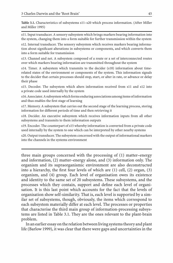
3 Charles Darwin and the ‘Root Brain’ 45
Table 3.1. Characteristics of subsystems s11–s20 which process information. (After Millerand Miller 1995)
s11. Input transducer. A sensory subsystem which brings markers bearing information intothe system, changing them into a form suitable for further transmission within the system
s12. Internal transducer. The sensory subsystem which receives markers bearing informa-tion about significant alterations in subsystems or components, and which converts theminto a form suitable for transmission
s13. Channel and net. A subsystem composed of a route or a set of interconnected routesover which markers bearing information are transmitted throughout the system
s14. Timer. A subsystem which transmits to the decider (s18) information about time-related states of the environment or components of the system. This information signalsto the decider that certain processes should stop, start, or alter in rate, or advance or delaytheir phase
s15. Decoder. The subsystem which alters information received from s11 and s12 intoa private code used internally by the system
s16.Associater.Asubsystemwhich formsenduringassociationsamong itemsof informationand thus enables the first stage of learning
s17. Memory. A subsystem that carries out the second stage of the learning process, storinginformation for different periods of time and then retrieving it
s18. Decider. An executive subsystem which receives information inputs from all othersubsystems and transmits to them information outputs
s19. Encoder. The counterpart of s15 whereby information is converted from a private codeused internally by the system to one which can be interpreted by other nearby systems
s20. Output transducer. The subsystem concerned with the output of informational markersinto the channels in the systems environment
three main groups concerned with the processing of (1) matter–energyand information, (2) matter–energy alone, and (3) information only. Theorganism and its supraorganismic environment are also deconstructedinto a hierarchy, the first four levels of which are (1) cell, (2) organ, (3)organism, and (4) group. Each level of organisation owes its existenceand identity to the same set of 20 subsystems. These subsystems, and theprocesses which they contain, support and define each level of organi-sation. It is this last point which accounts for the fact that the levels oforganisation show self-similarity. That is, each level is supported by a sim-ilar set of subsystems, though, obviously, the items which correspond toeach subsystem materially differ at each level. The processes or propertiesthat characterise the third main group of information-processing subsys-tems are listed in Table 3.1. They are the ones relevant to the plant-brainproblem.
Inanearlier essayon the relationbetween living systems theoryandplantlife (Barlow 1999), it was clear that there were gaps and uncertainties in the

46 P.W. Barlow
analysis. In particular, it was difficult to discover correspondences betweenthe subsystems which the theory suggested were involved in informationprocessing and any known processes, or plant structures. The questionis whether, in the light of new knowledge and the possible existence of
Table 3.2. Subsystems which process information at four levels of organisation that involveplant organisms
Subsystem LevelCell Organ Organism Group
s11. Input Plasma Statoliths Root cap, Sensitive plantstransducer membrane sites sensory hairs at the boundary
bearing AUX1 of the groupreceptorprotein
s12. Internal Endocytotic Statenchyme ← –transducer vesicles
s13. Channel Actin Plant synapses, Supersymplasm Vegetationand net cytoskeleton, pit fields patchwork
plasmodesmata
s14. Timer Biochemical Cells perceptive Canopy and Autumn coloursoscillators, of external timers its propertiesmitotic clock
s15. Decoder Release of auxin ← – –s16. Associater Crosstalk Cells with Organs that –
molecules inductive respond toproperties acclimation
and aptation
s17. Memory Short-lived Hysteresis loops, ← –gene regulators transcellular
electricalimpulses
s18. Decider Regulator genes, Transition Collective –osmoregulators zone cells plant brain,
target cells
s19. Encoder Genes and Auxin response Organ Plants withproteins involved processes aposematic open flowersin hormone markingresponse
S20. Output Plasma Cells which Attractor organs, Reproductivetransducer membrane export scent glands individual
and PIN informationproteins to motor cells
An arrow indicates that the subsystem is devolved to the next-lower organisational level

3 Charles Darwin and the ‘Root Brain’ 47
a plant nervous system, the corresponding plant subsystems can be moreconfidently identified; and if so, whether they are concordant with thoseanalysed by Miller and Miller (1995) for animal/human systems. Table 3.2summarises the present position for the various levels of plant organisation.
Of the information-processing subsystems listed in Table 3.1, s14, timer,is not directly relevant in the present context as it deals with the timing ofprocesses in relation to innate or external cues. At the levels of the cell andorgan, subsystems s16 and s17, associater and memory, respectively, mayhave correspondences with the fast electrical signals that can be propa-gated along plant organs. The remaining subsystems are those which eithertransmit information in the form of an auxin flow (especially s13, chan-nel and net, at the organ and organism levels), or are concerned with thetransduction of external information into the body of the plant and its con-version into internal information for eventual transduction as an outputresponse.
In whatever form information is perceived by the plant, it is internalisedandprocessed in suchaway that it canbeultimatelydirected towardsmove-ment or reproduction. The movement of plant organs involves tropisms(nasticmovements are excluded for themomentowing to their autonomousnature), the informational inputs for which are mainly gravity, illuminanceand moisture. Auxin is the key mediator of these growth events.
Although plant life can be decomposed into a series of hierarchical levels,these are more than merely classificatory conveniences but are systems ofdynamic interactions. Thus, the states of the lower organisational levelsprovide conditions for the emergence of larger-scale organisations such asthe group or community, and in particular for the physical movements ofthe latter whereby they – individuals, groups, communities – are able tooccupy new environments by means of propagules adapted and dispersedfor this purpose (seeds, abscinded plant parts, etc.). Finally, the communalmovement of plants aids in the construction of a green energy receptor andtransducer – and maybe something more (Miller and Miller 1982) – whichcovers the surface of the Earth. In some unfathomable way, this living skinmay even fulfil the role of a subsystem within some much higher level oforganisation and thereby participates in the ongoing creative evolution oforganic living systems.
3.6Conclusions and Future Prospects
Was Charles Darwin prescient in his use of the word brain in connectionwith his studies of the activities of the plant root apex and the implicationsthat might flow therefrom? We shall never know. Given his unease over the
48 P.W. Barlow
reception of his book The origin of species, he might well have kept anyideas about plants possessing brains strictly to himself.
One attribute of a brain, as the term is commonly understood, is thatit is an organ with a definite structure and location which gathers orcollects information, which was originally in the form of vibrations (heat,light, sound, chemical, mechanical, . . .) in the ambient environment andsomehow transforms them into an output or response. Interestingly, theexecution of these responses is by means of another vibrating system –the circumnutating root (Darwin 1880). Any change in the direction ofthe stimulus temporarily overrides the nutational process until the usualorientation of the plant organ is regained. Maybe this was how Darwinunderstood the situation. However, the absence of obvious nervous tissuewould have been a serious problem in his further pursuit of the matter. Butthe situation has changed in recent years, as other chapters in this bookwill show. With the discovery within plants of many of the componentsof animal-type nervous systems (Baluška et al. 2004), the question is howthis new knowledge can be conceptualised, and whether any paradigmwill emerge which acknowledges some sort of plant nervous system; andmoreover, whether this nervous system will have features which can begeneralised to all eukaryotic organisms.
In recognising the possession, by the root apex, of a brain-like function,a lacuna is filled in applying Miller’s comprehensive living systems theoryto plants. Consequently, there is now scope for analysing the information-processing subsystems and linking them with the other two major typesof subsystems that process matter and energy (Barlow 1999). This task canbe done at the various levels of organisation that characterise all biologicalconstructions.
The subsystems are not static artefacts of theory, but represent inherentlydynamic processes by which biological constructions act. They apply notonly to large constructions such as social communities, but also to smallermicrocosms such as cells. Indeed, the subsystems fulfil the role of the ‘coor-dinative conditions’ which the physicist and systems analyst Lancelot LawWhyte had seen as providing a key to understanding biology in general:“Until the coordinating conditions have been identified no theory of phylo-genesis, of ontogenesis, or of their relations, can be regarded as definitive.Moreover [they] hold the clue to the relation of physical laws and to theunity of the organism” (Whyte 1965, p. 67). For Whyte, these conditionsexpress “the biological spatio-temporal coordination, the rules of order-ing which must be satisfied . . . by the internal parts and processes of anyorganism capable of developing and surviving in some environment. Thecoordinating conditions are the expression of geometrical, 3D, or perhapskinematic rules determining the necessary 3D or spatio-temporal networkof the atoms, ions, molecules, organelles, etc. in a viable organism. They . . .

3 Charles Darwin and the ‘Root Brain’ 49
must cover all the fundamental aspects of the unity of organisms” (Whyte1965, p. 6). The recognition, as well as the appreciation of the place of theseconditions – or subsystems, as they would be in Miller’s living systemstheory – within the hierarchical construction that comprises plant life onEarth, provides a realistic and holistic way of perceiving and responding towhat our senses tell us, both directly and intuitively, about plant life.
References
Baluška F, Brailsford RW, Hauskrecht M, Jackson MB, Barlow PW (1993) Cellular dimor-phism in the maize root cortex: involvement of microtubules, ethylene and gibberellinin the differentiation of cellular behaviour in postmitotic growth zones. Bot Acta 106:394–403
Baluška F, Volkmann D, Barlow PW (1996) Specialized zones of development in roots: viewfrom the cellular level. Plant Physiol 112:3–4
Baluška F, Šamaj J, Volkmann D (2003) Polar transport of auxin: carrier-mediated flux acrossthe plasma membrane or neurotransmitter-like secretion? Trends Cell Biol 13:282–285
Baluška F, Mancuso S, Volkmann D, Barlow P (2004) Root apices as plant command centres:the unique ‘brain-like’ status of the root apex transition zone. Biologia (Bratislava) 59Suppl 13:7–19
Baluška F, Volkmann D, Menzel D (2005) Plant synapses: actin-based domains for cell-to-cellcommunication. Trends Plant Sci 10:106–111
Barlow PW (1999) Living plant systems: How robust are they in the absence of gravity? AdvSpace Res 23:1975–1986
Barlow PW, Volkmann D, Baluška F (2004) Polarity in roots. In: Lindsey K (ed) Polarity inplants. Blackwell, Oxford. pp 192–241
Blancaflor EB, Masson PH (2003) Plant gravitropism. Unraveling the ups and downs ofa complex process. Plant Physiol 133:1677–1690
Darwin C assisted by Darwin F (1880) The power of movements in plants. Murray, LondonDarwin C (1888a) Autobiography. In: Darwin F (ed) The life and letters of Charles Darwin
including an autobiographical chapter, vol 1. Murray, London, pp 26–107. Reprintedin 1983 In: de Beer G (ed) Charles Darwin and TH Huxley. Autobiographies. OxfordUniversity Press, Oxford, pp 8–88
Darwin C (1888b) Letter to Sir JD Hooker (November 23 1880). In: Darwin F (ed) The lifeand letters of Charles Darwin including an autobiographical chapter, vol 3. Murray,London, p 334
Falik O, Reides P, Gersani M, Novoplansky A (2003) Self/non-self discrimination of roots. JEcol 91:525–531
Friml J, Palme K (2002) Polar auxin transport – old questions and new concepts? Plant MolBiol 49:273–284
Friml J, Wisniewska J, Benková E, Mendgen K, Palme K (2002) Lateral relocation of auxinefflux regulator PIN3 mediates tropism in Arabidopsis. Nature 415:806–809
Friml J, Vieten A, Sauer M, Weijers D, Schwarz H, Hamann T, Offringa R, Jürgens G (2003)Efflux-dependent auxin gradients establish the apical-basal axis of Arabidopsis. Nature426:147–153
Fujii K (1895) On the nature and origin of so-called “chichi” (nipple) of Ginkgo biloba L.Bot Mag (Tokyo) 9:444–450
Galil J (1980) Kinetics of bulbous plants. Endeavour 5:15–20

50 P.W. Barlow
Iijima M, Kono Y (1991) Interspecific differences of the root system structures of four cerealspecies as affected by soil compaction. Jpn J Crop Sci 60:123–131
Kerk NM, Jiang K, Feldman LJ (2000) Auxin metabolism in the root apical meristem. PlantPhysiol 122:925–932
Kurczynska EU, Barlow PW (1999) Anatomical investigations of the ‘chi-chi’ of Ginkgobiloba: sites of cambial hyperactivity. J Exp Bot 50 (Suppl):34, abstract P3–58
Lodish H, Berk A, Zipinsky SL, Matsudaira P, Baltimore D, Darnell J (2000) Molecular cellbiology, 4th edn. Freeman, New York
Lyford WH (1974) Rhizography of non-woody roots of trees in the forest floor. In: TorreyJG, Clarkson DT (eds) The development and function of roots. Academic, London,pp 179–196
Massa GD, Gilroy S (2003) Touch modulates gravity sensing to regulate the growth ofprimary roots of Arabidopsis thaliana. Plant J 33:435–445
Miller JG (1978) Living systems. McGraw-Hill, New YorkMiller JG, Miller JL (1981) Systems science: an emerging interdisciplinary field. Center Mag
14:44–55Miller JG, Miller JL (1982) The Earth as a system. Behav Sci 27:303–322
(also available at the 1st international electronic seminar on wholeness, 2000,http://www.newciv.org/ISSS Primer/asem22jm.html)
Miller JL, Miller JG (1995) Greater than the sum of its parts III. Information processingsubsystems. Behav Sci 40: 171–182
Monshausen GB, Zieschang HE, Sievers A (1996) Differential proton secretion in the apicalelongation zone caused by gravistimulation is induced by a signal from the root cap.Plant Cell Environ 19:1408–1414
Müller A, Guan C, Gälweiler L, Tänsler P, Huijser P, Marchant A, Parry G, Bennett M, WismanE, Palme K (1998) AtPIN2 defines a locus of Arabidopsis for root gravitropism control.EMBO J 17:6903–6911
Ney D, Pilet P-E (1981) Nutation of growing and georeacting roots. Plant Cell Environ4:339–343
Ottenschläger I, Wolff P, Wolverton C, Bhalerao RP, Sandberg G, Ishikawa H, Evans ML,Palme K (2003) Gravity-regulated differential auxin transport from columella to lateralroot cap cells. Proc Natl Acad Sci 100:2987–2991
Ramón y Cajal S (1909) Histologie du système nerveux de l’homme et des vertébrés (Frenchtranslation by L Azoulay of Textura del sistema nervioso del hombre y de los vertebra-dos). Maloine, Paris
Rashotte A, Brady S, Read R, Ante S, Muday G (2000) Basipetal auxin transport is requiredfor gravitropism in roots of Arabidopsis. Plant Physiol 122:481–490
Schrader J, Baba K, May ST, Palme K, Bennett M, Bhalerao RP, Sandberg G (2003) Polar auxintransport in the wood-forming tissues of hybrid aspen is under simultaneous control ofdevelopmental and environmental signals. Proc Natl Acad Sci USA 100:10096–10101
SwarupR,Friml J,MarchantA,LjungK, SandbergG,PalmeK,BennettM(2001)Localizationof the auxin permease AUX1 suggests two functionally distinct hormone transportpathways operate in the Arabidopsis root apex. Genes Dev 15:2648–2653
Sztein AE, Cohen JD, Cooke TJ (2000) Evolutionary patterns in the auxin metabolism ofgreen plants. Int J Plant Sci 161:849–859
Takahashi N, Yamazaki Y, Kobayashi A, Higashitani A, Takahashi H (2003) Hydrotropisminteracts with gravitropism by degrading amyloplasts in seedling roots of Arabidopsisand radish. Plant Physiol 132:805–810
Thellier M, Le Sceller L, Norris V, Verdus M-C, Ripoll C (2000) Long-distance transport,storage and recall of morphogenetic information in plants. The existence of a sort ofprimitive plant ‘memory’. C R Acad Sci Ser III 323:81–91

3 Charles Darwin and the ‘Root Brain’ 51
Tortora GJ Grabowski SR (1996) Principles of anatomy and physiology. Harper Collins,New York
Whyte LL (1965) Internal factors in evolution. Tavistock, LondonWolverton C, Mullen JL, Ishikawa H, Evans ML (2002) Root gravitropism in response to
a signal originating outside of the cap. Planta 215:153–157

4 How Can Plants Choosethe Most Promising Organs?T. Sachs
Abstract Branch death is an important component of the processes that generate tree form.Many branches are formed and the plant appears to “choose” the most promising alter-natives for survival and further development. The purpose of this chapter is to considerhow this choice could occur in the absence of a central decision center. Experiments werecarried out on a model system of pea seedlings with two shoots. In most plants only oneof these shoots continued to grow. We studied the conditions that influence the choice ofthe “winning” branch. These included removing entire branches or leaves at different agesand modifying the environment of the entire plant or of individual shoots. The evidencefrom experiments and from comparative observations of many trees supports the followinghypothesis. All components of a shoot are sources of auxin and possibly of other signals.The level of auxin synthesis depends on the immediate environment and the developmentalstage of individual leaves. The responses to auxin include the orientation of vascular differ-entiation towards organs that are its strongest source. This oriented auxin response resultsin competition between alternative organs. This is a prominent example of “developmen-tal selection,” an alternative to developmental programs and prepatterns, which also hasvarious other roles in the generation of biological form.
4.1Introduction: Developmental Selectionof Branch Configurations
Plants develop their complicated forms by reiterating developmental pro-cesses, forming repeated structural metamers (Halle et al. 1978; Barlow1989). This can be readily verified by following the development of theyoung tips of leading branches. Reiteration is also necessary on theoret-ical grounds, for there is no other way in which the information codedin a genome could suffice to determine the detailed form of a large tree.Yet reiteration cannot be the whole story. Even a superficial observationof most trees suggests that while all buds are potential branches, most ofthem do not develop further. Those that do become branches develop atvaried rates that depend on their immediate environment and the presenceof neighboring branches, not only on a reiterated program. Further, themajority of the branches that start growing in a young canopy are shedwithin a few years (Sachs and Novoplansky 1995). Dead branches are read-ily found below large trees, especially following a storm. The shedding ofbranches is also the origin of bare trunks, which commonly develop from
Communication in PlantsF. Baluška, S. Mancuso, D. Volkmann (Eds.)© Springer-Verlag Berlin Heidelberg 2006

54 T. Sachs
branched systems, similar to those seen in the upper parts of the very sametrees. Reiterated developmental processes are thus only a framework thatis subject to long-term modification.
The purpose here is to consider the mechanisms that determine whichbranches are retained. This question calls for careful, repeated experi-ments in defined conditions, for which trees are hardly suitable. The workdescribed here is therefore based on a model system, using seedlings ofan annual plant. The results suggest that branches compete, and possiblemechanisms of this internal competition are briefly considered. The discus-sion proposes that competition and selection which follows rather than pre-cedes initial development is common at all levels of biological organization.
4.2An Experimental Model Demonstrates Branch Competition
4.2.1The Experimental System
The development of annual plants has been used extensively for studiesof the inhibitory influence of a growing shoot on bud growth (Snow 1937;Sachs 1991; Cline 1994). The choice of a successful branch from amonga number that have started growing does not, however, usually occur inannual plants. A possible evolutionary reason is that branch shedding is notadaptive inplantswitha short life span (Novoplanskyet al. 1994).Avaluablemodel system has nonetheless been developed in annuals (Snow 1931;Sachs 1966; Novoplansky et al. 1989). It uses the unstable state of a plantregenerating after severe damage. The first or seminal shoot was removedfrompea seedlings (Fig. 4.1a). This treatmentdistorted thebalancebetweenthe shoot and root systems of the growing plant, and, as in cut trees, shootnumber was increased (Sachs et al. 1993; Sachs 2004). Two buds grewinto more or less equal shoots or branches (Fig. 4.1b,c). Gradually thesetwo shoots became increasingly unequal (Fig. 4.1d). The smaller shootstopped growing and in some varieties it eventually died, returning theplant to a single axis state (Fig. 4.1e; Sachs 1966). The deterioration ofthe smaller shoot was prevented whenever the larger shoot was removed(Fig. 4.1f), demonstrating that interactions between the shoots are a majordeterminant of their individual fate.
“Two-shoot” pea seedling are an experimental system in which it is pos-sible to ask how the plant chooses the shoot that continues developing. Thefollowing simple experiments demonstrate that there is no key parame-ter. Instead, the plants appear to be integrating information from variedsources.

4 How Can Plants Choose the Most Promising Organs? 55
a b c d
e f g
Fig.4.1. The two-shoot pea experimental system. a The seminal shoot of a germinatingpea seedling was removed. b Within 3 days the removed shoot was replaced by the growthof inhibited buds and they became two new shoots. c Within four additional days thenew shoots carried expanded leaves and were generally somewhat unequal. d Inequalitybetween the shoots increased. At the time the experiments shown in Fig. 4.2 were startedthe wet-weight ratio of the smaller over the larger shoot was 0.37. e In some varieties thesmaller shoot actually died within 2 weeks. f Removal of the larger, dominant shoot beforethe smaller shoot had died. The declining shoot survived and developed rapidly. Its averagefreshweight, shown inmilligrams,didnotdiffermuch fromthatof anundisturbeddominantshoot (in e). g Dominance influences between shoots. Removing both the dominant shootand the upper part of the smaller shoot resulted in the growth of a bud in the axil of anexpanded leaf. This bud did not grow when the large shoot was left intact. (Based on Sachsand Hassidim 1996)

56 T. Sachs
4.2.2Stress Increases Competition
The two-shoot pea plants were stressed by removing parts of the reserveseed tissues (the cotyledons, Fig. 4.2b). This treatment, in addition to theobvious reduction in shoot growth, also increased shoot inequality (Sachsand Novoplansky 1997). When the larger shoot was removed the weakerone always survived and grew to regenerate a new plant (Fig. 4.1f). Thissuggests that the weaker shoot is responding not only to stress but also toan enhanced competition with the stronger shoot. Such results were notlimited to cases where there was a reduction of reserve storage materials.The stress of weak light and limited mineral nutrition had the same effects(Fig. 4.2c,d).
4.2.3Unequal Light Conditions
The two shoots could be subjected to different conditions, simulating en-vironmental heterogeneity at the level of an individual plant (Novoplanskyet al. 1989). Reducing the light available to one of the two shoots stronglyinhibited its growth and increased shoot inequality (Fig. 4.3e). When lightwas excluded from one shoot, it died within 2 weeks in 80% of cases(Fig. 4.3f). Here, again, the response depended on the presence of the shootthat remained in the light. When the more fortunate shoot was removed,the darkened shoot elongated rapidly, forming small, yellow leaves. Thisis the common form of seedlings whose entire shoot system is in darkconditions, part of the etiolation syndrome.
4.2.4The Rate of Shoot Development and Leaf Removal
The inequality of the two shoots tended to increase with time, and relationswere not reversed unless the individual shoots were subjected to additionaltreatments. This suggests that relative shoot size had an influence on thesubsequent development. Size is a parameter that can be readily manipu-lated, simulating natural herbivory. When the entire top part of one shootwas removed the shoot could continue development only by means of oneor more of the buds subtended by its remaining leaves. Such developmentoccurred readily, but only when the competing intact shoot was removed(Fig. 4.1g). A bud is thus at a pronounced disadvantage relative to a grow-ing shoot apex. This is, of course, the bud inhibition mentioned before, the

4 How Can Plants Choose the Most Promising Organs? 57
a b c
d e f
Fig.4.2. Responses of two-shoot peas to overall and to localized stress conditions. a Bothshoots developed in most control plants in optimal conditions. b Removing a large part ofthe cotyledons, and hence of the reserve materials, increased shoot inequality. c Growingplants in vermiculite without nutrient solutions (NS) increased shoot inequality. d Shadingboth shoots, which presumably limited photosynthesis, is a third example of the generalincrease of shoot inequality in response to stress. e, f Restricting the light reaching only oneof two young shoots greatly limited its development. When a shoot was darkened (f) ratherthen shaded (e) it died in 80% of the plants. The numbers refer to the dry weights of theshoots in milligrams. (They are not comparable to the fresh weights in Fig. 4.1.); (Based onSachs and Novoplansky 1997 and Novoplansky et al. 1989)

58 T. Sachs
a b c
d e f
g h i
difference here being that the bud is not morphologically below the leavesthat inhibit its development. Space limitations preclude the considerationof quantitative evidence that developmental rates are of critical importancein the criteria used by the plant (Novoplansky 1996, 2003).
Finally, individual leaves can be removed (Sachs and Hassidim 1996).When this was done to the larger of two somewhat unequal shoots therewas an increased chance of its losing a dominant position (Fig. 4.3a, cf.Fig. 4.1d). The removal of a developing leaf has a larger effect than the

4 How Can Plants Choose the Most Promising Organs? 59
� Fig.4.3. a–c The influence of simulated herbivory on shoot relations. The numbers arethe ratios of the fresh weights of the initially smaller over the initially larger shoot afteradding the expected weight of removed leaves; they should be compared with the controlof 0.37 (Fig. 4.1d). a Removing a mature leaf at an early stage from the somewhat largershoot resulted in the two shoots being more or less equal. b Removing a young, expandingleaf from the larger shoot had a larger effect than removing a mature, photosynthetic leaf.c An expanding leaf was removed from the larger shoot and a mature leaf from the smallershoot. The two shoots grew to be essentially equal, dramatically changing the outcome seenin the controls. d–f The evidence of vascular differentiation. The lines are only indicationsof overall structure, not a record of all the numerous vascular channels. d The cambialspace that had originally been occupied by vascular channels (broken lines) that led to theremoved seminal shoot (Fig. 1a) was taken over by new connections of the new growingshoots with the roots. There were no direct contacts between the new shoots. e Where thetwo shoots were unequal their vascular contacts and the space they occupied were correlatedwith their size and developmental rates. f When a weak shoot died the space occupied by itsvascular channels, but not the channels themselves, was taken over by the growing shoot.g–i. An hypothesis: the choice between shoots is mediated by hormonal relations with theroots. The shoots are the source of signals that inform the plant about their state. Theresponse to these signals that they receive from the roots limits their development. g Inthe cut plant essential supplies from the root are present in excess, and the shoots do notcompete. h As the shoots develop, their young and, to a lesser extent, their mature leavesincrease their signals and their requirements. A large proportion of the supplies from theroots are diverted towards the larger shoot and they enhance its continued development.i A positive feedback between development and continued development ensues and anincreasing proportion of the resources are diverted towards only one of the two shoots.(Based on Sachs and Novoplansky 1997)
removal of a photosynthetically active leaf (Fig. 4.3b). A plant forced to“choose” between two damaged branches discriminated against the onethat had lost a young rather than a mature leaf (Fig. 4.3c).
4.2.5Hypothesis: Branches Compete
The results of the experiments with pea seedlings suggest that branches ofthe same plant compete for resources, their genetic identity not withstand-ing (Sachs et al. 1993; Sachs 2004). This competition is temporarily liftedwhen the shoot/root balance is disrupted by the removal of the seminalshoot and is intensified when the plants are in stress conditions. Com-petition means that the plants not only respond to local environmentalconditions, such as low light and local leaf damage, but they also actually“choose” for continued development the most promising of the availablealternatives. An important criterion for this choice is rapid development,which predicts future rather than immediate photosynthetic performance.
60 T. Sachs
4.3Mechanisms of Competition
The statement that branches of the very same plant compete calls for con-crete mechanisms. These should allow for a comparison of branches andfor the selection of the more promising alternative. Such comparisons mustoccur in the absence of a central “brain” or computing center, an organwhich plants lack. Possible alternatives must take into account the fact thatplants can have numerous branches, and it is impossible for each to bethe source of a unique signal. Since competition is influenced by resourceavailability, an obvious possibility is that the stronger branches act as sinksthat divert transport towards themselves (Henriksson 2001). This is sup-ported by evidence that the vascular channels passively transport materialsto the sinks in which they are consumed. Yet both young and mature leaves,which have opposite sink effects, act to enhance the role of a strong branch(Fig. 4.3a–c). A more general reason that sink effects could not suffice isthat branch competition is a long-term process, during which new vascularchannels differentiate (Sachs et al. 1993). Vascular differentiation, unlikeshort-term transport, is actively oriented so that it connects the dominantbranches with the rest of the plant (Sachs 1991; Berleth and Sachs 2001).Since hormones and essential substrates move along the vascular tissues,their long-term transport is in fact oriented towards dominant branches.
When considered in terms of vascular differentiation it is easy to observethe “conflict” between the vascular connections of branches that developon opposite sides of the same axis (Fig. 4.3d–f). There are no direct vascu-lar contacts between branches: all channels within the plant axis are polar,connecting shoot and root tissues (Sachs 1991). The larger the branchthe larger its vascular supply and the more axial space this supply occu-pies. The causal relations between the branch development and orientedvascular differentiation can be readily confirmed by branch removal. Themolecular basis for processes of reoriented vascular differentiation couldbe dependent on changes in the localization of the products of PIN genes(Palme and Gälweiler 1999). The suggested role of vascular differentiationfocuses competition on the orientation and activity of cambial cells wheredeveloping vascular systems meet (Sachs et al. 1993). This is not a proof,however, since the evidence does not show that orientation precedes ratherthan follows branch development.
The hypothesis can be taken one step further (Fig. 4.3g–i). Leaves areknown to be sources of the hormone auxin (Sachs 1991; Berleth and Sachs2001; Ljung et al. 2001). This same auxin induces the differentiation of newvascular tissues along the axis connecting its source with the roots (Sachs1991; Berleth and Sachs 2001). Auxin is the only known signal whose localsource actually determines the orientation of these vascular tissues. The

4 How Can Plants Choose the Most Promising Organs? 61
formation of auxin occurs in all parts of a developing shoot, but especiallyin young expanding leaves (Ljung et al. 2001). Though the quantitative dataare meager, available knowledge about auxin synthesis and vascular differ-entiation suggest that its synthesis is enhanced by light. Since neighboringbranches shade one another, competition is environmental, not only inter-nal (Sachs et al. 1993). Yet if shade influences auxin formation both types ofcompetition could reflect the same internal mechanism by means of com-petitive vascular orientation. All this is in accordance with the hypothesisthat auxin is a signal that integrates the information about the location, size,environment and rate of development of a growing branch (Sachs 2004).
Are there other possible mechanisms of branch competition, in additionto auxin-induced orientation of vascular differentiation? These mecha-nisms need not be mutually exclusive and it is possible, or even likely, thatmore than one has a role in a process as central as the relations betweenplant organs. An alternative to oriented differentiation is the adaptation ofplant tissues to the higher level of auxin that is supplied by the strongerbranch (or other signals that a shoot may produce). The cambium thusresponds to the “best” branches – the ones that are the strongest sourcesof auxin – and “ignores” the weaker branches, whose vascular tissues de-teriorate without being replaced. This possibility is plausible and it wouldaccount for evidence that interactions between branches need not requirevascular reorientation (Snow 1937). However, there appears to be no con-crete evidence about its cellular basis and the way adaptation to high auxincould spread through plant tissues.
4.4Conclusions and Future Prospects
A general conclusion goes beyond the relations between tree branches. Sim-ilar processes of developmental selection could have a large role in biolog-ical pattern formation (Edelman 1987; Sachs 1988, 1991, 2002; Frank 1996,1997). This selection actually generates information about the location ofthe different structures, such as branches, during development itself. Inthis it differs from programs or prepatterns in which detailed informationprecedes actual differentiation. Developmental selection shares principleswith the Darwinian mechanism of evolution (Frank 1997), though thereis an important difference. The various branches of a tree are geneticallyidentical and the outcome of their selection is an adaptive form, not anevolution of a new genetic system. Conflicts of interest between alternativebranches or other structures of the same organism do not arise.
The specification of form by stochastic variation followed by selectionappears to be counterintuitive, but it is supported by direct observations of

62 T. Sachs
the development of varied biological patterns. The mature stomata of San-sevieria are more orderly or predictably spaced than the early, reversiblestages of stomata initiation (Kagan and Sachs 1991). “Neural Darwinism”is a mechanism by which appropriate nerve connections are selected fromexcess alternatives (Edelman 1987). Within cells, microtubules are main-tained according to functional roles that are established after they havebeen formed (Kirschner and Mitchison 1986).
It may be useful to consider the adaptive significance of the generationof tree form by developmental selection rather than strict programs. Com-petition between branches requires the wasteful development of structuresthat are not maintained. There is no doubt that dead branches below treesrepresent considerable organic material. Yet these branches did carry outphotosynthesis for as long as they were productive and resources such asbound nitrogen can be withdrawn from dying plant organs (Habib et al.1993). Losses of substrates could be balanced by the advantages of grad-ual selection, in which information about the value of an existing branchfeeds back to its survival and further development. This testing of variedpossibilities could provide for a robust outcome.
A major advantage of developmental section may be in its relation toplasticity. It is possible that in a predictable, ideal world strict programscould result in superior biological forms. The real world, however, is farfrom predictable. In the case of trees, the presence of competing neigh-bors, damage due to herbivores and parasites and possible local failuresin the development of the plant itself require a pronounced developmen-tal plasticity. Developmental selection uses the very same processes andgenetic information to generate form and to insure plastic responses to un-predictable conditions (Sachs 2002). This parsimony might have adaptivesignificance both because simpler developmental systems are required andbecause the various component are all optimally integrated.
References
Barlow PW (1989) Meristems, metamers and modules and the development of shoot androot systems. Bot J Linn Soc 100:255–279
Berleth T, Sachs T (2001) Plant morphogenesis: long-distance coordination and local pat-terning. Curr Opin Plant Biol 4:57–62
Cline MG (1994) The role of hormones in apical dominance. New approaches to an oldproblem in plant development. Physiol Plant 90:230–237
Edelman GM (1987) Neural Darwinism – the theory of neuronal group selection. BasicBooks, New York
Frank SA (1996) The design of natural and artificial adaptive systems. In: Rose MR, LauderGV (eds) Adaptation. Academic, San Diego, pp 451–505
Frank SA (1997) Developmental selection and self-organization. BioSystems 40:237–243

4 How Can Plants Choose the Most Promising Organs? 63
Habib R, Millard P, Proe MF (1993) Modelling the seasonal partitioning in young Sycamore(Acer pseudoplatanus) trees in relation to nitrogen supply. Ann Bot 71:453–459
Halle F., Oldeman, RAA, Tomlinson PB (1978) Tropical trees and forests. Springer, BerlinHeidelberg New York
Henriksson J (2001) Differential shading of branches or whole trees: survival, growth, andreproduction. Oecologia 126:482–486
Kagan M, Sachs T (1991) Development of immature stomata: epigenetic selection of a spac-ing pattern. Dev Biol 146:100–105
Kirschner M, Mitchison T (1986) Beyond self-assembly: from microtubules to morphogen-esis. Cell 45:329–342
Ljung K, Bhalerao RP, Sandberg G (2001) Sites and homeostatic control of auxin biosynthesisin Arabidopsis during vegetative growth. Plant J 28:465–475
Novoplansky A (1996) Hierarchy establishment among potentially similar buds. Plant CellEnviron 19:781–786
Novoplansky A (2003) Ecological implications of the determination of branch hierarchies.New Phytol 160:111–118
Novoplansky A, Cohen D, Sachs T (1989) Ecological implications of correlative inhibitionbetween plant shoots. Physiol Plant 77:136–140
Novoplansky A, Cohen D, Sachs T (1994) Responses of an annual plant to temporal changesin the light environment: an interplay between plasticity and determination. Oikos69:437–446
Palme K, Gälweiler L (1999) PIN-pointing the molecular basis of auxin transport. Curr OpinPlant Biol 2:375–381
Sachs T (1966) Senescence of inhibited shoots of peas and apical dominance. Ann Bot30:447–456
Sachs T (1988) Epigenetic selection: an alternative mechanism of pattern formation. J TheorBiol 134:547–59.
Sachs T (1991) Pattern formation in plant tissues. Cambridge University Press, CambridgeSachs T (2002) Consequences of the inherent developmental plasticity of organ and tissue
relations. Evol Ecol 16:243–265Sachs T (2004) Self-organization of tree form: a model for complex social systems. J Theor
Biol 230:197–202Sachs, T, Hassidim M (1996) Competition and mutual support of branches in damaged
plants. Vegetatio 127:25–30Sachs T, Novoplansky A (1995) Tree form: architectural models do not suffice. Isr J Plant Sci
43:203–212Sachs T, Novoplansky A (1997) What does aclonal organization suggest concerning clonal
plants? In: de Kroon H, van Groenendael (eds) The ecology and evolution of clonalplants. Backhuys, Leiden, pp 55–77
Sachs T, Novoplansky A, Cohen D (1993) Plants as competing populations of redundantorgans. Plant Cell Environ 16:765–770
Snow R (1931) Experiments on growth and inhibition. II. New phenomena of inhibition.Proc R Soc Lond Ser B 108:305–16
Snow R (1937) On the nature of correlative inhibition. New Phytol 36:283–300

5 The Role of Root Apices in Shoot GrowthRegulation: Support for Neurobiologyat the Whole Plant Level?Peter M. Neumann
Abstract This chapter reviews the potential and actual role of plant neurobiological activityfor integrating function at the whole plant level. Some support is provided for the drawing ofanalogies between the signaling roles of plant long-distance transport systems and animalnervous systems. However, the specific findings reviewed here do not indicate that rootapices function as essential neurobiological command centers involved in regulating shootgrowth responses to adverse changes in the root environment.
5.1IntroductionHow cell and tissue activities give rise to integrated function at the levelof the whole organism is a central question in biology. Hormonal signalmolecules and the neurobiological activities of interconnected nervoussystems and brains can provide the needed integrative capacity in animals.In plants, the activities of hormonelike substances are well characterizedbut neither morphologically distinct brains nor anatomically distinct nervecells appear to have evolved. Nevertheless, it has been suggested by Baluskaet al. (2004a, b) that the transition zone in the growing root tip [which alsoshows intense genetic (Birnbaum et al. 2003; Bassani et al. 2004), hormonal(Aloni et al. 2004) and ionic activities (Peters and Felle 1999; Fasanao etal. 2001; Fan and Neumann 2004)] together with the vascular transporttissues (xylem and phloem) that interconnect with practically all the partsof higher plants may be compared with the brains and nervous systems ofanimals.
A criterion for a good hypothesis is that it stimulates thought and theo-retical or experimental tests of validity: The suggestion that the root apexof higher plants is part of a cell body that has neurobiological parallels andcan be viewed as a “diffuse plant brain” connected via vascular strands (the“nerves”) to the plant shoot certainly meets this criterion.
When I, a plant stress physiologist, first considered the plant neurobiol-ogy concept, I was stimulated to clarify for myself the function of nervesand brains in animals and to evaluate the potential need for similar systemsin plants. I was also stimulated to look at the evolutionary progression ofplant morphology and anatomy in order to better evaluate the essentialityto plant life of roots and vascular tissues serving as neurobiological entities.
Communication in PlantsF. Baluška, S. Mancuso, D. Volkmann (Eds.)© Springer-Verlag Berlin Heidelberg 2006

66 P.M. Neumann
Finally, I examined the plant neurobiology concept in the light of severalexperiments from our laboratory. These involved the effects of partial rootexcision, or of treatments which inhibit root activity, on root-to-shoot sig-naling and shoot growth responses to environmental stresses. The findingsare presented in the following three sections.
5.2The Comparative Need for Rapid Neurobiological Activityin Animals and PlantsMulticellular animals and plants appear to have evolved from a commonancestral cell type and they share many basic molecules together with life-associated activities such as respiration, cell division and long-distancetransport systems. However, there are, of course, intrinsic differences be-tween animals and plants. For example, brains and networks of specializednerve cells have clearly evolved in animals but not in higher plants. Theanimal brain can be viewed as a device which rapidly processes sensoryinput signals, makes decisions and sends motor output. The human brainconsists of around one hundred billion neurons intimately connected viathe spinal chord to a nervous network which can rapidly transmit signals toand from every point in the body. The brain of higher animals is essential tolife since decapitation or brain inactivation immediately leads to death ofthe organism. In contrast, excision of roots or root apices of plants need nothave immediate adverse effects on the remainingparts (consider cutflowersand see Sect. 5.3). Centralized brains are, however, not essential for the ex-istence of some types of animals. For example, members of the marine phy-lum Cnidaria (e.g., hydra, jellyfishes, corals, sea anemones and Portugueseman-of-war) have survived and thrived for about a half billion years with-out brains. Body control in these organisms is based on a diffuse nerve netwhich provides the communication between sensory cells and the musclecells involved in essential movements. Cutting nerves in lower or higheranimals is expected to result in the paralysis of associated muscular func-tion and is likely to be life-threatening. In contrast, phloem girdling (i.e.,interruption of potentially nervelike tissues) in branches of fruit trees, aspracticed by farmers in Israel, does not appear to have much adverse effectand can result in the production of larger and sweeter fruits. Thus, plantsand animals can differ in their responses to disruption of signaling events.
A more important and major difference between animals and plants isthat the animals need and use contractile muscles to facilitate their het-erotrophic life style, while plants do not. A heterotrophic life style meansthat animals depend on obtaining preformed organic foodstuffs from theirenvironment. Regulated patterns of muscular activity and directional mo-

5 Essentiality of root-shoot communication 67
bilityareneeded toallowanimals tohunt, ingest anddigest essentialorganicfoodstuffs and to actively excrete resultant solid or liquid waste products.
In contrast, plants have an autotrophic life style, i.e., while remainingstationary they can synthesize all the life-essential organic materials theyneed fromreadily available suppliesof carbondioxide, light,water andmin-eral nutrients. Moreover, waste products can be stored internally by plantcell vacuoles. On the other hand, plants may produce up to 100,000 sec-ondary compounds needed, for example, as defense against herbivores anddiseases, or to help attract pollinators (Poethig 2001; Lev Yadun et al. 2002).
In animals, aural, olfactory, visual and tactile input signals are involvedin feeding and in additional essential activities such as prey capture, fight-ing, fleeing or mating. These varied inputs need to be continuously pro-cessed by nervous systems in order to produce the appropriate muscularcontractions within seconds. Speedy signal transmission and processingis clearly essential to animal survival. Plants appear to generally operatewithin a slower time scale than animals. For example, the important andrelatively fast stomatal changes induced by soil water deficit or darknesstake minutes rather than seconds to complete (Kramer and Boyer 1995).Similarly, significant geotropic responses to changes in root position take atleast 10 min to establish (Fasano et al. 2001; Aloni et al. 2004). Finally, stemgrowth inhibitionbya severe soilwaterdeficit ina cactus species tookweeksto establish (see later). Of course every rule has its exceptions and rapidmechanosensory responses such as leaf folding in Mimosa or trap closurein Venus’s-flytraps can take only seconds to complete. The action poten-tials and chemical stimuli associated with rapid leaf folding in Mimosa cancertainly be compared with the electrical and chemical processes involvedin nerve signal transmission. However, the velocity of propagation of elec-trical signals in plants is about 2 cm s−1, whereas action potentials travelalong nerve cells at tens of meters per second (Salisbury and Ross 1985).
In summary, the immobility of plants and their generally slower re-sponses to environmental changes suggest that a plant capacity for rapidlong-distance signal transmission via nervelike cells and rapid signal pro-cessing in brainlike centers of activity is less essential than it is in animals.
5.3Plants That Manage Without Roots, Root Apicesand Vascular TissuesThe plant neurobiology concept would be supported if functional essen-tiality of the root apex “brain” and associated vascular system “nerves” forthe continuity of plant life could be established. In this respect it is interest-ing to trace morphological and anatomical changes which occurred in the
68 P.M. Neumann
evolutionary progression from lower to higher plants. This progression in-dicates that lower forms of plant life without differentiated roots or vasculartissues have survived successfully to this day (Raven et al. 1999; Poethig2001). For example, multicellular brown algae such as the giant kelps foundin the Pacific Ocean show plasmodesmatal connections between cells asin higher plants and often consist of a leaflike blade connected to a stem-like stipe. Moreover, some kelps have elongated cells in the center of thestipe which resemble the phloem transport tissue of vascular plants. Somekelps may also develop a basal holdfast structure situated at the base of thestipe. This anchors the algae to rocks and superficially resembles a root.However, other kelps have no holdfast and form free-floating masses. Thus,largemarine algae appear to be able to function successfully with or withoutdifferentiated roots and vascular tissues.
The first multicellular terrestrial plants may have been similar to thesmall multicellular green algae Fritschiella, which has some common fea-tures with present-day higher plants. Fritschiella has plasmodesmata andforms multicellular rhizoids at the soil surface from which erect branchessprout (N.B. although rhizoids occupy the same position as roots they haveno specialized apices and no differentiated transport cells). Fritschiellacan therefore function successfully without specialized conductive tissuesto facilitate nutrient and signal transmission and without complex rootapices.
Sporophytes and gametophytes of bryophyte mosses are thought to rep-resent a subsequent stage in the evolution of higher plants and reveal moresimilarities. Thus, the cells of bryophytes are interconnected by plasmodes-mata and leaflike structures may be born on a stemlike structure which canbe attached to rhizoids at the base. Moreover, the stems of some bryophytesmay include central strands of nonlignified water-conducting tissues andof nutrient-conducting tissues. Although some bryophyte species utilizeprimitive conductive tissues, it is again noteworthy that others functionsuccessfully as multicellular plant organisms despite the absence of con-ductive tissues and of root apices.
In summary, various multicellular plant organisms can function with-out roots, root apices or vascular tissues (Table 5.1). In contrast, moderngymnosperm and angiosperm plants share a body architecture which in-cludes roots, root apices and vascular tissues. The relatively large size andworld-wide distribution achieved by gymnosperm and angiosperm plantssuggest that they represent a winning formula for ecological success. Nev-ertheless, many lower forms of plant life have retained an ability to surviveand propagate very successfully, in a world-wide variety of different envi-ronments, despite the absence of these attributes. The degree to which therelatively greater success of the higher plants is attributable to the facili-tation of plant neurobiological activity by root tip “brains” and vascular

5 Essentiality of root-shoot communication 69
Table 5.1. Presence or absence (+ or –) of characteristics which may be associated withneurobiological activity in photosynthetic organisms
Kelps Green algae Mosses Higher plants
Plasmodesmata + + + +Vascular tissue +/– – +/– +Hold fast + – – –Rhizoid – +/– + –Root – – – +
system “nerves”, remains to be determined. An obvious additional expla-nation for the success of the higher plants is that differentiated root systemsand vascular tissues increase plant capacity to effectively access and/or dis-tribute water, mineral nutrients and organic solutes to all parts of the wholeplant structure.
5.4Do Plant Shoot Responses to EnvironmentalStresses Require Rapid Root-to-Shoot Signaling?
Experiments in which any brainlike activity in the plant root apex or chem-ical and electrical signal transmissions via vascular tissues are inactivatedmay reveal the degree to which neurobiological activity is essentially in-volved in the functioning of the whole plant. In this section, three experi-ments which examine the effects of partial or complete inactivation of theroots on shoot growth responses to environmental change are reviewed.
In the first experiment we examined growth and physical characteristicsof the emerging first leaf of young maize seedlings with a single primaryroot, shortly after imposing a defined water deficit regime [addition to hy-droponic rootmediumof a nonpenetrating osmolyte, poly (ethyleneglycol)6000 (PEG) at –0.5-MPa water potential)]. Water deficit rapidly inhibitedleaf growth and this inhibition was maintained for hours and days. The wa-ter deficit treatment also induced (within minutes) associated decreases inthe extensibility characteristics of the expanding cell walls in the leaf elon-gation zone (Chazen and Neumann 1994). Most importantly in the presentcontext, similar leaf responses were observed when the seedling roots werekilled by freeze–thaw treatment with liquid nitrogen prior to imposing wa-ter deficit, i.e., prior to PEG addition. PEG does not penetrate the cell wallsof live or killed roots and effectively decreases water availability in eachcase (Chazen et al. 1995). Supportive findings were obtained in additionalexperiments on wheat seedlings (Neumann et al. 1997). An inescapable

70 P.M. Neumann
conclusion is that any neurobiological activity of a diffuse brain in the rootapex would have been disrupted by freeze–thaw treatment of the root andwas apparently unnecessary for the induction of the leaf growth inhibitionby water deficit. Similarly, the freeze -thaw treatment would have disruptedthe phloem transport system in the root so that active signal transmis-sion via phloem-related tissues acting as “nerves” was prevented. Chazenand Neumann (1994) concluded that neither electrical nor chemical sig-nals from the roots were essential for the rapid root-to-leaf transmissionof information concerning root water status. Instead they proposed thatpassive hydraulic signaling via the xylem transport system was involved.Rapid hydraulic signaling via pressure changes in the xylem may in thiscase represent an analogue to animal nervous systems. However, root apex“brains” are not required for such signaling.
In another set of experiments Snir and Neumann (1997, and unpublisheddata) investigated the short-term effects of altered mineral nutrient supplyon leaf growth in maize seedlings with a single primary root about 8 cmin length, a caryopsis and an emerging first true leaf. The interactive effectof excising a 1.5 cm section from the root apex was investigated with theaim of determining whether upward transport of electrical or chemicalsignals (e.g., plant hormones such as cytokinins) from the root tip mightbe involved in regulating leaf growth. Leaf growth was measured for 15 hfollowing partial root excision and under different nutrient supply regimes.Root excision caused a reduction in subsequent leaf growth in seedlingscontinuously supplied with mineral nutrient solution (Table 5.2). Thesefindings suggest that for seedlings supplied with mineral nutrient solution,root tip excision may have prevented the generation and transport to theshoot of growth essential hormone(s) or electrical signals from the roottip. Alternatively, tip excision simply reduced root length and surface area,thereby limiting rates of uptake and transport to the leaf growing zone ofgrowth-essential mineral nutrients.
When seedlings were grown on a minimal physiological solution (1 mMCaCl2 without additional mineral nutrients) leaf growth was slightly re-duced but root tip excision had no additional growth inhibitory effect. Thenutrients required for the ongoing growth of leaves in the low-nutrienttreatment were presumably transported from reserves in the caryopsis.The fact that excision of the root apex from low-nutrient seedlings hadno inhibitory effect on leaf growth indicates that hormonal or electricalsignals from any diffuse brain in the root tip were not essential for themaintenance of leaf growth under these conditions.
In a third experiment, relationships between water deficit and growthwere studied in succulent, photosynthetic stem cuttings of Hylocereus un-datus ((Haworth) Britton and Rose), a vine cactus from central America(Nerd and Neumann 2004). This species is grown as a fruit crop in Israel
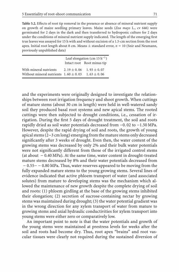
5 Essentiality of root-shoot communication 71
Table 5.2. Effects of root tip removal in the presence or absence of mineral nutrient supplyon growth of maize seedling primary leaves. Maize seeds (Zea mays L., cv 646) weregerminated for 2 days in the dark and then transferred to hydroponic culture for 2 daysunder the conditions of mineral nutrient supply indicated. The length of the emerging firsttrue leaves was assayed for 15 h with and without excision of a 1.5-cm section from the rootapex. Initial root length about 8 cm. Means ± standard error, n = 10 (Snir and Neumann,previously unpublished data)
Leaf elongation (cm 15 h−1)Intact root Root minus tip
With mineral nutrients 2. 19 ± 0. 06 1. 93 ± 0. 07Without mineral nutrients 1. 60 ± 0. 03 1. 63 ± 0. 06
and the experiments were originally designed to investigate the relation-ships between root irrigation frequency and shoot growth. When cuttingsof mature stems (about 30 cm in length) were held in well-watered sandysoil they produced basal root systems and new apical stems. The rootedcuttings were then subjected to drought conditions, i.e., cessation of ir-rigation. During the first 5 days of drought treatment, the soil and rootsrapidly dried as soil water potentials decreased from −0. 02 to −1.50 MPa.However, despite the rapid drying of soil and roots, the growth of youngapical stems (2−3 cm long) emerging from the mature stems only decreasedsignificantly after 3 weeks of drought. Even then, the water content of thegrowing stems was decreased by only 2% and their bulk water potentialswere not significantly different from those of the irrigated control stems(at about − 0.40 MPa). At the same time, water content in drought-treatedmature stems decreased by 8% and their water potentials decreased from− 0.55− − 0.80 MPa. Thus, water reserves appeared to be moving from thefully expanded mature stems to the young growing stems. Several lines ofevidence indicated that active phloem transport of water (and associatedsolutes) from mature to developing stems was the mechanism which al-lowed the maintenance of new growth despite the complete drying of soiland roots: (1) phloem girdling at the base of the growing stems inhibitedtheir elongation; (2) secretion of sucrose-containing nectar by growingstems was maintained during drought; (3) the water potential gradient wasin the wrong direction for any xylem transport of water from mature togrowing stems and axial hydraulic conductivities for xylem transport intoyoung stems were either zero or comparatively low.
An important point to note is that the water potentials and growth ofthe young stems were maintained at prestress levels for weeks after thesoil and roots had become dry. Thus, root apex “brains” and root vas-cular tissues were clearly not required during the sustained diversion of

72 P.M. Neumann
resources from mature to young stems that facilitated the maintenanceof growth. Conversely, the fact that a phloem girdle between mature andyoung stems inhibited the growth of the young stems reinforces the impor-tance of phloem transport tissues in interorgan communication. Althoughroot functioning did not appear to be essentially involved in stem growthregulation, phloem transport clearly was. The growth regulatory functionof phloem in this case could again be considered analogous to the signal-ing facilitated by the nerves of animals. However, the 3-week time scalerequired for this shoot growth response to relatively rapid dehydration ofthe root environment to become established was totally different from theseconds required for most animal responses to environmental changes.
5.5Conclusions and Future Perspectives
Some support is provided in this chapter for the drawing of analogiesbetween the signaling roles of plant long-distance transport systems andanimal nervous systems. However, the specific findings reviewed here donot indicate that root apices function as essential neurobiological com-mand centers involved in regulating shoot growth responses to adversechanges in the root environment. Cell-to-cell signaling via plasmodes-matal connections and variations in long-distance transport of hormones,essential nutrients and water via vascular tissues may conceivably providethe regulation needed to integrate most higher-plant growth responses toenvironmental changes.
References
Aloni R, Langhans M, Aloni E, Ulrich CI (2004) Role of cytokinin in the regulation of rootgravitropism. Planta 220:177–182
Baluska F, Volkmann D, Barlow PW (2004a) Cell bodies in a cage. Nature 428:371–371Baluska F, Mancuso S, Volkmann D, Barlow PW (2004b) Root apices as plant command cen-
tres: the unique ‘brain-like’ status of the root apex transition zone. Biologia (Bratislava)Suppl 59:7–19
Bassani M, Neumann PM, Gepstein S (2004) Differential expression profiles of growth-related genes in the elongation zone of maize primary roots. Plant Mol Biol 56:367–380
Birnbaum K, Shasha DE, Wang JY, Jung JW, Lambert GM, Galbraith DW, Benfey PN (2003)A gene expression map of the Arabidopsis root. Science 302:1956–1960
Chazen O, Hartung W, Neumann PM (1995) The different effects of PEG 6000 and NaClon leaf development are associated with differential inhibition of root water transport.Plant Cell Environ 18:727–735
Chazen O, Neumann PM (1994) Hydraulic signals from the roots and rapid cell wall hard-ening in growing maize leaves, are primary responses to PEG induced water deficits.Plant Physiol 104:1385–139

5 Essentiality of root-shoot communication 73
Fasano JM, Swanson SJ, Blancaflor EB, Dowd PE, Kao T, Gilroy S (2001) Changes in root cappH are required for the gravity response of the Arabidopsis root. Plant Cell 13:907–921
Kramer PJ, Boyer JS (1995) Water relations of plants and soils. Academic, New YorkLev-Yadun S, Inbar M, Izhaki I, Ne’eman G, Dafni A (2002) Colour patterns in vegetative
parts of plants deserve more research attention. Trends Plant Sci 7:59–60Nerd A, Neumann PM (2004) Phloem water transport maintains stem growth in a drought
stressed crop cactus (Hylocerus undatus). J Am Soc Hort Sci 129:486–490Neumann PM, Chazen O, Bogoslavsky L, Hartung W (1997) Role of root derived ABA in
regulating early leaf growth responses to water deficits. In: Altman A, Waisel Y (eds)Biology of root formation and development. Plenum, New York, pp 147–154
Peters WS, Felle HH (1999) The correlation of profiles of surface pH and elongation growthin maize roots. Plant Physiol 121:905–912
Poethig RS (2001) Life with 25,000 genes. Genome Res 11:313–316Raven PH, Evert RF, Eichhorn SE (1999) Biology of plants, 6th edn. Freeman, New YorkSalisbury FB, Ross CW (1985) Plant physiology, 3rd edn. Wadsworth, Belmont, CASnir N, Neumann PM (1997) Mineral nutrient supply, cell-wall adjustment and the control
of leaf growth. Plant Cell Environ 20:239–246

6 Signals and Targets Triggeredby Self-Incompatibility in Plants:Recognition of “Self ” Can Be DeadlyS.G. Thomas, S. Huang, C.J. Staiger, V.E. Franklin-Tong
Abstract Plants often respond to stimuli with dramatic rearrangements of their actincytoskeleton. Identificationof the signalsand transductionmachinery that lead tochanges inactin dynamics and organization is therefore of considerable interest. The self-incompatibil-ity (SI) response in the field poppy, Papaver rhoeas L., involves signal-mediated inhibitionof pollen tube growth in response to interaction with incompatible S proteins. This triggerslarges increases in [Ca2+]i and downstream a number of signalling components and targetsare modified in incompatible pollen. We have observed dramatic alterations to the actincytoskeleton in response to SI and established that this involves massive and sustainedactin depolymerization. We have begun to identify and characterize several actin-bindingproteins, including profilin and gelsolin, that may cooperate to transduce the signal froma Ca2+ wave into destruction of the cytoskeletal network that is essential for tip growth.Recently, we identified a role for programmed cell death (PCD) signalling cascades beingtriggered in SI and that a caspase-like activity is involved in mediating irreversible pollentube inhibition. Our data suggest that there is evidence for crosstalk between the SI-inducedsignalling cascades. We are currently investigating whether the signalling cascades for actinalterations and PCD are linked and whether the actin cytoskeleton functions as a sensorof cellular stress and can initiate PCD. Our current knowledge of the signalling cascade inP. rhoeas pollen therefore involves both early and late responses that work in concert toensure that pollen does not effect fertilization. Early cessation of tip growth is mediated bydestruction of the actin cytoskeleton, and this appears to cross-talk with a subsequent PCDcascade that commits the pollen to die.
6.1Introduction
Cell signalling mechanisms are vital for plants to adapt and survive ina harsh and challenging environment (Delledonne et al. 2005; Nürnbergeret al. 2005; Rhodes et al. 2005 in this volume). Plants respond to variousstimuli with dramatic rearrangements of their cytoplasm and these areoften mediated by a dynamic actin cytoskeleton. Thus, a major focus ofresearch is the identification of the signals and transduction machinerythat lead to changes in actin dynamics and organization.
The self-incompatibility (SI) response in the field poppy, Papaver rhoeasL., provides a good example of a genetically controlled system that employssignal-mediated inhibition of pollen tube growth in response to a specificstimulus. The ability to reproduce the SI response in Papaver pollen in
Communication in PlantsF. Baluška, S. Mancuso, D. Volkmann (Eds.)© Springer-Verlag Berlin Heidelberg 2006

76 S.G. Thomas et al.
vitro (Franklin-Tong et al. 1988) has allowed dissection of the signallingcascades triggered by SI (Franklin-Tong and Franklin 2003). Here we de-scribe dramatic alterations to the actin cytoskeleton that occur during SIand the characterization of several actin-binding proteins (ABPs) that mayplay central roles. We also show that SI involves signalling to programmedcell death (PCD) cascades. We have begun to explore whether the signallingcascades for actin alterations and PCD are linked and whether the actincytoskeleton functions as a sensor of cellular stress and can initiate PCD.
6.1.1Pollen–Pistil Interactions
Sexual reproduction is an excellent example of a process whereby plant cellsrespond to many different stimuli and utilize extensive signalling cascades.Pollination involves the deposition of pollen grains on a receptive stigma.This triggers numerous pollen–pistil interactions, including hydration ofthe pollen grain, germination and directional growth of the pollen tubethrough the pistil, and delivery of the sperm cells to the ovule, where theyeffect fertilization resulting in seed production. These events are tightlycontrolled at both the genetic and the biochemical level. Two recent reviewsdiscuss pollination in more detail (Edlund et al. 2004; Sanchez et al. 2004).
The pollen tube elongates via tip growth, which involves the precisecontrol of exocytosis and delivery of new plasma membrane and cell wallmaterials to the tube apex. It responds to chemical and physical signalsduring its journey to the ovule (reviewed by Franklin-Tong 1999a). Forexample, signals from the style include arabinogalactan proteins (Che-ung et al. 1995; de Graaf et al. 2003) triacylglycerides (Wolters-Arts et al.1998), chemocyanin (Kim et al. 2003), and γ-amino butyric acid (GABA)(Palanivelu et al. 2003). These signal molecules are believed to be involvedin the guidance of the pollen tube through the pistil. Within the pollentube, cytosolic free calcium ([Ca2+]i) has been shown to be an importantsecond messenger regulating pollen tube growth (reviewed by Franklin-Tong 1999b). Growing pollen tubes exhibit a tip-focused [Ca2+]i gradient(Rathore et al. 1991; Miller et al. 1992) that oscillates and is coordinated withgrowth (Holdaway-Clarke et al. 1997; Messerli et al. 1999, 2000). Althoughour understanding of this signalling cascade is incomplete, it is clear that[Ca2+]i oscillations allow spatio-temporal control of key processes involvedin pollen tube growth (reviewed by Holdaway-Clarke and Hepler 2003).

6 Targets of SI 77
6.1.2Self-Incompatibility
One aspect of pollen–pistil interactions that has been the focus of much re-search is SI. This is a genetically controlled mechanism which allows plantsto prevent inbreeding. Essentially, SI is a cell–cell recognition system thatallows the discrimination of genetically related pollen (“self” or incompat-ible pollen) from genetically unrelated pollen (“non-self” or compatiblepollen). For detailed reviews on the different types of SI, the reader is di-rected to three recent reviews (Franklin-Tong and Franklin 2003; Hiscockand McInnis 2003; Kao and Tsukamoto 2004).
In Papaver pollen, the cellular and molecular bases of the SI system havebeen investigated in detail (Franklin-Tong and Franklin 2003). The pistilcomponent, the S protein (Foote et al. 1994), is a small (about 15-kDa)secreted protein which acts as a ligand for the pollen component, which isthought to be a plasma membrane receptor (Franklin-Tong and Franklin2003). During an incompatible interaction, several events are triggered inthe pollen. A rapid influx of extracellular Ca2+ and large increases in [Ca2+]i
precede the dissipation of the tip-focused [Ca2+]i gradient, and comprisethe initial second messenger signals to downstream targets (Franklin-Tonget al. 1993, 1995, 1997, 2002). These targets include the rapid reorganizationand depolymerization of F-actin (Geitmann et al. 2000; Snowman et al.2002), which we discuss in more detail later. SI also involves the activationof several protein kinases leading to the phosphorylation of a number ofproteins. These include p26, a soluble inorganic pyrophosphatase (Rudd etal. 1996; Rudd and Franklin-Tong 2003). Phosphorylation of p26 and theinhibition of its activity by increases in [Ca2+]i are believed to interferewith pollen tube growth. A mitogen-activated protein (MAP) kinase, p56,is also phosphorylated (Rudd and Franklin-Tong 2003; Rudd et al. 2003).The timing of p56 activation (Rudd et al. 2003), after arrest of pollen tubegrowth, suggests that p56 is not involved in tip-growth inhibition, butinstead may signal to events occurring downstream. SI also triggers PCD(Jordan et al. 2000; Thomas and Franklin-Tong 2004) and this is discussedlater.

78 S.G. Thomas et al.
6.2The Actin Cytoskeleton and Self-Incompatibility
6.2.1Actin as a Sensor of Environmental Stimuli
A network of polymers, the actin cytoskeleton, provides a dynamic frame-work for multiple cell functions. At the protein level, it is composed of actinfilaments (F-actin)polymerized frommonomeric, globular actin (G-actin).Theactin cytoskeletonhasbeen shown toplay anessential role innumerousprocesses, including cell motility, organelle movement, vesicle traffickingand cytoplasmic streaming (reviewed by McCurdy et al. 2001; Staiger andHussey 2004). Actin filaments reorganize in response to various signallingevents and this mediates diverse responses to external environmental cues.A major focus of research on the cytoskeleton is the dissection of signallingcascades that regulate actin dynamics (reviewed by Staiger 2000). In incom-patible Papaver pollen, F-actin reorganizes rapidly (Geitmann et al. 2000;Snowman et al. 2002). Actin rearrangements in response to specific stimulihave also been described in stomatal guard cells responding to abscisic acidand light (Eun and Lee 1997), root hairs responding to Nod factors fromRhizobium bacteria (Cardenas et al. 1998; Miller et al. 1999) and epidermalcells responding to fungal and oomycete pathogens (Kobayashi et al. 1992,1993).
The dynamic nature of the actin cytoskeleton depends on the spatial dis-tribution and the local activity of a complex mixture of ABPs. The numberof ABPs identified in plants is growing rapidly and includes profilin, actin-depolymerizing factor (ADF)/cofilin, villin/gelsolin-like proteins, fimbrin,the Arp2/3 complex, AIP1, EF1α, and capping protein (reviewed by Staigerand Hussey 2004). The activity and regulation of these ABPs by secondmessengers suggest that they play key roles as transducers of extracellularstimuli; however, an involvement in specific cellular signalling processeshas yet to be demonstrated.
Pollen tube tip growth is particularly amenable to studying the signalsresponsible for mediating changes to the actin cytoskeleton. It is widelyassumed that the actin cytoskeleton helps guide or regulate the deliveryof secretory vesicles to the pollen tube apex, but exactly how this is ac-complished remains to be established. Furthermore, it is thought that thecontrol of pollen tube growth involves a complex interplay between the cy-toskeleton and signalling cascades, although there is currently little directevidence for this. However, early studies showed that increasing [Ca2+]i ar-tificially can disrupt actin filaments in pollen tubes (Kohno and Shimmen1987, 1988), suggesting that Ca2+ signals to alterations in actin organiza-tion.Analysisof a familyofRho-relatedGTPases inplants (ROPs) (reviewed

6 Targets of SI 79
by Yang 2002) has provided significant new insights into the interactionsbetween Ca2+ signals and actin dynamics. It has been demonstrated that thepollen-expressed ROP1, through its interacting partners RIC3 and RIC4,plays a role in two signalling cascades: one involving Ca2+ signalling, theother involving actin dynamics (Gu et al. 2003, 2005). These data begin toexplain how Ca2+ signals are linked to the spatio-temporal control of actindynamics and tip growth in pollen tubes.
It is well established that both actin organization and levels of F-actinmodulate pollen tube tip growth (Gibbon et al. 1999; Geitmann and Emons2000; Fu et al. 2001; Vidali et al. 2001; Lovy-Wheeler et al. 2005). A variety ofcytological approaches have allowed researchers to build a consensus viewof the distribution of the actin cytoskeleton in growing pollen tubes. Thereare three “zones” of F-actin in the pollen tube: long arrays of longitudinalactin filament bundles in the shank; a dense sub-apical meshwork of F-actin in a “basket-like” or ring configuration; and at the tip, a fine array ofdynamicfilaments. It is likely that the axial cables support reverse-fountain-pattern cytoplasmic streaming and that the apical arrays regulate secretoryvesicle trafficking. Figure 6.1a shows this arrangement for Papaver pollentubes.
6.2.2Actin as a Target for Self-Incompatibility Signalsin Incompatible Pollen
Evidence that SI signals to the actin cytoskeleton was first provided by theobservation that dramatic alterations to F-actin are triggered by SI induc-tion in incompatible pollen tubes (Geitmann et al. 2000; Snowman et al.2002). Detectable alterations were extremely rapid, occurring within 1 min.Thedistinctive sub-apical basket-like configurationdisappearedanda large“blob” of F-actin appeared at the pollen tube apical region (Fig. 6.1b). Theoverall intensity of phalloidin staining was reduced substantially, indicat-ing a loss of F-actin, and by 5−10 min after SI induction, the longitudinalF-actin bundles had largely disappeared (Fig. 6.1c,d). The remaining F-actin had a fine, speckled appearance (Fig. 6.1d,e), suggesting severing ordepolymerization of F-actin and pronounced cortical F-actin was evident(Fig. 6.1c–e). Further alterations were evident between 10 and 20 min; F-actin appeared as large aggregates or “punctate foci” (Fig. 6.1e,f) and thesepersisted for at least 3 h. These alterations were specific to induction of SIin incompatible pollen tubes, as control pollen tubes showed no changesto actin organization. Furthermore, the SI-induced rearrangement of actinwas demonstrated to be independent of growth arrest (Geitmann et al.2000).

80 S.G. Thomas et al.
Fig.6.1. Self-incompatibility (SI) induces dramatic rearrangements of actin cytoskeletonorganization in Papaver pollen. a The actin cytoskeleton of a normally growing pollen tubehas longitudinal arrays of F-actin in the pollen tube shank, a dense sub-apical network ofF-actin and a fine array of dynamic filaments at the tip. b Following SI induction rapidreorganization is observed. Within 1 min, F-actin accumulates in the tip of the pollen tube.c,d By 20 min after SI induction, the F-actin has disappeared from the cortex and is localizedto the periphery of grains and pollen tubes and “punctate foci” of F-actin begin to appear.e,f The punctate foci persist for greater than 60 min and increase in size over time. a,b,d–fare epifluorescence microscope images of rhodamine–phalloidin-labelled pollen tubes. cis a single optical section from confocal microscope imaging of rhodamine–phalloidin-labelled pollen grain. Bars represent 10 µm
6.2.3Self-Incompatibility Stimulates Rapidand Sustained Depolymerization of F-Actin
Because the actin reorganization observed during the early stages of the SIresponse suggested that actin depolymerization had occurred, a quantita-tive approach was used to investigate this further. A technique based onthe number of fluorescent-phalloidin binding sites (Howard and Oresajo1985; Lillie and Brown 1994) was used to determine the concentration ofactin in filamentous form in pollen (Gibbon et al. 1999; Snowman et al.2002). We found that the levels of F-actin in pollen throughout hydration,germination and early growth were constant (Snowman et al. 2002). UponSI induction, a rapid and sustained decrease in F-actin levels was induced.Within 1 min there was a significant reduction in F-actin. These levels con-tinued to decrease, so that by 5 min after SI induction, F-actin levels wereless than 50% of the controls and by 1 h were reduced by 74% (Snowman et

6 Targets of SI 81
al. 2002). Thus, SI induces massive and sustained F-actin depolymerizationin incompatible pollen.
6.2.4Increases in Cytosolic Calcium Lead to Changes in F-Actin
As we had established that Ca2+ acts as a second messenger during the SI re-sponse (Franklin-Tong et al. 1993, 1997), we wondered whether F-actin wasa target for Ca2+-dependent signalling. We therefore investigated whetherincreasing [Ca2+]i in pollen tubes, using the Ca2+ ionophore A23187 andmastoparan (Franklin-Tong et al. 1993, 1996), stimulated changes in F-actin.These treatments triggeredreorganizationof theF-actincytoskeletonthat appeared broadly similar to those stimulated by SI and led to quanti-tative reductions in F-actin levels (Snowman et al. 2002). This suggests thatduring SI increases in [Ca2+]i perturb the actin cytoskeleton.
6.2.5Profilin and Gelsolin: Mediators of Actin Alterations?
The marked changes to the actin cytoskeleton in the SI response raised thequestion of what molecular machinery transduces the changes in [Ca2+]i
into destruction of the F-actin. ABPs are stimulus-response modulators ofactin cytoskeleton reorganization (reviewed by Holdaway-Clarke and Hep-ler 2003; Staiger and Hussey 2004). F-actin depolymerization could resultfrom a loss of side-binding proteins, capping of filament ends, stimulationof filament severing, increased activity of a sequestering protein, or a com-bination of these activities. However, few ABPs in pollen tubes have beenwell characterized (reviewed by Hepler et al. 2001; McCurdy et al. 2001;Staiger and Hussey 2004). Because actin depolymerization can be achievedusing calcium ionophores, we attempted to model this in vitro with Ca2+-sensitive ABPs. Although plant ADF/cofilin can cause actin depolymeriza-tion and is regulated by Ca2+ indirectly, increases in [Ca2+]i are expected toinhibit the F-actin depolymerizing activity of ADF (Smertenko et al. 1998,Allwood et al. 2001), so its involvement in SI has not been examined.
Pollen profilin exhibits increased actin-sequestering activity at elevated,but physiologically relevant, Ca2+ concentrations (Kovar et al. 2000). A largepool of profilin was predicted to buffer G-actin and maintain the low actinpolymer level in pollen (reviewed by Staiger and Hussey 2004). However,experiments with native Papaver pollen profilin failed to account for thehigh level of depolymerization observed during SI (Snowman et al. 2002).We proposed that other ABPs may function in concert with profilin toachieve sustained actin depolymerization during SI.

82 S.G. Thomas et al.
6.2.6PrABP80 is Poppy Gelsolin
PrABP80 is a Ca2+-regulated ABP which may be involved in SI-mediatedactin depolymerization (Huang et al. 2004). PrABP80 was originally iden-tified by DNase I–Sepharose chromatography during attempts to isolatenative poppy pollen actin. Tandem mass spectrometry sequence analy-sis and kinetic actin polymerization assays showed that it belongs to thevillin/gelsolin family. Furthermore, the native protein was demonstratedto have potent Ca2+-dependent severing activity in vitro (Huang et al.2004). As shown in Fig. 6.2, PrABP80 significantly reduced the length ofpre-formed actin filaments after incubation in the presence of 160 µM freeCa2+ for 30 min (Fig. 6.2b) compared with incubation in the presence of15 nM free Ca2+ (Fig. 6.2a). It was also shown directly with time-lapsefluorescence imaging that PrABP80 could sever actin filaments in a Ca2+-dependent manner. PrABP80 could, therefore, assist F-actin depolymer-ization by Ca2+-activated severing and the creation of new pointed endsfor depolymerization by profilin. In addition, PrABP80 caps the barbedend of actin filaments, thereby blocking assembly of the profilin–actincomplex and allowing profilin to function as a simple actin sequesteringprotein. The feasibility of such a mechanism was supported by experimentsin which equimolar amounts of profilin were added to pre-existing actinfilaments together with nanomolar amounts of PrABP80. In these assays,polymer levels were monitored by fluorescence of pyrene-labelled actin inthe fluorimeter. The data from a typical experiment in the presence of 1 µMfree Ca2+, which mimics the [Ca2+]i observed in pollen tubes during theSI response, are shown in Fig. 6.2c. The extent and rate of actin depoly-merization were dramatically increased by substoichiometric amounts ofPrABP80, when compared with profilin alone; moreover, significant de-polymerization was not observed when free Ca2+ was reduced to 15 nM(Fig. 6.2c).
Thus, we have identified a potential mechanism that links SI-induced[Ca2+]i changes to actin depolymerization. We propose that PrABP80 func-tions at the centre of the SI response by creating new filament pointedends for disassembly and by blocking barbed ends from profilin–actinassembly. To test this molecular model thoroughly, we will need to deter-mine the cellular concentrations for PrABP80 in pollen, measure its affinityfor plant actin filament ends, and determine the efficiency of severing atphysiological [Ca2+]i.
The actin depolymerization observed during SI is somewhat unusual,since the level and extent of depolymerization are far greater than thoserequired to inhibit growth. Further alterations to the actin cytoskeletoncontinue long after the inhibition of tip growth, suggesting that SI-induced

6 Targets of SI 83
Fig.6.2. PrABP80 has potent calcium-dependent F-actin severing activity. a,b Actin fila-ments (4 µM) labelled with rhodamine–phalloidin were incubated with PrABP80 in thepresence of 15 nM free Ca2+ (a) or 160 µM Ca2+ (b) and images were collected after 30-minincubation. PrABP80 significantly reduced the length of actin filaments (mean 0. 4±0.4 µm)after incubation in the presence of 160 µMfree Ca2+(b). Reactions performed in the presenceof 15 nM free Ca2+ had a mean filament length of 4. 7±3.2 µm (a). The bar represents 10 µm.c The severing activity of PrABP80 was also shown by enhancement of profilin-mediateddepolymerization in the presence of 1 µM free Ca2+ as monitored by fluorescence of pyreneactin. Zea mays profilin alone (ZmPRO5; 2.5 µM) or ZmPRO5 together with PrABP80 wereadded to an F-actin solution prepared from 2.5 µM actin (40–50% pyrene-labelled). Depoly-merization was recorded as a reduction in pyrene fluorescence (arbitrary units) beginningfrom the addition of protein at time zero. The extent and rate of depolymerization wereincreased by substoichiometric amounts of PrABP80 + profilin (bottom curve), comparedwith profilin alone or PrABP80 alone (middle curves). Moreover, a similar level of depoly-merization was not observed when free calcium was reduced to 15 nM, a condition wherePrABP80 should function like a barbed-end cap (top curve)
signalling events continue after inhibition of growth. We therefore believethat some of these events may act to make growth arrest irreversible. Inyeast and some animal cells, sustained actin depolymerization is observedduring apoptosis and the actin cytoskeleton has been shown to play a func-tional role in the initiation of PCD by modulating PCD signalling cascades.(Kayalar et al. 1996; Levee et al. 1996; Janmey 1998; Korichneva and Ham-merling 1999; Rao et al. 1999; Morley et al. 2003; Gourlay et al. 2004; Gourlayand Ayscough 2005). We noted that the sustained actin depolymerizationobserved in incompatible pollen was highly reminiscent of apoptosis. Thissuggested that a PCD signalling cascade might be triggered by SI in incom-patible pollen.
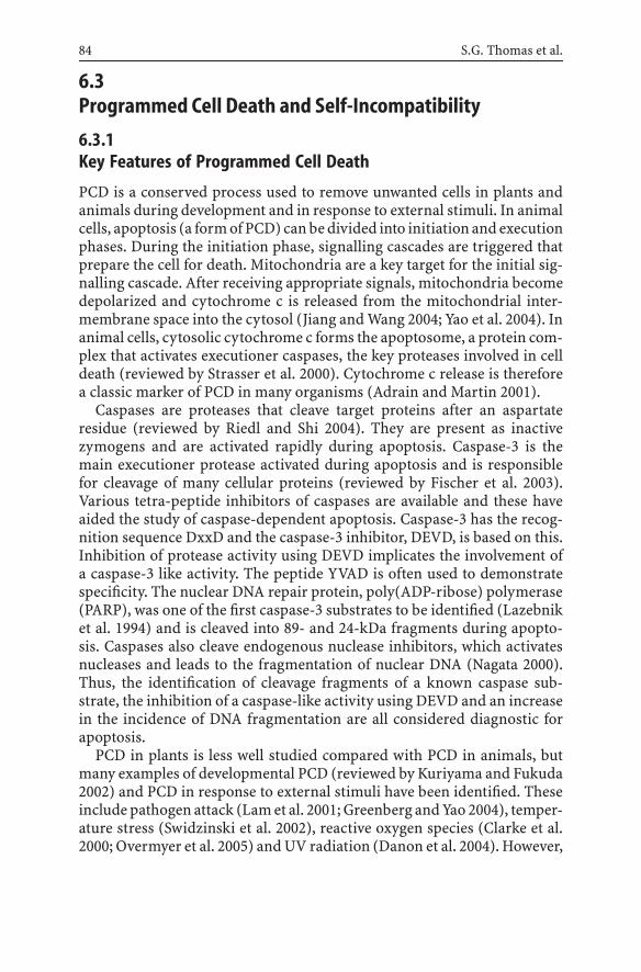
84 S.G. Thomas et al.
6.3Programmed Cell Death and Self-Incompatibility
6.3.1Key Features of Programmed Cell Death
PCD is a conserved process used to remove unwanted cells in plants andanimals during development and in response to external stimuli. In animalcells, apoptosis (a form of PCD) can be divided into initiation and executionphases. During the initiation phase, signalling cascades are triggered thatprepare the cell for death. Mitochondria are a key target for the initial sig-nalling cascade. After receiving appropriate signals, mitochondria becomedepolarized and cytochrome c is released from the mitochondrial inter-membrane space into the cytosol (Jiang and Wang 2004; Yao et al. 2004). Inanimal cells, cytosolic cytochrome c forms the apoptosome, a protein com-plex that activates executioner caspases, the key proteases involved in celldeath (reviewed by Strasser et al. 2000). Cytochrome c release is thereforea classic marker of PCD in many organisms (Adrain and Martin 2001).
Caspases are proteases that cleave target proteins after an aspartateresidue (reviewed by Riedl and Shi 2004). They are present as inactivezymogens and are activated rapidly during apoptosis. Caspase-3 is themain executioner protease activated during apoptosis and is responsiblefor cleavage of many cellular proteins (reviewed by Fischer et al. 2003).Various tetra-peptide inhibitors of caspases are available and these haveaided the study of caspase-dependent apoptosis. Caspase-3 has the recog-nition sequence DxxD and the caspase-3 inhibitor, DEVD, is based on this.Inhibition of protease activity using DEVD implicates the involvement ofa caspase-3 like activity. The peptide YVAD is often used to demonstratespecificity. The nuclear DNA repair protein, poly(ADP-ribose) polymerase(PARP), was one of the first caspase-3 substrates to be identified (Lazebniket al. 1994) and is cleaved into 89- and 24-kDa fragments during apopto-sis. Caspases also cleave endogenous nuclease inhibitors, which activatesnucleases and leads to the fragmentation of nuclear DNA (Nagata 2000).Thus, the identification of cleavage fragments of a known caspase sub-strate, the inhibition of a caspase-like activity using DEVD and an increasein the incidence of DNA fragmentation are all considered diagnostic forapoptosis.
PCD in plants is less well studied compared with PCD in animals, butmany examples of developmental PCD (reviewed by Kuriyama and Fukuda2002) and PCD in response to external stimuli have been identified. Theseinclude pathogen attack (Lam et al. 2001; Greenberg and Yao 2004), temper-ature stress (Swidzinski et al. 2002), reactive oxygen species (Clarke et al.2000; Overmyer et al. 2005) and UV radiation (Danon et al. 2004). However,

6 Targets of SI 85
many of the genes encoding components of the apoptotic machinery, in-cluding caspases, have either not yet been identified in plants or are simplynot present (reviewed by Woltering et al. 2002). Nevertheless, there is nodoubt that PCD occurs in plants and much of the evidence for it has comefrom biochemical studies (reviewed by van Doorn and Woltering 2005).We have used markers of PCD to investigate whether an incompatible SIresponse triggers PCD in Papaver pollen.
6.3.2Programmed Cell Death is Triggered Duringthe Papaver Self-Incompatibility Response
Cytochrome c release is an early event of SI-induced PCD. Since leakage ofcytochrome c into the cytosol is a key early marker for PCD, we investigatedwhether this occurred in pollen tubes undergoing incompatible SI. Weshowed that large increases in cytosolic cytochrome c were detected inincompatible pollen (Thomas and Franklin-Tong 2004). The increase incytochrome c release was rapid, beginning at 10 min and increasing upto 2 h, after SI induction. In controls, no release of cytochrome c wasobserved. The first detection of cytochrome c in the cytosol at around10 min corresponds to the point at which peak phosphorylation and MAPkinaseactivityofp56aredetected (Ruddet al. 2003).Ourdatamay thereforeindicate crosstalk between different signalling cascades and may representthe point at which the pollen tube becomes committed to death.
DNA fragmentation is stimulated by SI. Since DNA fragmentation is a clas-sic marker for PCD, we investigated whether DNA fragmentation occurredin incompatible Papaver pollen undergoing SI. DNA fragmentation wasdetected in incompatible pollen and was S-specific, as it was not observedin either untreated pollen tubes or compatible pollen tubes (Jordan etal. 2000). DNA fragmentation is first detected at 4 h and continues to in-crease for at least 16 h after SI induction. Pre-treatment of pollen tubeswith DEVD prior to the induction of SI caused a significant reduction inthe amount of DNA fragmentation, reducing it from levels of 71.6% inSI to 18.97% in pollen tubes with SI-induced in the presence of DEVD(Fig. 6.3a) (Thomas and Franklin-Tong 2004). YVAD (used as a negativecontrol) displayed only a small decrease in DNA fragmentation. These dataimplicate the involvement of a caspase-3 like activity in SI-mediated DNAfragmentation. Furthermore, DEVD pre-treatment of pollen tubes allowedgrowth to resume following SI induction, whilst YVAD did not alleviate SI-induced growth inhibition (Thomas and Franklin-Tong 2004). These datademonstrate that a caspase-3 like activity is also involved in mechanismsmediating SI-induced pollen tube inhibition.
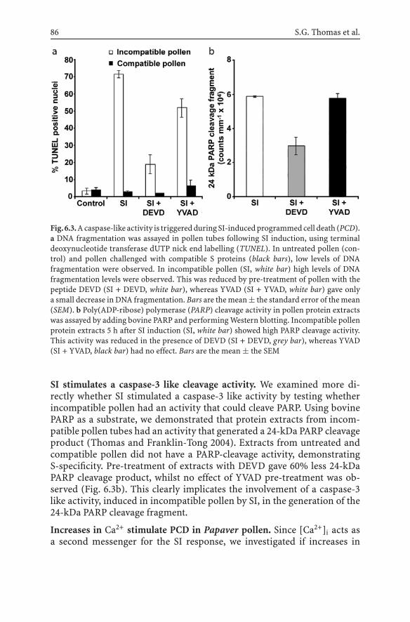
86 S.G. Thomas et al.
Fig.6.3.A caspase-like activity is triggered during SI-induced programmed cell death (PCD).a DNA fragmentation was assayed in pollen tubes following SI induction, using terminaldeoxynucleotide transferase dUTP nick end labelling (TUNEL). In untreated pollen (con-trol) and pollen challenged with compatible S proteins (black bars), low levels of DNAfragmentation were observed. In incompatible pollen (SI, white bar) high levels of DNAfragmentation levels were observed. This was reduced by pre-treatment of pollen with thepeptide DEVD (SI + DEVD, white bar), whereas YVAD (SI + YVAD, white bar) gave onlya small decrease in DNA fragmentation. Bars are the mean ± the standard error of the mean(SEM). b Poly(ADP-ribose) polymerase (PARP) cleavage activity in pollen protein extractswas assayed by adding bovine PARP and performing Western blotting. Incompatible pollenprotein extracts 5 h after SI induction (SI, white bar) showed high PARP cleavage activity.This activity was reduced in the presence of DEVD (SI + DEVD, grey bar), whereas YVAD(SI + YVAD, black bar) had no effect. Bars are the mean ± the SEM
SI stimulates a caspase-3 like cleavage activity. We examined more di-rectly whether SI stimulated a caspase-3 like activity by testing whetherincompatible pollen had an activity that could cleave PARP. Using bovinePARP as a substrate, we demonstrated that protein extracts from incom-patible pollen tubes had an activity that generated a 24-kDa PARP cleavageproduct (Thomas and Franklin-Tong 2004). Extracts from untreated andcompatible pollen did not have a PARP-cleavage activity, demonstratingS-specificity. Pre-treatment of extracts with DEVD gave 60% less 24-kDaPARP cleavage product, whilst no effect of YVAD pre-treatment was ob-served (Fig. 6.3b). This clearly implicates the involvement of a caspase-3like activity, induced in incompatible pollen by SI, in the generation of the24-kDa PARP cleavage fragment.
Increases in Ca2+ stimulate PCD in Papaver pollen. Since [Ca2+]i acts asa second messenger for the SI response, we investigated if increases in

6 Targets of SI 87
[Ca2+]i may signal to PCD in Papaver pollen. Treatment of pollen tubeswith mastoparan (Franklin-Tong et al. 1996) stimulated large increases incytochrome c release into the cytosol within 5 min (Thomas and Franklin-Tong 2004). These data showed that increases in [Ca2+]i in pollen act up-stream of cytochrome c release. Mastoparan-stimulated pollen extractsalso had high PARP-cleavage activity and the calcium channel blocker, La3+
(Franklin-Tong et al. 2002), gave a 70% decrease in this activity (Thomasand Franklin-Tong 2004), indicating that increases in [Ca2+]i can triggera caspase-3 like activity. Together these data suggest that the Ca2+ signalsstimulated by SI are part of the signalling cascade responsible for triggeringPCD in incompatible pollen.
6.3.3A Link Between Actin and Programmed Cell Death?
The F-actin depolymerization observed during SI certainly inhibits pollentube growth. However, the extent of depolymerization is much greater thanthe level required to stop tip growth. This suggested additional functionsfor SI-induced actin depolymerization. As discussed earlier, in animal andyeast cells there is evidence for actin dynamics signalling to PCD, butno-one has investigated this possibility in plants yet. We have exploreda possible link between actin depolymerization and PCD, using the G-actinbinding reagent latrunculin B to depolymerize pollen tube F-actin and thenmeasured PCD using DNA fragmentation. Preliminary data indicate thatmodest and transient reductions in actin polymer levels are sufficient totrigger caspase-3 mediated PCD in Papaver pollen tubes (Thomas, Huang,Staiger and Franklin-Tong, unpublished data). Thus, there is evidence forchanges in actin dynamics initiating PCD in Papaver pollen. This suggestscrosstalk exists between the actin and PCD signalling networks in pollen.
6.4Conclusions and Future Perspectives
Some of the major components and events that play a role in mediatingSI in Papaver pollen have now been identified. Figure 6.4 shows a modelthat highlights current knowledge about the signals and targets triggeredby SI in incompatible pollen. These include Ca2+, actin, PCD, and othercomponents not discussed in detail here. Following SI induction, which weassume involves the interaction of the stigmatic S protein with its cognatepollen S receptor, there is a rapid influx of Ca2+ accompanied by increases in[Ca2+]i in the pollen tube. The tip-focused [Ca2+]i gradient dissipates and

88 S.G. Thomas et al.
Fig.6.4. Signals and targets involved in SI in incompatible Papaver pollen. S proteins (S1)bind toaspecificpollenSreceptoronanS1 pollen tubeduringan incompatibleSI interaction.This stimulates influx of Ca2+ and increases in [Ca2+]i, which triggers an intracellular Ca2+
signalling cascade in the pollen tube. Depolymerization of F-actin is likely to be mediatedby Ca2+-regulated actin-binding proteins, including profilin and PrABP80. SI also tri ggersPCD in incompatible pollen. Hallmarks of PCD, including cytochrome c leakage frommitochondria, a caspase-3 like cleavage activity and caspase-mediated DNA fragmentationhave been observed in incompatible pollen. There is preliminary evidence for altered actindynamics or polymer levels acting as the trigger for PCD, which commits the pollen todie. Other SI-triggered signalling events in incompatible pollen include rapid increasesin the Ca2+-dependent phosphorylation of a number of proteins, including p26, a solubleinorganic pyrophosphatase and p56, a mitogen-activated protein kinase (MAPK)
tip growth is inhibited within a few minutes. We have shown that [Ca2+]i
acts as a second messenger and triggers a number of downstream events(Fig. 6.4). Because the tight spatio-temporal regulation of actin dynamicsin pollen tubes is crucial for growth, the cytoskeleton is an ideal target forthe Ca2+-signalling cascade during SI. Changes to the cytoskeleton dur-ing SI are probably mediated through the cooperative action of severalCa2+-regulated ABPs, such as profilin and PrABP80. The sustained actindepolymerization may function to maintain a block on tip growth, but pre-liminary data suggest it may also operate in crosstalk with PCD signallingcascades. The SI-induced PCD cascade includes leakage of cytochrome cfrom mitochondria, activation of a caspase-like activity and nuclear DNAfragmentation. Thus, the cytoskeleton is not only a target for signallingcascades, but may also be an initiator and transducer of signals. Activationof the p56 MAP kinase occurs after tip growth inhibition and, therefore,may signal to downstream events. Phosphorylation of the soluble inorganicpyrophosphatase, p26, is thought to act as a fail-safe mechanism ensuringthat tip growth remains inhibited. It is becoming clear, therefore, that there

6 Targets of SI 89
is crosstalk between the SI signalling cascades and that PCD probably func-tions as a decision-making point that results in incompatible pollen beingcommitted to death.
Thus, we are beginning to dissect a complex network of signals andtargets involved in signalling cascades triggered by SI. The challenge forthe future is not only to identify additional components, but also to eluci-date how these components interact and what crosstalk is utilized duringSI. In particular, the specific molecular interactions between [Ca2+]i, actinalterations and PCD signalling cascades are currently being investigated.Furthermore, hints that p56 MAP kinase activation may also cross-talk tothese elements are being pursued, as there is evidence for MAPK involve-ment in F-actin alterations and PCD in other systems (Zhang et al. 2000;Yang et al. 2001; Samaj et al. 2002). As the title of this book implies, itappears that plants have some aspects of a neuronal-like system of commu-nication and we are just beginning to scratch the surface of understandingthe intricacies of how cellular responses are achieved through networks ofsignalling intermediates.
Acknowledgements. Research in the laboratory of V.E.F.-T. is supportedby Biotechnology and Biological Sciences Research Council (BBSRC). Re-search in the laboratory of C.J.S. is supported by the US Department ofAgriculture, National Research Initiative grant no. 2002–35304–12412, andby the US Department of Energy – Energy Biosciences Division grant DE-FG02-04ER15526.
References
Adrain C, Martin SJ (2001) The mitochondrial apoptosome: a killer unleashed by thecytochrome seas. Trends Biochem Sci 26:390–397
Allwood EG, Smertenko AP, Hussey PJ (2001) Phosphorylation of plant actin-depolymerising factor by calmodulin-like domain protein kinase. FEBS Lett 499:97–100
Cardenas L, Vidali L, Dominguez J, Perez H, Sanchez F, Hepler PK, Quinto C (1998) Re-arrangement of actin microfilaments in plant root hairs responding to Rhizobium etlinodulation signals. Plant Physiol 116:871–877
Cheung AY, Wang H, Wu HM (1995) A floral transmitting tissue-specific glycoproteinattracts pollen tubes and stimulates their growth. Cell 82:383–393
Clarke A, Desikan R, Hurst RD, Hancock JT, Neill SJ (2000) NO way back: nitric oxide andprogrammed cell death in Arabidopsis thaliana suspension cultures. Plant J 24:667–677
Danon A, Rotari VI, Gordon A, Mailhac N, Gallois P (2004) Ultraviolet-C overexposureinduces programmed cell death in Arabidopsis, which is mediated by caspase-like ac-tivities and which can be suppressed by caspase inhibitors, p35 and defender againstapoptotic death. J Biol Chem 279:779–787
de Graaf BHJ, Knuiman BA, Derksen J, Mariani C (2003) Characterization and localizationof the transmitting tissue-specific PELPIII proteins of Nicotiana tabacum. J Exp Bot54:55–63

90 S.G. Thomas et al.
Edlund AF, Swanson R, Preuss D (2004) Pollen and stigma structure and function: the roleof diversity in pollination. Plant Cell 16:S84–97
Eun SO, Lee Y (1997) Actin filaments of guard cells are reorganized in response to light andabscisic acid. Plant Physiol 115:1491–1498
Fischer U, Jänicke RU, Schulze-Osthoff K (2003) Many cuts to ruin: a comprehensive updateof caspase substrates. Cell Death Differ 10:76–100
Foote H, Ride J, Franklin-Tong V, Walker E, Lawrence M, Franklin F (1994) Cloning andexpression of a distinctive class of self-incompatibility (S) gene from Papaver rhoeas L.Proc Natl Acad Sci USA 91:2265–2269
Franklin-Tong VE (1999a) Signaling in pollination. Curr Opin Plant Biol 2:490–495Franklin-Tong VE (1999b) Signaling and the modulation of pollen tube growth. Plant Cell
11:727–738Franklin-Tong VE, Franklin FCH (2003) Gametophytic self-incompatibility inhibits pollen
tube growth using different mechanisms. Trends Plant Sci 8:598–605Franklin-Tong VE, Franklin FCH (2003) The different mechanisms of gametophytic self-
incompatibility. Philos Trans R Soc Lond Ser B Biol Sci 358:1025–1032Franklin-Tong VE, Lawrence MJ, Franklin FCH (1988) An in vitro bioassay for the stigmatic
product of the self-incompatibility gene in Papaver rhoeas L. New Phytol 110:109–118Franklin-TongVE,Ride JP, FranklinFCH(1995)Recombinant stigmatic self-incompatibility
(S-) protein elicits a Ca2+ transient in pollen of Papaver rhoeas. Plant J 8:299–307Franklin-Tong VE, Hackett G, Hepler PK (1997) Ratio-imaging of Ca2+
i in the self-incompatibility response in pollen tubes of Papaver rhoeas. Plant J 12:1375–1386
Franklin-Tong VE, Ride JP, Read ND, Trewavas AJ, Franklin FCH (1993) The self-incompatibility response in Papaver rhoeas is mediated by cytosolic free calcium. PlantJ 4:163–177
Franklin-Tong VE, Drøbak BK, Allan AC, Watkins P, Trewavas AJ (1996) Growth of pollentubes of Papaver rhoeas is regulated by a slow-moving calcium wave propagated byinositol 1,4,5-trisphosphate. Plant Cell 8:1305–1321
Franklin-Tong VE, Holdaway-Clarke TL, Straatman KR, Kunkel JG, Hepler PK (2002) In-volvement of extracellular calcium influx in the self-incompatibility response of Papaverrhoeas. Plant J 29:333–345
Fu Y, Wu G, Yang Z (2001) Rop GTPase-dependent dynamics of tip-localized F-actin controlstip growth in pollen tubes. J Cell Biol 152:1019–1032
Geitmann A, Emons AMC (2000) The cytoskeleton in plant and fungal cell tip growth.J Microsc 198:218–245
Geitmann A, Snowman BN, Emons AMC, Franklin-Tong VE (2000) Alterations in the actincytoskeleton of pollen tubes are induced by the self-incompatibility reaction in Papaverrhoeas. Plant Cell 12:1239–1252
Gibbon BC, Kovar DR, Staiger CJ (1999) Latrunculin B has different effects on pollengermination and tube growth. Plant Cell 11:2349–2364
Gourlay CW, Ayscough KR (2005) Identification of an upstream regulatory pathway con-trolling actin-mediated apoptosis in yeast. J Cell Sci 118:2119–2132
Gourlay CW, Carpp LN, Timpson P, Winder SJ, Ayscough KR (2004) A role for the actincytoskeleton in cell death and aging in yeast. J Cell Biol 164:803–809
Greenberg JT, Yao N (2004) The role and regulation of programmed cell death in plant-pathogen interactions. Cell Microbiol 6:201–211
Gu Y, Vernoud V, Fu Y, Yang Z (2003) ROP GTPase regulation of pollen tube growth throughthe dynamics of tip-localized F-actin. J Exp Bot 54:93–101
Gu Y, Fu Y, Dowd P, Li S, Vernoud V, Gilroy S, Yang Z (2005) A Rho family GTPase controlsactin dynamics and tip growth via two counteracting downstream pathways in pollentubes. J Cell Biol 169:127–138

6 Targets of SI 91
Hepler PK, Vidali L, Cheung AY (2001) Polarized cell growth in higher plants. Annu RevCell Dev Biol 17:159–187
Hiscock SJ, McInnis SM (2003) Pollen recognition and rejection during the sporophyticself-incompatibility response: Brassica and beyond. Trends Plant Sci 8:606–613
Holdaway-Clarke TL, Hepler PK (2003) Control of pollen tube growth: role of ion gradientsand fluxes. New Phytol 159:539–563
Holdaway-Clarke TL, Feijo JA, Hackett GR, Kunkel JG, Hepler PK (1997) Pollen tube growthand the intracellular cytosolic calcium gradient oscillate in phase while extracellularcalcium influx is delayed. Plant Cell 9:1999–2010
Howard TH, Oresajo CO (1985) A method for quantifying F-actin in chemotactic peptideactivated neutrophils – study of the effect of Tboc peptide. Cell Motil Cytoskel 5:545–557
Huang S, Blanchoin L, Chaudhry F, Franklin-Tong VE, Staiger CJ (2004) A gelsolin-likeprotein from Papaver rhoeas pollen (PrABP80) stimulates calcium-regulated severingand depolymerization of actin filaments. J Biol Chem 279:23364–23375
Janmey PA (1998) The cytoskeleton and cell signaling: component localization and mechan-ical coupling. Physiol Rev 78:763–781
Jiang X, Wang X (2004) Cytochrome c-mediated apoptosis. Annu Rev Biochem 73:87–106Jordan ND, Franklin FCH, Franklin-Tong VE (2000) Evidence for DNA fragmentation trig-
gered in the self-incompatibility response in pollen of Papaver rhoeas. Plant J 23:471–479Kao T-h, Tsukamoto T (2004) The molecular and genetic basis of S-RNase-based self-
incompatibility. Plant Cell 16:S72–83Kayalar C, Ord T, Testa MP, Zhong LT, Bredesen DE (1996) Cleavage of actin by interleukin
1beta-converting enzyme to reverse DNase I inhibition. Proc Natl Acad Sci USA 93:2234–2238
Kim S, Mollet J-C, Dong J, Zhang K, Park S-Y, Lord EM (2003) Chemocyanin, a small basicprotein from the lily stigma, induces pollen tube chemotropism. Proc Natl Acad Sci USA100:16125–16130
Kobayashi I, Kobayashi Y, Yamaoka N, Kunoh H (1992) Recognition of a pathogen and a non-pathogen by barley coleoptile cells. 3. Responses of microtubules and actin-filaments inbarley coleoptile cells to penetration attempts. Can J Bot 70:1815–1823
Kobayashi Y, Kobayashi I, Kunoh H (1993) Recognition of a pathogen and a nonpathogen bybarley coleoptile cells. 2. Alteration of cytoplasmic strands in coleoptile cells caused bythe pathogen, Erysiphe graminis and the nonpathogen, E. pisi, prior to their penetration.Physiol Mol Plant Path 43:243–254
Kohno T, Shimmen T (1987) Ca2+-induced fragmentation of actin-filaments in pollen tubes.Protoplasma 141:177–179
Kohno T, Shimmen T (1988) Mechanism of Ca2+ inhibition of cytoplasmic streaming in lilypollen tubes. J Cell Sci 91:501–509
Korichneva I, Hammerling U (1999) F-actin as a functional target for retro-retinoids:a potential role in anhydroretinol-triggered cell death. J Cell Sci 112:2521–2528
Kovar DR, Drøbak BK, Staiger CJ (2000) Maize profilin isoforms are functionally distinct.Plant Cell 12:583–598
Kuriyama H, Fukuda H (2002) Developmental programmed cell death in plants. Curr OpinPlant Biol 5:568–573
Lam E, Kato N, Lawton M (2001) Programmed cell death, mitochondria and the planthypersensitive response. Nature 411:848–853
Lazebnik YA, Kaufmann SH, Desnoyers S, Poirier GG, Earnshaw WC (1994) Cleavage ofpoly(ADP-ribose) polymerase by a proteinase with properties like ICE. Nature 371:346–347
Levee MG, Dabrowska MI, Lelli JL Jr, Hinshaw DB (1996) Actin polymerization and depoly-merization during apoptosis in HL-60 cells. Am J Physiol Cell Physiol 271:C1981–1992
92 S.G. Thomas et al.
Lillie SH, Brown SS (1994) Immunofluorescence localization of the unconventional myosin,Myo2p, and the putative kinesin-related protein, Smy1p, to the same regions of polarizedgrowth in Saccharomyces cerevisiae. J Cell Biol 125:825–842
Lovy-Wheeler A, Wilsen KL, Baskin TI, Hepler PK (2005) Enhanced fixation reveals theapical cortical fringe of actin filaments as a consistent feature of the pollen tube. Planta221:95–104
McCurdy DW, Kovar DR, Staiger CJ (2001) Actin and actin-binding proteins in higher plants.Protoplasma 215:89–104
Messerli M, Danuser G, Robinson K (1999) Pulsatile influxes of H+, K+ and Ca2+ lag growthpulses of Lilium longiflorum pollen tubes. J Cell Sci 112:1497–1509
Messerli MA, Creton R, Jaffe LF, Robinson KR (2000) Periodic increases in elongation rateprecede increases in cytosolic Ca2+ during pollen tube growth. Dev Biol 222:84–98
Miller DD, Callaham DA, Gross DJ, Hepler PK (1992) Free Ca2+ gradient in growing pollentubes of Lilium. J Cell Sci 101:7–12
Miller DD, De Ruijter NCA, Bisseling T, Emons AMC (1999) The role of actin in roothair morphogenesis: studies with lipochito-oligosaccharide as a growth stimulator andcytochalasin as an actin perturbing drug. Plant J 17:141–154
Morley SC, Sun GP, Bierer BE (2003) Inhibition of actin polymerization enhances commit-ment to and execution of apoptosis induced by withdrawl of trophic support. J CellBiochem 88:1066–1076
Nagata S (2000) Apoptotic DNA fragmentation. Exp Cell Res 256:12–18Overmyer K, Brosche M, Pellinen R, Kuittinen T, Tuominen H, Ahlfors R, Keinanen M,
Saarma M, Scheel D, Kangasjarvi J (2005) Ozone-induced programmed cell death in theArabidopsis radical-induced cell death1 Mutant. Plant Physiol 137:1092–1104
Palanivelu R, Brass L, Edlund AF, Preuss D (2003) Pollen tube growth and guidance isregulated by POP2, an Arabidopsis gene that controls GABA levels. Cell 114:47–59
Rao JY, Jin YS, Zheng Q, Cheng J, Tai J, Hemstreet GP III (1999) Alterations of the actinpolymerization status as an apoptotic morphological effector in HL-60 cells. J CellBiochem 75:686–698
Rathore KS, Cork RJ, Robinson KR (1991) A cytoplasmic gradient of Ca2+ is correlated withthe growth of lily pollen tubes. Dev Biol 148:612–619
Riedl SJ, Shi Y (2004) Molecular mechanisms of caspase regulation during apoptosis. NatRev Mol Cell Biol 5:897–907
Rudd JJ, Franklin-Tong VE (2003) Signals and targets of the self-incompatibility responsein pollen of Papaver rhoeas. J Exp Bot 54:141–148
Rudd JJ, FranklinF,Lord JM,Franklin-TongVE(1996) Increasedphosphorylationof a 26-kDpollen protein is induced by the self-incompatibility response in Papaver rhoeas. PlantCell 8:713–724
Rudd JJ, Osman K, Franklin FCH, Franklin-Tong VE (2003) Activation of a putative MAPkinase in pollen is stimulated by the self-incompatibility (SI) response. FEBS Lett 547:223–227
Samaj J, Ovecka M, Hlavacka A, Lecourieux F, Meskiene I, Lichtscheidl I, Lenart P,Salaj J, Volkmann D, Bogre L, Baluska F, Hirt H (2002) Involvement of the mitogen-activated protein kinase SIMK in regulation of root hair tip growth. EMBO J 21:3296–3306
Sanchez AM, Bosch M, Bots M, Nieuwland J, Feron R, Mariani C (2004) Pistil factorscontrolling pollination. Plant Cell 16:S98–106
Smertenko AP, Jiang C-J, Simmons NJ, Weeds AG, Davies DR, Hussey PJ (1998) Ser6 inthe maize actin-depolymerizing factor, ZmADF3, is phosphorylated by a calcium-stimulated protein kinase and is essential for the control of functional activity. Plant J14:187–193

6 Targets of SI 93
Snowman BN, Kovar DR, Shevchenko G, Franklin-Tong VE, Staiger CJ (2002) Signal-mediated depolymerization of actin in pollen during the self-incompatibility response.Plant Cell 14:2613–2626
Staiger CJ (2000) Signalling to the actin cytoskeleton in plants. Annu Rev Plant Phys PlantMol Biol 51:257–288
Staiger CJ, Hussey PJ (2004) Actin and actin-modulating proteins. In: Hussey PJ (ed) Theplant cytoskeleton in cell differentiation and development, Blackwell, Oxford, pp 32–80
Strasser A, O’Connor L, Dixit VM (2000) Apoptosis signalling. Annu Rev Biochem 69:217–245
Swidzinski JA, Sweetlove LJ, Leaver CJ (2002) A custom microarray analysis of gene expres-sion during programmed cell death in Arabidopsis thaliana. Plant J 30:431–446
Thomas SG, Franklin-Tong VE (2004) Self-incompatibility triggers programmed cell deathin Papaver pollen. Nature 429:305–309
van Doorn WG, Woltering EJ (2005) Many ways to exit? Cell death categories in plants.Trends Plant Sci 10:117–122
Vidali L, McKenna ST, Hepler PK (2001) Actin polymerization is essential for pollen tubegrowth. Mol Biol Cell 12:2534–2545
Woltering EJ, van der Bent A, Hoeberichts FA (2002) Do plant caspases exist? Plant Physiol130:1764–1769
Wolters-Arts M, Lush WM, Mariani C (1998) Lipids are required for directional pollen-tubegrowth. Nature 392:818–821
Yang K-Y, Liu Y, Zhang S (2001) Activation of a mitogen-activated protein kinase pathwayis involved in disease resistance in tobacco. Proc Natl Acad Sci USA 98:741–746
Yang Z (2002) Small GTPases: versatile signaling switches in plants. Plant Cell 14:S375–388Yao N, Eisfelder BJ, Marvin J, Greenberg JT (2004) The mitochondrion – an organelle
commonly involved in programmed cell death in Arabidopsis thaliana. Plant J 40:596–610
Zhang S, Liu Y, Klessig DF (2000) Multiple levels of tobacco WIPK activation during theinduction of cell death by fungal elicitins. Plant J 23:339–347

7 Signal Perception and Transductionin Plant Innate ImmunityThorsten Nürnberger, Birgit Kemmerling
Abstract Innate immunity is an ancient form of defense against microbial infection thatis shared by plants, insects and vertebrates. Like animals, plants have evolved perceptionsystems for multiple, highly invariant pathogen-associated molecular patterns (PAMP) thattrigger basal or non-cultivar-specific defense responses in plants. Non-self-recognition isbrought about by specific plant receptors that are structurally similar to Toll-like receptorsmediating PAMP perception and activation of innate immune responses in animals. Inaddition to PAMP-mediated plant innate immunity, disease-resistance programs are oftenalso initiated through plant cultivar-specific recognition of microbial race-specific viru-lence factors, a recognition specificity that is not known from animals. Plant species andplant cultivar-specific resistance represent evolutionarily linked types of immunity that arecollectively referred to as the plant innate immune system. Signal transduction cascadesthat mediate activation of innate immune responses comprise elements that are common toboth forms of plant immunity, such as alterations in cytoplasmic calcium levels, mitogen-activated protein kinase activities or reactive oxygen species production. Forward geneticapproaches have, however, identified loci that contribute specifically to either plant speciesor plant cultivar-specific immunity.
7.1IntroductionImmunity of multicellular organisms to microbial invasion is based uponthe host’s ability to discriminate between “self” and potentially dangerous“nonself” structures (Hoebe et al. 2004). Phylogenetically ancient defensemechanisms, termed the innate immune system, make use of nonclonal,germline-encoded pattern recognition receptors that recognize pathogen-associated molecular patterns (PAMPs) (Medzhitov and Janeway 1997).These structures are unique to microbes (not found in potential hosts), areinvariant among microorganisms of a given class, and are indispensablefor microbial physiology (Medzhitov and Janeway 2002). Innate immunityappears to operate in virtually all higher organisms, including vertebrates,insects and plants (Medzhitov and Janeway 2002; Nürnberger and Brun-ner 2002). In contrast to this broad phylogenetic distribution of innateimmune systems, adaptive immunity arose only recently in evolution andis restricted to jawed vertebrates (Hoebe et al. 2004). This type of immu-nity is based on somatic recombination mechanisms that generate a largerepertoire of antigen receptors which are clonally distributed on T- andB-lymphocytes (Kasahara et al. 2004).
Communication in PlantsF. Baluška, S. Mancuso, D. Volkmann (Eds.)© Springer-Verlag Berlin Heidelberg 2006

96 T. Nürnberger, B. Kemmerling
Whileplants clearly lack adaptive immunity, their innate immune systemconsists of two mechanistically and evolutionarily related branches (Nürn-berger et al. 2004). PAMP-based activation of innate defense responses isconsidered to form the molecular basis of plant species-specific or basalresistance, a type of resistance that is effective in all plant cultivars ofa given species against all races of a pathogen species (Espinosa and Alfano2004; Thordal-Christensen 2003). A current model predicts that evolutionof pathogen race-specific virulence mechanisms that help pathogens toovercome basal defense responses facilitated the evolution of plant cultivar-specific disease resistance in plants (Espinosa and Alfano 2004). This typeof pathogen race/plant cultivar-specific resistance constitutes the secondbranch of the plant immune system and is governed by the direct or in-direct interaction of pathogen race-specific avirulence (Avr) factors andplant cultivar-specific resistance gene products. Thus, PAMPs and Avr fac-tors can clearly be distinguished by their natural distribution (microbialspecies-specific vs. microbial race-specific) and their plant species or plantcultivar-specific activity profile. In summary, plant species (or plant non-cultivar-specific) andplant cultivar-specific resistance represent apathogennon-race-specific as well as a pathogen race-specific way of coping with at-tempted microbial invasion, and should be considered as two distinct, butevolutionarily interrelated types of resistance that constitute plant innateimmunity.
7.2PAMPs as Triggers of Nonplant Cultivar-SpecificInnate Immune ResponsesPAMP-based activation of innate immunity is almost invariably found inmulticellular eukaryotes (Aderem and Ulevitch 2000; Cook et al. 2004;Imler and Hoffmann 2001; McGuinness et al. 2003). Major examples ofPAMPs triggering innate immune responses in various vertebrate and non-vertebrate organisms comprise the lipopolysaccharide (LPS) envelope ofGram-negative bacteria, peptidoglycans from Gram-positive bacteria, eu-bacterial flagellin, methylated bacterial DNA fragments and fungal cell wallderived glucans, chitins, mannans and proteins (Cook et al. 2004; Girardinet al. 2002). Remarkably, many of these molecules act as triggers of im-mune responses in various plant species as well (Boller 1995; Montesanoet al. 2003; Nürnberger et al. 2004; Vorwerk et al. 2004) (Tab. 7.1). Struc-tural conservation of the PAMPs recognized by plants and animals maysuggest a common ancestral origin of perception systems in the individ-ual hosts. However, such similarities seem not to extend to the minimumstructural requirements for eliciting immunity in both plants and animals.

7 Signal Perception and Transduction in Plant Innate Immunity 97
Table 7.1. Selection of pathogen-associated molecular patterns (PAMPs) and of plants sen-sitive to the respective PAMP
PAMP Pathogen(s) Plants sensitive Referenceto the respectivePAMP
Lipopolysaccharide Gram-negative bacteria Pepper, tobacco, Meyer et al. (2001),(xanthomonads, Arabidopsis Newman et al. (2002),pseudomonads) Zeidler et al. (2004)
Flagellin Gram-negative bacteria Tomato, Felix et al. (1999)Arabidopsis
Elongation factor Gram-negative bacteria Arabidopsis, Kunze et al. (2004)(EF-Tu) other BrassicaceaeHarpin (HrpZ) Gram-negative bacteria Tobacco, He et al. (1993),
(pseudomonads, Arabidopsis, Lee et al. (2001),Erwinia) parsley, cucumber Wei et al. (1992)
Cold-shock protein Gram-negative bacteria, Tobacco, tomato Felix andGram-positive bacteria Boller (2003)
Necrosis-inducing Bacteria (Bacillus spp.), All Bailey (1995),proteins fungi (Fusarium spp.), dicotyledonous Fellbrich et al. (2002),
oomycetes plants tested Mattinen et al. (2004),(Phytophthora spp., so far Pemberton andPythium spp.) Salmond (2004),
Qutob et al. (2002),Veit et al. (2001)
Transglutaminase Oomycetes Parsley, potato Brunner et al. (2002),(Phytophthora spp.) Halim et al. (2005),
Nürnberger et al. (1994)Lipid-transfer Oomycetes Tobacco Osman et al. (2001)proteins (elicitins) (Phytophthora spp,
Pythium spp.)Xylanase Fungi Tobacco, tomato Enkerli et al. (1999),
(Trichoderma spp.) Ron and Avni (2004)Invertase Yeast Tomato Basse et al. (1993)β-Glucans Fungi Legumes, tobacco, Fliegmann et al. (2004),
(Pyricularia oryzae), rice Klarzynski et al. (2000),oomycetes Yamaguchi et al. (2000)(Phytophthora spp.),brown algae
Sulfated fucans Brown algae Tobacco Klarzynski et al. (2003)Chitin All fungi Tomato, Baureithel et al. (1994),
Arabidopsis, Ito et al. (1997)rice, wheat, barley
Ergosterol All fungi Tomato Granado et al. (1995)Cerebrosides A, C Fungi Rice Koga et al. (1998)
(Magnaporthe spp.)

98 T. Nürnberger, B. Kemmerling
For example, an N-terminal 22 amino acid fragment of bacterial flagellin issufficient to activate immune responses in various plants (Felix et al. 1999),but the same domain is dispensable for flagellin perception by animal cells(Donnelly and Steiner 2002). Thus, flagellin recognition may have arisenindependently in the two phylae, and likely results from convergent evolu-tion. The evolutionary reason, however, why flagellin perception has arisentwice in plants and animals to activate innate defense systems is likelybecause flagellin represents a predominant and microbe-specific surfacesignature that facilitated non-self-recognition.
PAMPs constitute highly conserved determinants typical ofwhole classesof pathogens that are not found in potential host organisms and that are in-dispensable for the microbial lifestyle. PAMP-like elicitors of plant immuneresponses match these characteristics. For example, Pep-13 (Nürnbergeret al. 1994), a surface-exposed peptide motif of a Phytophthora cell walltransglutaminase (TGase) serves as a recognition determinant for the acti-vation of defense in various plants, such as parsley and potato, in responseto infection by various species of the genus Phytophthora (Brunner et al.2002; Halim et al. 2005). Pep-13 sequences are highly conserved amongPhytophthora TGases, but are not found in plant proteins and the Pep-13motif is essential not only for elicitor activity but also for TGase activity ofthe protein. Furthermore, TGase isoforms containing the Pep-13 motif areexpressed at all stages of the life cycle of Phytophthora infestans includingplant infection, suggesting a crucial role of these enzymes in Phytophthorabiology (Fabritius and Judelson 2003). In a similar study, Felix and Boller(2003) described a cold-shock-inducible RNA-binding protein from vari-ous Gram-positive bacteria (RNP-1) that induced innate immune responsesin tobacco. Within RNP-1 a central region was defined that was conservedamong all RNP-1 orthologs tested from various bacteria. Remarkably, thisregion was found to be indispensable not only for the RNA-binding activityof the protein but also for its immune-stimulating capacity.
7.3Plant Pattern Recognition Receptors MediatePAMP Perception and Activationof Non-Cultivar-Specific Plant Defense
In animals (ranging from crustaceans to insects to vertebrates), PAMP per-ception is brought about by pattern recognition receptors that distinguishself from conserved microbial structures (Aderem and Ulevitch 2000; Cooket al. 2004; Girardin et al. 2002; Medzhitov and Janeway 2002). DrosophilaToll and mammalian Toll-like receptors (TLRs) recognize PAMPs through

7 Signal Perception and Transduction in Plant Innate Immunity 99
an extracellular leucine-rich repeat (LRR) domain and transduce the PAMPsignal through a cytoplasmic Drosophila Toll/Interleukin-1 receptor (TIR)domain (Cook et al. 2004; Underhill and Ozinsky 2002). Plant binding pro-teins for numerous microbe-derived compounds have been kinetically andbiochemically characterized, but in only a very few cases purification ofthe receptor protein or isolation of the encoding gene has been achieved(Montesano et al. 2003; Nürnberger et al. 2004). Fabaceae species recognize1,6-β-linked, 1,3-β-branched heptaglucosides (HGs) from the cell walls ofthe phytopathogenic oomycete, Phytophthora sojae, for activation of plantdefense through binding to a 75-kDa HG-binding protein (HGP) (Mithöferet al. 1999). Intriguingly, soybean HGP harbors endoglucanase activity thatreleases oligomeric 1,3-β-D-oligoglucosides consisting of at least four moi-eties from complex glucans (Fliegmann et al. 2004). Thus, upon contactwith Phytophthora, HGP may facilitate release of oligoglucoside fragmentsfrom the oomycete cell wall that are appropriate PAMPs for binding tothe glucan-binding domain of the same protein. As HGP does not showany transmembrane domain it is likely to interact in concert with otherproteins in PAMP perception and intracellular signal transduction. Re-markably, PAMP perception systems in animals are often multicomponentcomplexes as well (Underhill and Ozinsky 2002).
The flagellin receptor, FLS2, from Arabidopsis thaliana constitutesa transmembranereceptorproteinkinasewithanextracellularLRRdomain(LRR-receptor-likekinase, LRR-RLK) (Gomez-GomezandBoller 2000).Ex-tracellular LRR domains are also found in the Drosophila Toll receptor andin the ten vertebrate TLRs (Cook et al. 2004; Underhill and Ozinsky 2002),all of which are implicated in the activation of innate immune responses.Little sequence similarity between the LRR domains of FLS2 and the animalflagellin receptor, TLR5, and the aforementioned fact that both receptorsrecognize different structures of eubacterial flagellin make it very likely thatboth receptors evolved independently as a result of convergent evolution(Gomez-Gomez and Boller 2000; Hayashi et al. 2001). This is even moreevident as FLS2 and TLR5 also differ in the structure of their cytoplas-mic domains. While FLS2 harbors a cytoplasmic Ser/Thr protein kinasedomain (Gomez-Gomez and Boller 2000), TLR5 carries an intracellularTIR domain that is indirectly associated with the interleukin-1-receptorassociated kinase (IRAK) via the adaptor protein MyD88 (Hayashi et al.2001). It is remarkable, however, that LRR domains have evolved indepen-dently for extracellular ligand recognition, which likely reflects the uniquefeatures of this domain for mediating intermolecular interactions. In thatrespect it is worth noting that the plant brassinosteroid receptor, BRI1,directly binds the brassinolide ( a plant steroid hormone) through one ofthe LRR stretches (Kinoshita et al. 2005). This suggests that LRR domainsmay not only facilitate protein–protein interactions, but may even act as

100 T. Nürnberger, B. Kemmerling
pattern recognition modules that universally mediate perception of ligandsof all chemical kinds. As plants possess 235 LRR-RLKs (Shiu and Bleecker2001), a significant number of those proteins are expected to serve PAMPperception in plants.
Loss of flagellin perception in Arabidopsis resulted in enhanced dis-ease susceptibility (reduced basal resistance) against the virulent bacterialstrain, Pseudomonas syringae pv. tomato DC3000 (Zipfel et al. 2004). Thisis important as it indicates that PAMP perception actively contributes tobasal (or plant species-specific) immunity and that single PAMP recogni-tion events already affect the severity of microbial infections on susceptiblehost plants. Eventually, this efficiency may explain why suppression ofPAMP-mediated immunity evolved as a major strategy of microorganismsto establish susceptibility on host plants.
7.4Pathogen Recognition in Host Cultivar-Specific Resistance
A current model suggests that PAMP-induced plant species (nonhost) resis-tance was incapacitated by microbial pathogens through the acquisition ofvirulence factors which enabled them to colonize susceptible hosts throughsuppression of innate immune defenses (Espinosa and Alfano 2004). Inturn, evolution of new pathogen race-specific virulence factors have driventhe coevolution of plant cultivar-specific resistance genes and thus develop-ment of phylogenetically more recent pathogen race/plant cultivar-specificdisease resistance (Dangl and Jones 2001; Van der Hoorn et al. 2002). Thegenetic basis for plant cultivar-specific disease resistance is determinedby gene pairs called pathogen-derived avirulence (Avr) genes and plant-derived resistance (R) genes. Avr gene-encoded proteins are likely (some-times dispensable) effectors that contribute to host infection, although theirbiochemical mode of action in many cases remains elusive. In those caseswhen Avr factors are recognized by resistant host plant cultivars throughinteraction with their complementary R gene-encoded protein counter-parts, they act as specific elicitors of plant defense rather than virulence orpathogenicity factors and betray the potential phytopathogen to the hostsurveillance system.
A simple biochemical interpretation of this gene-for-gene hypothesiswould be a receptor/ligand-like interaction between plant R gene productsand the corresponding pathogen-derived Avr gene products. Direct inter-action between AVR proteins and R proteins was indeed shown (Cohn etal. 2001; Jia et al. 2000), but nevertheless represents an exception ratherthan the rule in R-gene-dependent plant immunity. Molecular analyses ofnumerous plant–microbe interactions revealed that the situation is likely

7 Signal Perception and Transduction in Plant Innate Immunity 101
much more complex and that R proteins are unlikely to act as receptorsfor microbe-derived Avr proteins (Dangl and Jones 2001; Jones and Take-moto 2004). In many cases, binding sites for Avr proteins have been foundnot only in resistant but also in susceptible plant cultivars, suggestingthat those sites serve as virulence targets for microbial effectors duringattempted infection (Axtell and Staskawicz 2003; Dangl and Jones 2001;Mackey et al. 2002). These findings form the basis of the so-called guardhypothesis, which predicts that AVR proteins act as virulence factors thatcontact their cognate pathogenicity targets in host plants or even nonhostplants, but only function as elicitors of cultivar-specific plant resistancewhen the complementary R protein is recruited into a functional signalperception complex. Thus, R proteins monitor (“guard”) AVR-mediatedperturbance of cellular functions and may thus be considered adapter pro-teins that – owing to their presence – initiate signaling cascades that aresuppressed in susceptible plant cultivars that lack the appropriate R protein.A prime example constitutes the Arabidopsis RPM1 gene that confers resis-tance against P. syringae strains expressing the type III effectors, AvrRpm1and AvrB. RPM1 “guards” the plant against pathogens that manipulateRIN4 (the pathogenicity target in the plant) via AvrRpm1 or AvrB (bac-terial virulence factors) in order to suppress host defenses (Mackey et al.2002). RIN4 also appears to be the target for another P. syringae pv. tomatoderived AVR protein, AvrRpt2 (Axtell and Staskawicz 2003). However, incontrast to the previously described situation AvrRpt2 does not assemblewith RIN4 and RPM1, but with RIN4 and its cognate R protein, RPS2,which confers resistance against bacterial strains expressing AvrRpt2, butnot AvrRpm1 or AvrB, respectively. RIN4 likely acts as a negative regulatorof RPS2-mediated resistance in the uninfected plant. Upon infection, plantcyclophilin-assisted autocatalytic activation of the cysteine protease, Avr-Rpt2, resulted in AvrRpt2-mediated breakdown of RIN4 and subsequentactivation of RPS2-mediated plant immunity (Coaker et al. 2005).
Intracellular plant R proteins fall into one of two major structural classes.Motifs commonly found in R proteins of one class are coiled-coil (CC,leucine zipper) domains, LRRs and a nucleotide-binding site (CC-NBS-LRR) (Jones and Takemoto 2004). A second, widely found subset of plantR genes comprises a TIR domain in conjunction with a NBS and an LRRdomain (TIR-NBS-LRR) (Jones and Takemoto 2004). Plant plasma mem-brane R proteins are either composed of extracellular LRR domains onlyor are fused to protein kinase domains (LRR-RLK), such as in the Xa21gene from rice (Song et al. 1995). Notably, the domain structure of Xa21resembles that of the plant PAMP receptor, FLS2.
Importantly, similar structures are typical for the architecture of PAMPperception modules in animal cells as well (McGuinness et al. 2003;Medzhitov and Janeway 2002; Underhill and Ozinsky 2002). For example,

102 T. Nürnberger, B. Kemmerling
intracellular NBS-LRR proteins carrying an additional CARD domain(NOD1, NOD2) are implicated in intracellular PAMP sensing in animals(Girardin et al. 2002), while TIR-LRR proteins are implicated in PAMPsensing at the animal cell plasma membrane (Underhill and Ozinsky 2002).Thus, structural modules implicated in PAMP perception in animals do notonly resemble plant PAMP perception systems (FLS2 vs. TLR5), but alsoperception systems for pathogen race-specific Avr proteins.
7.5Intracellular Signal Transduction in Plant Innate Immunity
Signaling pathways mediating PAMP-induced defense responses employaltered cytoplasmic Ca2+ levels, reactive oxygen species (ROS), nitric ox-ide (NO) and posttranslationally regulated mitogen-activated protein ki-nase (MAPK) activity (Jonak et al. 2002; Nürnberger et al. 2004; Zhangand Klessig 2001). Intriguingly, most of these components are importantfor PAMP-induced activation of innate immune responses in animal cells(Barton and Medzhitov 2003). NO production appears to be a commoncharacteristic of plants that are under pathogen attack (Clarke et al. 2000;Delledonne et al. 1998; Durner et al. 1998). Although plant enzymes that arehomologous to human NO synthase (hNOS) have not been reported, phar-macological evidence has been presented for a plant enzyme that mecha-nistically resembles the human ortholog (Delledonne et al. 1998; Durneret al. 1998). Recently, Zeidler et al. (2004) showed that AtNOS1, a plant-specific NOS previously associated with hormone signaling in plants (Guoet al. 2003), mediated LPS-induced NO production and PR gene expressionin A. thaliana (Zeidler et al. 2004). Importantly, inactivation of the AtNOS1gene did not only abrogate LPS-induced NO production in these plants, butalso dramatically enhanced susceptibility of the mutant to P. syringae pv.tomato DC3000 infection.
MAPKs constitute central points of crosstalk in stress signaling, in-cluding those that result in protection against microbial invasion (Gomez-Gomez and Boller 2002; Jonak et al. 2002; Nürnberger and Scheel 2001;Zhang and Klessig 2001). Transient posttranslational activation of MAPKactivity has been reported from various pathogen-infected plants or fromplants thatwere infiltratedwithdifferentPAMPs. Inparticular, two isoformsof a total of 20 Arabidopsis MAPKs (AtMPK3 and AtMPK6) and their ho-mologous proteins in other plant species are responsive to PAMP treatmentor pathogen infection (Jonak et al. 2002; Zhang and Klessig 2001). In PAMP-treated parsley cells, PcMPK3 and PcMPK6 translocate to the nucleus (Leeet al. 2004; Ligterink et al. 1997), where they likely contribute to ROS-independent, WRKY transcription factor-dependent PR gene expression

7 Signal Perception and Transduction in Plant Innate Immunity 103
(Eulgem et al. 1999; Kroj et al. 2003). Moreover, Asai et al. (2002) identifieda flagellin-induced MAPK cascade and WRKY transcription factors that actdownstream of FLS2, and described a role of MAPK in activating early de-fense gene transcription likely through WRKY transcription factor activity.Although transcription factors are likely to be phosphorylation substratesof plant MAPKs, the only proteins known to be directly phosphorylatedby AtMPK6 are two isoforms of 1-aminocyclopropane-1-carboxylic acidsynthase (ACS), the rate-limiting enzyme of ethylene biosynthesis (Liu andZhang 2004). MPK6-catalyzed phosphorylation of ACS2 and ACS6 resultsin accumulation of ACS protein, elevated levels of cellular ACS activity,ethylene production and ethylene-induced plant defense phenotypes. Fur-ther functional links between MAPK activation and the plant immuneresponse, that is PR gene expression and the initiation of programmed celldeath, were established in Arabidopsis and tobacco, respectively (Jonak etal. 2002; Menke et al. 2004; Nürnberger et al. 2004; Ren et al. 2002; Zhangand Klessig 2001). For example, one study provided evidence that speciesimmunity in Nicotiana benthamiana was crucially dependent on NbSIPKand NbWIPK (orthologous to AtMPK6 and AtMPK3). Virus-induced genesilencing of either of the two isoforms resulted in significant reduction oftranscript levels that allowed massive growth of P. cichorii, which would notmultiply on wild-type N. benthamiana. Intriguingly, silencing of the heat-shock proteins HSP70 and HSP90 in the same system also compromisednonhost resistance to P. cichorii, suggesting a crucial role of chaperoneactivity in this particular type of plant resistance (Kanzaki et al. 2003). AsNbHSP90 was shown to interact with NbSIPK in a yeast two-hybrid sys-tem, it is conceivable that chaperones may constitute scaffolds that stabilizeplant MAPK cascades. A requirement for chaperone activity (a particularHSP90 isoform of Arabidopsis) has recently been demonstrated also forR-gene-dependent resistance (Takahashi et al. 2003).
Forward and reverse genetics approaches have contributed substantiallyto our current understanding of the molecular requirements of plant im-munity. The analysis of mutants impaired in hormone homeostasis re-vealed that jasmonic acid, ethylene and salicylic acid are not only crucialto cultivar-specific host plant resistance, but may also be indispensable forthe maintenance of nonhost resistance in specific plant–microbe combina-tions (Mysore and Ryu 2004). NPR1, a protein that modulates expressionof PR genes in a salicylic acid dependent manner, was recently shown tobe regulated by its redox status which facilitated complex formation withtranscription factors in the nucleus and subsequent binding to PR genepromoters and PR gene expression (Mou et al. 2003). EDS1 and PAD4are lipase-like proteins that were previously shown to be important forcultivar-specific host resistance governed by TIR-NBS-LRR genes, whileNDR1, a protein of unknown function is essential for cultivar-specific host

104 T. Nürnberger, B. Kemmerling
resistance mediated by CC-NBS-LRR resistance genes (Dangl and Jones2001; Jones and Takemoto 2004). SGT1 and RAR1, both components ofthe plant Skp1–Cullin–F-box (SCF) protein ubiquitin ligase complex (sup-posedly implicated in the removal of negative regulator plant resistancepathways), have been implicated in innate immunity both at the plantspecies and the plant cultivar level (Azevedo et al. 2002; Peart et al. 2002;Takahashi et al. 2003).
7.6Conclusions and Future Prospects
The last decade has seen an enormous increase in the knowledge baseon plant innate immunity. Cloning and characterization of more than 50plant resistance genes and isolation of the first genes encoding plant PAMPreceptors are among those achievements. Establishment and proof of the“guard hypothesis” and elucidation of the role of microbial effector pro-teins in susceptible (virulence factors) and resistant host plant cultivars(Avr factors) has further helped us to reconcile the concepts of PAMP-based immunity and R gene-dependent cultivar-specific plant immunity.Importantly, conceptual and structural similarities in the molecular orga-nization of plant and animal innate immunity have been unraveled. Whilethese findings underline the requirement for non-self-recognition and re-jection in all multicellular eukaryotic organisms, the currently availabledata set supports independent, convergent evolution of innate immunityin plants and animals.
In the future, a combination of powerful genetic screens and reverse ge-netics approaches, protein–protein interaction studies and genomic analy-ses (the latter based upon an increasing number of fully sequenced micro-bial genomes, including those from closely related pathovars) will signifi-cantly enhance the number of elements implicated in plant innate immu-nity. A more complete picture of the molecular basis of the two faces of thisphenomenon (plant species and plant cultivar-specific innate immunity)will enable us to determine the genetic requirements that are commonor specific to either type of plant immunity. Moreover, understanding themolecular organization of durable plant species immunity is likely to openavenues towards genetic engineering of durable pathogen resistance in cropplants.
ReferencesAderem A, Ulevitch R (2000) Toll-like receptors in the induction of the innate immune
response. Nature 406:782–787

7 Signal Perception and Transduction in Plant Innate Immunity 105
Asai T, Tena G, Plotnikova J, Willmann MR, Chiu W-L, Gomez-Gomez L, Boller T, AusubelFM, Sheen J (2002) MAP kinase signalling cascade in Arabidopsis innate immunity.Nature 415:977–983
Axtell MJ, Staskawicz BJ (2003) Initiation of RPS2-specified disease resistance in Arabidopsisis coupled to the AvrRpt2-directed elimination of RIN4. Cell 112:369–377
Azevedo C, Sadanandom A, Kitagawa K, Freialdenhoven A, Shirasu K, Schulze-Lefert P(2002) The RAR1 interactor SGT1, an essential component of R gene-triggered diseaseresistance. Science 295:2073–2076
Bailey B (1995) Purification of a protein from culture filtrates of Fusarium oxysporum thatinduces ethylene and necrosis in leaves of Erythroxylum coca. Phytopathology 85:1250–1255
Barton GM, Medzhitov R (2003) Toll-like receptor signaling pathways. Science 300:1524–1525
Basse CW, Fath A, Boller T (1993) High affinity binding of a glycopeptide elicitor to tomatocells and microsomal membranes and displacement by specific glycan suppressors.J Biol Chem 268:14724–14731
Baureithel K, Felix G, Boller T (1994) Specific, high affinity binding of chitin fragments totomato cells and membranes. J Biol Chem 269:17931–17938
Boller T (1995) Chemoperception of microbial signals in plant cells. Annu Rev Plant PhysiolPlant Mol Biol 46:189–214
Brunner F, Rosahl S, Lee J, Rudd JJ, Geiler S, Kauppinen S, Rasmussen G, Scheel D, Nürn-berger T (2002) Pep-13, a plant defense-inducing pathogen-associated pattern fromPhytophthora transglutaminases. EMBO J 21:6681–6688
Clarke A, Desikan R, Hurst RD, Hancock JT, Neill SJ (2000) NO way back: nitric oxide andprogrammed cell death in Arabidopsis thaliana suspension cultures. Plant J 24:667–677
Coaker G, Falick A, Staskawicz B (2005) Activation of a phytopathogenic bacterial effectorprotein by a eukaryotic cyclophilin. Science 308:548–550
Cohn J, Sessa G, Martin GB (2001) Innate immunity in plants. Curr Opin Immunol 13:55–62Cook DN, Pisetsky DS, Schwartz DA (2004) Toll-like receptors in the pathogenesis of human
disease. Nat Immunol 5:975–979Dangl JL, Jones JDG (2001) Plant pathogens and integrated defence responses to infection.
Nature 411:826–833Delledonne M, Xia Y, Dixon R, Lamb C (1998) Nitric oxide functions as a signal in plant
disease resistance. Nature 394:585–588Donnelly MA, Steiner TS (2002) Two nonadjacent regions in enteroaggregative Escherichia
coli flagellin are required for activation of toll-like receptor 5. J Biol Chem 277:40456–40461
Durner J, Wendehenne D, Klessig DF (1998) Defense gene induction in tobacco by nitricoxide, cyclic GMP and cyclic ADP ribose. Proc Natl Acad Sci USA 95:10328–10333
Enkerli J, Felix G, Boller T (1999) The enzymatic activity of fungal xylanase is not necessaryfor its elicitor activity. Plant Physiol 121:391–398
Espinosa A, Alfano JR (2004) Disabling surveillance: bacterial type III secretion systemeffectors that suppress innate immunity. Cell Microbiol 6:1027–1040
Eulgem T, Rushton PJ, Schmelzer E, Hahlbrock K, Somssich IE (1999) Early nuclear eventsin plant defence signalling: rapid gene activation by WRKY transcription factors. EMBOJ 18:4689–4699
Fabritius AL, Judelson HS (2003) A mating-induced protein of Phytophthora infestans isa member of a family of elicitors with divergent structures and stage-specific patternsof expression. Mol Plant Microbe Interact 16:926–935
Felix G, Duran JD, Volko S, Boller T (1999) Plants have a sensitive perception system for themost conserved domain of bacterial flagellin. Plant J 18:265–276

106 T. Nürnberger, B. Kemmerling
Felix G, Boller T (2003) Molecular sensing of bacteria in plants. The highly conserved RNA-binding motif RNP-1 of bacterial cold shock proteins is recognized as an elicitor signalin tobacco. J Biol Chem 278:6201–6208
Fellbrich G, Romanski A, Varet A, Blume B, Brunner F, Engelhardt S, Felix G, Kemmerling B,Krzymowska M, Nürnberger T (2002) NPP1, a Phytophthora-associated trigger of plantdefense in parsley and Arabidopsis. Plant J 32:375–390
Fliegmann J, Mithöfer A, Wanner G, Ebel J (2004) An ancient enzyme domain hidden inthe putative beta-glucan elicitor receptor of soybean may play an active part in theperception of pathogen-associated molecular patterns during broad host resistance.J Biol Chem 279:1132–1140
Girardin SE, Sansonetti PJ, Philpott DJ (2002) Intracellular vs extracellular recognition ofpathogens–common concepts in mammals and flies. Trends Microbiol 10:193–199
Gomez-Gomez L, Boller T (2000) FLS2: An LRR receptor-like kinase involved in the percep-tion of the bacterial elicitor flagellin in Arabidopsis. Mol Cell 5:1003–1011
Gomez-Gomez L, Boller T (2002) Flagellin perception: a paradigm for innate immunity.Trends Plant Sci 7:251–256
Granado J, Felix G, Boller T (1995) Perception of fungal sterols in plants. Plant Physiol107:485–490
Guo FQ, Okamoto M, Crawford NM (2003) Identification of a plant nitric oxide synthasegene involved in hormonal signaling. Science 302:100–103
Halim VA, Hunger A, Macioszek V, Landgraf P, Nürnberger T, Scheel D, Rosahl S (2005) Theoligopeptide elicitor Pep-13 induces salicylic acid-dependent and -independent defensereactions in potato. Physiol Mol Plant Pathol 64:311–318
Hayashi F, Smith KD, Ozinsky A, Hawn TR, Yi EC, Goodlett DR, Eng JK, Akira S, UnderhillDM, Aderem A (2001) The innate immune response to bacterial flagellin is mediated byToll-like receptor 5. Nature 410:1099–1103
He SY, Huang H-C, Collmer A (1993) Pseudomonas syringae pv. syringae HarpinPss: Aprotein that is secreted via the Hrp pathway and elicits the hypersensitive response inplants. Cell 73:1255–1266
Hoebe K, Janssen E, Beutler B (2004) The interface between innate and adaptive immunity.Nat Immunol 5:971–974
Imler J-L,Hoffmann JA(2001)Toll receptors in innate immunity.TrendsCell Biol 11:304–311Ito Y, Kaku H, Shibuya N (1997) Identification of a high-affinity binding protein for N-
acetylchitoologosaccharide elicitor in the plasma membrane of suspension-culturedrice cells by affinity labeling. Plant J 12:347–356
Jia Y, McAdams SA, Bryan GT, Hershey HP, Valent B (2000) Direct interaction of resistancegene and avirulence gene products confers rice blast resistance. EMBO J 19:4004–4014
Jonak C, Okresz L, Bogre L, Hirt H (2002) Complexity, cross talk and integration of plantMAP kinase signalling. Curr Opin Plant Biol 5:415–424
Jones DA, Takemoto D (2004) Plant innate immunity – direct and indirect recognition ofgeneral and specific pathogen-associated molecules. Curr Opin Immunol 16:48–62
Kanzaki H, Saitoh H, Ito A, Fujisawa S, Kamoun S, Katou S, Yoshioka H, Terauchi R (2003)Cytosolic HSP90 and HSP70 are essential components of INF1-mediated hypersensitiveresponse and non-host resistance to Pseudomonas cichorii in Nicotiana benthamiana.Mol Plant Pathol 4:383–391
Kasahara M, Suzuki T, Pasquier LD (2004) On the origins of the adaptive immune sys-tem: novel insights from invertebrates and cold-blooded vertebrates. Trends Immunol25:105–111
Kinoshita T, Cano-Delgado A, Seto H, Hiranuma S, Fujioka S, Yoshida S, Chory J (2005)Binding of brassinosteroids to the extracellular domain of plant receptor kinase BRI1.Nature 433:167–171

7 Signal Perception and Transduction in Plant Innate Immunity 107
Klarzynski O, Plesse B, Joubert JM, Yvin JC, Kopp M, Kloareg B, Fritig B (2000) Linear beta-1,3 glucans are elicitors of defense responses in tobacco. Plant Physiol 124:1027–1038
Klarzynski O, Descamps V, Plesse B, Yvin JC, Kloareg B, Fritig B (2003) Sulfated fucanoligosaccharides elicit defense responses in tobacco and local and systemic resistanceagainst tobacco mosaic virus. Mol Plant Microbe Interact 16:115–122
Koga J, Yamauchi T, Shimura M, Ogawa N, Oshima K, Umemura K, Kikuchi M, OgasawaraN (1998) Cerebrosides A and C, sphingolipid elicitors of hypersensitive cell death andphytoalexin accumulation in rice plants. J Biol Chem 273:31985–31991
Kroj T, Rudd JJ, Nürnberger T, Gäbler Y, Lee J, Scheel D (2003) Mitogen-activated proteinkinasesplayanessential role inoxidativeburst-independent expressionofpathogenesis-related genes in parsley. J Biol Chem 278:2256–2264
Kunze G, Zipfel C, Robatzek S, Niehaus K, Boller T, Felix G (2004) The N terminus ofbacterial elongation factor Tu elicits innate immunity in Arabidopsis plants. Plant Cell16:3496–3507
Lee J, Klessig DF, Nürnberger T (2001) A harpin binding site in tobacco plasma membranesmediates activation of the pathogenesis-related gene HIN1 independent of extracellularcalcium but dependent on mitogen-activated protein kinase activity. Plant Cell 13:1079–1093
Lee J, Rudd JJ, Macioszek VK, Scheel D (2004) Dynamic changes in the localization of MAPKcascade components controlling pathogenesis-related (PR) gene expression during in-nate immunity in parsley. J Biol Chem 279:22440–22448
Ligterink W, Kroj T, zur Nieden U, Hirt H, Scheel D (1997) Receptor-mediated activation ofa MAP kinase in pathogen defense of plants. Science 276:2054–2057
Liu Y, Zhang S (2004) Phosphorylation of 1-aminocyclopropane-1-carboxylic acid syn-thase by MPK6, a stress-responsive mitogen-activated protein kinase, induces ethylenebiosynthesis in Arabidopsis. Plant Cell 16:3386–3399
Mackey D, Holt BF III, Wiig A, Dangl JL (2002) RIN4 interacts with Pseudomonas syringaetype III effector molecules and is required for RPM1-mediated resistance in Arabidopsis.Cell 108:1–20
Mattinen L, Tshuikina M, Mäe, Pirhonen M (2004) Identification and characterization ofNip, necrosis-inducing virulence protein of Erwinia carotovora subsp. carotovora. MolPlant Microbe Interact 17:1366–1375
McGuinness DH, Dehal PK, Pleass RJ (2003) Pattern recognition molecules and innateimmunity to parasites. Trends Parasitol 19:312–319
Medzhitov R, Janeway C (1997) Innate immunity: the virtues of a nonclonal system ofrecognition. Cell 91:295–298
Medzhitov R, Janeway CA Jr (2002) Decoding the patterns of self and nonself by the innateimmune system. Science 296:298–300
Menke FL, van Pelt JA, Pieterse CM, Klessig DF (2004) Silencing of the mitogen-activatedprotein kinase MPK6 compromises disease resistance in Arabidopsis. Plant Cell 16:897–907
Meyer A, Pühler A, Niehaus K (2001) The lipopolysaccharides of the phytopathogen Xan-thomonas campestris pv. campestris induce an oxidative burst reaction in cell culturesof Nicotiana tabacum. Planta 213:214–222
Mithöfer A, Ebel J, Baghwat AA, Boller T, Neuhaus-Uhrl G (1999) Transgenic aequorinmonitors cytosoloc calcium transients in soybean cells challenged with β-glucan orchitin elicitors. Planta 207:566–574
Montesano M, Brader G, Palva T (2003) Pathogen-derived elicitors: searching for receptorsin plants. Mol Plant Pathol 4:73–79
Mou Z, Fan W, Dong X (2003) Inducers of plant systemic acquired resistance regulate NPR1function through redox changes. Cell 113:935–944

108 T. Nürnberger, B. Kemmerling
Mysore KS, Ryu CM (2004) Nonhost resistance: how much do we know? Trends Plant Sci9:97–104
Newman MA, von Roepenack-Lahaye E, Parr A, Daniels MJ, Dow JM (2002) Prior exposureto lipopolysaccharide potentiates expression of plant defenses in response to bacteria.Plant J 29:487–495
Nürnberger T, Nennstiel D, Jabs T, Sacks WR, Hahlbrock K, Scheel D (1994) High affinitybinding of a fungal oligopeptide elicitor to parsley plasma membranes triggers multipledefense responses. Cell 78:449–460
Nürnberger T, Scheel D (2001) Signal transmission in the plant immune response. TrendsPlant Sci 6:372–379
Nürnberger T, Brunner F (2002) Innate immunity in plants and animals: emerging par-allels between the recognition of general elicitors and pathogen-associated molecularpatterns. Curr Opin Plant Biol 5:318–324
Nürnberger T, Brunner F, Kemmerling B, Piater L (2004) Innate immunity in plants andanimals: striking similarities and obvious differences. Immunol Rev 198:249–266
Osman H, Vauthrin S, Mikes V, Milat ML, Panabieres F, Marais A, Brunie S, Maume B,Ponchet M, Blein JP (2001) Mediation of elicitin activity on tobacco is assumed byelicitin-sterol complexes. Mol Biol Cell 12:2825–2834
Peart JR, Lu R, Sadanandom A, Malcuit I, Moffett P, Brice DC, Schauser L, Jaggard DA,Xiao S, Coleman MJ, Dow M, Jones JD, Shirasu K, Baulcombe DC (2002) Ubiquitinligase-associated protein SGT1 is required for host and nonhost disease resistance inplants. Proc Natl Acad Sci USA 99:10865–10869
Pemberton CL, Salmond GPC (2004) The Nep1-like proteins – a growing family of microbialelicitors of plant necrosis. Mol Plant Pathol 5:353–359
Qutob D, Kamoun S, Gijzen M (2002) Expression of a Phytophthora sojae necrosis in-ducing protein occurs during transition from biotrophy to necrotrophy. Plant J 32:361–373
Ren D, Yang H, Zhang S (2002) Cell death mediated by MAPK is associated with hydrogenperoxide production in Arabidopsis. J Biol Chem 277:559–565
Ron M, Avni A (2004) The receptor for the fungal elicitor ethylene-inducing xylanase isa member of a resistance-like gene family in tomato. Plant Cell 16:1604–1615
Shiu SH, Bleecker AB (2001) Receptor-like kinases from Arabidopsis form a monophyleticgene family related to animal receptor kinases. Proc Natl Acad Sci USA 98:10763–10768
Song WY, Wang GL, Chen LL, Kim HX, Pi LY, Holsten T, Gardner J, Wang B, Zhai WX,Zhu LH, Fauquet C, Ronald P (1995) A receptor kinase-like protein encoded by the ricedisease resistance gene Xa21. Science 270:1804–1806
Takahashi A, Casais C, Ichimura K, Shirasu K (2003) HSP90 interacts with RAR1 and SGT1and is essential for RPS2-mediated disease resistance in Arabidopsis. Proc Natl Acad SciUSA 100:11777–11782
Thordal-Christensen H (2003) Fresh insights into processes of nonhost resistance. CurrOpin Plant Biol 6:351–357
Underhill DM, Ozinsky A (2002) Toll-like receptors: key mediators of microbe detection.Curr Opin Immunol 14:103–110
Van der Hoorn RA, De Wit PJ, Joosten MH (2002) Balancing selection favors guardingresistance proteins. Trends Plant Sci 7:67–71
Veit S, Wörle JM, Nürnberger T, Koch W, Seitz HU (2001) A novel protein elicitor (PaNie)from Pythium aphanidermatum induces multiple defense responses in carrot, Arabidop-sis, and tobacco. Plant Physiol 127:832–841
Vorwerk S, Somerville S, Somerville C (2004) The role of plant cell wall polysaccharidecomposition in disease resistance. Trends Plant Sci 9:203–209

7 Signal Perception and Transduction in Plant Innate Immunity 109
Wei ZM, Laby RJ, Zumoff CH, Bauer DW, He SY, Collmer A, Beer SV (1992) Harpin, elicitorof the hypersensitive response produced by the plant pathogen Erwinia amylovora.Science 257:85–88
Yamaguchi T, Yamada A, Hong N, Ogawa T, Ishii T, Shibuya N (2000) Differences in therecognition of glucan elicitor signals between rice and soybean: beta-glucan fragmentsfrom the rice blast disease fungus Pyricularia oryzae that elicit phytoalexin biosynthesisin suspension-cultured rice cells. Plant Cell 12:817–826
Zeidler D, Zähringer U, Gerber I, Dubery I, Hartung T, Bors W, Hutzler P, Durner J (2004)Innate immunity in Arabidopsis thaliana: lipopolysaccharides activate nitric oxide syn-thase (NOS) and induce defense genes. Proc Natl Acad Sci USA 101:15811–15816
Zhang S, Klessig DF (2001) MAPK cascades in plant defense signaling. Trends Plant Sci6:520–527
Zipfel C, Robatzek S, Navarro L, Oakeley EJ, Jones JD, Felix G, Boller T (2004) Bacterialdisease resistance in Arabidopsis through flagellin perception. Nature 428:764–767

8 Nitric Oxide Involvement in IncompatiblePlant–Pathogen InteractionsMatteo De Stefano, Alberto Ferrarini, Massimo Delledonne
Abstract Most plants resist potential parasite attack using a variety of biochemical re-sponses that often lead to a localized cell death termed the hypersensitive response, andinclude production of antimicrobial compounds, lignin formation, oxidative and nitrosativeburst, and increased expression of genes related to pathogenesis. In this framework, nitricoxide (NO) functions together with reactive oxygen species in triggering hypersensitive celldeath, and works independently of such intermediates in the induction of defense-relatedgenes. In this chapter, we will examine the synthesis of NO and its signaling functions inthe hypersensitive response and in the establishment of systemic acquired resistance.
8.1Introduction
Nitrogen monoxide or nitric oxide (NO) is a bioactive molecule that exertsa number of diverse activities in phylogenetically distant species (Beligniand Lamattina 2001). It is a gaseous free radical with a relatively short half-life, estimated to be less than 6 s (Bethke et al. 2004). This short half-lifereflects the highly reactive nature of NO. Its broad chemistry involves aninterplay between three species differing in their physical properties andchemical reactivity: the nitrosonium cation (NO+), the radical (NO•) andthe nitroxyl anion (NO−) (Neill et al. 2003). Typically, NO rapidly reactswith reactive oxygen species (ROS), proteins, especially with reactive aminoacids such as cysteine and tyrosine, as well as with various receptors andtranscription factors (Romero-Puertaset al. 2004).NOwasfirstdescribed inmammals as a major messenger in neurotransmission and is also involvedin vascular smooth muscle relaxation and regulation of vasoprotection.Macrophages and other circulating cells produce NO, which mediates theirbactericidal and tumoricidal effects (Delledonne et al. 2003). Research onNO in plants has gained considerable attention in recent years, and there isincreasing evidence of a role of this molecule in plant growth, developmentand defense (Romero-Puertas et al. 2004).
Communication in PlantsF. Baluška, S. Mancuso, D. Volkmann (Eds.)© Springer-Verlag Berlin Heidelberg 2006

112 M.D. Stefano, A. Ferrarini, M. Delledonne
8.2Activation of the Defense Response
Plants exhibit a wide array of both passive and active defense strategiesagainst pathogen attack. Preformed physical barriers (e.g., the cuticle andthe cell wall) and biochemical defenses (e.g., antimicrobial toxins) are of-ten insufficient to avoid spread of infection. Therefore, after recognitionof the invading pathogen, plants activate a very effective arsenal of in-ducible defense responses (hypersensitive response, HR) characterized byhypersensitive cell death, tissue reinforcement and production of antimi-crobial metabolites (McDowell and Woffenden 2003; Dangl et al. 1996;Hammond-Kosack and Jones 1996). The rapid recognition of pathogeninfection is supported by a sophisticated surveillance system that is capa-ble of distinguishing between self-generated signals and those emitted bypathogens (Holub 2001). It is followed by the establishment of systemicacquired resistance (SAR), a salicylic acid dependent long-lasting immu-nity against a broad spectrum of pathogens (Ryals et al. 1996). The HRrequires active host protein synthesis and is kept under tight genetic con-trol, being activated only if the plant detects a potential invader (Baker etal. 1997). In this way, plant cells autonomously maintain constant vigilanceagainst pathogens by expressing large arrays of “R genes” (R, resistance).R genes encode putative receptors that respond to the product of “Avrgenes” (Avr, avirulence) expressed by a pathogen during infection (Mc-Dowell and Woffenden 2003). This gene-for-gene interaction results fromeither direct or indirect interaction between the R gene and Avr geneproducts depending on the R–Avr gene pair. Evidence is emerging thatR proteins often do not recognize Avr proteins directly. In the “guardhypothesis,” R proteins activate resistance by interacting with anotherplant protein that is modified by the pathogen (McDowell and Woffenden2003).
R-gene-mediated activation of HR triggers a number of rapid cellularresponses, including perturbations in ion fluxes and the pattern of proteinphosphorylation, which precede the accumulation of ROS (mainly O−
2 andH2O2) and NO as well as the transcriptional activation of defense-relatedgenes (McDowell and Dangl 2000; Cohn et al. 2001) (Fig. 8.1). These initialresponses are followed by the production of phytoalexins, transcriptionalactivation of defense genes and hypersensitive cell death, which is a formof programmed cell death having regulatory and mechanistic features thatare similar to apoptosis in animal cells, such as membrane dysfunction,cytoplasmic vacuolization, chromatin condensation and endonucleolyticcleavage of DNA (Gilchrist 1998). The interplay between ROS and NOcontributes to theestablishmentof theHRand to thepotentiationofdefenseresponses (Blume et al. 2000). Downstream events are inhibited by some

8 Nitric Oxide Involvement in Incompatible Plant–Pathogen Interactions 113
Fig.8.1. Representation of NO signaling functions during the hypersensitive response.Dashed lines represent potential NO functions; solid lines represent experimental supportedresults. Avr avirulence signal, CHS chalcone synthase, C4H cinnamic acid-4-hydroxylase,CA cinnamic acid, Ca2+ calcium influx, cADPR cyclic ADP ribose, cGMP cyclic GMP, GSNOS-nitroso-l-glutathione, PAL phenylalanine ammonia lyase, PHE phenylalanine, PR pro-teins pathogenesis-related proteins, R receptor, SA salicylic acid, SAR systemic acquiredresistance, SOD superoxide dismutase
blockers for Ca2+ channels and anion channels, suggesting that the initialion fluxes are crucial for the induction of defense responses (Wendehenneet al. 2002).
8.3NO Production During the HypersensitiveDisease Resistance Response
In mammals, biosynthesis of NO is primarily catalyzed by NO synthase(NOS) by oxidation of l-arginine to l-citrulline and NO. In plants, NOcan be produced through both nonenzymatic and enzymatic routes. Theformer include the light-mediated conversion of NO2 to NO catalyzed bycarotenoids (Cooney et al. 1994). Liberation of NO occurs from nitriteat neutral pH (Yamasaki and Sakihama 2000). Other nonenzymatic reac-tions occur at acidic pH and in the presence of reducing agents such as
114 M.D. Stefano, A. Ferrarini, M. Delledonne
ascorbate and phenolic compounds (Romero-Puertas et al. 2004). Recently,the synthesis of NO via nonenzymatic reduction of apoplastic nitrite inseeds has been demonstrated (Bethke et al. 2004), although it is unlikelythat this route of production of NO is significant in response to pathogenattack as the apoplastic pH is likely to be too high (Bolwell et al. 2002). Theenzymatic routes of NO synthesis include reduction of nitrite by nitratereductase (NR), conversion of nitrite by xanthine oxidoreductase (Rockelet al. 2002) and conversion of arginine to citrulline by NOS (Guo et al.2003). As NR is involved in many physiological responses (Neill et al.2003), no significant differences in NO accumulation have been observedin response to infection with avirulent pathogens between wild-type Ara-bidopsis plants and a mutant impaired in NR activity (Zhang et al. 2003).Recently, AtNOS1 from Arabidopsis thaliana was identified on the basisof its sequence similarity to a protein implicated in NO synthesis in thesnail Helix pomatia (Guo et al. 2003). AtNOS1 displays flavin-, heme- andtetrahydrobiopterin-independent NOS activity and appears to be constitu-tively expressed (Zeidler et al. 2004).
In plants, NO production in response to infection has been shown underconditions in which the generation of ROS is also activated (Delledonne etal. 1998; Clarke et al. 2000; Foissner et al. 2000; Allan and Fluhr 1997). Thisevent is concomitantwith the avirulent gene-dependentoxidative burst thatoccurs immediately prior to the onset of hypersensitive cell death (Romero-Puertas et al. 2004). In A. thaliana and soybean, NO production rapidlyincreases followingchallengewithavirulentbacteriaand ismaintainedovera 6-h period (Clarke et al. 2000; Delledonne et al. 1998; Zhang et al. 2003).Moreover, direct contact of avirulent crown rust fungus with oat plantsinduces theproductionofNO inanearly stage in thedefense response (Tadaet al. 2004). In addition, epidermal tobacco cells treated with the fungalelicitor cryptogein (Foissner et al. 2000; Lamotte et al. 2004) and potatotubers treated with an elicitor from Phytophthora infestans (Yamamoto etal. 2004) accumulate NO within minutes. Lipopolysaccharides extractedfrom Gram-negative plant and animal pathogens have also been found toelicit a strong and rapid burst of NO in A. thaliana plants (Zeidler et al.2004).
8.4Experimental Approaches for Manipulationof Endogenous NO LevelsMost of the current information about the function of NO in plants relieson data obtained by pharmacological studies. These approaches includeapplication of NO donors or NO scavengers, and loss of NO synthesis

8 Nitric Oxide Involvement in Incompatible Plant–Pathogen Interactions 115
through inactivation of NO-generating enzymes (Neill et al. 2003). VariousNO donors have been employed to elevate the level of NO in plant cells.Although some compounds release NO, others are either iron nitrosylsubstances with a strong NO+ character or release equimolar amountsof NO and superoxide anion (O−
2 ) and should therefore be considered assources of peroxynitrite (ONOO−). As a consequence, data obtained us-ing these molecules are often difficult to interpret. Recently, it has beenshown that donors releasing NO in different redox forms have differentor even opposite effects on the expression of ferritin (Murgia et al. 2004).Sodium nitroprusside, a NO donor that releases NO+, induces the accu-mulation of ferritin transcripts in A. thaliana cell suspensions. In contrast,other NO donors such as S-nitroso-N-acetylpenicillamine and S-nitroso-L-glutathione (GSNO) were not able to induce such an accumulation (Murgiaet al. 2004). The NO scavengers 2-phenyl-4,4,5,5-tetramethylimidazoline-1-oxyl 3-oxide (PTIO) and carboxyPTIO are commonly used to reduce thelevels of NO (Neill et al. 2003). Mammalian NOS inhibitors are anothereffective approach widely used in modulating NO levels in plants (Neillet al. 2003). Recently, additional genetic-based approaches to manipulateNO levels in plants in order to clarify the NO involvement in incompat-ible plant–pathogen interaction have been proposed (Zeier et al. 2004).A. thaliana AtNOS1 mutant plants were found to be more susceptibleto virulent Pseudomonas syringae: these plants displayed a much severerdevelopment of disease symptoms and enhanced bacterial growth com-pared with wild-type (Zeidler et al. 2004). Furthermore, the expression inA. thaliana plants as well as in avirulent P. syringae of genes encoding bac-terial flavohemoglobins, which possess a strong NO denitrosylase activity,revealed that the removal of NO from either the inside or the outside causesa reduction in hypersensitive cell death (Zeier et al. 2004). Similarly, tobaccoplants that overproduce a nonsymbiontic hemoglobin from alfalfa, whichcan act as a NO scavenger, exhibited reduced cell death after inoculationwith avirulent pathogens (Seregelyes et al. 2003) although this protectionwas not observed in A. thaliana that express a nonsymbiotic hemoglobinchallenged with an avirulent strain of P. syringae (Perazzolli et al. 2004).
8.5NO and Cell Death
In animal cells, NO has been shown to cooperate with ROS to induce DNAfragmentation and cell lysis in murine lymphoma cells, hepatoma cells andendothelial cells. In this case, cell death is characterized by chromatin con-densation, vacuolization of the cytoplasm and loss of the mitochondrialmembrane electrical potential. Accumulating evidence also indicates a role

116 M.D. Stefano, A. Ferrarini, M. Delledonne
of NO in plant hypersensitive cell death. In A. thaliana suspension cells,NO donors induce cell death at concentrations similar to those generatedby cells challenged by avirulent bacteria (Clarke et al. 2000). In soybeancell suspensions, cell death appears to be mediated by relative levels ofNO and H2O2 (Delledonne et al. 2001). The same results were seen intobacco cultured cells where the addition of a NO donor and a H2O2 gen-erator separately had no effect on cell viability. However, when NO andH2O2 were added simultaneously, cells died in a concentration-dependentmanner (de Pinto et al. 2003). Whereas the mechanism by which NO andH2O2 interact for killing is still largely unknown, the reaction between NOand O−
2 produces ONOO− (Koppenol et al. 1992), a highly reactive oxidantmolecule that interacts with many molecular components and may mod-ulate downstream signaling (Beckman et al. 1990). In mammals, ONOO−
induces apoptosis and is also cytotoxic by causing oxidative tissue dam-age (Lin et al. 1995). In plants ONOO− does not appear to be an essentialintermediate of NO-induced cell death (Delledonne et al. 2001). However,ONOO− induces PR-1 accumulation in tobacco leaves (Durner and Klessig1999) and protein nitration leading to changes in the redox state of the cell(Delledonne et al. 2001).
8.6NO Signaling in the Plant Defense Response
The mobile nature of NO and its chemical reactivity with various cellulartargets means that the downstream effects of NO may be directly induced byinteraction of NO with various cellular components such as ion channels orproteins that modulate gene expression, or indirectly following interactionofNO with signaling proteins (Neill et al. 2003). Inmammalian systems, oneof the most important targets of NO is guanylate cyclase (GC). NO interactswith the heme prosthetic group of this enzyme, leading to its activation andresulting in an increased generation of intracellular cyclic GMP (cGMP). Inplants, a similar activation of GC has also been suggested (Hancock et al.2002).AdministrationofNOdonorsor recombinantmammalianNOSto to-bacco plants or suspension cells triggers the expression of PR-1 and pheny-lalanine ammonia lyase (PAL) (Durner and Klessig 1999). These genes arealso induced by cGMP, suggesting that the NO/cGMP-dependent signalingpathway present in animals may also exist in plants. In addiction, two in-hibitorsofNO-inducibleGCareable to suppress inductionofPAL in tobaccoplants, suggesting the involvement of cGMP-dependent components in NO-dependent defense gene activation. However GC inhibitors produced onlya partial suppression of PAL transcripts, suggesting the existence of bothcGMP-dependent and cGMP-independent pathways (Durner et al. 1998).

8 Nitric Oxide Involvement in Incompatible Plant–Pathogen Interactions 117
In both animal and plant cells, cyclic ADP ribose (cADPR) is anothermolecule that serves as a second messenger for NO signaling. In bothsystems, cADPR functions as a second messenger to stimulate Ca2+ re-lease through intracellular Ca2+-permeable ryanodine receptor channels.cADPR was shown to induce PAL and PR-1 gene expression in tobacco,and this induction was blocked by a cADPR-gated Ca2+ channel inhibitor(Durner and Klessig 1999). Moreover, a cADPR antagonist such as 8-Br-cADPR partially suppressed NO-dependent induction of PR-1 transcripts,suggesting that NO activation of the defense response may occur throughmore than one pathway (Delledonne et al. 2003). Since the expression ofPR-1 and PAL genes was increased when cGMP and cADPR were addedsimultaneously, these two second messengers appear to act synergisticallyto increase gene expression. Recently, Lamotte et al. have shown in tobaccocells that NO participates in the cryptogein-mediated elevation of cytosolicfree Ca2+ through the mobilization of Ca2+ from internal stores. In addition,cryptogein-mediated cytosolic Ca2+ elevation was shown to be partially in-hibited at low concentrations of an antagonist of RYR channels (Lamotte etal. 2004). These results suggest that NO contributes to the release of Ca2+
from internal pools.It is now recognized that NO and its related species can introduce post-
translational modification of proteins. These modifications are due to thehigh reactivity of NO with the thiol groups present in reactive amino acidssuch as cysteine and tyrosine as well as the transition metal centers ofa wide functional spectrum of proteins (Stamler et al. 2001). Tyrosinenitration and methionine oxidation can introduce irreversibile modifica-tion of proteins, leading to loss of function, while cysteine nitrosylation isa reversibile modification that can modulate protein functions (Sokolovskiand Blatt 2004). These modifications are both reversible and specific, al-lowing cells to flexibly and precisely alter protein function in response toenvironmental signals (Mannick and Schonhoff 2002).
8.7Systemic Acquired Resistance and NO
SAR is activated after pathogen infection and leads to the induction of theplant defense response in uninfected parts of the plant. As a result, theentire plant is more resistant to a secondary infection. Salicylic acid playsan important role during incompatible interactions for the amplificationsof early signals deriving from plant-pathogen recognition (Shirasu et al.1997). Exogenous application of salicylic acid has been found to mimicSAR and induces the transcription of PR genes. Several lines of evidencehighlight the role of NO in the modulation of signaling leading to SAR,

118 M.D. Stefano, A. Ferrarini, M. Delledonne
although its activity is fully dependent on the functions of salicylic acid.In tobacco, NO treatment has been shown to result in the accumulationof salicylic acid and its conjugates (Durner et al. 1998). PR-1 induction byNO is mediated by salicylic acid as it is blocked in NahG transgenic plantsthat are unable to accumulate salicylic acid (Durner et al. 1998). Moreover,treatment of tobacco plants with NOS inhibitors or NO scavengers partiallyinhibits salicylic acid induced SAR (Song and Goodman 2001).
Although salicylic acid moves through the plant, it is not an essen-tial signal that activates SAR (Mauch-Mani and Métraux 1998). Severalmolecules, such as short peptides and selected lipids and lipid derivatives,have been suggested to be putative short- or long-distance signals medi-ating the development of a variety of defense mechanisms. Yet anothercandidate for mobile signals is GSNO (Durner and Klessig 1999). GSNO isa major metabolite in phloem that is distributed throughout the plant andis believed to act as both an intracellular and an intercellular NO carrier. Inmammals NO has been shown to react with glutathione GSH to form GSNO,which can serve as a systemic source of NO. In addition, GSNO has beenshown to induce systemic resistance against tobacco mosaic virus infection(Song and Goodman 2001), and PAL expression in tobacco (Durner et al.1998). A GSNO-catabolizing enzyme (glutathione dependent formaldehydedehydrogenase, GS-FDH) and its encoding gene have recently been charac-terized. Mutant yeasts that lack this gene showed enhanced susceptibilityto nitrosative challenge, indicating an important biological role for this en-zyme. The identification of the GS-FDH gene both in pea and Arabidopsissuggests the ability of the plant to modulate GSNO bioactivity and signalingfunctions. (Neill et al. 2003).
8.8Conclusions and Future Prospects
NO is a gas with a broad chemistry that involves an array of interrelated re-dox forms with different chemical reactivities. NO was named “Molecule ofthe Year” in 1992 by the journal Science, a Nitric Oxide Society was founded,and a scientific journal devoted entirely to NO was created. A number of re-cent publications have evidenced the broad spectrum of cellular functionsmodulated by NO in plants. NO takes part in the regulation of several phys-iological processes, but the molecular mechanisms by which NO operatesare still largely unknown. A great effort is now needed for the identificationand characterization of the direct targets of NO. The understanding of NOsignaling functions at the biochemical, cellular and molecular levels willsoon make it possible to discern several important physiological and patho-logical processes of plants, as has already been demonstrated in mammals.

8 Nitric Oxide Involvement in Incompatible Plant–Pathogen Interactions 119
ReferencesAllan AC, Fluhr R (1997) Two distinct sources of elicited reactive oxygen species in tobacco
epidermal cells. Plant Cell 9:1559–1572Baker B, Zambryski P, Staskawicz B, Dinesh-Kumar SP (1997) Signalling in plant–microbe
interactions. Science 276:726–733Beckman JS, Beckman TW, Chen J, Marshall PA, Freeman BA (1990) Apparent hydroxyl
radical production by peroxynitrite: implications for endothelial injury from nitricoxide and superoxide. Proc Natl Acad Sci USA 87:1620–1624
Beligni MV, Lamattina L (2001) Nitric oxide in plants: the history is just beginning. PlantCell Environ 24:267–278
Bethke PC, Badger MR, Jonesa RL (2004) Apoplastic synthesis of nitric oxide by plant tissues.Plant Cell 16:332–341
Blume B, Nürnberger T, Nass N, Scheel D (2000) Receptor-mediated increase in cytoplasmicfree calcium required for activation of pathogen defense in parsley. Plant Cell 12:1425–1440
BolwellGP,BindschedlerLV,BleeKA,ButtVS,DaviesDR,GardnerSL,GerrishC,MinibayevaF (2002) The apoplastic oxidative burst in response to biotic stress in plants: a three-component system. J Exp Bot 53:1367–1376
Clarke A, Desikan R, Hurst RD, Hancock JT, Neill SJ (2000) NO way back: nitric oxide andprogrammed cell death in Arabidopsis thaliana suspension cultures. Plant J 24:667–677
Cohn J, Sessa G, Martin GB (2001) Innate immunity in plants. Curr Opin Immunol 13:55–62Cooney RV, Harwood PJ, Custer LJ, Franke AA (1994) Light-mediated conversion of nitrogen
dioxide to nitric oxide by carotenoids. Environ Health Persp 102:460–462Dangl JL, Dietrich RA Richberg MH (1996) Death don’t have mercy: cell death programs in
plant-microbe interactions. Plant Cell 8:1793–1807de Pinto MC, Lavermicocca P, Evidente A, Corsaro MM, Lazzaroni S, De Gara L (2003)
Exopolysaccharides produced by plant pathogenic bacteria affect ascorbate metabolismin Nicotiana tabacum. Plant Cell Physiol 44:803–810
Delledonne M, Polverari A, Murgia I (2003) The functions of nitric oxide-mediated signalingandchanges ingeneexpressionduring thehypersensitive response.AntioxidRedoxSign5:33–41
Delledonne M, Xia Y, Dixon RA, Lamb C (1998) Nitric oxide functions as a signal in plantdisease resistance. Nature 394: 585–588
Delledonne M, Zeier J, Marocco A, Lamb C (2001) Signal interactions between nitric ox-ide and reactive oxygen intermediates in the plant hypersensitive disease resistanceresponse. Proc Natl Acad Sci USA 98:13454–13459
Durner J, Klessig DF (1999) Nitric oxide as a signal in plants. Curr Opin Plant Biol 2:369–374Durner J, Wendehenne D, Klessig DF (1998) Defense gene induction in tobacco by nitric
oxide, cyclic GMP and cyclic ADP-ribose. Proc Natl Acad Sci USA 95:10328–10333Filep J, Lapierre C, Lachance S, Chan JST (1997) Nitric oxide co-operates with hydrogen
peroxide in inducing DNA fragmentation and cell lysis in murine lymphoma cells.Biochem J 321:897–901
Foissner I, Wendehenne D, Langebartels C, Durner J (2000) In vivo imaging of an elicitor-induced nitric oxide burst in tobacco. Plant J 23:817–824
Gilchrist DG (1998) Programmed cell death in plant disease: The purpose and promise ofcellular suicide. Annu Rev Phytopathol 36:393–414
Guo FQ, Okamoto M, Crawford NM (2003) Identification of a plant nitric oxide synthasegene involved in hormonal signaling. Science 302:100–103
Hammond-Kosack KE, Jones JDG (1996) Resistance gene dependent plant defense re-sponses. Plant Cell 8:1773–1791

120 M.D. Stefano, A. Ferrarini, M. Delledonne
Hancock JT, Desikan R, Clarke A, Hurst RD, Neill SJ (2002) Cell signalling followingplant/pathogen interactions involves the generation of reactive oxygen and reactivenitrogen species. Plant Physiol. Biochem. 40:611–617
Holub EB (2001) The arms race is ancient history in Arabidopsis, the wildflower. Nat RevGenet 2:516–527
Koppenol WH, Moreno JJ, Pryor WA, Ischiropoulos H, Beckman JS (1992) Peroxyni-trite, a cloaked oxidant formed by nitric oxide and superoxide. Chem Res Toxicol 5:834–842
Lamotte O, Gould K, Lecourieux D, Sequeira-Legrand A, Lebrun-Garcia A, Durner J, PuginA, Wandehenne D (2004) Analysis of nitric oxide signaling functions in tobacco cellschallenged by the elicitor cryptogein. Plant Physiol 235:516–529
Lin KT, Xue JY, Nomen M, Spur B, Wong PY (1995) Peroxynitrite induced apoptosis in HL-60cells. J Biol Chem 270:16487–16490
Mauch-Mani B, Métraux JP (1998) Salicylic acid and systemic acquired resistance topathogen attack. Ann Bot 82:535–540
McDowell JM, Dangl JL (2000) Signal transduction in the plant immune response. TrendsBiochem Sci 25:79–82
McDowell JM, Woffenden BJ (2003) Plant disease resistance genes: recent insights andpotential applications. Trends Biotechnol 21:178–183
Murgia I, de Pinto MC, Delledonne M, Soave C, De Gara L (2004) Comparative effects ofvariuos nitric oxide donors on ferritin regulation, programmed cell death, and cellredox state in plant cells J Plant Physiol 161:777–783
Neill SJ,DesikanR,HancockJT(2003)Nitricoxidesignalling inplants.NewPhytol 159:11–35Perazzolli M, Dominici P, Romero-Puertas M, Zago E, Zeier J, Sonoda M, Lamb C, Delle-
donne M (2004) Arabidopsis non-symbiotic hemoglobin AHb1 modulates nitric oxidebioactivity. Plant Cell 16:2785–2794
Rockel P, Strube F, Rockel A, Wildt J, Kaiser WM (2002) Regulation of nitric oxide (NO)production by plant nitrate reductase in vivo and in vitro. J Exp Bot 53:103–110
Romero-Puertas MC, Perazzolli M, Zago ED, Delledonne M (2004) Nitric oxide signallingfunctions in plant–pathogen interactions. Cell Microbiol 6:795–803
Ryals JA, Neuenschwander UH, Willits MG, Molina A, Steiner HY, Hunt MD (1996) Systemicacquired resistance. Plant Cell 8:1809–1819
Seregelyes C, Barna B, Hennig J, Konopka D, Pasternak TP, Lukacs N, Feher A, HorvathGV, Dudits D (2003) Phytoglobins can interfere with nitric oxide functions during plantgrowth and pathogenic response: a transgenic approach. Plant Sci 165:541–550
ShirasuK,NakajimaH,RajasekharVK,DixonRA,LambasC(1997) Salicylic acidpotentiatesan agonist-dependent gain control that amplifies pathogen signals in the activation ofdefense mechanisms. Plant Cell 9:261–270
Sokolovski S, Blatt MR (2004) Nitric oxide block outward-rectifying K+ channels indicatesdirect control by protein nitrosylation in guard cells. Plant Physiol 136:4275–4284
Stamler JS, Lamas S, Fang FC (2001) Nitrosylation: the prototypic redox-based signalingmechanism. Cell 106:675–683
Song, F, Goodman RM (2001) Activity of nitric oxide is dependent on, but is partiallyrequired for function of, salicylic acid in the signaling pathway in tobacco systemicacquired resistance. Mol Plant Microbe Interact 14:1458–1462
Tada Y, Mori T, Shinogi T, Yao N, Takahashi S, Betsuyaku S (2004) Nitric oxide and reactiveoxygen species do not elicit hypersensitive cell death but induce apoptosis in the adjacentcells during the defense response of oat. Mol Plant Microbe Interact 17:245–253
Wendehenne D, Lamotte O, Frachisse JM, Barbier-Brygoo H, Pugin A (2002) Nitrate efflux isanessential componentof the cryptogein signalingpathway leading todefense responsesand hypersensitive cell death in tobacco. Plant Cell 14:1937–1951

8 Nitric Oxide Involvement in Incompatible Plant–Pathogen Interactions 121
Yamamoto A, Katou S, Yoshioka H, Doke N, Kawakita K (2004) Involvement of nitric oxidegeneration in hypersensitive cell death induced by elicitin in tobacco cell suspensionculture. J Gen Plant Pathol 70:85–92
Yamasaki H, Sakihama Y (2000) Simultaneous production of nitric oxide and peroxynitriteby plant nitrate reductase: in vitro evidence for the NR-dependent formation of activenitrogen species. FEBS Lett 468:89–92
Zhang C, Czymmek KJ, Shapiro AD (2003) Nitric oxide does not trigger early programmedcell death events but may contribute to cell-to-cell signaling governing progression ofthe Arabidopsis hypersensitive response. Mol Plant Microbe Interact 16:962–972
Zeidler D, Zähringer U, Gerber I, Dubery I, Hartung T, Bors W, Hutzler P, Durner J (2004)Innate immunity in Arabidopsis thaliana: lipopolysaccharides activate nitric oxide syn-thase (NOS) and induce defense genes. Proc Natl Acad Sci USA 101:1581115816
Zeier J, Delledonne M, Severi E, Sonoda M, Lamb C (2004) Genetic elucidation of nitric oxidesignaling in incompatible plant-pathogen interactions. Plant Physiol 136:2875–2886

9 From Cell Division to Organ Shape:Nitric Oxide Is Involvedin Auxin-Mediated Root DevelopmentMaría Luciana Lanteri, Magdalena Graziano,Natalia Correa-Aragunde, Lorenzo Lamattina
Abstract Roots are plant organs that mainly function to acquire water and nutrients fromsoil. Root development is under the control of a regulated cell proliferation and morpho-genesis, and auxin is the central plant hormone that governs those processes. In this reviewwe discuss new aspects of the mechanisms that operate during root organogenesis. We par-ticularly emphasize the analyses of downstream signals involved in the auxin control of rootdevelopment. Nitric oxide (NO), an emerging chemical messenger that plays a significantrole in a broad spectrum of plant developmental processes, is a key component in the signaltransduction pathways that determine root architecture. Lateral root development as wellas adventitious root formation are strictly NO-dependent processes in the auxin-promotedroot organogenesis.
9.1Introduction
Roots are organs that accomplish a variety of biological functions. They arethe site of nutrient and water uptake and they also constitute the storageorgan in some species. Roots are in contact with a complex environmentincluding soil microorganisms in the rhizosphere and are, thereby, submit-ted to a fine modulation and cross talk of information, signals and stimuli.Roots are able to produce and to sense growth regulators, chemical mes-sengers and metabolites that communicate to the whole plant the result ofprocessing and integration of that information. In this review we presentrecent data concerning the downstream signals involved in auxin-regulatedmechanisms that control root growth and development. We focus on theinvolvement of nitric oxide (NO) as a signal molecule leading to lateral andadventitious root development with the aim of integrating available dataand to generate a clearer scenario for describing the cross talk betweenauxins and NO.
Communication in PlantsF. Baluška, S. Mancuso, D. Volkmann (Eds.)© Springer-Verlag Berlin Heidelberg 2006

124 M.L. Lanteri et al.
9.1.1Auxins Control Root Development
Hormones are essential players for transmitting information and connect-ing the whole plant. The action of hormones involves its perception, ini-tiation of both specific and nonspecific responses and, finally, the resultof a new steady state of growth and/or metabolic conditions. Among hor-mones, auxin plays an important role during plant growth and develop-mental processes; therefore, one of the essential aims of plant biologists isto understand its action, regulation and target molecules. As a critical planthormone, auxin modulates diverse processes such as tropic responses tolight and gravity, root and shoot morphogenesis, organ patterning, vas-cular development and growth in tissue culture (Davies 1995). Auxin isknown to influence cell division, cell expansion and cell differentiationand to have a profound influence on root morphology. The control of rootlength, the enhancement of lateral root (LR) formation and root hair den-sity, and the induction of adventitious root development are widely knownauxin-induced processes. Mutants that overproduce auxins tend to haveabundant lateral and adventitious roots (Boerjan et al. 1995; King et al.1995); conversely, mutants deficient in auxin responses are often charac-terized by long primary roots and few LRs (Estelle and Somerville 1987;Hobbie and Estelle 1995).
Although many tissues can synthesize auxin (Ljung et al. 2001), it ismainly produced in the shoot apical meristem. It was postulated that therate and direction of auxin movement through the cells and finally throughthe whole plant is the key fate in auxin-mediated responses (Leyser 1998).Auxin transport is complex and highly regulated, involving many identifiedproteins (Morris 2000). One of the most characteristic features of auxin isits polar cell-to-cell transport (Jones 1998). Both acropetal (Wilkins andScott 1968) and basipetal (Davies and Mitchell 1972) transport occurs inroots. Opposing directions of auxin transport in roots is achieved by spa-tial separation. Chemical and genetic approaches have revealed that trans-port of auxin to distant sites is clearly required for normal development(Friml 2003). For example, indole acetic acid (IAA, the main active auxinin plants) transport is necessary for proper LR development (Reed et al.1998; Bhalerao et al. 2002; Casimiro et al. 2001). Recent research suggeststhat polar transport of auxin is accomplished via vesicular secretion linkedto endocytotic and recycling processes (Baluska et al. 2003). This wouldimply that in addition to the hormone-like and morphogen-like properties,auxin could also have a neurotransmitter-like behavior (Baluska et al. 2003,2004). IAA biosynthesis, metabolism and transport are key features to or-chestrate plant development. Moreover, mechanisms downstream of auxintransport must necessarily transduce the auxin message and be relevant

9 Nitric Oxide and Auxin in Root Development 125
to complete its biological function. In spite of molecular, genetic and bio-chemical information generated through more than 100 years of studies onauxin biological functions, the auxin-controlled signaling pathways duringroot development as well as the specific roles for intermediate moleculesinvolved are not fully understood.
9.1.2Nitric Oxide Is a New Player in Auxin-MediatedRoot Development: Summary of Its Effects
NO is a diffusible second messenger first described in mammals, where itplays variable functions ranging from dilation of blood vessels to neuro-transmission and defense during immune response (Gow and Ischiropou-los 2001). Numerous investigations in the last decade have discovered newfunctions for this molecule in the plant kingdom (Lamattina el al. 2003;Neill et al. 2003). NO affects in a noticeable manner the morphology anddevelopmental pattern of roots. NO was shown to be involved in the promo-tion of lateral (Correa-Aragunde et al. 2004) and adventitious (Pagnussatet al. 2002) roots. High concentrations of NO produce root growth inhibi-tion (Correa-Aragunde et al. 2004), while low concentrations induce rootelongation (Gouvea et al. 1997; Hu et al. 2005). The density of root hairs isincreased by exogenous application of NO in various plant species (Lom-bardo and Lamattina, unpublished results). Recently, it was also demon-strated that NO mediates the gravitropic bending in soybean roots (Huet al. 2005). Figure 9.1 shows that NO effects on plant root morphologyand physiology take place at many different locations along the root or-gan. Interestingly, all these NO-mediated effects in roots are also under thecontrol of auxins. Moreover, local NO accumulation occurs in response toauxins during adventitious and LR emergence and also in the gravitropicresponse of roots (Pagnussat et al. 2002; Correa-Aragunde et al. 2004; Huet al. 2005).
Various NO sources have been identified in plants and many of them havebeen described in roots. Enzymatic sources that include nitrate reductaseand NO synthase like activities are involved in NO production (Rockelet al. 2002; Guo et al. 2003). In parallel, apoplastic synthesis of NO fromnitrite at an acidic pH might also be relevant (Bethke et al. 2004). Rootsare regularly exposed to nitrite, and acidification of root apoplast oftenoccurs in association with altered nutrient supply. Interestingly, cell walland apoplast acidification can also be induced by auxin (discussed byBethke et al. 2004). Additionally, a plasma membrane nitrite reductasespecific of roots has been described to catalyze the formation of NO fromnitrite (Stohr and Ulrich 2002). Finally, the potential NO source derived

126 M.L. Lanteri et al.
Fig.9.1. Schematic model showing nitric oxide (NO) participation in different processesduring root growth and morphogenesis. NO generated endogenously or applied exoge-nously has been implicated in signaling pathways associated with root physiology anddevelopment: (1) adventitious root formation from parenchyma cells at the root–shootjunction (Pagnussat et al. 2002); (2) lateral root development from differentiated pericyclecells (Correa-Aragunde et al. 2004); (3) root hair induction (Lombardo and Lamattina, un-published results); (4) root cell elongation, stimulation at low NO concentrations (Gouveaet al. 1997) and inhibition at high ones (Correa-Aragunde et al. 2004); (5) cell division,NO positively regulates cell cycle-promoting genes during lateral root primordia formation(Correa-Aragunde et al., submitted); (6) gravitropic response (Hu et al. 2005)
from the activity of the microorganisms living in the rhizosphere, amongthem the plant growth-promoting bacteria, must also be considered (Creuset al. 2005).
In this context, it becomes evident that NO could be synthesized inroots and that its synthesis depends on root environment and internalconditions. Evidence suggests that auxin could be an important factor indetermining NO production in roots. Here we summarize the tight linkagebetween auxins and NO signaling during root development, particularlyduring lateral and adventitious root formation (ARF).

9 Nitric Oxide and Auxin in Root Development 127
9.2Nitric Oxide Mediates Auxin-Induced LateralRoot Development
LRs play a major role in taking up nutrients and water from soil andstrongly contribute to the physical support for the plant. Therefore, theability of plants to develop a branched root architecture greatly increasestheir success in a particular environment (Malamy and Benfey 1997a).LRs originate from differentiated nondividing pericycle cells within theprimary root. The first event during LR primordia formation occurs whenindividual cells from the pericycle are induced to dedifferentiate and dividesymmetrically and asymmetrically to form the LR primordium. Finally, theLR primordium grows principally by elongation and emerges from the par-ent root (Malamy and Benfey 1997a). The initiation of LRs is dramaticallyinfluenced by information derived from a wide range of environmental, ge-netic and physiological factors (Malamy and Benfey 1997b; Casimiro et al.2003). Hence, the plant must integrate these signals and decide whether ornot to trigger LR initiation in a specific zone of the primary root. Thereby,several interesting questions arise: (1) how are these cues perceived andinterpreted?, (2) how are the signals transduced and (3) how is the localizedorganogenesis initiated? The identification of the nature of these signalsand the understanding of how they interact to regulate LR development areimportant challenges for plant biologists.
Auxin has been known for a long time to be the main plant hormoneinvolved in LR development. It was recently shown that NO is required forauxin-mediated LR formation (Correa-Aragunde et al. 2004). The appli-cation of the NO donor sodium nitroprusside induces LR development intomato (Lycopersicon esculentum L.) seedlings, while specific scavenging ofNO results in no LR formation (Fig. 9.2). The NO effect is dose-dependent,displaying a typical hormone dose–response curve. Moreover, NO is ableto induce LR primordia in auxin-depleted seedlings and it was found thatauxin-induced LR formation could be prevented by application of the NOscavenger 2-(4-carboxyphenyl)-4, 4,5,5-tetramethylimidazoline-1-oxyl-3-oxide, potassium salt (CPTIO) (Correa-Aragunde et al. 2004). These resultsstrongly support a lineal signal transduction cascade involving NO down-stream of auxins. NO is mainly produced in the pericycle cells that willgive place to an LR, indicating that NO is required during early stages ofLR development. According to this, depletion of endogenous NO with CP-TIO results in the complete inhibition of LR primordia formation in theCPTIO-treated seedlings. In parallel to the promotion of LR formation,it was clearly demonstrated an NO dose-dependent inhibition of primaryroot growth (Correa-Aragunde et al. 2004; Fig. 9.2). Microscopical analysis

128 M.L. Lanteri et al.
Fig.9.2. NO modulates root architecture. Photographs of tomato seedlings incu-bated in water (control), 1 mM of the NO scavenger 2-(4-carboxyphenyl)-4, 4,5,5-tetramethylimidazoline-1-oxyl-3-oxide, potassium salt (–NO) and 200 µM of the NO donorsodium nitroprusside (+NO) for 5 days. Bar: 1 cm. Insets Representative photographs offully elongated primary root cells. Bar: 50 µm
showed that the NO-dependent reduction in primary root length correlateswith an inhibition in root cell elongation (Fig. 9.2, inset).
LRs originate from a zone distal to the actively dividing primary rootmeristem. Hence, during the initiation of LRs, pericycle cells must dedif-ferentiate and reenter the cell cycle. The plant hormone auxin promotesthe G1-to-S phase transition through the expression of cell cycle regulatorygenes like cyclins and cyclin-dependent kinases (CDK) in some pericycle

9 Nitric Oxide and Auxin in Root Development 129
cells named founder cells. Moreover, auxin negatively regulates the expres-sion of two CDK inhibitors: Kip-related protein 1 (KRP1) and 2 (KRP2),involved in the inhibition of G1-to-S phase transition in Arabidopsis (Hi-manen et al. 2002). Interestingly, NO was able to induce G1-to-S phasetransition through the expression of cyclins and the downregulation ofKRP2 during LR formation in tomato (Correa-Aragunde et al., submitted).
Results clearly show that NO is involved in the control of LR initiationand development. Whether or not NO is the signal that integrates environ-mental, physiological and genetic factors that conduce to LR formation andhence modifies root architecture is a yet unanswered question. It remains tobe explored if any factor that stimulates LR development could also induceNO production in roots. For example, nutrients such as nitrate have animportant impact on LR development. In soils or media with patchy nitratedistribution, LRs preferentially proliferate in the nitrate-rich zone (Granatoand Raper 1989; Zhang and Forde 2000). Since enzymatic and nonenzy-matic nitrate and nitrite reduction could produce NO in roots (Rockel etal. 2002; Stohr and Ullrich 2002; Bethke et al. 2004), an interesting point toanalyze is whether NO could also be the signal that announces the presenceof nitrate in the soil and triggers the formation of LRs in the nutrient-richzone. Additionally, the nitrification and denitrification reactions producedby soil microorganisms are probably the most important NO sources inthe soil. Thus, the NO released by microorganisms could have a significantimpact on root architecture. Recently, it has been reported that the rhi-zobacterium Azospirillum brasilense produces NO and that it is involved inthe Azospirillum-mediated LR promotion in tomato (Creus et al. 2005).
9.3Nitric Oxide Is Required for Adventitious Root Formation
Auxin is the hormone responsible for the induction of ARF, a process thatinvolves cell division and root primordia development. Auxin promotesdedifferentiation of parenchyma cells and the entrance to cell division toform a new root meristem (De Klerk et al. 1995; Fujita and Syôno 1996).
A few years ago it was demonstrated that NO is involved in the auxinresponse during ARF in cucumber (Pagnussat et al. 2002). A transientincrease inendogenousNOconcentrationafter IAAtreatmentwas reportedin the basal region of the hypocotyl, where the new root meristems develop(Pagnussat et al. 2002). More recently, it was proposed that convergentand complex cyclic GMP (cGMP) dependent and independent signalingpathways are orchestrating the formation of a new root system when theprimary root is removed (Pagnussat et al. 2003, 2004).

130 M.L. Lanteri et al.
9.3.1Nitric Oxide Acts Downstream of Auxins to InduceAdventitious Root Formation
The auxin IAA is synthesized in the shoot apical meristem of the seedlingsand is basipetally transported via the polar transport system (Jones 1998).When the apical IAA production was disrupted by decapitation of cucum-ber explants and basipetal transport of auxins was inhibited by treatmentwith 1-naphthylphthalamic acid, ARF was significantly reduced (Pagnussatet al. 2003). Interestingly, this inhibitory effect due to blockage of auxin pro-duction and transport could be reversed by NO, suggesting that IAA andNO might be acting through a lineal signaling pathway. Accordingly, theauxin-induced ARF was prevented by the application of the NO scavengerCPTIO. Altogether, these results indicate that NO operates downstream ofauxins triggering ARF.
9.3.2Nitric Oxide Activates Cyclic GMP Dependent PathwaysDuring Adventitious Root Formation
In mammalian systems, one of the most studied targets of NO is the enzymeguanylate cyclase (GC). GC, together with the enzyme phosphodiesterase(PDE), regulates the endogenous concentration of the cellular messengercGMP. NO is able to transiently activate GC and increase the level of cGMP,while PDE activity is responsible for the breakdown of cGMP into 5′ GMP.cGMP is an important signaling molecule involved in mechanisms thatsense extracellular stimuli and transduce the signals into metabolic re-sponses (Reggiani 1997). The involvement of cGMP in different plant phys-iological processes has been assessed by the use of inhibitors of the enzymeGC like 6-anilino-5, 8-quinilinedione (LY83583; Mulsch et al. 1988; Donald-son et al. 2004). The GC inhibitor LY83583 was able to reduce ARF in bothIAA- and NO-treated cucumber explants. This inhibition was reversed bythe treatment with the permeable cGMP analog 8-Br-cGMP. In addition, thePDE inhibitor sildenafil citrate mimicked the NO effect on ARF (Pagnussatet al. 2003). This evidence strongly indicates that NO operates downstreamof IAA inducing ARF through the GC-catalyzed synthesis of cGMP.
A potential target for cGMP is a cGMP-dependent protein kinase (Pk-G).Although no plant Pk-G has been cloned yet, biochemical evidence fora Pk-G activity has recently been reported in the plant morning glory(Szmidt-Jaworska et al. 2003). cGMP can also act via cyclic ADP ribose(cADPR). cADPR was reported to regulate cytosolic Ca2+ concentration invarious plant systems (Sanders et al. 1999). Consequently, variations in Ca2+

9 Nitric Oxide and Auxin in Root Development 131
concentration may be involved in the signaling events leading to ARF incucumber. Results obtained in our laboratory indicate that Ca2+ and Ca2+-dependent protein kinase (CDPK) activity are downstream messengers inthe signaling pathways triggered by auxins and NO. Moreover, the availabil-ity of both intracellular and extracellular Ca2+ pools seems to be requiredfor the action of IAA and NO to trigger ARF (Lanteri et al., submitted).Thus, in addition to the function of Ca2+ as a mineral nutrient modulatingthe root growth (Druart 1997; Bellamine et al. 1998), evidence supports theinvolvement of Ca2+ as a second messenger linking both auxins and NO tothe activation of processes leading to ARF (Fig. 9.3).
9.3.3Nitric Oxide Induces Cyclic GMP Independent PathwaysDuring Adventitious Root Formation
Diverse signal transduction cascades rely on mitogen-activated proteinkinases (MAPKs) as intermediates to regulate a variety of cellular functionsin response to extracellular stimuli. Experimental evidence from differentplant species indicates that several MAPK pathways are implicated in theregulation of cell cycle and developmental processes, and in responses toenvironmental constraints and hormone treatments (Tena et al. 2001; Jonaket al. 2002). Evidence supports the activation of a MAPK signaling cascadeoccuring in response to auxins during ARF. Interestingly, this activationwas shown to be mediated by NO. The MAPK cascade seems to be cGMP-independent, since the NO-induced in vitro MAPK activity was not affectedby the GC inhibitor LY83583. However, the NO-induced MAPK activitydramatically decreased when measured in the presence of the MAPK kinaseinhibitor PD098059 (Pagnussat et al. 2004).
The MAPK signaling pathway could be regulating both mitotic processesand the expression of auxin-induced genes during the formation of newroots. Convincing evidence on the requirement of a MAPK cascade for plantcell division comes from experiments carried out in tobacco and Arabidop-sis cell cultures. It has been shown that the expression of the tobacco MAPKkinase kinase (MAPKKK) NPK1 (nucleus and phragmoplast-localized pro-tein kinase 1) is essential for phragmoplast expansion, and its absenceresults in the formation of multinucleate cells (Nishihama et al. 2001).In addition, Krysan et al. (2002) showed that members of the ArabidopsisNPK-like protein kinase family are involved in the control of cell division inArabidopsis. The induction of MAPK activity in response to auxins had pre-viously been described by Mizoguchi et al. (1994) and Mockaitis and How-ell (2000). Interestingly, studies of tobacco have revealed that NO activatesa MAPK involved in the plant defense responses (Kumar and Klessig 2000).

132 M.L. Lanteri et al.
Fig.9.3. Schematic representation that integrates the signaling pathways and moleculesinvolved in the indole acetic acid (IAA) – and the NO-induced adventitious root formation(ARF) in cucumber explants. The auxin IAA is synthesized in the shoot apical meristem ofthe seedling and is basipetally transported to the basal region of the hypocotyl. There, IAAtriggers a local and transient generation of NO in a yet unknown manner (Pagnussatet al. 2002). Thereafter, an NO-mediated cyclic GMP (cGMP) dependent pathway thatprobably includes the modulation of cytosolic Ca2+ concentration is operative to triggerARF (Pagnussat et al. 2003; Lanteri et al., submitted). In parallel, a cGMP-independentpathway that involves the activation of a mitogen-activated protein kinase signaling cascadeis required for ARF (Pagnussat et al. 2004). Both pathways could mediate downstreamresponses including the induction of cell division and differentiation resulting in ARF. NPA1-naphthylphthalamic acid, GC guanylate cyclase, PDE phosphodiesterase, ⊥ inhibition
Overall, the available data suggest a picture in which basipetal transportof auxins induces an NO burst in the basal region of the hypocotyl, wherethe adventitious root primordia develop. Then, NO triggers a bifurcatedsignaling cascade that includes both cGMP-dependent and -independentpathways. The activation of both pathways seems to be required for ARF

9 Nitric Oxide and Auxin in Root Development 133
in cucumber explants since the effect of both auxins and NO is abolishedwhen one of them is compromised (Fig. 9.3).
9.4Conclusions and Future Perspectives
We are clearly taking the first steps in order to describe at what differentlevels NO modulates root morphology and physiology. Nevertheless, themajor challenge will be the integration of the myriad of signals involvedin root development and the linkage between them and the different enzy-matic and nonenzymatic NO sources in roots.
Considerable effort and imagination will be necessary to design innova-tive approaches and experimental models which should include differentfactors affecting the signals associated with root growth. The availabilityof water, oxygen and nutrients, the interaction with soil microorganisms(pathogens and nonpathogens), the production and modification of plantgrowth-promoting compounds by those organisms together with the ge-netic background of each plant species should be considered and integratedin the near future. The application of genetic tools and the analysis of rootgrowth behavior under different soil conditions will surely contribute todeciphering the participation of root systems in plant productivity. In turn,NO which has been postulated as a key player in many of the known auxin-mediated processes associated with root development, should be consid-ered as a mammalian-like neurotransmitter when describing its behaviorin future discussions.
The basic knowledge discussed in this review will clearly have a tremen-dous impact owing to its potential application for improving programs ofrooting that are actually being used in nurseries. Undoubtedly, NO actionsshould be taken into account when studying the specific requirements ofeach plant species that determines its root growth pattern.
Acknowledgements. This work was supported by ANPCyT, CONICET, Fun-dación Antorchas and UNMdP, Argentina.
References
Baluška F, Šamaj J, Menzel D (2003) Polar transport of auxin: carrier-mediated fluxacross the plasma membrane or neurotransmitter-like secretion? Trends Cell Biol 13:282–285
Baluška F, Mancuso S, Volkmann D, Barlow P (2004) Root apices as plant command centres:the unique ‘brain-like’ status of the root apex transition zone. Biologia 59:7–19

134 M.L. Lanteri et al.
Bellamine J, Penel C, Greppin H, Gaspar T(1998) Confirmation of the role of auxinand calcium in the late phases of adventitious root formation. Plant Growth Regul26:191–194
Bethke PC, Badger MR, Jones RL (2004) Apoplastic synthesis of nitric oxide by plant tissues.Plant Cell 16:332–341
Bhalerao RP, Eklof J, Ljung K, Marchant A, Bennett M, Sandberg G (2002) Shoot-derivedauxin is essential for early lateral root emergence in Arabidopsis seedlings. Plant J29:325–332
Boerjan W, Cervera MT, Delarue M, Beeckman T, Dewitte W, Bellini C, Caboche M, Van On-ckelen H, Van Montagu M, Inze D (1995) Superroot, a recessive mutation in Arabidopsis,confers auxin overproduction. Plant Cell 7:1405–1419
Casimiro I, Marchant A, Bhalerao RP, Beeckman T, Dhooge S, Swarup R, Graham N, Inze D,Sandberg G, Casero PJ, Bennett M (2001) Auxin transport promotes Arabidopsis lateralroot initiation. Plant Cell 13:843–852
Casimiro I, Beeckman T, Graham N, Bhalerao R, Zhang H, Casero P, Sandberg G, Ben-nett MJ (2003) Dissecting Arabidopsis lateral root development. Trends Plant Sci8:165–171
Correa-Aragunde N, Graziano M, Lamattina L (2004) Nitric oxide plays a central role indetermining lateral root development in tomato. Planta 218:900–905
Correa-Aragunde N, Graziano M, Chevalier C, Lamattina L (2005) Nitric oxide positivelyregulates cell cycle progression during lateral root development. (submitted)
Creus C, Graziano M, Casanovas E, Pereyra M, Simontacchi M, Puntarulo S, Barassi C (2005)Nitric oxide is involved in the Azospirillum brasilense-induced lateral root formation intomato. Planta 221:297–303
Davies P (1995) Plant hormones and their role in plant growth development. Kluwer,Dordrecht
Davies P, Mitchell E (1972) Transport of indoleacetic acid in intact roots of Phaseoluscoccineus. Planta 105:139–154
De Klerk G-J, Keppel M, Ter Brugge J, Meekes H (1995) Timing of the phases in adventitiousroot formation in apple microcuttings. J Exp Bot 46:965–972
Donaldson L, Ludidi N, Knight MR, Gehring C, Denby K (2004) Salt and osmotic stresscause rapid increases in Arabidopsis thaliana cGMP levels. FEBS Lett 569:317–320
Druart P (1997) Optimization of culture media for in vitro rooting of Malus domesticaBorkh. cv. Compact Spartan. Biol Plant 39:67–77
Estelle M, Somerville C (1987) Auxin resistant mutants of Arabidopsis with an alteredmorphology. Mol Gen Genet 206:200–206
Friml J (2003) Auxin transport – shaping the plant. Curr Opin Plant Biol 6:7–12Fujita H, Syono K (1996) Genetic analysis of the effects of polar auxin transport inhibitors
on root growth in Arabidopsis thaliana. Plant Cell Physiol 37:1094–1101Gouvea CMCP, Souza JF, Magalhaes ACN, Martins IS (1997) NO releasing substances that
induce growth elongation in maize root segments. Plant Growth Regul 21:183–187Gow AJ, Ischiropoulos H (2001) Nitric oxide chemistry and cellular signaling. J Cell Physiol
187:277–282Granato TC, Raper CDJ (1989) Proliferation of maize (Zea mays L.) roots in response to
localized supply of nitrate. J Exp Bot 40:263–275Guo FQ, Okamoto M, Crawford NM (2003) Identification of a plant nitric oxide synthase
gene involved in hormonal signaling. Science 302:100–103Himanen K, Boucheron E, Vanneste S, de Almeida Engler J, Inze D, Beeckman T (2002)
Auxin-mediated cell cycle activation during early lateral root initiation. Plant Cell14:2339–2351

9 Nitric Oxide and Auxin in Root Development 135
Hobbie L, Estelle M (1995) The axr4 auxin-resistant mutants of Arabidopsis thaliana definea gene important for root gravitropism and lateral root initiation. Plant J 7:211–220
Hu X, Neill S, Tang Z, Cai W (2005) Nitric oxide mediates gravitropic bending in soybeanroots. Plant Physiol 137:663–670
Jonak C, Okresz L, Bogre L, Hirt H (2002) Complexity, cross talk and integration of plantMAP kinase signalling. Curr Opin Plant Biol 5:415–424
Jones AM (1998) Auxin transport: down and out and up again. Science 282:2201–2203King J, Stimart D, Fisher R, Bleecker A (1995) A mutation altering auxin homeostasis and
plant morphology in Arabidopsis. Plant Cell 7:2023–2037Krysan PJ, Jester PJ, Gottwald JR, Sussman MR (2002) An Arabidopsis mitogen-activated
protein kinase kinase kinase gene family encodes essential positive regulators of cytoki-nesis. Plant Cell 14:1109–1120
Kumar D, Klessig DF (2000) Differential induction of tobacco MAP kinases by the defensesignals nitric oxide, salicylic acid, ethylene, and jasmonic acid. Mol Plant MicrobeInteract 13:347–351
Lamattina L, Garcia-Mata C, Graziano M, Pagnussat G (2003) Nitric oxide: the versatility ofan extensive signal molecule. Annu Rev Plant Biol 54:109–136
Lanteri ML, Pagnussat G, Lamattina L (2005) Calcium and calcium-dependent proteinkinase activation are involved in nitric oxide-induced adventitious root formation incucumber. (submitted)
Leyser HMO (1998) Auxin, cell division and the control of plant development. In: Bryant JA,Chiantone D (eds) Plant cell proliferation and its regulation in growth and development.Wiley, New York, pp 59–65
Ljung K, Bhalerao RP, Sandberg G (2001) Sites and homeostatic control of auxin biosynthesisin Arabidopsis during vegetative growth. Plant J 28:465–474
Malamy JE, Benfey PN (1997a) Down and out in Arabidopsis: the formation of lateral roots.Trends Plant Sci 2:390–396
Malamy JE, Benfey PN (1997b) Organization and cell differentiation in lateral roots ofArabidopsis thaliana. Development 124:33–44
Mizoguchi T, Gotoh Y, Nishida E, Yamaguchi-Shinozaki K, Hayashida N, Iwasaki T, Ka-mada H, Shinozaki K (1994) Characterization of two cDNAs that encode MAP kinasehomologues in Arabidopsis thaliana and analysis of the possible role of auxin in acti-vating such kinase activities in cultured cells. Plant J 5:111–122
Mockaitis K, Howell SH (2000) Auxin induces mitogenic activated protein kinase (MAPK)activation in roots of Arabidopsis seedlings. Plant J 24:785–796
Morris DA (2000) Transmembrane auxin carrier systems – dynamic regulators of polarauxin transport. Plant Growth Regul 32:161–172
Mulsch A, Busse R, Liebau S, Forstermann U (1988) LY 83583 interferes with the release ofendothelium-derived relaxing factor and inhibits soluble guanylate cyclase. J PharmacolExp Ther 247:283–288
Neill SJ, Desikan R, Hancock JT (2003) Nitric oxide signalling in plants. New Phytol159:11–35
Nishihama R, Ishikawa M, Araki S, Soyano T, Asada T, Machida Y (2001) The NPK1 mitogen-activated protein kinase kinase kinase is a regulator of cell-plate formation in plantcytokinesis. Genes Dev 15:352–363
Pagnussat GC, Simontacchi M, Puntarulo S, Lamattina L (2002) Nitric oxide is required forroot organogenesis. Plant Physiol 129:954–956
Pagnussat GC, Lanteri ML, Lamattina L (2003) Nitric oxide and cyclic GMP are messengersin the indole acetic acid-induced adventitious rooting process. Plant Physiol 132:1241–1248

136 M.L. Lanteri et al.
Pagnussat GC, Lanteri ML, Lombardo MC, Lamattina L (2004) Nitric oxide mediates theindole acetic acid induction activation of a mitogen-activated protein kinase cascadeinvolved in adventitious root development. Plant Physiol 135:279–286
Reed RC, Brady SR, Muday GK (1998) Inhibition of auxin movement from the shoot intothe root inhibits lateral root development in Arabidopsis. Plant Physiol 118:1369–1378
Reggiani R (1997) Alteration of levels of cyclic nucleotides in response to anaerobiosis inrice seedlings. Plant Cell Physiol 38:740–742
Rockel P, Strube F, Rockel A, Wildt J, Kaiser W (2002) Regulation of nitric oxide (NO)production by plant nitrate reductase in vivo and in vitro. J Exp Bot 53:103–110
Sanders D, Brownlee C, Harper JF (1999) Communicating with calcium. Plant Cell11:691–706
Stohr C, Ullrich WR (2002) Generation and possible roles of NO in plant roots and theirapoplastic space. J Exp Bot 53:2293–2303
Szmidt-Jaworska A, Jaworski K, Tretyn A, Kopcewicz J (2003) Biochemical evidence fora cGMP-regulated protein kinase in Pharbitis nil. Phytochemistry 63:635–642
Tena G, Asai T, Chiu WL, Sheen J (2001) Plant mitogen-activated protein kinase signalingcascades. Curr Opin Plant Biol 4:392–400
Wilkins MB, Scott TK (1968) Auxin transport in roots. Nature 219:1388–1389ZhangH,FordeBG(2000)RegulationofArabidopsis rootdevelopmentbynitrate availability.
J Exp Bot 51:51–59

10 Neurotransmitters, Neuroregulatorsand Neurotoxins in PlantsSusan J. Murch
Abstract The transmission of signals between living cells is essential for the life of an organ-ism as it provides the mechanism by which cells respond to external stimuli. Plants producea wide range of phytochemicals that mediate cell function and translate environmentalcues for survival and many of these are human neuroregulatory molecules. For example,the human neurotransmitter melatonin (N-acetyl-5-methoxytryptamine) is a ubiquitous,highly conserved molecule associated with timing of circadian rhythms in many organisms,including higher plants. Other compounds such as hyperforin, now isolated from severalplant species, may function as serotonin transport or nonspecific cation channel activatorsin human brains and potentially in higher plants. A different group of neuroregulatorymolecules produced by plants overstimulate human neurons, resulting in neuronal celldamage and death. Excitotoxins such as β-methylamino-L-alanine not only affect humanhealth but are also regulatory molecules redirecting plant growth. Many fascinating ques-tions in future research will be defining the role of neurotransmitters, neuroregulators andneurotoxins in the growth and development of plants. As newer technologies emerge, itwill become possible to understand more about the role of neurological compounds in theinner workings of plant metabolism, plant environment interactions and the impact of plantneurosystems on human neurology.
10.1Neurotransmitters: Signaling Molecule in Plants?
Neurotransmitters found in plants to date include acetylcholine, epineph-rine, dopamine, levodopa, γ-aminobutyrate (GABA), glutamate, indole-3-aceticacid, 5-hydroxyindoleaceticacid,melatoninandserotonin(Fig. 10.1).The human “fight or flight” neurohormones, catecholamines, promoteflowering in short day plants (Khurana et al. 1987) and accumulate whenplants are stressed (Swiedrych et al. 2004). Other human neurohormones,such as GABA, function as mechanisms to conserve nutrients under stressconditions in plants or as antiherbivory compounds (Shelp et al. 2003).Another class of human neurohormones, indoleamines, can also influ-ence plant growth and development (Murch and Saxena 2002a). Melatonin(N-acetyl-5-methoxytryptamine) was first isolated from the bovine pinealgland in 1958 (Lerner et al. 1958). In the human brain, the highest levelsof melatonin are found during deep sleep and melatonin levels declinewith sunrise. The amplitude of the fluctuation of melatonin concentrationdeclines with age, a phenomenon associated with sleep rhythm disorders,
Communication in PlantsF. Baluška, S. Mancuso, D. Volkmann (Eds.)© Springer-Verlag Berlin Heidelberg 2006

138 S.J. Murch
HN
CH2
H2C
HN
OC
O
CH3H3C
Melatonin
HN
CH2
C
OH
O
Indole-3-acetic acid
HO
HO
C
CH2
HN
CH3
OH
H
Epinephrine
CH3
N
CH3
H3C
H2C
CH2
OCH3
O
Acetylcholine
OH
OH
NH2
Dopamine
OH
OH
NH2
C
OH
O
Levodopa
H2N
OH
O
γ-aminobutyric acid
Fig.10.1. Neurotransmitters found in a wide range of plants
geriatric insomnia, jet lag, epilepsy, seasonal affectivedisorder, pain, gastriculcers, cancer, body temperature irregularities, migraine and depression(Yu and Reiter 1993). The function of reproductive systems in photoperi-odically dependent rodents is largely controlled by the release of melatoninfrom the pineal gland (Wurtman et al. 1963; Reiter 1991). In addition,melatonin is an effective free-radical scavenger and antioxidant (Reiter etal. 1997).
Melatonin was first described in nutritional studies of fruits and vegeta-bles to determine if ingested melatonin could play a role in human health(Dubbels et al. 1995; Hattori et al. 1995). Low quantities of plant melatoninwere found in foodstuffs and melatonin is absorbed and active in animalmodel systems (Hattori et al. 1995). In 1997, significantly higher concen-trations of melatonin were detected in medicinal plants with traditionaland anecdotal evidence of efficacy as treatments for human neurologi-cal ailments (Murch et al. 1997). The discovery of melatonin in medicinalplants was interesting from several perspectives. It was the first report ofmelatonin in growing higher plants. The neurotransmitter was present ina broad range of plant species from different genera, different countriesand different types of ecosystems. Neurotransmitters could impact bothplant and human physiology. More recent studies have shown melatonin in

10 Neurotransmitters, Neuroregulators and Neurotoxins in Plants 139
high levels in 108 plant species used in traditional Chinese medicine (Chenet al. 2003). The fundamental questions underlying the discovery of mela-tonin continue to be centered on the role of melatonin in the metabolism ofhigher plants. It has been hypothesized that the role of melatonin in plantsmay be analogous to its function in mammals as a chemical messenger oflight and dark, calmodulin binding factor or an antioxidant (Balzer andHardeland 1996).
To investigate these questions, in vitro cultures of several medicinalplants were established to ensure that the quantified melatonin did not orig-inate froma bacterial or fungal contaminant. Melatonin has been quantifiedin axenic cultures of Hypericum perforatum, Arabidopsis thaliana, Scutel-laria baicalensis, Artemisia judaica and many others (Murch et al. 1977;Murch and Saxena, unpublished results). The recovery of radiolabel from14C-tryptophan as 14C-melatonin and 14C-serotonin in sterile St. John’s wortplantlets provided the first evidence of endogenous biosynthesis (Murchet al. 2000). Circadian rhythms in melatonin concentration were observedin Chenopodium rubrum (Kolar et al. 1997) and melatonin concentrationswere highest at specific stages of flower bud development (Murch and Sax-ena 2002b). In A. thaliana, melatonin levels were significantly higher insterile etiolated seedlings than in those seedlings that had been grown inlight (Fig. 10.2). Additionally, significantly higher levels of melatonin werefound in mature ripe tomatoes than in green tomatoes (Van Tassel et al.2001) and melatonin concentrations varied from 2 to 190 ng g in the seeds of
MelMel
0
50
100
150
200
250
Light Dark
Treatment
Concentr
ation
of
Mela
tonin
Fig.10.2. Melatonin detection and quantification in Arabidopsis thaliana. a Melatonin wasdetected by high-performance liquid chromatography separation with electrochemical de-tection as described previously (Murch et al. 2000). b Significantly more melatonin wasfound in sterile etiolated seedlings germinated and grown in complete darkness than wasquantified in seedlings grown with a 16-h photoperiod
140 S.J. Murch
edible plants (Manchester et al. 2000). Exogenous application of melatoninto plant tissues was shown to increase the birefringence and number ofmitotic spindles in lily cells (Jackson 1969) and to disrupt mitosis in onionroot cells (Banerjee and Margulis 1973). Melatonin also promoted vegeta-tive growth of etiolated hypocotyls in lupins (Lupinus albus) (Hernandez-Ruiz et al. 2004). Exogenously applied melatonin also affected the earlysteps in the transition to flowering, thereby reducing the number of flowersinitiated in Chenopodium rubrum (Kolar et al. 2003). These results indicatean association between melatonin and photoregulated metabolic processesin plants such as flowering, seed and root development and is indicative ofhow much there is left to discover about the inner mechanisms of plant life,plant signaling and plant behavior. However, signaling molecules, by theirnature, are short-lived, unstable, difficult to detect and difficult to quantify.
To further investigate the role of melatonin in plant metabolism, a se-ries of inhibitors of the melatonin synthesis pathway were evaluated. Thereare a wide range of compounds commercially available that mediate theserotonin–melatonin pathway as these are common pharmaceutical targetsfor medications for depression, migraine and attention-deficit disorders.Four inhibitors of auxin and indoleamine metabolism in plants were identi-fied from a group of more than 20 pharmaceutical compounds by exposureof axenic cultures and quantification of auxin, serotonin, melatonin and re-lated metabolites (Murch 2000). p-Chlorophenylalanine, D-amphetamine,fluoxetine (Prozac) and methylphenidate (Ritalin) were shown to alterthe rooting potential of stem and hypocotyl explants of St. John’s wort(Murch et al. 2001, 2004). In general, significant reductions in de novoroot regeneration were found to correspond with decreases in the poolof both indole acetic acid and melatonin. In one instance, root organo-genesis was impaired when melatonin concentration decreased but auxinconcentration remained unchanged, indicating that the compounds areinterdependent (Murch et al. 2001). Interestingly, p-chlorophenylalaninesignificantly decreased the endogenous concentration of melatonin to be-low detection limits with concurrent increases in serotonin concentration.p-Chlorophenylalanine is produced as a pharmaceutical compound thatdepletes mammalian serum serotonin concentrations (Yamada et al. 1999).In plants, p-chlorophenylalanine mediated the same the biochemical path-way and was manifested as a morphological response characterized bythe total inhibition of root organogenesis and the development of shoots(Fig. 10.3; Murch et al. 2001).
In 1957, Skoog and Miller hypothesized that the redirection of plantgrowth is initiated by changes in the relative ratio of plant growth regula-tors, viz., auxins and cytokinins. Auxin, often refereed to as “master con-troller” (Trewavas 1997), is among the best characterized metabolites oftryptophan (Fig. 10.4). Auxins induce a polarized growth, while cytokinins

10 Neurotransmitters, Neuroregulators and Neurotoxins in Plants 141
a b
Fig.10.3. Effect of the melatonin synthesis inhibitor p-chlorophenylalanine on morphogen-esis in St. John’s wort: a control explant, b explant cultured on medium supplemented withp-chlorophenylalanine. Quantification of indoleamines in St. John’s wort tissues exposed top-chlorophenylalanine demonstrated the complete inhibition of melatonin synthesis andelevated concentrations of serotonin
HN
CH2
CH C
OH
O
NH2
tryptophan
HN
CH2
CH C
OH
O
NH2
HO
5-hydroxytryptophan
HN
CH2
H2C
HN
OC
O
CH3H3C
melatonin
HN
CH2
C
OH
O
Indole-3-acetic acid
HN
CH2
H2C NH2
tryptamine
HN
CH2
H2C
HN
HOC
O
CH3
N-acetylserotonin
HN
CH2
H2C NH2
HO
serotonin
Fig.10.4. Radiolabel from 14C-tryptophan was recovered as 14C-indoleacetic acid, 14C-tryptamine, 14C-serotonin and 14C-melatonin. (Adapted from Murch et al. 2000, 2002)
attenuate auxin-generated growth. A high ratio of auxin to cytokinin willencourage the regeneration of long, thin tissues such as adventitious roots,whereas a low auxin-to-cytokinin ratio ensures the regeneration of thethicker, more isodiametric tissue forms that are associated with shoots.

142 S.J. Murch
However, over the last 45 years, there have been numerous reports ofother compounds that modulate the auxin:cytokinin responses or of syn-thetic compounds that induce regenerative responses in the absence ofexogenous auxin or cytokinin. As well, one of the problems inherent inattributing plant responses to the exogenous application of a plant growthregulator may be the failure to incorporate the metabolism of the moleculeinto the model. Therefore, the various plant responses to auxin may ac-tually represent the responses of plant tissue to a range of auxin-derivedor auxin-stimulated metabolites. An alternate potential role for melatoninmay be in the photoregulation of plant morphogenic processes, specificallythose associated with auxin. A classic bioassay of auxin activity in etio-lated lupin hypocotyls indicated that melatonin can play an auxenic rolein plant physiology (Hernandez-Ruiz et al. 2004). Further reexaminationof the classic systems of plant physiology may provide new clues to therole of melatonin that may be independent of other growth regulators, maymediate other growth regulators or may be involved in the expression ofresponses attributed to other compounds.
10.2Neuroregulators in Plants
Plants are among the most diverse of the biological kingdoms and the sheernumber of species provides for an almost endless potential for discovery.Plants are also limited by an inability to flee from hostile environmentsand have evolved a series of strategies for survival that include secondarymetabolism for the synthesis of numerous interesting molecules. Many ofthese molecules mediate neurological processes and therefore are used aspain medications, hallucinogens, antidepressants and poisons. It has beenestimated that plant species produce more than 35,000 unique biochemicalsthat are not required for normal growth or development. Since insectslack a blood brain barrier, neurologically targeted plant compounds canhave an immediate and often disruptive or fatal effect. The productionof secondary metabolites that are neuroregulating compounds providesplants with a defense system against predators as these compounds affectreceptors as agonists or antagonists or they modulate various steps inneuronal signal transduction such as ion channels or enzymes that take upor degrade neurotransmitters.
One of the essential shared experiences in virtually every human cultureis a gathering to consume a plant preparation that alters mood, experienceor feeling. We meet for coffee (Coffea arabica), share a cup of tea (Camelliasinensis), or gather to smoke khat (Celastrus edulis) or a cigarette (Nico-tiana tabacum). Traditional cultures in many parts of the world also use

10 Neurotransmitters, Neuroregulators and Neurotoxins in Plants 143
hallucinogenic plants such as Iboga (Tabernathe iboga), Yopo (Andenan-thera perigrina), Caapi (Banisteriopsis caapi), Angel’s trumpet (Brugman-sia sp.), Peyote (Lophorora williamsii) and Ebena (Virola theiodora). Stillothers exploit the capacity for manipulation of brain metabolism by plantssuch as marijuana (Cannabis sativa), morphine (Papaver somniferum) andcocaine (Erythroxylon coca).
The examination of even the commonest medicinal plants can revealsurprising chemical diversity. The efficacy of St. John’s wort (Hypericumperforatum L.) for treatment of human ailments has a long history, perhapsbeginning with the description of the plant by the Greek physician Hip-pocrates in the fifth century bce but one of the greatest challenges in St.John’s wort research has been the failure to discover a single molecule toaccount for the various medicinal efficacies. There have been 193 medic-inal metabolites reported in the literature on St. John’s wort (Hypericumperforatum L.) and compiled in the NAPRALERT database (Murch et al.,unpublished results). Hyperforin (Fig. 10.5) is thought to be involved in theantidepressant activity of St. John’s wort through inhibition of reuptake ofseveral neurotransmitters and activation of nonselective cation channels(Treiber et al. 2005). In vitro studies have shown that hyperforin interactswith neurotransmitter transporter proteins and reduces the accumulation
N
NN
N
O
H3C
O
CH3
CH3
CaffeineOH
OO
O
Hyperforin
O
H3C
H3C
OH
CH3
Δ-9-tetrahydrocannbinol
N
N
CH3
Nicotine
Fig.10.5. Commonly used neuroregulatory molecules from plants
144 S.J. Murch
of monoamine, amino acid and acetylcholine in synapses (Chatterjee et al.1998; Buchholzer et al. 2002). Evidence that hyperforin modulates mam-malian neurotransmitter metabolism has also been found in in vivo studies(Buchholzer et al. 2002). Interestingly, hyperforin is not unique to St. John’swort but was recently also found in a completely different medicinal plantspecies, Scutellaria baicalensis Georgi. (Murch et al. 2004b). Therefore,a role for hyperforin in plant metabolism seems likely.
The ability to detect, quantify and optimize production of neuroregula-tory molecules from plants is crucial to the discovery of new molecules andmechanisms for treatment of human diseases. Plant-derived cannabinoidshave a variety of medicinal properties, including analgesia, antiemesis,antiglaucoma via reduction of intraocular pressure, and reduction of in-jury (Biegon 2004). Plant symptoms of multiple sclerosis and mediationof the repercussions of traumatic brain-based neuroprotective compoundsare especially useful since the surgical repair of neurological damage isfrequently impossible. Neuroprotective agents from plants include antiox-idants, NO synthase inhibitors, AMPA antagonists, Ca2+ channel blockers,estrogen agonists and others (Levi and Brimble 2004).
In a recent study, we discovered 781 medicinal compounds in S. baicalen-sis, including 27 antibiotics, and new amino derivatives of baicalin andbaicalein that were not previously known (Murch et al. 2004b). The concen-tration and chemical profile of medicinal species was found to be dependenton both the germplasm (Murch et al. 2004b) and the growth environment(Murch et al. 2003; Zobayed et al. 2003). Indeed, St. John’s wort plantletsexposed to a metal ion stress completely lost the capacity to produce hyper-forin or hypericin (Murch et al. 2003); therefore, the neurological activityof a whole plant preparation could vary with the preparation of planttissues from different regions and different seed collections. As well, theoften-identified marker compounds thought to be characteristic of a singlespecies may not be unique and may not be related to the physiologicalresponse of a human. Much further study of the metabolome of medicinalplants has enormous potential for the discovery of new, neurologically ac-tive drugs for treatment and prevention of human diseases, but may alsolead to new understandings of the control of plant growth and development.
10.3Neurotoxins in Plants
There is another group of neuroregulating compounds that includes neuro-toxins and allergens and the investigation of these compounds can providenew insights into someof themost chronic anddevastatinghumandiseases.Many of these toxic plant compounds are unusual non-protein amino acids

10 Neurotransmitters, Neuroregulators and Neurotoxins in Plants 145
OH
O
NH
NH2
OH
O
O
ß-oxalylaminoalanine (BOAA)
OH
O
NH2HN
H3C
ß-methylaminoalanine (BMAA)
Fig.10.6. Neurotoxic, non-protein amino acids from plants
that can inhibit protein synthesis, disrupt urea cycle function or impairneurotransmission (Bell 2003). One example of a neurologically damagingamino acid from plants is β-oxalylaminoalanine (BOAA) found in Lathyrussativus. L. sativus (grasspea or chickling vetch, guaya in Ethiopia, khesariin India) is a small legume grown and eaten throughout many parts ofthe world. Consumption of seeds high in BOAA is thought to be the causeof lathyrism, a neurodegenerative disease characterized by spastic para-paresis. Taken in small quantities, pigeonpea is not neurotoxic but duringdroughts or when other food is limited, epidemics in lathyrism have oc-curred (Spencer, 1999). In 1977 an outbreak of lathyrism in Ethiopia causedmore than 2,500 people to become ill (Spencer and Palmer 2003). BothBOAA and a related compound BMAA (Fig. 10.6) can damage neuronsbut the potency and specificity of these amino acids is quite low (Lind-strom et al. 1990). The compounds are classified as excitotoxins, a toxicmolecule that overstimulates stimulates neurons resulting in cellular dam-age or cell death. In cell cultures, both BOAA and β-methylaminoalanine(BMAA) injure neurons in a dose-dependent manner and require variousmetabolic cofactors such as bicarbonate (Weiss et al. 1989). As a resultof both the general complexity of progressive neurodegenerative diseasesand the mechanisms of action of compounds that are not acutely toxic,the study of plant-based neurotoxins requires an interdisciplinary team ofneurologists, chemists and botanists. One example of this type of researchis the study of the epidemic of progressive neurodegenerative disease onGuam.

146 S.J. Murch
On Guam and in other Pacific locales, indigenous residents and immi-grants have experienced an epidemic of progressive neurodegenerative dis-eases that manifests as atypical parkinsonism, dementia, motor neuron dis-ease, or a combination of these three phenotypes. The Chamorros of Guamcall the disease lytico-bodig, while neuroscientists refer to it as the amy-otrophic lateral sclerosis/parkinsonism-dementia complex (ALS/PDC). Inthe 1950s, at the height of the epidemic, ALS/PDC was 100 times moreprevalent than ALS elsewhere in the world (Kurland and Mulder 1954; Mul-der et al. 1954). The disease of Chamorros is clinically and pathologicallylike classical ALS, with additional similarities to Alzheimer’s disease, andParkinson’s disease. It has often been called the “Rosetta Stone” of neuro-logical disease as it is thought that understanding ALS/PDC on Guam mayprovide the keys to understanding progressive neurodegenerative diseaseselsewhere. The exact cause of the disease remains uncertain but epidemi-ological research on Guam has implicated environmental factors ratherthan genetic predisposition, infectious agents or mineral imbalances. Atthe climax of the epidemic ALS/PDC was the main cause of death of adultChamorros but the incidence of the disease has steadily declined in recentdecades, potentially as a result of western influences on Chamorro culture.Today the disease occurs only in older people, and rarely in any individ-ual born after 1960. The study of this older generation of Chamorros andthe Chamorro culture is essential to understanding of the disease and thesearch for disease causes.
In1967,KurlandandWhitingbeganethnobotanical studies todetermineif the Chamorro culture, lifestyle and diet might be implicated as the causeof ALS/PDC. Flour made from the seeds of the indigenous cycad (Cycasmicronesica Hill.) was extensively investigated. The Chamorros knew thecycad seeds to be acutely toxic and detoxified the flour made from seedsby multiple washings over a 3-week period. Chemical examination of thecycad flour and cycad seeds led to the discovery of the unusual non-proteinamino acid BMAA (Vega and Bell 1969). BMAA is distributed across alltissues in cycad plants but is most concentrated in the reproductive tissueswhere it potentially protects gametes from herbivory and has ensured thesurvival of the species over three hundred million years (Banack and Cox2003a). Interestingly, new information indicates that the neurotoxin BMAAmay also play a role in plant physiology. A recent study with Arabidopsisseedlings demonstrated a 2–3-fold increase in hypocotyls elongation andinhibition of cotyledon opening when seeds were germinated in the pres-ence of low concentrations of BMAA (Brenner et al. 2000).
In animal systems, BMAA has an established toxicity of about 0.4 mg/g(Polsky et al. 1972). Spencer et al. (1986) suggested that BMAA might bea cause of ALS/PDC and fed monkeys with large doses of BMAA, caus-ing acute damage to motor neurons in the spinal cord producing a flaccid

10 Neurotransmitters, Neuroregulators and Neurotoxins in Plants 147
paralysis, and damaged neurons in the striatum and cortex producingparkinsonian and behavioral changes. However, Duncan et al. (1988) ar-gued that humans would need to eat massive amounts of cycad seed flouron a daily basis to generate a similar progressive neurological disease andas a result the BMAA hypothesis was largely discounted. In 2002, Cox andSachs hypothesized that biomagnification of BMAA through the Guamecosystem could effectively concentrate cycad neurotoxins and increasethe exposure of the Chamorro people. The hypothesis suggested that fly-ing foxes (Pteropus mariannus mariannus), large indigenous bats that feedon cycad seeds, biomagnified cycad neurotoxins in the Chamorro diet.In interviews many Chamorros identified flying foxes as the most prizedfood item. The population of flying foxes dramatically declined from about70,000 in the 1920s to just 58 individuals currently alive in Guam (Monsonet al. 2003). Flying foxes consume the outermost integument of cycad seeds,whereBMAAispresent at thehighest concentrations,masticate the tissue toextract the juices and spit out the pulp as “ejectapellets,” thereby effectivelyconcentrating the neurotoxin (Banack and Cox 2003b).Varying concentra-tions of BMAA in individual flying foxes, related to individual foragingpatterns, might have resulted in different cumulative doses of neurotox-ins among Chamorros who consumed equivalent numbers of flying foxes(Banack and Cox 2003b). Ecological surveys of cycads on Guam revealedthat cycads have modified root structures that are positively geotropic andhouse a cyanobacterial symbiont (Cox et al. 2003). Axenic cultures of nos-toc isolated from “coralloid” roots of cycads produced about 0.3 µg/g ofBMAA (Cox et al. 2003) and BMAA was found in the roots with heavycyanobacterial infections at concentrations up to 37 µg/g (Murch et al.2004c). Recently, BMAA was also found in diverse taxa of cyanobacteriacollected worldwide (Cox et al. 2005). Further, BMAA was detected in thebrain tissues of Chamorro patients who died of ALS/PDC and in the braintissues of two Canadians who died of Alzheimer’s disease (Murch et al.2004d) and chemical analysis of the Chamorro flour and flying fox samplesfrom Guam revealed that although BMAA is a non-protein amino acid,it is accumulated into proteins in cyanobacteria, cycads, flying foxes andhumans (Murch et al. 2004c). These data are fascinating as together theydescribe the biomagnification and persistence of a naturally occurring neu-rotoxin through various trophic levels of the ecosystem culminating withhuman exposure.

148 S.J. Murch
10.4Conclusions and Future Prospects
The study of plant cell communications and signaling mechanisms is inher-ently problematic. Signaling molecules are highly reactive, present in smallconcentrations and have short lifetimes within tissues. Similarly, receptorsof signals within plants can be unstable, short-lived and highly reactive.A variety of specific signals, amplification mechanisms, ion channels andgene transcriptions are likely involved in any morphological change. Fre-quently we are left to interpret the morphological outcome of a cascadeof metabolic reactions as the result of a single induction and the variousregulatory events at different steps in the metabolic cascade remain un-defined. This is the case with many of the auxin- or cytokinin-inducedmorphological responses. The advent of new technologies makes possiblethe generation of metabolic snapshots at specific points in time and willhelp in the generation of a clearer picture of the metabolic events in plantgrowth, development and morphogenesis. However, there is a need for bet-ter model systems and new approaches to plant development research thatare more inclusive of intercellular signaling in plants. Together, the devel-opment of new technologies and new model systems will allow for rapidadvancements in the next decade that will further our understanding of therole of neurologically active compounds in plants. It is likely that mecha-nisms of circadian rhythms are highly conserved across species and will befound to profoundly influence plant life. Similarly, many of the compoundscurrently considered to be secondary metabolites may actually be essentialfor the growth and development of plants and for plant survival. Finally,metabolome analysis of higher plants is revealing more chemical diversitythan was anticipated and will continue to provide novel compounds forprevention and treatment of neurological disorders.
References
Balzer I, Hardeland R (1996) Melatonin in algae and higher plants: possible new roles asa phytohormone and antioxidant. Bot Acta 109:180–183
Banerjee S, Margulis L (1973) Mitotic arrest by melatonin. Exp Cell Res 78:314–318Banack SA, Cox PA (2003a) Distribution of the neurotoxic nonprotein amino acid BMAA
in Cycas micronesica. Bot J Linn Soc 143:165–168Banack SA, Cox PA (2003b) Biomagnification of cycad neurotoxins in flying foxes: implica-
tions for ALS-PDC. Neurology 61:387–389Bell AE (2003) Nonprotein amino acids of plants: sgnificance in medicine, nutrition and
agriculture. J Agric Food Chem 51:2854–2865Biegon A (2004) Cannabinoids as neuroprotective agents in traumatic brain injury. Curr
Pharm Des 10:2177–2193

10 Neurotransmitters, Neuroregulators and Neurotoxins in Plants 149
Brenner ED, Martinez-Barboza N, Clark AP, Liang QS, Stevenson DW, Coruzzi GM (2000)Arabidopsis mutants resistant to S(+)-β-methyl-α, β-daminopropionic acid, a cycadderived glutamate receptor agonist. Plant Physiol 124:1615–1624
Buchholzer M-L, Dvorak C, Chatterjee SS, Klein J (2002) Dual modulation of striatal acetyl-choline release by hyperforin, a constituent of St. John’s wort. J Pharmacol Exp Ther301:714–719
Chatterjee SS, Bhattacharya SK, Wonnermann M, Singer A, Miller WE (1998) Hyperforinas a possible antidepressant component of Hypericum extracts. Life Sci 63:499–510
Chen G, Huo Y, Tan D-X, Liang Z, Zhang W, Zhang Y (2003) Melatonin in Chinese medicinalherbs. Life Sci 73:19–26
Cox PA, Banack SA, Murch SJ (2003) Biomagnification of cyanobacterial neurotoxins andneurodegenerative disease among the Chamorro people of Guam. Proc NY Acad Sci100:13380–13383
Cox PA, Banack SA, Murch SJ, Rasmussen U, Tien G, Bidigare RR, Metcalf JS, MorrisonLF, Codd GA, Bergman B (2005) Diverse taxa of cyanobacteria produce a commonneurotoxin. Proc Natl Acad Sci USA 102:5074–5078
Cox PA, Sacks OW (2002) Cycad neurotoxins, consumption of flying foxes, and ALS-PDCdisease in Guam. Neurology 58:956–959
Dubbels R, Reiter RJ, Klenke E, Goebel A, Schnakenberg E, Ehlers C, Schiwara HW,Schloot W (1995) Melatonin in edible plants identified by radioimmunoassay andby high performance liquid chromatography-mass spectrometry. J Pineal Res 18:28–31
Duncan MW, Kopin IJ, Lavine L, Garruto R, Markey SP (1988) The putative neurotoxinBMAA in cycad-derived foods is an unlikely cause of ALS-PD. Lancet 2:631–632
Hattori A, Migitata H, Masayuki I, Itoh M, Yamamoto K, Ritsuki O-K, Hara M, Suzuki T,Reiter RJ (1995) Identification of melatonin in plants and its effects on plasma melatoninlevels and binding to melatonin receptors in vertebrates. Biochem Mol Biol Int 35:627–634
Hernandez-Ruiz J, Cano A, Arano MB (2004) Melatonin: a growth stimulating compoundpresent in lupin tissues. Planta 220:140–144
Jackson WT (1969) Regulation of mitosis. II. Interaction of isopropyl N-phenylcarbamateand melatonin. J Cell Sci 5:745–755
Khurana JP, Tamot BK, Maheshwari N, Maheshwari SC (1987) Role of catecholaminesin promotion of flowering in short-day duckweed, Lemna paucicostata. Plant Physiol85:10–12
Kolar J, Machackova I, Eder J, Prinsen E, van Dongen W, van Onckelen H, Illnerova H (1997)Melatonin: occurrence and daily rhythm in Chenopodium rubrum. Phytochemistry44:1407–1413
Kolár J, Hohnson CH, Macháèková I (2003) Exogenously applied melatonin (N-acetyl-5-methoxytryptamine) affects flowering of the short-day plant Chenopodium rubrum.Physiol Plant 118:605–612
Kurland L, Mulder D (1954) Epidemiological investigations of amyotrophic lateral sclerosis:1. Preliminary report on geographic distribution with special reference to the Mar-iana Islands, including clinical and pathological observations. Neurology 4:355–378,438–448
Kurland LT (1972). An appraisal of the neurotoxicity of cycad and the etiology of amy-otrophic lateral sclerosis of Guam. Fed Proc 31:1540–1542
Levi MS, Brimble MA (2004) A review of neuroprotective agents. Curr Med Chem 11:2383–2397
Lerner AB, Case JD Takahashi Y (1958) Isolation of melatonin, a pineal factor that lightensmelanocytes. J Am Soc 80:2587

150 S.J. Murch
Lindstrom H, Luthman J, Mouton P, Spencer P, Olson L (1990) Plant-derived neurotoxicamino acids (β-N-oxalylamino-l-alanine and β-N-methylamino-L-alanine): effects oncentral monoamine neurons. J Neurochem 55:941–949
Manchester LC, Tan D-X, Reiter RJ, Park W, Monis K, Qi WB (2000) High levels of melatoninin the seeds of edible plants – possible function in germ tissue protection. Life Sci67:3023–3029
McGeer PL, Schwab C, McGeer EG, Haddock RL, Steele JC (1997). Familial nature and con-tinuing morbidity of the amyotrophic lateral sclerosis-parkinsonism dementia complexof Guam. Neurology 49:400–409
Monson CS, Banack SA, Cox PA (2003) Conservation implications of Chamorro consump-tion of flying foxes as a possible cause of amyotrophic lateral sclerosis-parkinsonismdementia complex in Guam. Conserv Biol 3:678–686
Mulder D, Kurland L, Iriarte L (1954). Neurological diseases on the island of Guam. US ArmForc Med J:1724–1739
Murch SJ, Simmons CB, Saxena PK (1997) Melatonin in feverfew and other medicinal plants.Lancet 350:1598–1599
Murch SJ (2000) Identification and characterization of melatonin in medicinal plants: fever-few, Huang-qin and St. John’s wort. PhD thesis. University of Guelph, Guelph, ON,Canada
Murch SJ, KrishnaRaj S, Saxena PK (2000) Tryptophan is a precursor for melatonin andserotonin biosynthesis in in vitro regenerated St. John’s wort (Hypericum perforatum L.cv. Anthos) plants. Plant Cell Rep 19:698–704
Murch SJ, Campbell SSB, Saxena PK (2001) The role of serotonin and melatonin in plantmorphogenesis: regulation of auxin-induced root organogenesis in in vitro culturedexplants of St. John’s wort (Hypericum perforatum L.). In Vitro Cell Dev Biol 37:786–793
Murch SJ, Saxena PK (2002a) Melatonin: a potential regulator of plant growth and develop-ment? In Vitro Cell Dev Biol Plant 38:531–537
Murch SJ, Saxena PK (2002b) Mammalian neurohormones: potential significance in sexualand asexual reproduction of St. John’s wort (Hypericum perforatum L.)? Naturwis-senschaften 89:555–560
Murch SJ, Haq K, Rupasinghe HPV, Saxena PK (2003) Nickel contamination affects growthand chemical composition of St. John’s wort (Hypericum perforatum L.) Environ ExpBot 45:251-257
Murch SJ, Saxena PK (2004a) Role of indoleamines in regulation of morphogenesis inin vitro cultures of St. John’s wort (Hypericum perforatum L.). Paper presented atthe XXVI international horticultural congress: the future for medicinal and aromaticplants
Murch SJ, Rupasinghe HPV, Goodenowe D, Saxena PK (2004b) A metabolomic analysis ofmedicinal diversity in Huang-qin (Scutellaria baicalensis Georgi) genotypes: discoveryof novel compounds. Plant Cell Rep 23:419–425
Murch SJ, Banack SB, Cox PA (2004c) A mechanism for slow release of biomagnifiedcyanobacterial neurotoxins and neurodegenrative disease in Guam. Proc Natl AcadSci USA 101:12228–12231
Murch SJ, Cox PA, Banack SA, Steele JC, Sacks OW (2004d) Occurrence of β-methylamino-l-alanine (BMAA) in ALS/PDC patients from Guam. Acta Neurol Scand 110:267–269
Polsky FI, Nunn PB, Bell AE (1972) Distribution and toxicity of α-amino-β-methylamino-propionic acid. Fed Proc 31:1473–1475
Reiter RJ (1991) Pineal melatonin: cell biology of its synthesis and of its physiologicalinteractions. Endo Rev 12:151–181
Reiter RJ, Tang L, Garcia JJ, Munoz-Hoyos A (1997) Pharmacological actions of melatoninin oxygen radical pathophysiology. Life Sci 60:2255–2271

10 Neurotransmitters, Neuroregulators and Neurotoxins in Plants 151
Skoog F, Miller CO (1957) Chemical regulation of growth and organ formation in planttissues cultured in vitro. Symp Soc Exp Biol 11:118–140
Shelp BJ, Van Cauwenberghe OR, Bown AW (2003) Gamma aminobutyrate: from intellectualcuriosity to practical pest control. Can J Bot 81:1045–1048
Spencer P, Nunn P, Hugon J (1987) Guam amyotrophic lateral sclerosis Parkinsonism de-mentia linked to a plant excitotoxin. Science 237:517–522
Spencer PS (1999) Food toxins, AMPA receptors and motor neuron diseases. Drug Met Rev31:561–587
Spencer PS, Palmer VS (2003) Lathyrism: aqueous leaching reduces grass-pea neurotoxicity.Lancet 362:1775–1776
Swiedrych A, Lorenc-Kukula K, Skirycz A, Szopa J (2004) The catecholamine biosynthesisroute in potato is affected by stress. Plant Physiol Biochem 42:593–600
Treiber K, Singer A, Henke B, Muller WE (2005) Hyperforin activates nonselective cationchannels (NSCCs). Br J Pharmacol 145:75–83
Trewavas, AJ (1997) Plant cyclic AMP comes in from the cold. Nature 390:657–658Van Tassel DL, Roberts N, Lewy A, O’Neill SD (2001) Melatonin in plant organs. J Pineal Res
31:8–15Weiss JH, Koh J, Choi DW (1989) Neurotoxicity of β-N-methylamino-L-alanine (BMAA) and
β-N-oxalylamino-L-alanine (BOAA) on cultured cortical neurons. Brain Res 497:64–71Wurtman RJ, Axelrod J, Chu EW (1963) Melatonin, a pineal substance: effect on the rat
ovary. Science 141:277–278Yu HS, Reiter RJ (1993) Melatonin: biosynthesis, physiological effects and clinical applica-
tions. CRC, Boca Raton, FLZobayed SMA, Murch SJ, Rupasinghe HPV, Saxena PK (2003) Elevated carbon supply altered
hypericin and hyperforin contents of St. John’s wort (Hypericum perforatum L. cv ’NewStem’) grown in bioreactors. Plant Cell Tiss Org Cult 75:143–149

11 Amino Acid Transport in Plantsand Transport of Neurotransmittersin Animals: a Common Mechanism?Tobias Müller, Wolfgang Koch, Daniel Wipf
Abstract Amino acids (AA) are essential elements in animals and plants. In a genome-wide analysis in yeasts, plants and animals five AA transporter superfamilies could beidentified. Transporters of one superfamily (ATF1, SLC38) which includes animal and plantmembers correspond to proteins involved in neurotransmitter transport in animals. Theirclose relation with plant genes suggests that a closely related transport mechanism couldbe involved in AA transport in plants. This chapter summarizes current knowledge onAA transport in animals and plants and the question of a possible common mechanism isdiscussed.
11.1Introduction
Amino acids (AA) are essential elements in animals and plants. They are in-volved in several functions such as protein synthesis, hormone metabolism,nerve transmission, cell growth, production of metabolic energy, nucleo-base synthesis, nitrogen metabolism and urea biosynthesis. AA trans-porters were characterized physiologically in animals as well as in plants.In a genome-wide analysis in yeasts, plants and animals Wipf et al. (2002)identified five AA transporter superfamilies. Two superfamilies (prefer-entially Na+-coupled transport) were only found in animals, whereas theother three superfamilies have members in both animal and plant genomes(Wipf et al. 2002). It is worthwhile noting that transporters of one super-family which includes animal (SLC32, 36 and 38, Sects. 11.2.5–11.2.7) andplant (AA transporter family 1, ATF1, Sect. 11.3.2) members correspondto proteins involved in neurotransmission in animals. The mechanismsof neurotransmission are well described and the close relation with plantgenes suggests that a closely related transport mechanismcould be involvedin AA transport in plants. This chapter summarizes current knowledge onneurotransmitter transport in animals and AA transport in plants and thequestion of a possible common mechanism is discussed.
Communication in PlantsF. Baluška, S. Mancuso, D. Volkmann (Eds.)© Springer-Verlag Berlin Heidelberg 2006

154 T. Müller, W. Koch, D. Wipf
11.2Amino Acid Transport in Animals
The proteins involved in AA transport are found in at least seven solutecarrier families (SLC): SLC7, SLC17, SLC32, SLC36 and SLC38 are phyloge-netically related to plant transport systems, whereas no homologs to SLC1and SLC6 could be found in the Arabidopsis genome.
11.2.1Sodium Dicarboxylate Symporter Family (SDS, SLC1)
The SLC1 family comprises five high-affinity glutamate transporters (sys-temX−
AG)SLCA1(EAAC1,EAAT3),SLC1A2(GlT1,EAAT2),SLC1A3(GLAST,EAAT1), SLC1A6 (EAAT4) and SLC1A7 (EAAT5) and two neutral AA trans-porters (system ASC) SLC1A4 (ASCT1, SATT) and SLC1A5 (ASCT2, AAAT,hATB0) (Kanai and Hediger 2004). Uptake of glutamate is coupled withcotransport of three sodium ions, one proton and the countertransport ofone potassium ion (Fig. 11.1) (Zerangue and Kavanaugh 1996). This cou-pling makes the transport against a concentration gradient more powerfuland speeds up the removal of excitatory AA from the synaptic cleft andin the surrounding neuroglia, protecting neurons against accumulation toneurotoxic levels (Fig. 11.1) (Kanai andHediger 2004). Theglutamate trans-porter EAAC1 was also shown to facilitate substrate exchange (Kanai andHediger 2004). SLC1 members regularly have ten transmembrane domainswith cytosolic C- and N- termini (Wipf et al. 2002). Recently Kanai andHediger (2004) developed a membrane model of glutamate transporterswith eight predicted transmembrane domains, a large extracellular gly-cosylated loop and a reentrant loop (two shorter hydrophobic domains)between domains 7 and 8, similar to the ion-permeating pore of ion chan-nels. The neutral AA transporter isoforms (ASCT1 and ASCT2; Fig. 11.1)transport L-alanine, L-serine, L-cysteine and L-threonine with high affin-ity in dependency to sodium ions. In addition to these substrates, ASCT2also has a high affinity to glutamine and asparagine and a lower affinityto methionine, leucine and glycine (Kanai and Hediger 2004). One of themain functions of ASC transporters is the exchange of AA, but they can alsoact as ligand-gated ion channels without being combined with potassiumcountertransport and proton cotransport (Broer et al. 2000; Zerangue andKavanaugh 1996). Most SLC1 members are found in brain tissues althoughrepresentatives of system B0 (Lynch and McGivan 1987), ASC (Vadgamaand Christensen 1984), ASCT1 and ASCT2 (Zerangue and Kavanaugh 1996)and X−
AG have also been found in nonbrain tissues (Wipf et al. 2002). For
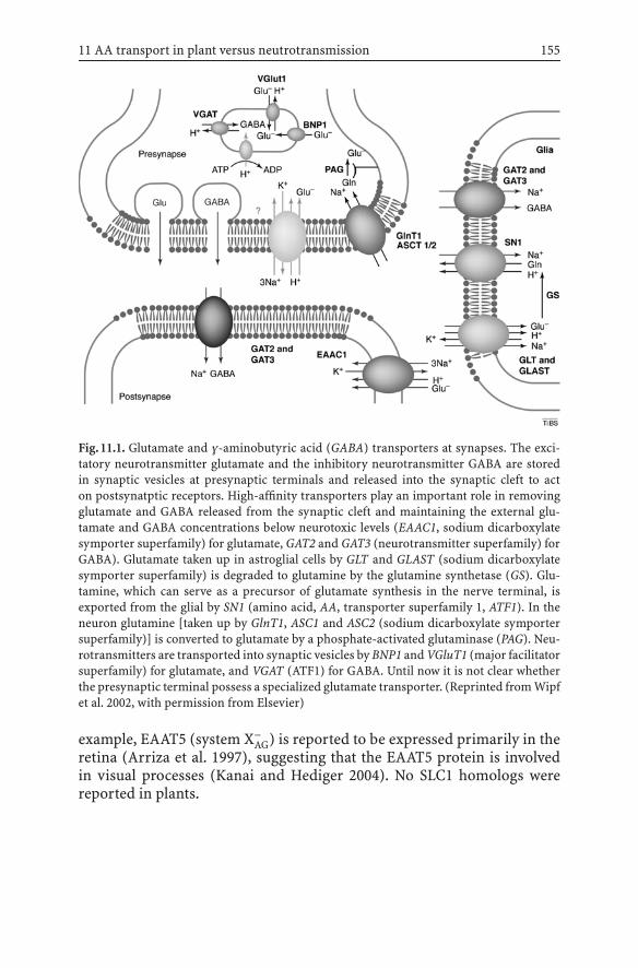
11 AA transport in plant versus neutrotransmission 155
Fig.11.1. Glutamate and γ-aminobutyric acid (GABA) transporters at synapses. The exci-tatory neurotransmitter glutamate and the inhibitory neurotransmitter GABA are storedin synaptic vesicles at presynaptic terminals and released into the synaptic cleft to acton postsynatptic receptors. High-affinity transporters play an important role in removingglutamate and GABA released from the synaptic cleft and maintaining the external glu-tamate and GABA concentrations below neurotoxic levels (EAAC1, sodium dicarboxylatesymporter superfamily) for glutamate, GAT2 and GAT3 (neurotransmitter superfamily) forGABA). Glutamate taken up in astroglial cells by GLT and GLAST (sodium dicarboxylatesymporter superfamily) is degraded to glutamine by the glutamine synthetase (GS). Glu-tamine, which can serve as a precursor of glutamate synthesis in the nerve terminal, isexported from the glial by SN1 (amino acid, AA, transporter superfamily 1, ATF1). In theneuron glutamine [taken up by GlnT1, ASC1 and ASC2 (sodium dicarboxylate symportersuperfamily)] is converted to glutamate by a phosphate-activated glutaminase (PAG). Neu-rotransmitters are transported into synaptic vesicles by BNP1 and VGluT1 (major facilitatorsuperfamily) for glutamate, and VGAT (ATF1) for GABA. Until now it is not clear whetherthe presynaptic terminal possess a specialized glutamate transporter. (Reprinted from Wipfet al. 2002, with permission from Elsevier)
example, EAAT5 (system X−AG) is reported to be expressed primarily in the
retina (Arriza et al. 1997), suggesting that the EAAT5 protein is involvedin visual processes (Kanai and Hediger 2004). No SLC1 homologs werereported in plants.
156 T. Müller, W. Koch, D. Wipf
11.2.2The Sodium- and Chloride-Dependent NeurotransmitterTransporter Family (NTF, SLC6)
Members of the neurotransmitter superfamily (SLC6) are key regulators ofextracellular neurotransmitter levels and are necessary for normal neu-rotransmission (Fig. 11.1) (Melikian 2004). SLC6 transporters (e.g., γ-aminobutyric acid, GABA, transporters GAT1-3, Fig. 11.1, dopamine trans-porter, DAT, etc.) regulate the level of extracellular solute concentrationsand are mainly found in the central and peripheral nervous system (Chenet al. 2004) where they are involved in the signalling pathway. They are,however, also found in many nonneural tissues with diverse physiologicalfunctions. For example, SLC6A6 (taurine transporter, TAUT) and SLC6A12(betaine transporter, BGT1) take part in osmoregulation in kidney (Chenet al. 2004). Genes of the SLC6 family encode proteins of approximately600 AA with a common structure of 12 transmembrane domains with pre-dicted intracellular N- and C-termini (Nelson 1998). The solute transportis coupled with the cotransport of Na+ and Cl−. The transport of neu-rotransmitter molecules against the concentration gradient uses energyproduced by the electrochemical Na+ gradient (Chen et al. 2004; Wipf etal. 2002). Na+ is absolutely necessary for transport activities in contrast toCl−, whose requirement for cotransport varies across members (Chen etal. 2004). Most of the SLC6 members are regulated by protein kinases, al-though some other mechanisms are assumed to exist (Blakely and Bauman2000). In organisms like yeasts, fungi and plants, that do not typically useNa+ gradients for metabolite uptake, no homologs to the SLC6 family couldbe found (Saier 1999).
11.2.3Cationic Amino Acid Transporters and HeteromericAmino Acid Transporters (SLC7)
The SLC7 family is divided into two subgroups: cationic AA transporters(CAT) (SLC7A1–4) and heteromeric AA transporters (HAT) (SLC7A5–11).Members of the CAT subgroup have 14 putative transmembrane domainsand are found in both animals and plants (Wipf et al. 2002). MammalianCATs mediate Na+-independent uptake of cationic AA (Closs et al. 1993).Transport properties of the CAT are those of system y+. The second sub-group (HAT) comprisesproteinswith12putative transmembranedomains.In contrast to what was observed for the CAT members, HAT transportersare quite diverse in terms of substrate selectivity, transporting neutral L-AA (large and small), negatively charged AA and cationic AA plus neutral

11 AA transport in plant versus neutrotransmission 157
AA (Verrey et al. 2004). Mammalian HATs are composed of a light subunit(LAT-1, y+LAT1, y+LAT-2, LAT-2, bo,+AT, Asc-1, xCT and two orphans) anda heavy subunit (“heavy chain, SLC3”) (Ganapathy et al. 2001, Palacin etal. 1998). The heavy subunit is required for the generation of active plasmamembrane AA transporters. This subunit is glycosylated, contains only oneputative transmembrane domain and plays a role in trafficking to targetmembranes, for example, in polarized epithelial cells (Palacin et al. 1998).Six of the known light chains associate with 4F2hc, the widespread heavysubunit, strictly basolateraly localized in epithelia (Verrey et al. 2004).A second heavy subunit, the brush border localized rBAT, was identified inmammals. So far, SLC3 homologs have not been found in plants.
11.2.4The Type I Phosphate Transporter Family (SLC17)
The transport of organic anions is the primary function of the SLC17family, which contains four type I phosphate transporters (SLC36A1–4),a lysosomal sialic acid transporter (SIALIN, SLC36A5) and, surprisingly,three vesicular glutamate transporters [VGLUT1 (or BNP1), VGLUT2–3,(SLC36A6–8)] occurring in synaptic vesicles (Fig. 11.1) (Reimer and Ed-wards 2004). All three VGLUT have been shown to have a Km for glu-tamate around 1 mM (Bai et al. 2001; Bellocchio et al. 2000; Fremeauet al. 2002). Homologs of SLC17 members have been identified in plantgenomes without being more closely characterized (Reimer and Edwards2004) (Sect. 11.3.2).
11.2.5The Vesicular Inhibitory Amino AcidTransporter Family (VIAAT, SLC32)
This family includes a single member, the vesicular inhibitory AA trans-porter (VIAAT) or vesicular GABA transporter (VGAT, Fig. 11.1) (Gasnier2004). VGAT is localized on synaptic vesicles (Fig. 11.1), where it mediatesthe uptake of GABA and glycine into synaptic vesicles and their exocytoticrelease (Gasnier 2004; McIntire et al. 1997).
11.2.6The Proton/Amino Acid Transporter Family (PAT, SLC36)
The SLC36 family comprises four members which encode around 500 AAproteins and have 11 predicted transmembrane domains (Boll et al. 2004).

158 T. Müller, W. Koch, D. Wipf
The first one (SLC36A1, LYAAT1, PAT1) was identified as a lysosomal AAtransporter, which mediates proton-coupled export of AA from lysosomes(Sagne et al. 2001). LYAAT1 was also localized to the apical membrane inintestine cells, where it mediates proton-driven uptake of small neutral AA(Thwaites et al. 1995). Like PAT1, SLC36A2 (PAT2) mediates proton symportuptake of small neutral AA. In addition, the SLC36 family comprises twoorphans, SLC36A3 and SLC36A4.
11.2.7The Sodium-Coupled Neutral Amino AcidTransporter Family (SNAT, SLC38)
In mammals transporters from the SLC38 family play various roles. For ex-ample, a role for SNAT2 in the provision of AA entering the gluconeogenicpathway could be suggested. SNAT3 may play a role in the response to aci-dosis in rats (Karinch et al. 2002) or could be involved in the detoxificationof ammonia from the portal blood in the liver (Mackenzie and Erickson2004). Recently Mackenzie and Erickson (2004) renamed the SLC38 mem-bers SNAT1–6. SNAT stands for sodium-coupled neutral AA transporterand also recalls the classic transport activities, i.e., system N/A trans-porter. SNAT1 (former ATA1, GlnT, NAT2, SA2, SAT1), SNAT2 (formerATA2, KIAA1382, SA1, SAT2) and SNAT4 (former ATA3, FLJ10191, NAT3,PAAT, SAT3) belong to the system A subfamily transporting aliphatic AAin correlation with an uptake of Na+ (Mackenzie and Erickson 2004).
System A members are pH-sensitive and underlie a sophisticated hor-monal and adaptive regulation (Häussinger and Kilberg 1992; McGivanand Pastor Anglada 1994; Shotwell et al. 1981). SNAT3 [former g17, NAT(mouse), SN1; Fig. 11.1 and SNAT5 (former JM24, SN2) correspond toa second kind of system, namely, system N. SNAT3 and SNAT5 are like sys-tem A Na+-dependent. Glutamine, histidine and asparagine belong to thesubstrates transported by system N, whereas system A is not that limited,transporting alanine, asparagine, cysteine, glutamine, glycine, methionineand serin (Mackenzie et al. 2004). Transport is also coupled with a H+
countertransport which produces a low extracellular pH that inhibits thesystem (Albers et al. 2001; Broer et al. 2002; Chaudhry et al. 1999, 2001, 2002;Nakanishi et al. 2001). As in system A, the regulation of system N is adaptive(Jacob et al. 1986; Kilberg et al. 1980). SNAT6 [former NAT-1 (human)], theorphan member of the SLC38 family has been isolated from a human braincomplementary DNA (cDNA) library (Mackenzie and Erickson 2004).
Note that SLC32, SLC36 and SLC38 members can be grouped in aneukaryotic specific superfamily, namely the ATF1 superfamily (Sect. 11.3.2)(Wipf et al. 2002; Fig. 11.3).

11 AA transport in plant versus neutrotransmission 159
It is important to mention that further AA transporters exist in animals.TAT1, a SLC16 (monocarboxylate transporter family) member specificallyis an aromatic AA transporter (Kim et al. 2001). Members of the mito-chondrial carrier family (SLC 25) potentially involved in AA transport arepresent in animals and plants (Wipf et al. 2002).
11.3Amino Acid Transport in Plants
For the distribution of AA in the plant several transport steps across mem-branes are necessary. AA have to cross the plasma membrane when takenup from the soil into root cells. In mycorrhized plants AA that pass thefungal layer or are produced within the fungus must be taken up from theplant root cells. AA synthesized in root tissue have to be exported to theshoot via xylem. Since mature xylem elements are dead cells, AA need tocross the plasma membrane to enter the xylem, potentially via facilitators,exchangers or antiporters. From the site of biosynthesis within the plant,mainly in the plastids of the leaf or root, AA have to pass several mem-branes to enter the long-distance traffic routes. Plastids are surrounded bytwo membranes, an outer and an inner envelope. AA cross the outer enve-lope of plastids via outer envelope proteins 16 and 14, which form channelspermeable to amines and AA (Pohlmeyer et al. 1997, 1998). For the innerenvelope, no AA transporters have been described so far. To leave the cyto-plasm of the cell, AA have to pass the plasma membrane. The concentrationof the AA in mesophyll cytoplasm and in the phloem is significantly highercompared with that in the apoplasm, suggesting an active transport of AAinto the phloem (Lohaus et al. 1995). In feeding experiments it was shownthat large parts of AA fed directly to the xylem sap appear unchanged inthe phloem sap. This indicates that AA can be exchanged between xylemand phloem (Atkins 2000; Pate and Sharkey 1975) and can cycle within theplant passing the membranes of multiple cells via transporters.
11.3.1Amino Acid–Polyamine–Choline Transporter Family
Uptake of AA is well characterized in yeasts (Fischer et al. 1998), where24 transporters of the AA–polyamine–choline (APC) family (pfam00324)have been functionally characterized. The members of the APC familycluster in subgroups reflecting the three kingdoms (yeast, plant and an-imal) (Fig. 11.2) (Wipf et al. 2002). Mammalian CATs are representativesof system y+, mediating Na+-independent uptake of cationic AA (Closs
160 T. Müller, W. Koch, D. Wipf
et al. 1993), and are characterized by a high affinity for cationic AA. Theplant membranes maintain a proton gradient over the plasma membranevia plasma membrane ATPases, and, correspondingly, the first plant mem-ber of the APC family, AtCAT1, mediated the accumulation of histidinein a pH-dependent manner (Frommer et al. 1995). However, it remainsunclear whether AtCAT1 functions as a uniporter or an exchanger, or asa secondary active proton cotransporter. Recent studies of a related pro-tein, AtCAT5, showed that the transport of basic AA mediated by AtCAT5 issensitive to protonophores, indicating that at least AtCAT5 is a proton sym-porter. AtCAT5 is a high-affinity transporter for arginin with an apparentKm value of 12 µM (Su et al. 2004). Other basic AA were good competitorsfor 14C arginine uptake into yeast cells expressing AtCAT5, while neutral oracidic AA like aspartate, proline, glutamate and glutamine were less effec-tive in uptake inhibition. The transport selectivity of AtCAT5 is similar tothat of AtCAT1, which also transports basic AA preferentially.
For other members of the CAT-family in Arabidopsis no direct transportactivity was demonstrated, nevertheless AtCAT3 and AtCAT6 expressedin yeast cells increased toxicity to the toxic analogue of glutamine, (S)-2amino-6-diazo-5-oxo-L-norleucine, but not to the arginine analog canava-nine (Su et al. 2004). The fact that arginine is not a substrate for AtCAT3and AtCAT6 already shows that not all members of the CAT family inArabidopsis can be supposed to be cationic AA transporters.
In animals, the APC-type AA transporters require an additional subunitfor functional expression at the plasma membrane (Sect. 11.2.3). Membersof the mammalian CAT family essentially transport cationic AA by facil-itated diffusion, while the HATs are in contrast quite diverse in terms ofsubstrate selectivity and function as obligatory exchangers. In plants no ho-mologs of secondary subunits have yet been identified, and one subbranchof the APC family, the LAT (Fig. 11.2), have still not been functionallycharacterized in plants.
11.3.2Amino Acid Transporter Family 1
Members of the ATF1 family were first described in plants as functionalAA transporters. Structurally related proteins were identified in yeastsand animals (Fischer et al. 1998). The superfamily contains plant-specificsubbranches, and branches that are more structurally related to mam-malian transporters (Fig. 11.3). Within the plant-specific subbranches,the best-characterized members are the Arabidopsis AA permeases (AAP;Figs. 11.3, 11.4). Several AAP have been isolated from Arabidopsis by com-plementation of yeast transport mutants with plant cDNAs (Fischer et al.
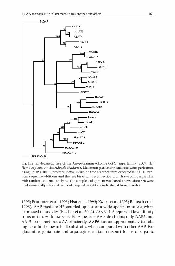
11 AA transport in plant versus neutrotransmission 161
Fig.11.2. Phylogenetic tree of the AA–polyamine–choline (APC) superfamily (SLC7) (HsHomo sapiens, At Arabidopsis thaliana). Maximum parsimony analyses were performedusing PAUP 4.0b10 (Swofford 1998). Heuristic tree searches were executed using 100 ran-dom sequence additions and the tree bisection–reconnection branch-swapping algorithmwith random sequence analysis. The complete alignment was based on 691 sites; 586 werephylogenetically informative. Bootstrap values (%) are indicated at branch nodes
1995; Frommer et al. 1993; Hsu et al. 1993; Kwart et al. 1993; Rentsch et al.1996). AAP mediate H+-coupled uptake of a wide spectrum of AA whenexpressed in oocytes (Fischer et al. 2002). AtAAP1-5 represent low-affinitytransporters with low selectivity towards AA side chains; only AAP3 andAAP5 transport basic AA efficiently. AAP6 has an approximately tenfoldhigher affinity towards all substrates when compared with other AAP. Forglutamine, glutamate and asparagine, major transport forms of organic

162 T. Müller, W. Koch, D. Wipf
Fig.11.3. Phylogenetic tree of the ATF1 superfamily. Maximum parsimony analyses wereperformed using PAUP 4.0b10 (Swofford 1998). Heuristic tree searches were executed us-ing 100 random sequence additions and the tree bisection-reconnection branch-swappingalgorithm with random sequence analysis. The complete alignment was based on 529 sites;498 were phylogenetically informative. Bootstrap values (%) are indicated at branch nodes
nitrogen, these transporters have Km values that are in a physiologicalrange with respect to the concentrations of these AA found in the phloemand xylem. Aspartate is transported by most AAP with an extremely lowaffinity and efficiency. Only AAP6 has a Km value for aspartate that is ina physiological range (Fischer et al. 2002).
Plants take up nitrate and ammonium but also AA as a nitrogen sourceand, more importantly, AA are the principal long-distance transport formsof organic nitrogen. In accordance with these facts, reporter gene analyses

11 AA transport in plant versus neutrotransmission 163
Fig.11.4. AA transporters in plants. Although a wide spectrum of proteogenic AA accumu-late in phloem (100–200 mM) and xylem sap (about tenfold lower concentration than inphloem), amines and AA predominate. The actual intracellular, apoplasmic, and phloemcomposition of AA is thought to be similar, thus the relatively unspecific AA permeases(AAP) might be responsible for functions like uptake of AA from the xylem into the cell andphloem loading. No organellar transporter has been identified to date. (Reprinted Wipf etal. 2002, with permission from Elsevier)
showed that the members of the AAP family are associated with the vas-cular tissue or the reproductive organs (Hirner et al. 1998; Okumoto etal. 2002, 2004). The properties of AAP described thus match the activitiesexpected for transport proteins involved in moving a wide spectrum ofAA into the phloem sap and are consistent with the properties describedfor uptake kinetics measured in plasma membrane vesicles of leaves (Liand Bush 1990a, b). Antisense repression of a related AA transporter frompotato, StAAP1, expressed in source leaves led to reduced concentration of

164 T. Müller, W. Koch, D. Wipf
AA in potato tubers. This reduced transport to sink organs supports the hy-pothesis of a role in phloem loading for some of the AAP (Koch et al. 2003).
Besides long-distance transport the delivery of reduced nitrogen to thereproductive organs is a key factor for seed development. The import of AAis an essential prerequisite for seed development since the accumulation ofstorage proteins must be preceded by AA import. It could be shown that theexpression of the AA transporter in seeds indeed precedes the expressionof the storage protein AtS1 in Arabidopsis (Hirner et al. 1998). A similar in-crease in the expressionof a legumeAAtransporterhasbeen shown inViciafaba (Miranda et al. 2001). In pea, it has been demonstrated that PsAAP1 isexpressed in the transfer cell layer of cotyledons and might be responsiblefor taking up AA released from the seed coat (Tegeder et al. 2000). Ectopicoverexpression of an AAP in pea seeds increases AA uptake and increasesstorage protein content by 40–50%. The results suggest that nitrogen supplylimits protein synthesis and storage in the seed (Rolletschek et al. 2005).
From the second plant-specific branch of the ATF family, the LHT(Fig. 11.3), two members have been functionally characterized so far.AtLHT1 has been described as a transporter specific for lysine and his-tidine, but other AA are also recognized (Chen and Bush 1997). LHT1is present in all tissues, with expression being strongest in young leaves,flowers and siliques. Recent characterization of a related protein, LHT2,shows that this transporter is more likely to be a general AAP with a highaffinity for proline (Km ∼ 10 µM) and aspartate (Km ∼ 72 µM). Inhibitionstudies indicate potentially similar affinities towards most AA except thebasic AA histidine, lysine and arginine (Lee and Tegeder 2004). Expressionof the gene in the tapetum tissue of the flower suggests a role in the transferof organic nitrogen to the tapetum tissue and in supplying the pollen withnutrients. The affinity of LHT2 towards proline is a factor of 10 higherthan the affinity of AAP6 and is, together with the high affinity of CAT5for arginine, among the highest determined for AA transporters in plants.AA concentrations in phloem, xylem, and supposedly in mesophyll cells,are highly variable depending on carbon supply, inorganic nitrogen andlight. Thus, it makes sense that the relatively immobile plants can respondto variations in AA concentrations with transporters that have different Km
values and transportation speed.ANT1, the only member characterized of a more distantly related branch
(Fig. 11.3) to the AAP and LHT mediates the transport of aromatic andneutral AA (Chen et al. 2001). The substrate specificity found in the thirdplant-specific branch of the ATF-family, the proline transporters (ProT;Fig. 11.3) is different. In contrast to the plant carriers just described, ProTpreferentially transport the compatible solutes proline (Km values of 0.427,0.500 and 1 mM for AtProT1–3, respectively), betaine (Km values of 0.115,0.267 and 0.290 mM for AtProT1–3) and GABA (4.5, 4.01 and 5.12 mM)

11 AA transport in plant versus neutrotransmission 165
(Grallath et al. 2005; Rentsch et al. 1996). AtProT1 is expressed in thephloem and phloem parenchyma cells, whereas AtProT2 expression isrestricted to the epidermis and cortex cells of roots. AtProT3 finally is onlyexpressed in the aerial parts of the plant, in the epidermal cells of the leaves.Induction of the expression of the AtProT upon salt stress implies a rolein stress adaptation or a function in organs of plants that desiccate likepollen and seeds (Rentsch and Frommer 1996; Schwacke et al. 1999). Inmangroves, where salt concentrations are permanently high, the transportof compatible solutes is thought to be an important factor for adaptationto the environment (Waditee et al. 2002).
In plants nothing is known about the compartimentation of AA or thetransport steps into the vacuole. Some plant ATF1 members might fulfila function in import or export of solutes in vesicles and vacuoles. A similarmechanism like the concentration of GABA in synaptic vesicles via VGAT(Fig. 11.1) is possible for the related uncharacterized plant proteins in theATF family.
The plant members of the ATF family investigated so far all function asH+-coupled systems, probably playing a role in accumulating AA withinthe plant cell. Subcellular localization studies could help to identify thetransporters involved in the compartimentation of AA into the vacuole.
11.4Conclusions and Future Prospects
Taken together, plant AA transporters can be grouped in three superfam-ilies with homologs from the animal kingdom (Lalonde et al. 2004; Wipfet al. 2002): (1) the ATF1 superfamily (SLC32, SLC36 and SLC38 and Ara-bidopsis ATF1 members) (Fig. 11.3); (2) the APC superfamily (SLC7, AtCATsand AtLATs) (Fig. 11.2); (3) AA transporters within the major facilitatorsuperfamily (MFS) (SLC17, homologs of unknown function in Arabidopsis(Wipf et al. 2002).
As described, several export steps are required for AA distribution inplants (Fig. 11.3): phloem-to-xylem transfer, release of AA into the apoplastof the leaf mesophyll as the first step for phloem loading, unloading in sinkorgans, supply of symplasmic isolated cells, e.g., growing embryo, guardcells and pollen. Multiple possibilities exist, including vesicular transportor carrier-mediated transport, but until now no exporter has been iden-tified in plants. Vesicular export is well established for AA and analogs inmammalian nerve cells. Carriers for the active uptake of AA into secretoryvesicles have been identified (Fig. 11.1) and homologous plant genes exist(Fig. 11.3). Furthermore, certain mammalian AA transporters are impli-cated in cellular export at the plasma membrane, especially from intestinal

166 T. Müller, W. Koch, D. Wipf
cells, and it has been speculated that the corresponding plant homologsmight fulfil similar functions (Wipf et al. 2002). Quite recently a mecha-nismsimilar to thevesicular secretionofneurotransmittershasbeen shownfor yeasts. A membrane protein localized to internal membranes is respon-sible for secretion of AA to the surrounding medium in yeasts (Velascoet al. 2004). The recent analysis of an Arabidopsis mutant hypersecretingglutamine from hydathodes led to the identification of a new class of mem-brane proteins possibly involved in this process (Pilot et al. 2004). Theseproteins, which contain only a single transmembrane domain, might helpto identify the mechanism for AA export from plant cells. However, thehigh number of proteins putatively involved in AA transport in plants (upto 53; Wipf et al. 2002) as well as in animals, and the different mechanismsprevented significant progress. To better understand the mechanisms un-derlying the loading and unloading of AA from phloem and xylem andthe exchange between the two systems, it will be essential to analyse eachmember individually.
Acknowledgements. We thank Felicity de Courcy for critical reading ofthe manuscript. Our work has been funded by grants from the DeutscheForschungsgemeinsschaft.
References
Albers A, Broer A, Wagner CA, Setiawan I, Lang PA, Kranz EU, Lang F, Broer S (2001)Na+ transport by the neural glutamine transporter ATA1. Pflugers Arch Eur J Physiol443:92–101
Arriza JL, Eliasof S, Kavanaugh MP, Amara SG (1997) Excitatory amino acid transporter 5,a retinal glutamate transporter coupled to a chloride conductance. Proc Natl Acad SciUSA 94:4155–4160
Atkins CA (2000) Biochemical aspects of assimilate transfers along the phloem path: N-solutes in lupins. Aust J Plant Physiol 27:531–537
Bai L, Xu H, Collins JF, Ghishan FK (2001) Molecular and functional analysis of a novelneuronal vesicular glutamate transporter. J Biol Chem 276:36764–36769
Bellocchio EE, Reimer RJ, Fremeau RTJ, Edwards RH (2000) Uptake of glutamate intosynaptic vesicles by an inorganic phosphate transporter. Science 289:957–960
Blakely RD, Bauman AL (2000) Biogenic amine transporters: regulation in flux. Curr OpinNeurobiol 10:328–336
Boll M, Daniel H, Gasnier B (2004) The SLC36 family: proton-coupled transporters forthe absorption of selected amino acids from extracellular and intracellular proteolysis.Pflugers Arch Eur J Physiol 447:776–779
Broer A, Albers A, Setiawan I, Edwards RH, Chaudhry FA, Lang F, Wagner CA, Broer S(2002) Regulation of the glutamine transporter SN1 by extracellular pH and intracellularsodium ions. J Physiol 539:3–14
Broer A, Wagner C, Lang F, Broer S (2000) Neutral amino acid transporter ASCT2 displayssubstrate-induced Na+ exchange and a substrate-gated anion conductance. Biochem J346:705–710

11 AA transport in plant versus neutrotransmission 167
Chaudhry FA, Krizaj D, Larsson P, Reimer RJ, Wreden C, Storm-Mathisen J, Copenhagen D,Kavanaugh M, Edwards RH (2001) Coupled and uncoupled proton movement by aminoacid transport system N. EMBO J 20:7041–7051
Chaudhry FA, Reimer RJ, Krizaj D, Barber D, Storm-Mathisen J, Copenhagen DR, EdwardsRH (1999) Molecular analysis of system N suggests novel physiological roles in nitrogenmetabolism and synaptic transmission. Cell 99:769–780
ChaudhryFA, SchmitzD,ReimerRJ, LarssonP,GrayAT,NicollR,KavanaughM,EdwardsRH(2002) Glutamine uptake by neurons: interaction of protons with system A transporters.J Neurosci 22:62–72
Chen L, Bush DR (1997) LHT1, a lysine- and histidine-specific amino acid transporter inArabidopsis. Plant Physiol 115:1127–1134
Chen L, Ortiz-Lopez A, Jung A, Bush DR (2001) ANT1, an aromatic and neutral amino acidtransporter in Arabidopsis. Plant Physiol 125:1813–1820
Chen NH, Reith ME, Quick MW (2004) Synaptic uptake and beyond: the sodium- andchloride-dependent neurotransmitter transporter family SLC6. Pflugers Arch Eur JPhysiol 447:519–531
Closs EI, Lyons CR, Kelly C, Cunningham JM (1993) Characterization of the third memberof the MCAT family of cationic amino acid transporters. Identification of a domainthat determines the transport properties of the MCAT proteins. J Biol Chem 268:20796–20800
Fischer WN, André B, Rentsch D, Krolkiewicz S, Tegeder M, Breitkreuz K, Frommer WB(1998) Amino acid transport in plants. Trends Plant Sci 3:188–195
Fischer WN, Kwart M, Hummel S, Frommer WB (1995) Substrate specificity and expressionprofile of amino acid transporters (AAPs) in Arabidopsis. J Biol Chem 270:16315–16320
Fischer WN, Loo DDF, Koch W, Ludewig U, Boorer KJ, Tegeder M, Rentsch D, Wright EM,Frommer WB (2002) Low and high affinity amino acid H+-cotransporters for cellularimport of neutral and charged amino acids. Plant J 29:717–731
Fremeau RT, Jr., Burman J, Qureshi T, Tran CH, Proctor J, Johnson J, Zhang H, Sulzer D,Copenhagen DR, Storm-Mathisen J, Reimer RJ, Chaudhry FA, Edwards RH (2002) Theidentification of vesicular glutamate transporter 3 suggests novel modes of signallingby glutamate. Proc Natl Acad Sci USA 99:14488–14493
Frommer WB, Hummel S, Riesmeier JW (1993) Expression cloning in yeast of a cDNAencoding a broad specificity amino acid permease from Arabidopsis thaliana. Proc NatlAcad Sci USA 90:5944–5948
Frommer WB, Hummel S, Unseld M, Ninnemann O (1995) Seed and vascular expression ofa high affinity transporter for cationic amino acids in Arabidopsis. Proc Natl Acad SciUSA 92:12036–12040
Ganapathy V, Ganapathy ME, Leibach FH (2001) Intestinal transport of peptides and aminoacids. In: Barrett KE, Donowitz M (eds) Current topics in membranes, vol 50. Academic,San Diego, CA, USA, pp 379–412
Gasnier B (2004) The SLC32 transporter, a key protein for the synaptic release of inhibitoryamino acids. Pflugers Arch Eur J Physiol 447:756–759
Grallath S, Weimar T, Meyer A, Gumy C, Suter-Grotemeyer M, Neuhaus JM, Rentsch D (2005)The AtProT family. Compatible solute transporters with similar substrate specificity butdifferential expression patterns. Plant Physiol 137:117–126.
Häussinger D, Kilberg MS (1992) Amino Acid transport in the liver. In: Häussinger D,Kilberg MS (eds) Mammalian amino acid transport: mechanisms and control. Plenum,New York, pp 133–148
Hirner B, Fischer WN, Rentsch D, Kwart M, Frommer WB (1998) Developmental controlof H+/amino acid permease gene expression during seed development of Arabidopsis.Plant J 14:535–544

168 T. Müller, W. Koch, D. Wipf
Hsu LC, Chiou TJ, Chen L, Bush DR (1993) Cloning a plant amino acid transporter byfunctional complementation of a yeast amino acid transport mutant. Proc Natl Acad SciUSA 90:7441–7445
Jacob R, Rosenthal N, Barrett EJ (1986) Characterization of glutamine transport by liverplasma membrane vesicles. Am J Physiol 251:E509-E514
Kanai Y, Hediger MA (2004) The glutamate/neutral amino acid transporter family SLC1:molecular, physiological and pharmacological aspects. Pflugers Arch Eur J Physiol447:469–479
Karinch AM, Lin CM, Wolfgang CL, Pan M, Souba WW (2002) Regulation of expression ofthe SN1 transporter during renal adaptation to chronic metabolic acidosis in rats. AmJ Physiol Renal Physiol 283:F1011-F1019
Kilberg MS, Handlogten ME, Christensen HN (1980) Characteristics of an amino acidtransport system in rat liver for glutamine, asparagine, histidine, and closely relatedanalogs. J Biol Chem 255:4011–4019
Kim DK, Kanai Y, Chairoungdua A, Matsuo H, Cha SH, Endou H (2001) Expres-sion cloning of a Na+-independent aromatic amino acid transporter with struc-tural similarity to H+/monocarboxylate transporters. J Biol Chem 276:17221–17228
Koch W, Kwart M, Laubner M, Heineke D, Stransky H, Frommer WB, Tegeder M (2003)Reduced amino acid content in transgenic potato tubers due to antisense inhibition ofthe leaf H+/amino acid symporter StAAP1. Plant J 33:211–220
Kwart M, Hirner B, Hummel S, Frommer WB (1993) Differential expression of two relatedamino acid transporters with differing substrate specificity in Arabidopsis thaliana.Plant J 4:993–1002
Lalonde S, Wipf D, Frommer WB (2004) Transport mechanisms for organic forms of carbonand nitrogen between source and sink. Annu Rev Plant Biol 55:341–372
Lee YH, Tegeder M (2004) Selective expression of a novel high-affinity transport systemfor acidic and neutral amino acids in the tapetum cells of Arabidopsis flowers. Plant J40:60-74
Li ZC, Bush DR (1990a) ∆pH-dependent amino acid transport into plasma membranevesicles isolated from sugar beet leaves. I. Evidence for carrier-mediated, electrogenicflux through multiple transport systems. Plant Physiol 94:268–277
Li ZC, Bush DR (1990b) ∆pH-dependent amino acid transport into plasma membranevesicles isolated from sugar beet leaves. II. Evidence for multiple aliphatic, neutralamino acid symports. Plant Physiol 96:1338–1344
Lohaus G, Winter H, Riens B, Heldt HW (1995) Further studies of the phloem loadingprocess in leaves of barley and spinach. The comparison of metabolite concentrations inthe apoplastic compartment with those in the cytosolic compartment and in the sievetubes. Bot Acta 108:270–275
Lynch AM, McGivan JD (1987) Evidence for a single common Na+-dependent transportsystem for alanine, glutamine, leucine and phenylalanine in brush-border membranevesicles from bovine kidney. Biochim Biophys Acta 899:176–184
Mackenzie B, Erickson JD (2004) Sodium-coupled neutral amino acid (System N/A) trans-porters of the SLC38 gene family. Pflugers Arch Eur J Physiol 447:784–795
McGivan JD, Pastor Anglada M (1994) Regulatory and molecular aspects of mammalianamino acid transport. Biochem J 299:321–334
McIntire SL, Reimer RJ, Schuske K, Edwards RH, Jorgensen EM (1997) Identification andcharacterization of the vesicular GABA transporter. Nature 389:870–876
Melikian HE (2004) Neurotransmitter transporter trafficking: endocytosis, recycling, andregulation. Pharmacol Therapeut 104:17–27

11 AA transport in plant versus neutrotransmission 169
Miranda M, Borisjuk L, Tewes A, Heim U, Sauer N, Wobus U, Weber H (2001) Amino acidpermeases in developing seeds of Vicia faba L.: expression precedes storage proteinsynthesis and is regulated by amino acid supply. Plant J 28:61–71
Nakanishi T, Kekuda R, Fei YJ, Hatanaka T, Sugawara M, Martindale RG, Leibach FH, PrasadPD, Ganapathy V (2001) Cloning and functional characterization of a new subtype ofthe amino acid transport system N. Am J Physiol Cell Physiol 281:C1757–C1768
Nelson N (1998) The family of Na+/Cl− neurotransmitter transporters. J Neurochem71:1785–1803
Okumoto S, Koch W, Tegeder M, Fischer WN, Biehl A, Leister D, Stierhof YD, Frommer WB(2004) Root phloem-specific expression of the plasma membrane amino acid protonco-transporter AAP3. J Exp Bot 55:2155–2168
Okumoto S, Schmidt R, Tegeder M, Fischer WN, Rentsch D, Frommer WB, Koch W (2002)High affinity amino acid transporters specifically expressed in xylem parenchyma anddeveloping seeds of Arabidopsis. J Biol Chem 277:45338–45346
Palacin M, Estevez R, Zorzano A (1998) Cystinuria calls for heteromultimeric amino acidtransporters. Curr Opin Cell Biol 10:455–461
Pate JS, Sharkey PJ (1975) Xylem to phloem transfer of solutes in fruiting shoots of legumes,studied by a phloem bleeding techniques. Planta 12:11–26
Pilot G, Stransky H, Bushey DF, Pratelli R, Ludewig U, Wingate VP, Frommer WB (2004)Overexpression of GLUTAMINE DUMPER1 leads to hypersecretion of glutamine fromhydathodes of Arabidopsis leaves. Plant Cell 16:1827–1840
Pohlmeyer K, Soll J, Grimm R, Hill K, Wagner R (1998) A high-conductance solute channelin the chloroplastic outer envelope from Pea. Plant Cell 10:1207–1216
Pohlmeyer K, Soll J, Steinkamp T, Hinnah S, Wagner R (1997) Isolation and characterizationof an amino acid-selective channel protein present in the chloroplastic outer envelopemembrane. Proc Natl Acad Sci USA 94:9504–9509
Reimer RJ, Edwards RH (2004) Organic anion transport is the primary function of theSLC17/type I phosphate transporter family. Pflugers Arch Eur J Physiol 447:629–635
Rentsch D, Frommer WB (1996) Molecular approaches towards an understanding of loadingand unloading of assimilates in higher plants. J Exp Bot 47:1199–1204
Rentsch D, Hirner B, Schmelzer E, Frommer WB (1996) Salt stress-induced proline trans-porters and salt stress-repressed broad specificity amino acid permeases identifiedby suppression of a yeast amino acid permease-targeting mutant. Plant Cell 8:1437–1446
Rolletschek H, Hosein F, Miranda M, Heim U, Gotz KP, Schlereth A, Borisjuk L, Saalbach I,Wobus U, Weber H (2005) Ectopic expression of an amino acid transporter (VfAAP1) inseeds of Vicia narbonensis and Pea increases storage proteins. Plant Physiol 137:1236–1249
Sagne C, Agulhon C, Ravassard P, Darmon M, Hamon M, El Mestikawy S, Gasnier B, Giros B(2001) Identification and characterization of a lysosomal transporter for small neutralamino acids. Proc Natl Acad Sci USA 98:7206–7211
Saier MH Jr (1999) A functional-phylogenetic system for the classification of transportproteins. J Cell Biochem (Suppl 32–33):84–94
Schwacke R, Grallath S, Breitkreuz KE, Stransky E, Stransky H, Frommer WB, Rentsch D(1999) LeProT1, a transporter for proline, glycine betaine, and γ-amino butyric acid intomato pollen. Plant Cell 11:377–392
Shotwell MA, Jayme DW, Kilberg MS, Oxender DL (1981) Neutral amino acid transportsystems in Chinese hamster ovary cells. J Biol Chem 256:5422–5427
Su YH, Frommer WB, Ludewig U (2004) Molecular and functional characterization ofa family of amino acid transporters from Arabidopsis. Plant Physiol 136:3104–3113

170 T. Müller, W. Koch, D. Wipf
Swofford DL (1998) PAUP*. Phylogenetic analysis using parsimony (*and other methods),version 4. Sinauer, Sunderland, MA, USA
TegederM,OfflerCE,FrommerWB,Patrick JW(2000)Aminoacid transporters are localizedto transfer cells of developing Pea seeds. Plant Physiol 122:319–325
Thwaites DT, McEwan GTA, Simmons NL (1995) The role of the proton electrochemicalgradient in the transepithelial absorption of amino acids by human intestinal Caco-2cell monolayers. J Membr Biol 145:245–256
Vadgama JV, Christensen HN(1984) Wide distribution of pH-dependent service of transportsystem ASC for both anionic and zwitterionic amino acids. J Biol Chem 259:3648–3652
Velasco I, Tenreiro S, Calderon IL, Andre B (2004) Saccharomyces cerevisiae Aqr1 is aninternal-membrane transporter involved in excretion of amino acids. Eukaryot Cell3:1492–1503
Verrey F, Closs EI, Wagner CA, Palacin M, Endou H, Kanai Y (2004) CATs and HATs: theSLC7 family of amino acid transporters. Pflugers Arch Eur J Physiol 447:532–542
Waditee R, Hibino T, Tanaka Y, Nakamura T, Incharoensakdi A, Hayakawa S, Suzuki S, Fut-suhara Y, Kawamitsu Y, Takabe T (2002) Functional characterization of betaine/prolinetransporters in betaine-accumulating mangrove. J Biol Chem 277:18373–18382
Wipf D, Ludewig U, Tegeder M, Rentsch D, Koch W, Frommer WB (2002) Conservation ofamino acid transporters in fungi, plants and animals. Trends Biochem Sci 27:139–147
Zerangue N, Kavanaugh MP (1996) Flux coupling in a neuronal glutamate transporter.Nature 383:634–637

12 GABA and GHB Neurotransmittersin Plants and AnimalsAaron Fait, Ayelet Yellin, Hillel Fromm
Abstract γ-Aminobutyric acid (GABA) is a four-carbon non-protein amino acid conservedfrom bacteria to plants and vertebrates. In the latter it is mainly known as a neurotrans-mitter. The enzymes that synthesize and catabolize GABA constitute a metabolic pathwayknown as the GABA shunt which bypasses two steps of the tricarboxylic acid cycle. Func-tional genomics tools using Arabidopsis as a model system revealed that the GABA shuntis imperative for normal plant development and for response to stress, and suggest rolesfor GABA as an important metabolite as well as a potential signaling molecule. Moreover,γ-hydroxybutyrate, a by-product of the GABA shunt and a neurotransmitter in animals, wasrecently discovered in plants. Here we discuss the possible roles of these two neurotrans-mitters in plants with focus on components that underlie their roles as signaling molecules.
12.1Introduction
γ-Aminobutyric acid (GABA) was discovered in plants over half a centuryago (Steward et al. 1949), and shortly after it was discovered in vertebrates.Interest in GABA shifted to animals when it was revealed that GABA occursat high levels in the brain, where it plays a major role in neurotransmission.Interest in the GABA shunt in plants emerged later, following observationsthat GABA is largely and rapidly produced in response to biotic and abi-otic stresses (reviewed by Shelp et al. 1999; Snedden and Fromm 1999;Kinnersley and Turano 2000). A breakthrough in studying GABA in plantscame from molecular studies, particularly following the cloning of theCa2+/calmodulin-regulated GABA-synthesizing enzyme glutamate decar-boxylase (GAD; Baum et al. 1993), and later by employing transgenic plants(Baum et al. 1996) and functional genomics tools in Arabidopsis to inves-tigate mutants of the GABA metabolic pathway (Bouché et al. 2003, 2004;Palanivelu et al. 2003; Fait et al. 2005).
GABA is mainly metabolized via a short pathway known as the GABAshunt, which bypasses two steps of the tricarboxylic acid (TCA) cycle(Fig. 12.1), and is composed of three enzymes: the cytosolic-localizedGAD, and the mitochondrial-localized GABA trans-aminase (GABA-T),and succinic semialdehyde dehydrogenase (SSADH). Owing to the fact thatmost of the knowledge about GABA as a signaling molecule comes frommammalian studies, we will briefly review the subject of GABA signalingin mammals as a basis for discussing aspects of neurotransmitter signaling
Communication in PlantsF. Baluška, S. Mancuso, D. Volkmann (Eds.)© Springer-Verlag Berlin Heidelberg 2006

172 A. Fait, A. Yellin, H. Fromm
Fig.12.1. Overview of the γ-aminobutyric acid (GABA) shunt and relevant metabolicbranches in plants and other organisms. GABA is metabolized via a short pathway knownas the GABA shunt, which bypasses two steps of the tricarboxylic acid cycle (TCA), andis composed of three enzymes: the cytosolic-localized Ca2+-calmodulin (CaM)-regulatedglutamate decarboxylase (GAD), and the mitochondrial-localized GABA trans-aminase(GABA-T) and succinic semialdehyde dehydrogenase (SSADH). The SSADH substratesuccinic semialdehyde (SSA) is also catabolized to γ-hydroxybutyrate (GHB) by GHB-dehydrogenase (GHBDH), which is cytosolic in mammals. GHB catabolism occurs in mam-mals either in the reverse direction by GHBDH/semialdehyde reductase (see text) or bythe mitochondrial-localized L-glucuronate reductase (GR; EC 1.1.1.19). In plants, GHBcatabolism has not been characterized. Chemical structures are given for GABA-shuntsubstrates, intermediates, and products. Unknown functions and transport systems aredepicted as dotted arrows and question marks. Glu glutamate, α-KG α-ketoglutarate, GABA-TK/P α-ketoglutaric acid/pyruvate-dependent GABA-T
in plants. We also address the occurrence, in plants, of γ-hydroxybutyrate(GHB), a by-product of the GABA shunt (Fig. 12.1) and a neurotransmitter,which was only recently discovered in plants (Allan et al. 2003; Breitkreuz etal. 2003; Fait et al. 2005). For further details on the different roles of GABAin plants the readers are referred to two recent reviews (Bouché et al. 2003;Bouché and Fromm 2004) and to earlier reviews by Shelp et al. (1999) andKinnersley and Turano (2000).

12 GABA and GHB Neurotransmitters in Plants and Animals 173
12.2The GABA Shunt and GABA Signaling
12.2.1Mammalian GABA Signaling
In the central nervous system of mammals GABA is the major inhibitoryneurotransmitter. GABA mediates inhibitory synaptic transmission bybinding to specialized receptors localized in presynaptic or postsynapticmembranes. Two types of receptors exist in brain cells: ionotropic recep-tors (GABAA and GABAC receptors), which are ligand-gated ion channels,and metabotropic receptors (GABAB receptors), which are coupled to G-proteins. GABA receptors are also expressed in nonexcitable cells in a va-riety of human tissues, such as heart, liver, lung, ovary, and testis (Calveret al. 2000). These findings imply that GABA could be a signaling moleculenot only in the brain but also in other organs. GABA receptors are alsofound in lower organisms such as Caenorhabditis elegans (Richmond andJorgensen 1999). Thus, GABA receptors seem to be widely distributed indiverse invertebrate and vertebrate organisms.
In addition to its neurotransmitter function in mature neurons, GABA isinvolved in the development of the nervous system by promoting neuronalmigration, proliferation, anddifferentiation (reviewed inOwens andKrieg-stein 2002). These effects are mediated by the activation of GABA receptors,which provoke depolarization of the membrane in the immature brain,where, in contrast to the adult brain, GABA is excitatory. Indeed, GABAis a chemoattractant that can influence neuronal growth in vitro. Duringcortical development, GABA can promote DNA synthesis and cell prolif-eration. Neurons become assembled into functional networks by growingaxons and dendrites, collectively called neurites. GABA regulates neuronaldifferentiation by promoting outgrowth of neurites. In conclusion, in themammalian brain GABA plays a major role in neural transmission and de-velopment, and it functions through interactions with specialized receptorsand transporters.
12.2.2GABA Signaling in Plants
In plants little is known about GABA functions despite the fact it was dis-covered over half a century ago (Steward et al. 1949). It has been shown thatGABA accumulates rapidly in plant tissues exposed to a variety of stressesincluding acidosis, cold, anoxia, heat, salt, and draught (reviewed by Sned-den and Fromm 1999; Kinnersley and Turano 2000). Nevertheless, recent
174 A. Fait, A. Yellin, H. Fromm
studies of Arabidopsis mutants revealed that disruption of the GABA shuntby functional knockout of the SSADH gene causes the accumulation ofhigh levels of reactive oxygen intermediates, necrosis, growth retardation,and hypersensitivity to environmental stresses, including UV radiationand heat (Bouché et al. 2003; Fait et al. 2005). However, from these studiesalone it is not clear if GABA functions only as a metabolite en route tothe TCA cycle and/or as a signaling molecule by interacting with special-ized receptors, and possibly by being transported to specific intracellularcompartments or translocated throughout the plant. Further evidence forthe possible role of GABA as a signaling molecule in plants came fromobservations that a gradient of GABA concentration along the pathway ofthe pollen tube from the stigma to the ovule is imperative for the properguidance of pollen tubes to the ovule, hence for reproduction (Palaniveluet al. 2003). An Arabidopsis mutant of GABA-T (designated pop2) showeddisruption of this GABA gradient and the concomitant inability of pollentubes to be directed to the ovule, hence causing infertility. This suggeststhat GABA plays a role in intercellular signaling in plants, possibly sim-ilar to its role in the immature brain of animals. If this is the case, itwould imply that GABA interacts with specialized plant receptors, andthat GABA-mediated cell–cell communication involves specialized GABAtransporters. Moreover, GABA, and known GABA agonists and antagonistsaffect growth in duckweed (Kinnersley and Lin 2000) and Stellaria longipes(Kathiresan et al. 1998). GABA activates arginine decarboxylase in soybean(Turano et al. 1997) and induces gene expression in S. longipes (Kathiresanet al. 1998). However, the possible existence of GABA receptors in plantsremains elusive. In this regard, genes that are highly homologous to theanimal GABA receptors are not found in the Arabidopsis genome. How-ever, Arabidopsis has a family of 20 genes encoding putative ionotropicglutamate receptors (AtGLRs) (Lacombe et al. 2001) based on their ex-tensive homology with mammal ionotropic glutamate receptors (iGluRs).Interestingly, iGluRs of mammals contain a domain (LIVBP-like domain)which shows structural homology with several receptors, including GABAB
receptors. Therefore, it is tempting to speculate that GABA could bind tothis domain and modulate the activity of some of the AtGLRs (Bouché etal. 2003).
In conclusion, there are clues supporting the notion that GABA is a sig-naling molecule in plants though much remains to be discovered. Furtherclues about the role of GABA could be obtained by studying the precisedynamic distribution and transport of GABA within plant cells as well asits long-distance translocation in the plant. The issue of neurotransmittertransport in plant and nonplant organisms is discussed in the following.

12 GABA and GHB Neurotransmitters in Plants and Animals 175
12.2.3GABA Transporters
There are two main types of GABA transport systems in living organ-isms: cell membrane GABA transporters (GATs) and vesicular GABA trans-porters (vGATs). A list of GABA transporters in different organisms is givenin Fig. 12.2. Four complementary DNAs (cDNAs) encoding high-affinityplasma membrane GABA transporters (GATs) have been isolated in rodentand human nervous systems (Borden 1996; Conti et al. 2004). The highlyhomologous GATs (GAT-1, GAT-2, GAT-3, and BGT-12) exhibit differentionic dependencies and inhibitor sensitivities, and are differentially dis-tributed within the central nervous system (Conti et al. 2004). A vGATwas first isolated from the nematode C. elegans, and was termed UNC-47.It is a multipass transmembrane protein with weak sequence homologyto plasma membrane amino acid transporters. It is expressed in GABAneurons and is located in the membrane of synaptic vesicles. In addition,the rat homolog of UNC-47 is capable of transporting GABA into vesicularcompartments of PC12 cells with the same kinetics of transport that wasshown for synaptic vesicles purified from rat brain (McIntire et al. 1997).Moreover, there is a plant homolog of UNC-47 (Fig. 12.2), but no plant ho-molog was found of any of the GATs. Our bioinformatics search for a GABAtransporter in the Arabidopsis genome revealed one putative transporter,which resembles the yeast GABA-specific transporter UGA4 (Fig. 12.2).
WhenSaccharomyces cerevisiaeGABAtransportwascharacterized, threedifferent GABA permeases were found: the general amino acid permease(GAP1), the proline permease (PUT4), and a specific GABA permease(UGA4). Triple mutant cells (gap1, put4, uga4) grow very poorly on GABAas the sole nitrogen source: their GABA uptake rate is very low under allgrowth conditions. Expression of one of the three genes is sufficient torestore growth on GABA (Jauniaux et al. 1987). Importantly, the GABA-specific UGA4 permease gene is activated only when yeast cells are exposedto GABA (Jauniaux et al. 1987; Vissers et al. 1989).
UGA4 is a 62-kDa protein, with 9–12 putative transmembrane regions,which shares significant sequence similarity with the yeast choline trans-porter (CTR gene), exhibiting but limited similarity to the previouslyreported GABA transporters, i.e., the yeast GAP1 and PUT4 permeases andthe rat brain GATs. Induction of UGA4 in the presence of GABA is exerted atthe level of UGA4 messenger RNA accumulation, most probably at the levelof transcription itself (Andre et al. 1993; Bermudez Moretti et al. 1995).
Arabidopsis proteins capable of transporting GABA were identified byheterologous complementation of a GABA transport-deficient yeast mutant(gap1, put4, uga4) (Breitkreuz et al. 1999). Expression of these Arabidopsistransporters allowed the growth of the GABA transport-deficient yeast

176 A. Fait, A. Yellin, H. Fromm
Fig.12.2. GABA transporters in different organisms. Vesicular and cell-membrane GABAtransporters from different organisms are listed in rows according to their type and aminoacid sequence homologies. Amino acid similarities (%) and identities (%) are presented forArabidopsis proteins in comparison with UNC-47 (vesicular transpoters) and UGA4 (cellmembrane transporters)
mutant (gap1, put4, uga4) on GABA as a sole nitrogen source (Breitkreuzet al. 1999). Two plant amino acid transporters, amino acid permease 3(AAP3) (Fischer et al. 1995) and the proline transporter 2 (ProT2) (Rentschet al. 1996), effectively mediated GABA transport in yeast. In addition,growth of Arabidopsis on a medium supplemented with GABA as the solenitrogen source provided evidence that plants possess proteins capableof transporting GABA. Recently the subcellular localization and tissue-specific expression of the AtProT protein family was reported (Grallathet al. 2005). Transient expression of green fluorescent protein fused toindividual AtProTs in tobacco protoplasts revealed that all three AtProTswere localized at the plasma membrane. Expression and functional analysisin a yeast mutant (gap1, put4, uga4) demonstrated that the affinity of allthree AtProTs was highest for glycine betaine, lower for Pro, and lowestfor GABA. Also, it has been shown that the expression patterns of thethree AtProTs are complementary. AtProT1 expression was found in thephloem, or in phloem parenchyma cells, throughout the plant, indicativeof a role in long-distance transport of compatible solutes. In contrast,AtProT2 expression was restricted to the epidermis and to the cortex cellsin roots, whereas in leaves expression could be demonstrated only afterwounding. AtProT3 expression was restricted to the aboveground parts ofthe plant and could be localized to the epidermal cells in leaves.

12 GABA and GHB Neurotransmitters in Plants and Animals 177
In conclusion, to date, two putative Arabidopsis GABA permeases havebeen characterized (ProT2, AAP3). However, their affinities and speci-ficities for GABA are relatively low. Therefore, other potential plant GABAtransporters should be explored. One promising candidate is the Arabidop-sis homolog of the yeast GABA-specific transporter UGA4 (Fig. 12.2).
12.3GHB, a By-Product of the GABA Shuntand a Neurotransmitter
12.3.1From Elixir of Life to Date-Rape Drug
GHB, a by-product of the GABA shunt (Fig. 12.1) has been at the center ofpublic debate since its discovery. Its structure is similar to that of GABA(Fig. 12.1) and it was, for a while, believed to be a wonder cure, and itspurported anabolic effect of enhancing body mass prompted its use in thefitness industry (Okun et al. 2001) until 1991, when the US Food and DrugAdministration (FDA) removed it from the market. GHB is still in use asan adjunct to anesthesia, and induces a trancelike state that mimics sleep.This property led to its use in sexual violence incidents in after-party hours,where it was mixed in alcoholic beverages. As such, it gained the negativenickname “date rape-drug” (Weir 2000).
GHB is found in all body tissues and, similar to GABA, occurs at high lev-els in the mammalian brain where it is synthesized and stored in neurons,and is released to the extracellular space upon Ca2+-dependent neuronaldepolarization. The mechanisms at the base of its proposed role as aninhibitory neuromodulator within the central nervous system are still be-ing evaluated. GHB has been suggested to be involved in the regulationof GABA, dopamine (Hedou et al. 2000; Howard and Feigenbaum 1997),5-hydroxytryptamine, acetylcholine, and serotonin metabolism (Gobailleet al. 2002). Moreover its pharmacological actions are thought to be me-diated through a receptor pathway mechanism, possibly by modulation ofGABAB receptors (Tunnicliff 1992; Mathivet et al. 1997; Lingenhoehl et al.1999) as well as through specific GHB receptors (Maitre 1997; Snead 2000;Andriamampandry et al. 2003).
The effects of GHB on animals are diverse. GHB produces deep reversibledepression of cerebral metabolism, increases dopamine concentrations, in-duces hypothermia (Maitre 1997), and decreases stroke volume and heartrate (Mamelak 1989). A protective mechanism mediated by a GHB-inducedtrancelike state has been proposed in cases of injury and hypoxia conditions

178 A. Fait, A. Yellin, H. Fromm
(Mamelak 1989, 1997; Boyd et al. 1990; Kolin et al. 1993). French scientistand philosopher Laborit reported on anticonvulsive effects of GHB (La-borit 1964). Laborit (1973) suggested that GHB causes the nervous systemto catabolize glucose mainly through the pentose shunt in glial cells, thusutilizing less oxygen, with consequential decrease in reactive oxygen inter-mediates (ROI), and the production of reducing power to counteract thehigh daytime glycolytic-mitochondrial metabolic activity (Genova et al.2004).
12.3.2SSADH Inborn Deficiency: the Dark Side of GHB
In the past 20 years, interest has grown toward GHB in connection to a rareinborn error of GABA catabolism in humans due to SSADH deficiency,an autosomal-recessive inherited disorder, of which there are likely lessthan 400 patients worldwide (Gupta et al. 2003). This pathology, whichmanifests physiologically as GHB–aciduria, has at least two neuroactivespecies, GABA and GHB. The understanding of the possible mechanismsbehind this pathology has been aided by the study of murine knockoutmodels. The complete absence of SSADH enzyme activity in neuronaland peripheral tissue leads to the birth of ssadh mice characterized bya phenotype reminiscent of the human disease (Gupta et al. 2003). Thepathological characters include neurological impairment and growth re-tardation, ataxia, and seizures, which eventually lead to 100%mortality, inaddition to a severe GHB accumulation (35–40-fold) and a minor increasein GABA (2–3-fold) in the mice urine, brain, and peripheral brain extracts.An intriguing reduction of glutamine was also reported. In contrast, othermetabolites linked to the GABA shunt, among them glutamate, the precur-sor of GABA synthesis, and intermediates of TCA cycle, have not shownsignificant changes (Hogema et al. 2001; Gibson et al. 2002).
The high accumulation of GHB raises questions on the efficiency of itscatabolism. The oxidation of GHB to SSA is a rate-limiting step, proceedingat approximately 1,000th of the rate at which SSA is oxidized to succinate bySSADH (Kaufman and Nelson 1991). In mammals two enzymes are thoughtto be responsible for the catabolism of GHB to SSA: (1) Schaller et al. (1999)suggest a role for SSA reductase/AFAR (also referred to as GHBDH) inthe reversible conversion of SSA to GHB; (2) Kaufman and Nelson (1991)have shown that aldehyde reductase (glucuronate reductase or l-hexonatedehydrogenase, EC 1.1.1.19, in their nomenclature) can oxidize GHB inunison with the reduction of glucuronate.

12 GABA and GHB Neurotransmitters in Plants and Animals 179
12.3.3The GABA Shunt and Redox Imbalance: from Bacteria to Humans
As indicated earlier, some of the effects of GHB in mammals suggest itsinvolvement in maintaining the redox state of cells. Studies in microor-ganisms (Poole and Allaway 2000) also suggested the GABA shunt as partof a metabolic network that helps maintain redox equilibrium. In anoxicenvironments bacteria of the orders Clostridiales (Firmicutes, phylum Bac-teria XIII) and Fusobacteriales (Fusobacteria, phylumBacteria XXI; Garrity2001) ferment amino acids in the anaerobic food chain, by which proteinsand other polymers are degraded to methane and CO2. Bacterial anaerobiccatabolism of glutamate to fatty acids involves the decarboxylation of glu-tamate to GABA and its conversion via GHB to acetate and butyrate (Buckel2001). In legume nodules glutamate appears in large quantities (Miller et al.1991; Vance and Heichel 1991; Salminen and Streeter 1992). It was shownthat this amino acid accumulates partly following the inhibition of the bac-terial enzyme 2-oxoglutarate dehydrogenase (2OGDH) during symbiosis(Walshaw et al. 1997), when high NADH levels are found in bacteroids, ow-ing to oxygen limitation. Thus, the removal of intermediates, reductants,or ATP from the bacteroids is imperative for TCA cycle efficiency (Lodwigand Poole 2003). Under hypoxic conditions the GABA shunt may bypassthe 2OGDH block and feed the TCA cycle with succinate or remove twoNADH molecules from the system by producing GHB (Fig. 12.1; Miller etal. 1991; Prell et al. 2002). In turn, GHB may be used as a precursor for thesynthesis of the carbon-sink polymer polyhydroxybutyrate (PHB). PHBalso accumulates during the stationary phase of growth of bacteroids whencells become limited for an essential nutrient but have excess carbon source(Udvardi and Day 1997; Poole and Allaway 2000).
In mammal nerve cells, the metabolic processes under hypoxic condi-tions are still under debate. Tretter and Adam-Vizi (2000) tested the relationbetween ROI level and enzymes of the TCA cycle in nerve terminals. Theirstudy revealed that, similarly to bacteroids, 2-OGDH is a rate-limiting en-zymes of the TCA cycle, and as such it may have a flux-controlling function(Tretter and Adam-Vizi 2000, and reference therein). Inhibition of 2OGDHplays a critical role in limiting the amount of reducing power in the formof NADH during H2O2-induced oxidative stress (Chinopoulos et al. 1999;Tretter and Adam-Vizi 2000). H2O2 modulates 2-OGDH activity possiblyby modifying sulfhydryl group(s) on 2OGDH (Nulton-Persson et al. 2003).The GABA shunt may function as a route to overcome the 2OGDH blockand further provide the system with NADH during the NAD+-dependentSSADH-driven reaction.
These and other pieces of evidence suggest the GABA shunt may beof significance in maintaining redox equilibrium. A deficient shunt and

180 A. Fait, A. Yellin, H. Fromm
concomitant overproduction of GHB may alter cell function by impairingits redox state (Gupta et al. 2003; Visser et al. 2002; Nakamura et al. 2001)or interfere with mitochondrial metabolism (Taberner et al. 1972; Godin etal. 1969), which might in turn lead to the amplification of oxidative stress(Genova et al. 2004).
12.3.4The GABA Shunt, GHB,and the Redox State in Plants
A recent study in Arabidopsis showed that SSADH deficiency leads to detri-mental effects on plant development under stress conditions (Bouché etal. 2003). The light-fluence-dependent appearance of necrotic lesions onleaves, and the accumulation of ROI in SSADH-deficient plants, particu-larly under high-fluence light, or in response to other stresses (e.g., heatshock) suggested a redox related function of the GABA shunt in plants(Bouché et al. 2003). Moreover, under conditions where SSADH is not func-tional, there might be a toxic accumulation of by-products of the TCA cycleand/or the GABA shunt. Similar to the situation in mammals, GHB mightaccumulate under these conditions, and cause tissue toxicity. Recently, anArabidopsis GHBDH cDNA was identified and characterized by functionalcomplementation of an SSADH-deficient yeast mutant (Breitkreuz et al.2003). The encoded enzyme synthesizes GHB from its precursor SSA. Re-cent evidence also showed the accumulation of GHB in plants in responseto hypoxic conditions (Allan et al. 2003; Breitkreuz et al. 2003). Earlier,Busch and Fromm (1999) showed that Arabidopsis SSADH is negativelyregulated by NADH to NAD+ ratios as well as by adenylate composition,AMP:ADP:ATP. Alterations in these metabolite ratios are characteristic ofhypoxic condition (Wigge et al. 1993; Shelp et al. 1995). Therefore, we mayspeculate that a decrease in SSADH activity would boost GHB productionas a consequence of the accumulation of the common unstable precursorSSA (Fig. 12.1). Moreover, similar to bacteria, increase in NADH/ROI inhypoxic cells would inhibit the 2-OGDH, leading to redirection of Glu and2OG through the GABA shunt.
The ssadh mutant in Arabidopsis provided an opportunity for furtherstudies of the physiological role of the GABA shunt and GHB in plants.In a recent pharmacological and metabolic study on Arabidopsis ssadhmutants, Fait et al. (2005) showed that GHB is abundant in the ssadhmutant. High-fluence light further increased GHB content in the mutant,which was associated with ROI accumulation and the appearance of lesionson the leaves. These observations suggested that (1) the increased level ofGHB in the mutant is likely caused by the metabolic block resulting from

12 GABA and GHB Neurotransmitters in Plants and Animals 181
the SSADH deficiency and the accumulation of SSA (Fig. 12.1) and (12.2)GHB levels might lead to redox imbalance and tissue toxicity. To test thelatter hypothesis the authors aimed at reducing GHB levels utilizing vinyl-GABA (Vigabatrin; VGB), a specific inhibitor of GABA-T (Fig. 12.1; Lewisand Wallace 2001). Indeed, VGB was found effective in reducing the levelsof GHB and the symptoms of SSADH deficiency, like it does in animals(Hogema et al. 2001; Gupta et al. 2002). Moreover, VGB prevented theaccumulation of H2O2 in the ssadh mutant (Fait et al. 2005). Thus, GHB istightly linked to ROI levels in plants.
12.4Conclusions and Future Perspectives
Recent studies reinforce the notion that the neurotransmitters GABA andGHB may function as signaling molecules in plants, playing a role in modu-lating carbon:nitrogenmetabolism, energybalance, and redoxequilibrium.Hence, not surprisingly, these two neurotransmitters are associated withstress responses in plants, as the levels of both change dramatically inresponse to stress situations. Moreover, like in animals, GABA seems toplay a role in cell guidance. However, the mode of action of GABA andGHB in plants is still unknown. Therefore, an effort to identify receptorsof plant neurotransmitters, as well as their intracellular and intercellulartransporters is crucial toward deciphering their functions and mode ofaction. Future studies should also attempt to investigate in vivo real-timedynamics and spatial distribution of neurotransmitters within plant cellsand throughout the plant under different physiological situations. Thesecellular studies with the help of functional genomic tools (e.g., mutants)andother whole-genomeapproaches are likely to unravel fascinating mech-anisms of neurotransmitter signaling in plants.
References
Allan WL, Peiris C, Brown AW, Shelp BJ (2003) Gamma-hydroxybutyrate accumulates ingreen tea and soybean sprouts in response to oxygen deficiency. Can J Plant Sci 83:951–953
Andre B, Hein C, Grenson M, Jauniaux JC (1993) Cloning and expression of the UGA4 genecoding for the inducible GABA-specific transport protein of Saccharomyces cerevisiae.Mol Gen Genet 237:17–25
Andriamampandry C, Taleb O, Viry S, Muller C, Humbert JP, Gobaille S, Aunis D, Maitre M(2003) Cloning and characterization of a rat brain receptor that binds the endogenousneuromodulator gamma-hydroxybutyrate (GHB). FASEB J 17:1691–1693
Baum G, Lev-Yadun S, Fridmann Y, Arazi T, Katsnelson H, Zik M, Fromm H (1996) Calmod-ulin binding to glutamate decarboxylase is required for regulation of glutamate andGABA metabolism and normal development in plants. EMBO J 15:2988–2996

182 A. Fait, A. Yellin, H. Fromm
Bermudez Moretti M, Correa Garcia S, Ramos EH, Batlle A (1995) GABA uptake in a Sac-charomyces cerevisiae strain. Cell Mol Biol 41:843–849
Borden LA (1996) GABA transporter heterogeneity: pharmacology and cellular localization.Neurochem Int 29:335–356
Bouché N, Fromm H (2004) GABA in plants: just a metabolite? Trends Plant Sci 9:110–115Bouché N, Fait A, Bouchez D, Moller SG, Fromm H (2003) Mitochondrial succinic-
semialdehyde dehydrogenase of the gamma-aminobutyrate shunt is required to restrictlevels of reactive oxygen intermediates in plants. Proc Natl Acad Sci USA 100:6843–6848
Bouché N, Lacombe B, Fromm H (2003) GABA signaling: a conserved and ubiquitousmechanism. Trends Cell Biol 13:607–610
Bouché N, Fait A, Zik M,. Fromm H (2004) The root-specific glutamate decarboxylase(GAD1) is essential for sustaining GABA levels in Arabidopsis. Plant Mol Biol 55:315–325
Boyd A, Sherman I, Sabil F (1990) The protective effect of gamma-hydroxybutyrate inregional intestinal ischemia in the hamster. Gastroenterology 99:860–862
Breitkreuz KE, Shelp BJ, Fischer WN, Schwacke R, Rentsch D (1999) Identification andcharacterization of GABA, proline and quaternary ammonium compound transportersfrom Arabidopsis thaliana. FEBS Lett 450:280–284
BreitkreuzKE,AllanWL,VanCauwenbergheOR, JakobsC,TalibiD,AndreB, ShelpBJ (2003)A novel gamma-hydroxybutyrate dehydrogenase: identification and expression of anArabidopsis cDNA and potential role under oxygen deficiency. J Biol Chem 278:41552–41556
Buckel W (2001) Unusual enzymes involved in five pathways of glutamate fermentation.Appl Microbiol Biotechnol 57:263–273
Busch K, Fromm H (1999) Plant succinic semialdehyde dehydrogenase. Cloning, purifica-tion, localization in mitochondria, and regulation by adenine nucleotides. Plant Physiol121:589–597
Calver AR, Medhurst AD, Robbins MJ, Charles KJ, Evans ML, Harrison DC, Stammers M,Hughes SA, Hervieu G, Couve A, Moss SJ, Middlemiss DN, Pangalos MN (2000) Theexpression of GABA(B1) and GABA(B2) receptor subunits in the CNS differs from thatin peripheral tissues. Neuroscience 100:155–170
Chinopoulos C, Tretter L, Adam-Vizi V (1999) Depolarization of in situ mitochondria dueto hydrogen peroxide-induced oxidative stress in nerve terminals: inhibition of alpha-ketoglutarate dehydrogenase. J Neurochem 73:220–228
Conti F, Minelli A, Melone M (2004) GABA transporters in the mammalian cerebral cor-tex: localization, development and pathological implications. Brain Res Brain Res Rev45:196–212
Fait A, Yellin A, Fromm H (2005) GABA shunt deficiencies and accumulation of reactiveoxygen intermediates: insight from Arabidopsis mutants. FEBS Lett 579:415–420
Fischer WN, Kwart M, Hummel S, Frommer WB (1995) Substrate specificity and expressionprofile of amino acid transporters (AAPs) in Arabidopsis. J Biol Chem 270:16315–16320
Genova ML, Merlo Pich M, Bernacchia A, Bianchi C, Biondi A, Bovina C, Ida Falasca A,Formiggini AG, Parenti Castelli G, Lenaz G (2004) The Mitochondrial production ofreactive oxygen species in relation to aging and pathology. Ann NY Acad Sci 1011:86–100
Gibson KM, Schor DS, Gupta M, Guerand WS, Senephansiri H, Burlingame TG, BartelsH, Hogema BM, Bottiglieri T, Froestl W, Snead OC, Grompe M, Jakobs C (2002) Focalneurometabolic alterations in mice deficient for succinate semialdehyde dehydrogenase.J Neurochem 81:71–79
Gobaille S, Schleef C, Hechler V, Viry S, Aunis D, Maitre M (2002) Gamma-hydroxybutyrateincreases tryptophan availability and potentiates serotonin turnover in rat brain. LifeSci 70:2101–2112

12 GABA and GHB Neurotransmitters in Plants and Animals 183
Godin Y, Mark J, Heiner H, Mandel P (1969) Catabolism of gamma-hydroxybutyrate in ratbrain. J Physiol (Paris) 61:134–135
Grallath S, Weimar T, Meyer A, Gumy C, Suter-Grotemeyer M, Neuhaus JM, Rentsch D (2005)The AtProT family: compatible solute transporters with similar substrate specificity butdifferential expression patterns. Plant Physiol 137:117–26
Gupta M, Greven R, Jansen EE, Jakobs C, Hogema BM, Froestl W, Snead OC, Bartels H,Grompe M, Gibson KM (2002) Therapeutic intervention in mice deficient for succinatesemialdehyde dehydrogenase (gamma-hydroxybutyric aciduria). J Pharmacol Exp Ther302:180–187
GuptaM,HogemaBM,GrompeM,BottiglieriTG,ConcasA,BiggioG, SoglianoC,RigamontiAE, Pearl PL, Snead OC 3rd, Jakobs C, Gibson KM (2003) Murine succinate semialdehydedehydrogenase deficiency. Ann Neurol 54:S81–90
Hedou G, Chasserot-Golaz S, Kemmel V, Gobaille S, Roussel G, Artault JC, Andria-mampandry C, Aunis D, Maitre M (2000) Immunohistochemical studies of the lo-calization of neurons containing the enzyme that synthesizes dopamine, GABA, orgamma-hydroxybutyrate in the rat substantia nigra and striatum. J Comp Neurol 426:549–560
Hogema BM, Gupta M, Senephansiri H, Burlingame TG, Taylor M, Jakobs C, SchutgensRB, Froestl W, Snead OC, Diaz-Arrastia R, Bottiglieri T, Grompe M, Gibson KM (2001)Pharmacologic rescue of lethal seizures in mice deficient in succinate semialdehydedehydrogenase. Nat Genet 29:212–216
Howard SG, Feigenbaum JJ (1997) Effect of gamma-hydroxybutyrate on central dopaminerelease in vivo. A microdialysis study in awake and anesthetized animals. BiochemPharmacol 53:103–110
Jauniaux JC, Vandenbol M, Vissers S, Broman K, Grenson M (1987) Nitrogen cataboliteregulation of proline permease in Saccharomyces cerevisiae: Cloning of the PUT4 geneand study of PUT4 RNA levels in wild-type and mutant strains. Eur J Biochem 164:601–606
Kanner BI, Kavanaugh MP, Bendahan A (2001) Molecular characterization of substrate-binding sites in the glutamate transporter family. Biochem Soc Trans 29:707–710
Kathiresan A, Miranda J, Chinnappa CC, Reid DM (1998) γ-Aminobutyric acid promotesstem elongation in Stellaria longipes: the role of ethylene. Plant Growth Regul 26:131–137
Kaufman EE, Nelson T (1991) An overview of gamma-hydroxybutyrate catabolism: the roleof the cytosolic NADP(+)-dependent oxidoreductase EC 1.1.1.19 and of a mitochondrialhydroxyacid-oxoacid transhydrogenase in the initial, rate-limiting step in this pathway.Neurochem Res 16:965–974
Kinnersley AM, Lin F (2000) Receptor modifiers indicate that GABA is a potential modulatorof ion transport in plants. Plant Growth Regul 32:65–76
Kinnersley AM, Turano FJ (2000) Gamma aminobutyric acid (GABA) and plant responsesto stress. Crit Rev Plant Sci 19:479–509
Kolin A, Brezina A, Mamelak M, Pandula E (1993) Cardioprotective action of sodiumgamma-hydroxybutyrate against isoproterenol induced myocardial damage. Int J ExpPathol 74:275–281
Laborit H (1964) Sodium 4-hydroxybutyrate. Int J Neuropharmacol 3:433–452Laborit H (1973) Gamma-hydroxybutyrate, succinic semialdehyde and sleep. Prog Neuro-
biol 1:255–274Lacombe B, Becker D, Hedrich R, DeSalle R, Hollmann M, Kwak JM, Schroeder JI, Le Novere
N, Nam HG, Spalding EP, Tester M, Turano FJ, Chiu J, Coruzzi G (2001) The identity ofplant glutamate receptors. Science 292:1486–1487
Lewis H, Wallace SJ (2001) Vigabatrin. Dev Med Child Neurol 43:833–835
184 A. Fait, A. Yellin, H. Fromm
Lingenhoehl K, Brom R, Heid J, Beck P, Froestl W, Kaupmann K, Bettler B, Mosbacher J(1999) Gamma-hydroxybutyrate is a weak agonist at recombinant GABA(B) receptors.Neuropharmacology 38:1667–1673
Lodwig E, Poole P (2003) Metabolism of Rhizobium bacteroids. Crit Rev Plant Sci 22:37–78Maitre M (1997) The gamma-hydroxybutyrate signalling system in brain organization and
functional implications. Prog Neurobiol 51:337–361Mamelak M (1989) Gammahydroxybutyrate: an endogenous regulator of energy
metabolism. Neurosci Biobehav Res 13:187–198Mamelak M (1997) Neurodegeneration, sleep, and cerebral energy metabolism: a testable
hypothesis. J Geriatr Psychiatr Neurol 10:29–32Mathivet P., Bernasconi R, De Barry J, Marescaux C, Bittiger H (1997) Binding characteristics
of gamma-hydroxybutyric acid as a weak but selective GABAB receptor agonist. Eur JPharmacol 321:67–75
McIntire SL, Reimer RJ, Schuske K, Edwards RH, Jorgensen EM (1997) Identification andcharacterization of the vesicular GABA transporter. Nature 389:870–876
Miller RW, McRae DG, Joy K (1991) Glutamate and gamma-aminobutyrate metabolism inisolated Rhizobium meliloti bacteroids. Mol Plant Microbe Interact 4:37–45
Nakamura K, Bindokas VP, Kowlessur D, Elas M, Milstien S, Marks JD, Halpern HJ, KangUJ (2001) Tetrahydrobiopterin scavenges superoxide in dopaminergic neurons. J BiolChem 276:34402–34407
Nulton-Persson AC, Starke DW, Mieyal JJ, Szweda LI (2003) Reversible inactivation of α-ketoglutarate dehydrogenase in response to alterations in the mitochondrial glutathionestatus. Biochemistry 42:4235–4242
Okun MS, Boothby LA, Bartfield RB, Doering PL (2001) GHB: an important pharmacologicand clinical update. Pharm Pharm Sci 4:167–75
Owens DF, Kriegstein AR (2002) Is there more to GABA than synaptic inhibition? Nat RevNeurosci 3:715–727
Palanivelu R, Brass L, Edlund AF, Preuss D (2003) Pollen tube growth and guidance isregulated by POP2, an Arabidopsis gene that controls GABA levels. Cell 114:47–59
Poole P, Allaway D (2000) Carbon and nitrogen metabolism in Rhizobium. Adv MicrobPhysiol 43:117–163
Prell J, Boesten B, Poole P, Priefer UB (2002) The Rhizobium leguminosarum bv. viciae VF39gamma-aminobutyrate (GABA) aminotransferase gene (gabT) is induced by GABA andhighly expressed in bacteroids. Microbiology 148:615–23
Rentsch D, Hirner B, Schmelzer E, Frommer WB (1996) Salt stress-induced proline trans-porters and salt stress-repressed broad specificity amino acid permeases identifiedby suppression of a yeast amino acid permease-targeting mutant. Plant Cell 8:1437–1446
Richmond JE, Jorgensen EM (1999) One GABA and two acetylcholine receptors function atthe C. elegans neuromuscular junction. Nat Neurosci 2:791–797
Salminen SO, Streeter JG (1992) Labelling of carbon pools in Bradyrhizobium japonicum andRhizobium leguminosarum bv. viciae bacteroids following incubation of intact noduleswith 14CO2. Plant Physiol 100: 597–604
Schaller M, Schaffhauser M, Sans N, Wermuth B (1999) Cloning and expression of suc-cinic semialdehyde reductase from human brain: identity with aflatoxin B1 aldehydereductase. Eur J Biochem 265:1056–1060
Shelp BJ, Walton CS, Snedden WA, Tuin LG, Oresnik IJ, Layzell DB (1995) GABA shunt indeveloping soybean seeds is associated with hypoxia. Physiol Plant 94:219–228
Snead OC (2000) Evidence for a G protein-coupled gamma-hydroxybutyric acid receptor.J Neurochem 75:1986–1996

12 GABA and GHB Neurotransmitters in Plants and Animals 185
Snedden WA, Fromm H (1999) Regulation of the γ-aminobutyrate-synthesizing enzyme,glutamate decarboxylase, by calcium/calmodulin: a mechanism for rapid activation inresponse to stress. In: Lerner HR (ed) Plant responses to environmental stresses: fromphytohormones to genome reorganization. Dekker, New York, pp 549–574
Steward FC, Thompson JF–Dent CE (1949) γ-Aminobutyric acid: a constituent of the potatotuber? Science 110:439–440
Taberner PV, Rick JT, Kerkut GA (1972) The action of gamma-hydroxybutyric acid oncerebral glucose metabolism. J Neurochem 19:245–254
Torres GE, Gainetdinov RR, Caron MG (2003) Plasma membrane monoamine transporters:structure, regulation and function. Nat Rev Neurosci 4:13–25
Tretter L, Adam-Vizi V (2000) Inhibition of Krebs cycle enzymes by hydrogen peroxide: a keyrole of α-ketoglutarate dehydrogenase in limiting NADH production under oxidativestress. J Neurosci 20:8972–8979
Tunnicliff G (1992) Significance of gamma-hydroxybutyric acid in the brain. Gen Pharmacol23:1027–1034
Turano FJ, Kramer GF, Wang C (1997) The effect of methionine, ethylene, and polyaminecatabolic intermediates on polyamine accumulation in detached soybean leaves. PhysiolPlant 101:510–518
Udvardi MK, Day DA (1997) Metabolite transport across symbiotic membranes of legumenodules. Annu Rev Plant Physiol Plant Mol Biol 48:493–523
Vance CP, Heichel GH (1991) Carbon in N2 fixation limitation or exquisite adaptation. AnnuRev Plant Physiol Plant Mol Biol 42:373–392
Visser JE, Smith DW, Moy SS, Breese GR, Friedmann T, Rothstein JD, Jinnah HA (2002)Oxidative stress and dopamine deficiency in a genetic mouse model of Lesch–Nyhandisease. Brain Res Dev Brain Res 133:127–139
Vissers S, Andre B, Muyldermans F, Grenson M. (1989) Positive and negative regulatory ele-ments control the expression of the UGA gene coding for the inducible 4-aminobutyric-acid-specific permease in Saccharomyces cerevisiae. Eur J Biochem 181:357–361
Walshaw DL, Wilkinson A, Mundy M, Smith M, Poole PS (1997) Regulation of the TCA cycleand the general amino acid permease by metabolism in Rhizobium leguminosarum.Microbiology 143:2209–2221
Weir E (2000) Raves: a review of the culture, the drugs and the prevention of harm. CMAJ162:1843–1848
Wigge B, Kroemer S, Gardestroem P (1993) The redox levels and subcellular distribution ofpyridine nucleotides in illuminated barley leaf protoplasts studied by rapid fractiona-tion. Physiol Plant 88:10–18

13 The Arabidopsis thalianaGlutamate-like Receptor Family (AtGLR)Matthew Gilliham, Malcolm Campbell, Christian Dubos,Dirk Becker, Romola Davenport
Abstract The 20 genes that encode the Arabidopsis thaliana glutamate-like receptor family(AtGLR) share significant similarity in amino acid coding sequence and predicted sec-ondary structure with animal ionotropic glutamate receptor (iGluR) subunits. In animals,iGluR subunits form glutamate-gated non-selective cation channels (NSCCs) catalysing Na+
and/or Ca2+ influx into cells; in one iGluR subfamily glycine also is required as a coagonist. InArabidopsis, both glutamate and glycine have been demonstrated to depolarise the plasmamembrane and increase [Ca2+]cyt, and iGluR antagonists blocked these effects. AtGLRs aretherefore predicted to function in an analogous manner to iGluR. Attempts to functionallycharacterise AtGLRs in heterologous expression systems have proved inconclusive with noligand-gated activity detected. Research into the glutamate receptor-like family has beenhindered by the lack of phenotypes associated with the AtGLR genes but several phenotypesassociated with AtGLR overexpression and knockout have recently given hints as to theirfunction. AtGLR have been implicated in light and C:N signalling, hypocotyl detiolation,root growth, abscisic acid (ABA) metabolism, stress responses, and general ion transport.This review will concentrate on recent developments in the AtGLR field, including the rolesand effects of glutamate and glycine and related metabolites in plant physiology relative topotential roles for AtGLRs. It will examine progress made toward defining the functionsof particular AtGLRs and will conclude by recommending potentially fruitful avenues offuture research.
13.1Introduction
Twenty genes in the Arabidopsis thaliana genome encode subunits ofglutamate-like receptors (AtGLRs) (Lametal. 1998;Lacombe2001a).AtGLRsubunits are so named owing to their similarity in amino acid coding se-quence, and predicted secondary structure, to animal ionotropic glutamatereceptor (iGluR) subunits (Lam et al. 1998; Fig. 13.1). Phylogenetic analysissuggests that the evolution of the AtGLR subunits predates the divergenceof plants and animals (Chiu et al. 1999, 2002). It is believed that their evo-lutionary precursor evolved from the insertion of an inverted K+-selectiveion channel into an amino acid binding protein (Wo and Oswald 1995). Thediscovery of a functional glutamate-activated K+-selective channel (GluR0)in the prokaryotic cyanobacteria Synechocystis provides compelling sup-port for both hypotheses (Chen et al. 1999). To date, the functional roles ofAtGLR subunits have not been clearly defined.
Communication in PlantsF. Baluška, S. Mancuso, D. Volkmann (Eds.)© Springer-Verlag Berlin Heidelberg 2006
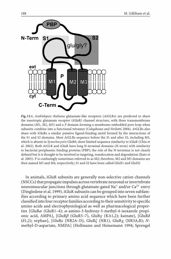
188 M. Gilliham et al.
Fig.13.1. Arabidopsis thaliana glutamate-like receptors (AtGLRs) are predicted to sharethe ionotropic glutamate receptor (iGluR) channel structure, with three transmembranedomains (M1, M2, M3) and a P domain forming a membrane-embedded pore loop whensubunits combine into a functional tetramer (Colquhoun and Sivilotti 2004). AtGLRs alsoshare with iGluRs a similar putative ligand-binding motif formed by the interactions ofthe S1 and S2 domains. Most AtGLRs sequence before the S1 and after S2, including M3,which is absent in Synechocystis GluR0, show limited sequence similarity to iGluR (Chiu etal. 2002). Both AtGLR and iGluR have long N-terminal domains (N-term) with similarityto bacterial periplasmic binding proteins (PBP); the role of the N-terminus is not clearlydefined but it is thought to be involved in targeting, translocation and degradation (Kato etal. 2005). P is confusingly sometimes referred to as M2; therefore, M2 and M3 domains arethen named M3 and M4, respectively; S1 and S2 have been called GlnH1 and GlnH2
In animals, iGluR subunits are generally non-selective cation channels(NSCCs) thatpropagate impulses across vertebrateneuronal or invertebrateneuromuscular junctions through glutamate-gated Na+ and/or Ca2+ entry(Dingledene et al. 1999). iGluR subunits can be grouped into seven subfam-ilies according to primary amino acid sequence which have been furtherclassified into four receptor families according to their sensitivity to specificamino acids and electrophysiological as well as pharmacological proper-ties [GluRα (GluR1–4); α-amino-3-hydroxy-5-methyl-4-isoxazole propi-onic acid, AMPA], [GluRβ (GluR5–7), GluRγ (KA1,2); kainate], [GluRδ(δ1,2); orphan], [GluRε (NR2A–D), GluRζ (NR1), GluRχ (NR3A,B); N-methyl-D-aspartate, NMDA] (Hollmann and Heinemann 1994; Sprengel

13 The Arabidopsis thaliana Glutamate-like Receptor Family(AtGLR) 189
and Seeburg 1995; Dingledene et al. 1999). In addition to glutamate, NMDAreceptors require glycine as a coagonist (Dingledine et al. 1999). AtGLRshave been divided into three clades according to sequence (AtGLR1.1–1.4;2.1–2.9; 3.1–3.7) (Lacombe et al. 2001a; Chiu et al. 2002).
This brief review will concentrate on recent developments in the field,specifically exploring the roles and effects of glutamate and glycine, and re-lated metabolites, in plant physiology relative to potential roles for AtGLRs.It will then examine the progress made toward defining the functions ofparticular AtGLRs and will conclude by recommending potentially fruitfulfuture avenues of research.
13.2Roles (and Effects) of Glutamate, Glycineand Interrelated Amino Acids in Plants
13.2.1Effects of Amino Acids on Plant Development
Plant development is sensitive both to light and to tissue C:N, which deter-mines the allocation of biomass between root and shoot (Thum et al. 2003).The molecules that signal this ratio, and the receptors that detect it, arelargely unknown, but it is thought that certain amino acids signal nitrogenstatus (Thum et al. 2003). Attempts to relate AtGLRs to C:N signalling haveshown that iGluR agonists and inhibitors affect hypocotyl development.6,7-Dinotroquinoxaline 2,3-(1H,4H)-dione (DNQX) caused an etiolatedphenotype in seedlings grown in light but not those grown in the dark(Lam et al. 1998) and this was reversed by glutamate and/or glycine (Du-bos et al. 2003). S(+)-β-Methyl-α,β-diaminopropionic acid (BMAA) alsocaused etiolation in light but inhibited hypocotyl elongation in the dark,and both effects were reduced by glutamate and glutamine (Brenner et al.2000).
Rhizosphere amino acids may signal the location of nutrient-rich or-ganic matter (Filleur et al. 2005). Both glutamate and glycine have beendemonstrated to modify root elongation. Micromolar levels of glutamateinduced root bending toward a glutamate source, whereas greater concen-trations inhibited primary root growth (Filleur et al. 2005). Glycine has alsobeen demonstrated to exert positive (White 1939; Fries 1953; Skinner andStreet 1953) and negative (Skinner and Street 1953) effects on root growth.Amino acids also function in developmental and stress responses bothas signals and as osmoprotectants. For instance, the glutamate-derived γ-aminobutyric acid (GABA) and glycine-derived glycine betaine function

190 M. Gilliham et al.
as compatible solutes under salt and water stress in Arabidopsis (Chenand Murata 2002; Kaplan et al. 2004). GABA is a key neurotransmitterin animals and is synthesised rapidly in plants in response to biotic andabiotic stresses, including anoxia, oxidative stress, and mechanical stress(Scott-Taggart et al. 1999). Changes in GABA levels are also implicated inplant development, and a gradient of GABA directs pollen tube formation(Palanivelu et al. 2003). β-Aminobutyric acid (BABA) potentiates plantdefences against pathogen attack (Zimmerli et al. 2000). Glycine is also in-volved in the movement response in Mimosa pulvinus cells (Otsiogo-Oyabiand Roblin 1985).
13.2.2Glutamate and Glycine as Signalling Molecules
Recent evidence supports the hypothesis that glutamate and glycine func-tion as signalling molecules in Arabidopsis (Dubos et al. 2003). Aside fromtheir obvious effects on plant growth and development glutamate can in-duce an atypical depolarisation of the electrical potential gradient acrossthe plasma membrane (Em) of Arabidopis root, hypocotyl, and leaf meso-phyll cells when compared with other amino acids (Dennison and Spalding2000; Sivaguru et al. 2003; Meyerhoff et al. 2005) and new data suggest thatthis occurs with glycine as well (Qi et al. 2004; M. Gilliham, unpublished).The depolarisation is multiphasic, consisting of an initial rapid, substantialtransient with duration in the order of tens of seconds followed by a smallersecondary phase that plateaus until removal of the agonist. The secondaryphase seen in the presence of other amino acids probably reflects the ac-tivity of proton-coupled amino acid symporters (Kinraide and Etherton1980; de Jong and Borstlap 2000), whereas the primary transient is proba-bly due, at least in part, to the passage of Ca2+ across the plasma membrane.The primary depolarisation by glutamate is, at least initially, coincidentalwith a transient rise in [Ca2+]cyt, as indicated by whole seedlings consti-tutively expressing the chemiluminescent Ca2+-binding protein aequorin,with both phenomena being abolished by the non-specific calciumchannelblocker La3+ (Dennison and Spalding 2000; Sivaguru et al. 2003; Meyerhoffet al. 2005).
Both theglutamate-inducedprimaryEm transientandtherise in [Ca2+]cyt
appear to be concentration-dependent, first detectable around 50 µM andsaturate in the low millimolar range (Demidchik et al. 2004; Meyerhoff etal. 2005). As has been observed in animals, glutamate and glycine appliedtogether synergistically decrease the amount of agonist needed to increase[Ca2+]cyt to 10 µM (Dubos et al. 2003). Subsequent applications of agonistdecrease both these responses (Meyerhoff et al. 2005; Qi et al. 2005; M.
13 The Arabidopsis thaliana Glutamate-like Receptor Family(AtGLR) 191
Gilliham, unpublished), possibly indicating a desensitisation of response(Everts, et al. 1999; Sun et al. 2002). Unpublished results of Qi et al (2005)suggest that glutamatedesensitised the response toglycine in thehypocotyl,but not vice versa, whereas no such specificity in desensitisation responsehas been observed in roots (M. Gilliham, unpublished). De novo synthesisof proteins may be required for full recovery of the response, since recoveryis inhibited by cycloheximide (Meyerhoff et al. 2005).
Neither response is initiated by GABA, NMDA, AMPA or KA, support-ing the divergence of AtGLR and iGluR before they developed unique ligandspecificity (Chiuetal. 1999).However,DNQXand6-cyano-7-nitroquinoxal-ine-2,3-dione (CNQX) can abolish the glutamate-induced and/or the gly-cine-induced [Ca2+]cyt increase (Dubos et al. 2003; Meyerhoff et al. 2005).The cold-shock-induced rise in [Ca2+]cyt is unaffected by DNQX (or previ-ous application of glutamate) (Dubos et al. 2003), indicating separate Ca2+
response pathways but also specificity in the CNQX/DNQX antagonism.The NMDA/glutamate site binding iGluR antagonist AP-5 also blocks bothEm depolarisation and the Ca2+-dependent microtubule depolymerisationinduced by glutamate (Sivaguru et al. 2003), however, its effect on [Ca2+]cyt
has not been measured directly. Interestingly NMDA also depolymerisesmicrotubules despite not inducing a [Ca2+]cyt or an Em response (Sivaguruet al. 2003), suggesting that caution must be exercised when using iGluR ag-onists/antagonists as indication of AtGLR involvement in these processes.It should also be noted that NMDA-type iGluRs, the only iGluR subtype tobind glycine, are not inhibited by CNQX/DNQX which bind at the glutamatebinding site of AMPA/KA iGluR.
L-glutamate also induced the influx of Na+ and Ca2+ in a small percent-age of patch-clamped protoplasts derived from Arabidopsis root cells byactivating ‘spiky’ inward currents at hyperpolarised potentials with exter-nal Ca2+ or less erratic currents with Na+ (Demidchik et al. 2004). Glycinecan also activate an instantaneous current in patch-clamped protoplastsderived from wheat root cells (M. Gilliham, unpublished). The effect ofglutamate on unidirectional influx of Na+ into Arabidopsis roots was in-conclusive (Demidchik et al. 2004). Because Na+ influx is mainly via NSCCs,which are permeable to other monovalent cations, the movement of Na+,K+, Cs+, and NH+
4 , although not dependent upon a glutamate-induceddepolarisation may contribute to it.
Sivaguru et al. (2003) have suggested that AtGLRs are involved in rootresponses to toxic Al3+. They propose that Al3+, known to induce organicanion release in some species (Ma and Furukawa 2003), stimulates gluta-mate release, which activates Ca2+ influx via plasma-membrane-localisedAtGLR, leading to Em depolarisation and microtubule depolymerisation.They observed that 5-nitro-2-(3-phenylpropylamino)-benzoate (NPPB),known to inhibit the Al3+-activated organic anion channel of the root

192 M. Gilliham et al.
apex (Zhang et al. 2001), blocks Al3+-induced Em depolarisation and Ca2+-dependent microtubule depolymerisation but does not block these in re-sponse to L-glutamate. An alternative sequence of events in response toglutamate is suggested by plant action potentials. The action potential ofgiant algal cells is dependent on three phases: (1) an initial small Ca2+ influxthat (2) stimulates a substantial Cl− efflux to below EK which (3) stimulatesan efflux of K+ repolarising Em (Shepherd et al. 2002). It is entirely possiblethat thedepolarisation inducedbyglutamatecould involvea similar chainofevents. Qi et al. (2005) have reported that, in contrast to lone applications,a combination of NPPB and 1,2-bis(o-aminophenoxy)ethane-N,N,N ′,N ′-tetraacetic acid (BAPTA) (a Ca2+ chelator) can decrease the glutamate-induced Em depolarisation, supporting the hypothesis that ligand treat-ment affects membrane permeability to multiple ions. Taken together withthe putative role of AtGLR in de-etiolation it is tempting to suggest aninvolvement of AtGLR in the blue-light induced Em depolarisation of thehypocotyl (Cho and Spalding 1996).
13.3Roles of AtGLR
13.3.1Expression
Individual AtGLRs show complex spatio-temporal expression patterns dur-ing plant development and a diversity of responses to stresses (Sect. 13.3.5).Data from multiple sources (RT-PCR, promoter:reporter fusions, expressedsequence tags, microarrays) indicate widespread AtGLR transcription, typ-ically with low messenger RNA (mRNA) abundance. RT-PCR indicatedthat most AtGLRs were transcribed in root, shoot, flower, and silique in8-week-old plants, although transcripts of several clade 2 genes (2.1–2.3,2.9) were detected roots only (Chiu et al. 2002). At the single-cell level,individual mature leaf epidermal and mesophyll cells contained three tonine AtGLR transcripts, with no consistent pattern except that AtGLR3.7was ubiquitous (Roy et al. 2004). The putative promoter of AtGLR3.7 wasfused to the β-glucuronidase (GUS) reporter and indicated widespreadtranscription in all tissues in seedlings and in root vasculature, leaves,siliques (but not seeds), and flowers of mature plants (Essah 2002). Thenear ubiquity of AtGLR3.7 transcripts raises the possibility that AtGLR3.7could be a key subunit involved in a variety of AtGLR heteromeric pro-teins, analogous to NMDAR1 amongst NMDA receptors. Other AtGLRsmay be more confined in their expression, for instance, localisation of At-GLR3.2 by promoter:GUS fusion, RT-PCR, and an antibody raised to the

13 The Arabidopsis thaliana Glutamate-like Receptor Family(AtGLR) 193
C-terminus indicated strongest expression in developing floral stems andvasculature (Kim et al. 2001; Turano et al. 2002). A number of AtGLRsshow high expression in senescent leaves, including all AtGLR1, AtGLR2.5,AtGLR2.7–9, and AtGLR3.4 (Zimmermann et al. 2004; Meyerhoff et al.2005). Promoter:GUS fusions for AtGLR2.8, AtGLR2.9, and AtGLR3.4 haveindicated their expression throughout the leaf mesophyll but are greatestsurrounding vascular bundles as well as hydathodes, suggestive of a role inmetabolite re-mobilisation during senescence (O. Meyerhoff and D. Becker,unpublished).
Most AtGLRs have a hydrophobic N-terminus predicted to be cleavedafter targeting the nascent protein to the endoplasmic reticulum for pro-cessing (Davenport 2002). No clear picture has yet emerged concerning thesubcellular distribution of individual plant glutamate receptors. However,using green fluorescent protein (GFP) or GUS–GFP tags several AtGLR(AtGLR3.4, 3.7, and a radish GLR) have been localised to the plasmamembrane of onion epidermis cells, Arabidopsis and tobacco protoplasts(D. Becker, unpublished; R. Davenport, unpublished; Kang and An 2003).However, these studies should be interpreted with caution since incorpo-ration of tags and/or overexpression can cause mistargeting (AtGLR3.4 hasalso been reported to be expressed in plastids) (Szabo et al. 2004). Anantibody to the C-terminus localised AtGLR3.2 to the membrane fractionbut was not used to distinguish the membrane involved (Turano et al.2002).
13.3.2Amino Acid Binding and AtGLR Regulation
AtGLRs have been predicted to function in an analogous manner to iGluRs.Phylogenetic analysis predicts that AtGLRs share greatest sequence simi-larity with NMDA receptors despite sharing more invariant residues withGluRδ receptors (Chiu et al. 1999, 2002). Subunit–ligand interactions forAtGLRweremodelledby superimposingalignedsequencesonto the ligand-binding domains of a rat iGLRα crystal structure (Dubos et al. 2003). Thedocking algorithm OXDOCK (Glick et al. 2002) found that only AtGLR1.1would be predicted to bind glutamate, whereas the remaining 19 predictedAtGLR subunits would not owing to steric hindrance in the ligand-bindingsite (Dubos et al. 2003). Instead, these 19 subunits share a high degree ofsimilarity with glycine-binding domains, and are predicted to bind glycine.It is intriguing that Arabidopsis possesses a greater number of AtGLR sub-units that are predicted to bind glycine, as opposed to glutamate, and maysuggest that glycine plays a more important role in signalling than hadpreviously been suspected.

194 M. Gilliham et al.
13.3.3Are AtGLRs Ion Channels?
AtGLRs are thought to encode subunits of plant NSCCs (Davenport 2002;Demidchik et al. 2002; Demidchik, this volume). However, despite profoundamino acid sequence similarity with iGluRs the residues within the putativepore region of AtGLRs are unlike any other ion channel known (Davenport2002). This makes predictions about their ion selectivity, if they are indeedchannels, impossible. Furthermore, although AtGLRs are divided into threeclear clades upon the sequence similarity of their whole sequence if only theM1, P, and M2 domains are compared then the similarity in and betweenclades breaks down. This may indicate either a loss of channel functionor similar selectivity and/or gating properties between AtGLR clades, incontrast to iGluR subfamilies (Chiu et al. 2002).
Attempts to functionally characterise AtGLRs in heterologous expres-sion systems have so far proved inconclusive. The expression of severalgroup 2 members in Xenopus oocytes did not result in any detectable cur-rent, even following fusion with the signal peptide from the rat GluR6 whichfacilitated the functional characterisation of the previously non-functionalSynechocystis GluR0 (Lacombe et al. 2001b; B.R. Davies and R. Davenport,unpublished). The injection of AtGLR3.4 and AtGLR3.7 complementaryRNA (cRNA) results in the activation of cation-permeable conductances atthe oocyte plasma membrane and the consecutive activation of the intrinsicCa2+-activated chloride current (IClCa) (D. Becker, unpublished; Cheffings2001; Gilliham 2005, unpublished; Lacombe et al. 2001b). Activation of theIClCa does indicate that [Ca2+]cyt has been elevated but it is not proof thatthe injected cRNA forms a channel that catalyses the influx of Ca2+ acrossthe plasma membrane. IClCa activation may be the sole result of upregula-tion of additional endogenous channels in intracellular compartments ortheplasmamembrane suchasCa2+-permeablehyperpolarisation-activatedcation channels (Tzounopoulos 1995; Kuruma et al. 2000) or the disruptionof cellular homeostasis (Weber 1999). Cheffings (2001) has suggested thatAtGLR3.7 is constitutively active and catalyses the voltage-independentmovement of Na+, K+, and Ca2+ across the plasma membrane when ex-pressed in Xenopus oocytes. Injection of the AtGLR3. 7 coding sequencecontaining stop codons reduced the instantaneous current component at allvoltages to wild-type levels, indicating that AtGLR3. 7 may encode a chan-nel. The possibility of constitutive AtGLR channel activity is supported bythe sequence of AtGLR M2 domains, which resemble sequences known toinduce constitutive activity in some iGluRs (Chiu et al. 1999; Yuzaki 2003).
Disappointingly,nocurrentshavebeenactivatedby L-glutamate, glycine,or a combination of both, at any voltage, at various pHext, with a range ofexternal cations, in Xenopus oocytes at levels over control following the

13 The Arabidopsis thaliana Glutamate-like Receptor Family(AtGLR) 195
injection of any AtGLRs. Additionally, ABA, BMAA, GABA, NMDA, andglutamine have not activated any currents in any heterologous system con-taining any AtGLRs as tested so far (Cheffings 2001; Lacombe et al. 2001b;Meyerhoff et al. 2005; M. Gilliham, unpublished), although incubation ofthe AtGLR3. 4 injected oocytes with DNQX did reduce the Na+-permeable“leak” current (M. Gilliham, unpublished).
Although at least AtGLR3.4 is expressed at the plasma membrane inheterologous systems (D. Becker, unpublished), it is possible that AtGLRsmay require plant-specific processing for proper function. It is possible thatthe activity of some AtGLRs is modified by endogenous subunits in oocytesand other heterologous systems, resulting in activation of atypical currentsor loss of AtGLR channel function (Green et al. 2002; Anantharam et al.2003). Other proteins or cofactors may also be needed for proper activityof AtGLR in heterologous systems, for instance, SOL-1, a CUB-like protein,colocalises at the neuron cell surface with GLR1 in Caenorhabditis elegansand is required for ligand-gated GLR1 activity (Zheng et al. 2004).
In planta evidence for ion channel activity of glutamate receptors is lim-ited. Kim et al. (2001) constitutively overexpressed AtGLR3. 2, which led tosigns of Ca2+ deficiency and a hypersensitivity to K+ and Na+. Althoughtotal tissue Ca2+ was the same in wild-type plants and plants overexpressingAtGLR3.2, medium supplemented with Ca2+ could rescue the mutant phe-notype. On the basis of these findings AtGLR3.2, which is most abundantlyexpressed in vascular tissues, has a proposed role in Ca2+ distributionwithin the plant (Kim et al. 2001; Turano et al. 2002). Antisense AtGLR1.1plants did not show hypersensitivity to K+ and Na+ compared with wild-type plants, but root growth was more inhibited by high levels of Ca2+ (Kangand Turano 2003). In unpublished work, Qi et al. (2004) have reported thatAtGLR T-DNA insertional mutants have aberrant growth rate responses toglutamate or light. Knockouts of AtGLR3.3 also displayed reduced depo-larisations in the root and hypocotyl associated with glutamate or glycine,whereas knockouts of AtGLR3.4 showed reduced response to glycine only(Qi et al. 2005).
13.3.4C:N Signalling
Phenotypic analysis of antisense AtGLR1.1 (antiAtGLR1.1) plants has im-plicated a role for AtGLR1.1 in C:N perception and signalling (Kang and Tu-rano 2003; Kang et al. 2004). antiAtGLR1.1 seed germination was inhibitedby sucrose, but rescued by supplemental nitrate. Compared with wild-type,seed germination of antiAtGLR1.1 plants was also more susceptible to inhi-bition by DNQX, possibly because antiAtGLR1.1 suppressed AtGLR activity

196 M. Gilliham et al.
sufficiently to make plants more susceptible to DNQX. BMAA counteractedthe inhibitory effect of DNQX on antiAtGLR1.1 seed germination despiteprevious observations that DNQX and BMAA have superficially analogouseffects on plant growth and development (Brenner et al. 2000; Sect. 13.2.1),suggesting one must be cautious in ascribing a precise mode of actionto either of these compounds in plants. Glutamate (and to a lesser extentglutamine) rescued germination of antiAtGLR1.1 seed by DNQX, but bothwere less effective than BMAA. It was proposed that glutamate uptake ordistribution was poor compared with DNQX, or that glutamate is not thenatural ligand for AtGLRs. However, as AtGLR1.1 encodes the only subunitpredicted to bind glutamate (Dubos et al. 2003) antiAtGLR1.1 plants wouldbe expected to be particularly impaired in glutamate perception.
The expression of genes and the accumulation of proteins related tocarbonandnitrogenmetabolism,aswell asABAresponsesandmetabolism,were altered in antiAtGLR1.1 plants (Kang and Turano 2003; Kang et al.2004). For instance, antiAtGLR1.1 plants had elevated ABA levels, increasedtranscript abundanceofABAbiosynthetic genes, enhancedABAsensitivity,reduced stomatal apertures, and increased drought resistance. Given thatABA has been implicated in sugar signalling (Gibson 2005), the alterationof ABA metabolism and response in antiAtGLR1.1 plants provides anotherlink to carbon sensing in these plants.
A model has been proposed that places AtGLR1.1 at the interface ofsucrose and amino acid signalling, suggesting AtGLR1.1 is stimulated byamino acids and suppresses ABA biosynthesis and sugar signalling, allow-ing seedgermination tooccur (KangandTurano2003;Kanget al. 2004).Themodel also suggests that AtGLR1.1 is inhibited by sucrose, which, in turn,allows sugar signalling and ABA biosynthesis to occur and results in inhi-bition of seed germination. However, the strict involvement of AtGLR1.1 inC:N signalling needs to be confirmed. The segment of the AtGLR1.1 genethat was used to generate the antisense construct shares sequence identitywith several AtGLR genes, including AtGLR1.2, AtGLR1.4, and AtGLR3.6. Itmay be that these other members of the AtGLR family were also suppressedby the antisense construct and therefore they may also contribute to thephenomena observed in the antiAtGLR1.1 lines.
13.3.5Stress Responses
The GENEVESTIGATOR microarray toolbox suggests that, with respect toabiotic stress, there is no single stress factor or plant hormone that inducesor represses expression of AtGLR in general (Zimmermann et al. 2004).For instance, while AtGLR2.5 and AtGLR3.2 were upregulated by ABA,
13 The Arabidopsis thaliana Glutamate-like Receptor Family(AtGLR) 197
AtGLR2.1 and AtGLR3.5 were downregulated. AtGLR1.3 and AtGLR1.1seemed not to respond at all to this stress hormone despite the latter beingimplicated in the regulation of ABA biosynthesis and antiAtGLR1.1 plantsexhibiting elevated ABA levels (Kang and Turano 2003; Sect. 3.3). Maathuiset al. (2003) demonstrated that members of the AtGLR family showed time-dependent changes in transcription in response to Ca2+ deprivation andexcess Na+, but not K+ deprivation, supporting the prediction that theyfunction as NSCCs.
Analysis of GUS expression under control of the AtGLR3.4 promoter sug-gested that AtGLR3.4 expression was induced by mechanical stress such aswounding or touch and cold (Meyerhoff et al. 2005). The upregulation ofAtGLR3.4 transcription by wounding and during senescence could suggestan involvement of methyljasmonate (Sect. 13.3.1), a common componentof both processes (Devoto and Turner 2005). Quantitative RT-PCR analysesof AtGLR3.4 expression revealed that cold-induced transcription was fast,reaching peak transcript levels after 30 min. Additionally, La3+ inhibitedcold-induced AtGLR3.4 transcription, suggesting the involvement of Ca2+.AtGLR1.2 and AtGLR2.5 are also upregulated by cold treatment (Zimmer-mann et al. 2004).
13.4Conclusions and Future Perspectives
Research into the glutamate receptor-like family has been hindered by thelack of phenotypes associated with the AtGLR genes. The novel pore regionof the predicted proteins and the paucity of understanding of NSCC func-tion in vivo have made it difficult to predict function. However there arenow several phenotypes associated with AtGLR overexpression (Kim et al.2001) and knockout (Kang and Turano 2003; Kang et al. 2004), and system-atic electrophysiological characterisation of single AtGLR knockout plantsis underway (Qi et al. 2004, 2005; Gilliham 2005). Despite recent progress,several fundamental questions still remain regarding AtGLR function.
13.4.1Expression
One promising approach to predicting AtGLR function is the study ofAtGLR gene expression in response to developmental and environmentalcues. In particular, very rapid changes in gene expression in response to en-vironmental signals (such as application of a ligand, or stresses) implicatethe gene product in specific responses to stimuli (Meyerhoff et al. 2004,

198 M. Gilliham et al.
2005). It is also desirable to follow changes in abundance at the proteinlevel (to substantiate, for instance, the possibility of rapid protein turnoverin response to stresses), and to determine the membrane localisation ofAtGLR with confidence. GFP tags can interfere with normal protein tar-geting, especially in the case of endomembrane proteins where both theN- and the C-termini as well as internal signals may be involved in tar-geting, and it is desirable to have a test of retention of functionality orphenotype of the tagged protein (which we do not have at present formost AtGLRs). Alternatives to large tags such as GFP include antibodiesor smaller tags (which could be incorporated internally in the AtGLR pro-tein to allow in vivo monitoring of membrane protein dynamics as in thecase of iGluR tagged with a toxin binding site; Sekine-Aizawa and Huganir2004).
13.4.2Ligand Binding and Regulation
The ligand-binding profile of AtGLR needs to be examined experimentally.Although glutamate and glycine binding has been modelled, no ligand-binding assays have been performed in vitro to confirm the prediction ofthe model. It is possible that other ligands exist for AtGLR and elucidationof these could inform phenotyping studies (Sect. 13.4.3). In the absenceof a functional expression system, ligand binding could be explored byconstructing chimaeras consisting of only the soluble ligand-binding do-mains and using these to measure affinity of binding, as has been donewith S1,S2 domains from animal iGluRs (Kuusinen et al. 1995). In addition,the proposed signalling role of glutamate or glycine should be substanti-ated by high-resolution quantification of changes in apoplastic concentra-tion (e.g. by fluorescence-based techniques; Acher et al. 2005; Fehr et al.2005).
13.4.3Knockout and Overexpression Phenotyping
As there are 20 AtGLRs, knockout of some members may result in sub-tle or no phenotypes owing to specificity of function or compensationby other members, making it necessary to reduce expression of severalmembers simultaneously to readily identify phenotypes. This could beachieved by pyramiding knockouts or by employing an RNA interfer-ence strategy to silence several AtGLRs simultaneously (Essah et al. 2005).Overexpression of AtGLRs is also a desirable strategy, since it is possible

13 The Arabidopsis thaliana Glutamate-like Receptor Family(AtGLR) 199
that excess of one subunit could disrupt the formation of normal het-eromers and may have relatively widespread effects, which might be dif-ficult to interpret but would give crude information about the processesin which AtGLRs are involved. AtGLRs have been suggested to have wide-ranging functions so phenotyping requires testing of plant responses toa range of different ligands (informed by Sect. 13.4.2), including gluta-mate, glycine, GABA, ABA, and other amino acids, as well as agonists andantagonists of animal iGluRs. Plant responses may involve germination,root growth, circadian-asssociated phenotypes such as aberrant hypocotylangle or length, responses to light, nutrients and toxic ions, solute accu-mulation, and electrical and calcium signalling. Phenotyping needs to beinformed to some extent by the reported physiological effects of aminoacids on plant behaviour, but conversely physiological characterisationwill depend partly on mutant characterisation to predict plant responsesto ligands.
13.4.4Heterologous Expression
Heterologous expression is desirable to demonstrate ion channel functionof AtGLRs. However expression in Xenopus oocytes has been uninfor-mative (Sect. 13.3.3). Chimaeric iGluRs constructed by transplanting thepore domain of apparently non-functional iGluRs (such as KA-bindingproteins and C. elegans iGluR) into rat iGluRs have been used to demon-strate functionality of the pore (Villmann et al. 1997; Strutz-Seebohm etal. 2003). Although not proof of function in vivo this approach could indi-cate whether the pore region is capable of ion conduction and its potentialselectivity. Use of homologous or heterologous plant systems such as cul-tured mesophyll cells may result in correct processing. Alternatively thepossible toxicity of some AtGLRs in yeasts and E. coli, which suggestsfunctional expression (Davenport 2002), could be used to advantage by in-ducible expression of toxic AtGLRs, which would permit at least ion uptakestudies. In some cases coexpression of subunits in heterologous systemsmay be required to produce functional ion channels. iGluRs function asheteromers and some subunits show no channel activity when expressedas homomers (Dingledine et al. 1999). The large size of the AtGLR familyand the evidence for simultaneous expression of many different AtGLRsin single cells (Sect. 13.3.1) makes it difficult to predict likely heteromericcombinations, and it may be desirable to coexpress large pools of sub-units simultaneously, or to use a yeast two-hybrid approach to identifyinteracting subunits.

200 M. Gilliham et al.
13.4.5NSCC Characterisation
The inadequacy of NSCC characterisation in planta and the uncertaintyregarding the definitive identification of ions involved in (or the sig-nalling components downstream of) the ligand gated Em depolarisation inplanta make it difficult to match the characteristics of putative heteromer-dependent AtGLR currents to native currents. The use of ion channel block-ers and ion substitution in microelectrode impalements are producingsome insights (Sivaguru et al. 2003; Qi et al. 2005) and greater resolutioncould be provided by further patch clamp experiments (Demidchik et al.2004) where greater control of solutions could also reveal whether cytosoliccomponents are involved. Selection of fluorescent protoplasts expressingmarker:AtGLR promoter fusions indicating expression of specific AtGLRswithin protoplasts may allow the tentative identification of currents withgene products (Kiegle et al. 2000). Alternatively, mRNA could be extractedfrom protoplasts after electrophysiological investigation and AtGLR tran-script abundance correlated with membrane conductance (Gehwolf et al.2002).
ReferencesAcher FC, Bertrand HO (2005) Amino acid recognition by Venus flytrap domains is encoded
in an 8-residue motif. Biopolymers 80:357–66AnantharamA,LewisA,PanaghieG,GordonE,McCrossanZA,LernerDJ,AbbottGW(2003)
RNAinterference reveals that endogenous Xenopus MinK-relatedpeptides governmam-malian K+ channel function in oocyte expression studies. J Biol Chem 278:11739–11745
Brenner ED, Martinez-Barboza N, Clark AP, Liang QS, Stevenson DW, Coruzzi GM (2000)Arabidopsis mutants resistant to S(+)-beta-methyl-alpha,beta-diaminopropionic acid,a cycad-derived glutamate receptor agonist. Plant Physiol 124:1615–1624
Cheffings CM (2001) Calcium channel activity of a plant glutamate receptor homologue. Pa-per presented at the 12th International Workshop on Plant Membrane Biology. Madison,WI, USA
Chen THH, Murata N (2000) Enhancement of tolerance of abiotic stress by metabolicengineering of betaines and other compatible solutes. Curr Opin Plant Biol 5:250–257
Chen G-Q, Cui C, Mayer ML, Gouax E (1999) Functional characterisation of a potassium-selective prokaryotic glutamate receptor. Nature 402:817–821
Chiu J, DeSalle R, Lam HM, Meisel L, Coruzzi G (1999) Molecular evolution of glutamatereceptors: a primitive signaling mechanism that existed before plants and animalsdiverged. Mol Biol Evol 16:826–838
Chiu JC, Brenner ED, DeSalle R, Nitabach MN, Holmes TC, Coruzzi GM (2002) Phyloge-netic and expression analysis of the glutamate-receptor-like gene family in Arabidopsisthaliana. Mol Biol Evol 19:1066–1082
Cho MH, Spalding EP (1996) An anion channel in Arabidopsis hypocotyls activated by bluelight. Proc Natl Acad Sci USA 93:8134–8138
Colquhoun D, Silivotti LG (2004) Function and structure in glycine receptors and some oftheir relatives. Trends Neurosci 27:337–344

13 The Arabidopsis thaliana Glutamate-like Receptor Family(AtGLR) 201
Davenport R (2002) Glutamate receptors in plants. Ann Bot 90:549–557de Jong A, Borstlap AC (2000) Transport of amino acids (L-valine, L-lysine, L-glutamic acid)
and sucrose into plasma membrane vesicles isolated from cotyledons of developing peaseeds. J Exp Bot 51:1663–1670
Demidchik V, Davenport RJ, Tester M (2002) Nonselective cation channels in plants. AnnuRev Plant Physiol Plant Mol Biol 53:67–107
Demidchik V, Essah P, Tester M (2004) Glutamate activates sodium and calcium currents inthe plasma membrane of Arabidopsis root cells. Planta 219:167–175
Dennison KL, Spalding EP (2000) Glutamate-gated calcium fluxes in Arabidopsis. PlantPhysiol 124:1511–1514
Devoto A, Turner JG (2005) Jasmonate-regulated Arabidopsis stress signalling network.Physiol Plant 123:161–172
Dingledine R, Borges K, Bowie D, Traynelis SF (1999) The glutamate receptor ion channels.Pharmacol Rev 51:7–61
Dubos C, Huggins D, Grant GH, Knight M.R, Campbell MM (2003) A role for glycine in thegating of plant NMDA-like receptors. Plant J 35:800–810
Essah PA (2002) Sodium transport and accumulation in A. thaliana. PhD thesis. Universityof Cambridge, http://plantscience.acpfg.com.au/upload/essah/PAEPHD.pdf
Essah PA, Liu LH, Davenport R, Leigh R, Tester M (2005) Using the chemical-regulatedinducible inverted-repeat RNA (RNAi) system to elucidate the function(s) of glutamatereceptor homologues in Arabidopsis thaliana. Abstract, SEB main meeting, July 11–15,Barcelona, Spain
Everts I, Petroski R, Kizelsztein P, Teichberg VI, Heinemann SF, Hollmann M (1999) Lectin-induced inhibition of desensitization of the kainate receptor GluR6 depends on theactivation state and can be mediated by a single native or ectopic N-linked carbohydrateside chain. J Neurosci 19:916–927
Fehr M, Okumoto S, Deuschle K, Lager I, Looger LL, Persson J, Kozhukh L, Lalonde S,Frommer WB (2005) Development and use of fluorescent nanosensors for metaboliteimaging in living cells. Biochem Soc Trans 33:287–290
Filleur S, Walch-Liu P, Gan Y, Forde, BG (2005) Nitrate and glutamate sensing by plant roots.Biochem Soc Trans 33:283–286
FriesN(1953)Limiting factors in thegrowthof thepea seedling root. Physiol Plant 6:292–300Gehwolf R, Griessner M, Pertl H, Obermeyer G (2002) First patch, then catch: measuring
the activity and the mRNA transcripts of a proton pump in individual Lilium pollenprotoplasts FEBS Lett 512: 152–156
Gibson SI (2005) Control of plant development and gene expression by sugar signaling.Curr Opin Plant Biol 93–102
Gilliham M (2005) Plant glutamate receptors – progress to date. Book of Abstracts, PlantNeurobiology, 17–20 May, Florence, Italy
Glick M, Robinson DD, Grant GH, Richards WG (2002) Identification of ligand bind-ing sites on proteins using a multi-scale approach. J Am Chem Soc 124:2337–2344
Green T, Rogers CA, Contractor A, Heinemann SF (2002) NMDA receptors formed by NR1 inXenopus laevis oocytes do not contain the endogenous subunit XenU1. Mol Pharmacol61:326–333
Hollmann M, Heinemann S (1994) Cloned glutamate receptors. Annu Rev Neurosci 17:31–108
Kang J, Turano FJ (2003) The putative glutamate receptor 1.1 (AtGLR1.1) functions asa regulator of carbon and nitrogen metabolism in Arabidopsis thaliana. Proc Natl AcadSci USA 100:6872–6877

202 M. Gilliham et al.
Kang J, Mehta S, Turano FJ (2004) The putative glutamate receptor 1.1 (AtGLR1.1) inArabidopsis thaliana regulates abscisic acid biosynthesis and signaling to control devel-opment and water loss. Plant Cell Physiol 45:1380–1389
Kang S, An CS (2003) Localisation and functional analysis of the glutamate receptor fromsmall radish. Book of abstracts, plant biology 2003, June 25–30, Honolulu, HI, USA
Kaplan F, Kopka J, Haskell DW, Zhao W, Schiller KC, Gatzke N, Sung DY, Guy CL (2004)Exploring the temperature-stress metabolome of Arabidopsis. Plant Physiol 136:4159–4168
Kato A, Rouach N, Nicoll RA, Bredt DS (2005) Activity-dependent NMDA receptor degra-dation mediated by retrotranslocation and ubiquitination. Proc Natl Acad Sci USA102:5600–5605
Kiegle E, Gilliham M, Haseloff J, Tester, M (2000) Hyperpolarisation-activated calciumcurrents found only in cells from the elongation zone of Arabidopsis thaliana roots.Plant J 21:225–229
Kim SA, Kwak JM, Jae S-K, Wang M-H, Nam HG (2001) Overexpression of the AtGluR2 geneencoding an Arabidopsis homolog of mammalian glutamate receptors impairs calciumutilisation and sensitivity to ionic stress in transgenic plants. Plant Cell Physiol 42:74–84
Kinraide TB, Etherton B (1980) Electrical evidence for different mechanisms of uptake forbasic, neutral and acidic amino acids in oat coleoptiles. Plant Physiol 65:1085–1089
Kuruma A, Hirayama Y, Hartzell HC (2000) A hyperpolarization- and acid-activated nons-elective cation current in Xenopus oocytes. Am J Physiol Cell Physiol 279:C1401–C1413
Kuusinen A, Arvola M, Keinanen K (1995) Molecular dissection of the agonist binding siteof an AMPA receptor. EMBO J 14:6327–6332
Lacombe B, Becker D, Hedrich R, DeSalle R, Hollmann M, Kwak JM, Schroeder JI, Le NovereN, Nam HG, Spalding EP, Tester M, Turano FJ, Chiu J, Coruzzi G (2001a) The identity ofplant glutamate receptors. Science 292:1486–1487
Lacombe B, Meyerhoff O, Steinmeyer R, Becker D, Hedrich R (2001b) Role of Arabidopsisionotropic glutamate receptors. Abstract. Association ‘Canaux Ioniques’ 12eme col-loque, La Londe les Maures
Lam HM, Chiu J, Hsieh MH, Meisel L, Oliveira IC, Shin M, Coruzzi G (1998) Glutamate-receptor genes in plants. Nature 396:125–126
Ma JF, Furukawa J (2003) Recent progress in the research of external Al detoxification inhigher plants: a minireview. J Inorg Biochem 97:46–51
Maathuis FJM, Filatov V, Herzyk P, Krijger GC, Axelsen KB, Chen S, Green BJ, Li Y, MadaganKL, Sanchez-Fernandez R, Forde BG, Palmgren MG, Rea PA, Williams LE, Sanders D,Amtmann A (2003) Transcriptome analysis of root transporters reveals participation ofmultiple gene families in the response to cation stress. Plant J 35:675–92
Meyerhoff O, Müller K, Roelfsema MRG, Latz A, Lacombe B, Hedrich R, Dietrich P, Becker D(2004) AtGLR3.4 represents an Arabidopsis glutamate-, touch-, and cold-sensitive re-ceptor. Book of abstracts, 13th International Workshop on Plant Membrane Biology,July 6–10, Montpellier, France
Meyerhoff O, Müller K, Roelfsema MRG, Latz A, Lacombe B, Hedrich R, Dietrich P, Becker D(2005) AtGLR3. 4 represents an Arabidopsis glutamate-, touch-, and cold-sensitive Re-ceptor gene. Planta (in press)
Otsiogo-Oyabi H. Roblin G (1985) Changes in membrane potential related to glycine uptakein the motor cell of the pulvinus of Mimosa pudica. J Plant Physiol 119:19–24
Palanivelu R, Brass L, Edlund AF, Preuss D (2003) pollen tube growth and guidance isregulated by POP2, an Arabidopsis gene that controls GABA levels. Cell 114:47–59
Qi Z, Durham TL, Stephens NR, Spalding EP (2004) Determining functions of Arabidopsisglutamate receptors using electrophysiology and reverse genetics. American Society ofPlant Biologists meeting book of abstracts, FL, USA

13 The Arabidopsis thaliana Glutamate-like Receptor Family(AtGLR) 203
Qi Z, Stephens NR, Durham TL, Spalding EP (2005) AtGLR3.3 and 3.4 differentially mediateglutamate and glycine-induced depolarization in the Arabidopsis root and hypocotylcells. Midwest ASPB sectional division meeting abstract, Donald Danforth Plant ScienceCenter, St Louis, MO
Roy SJ, Widdowson L, Richardson PJ, Leigh RA, Tester M (2004) Single cell gene expressionand microarray analysis of GLRs. Book of abstracts, 13th International Workshop onPlant Membrane Biology, July 6–10, Montpellier, France
Scott-Taggart CP, Van Cauwenberghe OR, McLean MD, Shelp BJ (1999) Regulation ofgamma-aminobutyric acid synthesis in situ by glutamate availability. Physiol Plant106:363–369
Sekine-AizawaY,HuganirR(2004) Imagingof receptor traffickingbyusingα-bungarotoxin-binding-site-tagged receptors. Proc Natl Acad Sci USA 101:17114–17119
Shepherd VA, Beilby MJ, Shimmen T (2002) Mechanosensory ion channels in charophytecells: the response to touch and salinity stress. Eur Biophys J 31:341–355
SivaguruM,PikeP,GassmannW,BaskinTI (2003)Aluminumrapidlydepolymerizes corticalmicrotubules and depolarizes the plasma membrane: evidence that these responses aremediated by a glutamate receptor. Plant Cell Physiol 44:667–675
Skinner JC, Street HE (1953) Studies on the growth of excised roots. II. Observations on thegrowth of excised groundsel roots. New Phytol 53:44–67
Sprengel R, Seeburg PH (1995) Ionotropic glutamate receptors. In North RA (ed) Handbookof receptors and channels: ligand- and voltage-gated ion channels. CRC, Boca Raton,FL, pp 213–263
Strutz-Seebohm N, Werner M, Madsen DM, Seebohm G, Zheng Y, Walker CS, Maricq AV,Hollmann M (2003) Functional analysis of Caenorhabditis elegans glutamate receptorsubunits by domain transplantation. J Biol Chem 278:44691–44701
SunY, Olson R, Horning M, Armstrong N, Mayer M, Gouaux E (2002) Mechanism of gluta-mate receptor desensitization. Nature 417:245–253
Szabo I, Formentin E, Segalla A, DeMarco V, Zanetti M, Rabito G, Marin O, Lo Schiavo F,Giacometti GM (2004) Study of two putative ion channels of chloroplast inner envelopemembrane.Bookof abstracts, 13th InternationalWorkshoponPlantMembraneBiology,July 6–10, Montpellier, France
Thum KE, Shasha DE, Lejay LV, Coruzzi GM (2003) Light- and carbon-signaling pathways.Modeling circuits of interactions. Plant Physiol 132:440–452
Turano FJ, Muhitch MJ, Felker FC McMahon MB (2002) The putative glutamate receptor3.2 from Arabidopsis thaliana (AtGLR3.2) is an integral membrane peptide that accu-mulates in rapidly growing tissues and persists in vascular-associated tissues. Plant Sci163:43–51
Tzounopoulos T, Maylie J, Adelman JP (1995) Induction of endogenous channels by highlevels of heterologous membrane proteins in Xenopus oocytes. Biophys J 69:904–908
Villmann C, Bull L, Hollmann M (1997) Kainate binding proteins possess functional ionchannel domains. J Neurosci 17:7634–7643
Weber WM (1999) Endogenous ion channels in oocytes of Xenopus laevis: recent develop-ments. J Membr Biol 170:1–12
White PR (1939) Glycine in the nutrition of excised tomato roots. Plant Physiol 14:527–538Wo ZG, Oswald RE (1995) Unravelling the modular design of glutamate-gated ion channels
Trends Neurosci 18:161–168Yuzaki M (2003) New insights into the structure and function of glutamate receptors: the
orphan receptor δ2 reveals its family’s secrets. Keio J Med 52:92–99Zhang WH, Ryan PR, Tyerman SD (2001) Malate-permeable channels and cation chan-
nels activated by aluminum in the apical cells of wheat roots. Plant Physiol 125:1459–1472

204 M. Gilliham et al.
Zheng Y, Mellem JE, Brockie PJ, Madsen DM, Maricq AV (2004) SOL-1 is a CUB-domain pro-tein required for GLR-1 glutamate receptor function in C. elegans. Nature 472:451–457
Zimmerli L, Jakab G, Métraux JP, Mauch-Mani B (2000) Potentiation of pathogen-specificdefense mechanisms in Arabidopsis by beta-aminobutyric acid. Proc Natl Acad Sci USA97:12920–12925
Zimmermann P, Hirsch-Hoffmann M, Hennig L, Gruissem W (2004) GENEVESTIGATOR.Arabidopsis microarray database and analysis toolbox. Plant Physiol 136:2621–2632

14 Similarities Between EndocannabinoidSignaling in Animal Systemsand N-Acylethanolamine Metabolismin PlantsElison B. Blancaflor, Kent D. Chapman
Abstract N-Acylethanolamines (NAEs) are minor lipid constituents of plant and animalcells, and their roles in mammalian physiology and neurobiology have been studied inten-sively for many years. However, corresponding studies on the function of NAEs in plantshave appeared only recently. Within the last decade significant progress has been madein quantifying NAEs in plant tissues, characterizing their potential targets in plant cellsand identifying the relevant enzyme involved in their degradation, but much remains tobe determined regarding the role of these fatty acid amides in plant physiology. Althoughour understanding of the specific functions of NAE in plants is far from complete, recentadvances in plant NAE biochemistry are pointing to intriguing similarities between animalsand plants in the metabolism and perception of NAE. In this chapter we discuss NAEs asprospective signaling and regulatory molecules in plant cells. Advances in mammalian NAEresearch are presented when appropriate in order to draw parallels as well as to highlightdifferences between plant NAE metabolism and the endocannabinoid signaling system, themajor pathway by which NAE exerts its physiological effects in animal cells.
14.1Introduction and Overviewof Mammalian Endocannabinoid Signaling
For many centuries derivatives of marijuana (Cannabis sativa) have beenknown to possess medicinal properties. Identification of 9-tetrahydrocan-nabinol (THC; Fig. 14.1a) as one of the active ingredients of marijuanaled to the discovery of a group of G-protein coupled receptors that bindto THC. Two cannabinoid (CB) receptors, CB1 and CB2, have been clonedthus far, with CB1 being the most highly expressed neuromodulatory re-ceptor in the brain. CB2 receptors, on the other hand, are absent in thebrain but predominate in tissues of the immune system (Wilson and Nicoll2002). Low levels of N-acylethanolamines (NAEs) were first identified inmammalian brain in 1965 (Bachur et al. 1965) but the function of thisfamily of lipids remained unclear until N-arachidonylethanolamine (anan-damide; NAE20:4; Fig. 14.1b) was discovered to be an endogenous ligandfor CB1 receptors (Devane et al. 1992). This finding stimulated a flurry ofresearch on the metabolism and biological activities of NAEs in severalanimal models, which eventually solidified the status of anandamide asan endogenous neurotransmitter (Self 1999). Anandamide as a ligand for
Communication in PlantsF. Baluška, S. Mancuso, D. Volkmann (Eds.)© Springer-Verlag Berlin Heidelberg 2006

206 E.B. Blancaflor, K.D. Chapman
Fig.14.1. Structures of selected cannabinoids. a ∆9-Tetrahydrocannabinol, the major activeingredient in marijuana, is a strong ligand for cannabinoid 1 receptors in mammalianbrains. b Anandamide is an N-acylethanolamine (NAE) with a 20-carbon acyl chain andfour double bonds. It has a wide range of neuromodulatory and physiological functionsin vertebrates, and is drawn in two different conformations. c NAE types identified andquantified in plants
CB receptors is somewhat surprising given the lack of obvious structuralsimilarity with THC, but nearly all physiological effects elicited by mar-ijuana have been attributed to anandamide and the metabolism of NAEsis now considered an integral part of the “endocannabinoid signaling sys-tem”. The activity of anandamide at the CB1 receptor has been attributedby some (Reggio 2003) to its ability to adopt alternative conformations(Fig. 14.1b).
Although anandamide is the most studied NAE type in vertebrates,other endogenous NAEs such as N-oleoylethanolamine (NAE18:1) andN-palmitoylethanolamine (NAE16:0; Fig. 14.1c) elicit a number of biolog-

14 N-Acylethanolamines in Plants 207
ical effects in animals, including satiety (LoVerme et al. 2005) and anti-inflammatory responses (Fowler 2003), respectively. Some of the cellularprocesses mediated by NAEs appear to be mediated by both receptor-dependent and receptor-independent pathways. For example, anandamidebinding to CB1 receptors is known to stimulate Gi/o proteins, which inturn influences the activity of a series of second messengers and proteins,including adenylate cyclase, voltage-gated calcium channels, K+ channels,phosphatidyl 3-kinase and mitogen-activated protein (MAP) kinase (DePetrocellis et al. 2004; De Fonseca et al. 2005). On the other hand, experi-ments with mouse cell cultures showing that NAE induction of transcrip-tion and MAP kinase phosphorylation are not inhibited by Gi/o proteinblockers or CB1 receptor antagonists can be taken as evidence for receptor-independent signaling (Berdyshev et al. 2001).
The widespread occurrence of NAEs in various plant species (Chapman2004), the recent cloningof aplant enzyme involved in thehydrolysisofNAE(Shrestra et al. 2003) and the identification of high-affinity NAE bindingproteins in plant tissues whose binding appears to be influenced by CBreceptor antagonists (Tripathy et al. 2003) point to intriguing parallels tomammalianendocannabinoidsignaling. In this chapter,wereviewprogressin plant NAE research focusing on metabolism and prospective functionswhile highlighting avenues for future work with the hope of generatingexcitement in this emerging field within plant physiology.
14.2NAE Structure and Occurrence in Plants
NAEs are fatty acid derivatives with amide linkages to ethanolamine(Fig. 14.1). As such, they are electrically neutral, and as expected havelimited solubility in aqueous solutions. Various NAE types have been iden-tified in animals (Schmid et al. 1990), higher plants (Chapman 2004) andsome microorganisms (Schmid et al. 1990). For the most part the acylmoieties of NAEs are a reflection of the fatty acid compositions of theparent organisms, although the precise profiles (and quantities) of NAEsin a given organism vary from tissue to tissue. Generally NAEs are presentin amounts approximating nanograms per gram of fresh weight, but undercertain pathological conditions, NAE levels accumulate to micrograms pergram of fresh weight (Schmid et al. 1990; Chapman 2004). This is particu-larly striking under conditions of ischemia in mammalian heart and brain,where NAEs accumulate in damaged tissues, but are barely detectable innearby unaffected tissues (Schmid et al. 1990).
In plants, NAEs were first identified in processed, seed-derived prod-ucts in the 1950s, but not until the 1990s did it become evident that

208 E.B. Blancaflor, K.D. Chapman
NAEs were endogenous metabolites in living plant cells. The NAE typesidentified in seeds were 12–18 carbons in length with zero to two dou-ble bonds, and the general abundance of these NAEs in desiccated seedswas on the order of micrograms per gram of fresh weight (Fig. 14.1c).Generally, N-linoleoylethanolamine (NAE18:2), NAE16:0, NAE18:1 and N-lauroylethanolamine (NAE12:0)were the fourmostabundant typesofNAEsdistributed in seeds, but their order of abundance varied in seeds of differ-ent plant species (Chapman et al. 1999). Moreover, improved quantitativemethods have allowed for identification to be expanded to include N-linolenoylethanolamine (NAE18:3), which now can be classified as a com-mon NAE present in most plant tissues. Total NAE content was found tovary over orders of magnitude in desiccated seeds from different legumes,but there was no obvious taxonomic relationship of NAE profiles (Venableset al. 2005). Instead NAE profiles seemed to reflect the general total fattyacid profiles in acyl lipids from the species of origin. Of particular signifi-cance is that the major NAE types that occur in plant tissues are identicalto the major NAE types that occur in animal tissues, including NAE16:0,NAE18:1 and NAE18:2 – and the main differences occur amongst the minorNAE types such as NAE12:0 (reported only in plant systems) and NAE20:4(reported only in animal systems).
14.3NAE Metabolism in Plants
14.3.1NAE Formation
In mammals, NAEs are derived from the hydrolysis of the membrane phos-pholipid, N-acylphosphatidylethanolamine (NAPE), by a Ca2+-stimulatedphospholipase D (PLD; Schmid et al. 1996, 2002; Fig. 14.2). A novel NAPE-selective PLD was cloned recently from mammals and the recombinant pro-tein exhibited biochemical properties distinct from the well-characterizedmammalian PLD1 and PLD2 proteins (Okamoto et al. 2004). The mam-malian NAPE-PLD belongs to the zinc metallohydrolase family and exhib-ited activity only toward NAPE, and not toward the more abundant mem-brane phospholipids, phosphatidylcholine (PC) and phosphatidylethanol-amine (PE). Overexpression of this NAPE-PLD resulted in decreased NAPElevels and increased NAE levels (Okamoto et al. 2005), consistent with thenotion that this enzyme hydrolyzes NAPE to form NAEs.
No obvious homologs of the mammalian NAPE-PLD are apparent inplants. However, radiolabeling experiments indicated that NAPE is the pre-cursor for NAEs in plant cells and that the NAE types produced by plants are

14 N-Acylethanolamines in Plants 209
Fig.14.2. Schematic comparison of NAE metabolism in plants and animals. NAEs are me-tabolized to NAE oxylipins (by lipoxygenase, LOX), or are hydrolyzed to free fatty acids(FFAs) and ethanolamine (by fatty acid amide hydrolase, FAAH) in both plant and ani-mal systems. FFAs are incorporated into N-acylphosphatidylethanolamine (NAPE) directlyby an NAPE synthase enzyme in plants, or by coordinated acyltransferase–transacylasereactions in animal systems. FFAs can be derived by FAAH-mediated NAE hydrolysis orfrom phospholipid acylhydrolases (PLA). NAPE is hydrolyzed by a phosphodiesterase ac-tivity to yield NAEs in both animal and plant systems. In plants, NAE formation is by anHKD/phosphatidyltransferase (PLDβ or PLDγ isoforms, where PLD is phospholipase D),whereas in animals NAE formation is by a specific NAPE-PLD zinc-metallohydrolase. PCphosphatidylcholine, CoASH coenzyme A, PE phosphatidylethanolamine, AOS allene oxidesynthase, COX cylooxygenase. For clarity not all metabolites are shown
a reflection of the N-acyl composition of the NAPE precursor (Chapman2000). Previous work demonstrated that two recombinant Arabidopsis PLDisoforms, PLDβ and PLDγ, were capable of hydrolyzing NAPE to NAE invitro (Pappan et al. 1998). Five PLD isoforms (encoded by 12 genes) havebeen identified in Arabidopsis (Wang 2004). The most studied and mosthighly expressed PLD isoform in plants, PLDα, did not hydrolyze NAPE in

210 E.B. Blancaflor, K.D. Chapman
vitro (Pappan et al. 1998), but instead was inhibited by NAE in terms ofits activity toward other phospholipid substrates. PLDδ did not form NAEfrom NAPE in vitro, and its activity toward other phospholipid substrateswas not affected by NAEs (Chapman and McHugh, unpublished obser-vations). PLDζ showed strict specificity for PC in vitro (Wang 2004), butNAPE was not tested as a potential substrate for this PLD family member. Atthis point the data suggest that PLDβ and/or PLDγ isoforms may catalyzethe formation of NAEs in vivo, although this has not been demonstrateddirectly. Since there are two PLDβ genes and three PLDγ genes in Ara-bidopsis and these enzymes, unlike the mammalian NAPE-PLD, hydrolyzemultiple phospholipid substrates, sorting out the precise PLD isoform(s)in plants responsible for NAE formation in vivo represents a considerablechallenge.
A comparison of the mammalian machinery for NAE formation to thatin plants reveals subtle but perhaps significant differences. While plantsand animals both produce NAEs from NAPEs by phosphodiesterase activi-ties (Fig. 14.2), the enzymes that catalyze this reaction operate by differentmechanisms. The mammalian enzyme is a member of the zinc metallohy-drolase family and hydrolyzes NAPE to NAE and phosphatidic acid (PA),but does not demonstrate transphosphatidylation activity that is charac-teristic of the PLD family of enzymes. The plant NAPE-PLDs belong tothe HKD/phosphatidyltransferase family and display activity toward otherphospholipid substrates in addition to NAPE (Wang 2002). These subtledifferences may be found to have functional significance in the regulationof NAE formation in these two groups of multicellular eukaryotes.
14.3.2NAE Hydrolysis
In mammalian systems, endocannabinoid signaling is believed to be termi-nated by the uptake of anandamide into the cell where it is hydrolyzed intoarachidonic acid and ethanolamine (De Fonseca et al. 2005). The enzymaticdegradation of NAEs in animal cells is facilitated by a single enzyme calledfatty acid amide hydrolase (FAAH; McKinney and Cravatt 2005; Fig. 14.2).Cloning of rat FAAH in 1996 (Cravatt et al. 1996) eventually led to theidentification of FAAH orthologs in other mammalian species. The mam-malian FAAH enzymes are 579 amino acids in length and belong to a largegroup of proteins containing conserved amidase signature (AS) sequences.In contrast to other enzymes belonging to the AS family, FAAH is distinctin that it carries an N-terminal transmembrane domain that predicts it tolocalize to cellular membranes (Cravatt et al. 1996; McKinney and Cravatt2005).

14 N-Acylethanolamines in Plants 211
A breakthrough in plant NAE research was accomplished by the re-cent identification of a FAAH homolog (At5g64440) from the Arabidopsisgenome based upon the occurrence of the AS domain and conservationof several key catalytic residues (Shrestha et al. 2003). The ArabidopsisFAAH complementary DNA (cDNA) was predicted to encode a protein of607 amino acids and the AS domain was nearly 60% identical to that ofthe mammalian FAAH. Moreover, like mammalian FAAHs, the Arabidop-sis FAAH protein contained a putative transmembrane domain near theN-terminus and shared three domains of unknown function near the C-terminus (Shrestha et al 2003). The recombinant Arabidopsis FAAH wasshown by biochemical analysis to hydrolyze a variety of plant NAEs and,interestingly, hadahigher affinity for anandamide. Furthermore,Arabidop-sis FAAH activity was strongly inhibited by methylarchidonyl fluorophos-phonate, an active-site-directed inhibitor of mammalian FAAH (Shrestraet al. 2003). Thus, despite the low primary sequence similarity betweenmammalian and Arabidopsis FAAH (just 18.5% identity over their entirelengths), molecular and biochemical evidence indicate that the Arabidop-sis At5g64440 gene encodes a functional NAE amidohydrolase. Hence, themachinery for degradation of NAEs appears to be functionally conservedbetween plants and animals (Fig. 14.2).
The cloning of mammalian FAAH paved the way for the generation ofanimal models to characterize the role of this enzyme in endocannabinoidsignaling. As predicted, FAAH knockout mice exhibited a tenfold elevationof anandamide and other NAEs when compared with wild-type mice. Inaddition to exhibiting increased sensitivity to exogenously applied anan-damide, FAAH knockout mice displayed a variety of physiological and be-havioral abnormalities consistent with altered endocannabinoid signaling,such as hypothermia and reduced response to pain (Cravatt et al. 2001).Similar to the scenario in FAAH knockout mice, Arabidopsis seedlingsdownregulating or overexpressing FAAH are altered in their sensitivity toexogenously applied NAE. Preliminary results with FAAH transgenics arebeginning to indicate that the formation and degradation of NAE may beimportant in mediating plant responses to the environment (Wang, Blan-caflor and Chapman, unpublished data).
14.3.3NAE Oxidation
NAE action in plants could be accomplished via the formation of oxylipins,which are cyclic oxidation products derived from the catabolism of freefatty acids (FFAs). In mammalian systems, oxidation products of polyun-saturated NAEs, including eicosanoid ethanolamides, prostaglandins and

212 E.B. Blancaflor, K.D. Chapman
leukotrienes, comprise an important class of signaling molecules that par-ticipate in diverse physiological processes (De Petrocellis et al. 2004). Asin the case of animals, polyunsaturated NAEs found in plants such asNAE18:2 and NAE18:3 can serve as substrates for enzymatic oxidation bythe lipoxygenase (LOX) pathway to produce novel plant oxylipins (van derStelt et al. 2000; Fig. 14.2), raising the possibility that NAE oxylipins maymediate certain physiological processes in plants. In fact, jasmonic acidand its methyl esters represent a class of plant oxylipins that function assignaling compounds during different stages of plant development (Turneret al. 2002). Also, 12-oxo-13-hydroxy octadecenoylethanolamide is formedduring cottonseed imbibition but the physiological significance of theseNAE-derived metabolites remains unclear (Shrestha et al. 2002). Addi-tional work is needed to fully understand the significance of NAE oxylipinformation during seed germination and response to pathogens.
14.3.4NAPE Formation
NAPE is the membrane phosph olipid precursor for NAE, and the typesof NAEs that are formed are almost always a reflection of the acyl groupspresent in the NAPE precursor pool (Schmid et al. 1996). In other wordsthere is little, if any, selectivity for NAPE molecular species by the NAPE-PLDs (Okamoto et al. 2004, 2005). This suggests that the synthesis of NAPEprecursors may play an important part in the endocannabinoid signalingpathway by determining the profile of available NAEs.
The synthesis of NAPE in animal systems proceeds by an energy-inde-pendent transacylation reaction whereby the sn-1-O-acyl moiety of PC istransferred to the N-position of PE without a FFA intermediate (Schmidand Berdyshev 2002). This transacylase activity has been characterized invitro in several animal systems, but has not yet been cloned from any or-ganism. Because arachidonic acid is seldom found at the sn-1-position ofmammalian PC, some controversy has developed over this proposed mech-anism in terms of its ability to account for the accumulation of anandamide.It is generally believed that acyl groups (including arachidonic acid) areshuttled into the sn-1 position of lysoPC from a fatty acid pool generated byphospholipase A activity on membrane phospholipids (Fig. 14.2). Hence,this proposed pathway involves the incorporation of fatty acids into the N-position of NAPE by the coordinate action of acyltransferase and transcy-lase activities. By contrast, plants synthesize NAPE directly from FFAs, bya membrane-bound enzyme designated NAPE synthase (Chapman 2000).Notably, acyl moieties from acyl coenzyme A or PC were 100–1,000 timesless efficiently incorporated in NAPE than unesterified FFAs (Chapman

14 N-Acylethanolamines in Plants 213
and Moore 1993). The NAPE synthase enzyme has been purified to homo-geneity from cottonseed microsomal membranes, but no cDNA sequenceshave been identified yet that encode this enzyme. Because this enzyme uti-lizes FFAs and PE, two membrane bilayer-destabilizing lipids, as substrates,and synthesizes NAPE, a bilayer-stabilizing lipid (Schmid et al. 1990), thismight be a mechanism for scavenging FFAs and protecting membrane in-tegrity during plant cell stress. In fact work in potato cells suggests thatthis mechanism may operate in vivo wherein FFAs accumulate in responseto hypoxic stress, followed by a rapid elevation of NAPE (Rawyler andBraendle 2001).
Plants and animals ultimately synthesize NAPE using the same metabo-lites (FFA and PE), but have developed different enzymatic machinery toaccomplish this synthesis. These metabolic differences might relate to dif-ferent mechanisms of regulation of overall NAE metabolism in plants andanimals. Reconciliation of these proposed pathways and a better under-standing of the role of NAPE biosynthesis in NAE signaling will be greatlyenhanced by the molecular identification of DNA sequences encoding theenzymes involved in this pathway.
14.4Prospective Functions of NAE in Plants
14.4.1NAEs in Plant Defense Responses
As noted already, the accumulation of NAEs under neurodegenerative con-ditions led to the proposal that NAEs might have neuroprotective rolesin animals (Fowler 2003). Evidence supporting the notion that plants andanimals might share common NAE signaling pathways comes from theobservation that plant cells also accumulate NAEs in response to stress(Chapman et al. 1998). For instance, the fungal elicitor xylanase stimulatedtobacco suspension cells to release NAEs into the culture medium. How-ever, in contrast to mammalian cells, which released mostly long-chain,saturated and unsaturated acyl chain NAEs (NAE16:0 and NAE20:4; Bergeret al. 2004), the NAEs that accumulated following fungal elicitation wereof the shorter acyl chain types (NAE12:0 and N-myristoyethanolamine,NAE14:0; Chapman et al. 1998). This could be reflective of differences inthe physiological effects and/or targets of these NAE types in plants andanimals. In mammalian systems, the action of one NAE type modulatesthe activity of another NAE species by prolonging its signaling activity(Fowler 2003). This “entourage” effect is best illustrated with NAE16:0 and

214 E.B. Blancaflor, K.D. Chapman
NAE20:4. Although NAE16:0 does not bind to CB1 receptors, it is capableof downregulating FAAH expression, leading to the enhancement of anan-damide effects in the cell (De Petrocellis et al. 2002). It will be interestingto see whether a similar scenario occurs between the different NAE typesin plants.
Tobacco cell cultures exhibit a short-term alkalinization of the culturemedium upon exposure to xylanase (Felix et al. 1993). When added with-out elicitors, NAE14:0 did not affect the alkalinization response, but whenadded together with xylanase, the elicitor-induced alkalinization responsewas inhibited. Most plant NAE types, including the mammalian NAE anan-damide, showed this inhibitory effect toward xylanase-induced alkaliniza-tion (Tripathy et al. 1999). Moreover, NAE14:0 inhibited the alkaliniza-tion response induced by other pathogen elicitors and the effect of addedNAE14:0 was dependent on concentration as well as the timing of addition(Tripathy et al. 1999).
There is also evidence that NAEs participate in gene expression changesin plants responding to pathogens. The induction of phenylalanine ammo-nia lyase (PAL) gene expression, a critical enzyme in the phenylpropanoidpathway, typically accompanies pathogen attack (Dixon et al. 2002). Thesechanges in PAL transcript abundance are likely related to the cell’s ability toestablish a coordinated defense response to ward off the invading pathogen.Interestingly, PAL2 gene expression was induced by NAE14:0 in a mannerthat mirrored the induction by elicitors. More importantly, NAE14:0 accu-mulated 10–50-fold in elicitor-treated tobacco leaves and these concentra-tions were capable of activating PAL2 gene expression. On the basis of theseobservations it is possible that NAE formation following elicitor treatmentis part of the signal transduction machinery that leads to plant defenseresponses.
As discussed earlier, NAEs in vertebrates are perceived by transmem-brane CB receptors at the cell surface (Wilson and Nicoll 2002). A similarmechanism for NAE perception might exist in plants to facilitate signalingduring pathogen elicitor perception. Evidence in support of this assump-tion comes from the recent identification of a high-affinity NAE bindingprotein in cell suspensions and microsomal membranes from a varietyof plant sources (Tripathy et al. 2003). Interestingly, CB receptor antago-nists were able to reduce NAE14:0-specific binding when included in theassays and these antagonists diminished the NAE14:0-induced PAL2 ex-pression in leaves of tobacco plants as well as the NAE inhibition of theshort-term elicitor-induced alkalinization response (Tripathy et al. 2003).Taken together these results support the concept of a CB receptor-likeNAE binding protein participating in defense signal transduction. Thesuccessful isolation of a functional protein and the generation of knock-outs/overexpressors for this binding protein will represent an invaluable

14 N-Acylethanolamines in Plants 215
step toward determining whether NAE receptors are responsible for medi-ating NAE effects during plant defense signaling.
14.4.2NAE in Seed Germination and Seedling Growth
NAEs and their precursor NAPEs are actively metabolized during seedgermination (Sandoval et al. 1995; Chapman et al. 1999; Shrestha et al.2002), raising the possibility that the rapid metabolism of these fatty acidethanolamides during seed imbibition is necessary for the establishmentof normal seedling growth. Indeed, one of the more dramatic biologicaleffects of NAEs in plants is the recent observation that exogenous NAE12:0impairs normal seedling development in Arabidopsis. These defects inseedling development included a reduction in primary root elongation,inhibition of root hair initiation and an alteration of cell division patternsin the root meristem. The disruption of normal root development wasreflected at the cellular level as defects in endomembrane dynamics, andmicrotubule and actin organization (Blancaflor et al. 2003; Motes et al.2005). Interestingly, 1-butanol, a nonselective inhibitor of PLD because itblocks the transphosphatidylation reaction catalyzedby all PLDs, is capableof inducing similar defects in root development as NAE12:0 (Gardiner etal. 2003; Motes et al. 2005). The previously mentioned studies suggestthat some of the physiological effects of NAE on seedling developmentcould be related to impaired PLD function. Indeed, downregulating PLDζthrough interference RNA induces abnormalities in root hair development(Ohashi et al. 2003) and knockouts to a gene involved in PC synthesis,an intermediate lipid in the synthesis of PA by PLD, leads to defects inroot development (Cruz-Ramirez et al. 2004). The effect of 1-butanol onthe organization of the microtubule cytoskeleton has been suggested to bethe result of a two-step transphosphatidylation reaction catalyzed by PLD(Dhonukshe et al. 2003).
Perhaps a more convincing argument supporting PLD as a target of NAEduring seedling development is a report showing that NAEs are potent,noncompetitive inhibitors of PLDα activity in vitro. Consistent with theseresults is the observation that low concentrations of NAE inhibit abscisicacid induced stomatal closure in leaf epidermal peels (Austin-Brown andChapman 2002), a process that is known to be mediated by PLDα (Zhanget al. 2004). A closer analysis of the effects of 1-butanol and NAE on specificcellular processes as well as studies of single or multiple PLD knockouts(Wang 2002) particularly with regard to their responses to NAE shouldprovide clues as to how NAE interacts with PLD in modulating seed germi-nation.

216 E.B. Blancaflor, K.D. Chapman
14.5Conclusions and Future Prospects
Since the discovery of anandamide as an endogenous neurotransmitter,there has been unprecedented progress in our understanding of NAE sig-naling in animals. As a result of these discoveries, new drugs have beendesigned that could potentially be used to treat medical conditions re-sulting from compromised endocannabinoid signaling (De Fonseca et al.2005). Although plant NAE research is still in its infancy, it appears thatplants share many important features of animal endocannabinoid signal-ing (Fig. 14.2). For example, the accumulation of NAEs during pathogenelicitation and anoxic stress parallels the elevation of NAEs seen in mam-malian cells undergoing neurological degeneration. Also there is evidencethat plants, like animals, metabolize NAEs via amidohydrolase and LOXpathways. Finally, perception of NAE in plants may be facilitated by trans-membrane proteins that could function in a manner analogous to the CBreceptors in mammals. We anticipate that studies in this historically un-derpopulated field would be bolstered by new approaches such as lipidmetabolic profiling (Saghatelian et al. 2004) and the more traditional for-ward/reverse genetic techniques to alter specific steps in NAE metabolismand perception. Moreover, the combination of sensitive quantitative pro-cedures for endogenous NAE metabolites along with the identification ofconditions that influence NAE accumulation should remain an importantarea of focus in the future.
Acknowledgements. NAE research in the authors’ laboratories is supportedby DOE (grant DE-FG02-05ER15647), USDA (grant NRICGP-99-35304–8002), the Noble Foundation and in part by NSF (grant DBI-0400580). Wethank Charlene Case for preparing the figures.
References
Austin-Brown S, Chapman KD (2002) Inhibition of phospholipase Dα by N-acylethanolamines. Plant Physiol 129:1892–1898
Bachur NR, Masek K, Melmon KL, Undenfriend S (1965) Fatty acid amides of ethanolaminein mammalian tissues. J Biol Chem 240:1019–1024
Berdyshev EV, Schmid PC, Krebsbach RJ, Hillard CJ, Huang C, Chen N, Dong Z, SchmidHH (2001) Cannabinoid-receptor-independent cell signalling by N-acylethanolamines.Biochem J 360:67–75
Berger C, Schmid PC, Schabitz WR, Wolf M, Schwab S, Schmid HHO(2004) Massive accumu-lation of N-acylethanolamines after stroke. Cell signalling in acute cerebral ischemia?J Neurochem 88:1159–1167
14 N-Acylethanolamines in Plants 217
Blancaflor EB, Hou G, Chapman KD (2003) Elevated levels of N-lauroylethanolamine, anendogenous constituent of desiccated seeds, disrupt normal root development in Ara-bidopsis thaliana seedlings. Planta 217:206–217
Chapman KD, Moore TS (1993) Catalytic properties of a newly discovered acyltrans-ferase that synthesizes N-acylphosphatidylethanolamine in cottonseed (Gossypiumhirsutum L.) microsomes. Plant Physiol 102:761–769
Chapman KD, Tripathy S, Venables B, Desouza A (1998) N-Acylethanolamines: formationand molecular composition of a new class of plant lipids. Plant Physiol 116:1163–1168
Chapman KD, Venables B, Blair R Jr, Bettinger C (1999) N-Acylphosphatidylethanolaminesin seeds: quantification of molecular species and their degradation upon imbibition.Plant Physiol 120: 1157–1164
Chapman KD (2000) Emerging physiological roles for N-acylphosphatidylethanolaminemetabolism in plants: signal transduction and membrane protection. Chem Phys Lipids108:221–230
Chapman KD (2004) The occurrence, metabolism and prospective functions of N-acylethanolamines in plants. Prog Lipid Res 43:302–327
Cravatt BF, Giang DK, Mayfield SP, Boger DL, Lerner RA, Gilula NB (1996) Molecularcharacterizationofanenzymethatdegradesneuromodulatory fatty-acidamides.Nature384:83–87
Cravatt BF, Demarest K, Patricelli MP, Bracey MH, Giang DK, Martin BR, Lichtman AH(2001) Supersensitivity to anandamide and enhanced endogenous cannabinoid sig-naling in mice lacking fatty acid amide hydrolase. Proc Natl Acad Sci USA 98:9371–9376
Cruz-Ramirez A, Lopez-Bucio J, Ramirez-Pimentel G, Zurita-Silva A, Sanchez-Calderon L,Ramirez-Chavez E, Gonzalez-Ortega E, Herrera-Estrella L (2004) The xipotl mutant ofArabidopsis reveals a critical role for phospholipid metabolism in root system develop-ment and epidermal cell integrity. Plant Cell 16:2020–2034
De Fonseca FR, Del Arco I, Bermudez-Silva FJ, Bilbao A, Cippitelli A, Navarro M (2005) Theendocannabinoid system: physiology and pharmacology. Alcohol 40:2–14
De Petrocellis L, Cascio MG, Di Marzo V (2004) The endocannabinoid system: a generalview and latest additions. Br J Pharmacol 141:765–774
De Petrocellis L, Bisogno T, Ligresti A, Bifulco M, Melck D, Di Marzo V (2002) Effecton cancer cell proliferation of palmitoylethanolamide, a fatty acid amide interactingwith both the cannabinoid and vanilloid signalling systems. Fundam Clin Pharmacol.16:297–302
DevaneWA,HanusL,BreuerA,PertweeRG,StevensonLA,GriffinG,GibsonD,MandelbaumA, Etinger A, Mechoulam R (1992) Isolation and structure of a brain constituent thatbinds to the cannabinoid receptor. Science 258:1946–1949
Dhonukshe P, Laxalt AM, Goedhart J, Gadella TW, Munnik T (2003) Phospholipase Dactivation correlates with microtubule reorganization in living plant cells. Plant Cell15:2666–2679
Dixon RA, Achnine L, Kota P, Liu CJ, Reddy MS, Wang L (2002). The phenylpropanoidpathway and plant defense – a genomics perspective. Plant Mol Pathol 3:371–390
Felix G, Regenass M, Boller T (1993) Specific perception of subnanomolar concentrationsof chitin fragments by tomato cells: induction of extracellular alkalinization, changes inprotein phosphorylation, and establishment of refractory state. Plant J 4:307–316
Fowler CJ (2003) Plant-derived, synthetic and endogenous cannabinoids as neuroprotectiveagents non-psychoactive cannabinoids, ‘entourage’ compounds and inhibitors ofN-acylethanolamine breakdown as therapeutic strategies to avoid psychotropic effects. BrainRes Rev 41:26–43

218 E.B. Blancaflor, K.D. Chapman
Gardiner JC,CollingsDA,Harper JD,Marc J (2003)TheeffectsofphospholipaseDantagonist1-butanol on seedling development and microtubule organization in Arabidopsis. PlantCell Physiol 44:687–696
Lo Verme J, Gaetani S, Fu J, Oveisi F, Burton K, Piomelli D (2005) Regulation of food intakeby oleoylethanolamide. Cell Mol Life Sci 62:708–716
McKinney MK, Cravatt BF (2005) Structure and function of fatty acid amide hydolase. AnnuRev Biochem 74:411–432
Motes CM, Pechter P, Yoo C-M, Wang Y-S, Chapman KD, Blancaflor EB (2005) Differentialeffects of two phospholipase D inhibitors, 1-butanol and N-acylethanolamine (NAE),on in vivo cytoskeletal organization and Arabidopsis seedling growth. Protoplasma (inpress)
Ohashi Y, Oka A, Rodriguez-Pousada R, Possenti M, Ruberti I, Morelli G, Aoyama T (2003)Modulation of phospholipids signaling by GLABRA2 in root-hair pattern formation.Science 300:1427–1430
Okamoto Y, Morishita J, Tsuboi K, Tonai T, Ueda N (2004) Molecular characterization ofa phospholipase D generating anandamide and its congeners. J Biol Chem 279:5298–5305
Okamoto Y, Morishita J, Wang J, Schmid PC, Krebsbach RJ, Schmid HHO, Ueda N(2005) Mammalian cells stably overexpressing N-acylphosphatidylethanolamine-hydrolyzing phospholipase D exhibit dramatically reduced levels of N-acylphosphatidylethanolamines. Biochem J 389:241–247
Pappan K, Austin-Brown S, Chapman KD, Wang X (1998) Substrate selectivities and lipidmodulation of plant phospholipase Dα, β and γ. Arch Biochem Biophys 353:131–140
Rawyler AJ, Braendle RA (2001) N-Acyphospahtidylethanolamine accumulation in potatocells upon energy shortage caused by anoxia or respiratory inhibitors. Plant Physiol127:240–251
Reggio PH (2003) Pharmacophores for ligand recognition and activation/inactivation of thecannabinoid receptors. Curr Pharm Des. 9:1607–33
Sandoval JA, Huang Z-H, Garret DC, Gage DA, Chapman KD (1995) N-Acylphosphatidylethanolamine in dry and imbibing cotton (Gossypiumhirsutum L.) seeds: amounts, molecular species and enzymatic synthesis. PlantPhysiol 109:269–275
Saghatelian A, Trauger SA, Want EJ, Hawkins EG, Siuzdak G, Cravatt BF (2004) Assign-ment of endogenous substrates to enzymes by global metabolite profiling. Biochemistry43:14332–14339
Schmid HH, Schmid PC, Natarajan V (1990) N-Acylated glycerophospholipids and theirderivatives. Prog Lipid Res 29:1–43
Schmid HH, Schmid PC, Natarajan V (1996) The N-acylation-phosphodiesterase pathwayand cell signaling. Chem Phys Lipids 80:133–142
Schmid HH, Schmid PC, Berdyshev EV (2002) Cell signaling by endocannabinoids andtheir congeners: questions of selectivity and other challenges. Chem Phys Lipids 121:111–134
Schmid HH, Berdyshev EV (2002) Cannabinoid receptor-inactive N-acylethanolamines andother fatty acid amides: metabolism and function. Prostaglandins Leukot Essent FattyAcids 66:363–376
Self DW (1999) Anandamide: a candidate neurotransmitter heads for the big leagues. NatNeurosci 2:303–304
Shrestha R, Noordimeer M, van der Stelt M, Veldink G, Chapman KD (2002) N-Acylethanolamines are metabolized by lipoxygenase and amidohydrolase in two com-peting pathways during cotton (Gossypium hirsutum L) seed imbibition. Plant Physiol130:391–401

14 N-Acylethanolamines in Plants 219
Shrestha R, Dixon RA, Chapman KD (2003) Molecular identification of a functional ho-mologue of the mammalian fatty acid amide hydrolase in Arabidopsis thaliana. J BiolChem 278:34990–34997
Tripathy S, Venables BJ, Chapman KD (1999) N-Acylethanolamines in signal transductionof elicitor perception: attenuation of alkalinization response and activation of defensegene expression. Plant Physiol 121:1299–1308
Tripathy S, Kleppinger-Sparace K, Dixon RA, Chapman KD (2003) N-Acylethanolamine sig-naling in tobacco is mediated by a membrane associated, high-affinity binding protein.Plant Physiol 131:1781–1791
Turner JG, Ellis C, Devoto A (2002) The jasmonate signaling pathway. Plant Cell 14:S153–S164
Venables BJ, Waggoner CA, Chapman KD (2005) N-Acylethanolamines in selected legumes.Phytochem (in press)
Wang X (2002) Phospholipase D in hormonal and stress signaling. Curr Opin Plant Biol5:403–414
Wang X (2004) Lipid signaling. Curr Opin Plant Biol 7:329–36Wilson RI, Nicoll RA (2002) Endocannabinoid signaling in the brain. Science 296:678–682van der Stelt M, Noordermeer MA, Kiss T, van Zadelhoff G, Merghart B, Veldink GA,
Vliegenthart JFG (2000) Formation of a new class of oxylipins from N-acyl (ethanol)amines by the lipoxygenase pathway. Eur J Biochem 267:2000–2007
Zhang W, Qin C, Zhoa J, Wang X (2004) Phospholipase Dα 1-derived phosphatidic acidinteracts with ABI1 phosphatase 2C and regulates abscisic acid signaling. Proc NatlAcad Sci USA 101:9508–9513

15 Regulation of Plant Growthand Developmentby Extracellular NucleotidesStanley J. Roux, Charlotte Song, Collene Jeter
Abstract The traditionally emphasized roles of ATP and other nucleotides are in energymetabolism, and they all happen within the borders of the plasma membrane of cells. How-ever, recent findings, first in animals and then in plants, indicate that ATP and ADP can havesignificant roles outside the plasma membrane as agonists that do not have to be hydrolyzedto induce diverse signaling responses in cells. This chapter reviews results obtained mainlyin Arabidopsis that point to a likely role of extracellular nucleotides as regulators of plantgrowth and development. Concentrations of ATP in the 40–60-µM range are released fromcells into intercellular spaces of plant tissues by wounds, and lower levels of applied ATP, butnot AMP or phosphate, can induce increases in the concentration of cytoplasmic calciumions and such calcium-dependent downstream responses as increased superoxide produc-tion and increased levels of messenger RNAs associated with wound responses such aslipoxygenase and 1-aminocyclopropane-1-carboxylic acid synthase. Inhibitors of calciumsignaling (calcium chelators, La3+, Gd3+, calmodulin antagonists) and inhibitors of animalpurinoceptors suppress the inductive effects of ATP in plants. Because growth zones ofplants are regions of high secretory activity and because secretory vesicles contain millimo-lar levels of ATP that are delivered to the plant extracellular matrix when they fuse with theplasma membrane, extracellular ATP could also play a signaling role during growth of un-wounded tissues, and published evidence for this is reviewed and discussed here. Althoughextracellular nucleotides are well-established agonists in animal cells, the evidence for theirrole in plants is thus far only indirect. The discovery and characterization of purinoceptorswas the breakthrough that confirmed extracellular nucleotides were signaling agents inanimals, and a similar identification of ATP-/ADP-responsive receptors in plants will berequired to confirm that these agents are extracellular regulators of plant metabolism.
15.1IntroductionAlthough most biologists think of ATP as the principal energy currency ofthe cell, functioning entirely inside of cells, there is a significant literaturedocumenting an extracellular function of ATP as an agonist that does nothave to be hydrolyzed to activate responses in cells (Burnstock and Knight2004). This literature originally was restricted to studies of animal cells,where adenine nucleoside triphosphate and diphosphate mediate a widevariety of biological processes in the extracellular matrix (ECM) at special-ized receptors known as P2 purinoceptors. The main types of these multi-gene family receptors in animals are P2X, 2-pass transmembrane (TM)subunits which oligomerize to form ligand-gated ion channels, and P2Y,
Communication in PlantsF. Baluška, S. Mancuso, D. Volkmann (Eds.)© Springer-Verlag Berlin Heidelberg 2006

222 S.J. Roux, C. Song, C. Jeter
7-pass TM heterotrimeric G-protein linked receptors. The binding of ATPor ADP (among other NTPs and NDPs) activates these receptors, initiatingsecondary messenger systems and downstream signaling cascades, therebyaffecting changes in gene expression and culminating in the induction ofcell-type specific responses.
The first recognized physiological activity of these signaling agents wasthat of a co-neurotransmitter, and so this type of signaling was originallyknown as purinergic transmission (Burnstock 1972). Derivatives of ATP,including ADP and adenosine, were also shown to have biological effects.For example, adenosine functions as a negative regulator of neurotransmit-ter release at specialized P1 purinceptors, functioning together with ATPto modulate smooth muscle contraction; and ADP signals blood plateletaggregation during thrombosis (reviewed in Burnstock 1996; Ralevic andBurnstock 1998).
Extracellular ATP (eATP) has been reported to have numerous effectson the physiology of plants, too, altering both developmental programsand responses to environmental stimuli. Early studies showed that ex-ogenous application of ATP could induce the closure of the Venus’s-flytrap(Jaffe 1973), affect cytoplasmic streaming in Chara cells (Williamson 1975),modulate stomatal aperture in Commelina communis (Nejidat et al. 1983),and stimulate pollen tube generative nuclear divisions in Lilium lingiflorum(Kamizyo and Tanaka 1982). However, most of these reports assumed thatthe applied ATP was somehow, directly or indirectly, altering the energycharge of the cell, and thus was still playing its standard role of drivingenergy-dependent reactions.
More recent results have revealed that the hydrolysis of eATP is notrequired to induce responses in plant cells (Demidchik et al. 2003; Jeteret al. 2004). This has led plant scientists to recognize the potential roleof eATP as an agonist, exerting its effects through interaction with cellsurface receptors, similar to what happens in animal cells. The purpose ofthis chapter is to review this more recent literature and discuss potentialmechanisms by which extracellular nucleotides could alter plant growthand development as cell–cell signaling molecules.
15.2Rapid Responses of Plants to Applied Nucleotides
15.2.1Induced Changes in the Concentration of Cytoplasmic Calcium Ions
In animal cells, activation of purinoceptors by extracellular nucleotidesrapidly leads to changes in membrane potential and increases in the con-

15 Regulation of Plant Growth and Development by Extracellular Nucleotides 223
centration of cytoplasmic calcium ions ([Ca2+]cyt). Not surprisingly, then,scientists interested in testing the signaling roles of eATP and extracellularADP (eADP) in plants assayed the effects of these nucleotides on membranetransport properties.
The first report of the rapid induction of membrane potential changesby extracellular nucleotides was that of Lew and Dearnaley (2000), whodemonstrated that both eADP and eATP could induce large membranedepolarization changes in root hairs of Arabidopsis thaliana within sec-onds after the application. Phosphate application had no effect, negatingthe explanation that the depolarization was the result of phosphate re-leased by hydrolysis of the applied nucleotides. Applied ATP, GTP, andADP all induced large depolarizations, but AMP, TTP, and CTP did not.Dose-response tests revealed that half-maximal depolarization happenedat 0.4 mM for ATP, but at only 10 µM for ADP, indicating that eADP was themore effective inducer of this response. Given that in animals nucleotidebinding induces increased [Ca2+]cyt, whether the receptor is either a P2Xor a P2Y type, the authors tested for a change in [Ca2+]cyt. Using dextran-conjugated calcium green as the reporter of ∆[Ca2+]cyt, they reported nochange in [Ca2+]cyt induced by either eATP or eADP. An increase in [Ca2+]cyt
would be expected to decrease cytoplasmic streaming in the root hairs, butapplied nucleotides had no effect on this, either. Interestingly, eADP, butnot eATP, induced a slight (22–38%) increase in root hair growth.
Even 10 µM ADP is a much higher concentration than would be expectedin the ECM of any plant or animal cell, so Lew and Dearnaley (2000)speculated that the most likely naturally occurring situation that wouldexpose root hairs to high concentrations of eATP would be root wounding,which would release cytoplasmic ATP to the outside of the cell. Severalauthors have measured cytoplasmic ATP concentrations at between 1 and2 mM(Gout et al. 1992). Although ATP released fromroot cells by woundingwould be rapidly hydrolyzed by wall-localized apyrases and phosphatases,it could be expected to reach and remain above 10 µM long enough toinduce membrane depolarization changes.
If extracellular nucleotides were activating receptors in root hair cells,their failure to induce changes in the [Ca2+]cyt of these cells suggested thatthe signaling pathways they induced were different from those induced bypurinoceptors in animals. Alternatively, it was possible that the experimen-tal setup used by Lew and Dearnaley was not sensitive enough to detect thechanges in [Ca2+]cyt induced by eATP and eADP. Demidchik et al. (2003)tested this possibility by using a very different methodology to assess theeffects of extracellular nucleotides on [Ca2+]cyt. They used transgenic Ara-bidopsis plants constitutively expressing apoaequorin (Knight et al. 1996).When these plants take up the luminophore coelenterazine, the apoae-quorin they are expressing is converted to the bioluminescent calcium

224 S.J. Roux, C. Song, C. Jeter
sensor aequorin, which can then sensitively report changes in [Ca2+]cyt bygiving off light.
Demidchik et al. (2003) applied nucleotides to excised roots bathed ina buffered solution containing 10 mM CaCl2 and found that as little as300 nM ATP could induce a twofold increase in [Ca2+]cyt in less than 10 s.The nonhydrolyzable 2-meATP was almost as effective as ATP, indicatingthat agonist hydrolysis was not required for the response (Fig. 15.1). NeitherAMP nor phosphate induced any significant change in [Ca2+]cyt, and thepyrimidine UTP was ineffective below 100 µM, demonstrating the speci-ficity of the nucleotide action. The P2-receptor antagonist pyridoxalphos-phate-6-azophenyl-2′,4′-disulfonic acid (PPADS) and the calcium channelblocker gadolinium, both of which inhibit eATP action at some P2 recep-tors in animal cells, completely suppressed the response of roots to eATP.Taken together, the dose responsiveness, substrate specificity and pharma-cological profile suggested that the response was mediated by a distinct cellsurface receptor, possibly an ion channel. The magnitude of the increase in[Ca2+]cyt, from approximately 100 nM to over 400 nM after treatment with1 µM ATP, was certainly sufficient to turn on calcium-dependent signalingpathways in plants.
Fig.15.1. Externally applied purines and UTP induce an increase in the concentrationof cytoplasmic calcium ions ([Ca2+]cyt) in Arabidopsis roots. Shown are the mean dose-response curves for effects of different agonists on [Ca2+]cyt, as estimated by aequorinluminescence. ATP analogs tested included BzATP, an analog with a modified Rib moiety;αβmeATP, which has a methylene insert between the first and second phosphates; andβγmeATP, which has a methylene insert between the second and third phosphate. (Takenfrom Demidchik et al. 2003, with permission)

15 Regulation of Plant Growth and Development by Extracellular Nucleotides 225
Differences between the results of Lew and Dearnaley (2000) and Demid-chik et al. (2003) can be attributed mainly to the different methodologiesused (e.g., intact vs. excised roots; root hair response vs. whole root re-sponse; calcium green vs. aequorin reporter), but both results arguedstrongly for the conclusion that ATP is a signaling agent in plants, justas it is in animals. Both results, however, begged the question: Do thesenucleotide-induced changes lead to downstream responses that are knownto be regulated by ∆[Ca2+]cyt?
A clear answer to this question came from the results of Jeter et al.(2004). These authors, using the same type of aequorin-expressing Ara-bidopsis plants employed by Demidchik et al. (2003), found that appliednucleotides could induce significant increases in [Ca2+]cyt in intact Ara-bidopsis seedlings, with most of the luminescent signal coming from theaerial parts of the plant. They used similar controls as Demidchik et al.(2003), including all of the naturally occurring ATP derivatives such asAMP and phosphate, but using different poorly hydrolyzable nucleotideP2-receptor agonists (ATPγS, ADPβS, and AMPS). They observed similarresults, demonstrating other plant tissues besides the root can also respondto exogenously applied nucleotide derivatives, and confirming the speci-ficity of the nucleotide effects. Further studies with calcium flux inhibitors,especially the use of the calcium chelator 1,2-bis(2-aminophenoxy)ethane-N,N,N’,N’-tetraacetic acid (BAPTA), strongly argue for an influx of cal-cium from the ECM, and, together with previous reports, suggest the pres-ence of a plasma membrane-localized ion channel mediating the increased[Ca2+]cyt. The magnitude of the ∆[Ca2+]cyt induced by nucleotides in in-tact seedlings was lower than that observed by Demidchik et al. (2003) inexcised roots (Jeter et al. 2004). This and the apparent higher thresholdfor induction observed in the seedlings may have been due to intrinsicdifferences in the responsiveness of the two tissues, or to differences in theprotocols employed (e.g., excised vs. intact tissue; 10 mM Ca2+ in measur-ing medium for roots vs. less than 100 µM Ca2+ in measuring medium forseedlings).
Jeter et al. (2004) also documented that the eATP treatments induceddownstream gene expression changes known to influence hormone andstress responses, thus linking the initial [Ca2+]cyt changes to later geneticchanges that could mediate the growth and development of respondingplants. The application of 500 µM ATP or ADP, but not that of AMP or buffer,induced an increased abundance of messenger RNAs encoding variousmitogen-activated protein kinases (ATMEKK1, ATMPK3, ATPK19), and theethylene-related ERF2, ERF3, ERF4, and ACS6 genes. These gene expressionchanges were partially blocked in cells pretreated with Gd3+ or a calciumchelator, revealing their dependence on an increase in [Ca2+]cyt. The samegenes had been shown to be up-regulated by touch and osmotic stresses

226 S.J. Roux, C. Song, C. Jeter
(Arteca and Arteca 1999; Reymond et al. 2000), mechanical stimuli knownto induce animal cells to release ATP into the ECM (Sauer et al. 2000), so theauthors tested whether these mechanical stresses could induce the sametissues that respond to externally applied nucleotides to release ATP.
As assayed by the sensitive luciferin–luciferase method, stress stimulido indeed induce measurable ATP release from young seedlings (Jeter etal. 2004; Fig. 15.2). The touch stimulus was applied by gently shaking theseedlings, and the hypertonic stress was applied by briefly submerging theseedlings in 300 mM NaCl. Seedlings recovered from the stresses appliedandappearednormalwithin24 h, indicating that itwasunlikely the releasedATP came from damaged cells.
Pathogen attack is another form of stress, and plants typically respond tothis stress by the release of oligogalacturonides (OGA) in their ECM. ThisOGA serves as a signaling molecule to induce defense responses througha transduction pathway that includes increase in [Ca2+]cyt as an early step.SinceATPrelease fromdamagedcellswouldbeanother likely event inducedby pathogen attack, could ATP and OGA function together to elicit woundresponses?
Jeter et al. (2004) investigated this point and found that OGA and ATPγSmutually enhanced each other’s effects on the ∆[Ca2+]cyt. ADPβS with OGAsimilarly increased [Ca2+]cyt together, whereas AMPS had a significantinhibitory effect. These results suggested that nucleotide derivatives couldfunction together with OGA to modulate plant responses to wounding orherbivory, but they raise the question of the relationship between ATP andOGA signaling pathways.
Cessna and Low (2001) concluded that OGA induces increased [Ca2+]cyt
by Ca2+ release from internal stores, whereas the results of both Demid-
Fig.15.2. Various stress stimuli induce ATP release from Arabidopsis seedlings. The relativeATP level in the medium surrounding the seedlings was measured using the luciferin–luciferase assay (Taken from Jeter et al. 2004, with permission)

15 Regulation of Plant Growth and Development by Extracellular Nucleotides 227
chik et al. (2003) and Jeter et al. (2004) showed that ATP induces in-creased [Ca2+]cyt primarily by promoting Ca2+ uptake from the ECM. Thus,pathogen attack appears to lead to the release of at least two different sig-naling agents, ATP and OGA, that act by different pathways to reinforceeach other’s stimulation of an early defense response, increased [Ca2+]cyt,that is critical for downstream defense activities of the plant.
15.2.2Induced Changes in Superoxide Production
This themeofATP involvement indefense responses is supported furtherbythe pioneering work of Song and collaborators (Song 2004; Song et al. 2004).They investigated whether the level of ATP that accumulates in the apoplas-tic space of a wound site is sufficient to induce superoxide production anddownstreamwoundsignaling responses. They usedmicropipettes to collectmultiple samples (several hundred femtoliters each) of fluid accumulatingat wound sites of Arabidopsis leaves and measured the concentration ofATP in these samples using the luciferin–luciferase method. They foundthat the concentration was consistently in the 30−50 µM range.
Song (2004) also found that delivery of ATP samples as low as 500 nM intothe intercellular spaces of Arabidopsis leaves was sufficient to rapidly inducesignificant superoxide production in them, as measured by the colorimetricmethodof Jabs et al. (1996),whichusesnitroblue tetrazoliumas the stainingagent. Delivery of equivalent concentrations of phosphate buffer or of AMPinto the leaves had no significant effect on this response. That the responserequired the participation of NADPH oxidase, a key enzyme that catalyzessuperoxide production in plants (Mahalingam and Federoff 2003) and inanimals (Vignais 2002), was demonstrated by the authors’ observation thatmutants disrupted in two genes that encode subunits of an NADPH oxidasehomolog also do not accumulate significant superoxide in response to eATP(Song et al. 2004).
Various inhibitor treatments provided insight into the signaling path-way leading from ATP to superoxide production. Inhibitors of receptorsthat initiate eATP responses in animals, such as PPADS, were able to blockthe response. Cation channel blockers, calcium chelators, and calmod-ulin antagonists also blocked this ATP response, implicating increases in[Ca2+]cyt and the activation of calmodulin as intermediate signaling steps(Fig. 15.3). Perhaps most importantly, pretreatment of leaves with relativelylow concentrations of potato apyrase (Sigma), an enzyme that efficientlyhydrolyzes ATP, significantly reduced the level of superoxide induced bywounding, arguing that it is likely ATP itself, and not a breakdown product,that elicits the response (Wang and Roux, unpublished).

228 S.J. Roux, C. Song, C. Jeter
Fig.15.3. Inhibitors of animal P2 receptors, Ca2+ signaling, and adenosine reduce the super-oxide accumulation induced by ATP. Shown are images of representative Arabidopsis leavesstained for superoxide production (Jabs et al. 1996) after they had been treated with buffer,ATP, or ATP plus inhibitor
Genes that are induced by various stresses, including genes involvedin the biosynthesis of jasmonates and ethylene, LOX2 and ACS6, respec-tively, were up-regulated by eATP at the same micromolar concentrationsthat induced superoxide production, further supporting a role for eATPas a signal. Message abundance for PAL1, which is a well-studied stress-induced and reactive oxygen species induced gene, was also increased byeATP, and this effect was blocked by P2-receptor antagonists (Song 2004).
Taken together these results strongly suggest that the release of ATP atwound sites can serve as an early signal to induce superoxide productionand downstream gene expression changes typically induced by the woundstimulus. More rigorous support for this hypothesis would require testingwhether the knockout of genes encoding enzymes that could regulate theeATP concentration at wound sites, such as ectoapyrase enzymes (Thomaset al. 2000), alter the occurrence and intensity of signaling steps normallyinduced by the wounding stimulus.
15.3Slower Growth Response Changes Induced by eATP
Given that superoxide production can have growth regulatory effects thatcan be both promotive and inhibitory of growth (Liskay et al. 2004; Mittler
15 Regulation of Plant Growth and Development by Extracellular Nucleotides 229
et al. 2004), this leads to the question of whether eATP, which can inducesuperoxide production when applied to unwounded leaves, has any growtheffects. Tang and colleagues (Tang et al. 2003; Tang 2004) provided aninitial answer to this question. They demonstrated that relatively highconcentrations of applied ATP and ADP (3 mM) and lower concentrationsof the relatively nonhydrolyzable nucleotides ATPγS and ADPβS (0.3 mM)could inhibit the straight growth of roots, and that concentrations of thesenucleotides about 3 times lower could inhibit gravitropic growth of rootswithout significantly inhibiting their straight growth.
Becauseof thestrongrelationshipofauxin transport togrowth,Tangetal.(2003) tested whether the growth inhibition induced by applied ATP couldbe mediated by the eATP effects on auxin transport. Their results indicatedthat both in maize and in Arabidopsis roots the same concentrations of ATPthat inhibited gravitropic growth also inhibited auxin transport.
To investigate by what mechanism ATP was having its effects on growth,Tang et al. (2003) carried out a variety of controls to render unlikely sometrivial explanations, such as that the effects were due to ATP-induced pHchanges or chelation of divalent cations or to phosphate released from thehydrolysis of the applied nucleotides. They proposed two possible mecha-nisms of ATP action. One, based on the results of Thomas et al. (2000), wasthat eATP could reduce the steepness of the ATP gradient across the plasmamembrane and thus inhibit the transport effectiveness of a multidrug re-sistance transporter that has been implicated in auxin transport (Noh etal. 2001). The other was that the applied ATP could be acting through themore traditional mechanism of activating a P2 purinoceptor.
Regarding the receptor hypothesis, none have been identified so far inany plant. If one were identified as mediating the growth effects reportedby Tang et al. (2003), either it would have to be far less sensitive than themammalian ones, or only a small fraction of the applied ATP and ADPwould be reaching the receptor site, with the rest being rapidly hydrolyzedor otherwise altered. In the animal literature P2X purinoceptors were orig-inally thought to be quite insensitive, responding only to millimolar levelsof ATP, but now it appears that P2X receptors can respond to nanomo-lar levels of ATP, but these same levels desensitize the receptors so thatthey subsequently will respond only to much higher (millimolar) doses(Rettinger and Schmalzing 2004). The same desensitization phenomenoncould significantly raise the threshold of plant responsiveness to externalnucleotides.
Schopfer (2001) has presented data favoring a promotive role for super-oxide in plant growth. Can the low levels of eATP that promote superoxideproduction in Arabidopsis leaves promote growth of Arabidopsis? Tang(2004) showed that concentrations of ATP in the range 100−200 µM couldsignificantly promote hypocotyl growth in etiolated seedlings of Arabidop-

230 S.J. Roux, C. Song, C. Jeter
Fig.15.4. Effects of applied of ATPγS and ADPβS on the elongation of hypocotyls of etiolatedArabidopsis seedlings. The open circle at 1.0 mM indicates that at this concentration AMPhad no effect on growth. Error bars represent standard errors
sis. Figure 15.4 shows that even lower doses of ATPγS and ADPβS, inthe 10−20 µM range, can stimulate hypocotyl growth, while doses above400 µM inhibit growth. Comparing these results with the growth resultsshown in Tang et al. (2003), we find that the threshold for inhibiting growthis about 10 times lower when the poorly hydrolyzable nucleotides are ap-plied. Taken together, the data show that nucleotide hydrolysis is not re-quired for its growth effects, and suggest that the less sensitive responsesto ATP and ADP may be due to the rapidity with which these nucleotidesare hydrolyzed in the plant cell wall, where acid and alkaline phosphatasesabound.
To relate the previously mentioned results to physiology, Coco et al.(2003) proposed that sites of active delivery of secretory vesicles are sitesof release of high concentrations of ATP, because secretory vesicles containconcentrations of ATP near millimolar (Pugielli et al. 1999), and they wouldunload this cargo of ATP into the ECM when they fuse with the plasmamembrane. If this is true also in plants, then one would predict that thegrowth of plant cells would be accompanied by the delivery of ATP to plantcell walls, because sites of active growth in plants are also sites of activedelivery of secretory vesicles (Clark et al. 2005).
Since high levels of ATP can inhibit growth, one could also postulatethat control of eATP concentration at growth sites would be a mechanismof growth control. Consistent with this hypothesis, treatment of wild-typepollen with either micromolar levels of ATPγS or with inhibitors of the ATP-hydrolyzing enzyme apyrase inhibits pollen germination (Steinebrunneret al. 2003). Knocking out AtApy1 and AtApy2, two closely related apyrases

15 Regulation of Plant Growth and Development by Extracellular Nucleotides 231
that are both strongly expressed in wild-type pollen, also blocks pollengermination (Steinebrunner et al. 2003). However, although it is likelythat Arabidopsis, like peas (Thomas et al. 1999) and like animals, has ec-toapyrases that control the extracellular concentration of ATP, the subcel-lular locale of AtApy1 and AtApy2 in Arabidopsis has not been confirmed,so their regulation of pollen germination may or may not be through theirmaintenance of a growth-promoting level of eATP concentration in pollenwalls. By sequence analysis there appear to be seven different apyrases inArabidopsis, and it will be important to determine which of these functionas ectoapyrases, and thus as potential regulators of extracellular nucleotideagents that may serve as growth regulators in plants.
Legume apyrases have been strongly implicated in the process of Nodsignaling (Cohn et al. 2001), and they also appear to play a role in plantdefense against pathogens (Kawahara et al. 2003). Although these enzymeshave not been a focus of this review, to the extent that members of thisfamily control eATP concentration they could obviously play major roles ingrowth control. Progress in defining the role(s) of apyrases in nodulationwill be synergistic with progress in defining the role(s) of apyrases ingrowth control in Arabidopsis and other non-nodulating species. Quitelikely, the apyrase studies in legumes may also catalyze more rapid progressin defining the role(s) of extracellular nucleotides in plant growth anddevelopment.
15.4Conclusions and Future Perspectives
Thedata athandstrongly favor the conclusion that extracellularnucleotidescan induce signaling changes in plant cells that are potentially growthregulating. Akey limiting factor for future progress in this exciting newfieldis the lackofa clearly identifiedP2-likepurinoceptor.Theprimarystructureof these receptors is not highly conserved even in mammals, and a closehomolog of the mammalian P2 purinoceptors has not been identified eitherin plants or in other animals like Drosophila or Caenorhabditis elegans.However, Demidchik et al. (2003) hinted that they were making progress onthis front, and if theyare successful in identifyingareceptor forextracellularnucleotides, this would represent a major advance.
Another key area critical for future progress is defining the mechanismsand physiological situations that promote the release of nucleotides to thewall of plant cells and that control their concentration there. A prerequisitefor this progress will be developing techniques for rapidly and quantita-tively measuring changes in the concentration of extracellular nucleotidesin plant cell walls. Progress in this area has been relatively slow even in

232 S.J. Roux, C. Song, C. Jeter
animal cells, although several works have used luciferin–luciferase-basedreporters localized to theECMtoobtain initial dataon this criticalmeasure-ment (Joseph et al. 2003), and plant scientists are testing this approach alsoin plants (Gary Stacey, University of Missouri, personal communication).
If and when a purinoceptor is identified in plants and methods formeasuring changes in eATP concentration are perfected, the field of nu-cleotide signaling will rapidly expand in significance, as will the field ofectoapyrase studies. Since virtually all growth-signaling pathways are in-terlinked, future breakthroughs on the impact of extracellular nucleotideson plant growth and development will also inform diverse other fields thatfocus on growth control (e.g., hormone physiology, photomorphogenesis,gravitropism/phototropism, plant reproduction). The field of extracellularnucleotide signaling in plants is in its infancy, and we expect the mostexciting insights are yet to come.
Acknowledgements. The authors thank Wenqiang Tang, Timothy Butter-field, X. Wang, and Stephen Stout for data and valuable discussion. Researchof the authors reviewed here was supported by grants from the NationalScience Foundation (IBN 0344221) and the National Aeronautics and SpaceAdministration (NAG2-1586 and NAG10-295).
References
Arteca, J, Arteca N (1999) A multi-responsive gene encoding 1-aminocyclopropane-1-carboxylate synthase (ACS6) in mature Arabidopsis leaves. Plant Mol Biol 39:209–219
Burnstock G (1972) Purinergic nerves. Pharmacol Rev 24:509–581Burnstock G (1996) Purinoceptors: ontogeny and phylogeny. Drug Dev Res 39:204–242Burnstock G, Knight GE (2004) Cellular distribution and functions of P2 receptor subtypes
in different systems. Int Rev Cytol 240:31–51Clark G, Lee D, Dauwalder M, Roux SJ (2005) Immunolocalization and histochemical evi-
dence for the association of two different Arabidopsis annexins with secretion duringearly seedling growth and development. Planta 220:621–631
Coco S, Calegari F, Pravettoni E, Pozzi D, Taverna E, Rosa P, Matteoli M, Verderio C (2003)Storage and release of ATP from astrocytes in culture. J Biol Chem 278:1354–1362
Cohn JR, Uhm T, Ramu S, Nam Y-W, Kim D-J, Penmetsa V, Wood TC, Denny RL, Young ND,Cook, DR, Stacey G (2001) Differential regulation of a family of apyrase genes fromMedicago truncatula. Plant Physiol 125:2104–2119
Demidchik V, Nichols C, Oliynyk M, Dark A, Glover, BJ, Davies JM (2003) Is ATP a signalingagent in plants? Plant Physiol 133:456–461
Gout E, Bligny R, Douce R (1992) Regulation of intracellular pH values in higher plantcells. Carbon-13 and phosphorus-31 nuclear magnetic resonance studies. J Biol Chem267:13903–13909
Jabs T, Dietrich RA, Dangl JL (1996) Initiation of runaway cell death in an Arabidopsismutant by extracellular superoxide. Science 273:1853–1856
Jaffe MJ (1973) The role of ATP in mechanically stimulated rapid closure of the Venus’s-flytrap. Plant Physiol 51:17–18

15 Regulation of Plant Growth and Development by Extracellular Nucleotides 233
Jeter C, Tang W, Henaff E, Butterfield T, Roux SJ (2004) Evidence of a novel cell signalingrole for extracellular adenosine triphosphates and diphosphates in Arabidopsis. PlantCell 16:2652–2664
Joseph SM, Buchakjian MR, Dubyak GR (2003) Colocalization of ATP release sites andecto-ATPase activity at the extracellular surface of human astrocytes. J Biol Chem278:23331–23342
Kamizyo A, Tanaka N (1982) Studies on the generative nuclear divisions. III. Effects ofexogenous ATP on the generative nuclear divisions in Lilium longiflorum. Cytologia47:195–205
Kawahara T, Toyoda K, Kiba A, Miura A, Ohgawara T, Yamamoto M, Inagaki Y, Ichinose Y,and Shiraishi T (2003) Cloning and characterization of pea apyrase: involvement ofPsAPY1 in response to signal molecules from the pea pathogen Mycosphaerella pinodes.J Gen Plant Pathol 69:33–38
KnightH,TrewavasAJ,KnightMR(1996)Coldcalciumsignaling inArabidopsis involves twocellular pools and a change in calcium signature after acclimation. Plant Cell 8:489–503
Lew RR, Dearnaley JDW (2000) Extracellular nucleotide effects on the electrical propertiesof growing Arabidopsis thaliana root hairs. Plant Sci 153:1–6
Liszkay A, van der Zalm E, Schopfer P (2004) Production of reactive oxygen intermediates(O· −
2 , H2O2, and ·OH) by maize roots and their role in wall loosening and elongationgrowth. Plant Physiol 136:3114–3123
Mahalingam R, Fedoroff N (2003) Stress response, cell death and signalling: the many facesof reactive oxygen species. Physiol Plant 199:56–68
Mittler R, Vanderauwera S, Gollery M, Van Breusegem F (2004) Reactive oxygen genenetwork of plants. Trends Plant Sci 9:490–498
Nejidat A, Itai C, Roth-Bejerano N (1983) Stomatal response to ATP mediated by phy-tochrome. Physiol Plant 57:367–370
Noh B, Bandyopadhyay A, Peter W, Spalding E, Murphy A (2003) Enhanced gravi- andphototropism in plant mdr mutants mislocalizing the auxin efflux protein PIN1. Nature423:999–1002
Ralevic V, Burnstock G (1998) Receptors for purines and pyrimidines. Pharm Rev 50:413–492
Reymond P, Weber H, Damond M, Farmer EE (2000) Differential gene expression in re-sponse to mechanical wounding and insect feeding in Arabidopsis. Plant Cell 12:707–719
Sauer H, Hescheler J, Wartenberg M (2000) Mechanical strain-induced Ca2+ waves arepropagated via ATP release and purinergic receptor activation. Am J Physiol Cell Physiol279:C295-C307
Schopfer P (2001) Hydroxyl radical-induced cell-wall loosening in vitro and in vivo: impli-cations for the control of elongation growth. Plant J 28:679–688
Song C (2004) Extracellular ATP signaling: induction of superoxide accumulatin and possi-ble regulation by ectopayrases in Arabidopsis thaliana. PhD dissertation. The Universityof Texas at Austin
Song C, Stout S, Roux SJ (2004) Extracellular nucleotide signaling induces the accu-mulation of superoxide in Arabidopsis thaliana. Plant Biology 2004, abstract 450.http://216.133.76.127/pb2004/public/P51/7845.html
Steinebrunner I, Wu J, Sun Y, Corbett A, Roux SJ (2003) Disruption of apyrases inhibitspollen germination in Arabidopsis. Plant Physiol 131:1638–1647
Tang WQ (2004) Effects of extracellular ATP and ADP on growth and development ofArabidopsis seedlings. PhD dissertation. The University of Texas at Austin
Tang WQ, Brady SR, Sun Y, Muday GK, Roux SJ (2003) Extracellular ATP inhibits rootgravitropism at concentrations that inhibit polar auxin transport. Plant Physiol 131:147–154

234 S.J. Roux, C. Song, C. Jeter
Thomas C, Sun Y, Naus K, Lloyd A, Roux SJ (1999) Apyrase functions in plant phosphatenutrition and mobilizes phosphate from extracellular ATP. Plant Physiol 119:543–551
Thomas C, Rajagopal A, Windsor B, Dudler R, Lloyd A, Roux SJ (2000) A role for ectophos-phatase in xenobiotic resistance. Plant Cell 12:519–533
Vignais PV (2002) The superoxide-generating NADPH oxidase: Structural aspects andactivation mechanism. Cell Mol Life Sci 59:1428–1459
Williamson RE (1975) Cytoplasmic streaming in chara: A cell model activated by ATP andinhibited by cytochalasin B. J Cell Sci 17:655–668

16 Physiological Roles of NonselectiveCation Channels in the PlasmaMembrane of Higher PlantsVadim Demidchik
Abstract Nonselective cation channels (NSCC) in the plasma membrane of higher plantsform a large and diverse group of plant cation channels which are permeable for K+, Na+
and Ca2+. They include four classes: (1) constitutive NSCC; (2) NSCC activated by reactiveoxygen species (ROS); (3) ligand-activated NSCC; and (4) mechanosensitive NSCC. Ourunderstanding of physiological functions of NSCC has significantly progressed in the lastfew years. NSCC were demonstrated to be involved in nutritional uptake of K+, NH+
4 , Ca2+,Mg2+, micronutrients and trace elements, toxic Na+ influx, in ROS-, amino acid, purine- andcyclic nucleotide induced signalling, growth and development. Here, physiological roles ofplant NSCC are reviewed and discussed.
16.1Introduction
Plant ion channels transporting cations through the plasma membranecan be divided in to three main groups. The first identified group is K+
channels that includes two large classes, namely outwardly and inwardlyrectifying K+ channels (KOR and KIR respectively) (reviewed by Very andSentenac 2003). These channels are highly selective for K+ and NH+
4 overother cations and are involved in K+ and NH+
4 uptake, release and redistri-bution in plants. The second group of cation channels is Ca2+ channels. Thisgroup embraces depolarisation-activated (Thuleau et al. 1994; Thion et al.1996) and hyperpolarisation-activated (Gelli and Blumwald 1997; Kiegleet al. 2000; Very and Davies 2000; Demidchik et al. 2002) Ca2+ channels(DACC and HACC respectively). Cation selectivity of Ca2+ channels hasbeen properly tested only in a few studies; therefore, it is unclear whetherthese channels are highly selective for Ca2+ over all other cations (like theanimal Ca2+ channel) or not. Besides, genes-encoding classic voltage-gatedCa2+ channels (known from animal physiology) have not been identifiedin plants (White et al. 2002). So, many plant Ca2+-permeable channels verylikely belong to nonselective cation channels (NSCC). NSCC form the thirdgroup of plant cation channels with about equal permeability for K+ andNa+ (PK/PNa<3) and high Ca2+ permeability (PCa/PK> 0.1 – 0.2) (Demid-chik et al. 2002). The focus of this review is on NSCC with a particularemphasis on their physiological functions in plants. Our understanding ofthe physiological roles of NSCC has improved significantly, but not much
Communication in PlantsF. Baluška, S. Mancuso, D. Volkmann (Eds.)© Springer-Verlag Berlin Heidelberg 2006

236 V. Demidchik
progress has been made with respect to molecular characterisation of can-didate genes for NSCC since the first putative plasma membrane NSCCwere discovered in plants (Lam et al. 1998).
16.2Physiological Roles of Animal NSCC
In animals, NSCC are a diverse group of channels with different structuresand physiological roles (Hescheler and Schultz 1993). A modern NSCCclassification has been published on the web (http://www.neuro.wustl.edu/neuromuscular/mother/chan.html). These channels play a major role inrecognition of hormones and neurotransmitters and catalyse early cel-lular responses to these substances. NSCC are involved in volume regu-lation, sensing H+, Ca2+, reactive oxygen species (ROS) and intracellu-lar cyclic nucleotides. Animal glutamate-, purine-, cyclic-nucleotide-, andacetylcholine-activated ionotropic receptors are NSCC. They mediate com-munication between cells and establish a basis for a high nervous activity.Depolarisation (due to K+ and Na+ fluxes) and/or increase in the cytosolicCa2+ activity ([Ca2+]cyt) are frequently consequences of activation of NSCCat the cellular level. These NSCC-mediated effects can directly or indirectly(through the second messengers and changes in gene expression) alterphysiological processes at the organismal level.
16.3Functional Classification of Plant NSCC
Plant NSCC exist both in the plasma membrane and in the tonoplast.Tonoplast NSCC are well studied at the physiological level in many plantspecies (reviewed by Demidchik et al. 2002). Plasma membrane NSCC em-brace (1) constitutive NSCC, (2) ROS-activated NSCC, (3) ligand-activatedNSCC, and (4) mechanosensitive NSCC. Constitutive NSCC are active per-manently without activating factors. Ligand-activated NSCC open afterinteraction with a specific chemical substance (ligand). ROS-activated andmechanosensitive NSCC require the presence of ROS or stretching foractivation, respectively. All these channels vary in biophysical properties(reviewed by Demidchik et al. 2002).

16 Nonselective cation channels 237
16.4The Role of NSCC in Plant Mineral Nutrition
Uptake of cations is a crucial physiological process that is involved in plantcell metabolism, photosynthesis, productivity and resistance to stresses(Bergmann 1992; Marschner 1995). Four macronutrients can be taken upby roots in cationic form – N, K, Ca, and Mg (as NH+
4 , K+, Ca2+ and Mg2+).Most micronutrients are cations that can potentially permeate NSCC (Na+,Fe2+, Cu2+, Ni2+, Co2+, Zn2+, and Mn2+). With the exception of K+ and NH+
4 ,passive influx of cations in plant roots is probably dominated by the activityof NSCC. Cations can be transported into the cytosol by hyperpolarisation-activated and voltage-insensitive NSCC that are functional at voltages al-lowing passive cation influx. Depolarisation-activated NSCC are unlikelyto play significant roles in the influx of monovalent cations, but they couldtransport divalent cations (because the activity of divalent cations in thecytosol is low and they cross the plasma membrane passively at positivemembrane potentials).
16.4.1Potassium and Ammonium
Many constitutive NSCC are permeable to K+ (reviewed by Demidchik etal. 2002). With few exceptions, for example, NSCC in pea leaf epidermis(Elzenga and van Volkenburgh 1994) and maize root cortex (Roberts andTester 1997), K+ permeates through NSCC better than other cations. Per-meability to NH+
4 has been measured in several preparations (Cerana andColombo 1992; Demidchik and Tester 2002; Roberts and Tyerman 2002;Zhang et al. 2002), often with a slightly lower permeability than for K+
PNH+4/PK ≈ 0. 7. However, a slightly greater selectivity for NH+
4 over K+ wasfound in the symbiosome membrane of Lotus japonicus (Roberts and Ty-erman 2002). In this membrane NSCC probably transport fixed NH+
4 fromRhizobia bacteria and the legume host.
According to Hirsch et al. (1998) the 86Rb+ uptake rate was about 10,5 and 2 times higher at 10, 100 and 1,000 µM external Rb+ respectively inwild-type than in knockout lacking inwardly rectifying K+ channels (akt1).So, the role of KIR in K+ uptake is very high at moderately low externalK+ concentrations but, under average soil K+ concentrations (0.1−1 mM;Bergmann 1992) NSCC could mediate about 20–35% of total accumulatedK+. Many KIR are highly permeable for NH+
4 but not all of them (Bertl etal. 1997); therefore, in some cases, passive NH+
4 influx is likely mediated byNSCC (Kronzucker et al. 2001).

238 V. Demidchik
Ligand-activatedandROS-activatedNSCCcanpotentiallyplayan impor-tant role in the nutritional K+ influx. Demidchik et al. (2004) have demon-strated that glutamate-activated NSCC in Arabidopsis root epidermis catal-yse significant K+ influx currents. High K+ permeability of glutamate-activated NSCC is probably responsible for a glutamate-induced depolar-isation of the plasma membrane in Arabidopsis root cells (Dennison andSpalding 2000). OH•-activated NSCC were shown to mediate large K+ andNH+
4 influx currents (Demidchik et al. 2003). Mechanosensitive NSCC inAllium cepa bulb epidermis are well permeable to K+ (Ding and Pickard1993) but their role in nutritional K+ uptake is difficult to predict owing tovery limited data on this group of channels.
16.4.2Calcium and Magnesium
Extracellular Ca2+ and Mg2+ concentrations are similar (between 0.1–2 mM) but [Ca2+]cyt (about 100 nM) is dramatically lower than cytosolicMg2+ activity (0.4−2 mM)(Bergmann 1992; Marschner 1995). This resultsin very positive ECa (from 87 to 125 mV) and EMg about 0 mV. At negativemembrane potentials, both ions can be passively transported to the cell butonly Ca2+ can pass a membrane through the channels at positive potentials.
According to Romano et al. (1998) depolarisation-activated K+-perme-able channels in Arabidopsis mesophyll protoplasts can catalyse a signif-icant Ca2+ influx. Similar results were found in root protoplasts (Robertsand Tester 1997; Gilliham et al. 2004). Selectivity series for single Ca2+-permeableoutwardly rectifyingconductances in themaize root showedthatthey were catalysed by NSCC (Roberts and Tester 1997). So, depolarisation-activated NSCC could catalyse some Ca2+ influx.
Passive transport of Ca2+ for nutritional needs is a key question of plantmineral nutrition (Marschner 1995). HACC activate at membrane poten-tials more negative than −150 mV (Véry and Davies 2000). When [Ca2+]cyt
increases HACC activate at more positive voltages and therefore probablyplay a significant role in Ca2+ uptake in growing tissues (where [Ca2+]cyt iselevated) (Véry and Davies 2000; Demidchik et al. 2002). DACC have lowamplitude and are only functional in about 35% of protoplasts (Thion et al.1996). DACC reveal maximal activation at −70 to −80 mV and could be in-volved in signalling, havingnosignificant impactonnutritionalCa2+ influx.Typically plasma membrane potentials in Arabidopsis root epidermal cellsvary between −101 and −144 mV (Maathuis and Sanders 1993; Demidchiket al. 2002), thus HACC or DACC cannot be a major Ca2+ influx path-way in these conditions. A key role of constitutive Ca2+-permeable NSCCin nutritional Ca2+ influx has recently been demonstrated in Arabidopsis

16 Nonselective cation channels 239
root epidermis (Demidchik et al. 2002). These channels were clearly dom-inant in Ca2+ influx currents at resting membrane potentials. 44Ca2+ fluxmeasurements showed that the Ca2+ uptake rate was not sensitive to ve-rapamil (HACC blocker) but was inhibited by Gd3+ (nonspecific blockerof cation channels). In addition, basal [Ca2+]cyt revealed linear voltagedependence (NSCC have linear current–voltage relationships) that againimplicates NSCC in Ca2+ uptake. Constitutive Ca2+-permeable NSCC werepermeable to Mg2+, and could therefore participate in nutritional Mg2+
influx into the root.Glutamate-activatedCa2+ influxchannelshave recentlybeen identified in
Arabidopsis root epidermal protoplasts (Demidchik et al. 2004). Glutamate-activated Ca2+ conductance appeared as “spiky” currents in about 20%of protoplasts. Protoplasts isolated from roots of aequorin-transformedArabidopsisplantsdemonstrated steady-state (measuredover 2 h) elevationof [Ca2+]cyt in response to 0.2−2 mMglutamate, suggesting a significant roleof glutamate-activated channels in the regulation of the basal cytosolic Ca2+
activity. Apoplastic glutamate concentration is between 0.3 and 1.3 mM, asmeasured in a range of tissues and species (Lohaus et al. 1995; Lohaus andHeldt 1997). Therefore, it is possible that apoplastic glutamate, and maybeglycine too (Dubos et al. 2003), functions as a “permanent” activator ofCa2+ influx into the plant cells.
Purine-induced Ca2+ influx has recently been discovered in Arabidopsisroots (Demidchik et al. 2003). Application of ATP and its nonhydrolysablederivatives resulted in a manifold transient increase in [Ca2+]cyt fully re-covered within 10−15 min after the application of purines. This suggeststhat, in contrast to glutamate, purines do not cause steady-state changes inCa2+ influx and do not affect nutritional Ca2+ uptake.
Arabidopsis root and guard cell ROS-activated NSCC catalysed signifi-cant Ca2+ and Mg2+ influx that could play a role in nutritional uptake ofthese cations (Pei et al. 2000; Demidchik et al. 2003; Foreman et al. 2003).
16.4.3Microelements and Trace Elements
It is generally accepted that Na+ influx is catalysed by root epidermalNSCC (reviewed by Demidchik et al. 2002; Tester and Davenport 2003).Constitutive Na+-permeable NSCC have been demonstrated in a range oftissues and species (Stoeckel and Takeda 1989; Elzenga and van Volken-burgh 1994; White and Lemtiri-Chlieh 1995; Tyerman et al. 1997; Robertsand Tester 1997; Amtmann et al. 1997; Véry et al. 1998; Demidchik andTester 2002). In the majority of preparations these channels were weaklyselective for monovalent cations, voltage-insensitive (or slightly voltage

240 V. Demidchik
dependent), blocked by lanthanides, insensitive to Ca2+ and K+ channelinhibitors (such as nifedipine, verapamil and tetraethylammonium), andshowed fast activation kinetics (in the millisecond range).
In addition to permeating constitutive NSCC, Na+ can also perme-ate a membrane through glutamate-activated conductance, as was re-cently characterised in Arabidopsis root epidermis (Demidchik et al. 2004).Glutamate-induced Na+ influx currents were present in a low proportionof protoplasts (about one tenth of the protoplasts at 0.1 mM and about onethird at 3 mM glutamate), were weakly voltage dependent, permeable toK+, Cs+ and Ca2+ and rapidly activating. However, the role of Na+ influxthrough glutamate-activated channels is obscure because 44Na+ was onlyslightly affected by glutamate (Demidchik et al. 2004).
ROS-activated NSCC are permeable to Na+ (PK/PNa ≈ 0. 7) (Demidchiket al. 2003). NaCl was shown to stimulate the production of extracellularROS (Demidchik et al. 2003). I suggest that NaCl-induced ROS produc-tion can activate Na+ uptake through ROS-activated cation channels. Thismechanism can potentially be very important for toxic Na+ influx undersalinity.
Welch (1995) suggested that “nonspecific” cation channels are involvedin the plant uptake of cation micronutrients and toxic metals, and sincethen a number of reports have been published about the NSCC-mediatedinflux of different metals in plants (Demidchik et al. 2002, 2003; Reid andLiu, 2004). The uptake of micronutrients, toxic and trace elements couldbe an important physiological function of NSCC. This function is based onNSCC domination in divalent cation influx at resting membrane voltages(Demidchik et al. 2002). Constitutive NSCC were shown to conduct largeinflux currents of Zn2+ (PCa/PZn ≈ 0. 51) (Demidchik et al. 2002). ROS-activated NSCC were also permeable to this cation (Foreman et al. 2003).Experimental evidence supporting the involvement of cyclic nucleotide-gated channels (CNGC) in transport of Ni2+ and Pb2+ has recently beenreported for tobacco plants (Arazi et al. 1999; Sunkar et al. 2000). Reidand Liu (2004) have demonstrated that when extracellular radio-labelledCo2+ concentrations were between 1 and 10 µM this micronutrient wastaken up into plants exclusively by NSCC. NSCC also play a crucial role inan accumulation of Chernobyl pollutant 37Cs+ (White and Broadley 2000;Broadley et al. 2001; Demidchik et al. 2002).
16.5The Role of NSCC in Plant Signalling
Figure 16.1 shows a model of possible NSCC involvement in plant sig-nalling and communication processes. These processes are necessary for

16 Nonselective cation channels 241
Fig.
16.1
.Pos
sibl
ero
les
ofno
nsel
ecti
veca
tion
chan
nels
(NSC
C)
inpl
ants
igna
lling

242 V. Demidchik
adaptation to new environments, stress, gravitropic and hormonal re-sponses, interaction with pathogens, and coordinated behaviour of cellsduring growth and development. Ligand- and ROS-activated NSCC catal-yse Ca2+ influx to the cytosol in response to signalling substances (lig-ands) that could result in generation of a transient increase in Ca2+ activ-ity having signal-specific shape. Calcium transients probably trigger bothsignal-specific gene expression and direct Ca2+-mediated changes in in-tracellular reactions. Here, I briefly describe ligand- and ROS-activatedCa2+-permeable NSCC that have been found in plants.
ROS (hydrogen peroxide, superoxide and hydroxyl radical) are impor-tant signalling agents in plants that are involved in many physiologicalphenomena (Apel and Hirt 2004). Plant ROS-activated NSCC were foundfor the first time in intact cells of the freshwater alga Nitella flexilis (Demid-chik et al. 1997, 2001). Extracellular Cu2+, which can generate OH• in intactcell walls (Fry et al. 2002), caused significant influx currents that were non-selective for monovalent cations, voltage-independent and instantaneouslyactivating (Demidchik et al. 1997). Calcium permeability of ROS-activatedNSCC was found for the first time in Arabidopsis guard cells (Pei et al.2000). H2O2 activated significant Ca2+ influx currents that regulated stom-atal closing. In contrast to the case for guard cells, H2O2 did not activatecation conductances in Arabidopsis root protoplasts, however OH• was ef-fective in this preparation (Demidchik et al. 2003; Foreman et al. 2003).Arabidopsis root OH•-activated channels were permeable for monovalentand divalent cations, showed inward rectification and slow kinetics, andwere sensitive to lanthanides. They mediated an increase in [Ca2+]cyt whichcould encode information about stresses (Demidchik et al. 2003).
Glutamate and glycine are animal neurotransmitters and important sig-nalling agents regulating a number of physiological functions (reviewedby Dingledine et al. 1999). In plants, they can induce NSCC-mediated Ca2+
influx conductances, plasma membrane depolarisation and cytosolic Ca2+
transients (Dennison and Spalding 2000; Dubos et al. 2003; Demidchik etal. 2004), most likely through the activation of plant ionotropic glutamatereceptors (Lam et al. 1998; Lacombe et al. 2001). Glutamate-activated Ca2+
transients were greater in the mature epidermis than in inner layers of rootcells (Demidchik et al. 2004), suggesting that glutamate can be an externalstimulus from rhizosphere (from other plants, pathogens and parasites).Apoplastic glutamate and glycine concentrations are at least 0.1−0.3 mM, asmeasured in Arabidopsis roots (Demidchik et al. unpublished), thereforethey could play signalling roles at higher concentrations (about 1 mM).Systems excreting and breaking down apoplastic amino acids have not yetbeen identified in plants. Therefore, signalling roles of glutamate-/glycine-activated NSCC are very likely restricted by perception of external stimulifrom other organisms (Fig. 16.1).

16 Nonselective cation channels 243
Molecular characterisation of plant glutamate receptors is in progress.Knocking out AtGLR1.1 (one of Arabidopsis glutamate receptor homo-logues) showed a possible involvement of glutamate receptors in the seedgermination, N and C nutrition, and in abscisic acid regulation of the rootgrowth (Kang and Turano 2003; Kang et al. 2004). Changes in glutamate-induced cation fluxes have not been tested in Atglr1.1. Kim et al. (2001) haveshown that AtGLR2 could be involved in Ca2+ transport and utilisation inArabidopsis. According to Turano et al. (2002) AtGLR3.2 is expressed ingrowing tissues and vessels where it could be implicated in Ca2+ trans-port. Phylogenetic and expression analyses of the Arabidopsis glutamate-receptor-like gene family suggest a particular role of glutamate receptorsin the plant root physiology because all 20 AtGLRs were expressed in roots(Chiu et al. 2002).
Cyclic nucleotides (cAMP and cGMP) are ubiquitous signalling molecu-les in all living organisms. CNGC are thought to be a target for thesesignalling agents (Talke et al. 2003). Genes encoding CNGC have beenidentified in plants (reviewed by Demidchik et al. 2002). Knocking-outAtCNGC2 (dnd1) and AtCNGC4 (hlm1) suppressed hypersensitive responseand altered resistance to pathogens (Clough et al. 2000; Balague et al. 2003;Jurkowski et al. 2004). This suggests that CNGC participate in plant Ca2+
transport. Indeed, heterologously expressed plant CNGC demonstrate ac-tivation by cyclic nucleotides and permeability to monovalent and divalentcations (Leng et al. 1999, 2002). In Arabidopsis guard cells and mesophyll,Lemtiri-Chlieh and Berkowitz (2004) have recently characterised Ca2+-permeable CNGC with inward rectification and sensitivity to lanthanides.However, Maathuis and Sanders (2001) reported an inhibitory effect ofcyclic nucleotides on constitutive Na+-permeable NSCC in Arabidopsisroot-derived protoplasts. Therefore, the precise role of CNGC in the plantsignalling remains obscure.
Purines play a central role in energy metabolism: however, in animals,they also function as signalling molecules activating ionotropic purinergicreceptors (which are NSCC) (Ralevic and Burnstock 1998). In plants, ABCtransporters could release ATP and ADP to the extracellular space, whilstectoapyrases could break these molecules down (Thomas et al. 1999). Thesesystemscanpotentiallyprovide rapid release/removalofpurines that isnec-essary for purinergic signalling (Ralevic and Burnstock 1998). Does purinesignalling exist in plants? Three recent reports show that such signallingmay exist in plants. Lew and Dearnaley (2000) have found that externallyapplied ATP and ADP depolarise the plasma membrane of Arabidopsisroot hair. Demidchik et al. (2003) have demonstrated that different purines(including nonhydrolysable ATP analogues) induce transient elevationsin [Ca2+]cyt. Jeter et al. (2004) have additionally shown that purines acti-vate Ca2+-mediated cytosolic signalling cascades. Purine-induced [Ca2+]cyt

244 V. Demidchik
transients were blocked by inhibitors of animal purinoceptors (suraminand pyridoxalphosphate-6-azophenyl-2′,4′-disulfonic acid) (Demidchik etal. 2003). Overall, these data suggest that plants evolved systems for purinesignalling which are very similar to animal purinoceptors.
16.6The Role of NSCC in Plant Growth and Development
Despite a number of observations showing the indirect involvement ofligand-gated NSCC (reviewed by Davenport 2002; Talke et al. 2003) in plantgrowth and development, data demonstrating direct evidence for suchinvolvement are very limited (Demidchik et al. 2002, 2003; Foreman et al.2003). It was shown that elongation of cells in the elongation zone andelongation of root hairs is driven by an ROS/Ca2+-dependent mechanismthat is mediated by plasma membrane NADPH oxidase and ROS-activatedCa2+-permeable NSCC (Demidchik et al. 2003; Foreman et al. 2003). Inaddition, constitutive NSCC were shown to have higher activity in the rootelongation zone that could promote further [Ca2+]cyt elevation throughactivation of HACC (Demidchik et al. 2002).
16.7Conclusions and Future Perspectives
Our understanding of physiological roles of NSCC has significantly pro-gressed in the last few years. Apart from toxic Na+ influx, this group ofchannels was shown to be involved in nutritional uptake of K+, NH+
4 , Ca2+,Mg2+, micronutrients and trace elements, in ROS-, amino acid, purine- andcyclic nucleotide induced signalling, growth and development. The nextstep in studying NSCC will be the delineation of their involvement in com-munication between plant cells, and between plants and other organisms.Another important task for future research is to find genes encoding plantpurinoceptors, ROS-activated NSCC and constitutive NSCC.
References
Amtmann A, Laurie S, Leigh RA, Sanders D (1997) Multiple inward channels provideflexibility on Na+/K+ discrimination at the plasma membrane of barley suspensionculture cells. J Exp Bot 48:481–497
Apel K, Hirt H (2004) Reactive oxygen species: metabolism, oxidative stress, and signaltransduction. Annu Rev Plant Biol 55:373–399

16 Nonselective cation channels 245
Arazi T, Sunkar R, Kaplan B, Fromm H (1999) A tobacco plasma membrane calmodulin-binding transporter confers Ni2+ tolerance and Pb2+ hypersensitivity in transgenicplants. Plant J 20:171–182
Balague C, Lin BQ, Alcon C, Flottes G, Malmstrom S, Kohler C, Neuhaus G, Pelletier G,GaymardF,RobyD(2003)HLM1, anessential signaling component in thehypersensitiveresponse, is a member of the cyclic nucleotide-gated channel ion channel family. PlantCell 15:365–379
Bergmann W (1992) Nutritional disorders of plants – development, visual and analyticaldiagnosis. Fisher, Jena
Bertl A, Reid JD, Sentenac H, Slayman CL (1997) Functional comparison of plant inward-rectifier channels expressed in yeast. J Exp Bot 48:405–413
Broadley MR, Escobar-Gutierrez AJ, Bowen HC, Willey NJ, White PJ (2001) Influx andaccumulation of Cs+ by the akt1 mutant of Arabidopsis thaliana (L.) Heynh. lackinga dominant K+ transport system. J Exp Bot 52:839–844
Cerana R Colombo R (1992) K+ and Cl−− conductance of Arabidopsis thaliana plasmamembrane at depolarised voltages. Bot Acta 105:273–277
Chan CWM, Schorrak LM, Smith RK, Bent AF, Sussman MR (2003) A cyclic nucleotide-gated ion channel, CNGC2, is crucial for plant development and adaptation to calciumstress. Plant Physiol 132:728–731
Chiu JC, Brenner ED, DeSalle R, Nitabach MN, Holmes TC, Coruzzi GM (2002) Phyloge-netic and expression analysis of the glutamate-receptor-like gene family in Arabidopsisthaliana. Mol Biol Evol 19:1066–1082
Clough SJ, Fengler KA, Yu IC, Lippok B, Smith RK, Bent AF (2000) The Arabidopsis dnd1“defense, no death” gene encodes a mutated cyclic nucleotide-gated ion channel. ProcNatl Acad Sci USA 97:9323–9328
Davenport R (2002) Glutamate receptors in plants. Ann Bot 90:549–557Demidchik V, Bowen HC, Maathuis FJM, Shabala SN, Tester MA, White PJ, Davies JM (2002)
Arabidopsis thaliana root non-selective cation channels mediate calcium uptake andare involved in growth. Plant J 32:799–808
Demidchik V, Davenport RJ, Tester MA (2002) Nonselective cation channels in plants. AnnuRev Plant Biol 53:67–107
Demidchik V, Essah PA, Tester M (2004) Glutamate activates cation currents in the plasmamembrane of Arabidopsis root cells. Planta 219:167–175
Demidchik V, Nichols C, Oliynyk M, Glover B, Davies JM (2003) Is extracellular ATP a sig-nalling agent in plants? Plant Physiol 133:456–461
Demidchik V, Shabala SN, Coutts KB, Tester MA, Davies JM (2003) Free oxygen radicalsregulate plasma membrane Ca2+ and K+-permeable channels in plant root cells. J CellSci 116:81–88
Demidchik V, Tester MA (2002) Sodium fluxes through nonselective cation channels inthe plant plasma membrane of protoplasts from Arabidopsis roots. Plant Physiol128:379–387
Dennison KL, Spalding EP (2000) Glutamate-gated calcium fluxes in Arabidopsis. PlantPhysiol 124:1511–1514
Ding JP, Pickard BG (1993) Mechanosensory calcium-selective cation channels in epidermalcells. Plant J 3:83–110
Dingledine R, Borges K, Bowie D, Traynelis SF (1999) The glutamate receptor ion channels.Pharmacol Rev 51:7–61
Dubos C, Huggins D, Grant GH, Knight MR, Campbell MM (2003) A role for glycine in thegating of plant NMDA-like receptors. Plant J 35:800–810

246 V. Demidchik
Elzenga JTM, van Volkenburgh E. (1994) Characterization of ion channels in the plasmamembrane of epidermal cells of expanding pea (Pisum sativum arg) leaves. J MembrBiol 137:227–235
Foreman J, Demidchik V, Bothwell JHF, Mylona P, Miedema H, Torres MA, Linstead P,Costa S, Brownlee C, Jones JDG, Davies JM, Dolan L (2003) Reactive oxygen speciesproduced by NADPH oxidase regulate plant cell growth. Nature 422:442–446
Fry SC, Miller JG, Dumville JC (2002) A proposed role for copper ions in cell wall loosening.Plant Soil 247:57–67
Gelli A, Blumwald E (1997) Hyperpolarization-activated Ca2+-permeable channels in theplasma membrane of tomato cells. J Membr Biol 155:35–45
Gilliham M, SullivanW, Tester M, Tyerman SD (2004) Simultaneous flux and current mea-surements from plant protoplasts. Abstracts of the 13th international workshop on plantmembrane biology, Montpellier, P15–16, p 282
Hescheler J, Schultz G (1993) Nonselective cation channels: physiological and pharmaco-logical modulations of channel activity. EXS 66:27–43
Hirsch RE, Lewis BD, Spalding EP, Sussman MR (1998) A role for the AKT1 potassiumchannel in plant nutrition. Science 280:918–921
Jeter CR, Tang WQ, Henaff E, Butterfield T, Roux SJ (2004) Evidence of a novel cell signalingrole for extracellular adenosine triphosphates and diphosphates in Arabidopsis. PlantCell 16:2652–2664
Jurkowski GI, Smith RK, Yu IC, Ham JH, Sharma SB, Klessig DF, Fengler KA, Bent AF(2004) Arabidopsis DND2, a second cyclic nucleotide-gated ion channel gene forwhich mutation causes the “defense, no death” phenotype. Mol Plant Microbe Interact17:511–520
Kang JM, Mehta S, Turano FJ (2004) The putative glutamate receptor 1.1 (AtGLR1.1) inArabidopsis thaliana regulates abscisic acid biosynthesis and signaling to control devel-opment and water loss. Plant Cell Physiol 45:1380–1389
Kang JM, Turano FJ (2003) The putative glutamate receptor 1.1 (AtGLR1.1) functions asa regulator of carbon and nitrogen metabolism in Arabidopsis thaliana. Proc Natl AcadSci USA 100:6872–6877
Kiegle E, Gilliham M, Haseloff J, Tester M (2000) Hyperpolarisation-activated calciumcurrents found only in cells from the elongation zone of Arabidopsis thaliana roots.Plant J 21:225-229
Kim SA, Kwak JM, Jae SK, Wang MH, Nam HG (2001) Overexpression of the AtGluR2 geneencoding an arabidopsis homolog of mammalian glutamate receptors impairs calciumutilization and sensitivity to ionic stress in transgenic plants. Plant Cell Physiol 42:74–84
Kronzucker HJ, Britto DT, Davenport RJ, Tester M (2001) Ammonium toxicity and the realcost of transport. Trends Plant Sci 6:335–337
Lacombe B, Becker D, Hedrich R, DeSalle R, Hollmann M, Kwak JM, Schroeder JI, LeNovere N, Nam HG, Spalding EP, Tester M, Turano FJ, Chiu J, Coruzzi G (2001) Theidentity of plant glutamate receptors. Science 292:1486–1487
Lam HM, Chiu J, Hsieh MH, Meisel L, Oliveira IC, Shin M, Coruzzi G (1998) Glutamate-receptor genes in plants. Nature 396:125–126
Leng Q, Mercier RW, Hua BG, Fromm H, Berkowitz GA (2002) Electrophysiological analysisof cloned cyclic nucleotide-gated ion channels. Plant Physiol 128:400–410
Leng Q, Mercier RW, Yao WZ, Berkowitz GA (1999) Cloning and first functional character-ization of a plant cyclic nucleotide-gated cation channel. Plant Physiol 121:753–761
Lemtiri-Chlieh F, Berkowitz GA (2004) Cyclic adenosine monophosphate regulates calciumchannels in the plasma membrane of Arabidopsis leaf guard and mesophyll cells. J BiolChem 279:35306–35312

16 Nonselective cation channels 247
Lew RR, Dearnaley JDW (2000) Extracellular nucleotide effects on the electrical propertiesof growing Arabidopsis thaliana root hairs. Plant Sci 153:1–6
Lohaus G, Heldt H-W (1997) Assimilation of gaseous ammonia and the transport of itsproducts in barley and spinach leaves. J Exp Bot 48:1779–1786
Lohaus G, Winter H, Riens B, Heldt H-W (1995) Further studies of the phloem loadingprocess in leaves of barley and spinach – the comparison of metabolite concentrationsin the apoplastic compartment with those in the cytosolic compartment and in the sievetubes. Bot Acta 108:270–275
Maathuis FJM, Sanders D (2001) Sodium uptake in arabidopsis roots is regulated by cyclicnucleotides. Plant Physiol 127:1617–1625
Marschner H (1995) Mineral nutrition of higher plants, 2nd edn. Academic, LondonPei ZM, Murata Y, Benning G, Thomine S, Klusener B, Allen GJ, Grill E, Schroeder JI (2000)
Calcium channels activated by hydrogen peroxide mediate abscisic acid signalling inguard cells. Nature 406:731–734
Ralevic V, BurnstockG (1998) Receptors for purines and pyrimidines. PharmRev 50:413-492Reid R, Liu JH (2004) Measurement of trace metal influx in plants: a case study with Co.
Funct Plant Biol 31:941–947Roberts DM, Tyerman SD (2002) Voltage-dependent cation channels permeable to NH+
4 ,K+, and Ca2+ in the symbiosome membrane of the model legume Lotus japonicus. PlantPhysiol 128:370–378
Roberts SK, Tester M (1997) Patch clamp study of Na+ transport in maize roots. J Exp Bot48:431–440
Roberts SK, Tester M (1997) Permeation of Ca2+ and monovalent cations through an out-wardly rectifying channel in maize root stelar cells. J Exp Bot 48:839–846
Romano LA, Miedema H, Assmann SM (1998) Ca2+-permeable, outwardly-rectifying K+
channels in mesophyll cells of Arabidopsis thaliana. Plant Cell Physiol 39:1133–1144Rubio F, Flores P, Navarro JM, Martinez V (2003) Effects of Ca2+, K+ and cGMP on Na+
uptake in pepper plants. Plant Sci 165:1043–1049Stoeckel H, Takeda K (1989) Calcium-activated, voltage-dependent, non-selective cation
currents in endosperm plasma membrane from higher plants. Proc R Soc Lond Ser B237:213–231
Sunkar R, Kaplan B, Bouche N, Arazi T, Dolev D, Talke IN, Maathuis FJM, Sanders D,Bouchez D, Fromm H (2000) Expression of a truncated tobacco NtCBP4 channel intransgenic plants and disruption of the homologous Arabidopsis CNGC1 gene conferPb2+ tolerance. Plant J 24:533–542
Talke IN, Blaudez D, Maathuis FJM, Sanders D (2003) CNGCs: prime targets of plant cyclicnucleotide signalling? Trends Plant Sci 8: 286–293
Tester M, Davenport R (2003) Na+ tolerance and Na+ transport in higher plants. Ann Bot91:503–527
Thion L, Mazars C, Thuleau P, Graziana A, Rossignol M, Moreau M, Ranjeva R (1996)Activation of plasma membrane voltage-dependent calcium-permeable channels bydisruption of microtubules in carrot cells. FEBS Lett 393:13–18
Thomas C, Sun Y, Naus K, Lloyd A, Roux S (1999) Apyrase functions in plant phosphatenutrition and mobilizes phosphate from extracellular ATP. Plant Physiol 119:543–551
Thuleau P, Ward JM, Ranjeva R, Schroeder JI (1994) Voltage-dependent calcium-permeablechannels in the plasma membrane of a high plant cell. EMBO J 13:2970–2975
Turano FJ, Muhitch MJ, Felker FC, McMahon MB (2002) The putative glutamate receptor3.2 from Arabidopsis thaliana (AtGLR3.2) is an integral membrane peptide that accu-mulates in rapidly growing tissues and persists in vascular-associated tissues. Plant Sci163:43–51

248 V. Demidchik
Tyerman SD, Skerrett M, Garrill A, Findlay GP, Leigh RA (1997) Pathways for the permeationof Na+ and Cl into protoplasts derived from the cortex of wheat roots. J Exp Bot48:459–480
Véry A-A, Davies JM (2000) Hyperpolarization-activated calcium channels at the tip ofArabidopsis root hairs. Proc Natl Acad Sci USA 97:9801–9806
Véry A-A, Robinson MF, Mansfield TA, Sanders D (1998) Guard cell cation channels areinvolved in Na+-induced stomatal closure in a halophyte. Plant J 14:509–521
Véry A-A, Sentenac H (2003) Molecular mechanisms and regulation of K+ transport inhigher plants. Annu Rev Plant Biol 54:575–603
Welch RM (1995) Micronutrient nutrition of higher plants. Crit Rev Plant Sci 14:49–82White PJ, Broadley MR (2000) Mechanisms of caesium uptake by plants. New Phytol
147:241–256White PJ, Bowen HC, Demidchik V, Nichols C, Davies JM (2002) Genes for calcium-
permeable channels in the plasma membrane of plant root cells. Biochim BiophysActa Rev Biomembr 1564:299–309
White PJ, Lemtiri-Chlieh F (1995) Potassium currents across the plasma membrane ofprotoplasts derived from rye roots: a patch-clamp study. J Exp Bot 46:497–511
Zhang WH, Skerrett M, Walker NA, Patrick JW, Tyerman SD (2002) Nonselective currentsand channels in plasma membranes of protoplasts from coats of developing seeds ofbean. Plant Physiol 128:388–399

17 Touch-Responsive Behaviorsand Gene Expression in PlantsElizabeth McCormack, Luis Velasquez,Nikkí A. Delk, Janet Braam
Abstract The abilities of plants to perceive stimuli in their environment are often over-looked. Yet hints of exquisite sensitivities abound. Indeed, although it is not generallyappreciated, probably all plants can perceive and respond to simple mechanostimulationlike touch. We will briefly review some of the more spectacular touch responses of special-ized plants and then discuss developmental and molecular responses to touch that occurin nonspecialized plants. Many of these data and concepts have recently been reviewed byBraam (New Phytol. 165:373–389, 2005).
17.1Specialized Plants – Touch Responses That Catch Attention
Fast reactions are easy to see and therefore the touch responses of plantslike Venus’ flytrap, sundew and Mimosa are well recognized. Venus’ flytrap(Dionaea muscipula) and sundew (e.g., Drosera rotundifolia) use touch-sensitive responses to carry out carnivory by cleverly trapping insects;these touch responses enable them to thrive in nitrogen poor soils. Venus’flytrap awaits potential prey by spreading wide its specialized bi-lobedleaves edged with needle-shaped tines (Fig. 17.1a). Trigger hairs on theventral leaf surface must be touch-stimulated multiple times to induce trapclosure (Curtis 1834). Recent examination of the changes that take placein leaf geometry upon rapid closing reveal that the rapid closure comesfrom a snap buckling of the leaves as they transition from open to closedstate (Forterre et al. 2005). The signaling that must occur between the trig-ger hairs and the closure response is still somewhat mysterious; however,intercellular electrical changes are detected and may act as intermediarysignals (Burdon-Sanderson 1873; Jacobs 1954; Jacobson 1965; Simons 1981;Fagerberg and Allain 1991).
The sundew attracts insects with its glistening mucilage-laden tentacles(Fig. 17.1b). Insects that alight upon the tentacle surfaces find themselvesboundby theglueymucilage.Theagitatedmovementsof the insect attempt-ing to loosen itself from the sticky surface lead to touch-induced tropic andnastic movements of neighboring tentacles (Darwin 1893; Lloyd 1942).An indentation of the leaf generates a cup-shaped enclosure (Fig. 17.1b)where the meal is dissolved. The selective sensitivity of the tentacles isremarkable. Darwin (1880, 1893) reported that the tentacles are capable of
Communication in PlantsF. Baluška, S. Mancuso, D. Volkmann (Eds.)© Springer-Verlag Berlin Heidelberg 2006

250 E. McCormack et al.
Fig.17.1. a Potential prey nears a Dionaea muscipula (Venus’ flytrap). b Open Drosera(sundew) leaf before and after touch stimulation. c Utricularia inflata (bladderwort) sideview with tentacles visible at right near trap door. The doubly compound leaves of Mimosapudica (sensitive plant) d open before stimulation and e closed after stimulation. f Repetitivetouch stimulation leads to a delay in flowering and an inhibition of inflorescence elongationin Arabidopsis. The plants on the right were touched twice daily; the plants on the left areuntreated controls. (Reproduced with permission of the New Phytologist Trust; Braam 2005)
detecting a strand of human hair weighing less than a microgram and yetrain droplets have little effect in activating the movements.
The trap of the bladderwort (Utricularia) may operate with similarmechanisms described recently for Venus’ flytrap (Forterre et al. 2005).This aquatic rootless plant uses a thin-walled hollow sac and a watertighttrapdoor as its prison and digestion chamber (Fig. 17.1c) (Lloyd 1942). Theouter walls are curved inward, pulled perhaps by the negative hydrostaticpressure inside the bladder. Touch-sensitive appendages extend out nearthe trapdoor. When these triggers sense touch, by waterfleas or other small

17 Touch-Responsive Behaviors and Gene Expression in Plants 251
creatures that have ventured close to the bladder opening, the door burstsopen within 30 ms and water, and any unfortunate creatures caught in thecurrent, rushes the chamber as the outer walls flip from concave to slightlyconvex in shape.
The touch-induced leaflet movements of the sensitive plant (Mimosapudica) are more likely to occur for protective rather than aggressive pur-poses and adjacent, but untouched, leaflets fold up (Fig. 17.1d,e). If all theleaflets of the compound leaf undergo this movement, as in response toa strong mechanical stimulation such as wounding, the whole leaf nearlydisappears (Simons 1981; Malone 1994). Electrical, hydraulic and chemicalsignals have all been implicated in carrying the long-range information thatenables responses even in unwounded leaves (Ricca 1916; Houwinck 1935;Simons 1981; Fromm and Eschrich 1988; Malone 1994; Fleurat-Lessard etal. 1997; Stahlberg and Cosgrove 1997).
17.2Thigmotropism – Vines, Tendrils and Roots
Vines, tendrils and roots show expert thigmotropic behaviors. The Mon-stera vine uses both darkness and touch as growth and differentiationsignals. The young vine searches the forest floor for darkness and growstoward dark shadows, a behavior called skototropism, to reach the baseof its chosen host tree (Strong and Ray 1975). Upon touching the tree,the vine turns to grow upward and undergoes morphogenetic alterationssuch as leaf development and expansion as it ascends (Strong and Ray1975).
Tendril coiling also enables plants to reach sunlit heights that wouldotherwise require the expensive generation and maintenance of a tall sup-porting trunk. The touch-sensitive tip coiling behaviors of tendrils beginwithin seconds to minutes; not as fast as the trapping responses describedbefore, but with a time frame that selectively enables winding around andattachment to stable objects perceived in the local environment (Jaffe andGalston 1968). Indeed, tendrils that respond to transient touch stimula-tion are able to reverse their behavior by unwinding (Jaffe and Galston1968). Tendrils are highly sensitive, being able to perceive stimuli of a fewmilligrams or less (Darwin 1906; Simons 1992). In addition, tendrils arecapable of distinguishing potentially productive and nonproductive per-turbations; tendrils fail to coil in response to water droplets (Jaffe andGalston 1968). Tendrils can be thigmotropic, displaying coiling in a direc-tion dependent upon the site of stimulation, or thigmonastic in that thedirection of the coiling is predetermined and the touch stimulation simplytriggers the response (Jaffe and Galston 1968). Octadecanoids and auxin

252 E. McCormack et al.
(indole-3-acetic acid) have been implicated as mediators of the differentialgrowth effecting coiling (Jaffe and Galston 1968; Jaffe 1985; Weiler et al.1993, 1994; Stelmach et al. 1998; Blechert et al. 1999).
Root tips are also highly sensitive to touch stimulation; perception oftouch is thought to enable roots to avoid obstacles as they penetrate soils.Darwin (1880) and more recently Massa and Gilroy (2003) have observedthat when root tips encounter an impenetrable surface such as a glassplate, the root tip flattens and turns 90 ◦C to grow over the surface untilit is once again allowed to display positive gravitropism and turn to growdownward. Remarkably, perception of touch appears to delay the gravity-induced downward movement of columella cell starch granules and in thisway may interfere with root gravitropism (Massa and Gilroy 2003).
17.3Thigmomorphogenesis – Plasticity of Shoot Growth
Not only do the roots of nonspecialized plants sense and respond to touch,but the shoots are alsomechanosensitive andmechanoresponsive.Mechan-ical perturbations, like touch or wind, generate gradual morphogeneticalterations in most, if not all, plants (Jaffe 1973). Plants subjected to repet-itive mechanical stimuli develop with shorter and often stockier pheno-types (Fig. 17.1f). These changes occur slowly over time and thereforeare not often recognized, but they can result in quite dramatic morpho-genetic alterations. Jaffe, who conducted much of the pioneering work onthis phenomenon, called the touch-induced changes in growth “thigmo-morphogenesis” (Jaffe 1973). Thigmomorphogenesis likely evolved as anadaptive response to environmental stresses like wind and often results inincreased rigidity or increased flexibility, depending upon the species, andmay therefore improve resistance to further mechanical perturbation (Jaffeet al. 1984; Biddington 1986; Telewski and Jaffe 1986; Depege et al. 1997;Coutand et al. 2000).
Mechanical perturbation, like wind or touch, is likely perceived throughthe resulting longitudinal strain experienced by the shoot tissue. The ex-tent of thigmomorphogenetic changes correlates strongly with the degreeof longitudinal strain (Coutand et al. 2000). Furthermore, because thig-momorphogenetic alterations can affect subsequent strain, there are likelydirect feedback pathways at play. The degree of thigmomorphogenesiswould thus be tailored appropriately to the degree required for acclimationto the condition. For example, tobacco plants engineered to have weakenedxylem composition would be predicted to have reduced tensile stiffnessand should experience greater strain than wild-type plants. However, thesetransgenic tobacco are nearly indistinguishable from the wild-type control

17 Touch-Responsive Behaviors and Gene Expression in Plants 253
in overall stiffness because the transgenics generate additional xylem tissueas apparent compensation for the material defects (Hepworth and Vincent1999). These experimentally induced conditions are probably closely re-lated to those that occur during natural progressive plant growth. Increasedtissue strain would undoubtedly occur as plant size increases if there wereno accompanying compensatory changes. Indeed, increased height in Ara-bidopsis is correlated with enhanced xylem production (Ko et al. 2004). Thetissue alterations are likely induced by growth-associated strain increases.Consistent with these ideas, enhanced cambium differentiation can be in-duced by applying weight to immature Arabidopsis inflorescences (Ko et al.2004), and cultured callus cells differentiate with cambium-like character-istics when subjected to compressive forces (Lintilhac and Vesecky 1984;Barnett and Asante 2000). Similar compensatory changes likely occur ata cellular level and can be observed when cell wall composition is alteredby either mutation or inhibitors. Reduction in cellulose leads to enhancedpectin deposition (His et al. 2001); loss of lignin results in overaccumula-tion of cellulose (Hu et al. 1999). Thus, plants likely use strain as a measureof structural integrity at both the tissue and the cellular levels and canactivate pathways that reinforce walls, tissues or organs in compensatorymanners. The mechanosensory pathway is therefore undoubtedly criticalfor overall plant growth and development.
17.4Mechanosensitive Gene Expression
What are the growth alterations that result in thigmomorphogenesis andhow are they triggered? One clue may come from the dramatic changesin gene expression documented to occur very rapidly in plants subjectedto mechanical perturbation. The first touch-inducible genes were discov-ered rather unintentionally (Braam and Davis 1990) and since then manygenes have been found to show mechanosensitive expression regulation(reviewed in Lee et al. 2005). Microarray technology permits a deliberateapproach to investigating the prevalence of touch-inducible genes in theplant genome. The Affymetrix chip (Affymetrix, Santa Clara, CA, USA) forthe Arabidopsis thaliana genome enables the probing of 22,810 genes forinducible expression. Over 2.5% of the total, 589 genes, are upregulated inexpression at least twofold within 30 min of a touch stimulation (Lee et al.2005). Expression of 171 genes is reduced (Lee et al. 2005).
In addition to many genes of unknown function, genes encoding poten-tial calcium (Ca2+)-binding proteins, cell wall synthesis and modificationenzymes, protein kinases, transcription factors and disease-resistance pro-teins are most highly enriched in representation among those with twofold

254 E. McCormack et al.
or greater touch-inducible expression (Lee et al. 2005). Because calmod-ulin (CaM) and CaM-like proteins were among the first touch-induciblegenes discovered (Braam and Davis 1990), it was not unexpected that thesewould be among those identified by the microarray as touch-inducible.Overall there is a 3.3-fold enrichment of Ca2+-binding protein encodinggenes among the genes upregulated at least twofold by touch (Braam andDavis 1990). Furthermore, Ca2+ has been implicated as a second messengerin mechanosensory signaling (Batiza et al. 1996; Calaghan and White 1999).Touch and wind trigger rapid cellular Ca2+ increases in plants (Knight et al.1991). Thus, expression upregulation of Ca2+-binding protein genes may bethe result of a feedback pathway designed to produce more Ca2+ receptorswhen stimuli that use Ca2+ as a second messenger are perceived. Further-more, because these proteins are potential Ca2+ sensors (McCormack andBraam 2003), they may function to transduce Ca2+ signals into cellular re-sponses through Ca2+-dependent changes in target protein activities. Themicroarray and subsequent real-time PCR data indicate that expression lev-els of at least one CAM (CAM2) of the seven CAMs and 14 of the 50 CMLsare significantly upregulated by touch stimulation (Lee et al. 2005). Thephysiological functions of these touch-inducible genes encoding potentialCa2+ sensors remain largely unknown. However, transgenic plants withreduced CML24/TCH2 protein accumulation are defective in germinationand seedling responses to abscisic acid, day-length regulated transition toflowering and growth inhibition induced by various salts (Delk et al. 2005).
Thigmomorphogenesis and the accompanying changes in structuralproperties would be predicted to require cell wall modifications. Cellwalls are a major determinant of plant tissue integrity and form and thuschanges occurring in thigmomorphogenesis likely involve alterations tothe wall. Indeed, genes encoding cell wall synthesis and modification en-zymes are among those most highly represented in the group of touch-inducible genes, undergoing a 2.5-fold increase in enrichment among thetouch-inducible genes (Lee et al. 2005). Thus, touch-inducible expressionof cell-wall-associated enzymes may underlie mechanostimulus-evokedmorphogenetic changes.
Transcription factors and protein kinases are regulatory proteins whoseproduction levels are often themselves regulated in response to diversestimuli. Since these proteins can act as cellular switches to control physio-logical changes, their genes are typically among those most highly alteredin expression in microarray experiments. Indeed, the touch-inducible genecluster includes approximately twofold enrichment in both transcriptionfactor and protein kinase genes (Lee et al. 2005).
Perhaps more unexpectedly, a major class of genes upregulated in ex-pression by touch includes those that have been implicated in diseaseresistance (Lee et al. 2005). There is some evidence that suggests that

17 Touch-Responsive Behaviors and Gene Expression in Plants 255
repetitive mechanical stimulation can lead to enhanced disease resistance(Biddington 1986); however, the basis of this potential resistance is notwell understood. Notably a number of the disease-resistance-related genesupregulated in expression by touch are members of the nucleotide bind-ing site (NBS) leucine-rich repeat (LRR) gene family. This family containsthe majority of plant disease-resistance genes (R genes) identified to date(Cannon et al. 2002). The NBS domain is thought to contribute in signaltransduction, and the LRR domain may be responsible chiefly for elici-tor recognition (Cannon et al. 2002). The upregulation of expression ofthese genes suggests a potentiation of defense in touch-stimulated plants.Whether this potentiation results in an enhanced resistance response byplants is still to be determined.
It is also intriguing that many of the touch-inducible genes have beenassociated with the jasmonic acid (JA) pathway. In addition to genes encod-ing enzymes involved in JA biosynthesis, other touch-inducible genes arethose that have been used as JA-dependent resistance markers, includingthose encoding proteinase inhibitors that block digestive enzymes of someinsect herbivores. Recent literature indicates that plant defense responsesagainst insect herbivores and some microbial pathogens are coordinatedby JA signaling pathways (Howe 2001). JA-mediated defenses are typicallypreceded by accumulation of JA in response to biotic stress (Wasternackand Hause 2002). Whether touch results in the accumulation of JA and/orthe establishment of an enhanced disease resistance state awaits furtherstudy.
Touch-inducible genes provide important tools for investigating howplants perceive mechanical stimuli; however, to date, most genes foundto be touch-inducible in expression are also upregulated in expression byother types of stimuli. The original TCH genes, encoding CaM2, CML12,CML24, and XTH22, are not only upregulated in expression by variousforms of stimuli that have mechanical properties, such as touch, wind andwounding, but also by stimuli such as cold, heat, and darkness that, atleast superficially, do not appear to be mechanical in nature (Braam 1992,2000; Sistrunk et al. 1994; Xu et al. 1995; Polisensky and Braam 1996). Toaddress the question whether all touch-inducible genes share this propertyof being upregulated by diverse stimuli, microarray experiments were doneusing darkness as a stimulus (Lee et al. 2005). More than half of all touch-inducible genes were also upregulated in expression by darkness, and thecoregulation was most apparent in those genes with the greatest fold touchinducibility (Lee et al. 2005). Indeed, among the top 60 touch-induciblegenes with at least tenfold upregulation, only four were not at least twofoldupregulated in plants treated with darkness (Lee et al. 2005). All but threeof the 68 genes that are most strongly upregulated by darkness are alsotouch-inducible (Lee et al. 2005). These findings are consistent with an

256 E. McCormack et al.
interpretation that darkness and perhaps the other stimuli that induceexpression of the TCH genes are related in some way. One possibility is thatall these stimuli share the property of causing mechanical perturbationsand in this way trigger a common mechanosensory pathway that leads togene upregulation of expression (Braam 2000).
If the diverse stimuli that trigger TCH upregulation of expression actthrough a common pathway, then one would expect that a single cis regu-latory element would be responsible for conferring the complex expressionregulation. Indeed, a 102 base-pair region located upstream of the TCH4transcriptional start site is sufficient to confer touch, darkness, cold heatand epi-brassinolide upregulation of expression to reporter genes (Iliev etal. 2002). Sequences within this region share some similarities to sequencesidentified as important for conferring cold and touch inducibility to theCBF2 gene (Zarka et al. 2003). The mechanism by which these sequencesmay act to regulate gene expression is currently unknown.
Distinct methods have been used to monitor TCH expression duringplant morphogenesis. TCH::reporter transgenics have been characterizedin addition to the more direct methods of immunolocalization and reversetranscription PCR (Sistrunk et al. 1994; Antosiewicz et al. 1995, 1997; Xu etal. 1995; Delk et al. 2005). In general,TCH expression is enrichedat sites thatmay be predicted to experience mechanical stress. TCH protein accumu-lates and/or TCH::reporter transgenes are expressed in the ruptured seedcoat, branch points, the root–shoot junction, elongating hypocotyls androots, and developing trichomes and silique abscission zones. In addition,plants subjected to enhanced weight on the inflorescence have increasedTCH2 and TCH4 expression (Ko et al. 2004). These data indicate that TCHexpression may not only be upregulated in response to externally appliedmechanical perturbations such as touch, but also by mechanical forces thatbecome manifest during normal plant morphogenesis.
17.5Conclusions and Future Prospects
Plants perceive much more of their environment than is often apparent tothe casual observer. Touch can induce profound rapid responses and moreslowly acquired growth alterations. Rapid touch-induced plant movementin specialized plants is often associated with predation or protection. Thespeed of these responses is an essential component of the response in thesesituations. Plants that acclimate over their lifetime to touch or wind stimulialso undergo dramatic touch-induced changes, but these, at least overtly,occur slowly over time. Molecular responses in nonspecialized plants can,however, occur quite rapidly. In Arabidopsis, changes in gene expression are

17 Touch-Responsive Behaviors and Gene Expression in Plants 257
seen within minutes after touch, and over 700 genes have altered transcriptlevels within 30 min. The prevalence of touch-responsive genes and therapidity with which such changes in gene expression occur are indicationsthat nonspecialized plants do possess capacity for rapid responses to touch.
Recent research reveals the types of genes upregulated by touch andimplicate Ca2+ signaling, cell wall modification and disease resistance aspotential downstream responses. Further work is needed to reveal thephysiological relevance of these touch-induced changes in gene expres-sion. Furthermore, touch-induced genes are powerful molecular tools forthe dissection of the perception and response pathways that enable plantsto perceive mechanical stimuli and manifest appropriate responses. Per-ception mechanisms and intercellular and intracellular transduction ma-chinery and signals await future discovery.
Acknowledgements. Our research in this area is supported by the Depart-ment of Energy (grant no. DE-FG02-03ER15394) and the National ScienceFoundation (grant nos. IBN-9982654, IBN-0321532 and IBN-0313432).
ReferencesAntosiewicz DM, Polisensky DH, Braam J (1995) Cellular localization of the Ca2+ binding
TCH3 protein of Arabidopsis. Plant J 8:623–636Antosiewicz DM, Purugganan MM, Polisensky DH, Braam J (1997) Cellular localization of
Arabidopsis xyloglucan endotransglycosylase-related proteins during development andafter wind stimulation. Plant Physiol 115:1319–1328
Barnett JR, Asante AK (2000) The formation of cambium from callus in grafts of woodyspecies. In: Savidge RA, Barnett JR, Napier R (eds) Cell and molecular biology of woodformation. BIOS, Oxford, pp 155–167
Batiza AF, Schulz T, Masson PH (1996) Yeast respond to hypotonic shock with a calciumpulse. J Biol Chem 271:23357–23362
Biddington NL (1986) The effects of mechanically-induced stress in plants – a review. PlantGrowth Regul 4:103–123
Blechert S, Bockelmann C, Füsslein M, v Schrader T, Stelmach BA, Niesel U, Weiler EW(1999) Structure-activity analyses reveal the existence of two separate groups of activeoctadecanoids in elicitation of the tendril-coiling response of Bryonica dioica Jacq.Planta 207:470–479
Braam J (1992) Regulated expression of the calmodulin-related TCH genes in culturedArabidopsis cells: induction by calcium and heat shock. Proc Natl Acad Sci USA 89:3213–3216
Braam J (2000) The Arabidopsis TCH genes: regulated in expression by mechanotrans-duction? In: Cherry JH, Locy RD, Rychter A (eds) Plant tolerance to abiotic stresses inagriculture: role of genetic engineering, vol 83, Kluwer, Dordrecht, pp 29–37
Braam J (2005) In touch: plant responses to mechanical stimuli. New Phytol 165:373–389Braam J, Davis RW (1990) Rain-, wind- and touch-induced expression of calmodulin and
calmodulin-related genes in Arabidopsis. Cell 60:357–364Burdon-Sanderson J (1873) Note on the electrical phenomena which accompany stimulation
of leaf of Dionaea muscipula. Proc Philos Trans R Soc Lond 21:495–496
258 E. McCormack et al.
Calaghan SC, White E (1999) The role of calcium in the response of cardiac muscle to stretch.Prog Biophys Mol Biol 71:59–90
Cannon SB, Zhu HY, Baumgarten AM, Spangler R, May G, Cook DR, Young ND (2002)Diversity, distribution, and ancient taxonomic relationships within the TIR and non-TIR NBS-LRR resistance gene subfamilies. J Mol Evol 54:548–562
Coutand C, Julien JL, Moulia B, Mauget JC, Guitard D (2000) Biomechanical study of theeffect of a controlled bending on tomato stem elongation: global mechanical analysis. JExp Bot 51:1813–1824
Curtis MA (1834) Enumeration of plants around Wilmington, NC. Boston J Nat Hist1:123-237
Darwin C (1880) The power of movement in plants. Clowes, LondonDarwin C (1893) Insectivorous plants. Murray, LondonDarwin C (1906) The movements and habits of climbing plants. Murray, LondonDelk NA, Johnson KA, Chowdhury N, Braam J (2005) CML24, regulated in expression by
diverse stimuli, encodes a potential Ca2+ sensor that functions in responses to ABA, daylength and ion stress. Plant Physiol
Depege N, Thonat C, Coutand C, Julien J-L, Boyer N (1997) Morphological responsesand molecular modifications in tomato plants after mechanical stimulation. Plant CellPhysiol 38:1127–1134
Fagerberg WR, Allain D (1991) A quantitative study of tissue dynamics during closure inthe traps of Venus’s flytrap Dionaea muscipula Ellis. Am J Bot 78:647–657
Fleurat-Lessard P, Bouche-Pillon S, Leloup C, Bonnemain J-L (1997) Distribution and activ-ity of the plasma membrane H+-ATPase in Mimosa pudica L. in relation to ionic fluxesand leaf movements. Plant Physiol 113:747–754
Forterre Y, Skotheim JM, Dumais J, Mahadevan L (2005) How the Venus flytrap snaps.Nature 433:421–425
Fromm J, Eschrich W (1988) Transport processes in stimulated and nonstimulated leaves ofMimosa pudica. II. Energesis and transmission of seismic stimulations. Trees 2:18–24
Hepworth DG, Vincent JFV (1999) The growth response of the stems of genetically mod-ified tobacco plants (Nicotiana tabacum “Samsun”) to flexural stimulation. Ann Bot83:39–43
His I, Driouich A, Nicol F, Jauneau A, Höfte H (2001) Altered pectin composition in primarycell walls of Korrigan, a dwarf mutant of Arabidopsis deficient in a membrane-boundendo-1,4-beta glucanase. Planta 212:348–358
Houwinck AL (1935) The conduction of excitation in Mimosa pudica. Recl Trav Bot Neerl32:51–91
Howe GA (2001) Cyclopentenone signals for plant defense: remodeling the jasmonic acidresponse. Proc Natl Acad Sci USA 98:12317–12319
Hu WJ, Harding SA, Lung J, Popko JL, Ralph J, Stokke DD, Tsai CJ, Chiang VL (1999) Repres-sion of lignin biosynthesis promotes cellulose accumulation and growth in transgenictrees. Nat Biotech 17:808–812
IlievE,XuW,PolisenskyDH,OhM-H,ToriskyRS,ClouseSD,BraamJ (2002)Transcriptionaland posttranscriptional regulation of Arabidopsis TCH4 expression by diverse stimuli.Roles of cis regions and brassinosteroids. Plant Physiol 130:770–783
Jacobs MR (1954) The effect of wind sway on the form and development of Pinus radiata D.Don Aust J Bot 2:35–51
Jacobson SL (1965) Receptor response in the Venus’s flytrap. J Gen Physiol 49:117–129Jaffe MJ (1973) Thigmomorphogenesis: the response of plant growth and development to
mechanical stimulation. Planta 114:143–157Jaffe MJ (1985) Ethylene and other plant hormones in thigmomorphogenesis and tendril
thigmonasty. Adv Agric Biotechnol 16:353–367

17 Touch-Responsive Behaviors and Gene Expression in Plants 259
Jaffe MJ, Galston AW (1968) The physiology of tendrils. Annu Rev Plant Physiol 19:417–434Jaffe MJ, Telewski FW, Cooke PW (1984) Thigmomorphogenesis: on the mechanical prop-
erties of mechanically perturbed bean plants. Physiol Plant. 62:73–78Knight MR, Campbell AK, Smith SM, Trewavas AJ (1991) Transgenic plant aequorin re-
ports the effects of touch and cold-shock and elicitors on cytoplasmic calcium. Nature352:524-526
Ko J-H, Han K-H, Park S, Yang J (2004) Plant body weight-induced secondary growth inArabidopsis and its transcription phenotype revealed by whole-transcriptome profiling.Plant Physiol 135:1069–1083
Lee D, Polisensky DH, Braam J (2005) Genome-wide identification of touch- and darkness-regulated Arabidopsis genes: a focus on calmodulin-like and XTH genes. New Phytol165:429–444
Lintilhac PM, Vesecky TB (1984) Stress-induced alignment of division plane in plant tissuesgrown in vitro. Nature 307:363–364
Lloyd FE (1942) The carnivorous plants. Chronica Botanica, Waltham, MAMalone M (1994) Wound-induced hydraulic signals and stimulus transmission in Mimosa
pudica L. New Phytol 128:49–56Massa GD, Gilroy S (2003) Touch modulates gravity sensing to regulate the growth of
primary roots of Arabidopsis thaliana. Plant J 33:435–445McCormack E, Braam J (2003) Calmodulins and related potential calcium sensors of Ara-
bidopsis. New Phytol 159:585–598Polisensky DH, Braam J (1996) Cold-shock regulation of the Arabidopsis TCH genes
and the effects of modulating intracellular calcium levels. Plant Physiol 111:1271–1279
Ricca U (1916) Soluzione d’un problema di fisiologia: la propagazione di stimulo nellaMimosa. Nuovo G Bot Ital 23:51–170
Simons P (1992) The action plant. Blackwell, OxfordSimons PJ (1981) The role of electricity in plant movements. New Phytol 87: 11–37Sistrunk ML, Antosiewicz DM, Purugganan MM, Braam J (1994) Arabidopsis TCH3 encodes
a novel Ca2+ binding protein and shows environmentally induced and tissue-specificregulation. Plant Cell 6:1553–1565
Stahlberg R, Cosgrove DJ (1997) The propagation of slow wave potentials in pea epicotyls.Plant Physiol 113:209–217
Stelmach BA, Müller A, Hennig P, Laudert D, Andert L, Weiler EW (1998) Quantitation ofthe octadecanoid 12-oxo-phytodienoic acid, a signalling compound in plant mechan-otransduction. Phytochemistry 47:539–546
Strong DRJ, Ray TSJ (1975) Host tree location behavior of a tropical vine (Monstera gigantea)by skototropism. Science 190:804–806
Telewski FW, Jaffe MJ (1986) Thigmomorphogenesis: field and laboratory studies of Abiesfraseri in response to wind or mechanical perturbation. Physiol Plant 66:211–218
Wasternack C, Hause B (2002) Jasmonates and octadecanoids: Signals in plant stress re-sponses and development. Prog Nucl Acid Res Mol Biol 72:165–221
Weiler EW, Albrecht T, Groth B, Xia Z-Q, Luxem M, Liss H, Andert L, Spengler P (1993)Evidence for the involvement of jasmonates and their octadecanoid precursors in thetendril coiling response of Bryonia dioica. Phytochemistry 32:591–600
Weiler EW, Kutchan TM, Gorba T, Brodschelm W, Niesel U, Bublitz F (1994) The Pseu-domonas phytotoxin coronatine mimics octadecanoid signalling molecules of higherplants. FEBS Lett 345:9–13
Xu W, Purugganan MM, Polisensky DH, Antosiewicz DM, Fry SC, Braam J (1995) Ara-bidopsis TCH4, regulated by hormones and the environment, encodes a xyloglucanendotransglycosylase. Plant Cell 7:1555–1567

260 E. McCormack et al.
Zarka DG, Vogel JT, Cook D, Thomashow MF (2003) Cold induction of Arabidopsis CBFgenes involves multiple ICE (inducer of CBF expression) promoter elements and a cold-regulatory circuit that is desensitized by low temperature. Plant Physiol 133:910–918

18 Oscillations in PlantsSergey Shabala
Abstract Despite the fact that the rhythmical behaviour is a quintessential pattern of lifeitself, most researchers still treat oscillations in plants as some unwanted “physiologicalnoise”. In this review, an attempt is made to summarise recent progress in this area andhighlight the paramount role of oscillatory processes in plant life. First, diversity and hierar-chy of oscillations in plants are examined, then a general overview of oscillatory phenomenais given, with the main emphasis on the physiological role of oscillatory processes in plants.The areas covered include leaf and stomata movement, nutations, nutrient acquisition,growth and differentiation, photosynthesis and osmotic adjustment. A possible role forultradian rhythms in timekeeping is also briefly discussed. The importance of ultradian os-cillations is further illustrated by discussing their involvement in the encoding mechanism,mediating plant–environment interaction. Finally, advantages and principles of oscillatorycontrol are considered in the context of plant physiology, with a major emphasis on feed-back control and self-sustained oscillations, as well as on deterministic chaos and “strange”behaviour in plants.
18.1Introduction
With a possible exception of plant movements (such as those for leaves orplant axial organs) and oscillations in the stomatal aperture, many plantphysiologists still treat oscillations as some unwanted or physiologicallyunrelated “noise” (Giersch 1994). More recently, a breakthrough in under-standing of the signalling role of Ca2+ in cell metabolism caused a vividinterest in calcium oscillations in stomatal guard cells, as reflected by a largenumber of excellent reviews (McAinsh and Hetherington 1998; Blatt 2000).Thephysiological roleofotheroscillations found inplant tissues andorgansis yet to be fully revealed. In this review, an attempt is made to summarisethe recent progress in this area and highlight the paramount importanceof oscillatory processes in plant life. The major focus is on ultradian (fast)oscillations, in the minute range of time periods. This is largely due tothe fact that, despite a significant interest and breakthrough in our under-standing of mechanisms of circadian oscillations in plants (Webb 2003),“astonishingly little research effort is currently devoted to ultradian highfrequency oscillations in plant biology” (Lüttge and Hütt 2004).
Communication in PlantsF. Baluška, S. Mancuso, D. Volkmann (Eds.)© Springer-Verlag Berlin Heidelberg 2006

262 S. Shabala
18.2Diversity and Hierarchy of Plant Oscillators
18.2.1Spatial and Temporary Hierarchy
Oscillations in the plant kingdom cover an extremely broad range of peri-ods (Fig. 18.1) and occur at various levels of plant structural organisation.Consequently, we are talking about spatial and temporal hierarchy of oscil-lations in plants.
Spatial hierarchy implies that oscillations are present at different levelsof plant structural organisation, from the molecular level to that for thewhole plant or even of plant ecosystem (Aschoff 1981). The physiologi-cal significance of such spatial hierarchy is that the oscillating process inone “compartment” is optimised for a certain range of conditions and isnot interfering with other “compartments”. A good example is stomatapatchiness and oscillations in leaf photosynthesis (Sect. 18.2.2.2).
Growth
Enzymatic activity; protein synthesis
ATP content
Membrane transport
Protoplasmic movement
Leaf movement
Membrane electric activity
Conformational changes in molecules Seed germination
10-2 10-1 100 101 102 103 104 105 106 107 108
Period of oscillations, sec
1 sec 1 min 1 h 1 d 1 month 1 year
Fig.18.1. Diversity and temporal hierarchy of plant oscillators

18 Oscillations in Plants 263
Temporal hierarchy implies that several oscillatory processes, with verydifferent periods, may occur at the same cellular compartment, but becauseof the difference in the time scales in many cases one of these processescan be considered as stationary. The most apparent example is coexistenceof circadian and ultradian oscillations for the same process. Oscillations instomatal conductance (Barrs 1971; Cowan 1972) or leaf movement (Engel-mann and Antkowiak 1998) are a good example. Another illustration maybe net H+ flux oscillations in the elongation zone of corn roots, with 7-minand 1.5-h period components (Shabala et al. 1997a).
The period range of reported oscillations in plants is extremely broad(Fig. 18.1), from a few milliseconds (oscillations in ion channel con-ductance; conformational changes in macromolecules) to several years(flowering cycles in some species) (Ashoff 1981; Shabala 1989, and refer-ences within). Most reported oscillations are between these two extremes(Fig. 18.1). These can be further divided into two major groups. One suchgroup is circadian oscillations, with free-running periods between 20 and28 h (Aschoff 1981; Webb 2003). The other (larger) group is oscillations inthe minute range of periods. The latter group, the ultradian oscillations, isa major focus of this paper.
18.2.2Functional Expression
Despite the ubiquitous presence of ultradian oscillations, their physiolog-ical role in plants is not always clear. Most published reports are phe-nomenological in nature, with no clear evidence for “functionality”. In thissection, I review some of the known evidence for ultradian oscillations inplants, with a major emphasis on the physiological significance of observedphenomena.
18.2.2.1Leaf MovementOscillatory leaf movements, with periods from several minutes to 1−2 hhave been reported for many species (Antkowiak and Engelmann 1995;Sharma et al. 2003). It is believed that reversible changes in leaf anglein plants bearing pulvini are caused by changes in the size and turgor ofpulvini motor cells; these in turn are regulated by the rhythmical movementof K+ and Cl− into and out of such cells (Kim et al. 1993; Antkowiak andEngelmann 1995; Engelmann and Antkowiak 1998).
The functional role of ultradian oscillations in leaf movement is yet to bedemonstrated in direct experiments. Several authors suggest that fast ul-tradian leaf movements may serve to regulate the light intensity, optimise

264 S. Shabala
water and nutrient supply and reduce leaflet temperature by increasingtranspiration (Engelmann and Antkowiak 1998; Sharma et al. 2003). Thelatter authors also reported that the period of oscillations in leaflet move-ment rhythm in Desmodium was strongly dependent on light intensity.Thus, theseoscillations could serve thepurposeofoptimising theamountoflight falling on the leaflet or/and facilitating transpiration of water throughstomata.
18.2.2.2Stomatal ControlStomatal oscillations originate from several feedback loops regulating leafwater and gas status (Raschke 1975; Willmer 1988). This is undoubtedly thebest-known class of ultradian oscillators in plants which have been knownfor a long time (Barrs 1971; Cowan 1972). These oscillations usually occurwithin a 10−90 min range (Raschke 1975) and are caused by abrupt changesof externalparameters suchas light,CO2,O2,H2Oandtemperature (Giersch1994; Luttge and Hutt 2004). Stomatal oscillations are an intrinsic featureof plant leaves, and the major reason these oscillations are not observed inevery experiment is the fact that various leaf parts tend to oscillate out ofphase (Siebke and Weis 1995). Ionic mechanisms of guard cell signallinghave been the subject of numerous and comprehensive reviews (McAinshand Hetherington 1998; Blatt 2000). The important question addressed hereis: what are physiological implications of such oscillations?
Stomatal “patchiness” is a widely reported phenomenon (Cardon et al.1994; Mott and Buckley 1998). Importantly, such “patchiness” is usuallylimited to a region bound by veins. The latter may be a result of a specificionic (particularly, K+) “microenvironment” in such regions, as revealedby correlating fine leaf anatomical structure and net K+ and H+ flux pro-files (Shabala et al. 2002). Taken together, it may be suggested that such“small-scale” oscillations reported in plants (Siebke and Weis 1995) arerequired for “fine-tuning” of guard cell osmotic balance and optimisingleaf photosynthesis for a specific light and ionic environment.
18.2.2.3NutationsNutations of plant axial organs, with periods ranging from a few minutesto several hours, are found in both roots and shoots of a large numberof species (reviewed by Barlow et al. 1994). These nutations are a resultof differential growth, with membrane-transport processes being a likely“controlling mechanism” (Hejnowicz and Sievers 1995; Barlow et al. 1994;Shabala and Newman 1997). In nutating organs, cells on the convex sidedisplaya turgorpressure10%greater thanthoseof theconcaveside (Vanden

18 Oscillations in Plants 265
Driessche 2000). Recent experiments in our laboratory have found a strong(R > 0. 9) correlation between root circumnutation patterns and rhythmicchanges in K+ uptake in vertically grown maize roots (Shabala 2003), withperiodical (t ∼ 50 min) K+ flux changes from influx to efflux at two oppositesides in opposite phase.
Plant circumnutations showstrongdependenceon temperature (Q10 ∼ 2;Schuster and Engelmann 1997), light intensity (Anderson-Bernadas et al.1997) and quality (Schuster and Engelmann 1997), mechanical stimula-tion (Anderson-Bernadas et al. 1997) and chemical composition of themedium (Buer et al. 2000). All these observations point out the possibilityof “encoding” environmental information by nutational patterns.
The traditional view of the physiological role of plant nutations is thatnutational growth of an organ maximises the penetration of the medium inwhich it grows (Barlow et al. 1994). Recently, Inoue et al. (1999) showed thatin rice root circumnutation was important in creating a larger pushing forceof the seminal root without causing floating of the seedling on flooded andvery soft soil, thus leading to a higher seedling establishment percentage inthe field. More direct evidence is needed to fully reveal the role of nutationsin plant axial organs.
18.2.2.4Root Nutrient AcquisitionDespite the widely reported substantial diurnal changes in the rates of nu-trient uptake by roots (reviewed by Shabala 2003), ultradian oscillations inroot nutrient acquisitions remains underexplored. Nonetheless, such os-cillations have been reported in several plant species (Knaritonashvili et al.1997; Macduff and Dhanoa 1996; Shabala et al. 1997; Shabala and Knowles2002; Shabala 2003), with periods ranging from 5 min to several hours.Importantly, such oscillations appear to be extremely sensitive to environ-mental conditions, with their periods showing a strong dependence on so-lution pH, temperature and osmolality (Shabala 2003). This may be strongevidence for a frequency-encoding mechanism operating in plant roots,similar to one reported for guard cells (McAinsh et al. 1995; Blatt 2000).
18.2.2.5GrowthThere is increasing evidence that axial growth of many plant organs is alsorhythmically modulated. Ultradian oscillations in stem growth with peri-ods of several minutes were reported for stems, hypocotyls, roots and spo-rangiphores (see Shabala 2003 for references). Oscillations in membrane-transport activity are likely to be the major driving force (Shabala et al.1997a; Tyerman et al. 2001). It has been suggested that periodical fluctua-

266 S. Shabala
tions in net H+ efflux and K+ uptake into the cell may be a useful strategyto balance cell wall loosening and turgor-driven cell expansion, given theobserved phase shift between these two oscillatory cycles (Shabala 2003).
Another plant system exhibiting pronounced growth oscillations is pol-len tubes. A pulsating growth of pollen tubes has been reported else-where (Feijo et al. 2001; Holdaway-Clarke and Hepler 2003). Both period-ical changes in cell wall strength (Holdaway-Clarke and Hepler 2003) andrhythmical changes in cell turgor pressure (Messerli and Robinson 2003)are likely to control such oscillatory growth. The direct link and the specificmechanisms of growth oscillations and those in Ca2+, H+, K+ and Cl− fluxesfrom growing pollen tubes (Holdaway-Clarke and Hepler 2003) remain tobe revealed.
18.2.2.6Cell Differentiation and MorphogenesisAnother important function of ultradian cellular oscillations may be theirinvolvement in cell differentiation and morphogenesis. There is no short-age of models suggesting that pattern formation in developing organismsmay be the result of oscillatory dynamics (Jaeger and Goodwin 2001). Ori-entation of cell division and other cambial effects in trees was suggestedto be a result of superposition of waves resulting from interacting cellu-lar oscillators (Hejnowicz 1975). The idea that the ultradian clocks maycontrol the cell division process is well established in microbiology (Lloydand Stupfel 1991). It has been shown that the ultradian clock exerts controlover energy-yielding processes, protein turnover, motility and the tim-ing of cell-division processes in a large number of unicellular organisms(Lloyd and Kippert 1993). A key role of cyclic-AMP-dependent oscillationsin the cellular differentiation processes of Dictyostelium was demonstratedelsewhere (Goldbeter et al. 1990).
For higher plants, direct experimental evidence is still lacking. Earlier weshowed the evidence for large-amplitude ultradian Ca2+ flux oscillationsin the meristematic region of corn roots (Shabala and Newman 1997).Unlike those in the elongation zone, these oscillations did not correlate withroot nutational movement. It was suggested that such oscillations serve asa synchronising factor for cell division in the root meristem (Shabala andNewman 1997). More direct evidence is needed to verify this hypothesis.
18.2.2.7PhotosynthesisIn the natural environment, light is probably the most widely and rapidlyfluctuating factor. During the course of a day, forest understorey plantsare exposed to brief periods of high light superimposed on a low-light

18 Oscillations in Plants 267
background with a period ranging from a few seconds to 10 min (Pearcy1990). On a larger scale, light fluctuations caused by cloud movementsinfluence all plants growing in a particular area (Cardon et al. 1994), result-ing in light fluctuations in the minute range of periods. It is not surprising,therefore, that leaf photosynthetic machinery is adjusted to such a “fluctu-ating” regime.
Oscillations in photosynthesis are well-reported phenomena (Kocks andRoss 1995; Lüttge and Hütt 2004) and have been found at various levels oforganisation. Rhythmical changes in chlorophyll a fluorescence, phospho-rylation, oxygen evolution and CO2 assimilation are all examples of suchoscillatory behaviour. The period range for such oscillations is usuallyaround several minutes (Siebke et al. 1992). In most cases, such oscilla-tions are strongly damped and, being induced by sudden changes in oneof the environmental variables (light, CO2, etc.), disappear after several cy-cles (Siebke et al. 1992; Kocks and Ross 1995). However, non-damped (forseveral hours) oscillations have also been reported (Siebke and Weis 1995).
18.2.2.8Osmotic AdjustmentAs discussed before, both leaf and axial organ movements are mediatedby turgor and volume changes in the epidermal (in the case of nutations)or pulvini (in the case of leaf movement) cells. In addition, oscillatoryion transport mechanisms were shown to operate in planktonic diatomsfor adjustment of buoyancy by appropriate uptake and release of ions(Gradmann and Boyd 1995). In higher plants, rapid (1−2 min period) cyclesof K+ uptake and release in osmotically stressed leaf mesophyll cells werereported (Shabala et al. 2000). All these facts point out the possibility of the“fine-tuning” mechanisms of osmotic adjustment being realised throughoscillatory ion uptake across the plasma membrane.
18.2.2.9Ultradian Rhythms in Time-KeepingMolecular and genetics aspects of circadian rhythms have been the sub-ject of recent reviews (Webb 2003). Transcriptional/translational geneticsmodels are favoured (Dunlap 1998). Several clock genes have been identi-fied (Webb 2003). However, it appears that circadian systems will almostcertainly be made up of more than one interconnected feedback loop, andthere are many discomforting facts for the current genetics model. Dunlap(1998) himself called it as a “pleasing caricature of reality”.
One of the striking features of oscillations in plants is the coexistenceof ultradian and circadian modulations of the same physiological process.Can ultradian rhythms be a part of the circadian clock mechanism?

268 S. Shabala
The idea of the circadian clock being composed of a population ofstrongly coupled ultradian oscillators is rather old (Pavlidis 1971). Accord-ing to this scenario, a 24 h rhythmicity can be achieved as a result of thefrequency reduction (“beating”) in an ensemble of interacting oscillatorswhose periods are much faster (in a minute range). The major drawback forall coupling models is the scale invariance. This problem was successfullyresolved by suggesting the hierarchical coupling model (Barrio et al. 1996).If ultradian oscillators are assumed to be membrane-based, the tempera-ture compensation problem may be achieved by temperature adaptationof membrane lipids as suggested in recent experiments on Neurospora(Lakin-Thomas 1998).
18.2.2.10Oscillations as a Part of an Encoding MechanismThe concept of frequency-encoded environmental information in plantcells has become rather popular among physiologists in relation to os-cillatory Ca2+ spikes in guard cells (McAinsh et al. 1995; McAinsh andHetherington 1998); however, it appears that this is also true for manyother rhythmical processes in plants. Different environmental factors suchas low temperatures, N and Fe starvation, mechanical stress, UV and salin-ity are known to modify characteristics of plant rhythms (Erdei et al.1998). Cytosolic Ca2+ spikes in response to the nod factor were found inwild-type alfalfa plants, but not in non-nodulating mutant (Ehrhardt et al.1996). Oscillations in root ion fluxes showed a clear dependence on solu-tion pH, osmolality and nutrient availability (Shabala 2003). Answering thequestion about decoding mechanisms remains a great challenge for futureresearch.
18.3Advantages and Principles of Oscillatory Control
18.3.1Feedback Control, Damping and Self-Sustained Oscillations
Why do plants (or plant components) exhibit oscillatory behaviour? Theanswer is rather simple: such behaviour is an intrinsic feature of everyfeedback-controlled system.
Virtually every aspect of plant metabolism is controlled by a large num-ber of positive and negative feedback systems. Thus, it is expected that everyphysiological parameter in plants will oscillate, with some characteristicfrequency (period), under certain conditions. What are these conditions?

18 Oscillations in Plants 269
The foundations of the systems theory suggest that if the relationshipbetween several parameters within the system is described by linear equa-tions, such a system should eventually reach its stable state (Stucki andSomogyi 1994). Once disturbed, such a system will eventually return toa new steady state through a series of damped oscillations. This is often ob-served for plant photosynthetic responses (Kocks and Ross 1995), changesin stomatal aperture (Barrs 1971) or cell electrophysiological characteris-tics (Tyerman et al. 2001).
The story is quite different if the system is governed by non-linear mech-anisms. In that case, a limited cycle (a two-dimensional attractor), ratherthan a singular point, will be a stable condition (Stucki and Somogyi 1994).Thus, self-sustainedoscillationsare expected tobe found in suchnon-linearsystems.
There is no doubt that most physiological processes in plants are gov-ernedbynon-linearmechanisms.Physiologically itmeans thatwithin somenarrow range of parameters (light intensity, ambient temperature, waterand nutrient availability, etc.), plant responses might be linear. In thiscase, a sudden perturbation within this range will cause only a brief se-ries of damped oscillations in plant physiological responses. As soon asthe disturbance is beyond the range of the linear response, non-dampingself-sustained oscillations are expected.
18.3.2Advantages of Oscillatory Strategy
Every plant physiologist will probably agree that circadian rhythmicityin plants is a result of evolutionary adaptation of energy metabolism tooptimise energy conservation with respect to daily environmental cycles ofenergy supply (Wagner et al. 1975). Researchers are less in agreement whendiscussing the functional role of ultradian oscillations in plants. Althoughthere is no lack of theoretical investigations (see later) showing advantagesof oscillatory strategy, direct evidence for plants is still rather rudimentary.Earlier Rapp (1987) described at least five positive functional advantageswhich periodic behaviour confers to living organisms. These are revisitednext.
18.3.2.1Temporal OrganisationSeparate temporal compartments may be necessary where mutually in-compatible biochemical reactions occur in an identical spatial (subcellular)compartment. Examples may include protein synthesis and degradation.
270 S. Shabala
18.3.2.2Spatial OrganisationOscillations may provide synchronisation of events widely separated inspace between different cells, or between subcellular compartments (Lloydand Stupfel 1991). At the whole-plant level, frequency-coded signals arebelieved to play an important role in communication between the differ-ent organs and tissues (Wagner et al. 1998). Oscillations may also providea specification of positional information during morphogenetical develop-ment (Hejnowicz 1975; Lloyd and Stupfel 1991).
18.3.2.3Prediction of Repetitive EventsOscillatory systems are able to respond rapidly to inputs received from theirenvironmental surroundings (Lloyd and Stupfel 1991). A plant’s ability toutilise sunflecks is one such example (Pearcy 1990). Also consistent withthis idea, plant adaptation to a changing environment means resetting toa new circadian and/or ultradian rhythm (Erdei et al. 1998).
18.3.2.4EfficiencyTheoretical models show that the energetic efficiency of oscillatory pro-cesses may be greater in the oscillatory mode than when steady-state be-haviour prevails (Termonia and Ross 1982; Richter and Ross 1981), provid-ing an evolutionary advantage over those organisms incapable of anythingother than steady-state behaviour. Another advantage of an oscillatorystrategy is that oscillations may enhance sensitivity to weak external stim-uli (Dolmetsch et al. 1998).
18.3.2.5Precision of ControlIt is widely accepted that oscillations may act as a filter to discriminate truesignals from environmental noise (Tsien and Tsien 1990; Lloyd and Stupfel1991). Theoretical findings by Rapp et al. (1981) suggest that, being morerobust to environmental perturbations, an oscillatory strategy providessignificant functional advantages for living cells.
18.3.3Deterministic Chaos and “Strange” Behaviour
Theoretical studies show that non-linear systems will possess complex dy-namics leading to “strange” behaviour such as bifurcation and chaos (com-
18 Oscillations in Plants 271
prehensively reviewed by Lüttge and Hütt 2004). Termed as “deterministicchaos”, such behaviour is routine in many physical and hydrodynamicssystems. It still remains unclear, however, to what extent biological systemsfunctionally exploit this behaviour (Lüttge and Hütt 2004).
Experimental evidence on chaos in plants is only gradually accumulat-ing. The period doubling of leaf electric and temperature oscillations inresponse to rhythmical light (Shabala et al. 1986, 1997b), aperiodic leafletmovement reminiscent of homoclinic chaos (Chen et al. 1995) and chaoticbehaviour in CO2 exchange (Lüttge and Beck 1992) were reported. Chaoticoscillations in the plant cell expansion rate were predicted by Kellershohn etal. (1996), and bifurcational regimes in stomatal oscillations were modelledby Rand et al. (1981).
Such a dearth of experimental evidence of deterministic chaos in plantsis more than surprising. Biological systems may gain benefits from exhibit-ing chaotic dynamics as it may contribute to the generation of diversityand hence adaptability (Lüttge and Hütt 2004). Advantages of chaotic dy-namics were recently discussed (Lloyd 1997) and include greater flexibilityin response towards external influences, phenotypic diversity, larger func-tional independence from external entrainment and higher dissipation ofdisturbance. Overall, exploitation of these benefits ensures the evolution-ary survival of chaotic dynamics; thus, it can make “chaotic” behaviour inplants physiologically important.
18.3.4Resonant Regimes
The Dictionary of Physics defines resonance as a “condition in which a vi-brating system responds with maximum amplitude to an alternating driv-ing force”. Resonance phenomena are widely used in various areas of mod-ern life (engineering, medicine, etc.). Keeping in mind the wide range ofoscillatory activity in plants, we have to answer two questions: (1) are reso-nant responses possible in plants? and (2) will such behaviour be beneficialto plants?
The answer to both questions is “yes”. There is no shortage of theoreti-cal models predicting resonant plant responses to periodical disturbance.Examples include photosynthetic responses (Kocks and Ross 1995), mem-brane transport activity (Markevich and Sel’kov 1986; Tsong 1990) andenergy transduction in glycolysis (Termonia and Ross 1982). These modelpredictions are further confirmed by experimental studies showing theresonant type of responses in stomatal conductance, water uptake and leafsurface electric potential to rhythmical light (Shabala 1989, 1997; Cardonet al. 1994) or root medium environment (Shabala et al. 1991). Preliminary

272 S. Shabala
experiments suggested that seed germination rate, root regeneration abil-ity and plant growth rate were all significantly increased when plants weregrown under a resonant rhythmical light regime (Shabala et al. 1989; Sha-bala 1989). It is also worth mentioning that a 2.5-fold increase in 31P uptakeby plant roots was observed under these conditions (Shabala 1989). Thesefindings may eventually bring studies on plant oscillations from a categoryof being merely “curious phenomena” to an important practical aspect ofcontrolling plant growth and development.
18.4Conclusions and Future Perspectives
It is obvious that ultradian oscillations in plants are more widespread thanmight appear at first glance. Being governed by non-linear mechanisms,virtually every physiological parameter in a plant may (and should) displayoscillatory behaviour under certain conditions. The evidence for importantphysiological roles of such oscillations is beginning to emerge. Future re-search should be focused on molecular and cellular aspects of mechanisms,underlying such oscillatory behaviour at various levels of plant structuralorganisation, as well as on more direct evidence for the physiological role ofoscillations in plant growth, development and adaptive responses to the en-vironment. As for now, despite the phenomena being known for hundredsof years, we have only examined the tip of the iceberg.
References
Anderson-Bernadas C, Cornelissen G, Turner CM, Koukkari WL (1997) Rhythmic nature ofthigmomorphogenesis and thermal stress of Phaseolus vulgaris L shoots. J Plant Physiol151:575–580
Antkowiak B, Engelmann W (1995) Oscillations of apoplasmic K+ and H+ activities inDesmodium motorium (Houtt.) Merril. pulvini in relation to the membrane potential ofmotor cells and leaflet movements. Planta 196:350–356
Ashoff J (1981) A survey on biological rhythms. Hand Behav Neurobiol 3–10Barlow PW, Parker JS, Brain P (1994) Oscillations of axial plant organs. Adv Space Res
14:149–158Barrio RA, Zhang LM, Maini PK (1997) Hierarchically coupled ultradian oscillators gener-
ating robust circadian rhythms. Bull Math Biol 59:517–532Barrs HD (1971) Cyclic variations in stomatal aperture, transpiration, and leaf water po-
tential under constant environmental conditions. The phenomenon. Annu Rev PlantPhysiol 22:223–236
Blatt MR (2000) Cellular signalling and volume control in stomatal movements in plants.Annu Rev Cell Dev Biol 16:221–241
Buer CS, Masle J, Wasteneys GO (2000) Growth conditions modulate root-wave phenotypesin Arabidopsis. Plant Cell Physiol 41:1164–1170

18 Oscillations in Plants 273
Cardon ZG, Mott KA, Berry JA (1994) Dynamics of patchy stomatal movements, and theircontribution to steady-state and oscillating stomatal conductance calculated using gas-exchange techniques. Plant Cell Environ 17:995–1007
Chen JP, Engelmann W, Baier G (1995) Nonlinear dynamics in the ultradian rhythm ofDesmodium motorium. Z Naturforsch 50:1113–1116
Cowan IR (1972) Oscillations in stomatal conductance and plant functioning associatedwith stomatal conductance: observations and a model. Planta 106:185–219
Dolmetsch RE, Xu K, Lewis RS (1998) Calcium oscillations increase the efficiency andspecificity of gene expression. Nature 392:933–936
Dunlap J (1998) An end in the beginning. Science 280:1548–1549Ehrhardt DW, Wais R, Long SR (1996) Calcium spiking in plant root hairs responding to
rhizobium nodulation signals. Cell 85:673–681Engelmann W, Antkowiak B (1998) Ultradian rhythms in Desmodium. Chronobiol Int
15:293–307Erdei L, Szegletes Z, Barabas KN, Pestenacz A, Fulop K, Kalmar L, Kovacs A, Toth B, Der A
(1998) Environmental stress and the biological clock in plants – changes of rhythmicbehavior of carbohydrates, antioxidant enzymes and stomatal resistance by salinity. JPlant Physiol 152:265–271
Feijo JA, Sainhas J, Holdaway-Clarke T, Cordeiro MS, Kunkel JG, Hepler PK (2001) Cel-lular oscillations and the regulation of growth: the pollen tube paradigm. Bioassays23:86–94
Giersch C (1994) Photosynthetic oscillations: observations and models. Comments TheorBiol 3:339–364
Goldbeter A, Li YX, Dupont G (1990) Oscillatory dynamics in intercellular communication.Biomed Biochim Acta 49:935–940
Gradmann D, Boyd CM (1995) Membrane voltage of marine-phytoplankton, measured inthe diatom Coscinodiscus radiatus. Marine Biol 123:645–650
Hejnowicz Z (1975) A model for morphogenetic map and clock. J Theor Biol 54:345–362Hejnowicz Z, Sievers A (1995) Proton efflux from the outer layer of the peduncle of tulip in
gravitropism and circumnutation. Bot Acta 108:7–13Holdaway-Clarke TL, Hepler PK (2003) Control of pollen tube growth: role of ion gradients
and fluxes. New Phytol 159:539–563Inoue N, Arase T, Hagiwara M, Amano T, Hayashi T, Ikeda R (1999) Ecological significance
of root tip rotation for seedling establishment of Oryza sativa L. Ecol Res 14:31–38Jaeger J, Goodwin BC (2001) A cellular oscillator model for periodic pattern formation. J
Theor Biol 213:171–181Kellershohn N, Prat R, Ricard J (1996) Aperiodic (‘chaotic’) behaviour of plant cell wall
extension. 1. Nonlinear chemical model of periodic and aperiodic oscillations of cellwall growth. Chaos Sol Fractals 7:1103–1117
Kharitonashvili EV, Lebedeva GV, Plyusnina TY, Riznichenko GY, Alekhina ND (1997)Empirical model of nitrate metabolism regulation in the roots of wheat seedlings. RussJ Plant Physiol 44:493–499
Kim HY, Cote GG, Crain RC (1993) Potassium channels in Samanea saman protoplastscontrolled by phytochrome and the biological clock. Science 260:960–962
Kocks P, Ross J (1995) Kinetic-model for (damped) oscillations of transthylakoid pH inplants. J Phys Chem 99:16490–16497
Lakin-Thomas PL (1998) Choline depletion, frq mutations, and temperature compensationof the circadian rhythm in Neurospora crassa. J Biol Rhythms 13:268–277
Lloyd D (1997) Chaos and ultradian rhythms. Biol Rhythms Res 28:134–143Lloyd D, Kippert F (1993) Intracellular coordination by the ultradian clock. Cell Biol Int
17:1047–1052

274 S. Shabala
Lloyd D, Stupfel M (1991) The occurrence and functions of ultradian rhythms. Biol RevCambr Philos Soc 66:275–299
Lüttge U, Beck F (1992) Endogenous rhythms and chaos in crassulacean acid metabolism.Planta 188:28–38
Lüttge U, Hütt M-T (2004) High frequency or ultradian rhythms in plants. Prog Bot 65:235–263
Macduff JH, Dhanoa MS (1996) Diurnal and ultradian rhythms in K+ uptake by Trifoliumrepens under natural light patterns – evidence for segmentation at different root tem-peratures. Physiol Plant 98:298–308
Markevich NI, Sel’kov EE (1986) Mathematical model of resonance amplification on externaleffects on the membranes. Biofizika 31:662–666
McAinsh M, Hetherington A (1998) Encoding specificity in Ca2+ signalling systems. TrendsPlant Sci 3:32–36
McAinsh MR, Webb AAR, Taylor JE, Hetherington AM (1995) Stimulus-induced oscillationsin guard-cell cytosolic-free calcium. Plant Cell 7:1207–1219
Messerli MA, Robinson KR (1998) Ionic and osmotic disruptions of the lily pollen tubeoscillator: testing proposed models. Planta 217:147–157
Mott KA, Buckley TN (1998) Stomata heterogenity. J Exp Bot 49:407–417Pavlidis T (1971) Population of biochemical oscillators as circadian clocks. J Theor Biol
33:319–338Pearcy RW (1990) Sunflecks and photosynthesis in plant canopies. Annu Rev Plant Physiol
Plant Mol Biol 41:421–453Rand RH, Upadhyaya SK, Cooke JR, Storti DW (1981) Hopf bifurcation in a stomatal
oscillator. J Math Biol 12:1–11Rapp PE (1987) Why are so many biological systems periodic? Prog Neurobiol 29:261–273Rapp PE, Mees AI, Sparrow CT (1981) Frequency encoded biochemical regulation is more
accurate than amplitude dependent control. J Theor Biol 90:531–544Raschke K (1975) Stomatal action. Annu Rev Plant Physiol 26:309–340Richter P, Ross J (1981) Oscillations and efficiency in glycolysis. Biophys Chem 12:285–
297Schuster J, Engelmann W (1997) Circumnutations of Arabidopsis thaliana seedlings. Biol
Rhythm Res 28:422–440Shabala S (1986) Forced oscillations of higher plant biopotentials. Izv Timiryazev S-kh Akad
4:86–89 (in Russian)Shabala S (1989) Light-induced oscillations of plant bioelectric potentials and their rela-
tion to physiological processes in plants. PhD thesis. Institute of Experimental Botany,Minsk
Shabala S (1997) Leaf bioelectric responses to rhythmical light: identification of the contri-bution from stomatal and mesophyll cells. Aust J Plant Physiol 24:741–749
Shabala S (2003) Physiological implications of ultradian oscillations in plant roots. PlantSoil 255:217–226
Shabala S, Knowles A (2002) Rhythmic patterns of nutrient acquisition by wheat roots.Funct Plant Biol 29:595–605
Shabala S, Newman IA (1997) Proton and calcium flux oscillations in the elongation regioncorrelate with root nutation. Physiol Plant 100:917–926
Shabala S, Maslobrod SN, Zhakote AG (1989) Effects of periodic light, given at a resonancefrequency of oscillations in leaf bioelectric potentials, on growth and development ofplants. Fiziol Biokhim Kult Rast 21:27–31 (in Russian)
ShabalaS, ShevchenkoVA,MaslobrodSN,TretyakovNN(1991)Endogenousoscillationsandthe resonant phenomena in plants under rhythmical changes of the root environmentalparameters. Izv Timiryazev S-kh Akad 6:87–97 (in Russian)

18 Oscillations in Plants 275
Shabala S, Newman IA, Morris J (1997a) Oscillations in H+ and Ca2+ ion fluxes around theelongation region of corn roots and effects of external pH. Plant Physiol 113:111–118
Shabala S, Delbourgo R, Newman I (1997b) Observations of bifurcation and chaos in plantphysiological responses to light. Aust J Plant Physiol 24:91–96
Shabala S, Babourina O, Newman I (2000) Ion-specific mechanisms of osmoregulation inbean mesophyll cells. J Exp Bot 51:1243–1253
ShabalaS, SchimanskiLJ,KoutoulisA(2002)Heterogeneity inbean leafmesophyll tissueandion flux profiles: Leaf electrophysiological characteristics correlate with the anatomicalstructure. Ann Bot 89:221–226
Sharma VK, Bardal TK, Johnsson A (2003) Light-dependent changes in the leaflet movementrhythm of the plant Desmodium gyrans. Z Naturforsch 58:81–86
Siebke K, Weis E (1995) Assimilation images of leaves of Glechoma hederacea: analysis ofnon-synchronous stomata lelated oscillations. Planta 196:155–165
Siebke K, Yin ZH, Raghavendra AS, Heber U (1992) Vacuolar pH oscillations in mesophyllcells accompany oscillations of photosynthesis in leaves – interdependence of cellularcompartments, and regulation of electron flow in photosynthesis. Planta 186:526–531
Stucki JW, Somogyi R (1994) A dialog on Ca2+ oscillations – an attempt to understand theessentials of mechanisms leading to hormone-induced intracellular Ca2+ oscillations invarious kinds of cell on a theoretical level. Biochim Biophys Acta 1183:453–472
Termonia Y, Ross J (1982) Entrainment and resonance in glycolysis. Proc Natl Acad Sci USA79:2878–2881
Tsien RW, Tsien RY (1990) Calcium channels, stores, and oscillations. Annu Rev Cell Biol6:715–760
Tsong TY (1990) Electrical modulation of membrane proteins: enforced conformationaloscillations and biological energy and signal transductions. Annu Rev Biophys BiophysChem 19:83–106
Tyerman SD, Beilby M, Whittington J, Juswono U, Newman I, Shabala S (2001) Oscillationsin proton transport revealed from simultaneous measurements of net current and netproton fluxes from isolated root protoplasts: MIFE meets patch-clamp. Aust J PlantPhysiol 28:591–604
Vanden Driessche T (2000) Nutations in shoots and in Desmodium lateral leaflets, nyctinas-tism and seismonastism in Mimosa pudica. Comparison and evolution of morphologyand mechanism. Biol Rhythm Res 31:451–468
Wagner E, Deitzer GF, Fischer S, Frosh S, Kempf O, Stroebele L (1975) Endogenous os-cillations in pathways of energy transduction as related to circadian rhythmicity andphotoperiodic control. BioSystems 7:68–76
Wagner E, Normann J, Albrechtova JTP (1998) Photoperiodic control of flowering: ‘Flori-gen’ – A frequency-coded electric signal? Endocyt Cell Res 12:204–204
Webb AAR (2003) The physiology of circadian rhythms in plants. New Phytol 160:281–303Willmer CM (1988) Stomatal sensing of the environment. Biol J Linn Soc 34:205–321

19 Electrical Signals in Long-DistanceCommunication in PlantsKazimierz Trebacz, Halina Dziubinska, Elzbieta Krol
Abstract Action potentials (APs) belong to long-distance signals in plants. They fulfill theall-or-none law, propagate without decrement and their generation is limited by refractoryperiods. The ion mechanism of APs was elaborated in giant Characean algae and extendedby another model plant – the liverwort Conocephalum conicum. It consists of an increasein cytoplasmic Ca2+ concentration ([Ca2+]cyt) which activates anion channels responsiblefor Cl− efflux and for membrane depolarization. Repolarization occurs after the openingof potassium channels and K+ efflux. The resting potential is restored by the electrogenicproton pump. A number of ion channels which may play a role in AP were identified bythe patch-clamp technique. APs propagate on the principle of local electrical circuits. Theycover whole plants, plant organs or definite tissues, mainly phloem, phloem parenchymaand protoxylem. APs mediate between local stimulation and movements in carnivorousDionaea muscipula, Aldrovanda vesiculosa, and tigmonastic Mimosa pudica. The role ofAPs in regulation of respiration, photosynthesis, growth, pollination, fertilization and geneexpression is well documented. An AP-coupled increase in [Ca2+]cyt seems to play a centralrole in signal transduction.
19.1Action Potentials
19.1.1General Characteristics
Actionpotentials (APs)arewidespreadsignalingphenomena,knownmain-ly from excitable animal tissues comprising nerves, muscles and epithelia.They exist in plants and fungi as well (Pickard 1973; Davies 1987). There areseveral criteria that allow APs to be distinguished from other phenomenahaving electrical components.
– APs can propagate with fairly constant velocity and without decrement.
– They are of all-or-none character, i.e., stimuli weaker than a certainthreshold do not evoke APs, whereas overthreshold stimuli trigger APsof constant amplitude.
– Refractory periods: absolute and relative follow AP generation.
APs are also characterized by a regular time course, in contrast to wound-induced variation potentials (VPs) (Fig. 19.1). In APs the fast depolarizing
Communication in PlantsF. Baluška, S. Mancuso, D. Volkmann (Eds.)© Springer-Verlag Berlin Heidelberg 2006

278 K. Trebacz, H. Dziubinska, E. Krol
10 cm
E1
E1E1
E2
E2
E2
E3E3
E3E4
E4 E4
_
_+
+
W
W
10 min
50 mV
AP VP
Fig.19.1. Action potentials (APs) and variation potentials (VPs) recorded in the stem ofHelianthus annuus by extracellular electrodes, E1–E4. The AP was elicited by electricalstimulation (±), and the VP by wounding (W). Vertical arrows indicate the moment ofstimulation. Arrowheads point to the direction of propagation (After Stankovic et al. 1998)
phase is followed by slower repolarizing and after-hyperpolarizing phases.APs can be evoked by relatively weak, nondamaging stimuli. The sameplace can be stimulated many times without visible damage. APs spread onthe basis of local electrical circuits.
19.1.2Ion Mechanism of Action Potentials
The ion mechanism of APs was intensely studied in giant Characean cells.The huge dimensions of internodal cells enable application of many exper-imental techniques, including voltage-clamp, patch-clamp with a patch-pipette attached to the plasma membrane through a “window” cut in a cellwall, and calcium imaging with aequorin or fura microinjected into a cell.The cells can be surgically modified by cutting off the nodes and subsequentexchange of the vacuolar sap and even tonoplast decomposition (Tazawaand Shimmen 1987).
In resting cells Ca2+ and Cl− are kept far from the electrochemical equi-librium. These two ion species are the best candidates as depolarizing ions.Indeed, according to the generally accepted model, an AP is initiated bycalcium influx into the cytosol followed by Cl− efflux (Williamson andAshley 1982; Lunevsky et al. 1983). Chloride ions leave the cell down theirelectrochemical potential gradient through Ca2+-activated anion channels.Repolarization occurs after opening of voltage-gated potassium channels of

19 Electrical Signals in Long-Distance Communication in Plants 279
Kout type allowing K+ efflux. Chloride and potassium fluxes in Chara dur-ing one AP are much higher than the theoretical value of 1−2 pmol cm−2
and reach up to 10,000 pmol cm−2 (Oda 1976).Recently, the question arose of what is the source of Ca2+ ions entering
the cytosol (Tazawa and Kikuyama 2003). There is strong evidence thatCa2+ influx originates from the apoplast. Internal stores, mainly endoplas-mic reticulum (ER), are indicated as an alternative source of Ca2+ ions.The former conclusion is supported, among others, by experiments with45Ca2+ and near-Nernstian dependence of the AP amplitude on externalCa2+ concentration (Hayama et al. 1979). The latter statement is basedmostly on evidence that showed (1) a reduction of Ca2+ currents after ap-plication of IP3 modulating factors (Biskup et al. 1999) and (2) quenchingof fura fluorescence in cells preincubated with Mn2+ which coaccumulateswith Ca2+ in the ER (Plieth et al. 1998). Combining these two sourcesof Ca2+ is also possible in a process of calcium-induced calcium release.According to it, a small portion of Ca2+ crossing the plasma membranefrom the apoplast would lead to Ca2+ release from the ER and possiblyother internal stores, including the vacuole. An increase in cytoplasmicCa2+ concentration ([Ca2+]cyt) is postulated to mediate between the AP onthe plasmalemma and that on the tonoplast. The regulation of transient[Ca2+]cyt increase during the AP is very precise and occurs according to theall-or-none rule (Wacke and Thiel 2001). Involvement of voltage-dependentIP3 production in the mechanism of the AP is postulated (Wacke et al.2003).
There were numerous attempts to characterize ion channels carryingindividual ion fluxes coupled with the AP, using the patch-clamp technique.At least threecalcium-dependentanionchannelshavebeencharacterized inthe Chara plasmalemma. Channels of unitary conductance of 9 pS (Okiharaet al. 1991), 17 and 38 pS (Homann and Thiel 1994) have been examined.The 9-pS channel has a narrow [Ca2+]cyt dependence with a maximumat approximately 1 µM. Addition of calmodulin transiently activates thechannel, while calmodulin inhibitors W-7 and chlorpromazine cause itsblockage (Okihara et al. 1993). The other two channel types show burstingbehavior in a cell-attached mode. Quantal release of Ca2+ from stores inthe vicinity of the plasmalemma was postulated to activate these channels(Thiel et al. 1993; Thiel and Dityatev 1998).
A possible role in conducting an outward current in the repolarizationphase of the AP was attributed to the 40 pS potassium channel in Chara(Homann and Thiel 1994). In Nitellopsis a 25−50 pS K+ channel was found(Katsuhara et al. 1990). The channel is regulated by external Ca2+ concen-tration and by ATP. Other nucleotides like AMP and the nonhydrolyzableATP analogue are equally active in decreasing the channel open probability(Katsuhara and Tazawa 1992).
280 K. Trebacz, H. Dziubinska, E. Krol
The picture is not so clear when calcium channels are considered. Thiel etal. (1993) found 4 pS Ca2+-permeable channels in the Chara plasmalemma.It is postulated that they serve in Ca2+ accumulation in the vicinity of theplasmalemma. The channels do not show clear voltage dependence.
There is a general problem with application of the patch-clamp techniqueto study the channels operating during the AP. In most cases protoplasts areunexcitable. Thus, voltage-clamp experiments on intact turgid cells seemmore reliable. The early studies by Lunevsky et al. (1983) and subsequentstudies by Tsutsui and Ohkawa (1993) allowed the Ca2+ currents to becharacterized in more detail. The pharmacology of channels carrying Ca2+
resembles that of L-type calcium channels in animal cells.Characean cells offer technical advantages over small cells of higher
plants, but because of their enormous dimensions they are highly special-ized. Thus, their usefulness as model cells in explaining excitation in plantsis often questionable. Conocephalum conicum – a liverwort – seems a bet-ter model system for studying APs in terrestrial higher plants. C. conicumbelongs to the evolutionarily oldest land plants. The thallus has a simplestructure with relatively large cells connected symplasmically. It has noconducting bundles. All cells including rhizoids are excitable. APs can beevoked, among others, by electrical stimulation, illumination and cool-ing. Wounding often leads to the generation of long-lasting trains of APs(Paszewski et al. 1982). APs fulfill all basic electrophysiological principles.They are generated according to the all-or-none law and they propagatethroughout the thallus with a velocity of 2.5−9 cm min−1. Immersion, de-pendingon the resistanceof the solution, causesaccelerationofpropagationto more than 30 cm min−1 (Zawadzki and Trebacz 1985). Excitation is al-ways followed by refractory periods: absolute 2−4 min and relative 6−8 min(Dziubinska et al. 1983). Spatial and temporal summation of subthresholdstimuli takes place (Trebacz and Zawadzki 1985). C. conicum can be excitedby light stimuli. When shaded, its thallus cells hyperpolarize and depolarizeupon reillumination in a dose-dependent manner. Light-induced generatorpotentials, when strong enough, lead to exceeding of a threshold and APgeneration (Trebacz and Zawadzki 1985).
Intracellular microelectrodes combined with application of ion channeland proton pump inhibitors allowed qualitative determination of ions par-ticipating in resting potentials and APs. In C. conicum the resting potentialconsists of passive and active components. The first is connected with theplasma membrane permeability to potassium and the other with operat-ing of the electrogenic proton pump. Abolishing the active component byproton pump inhibitors causes depolarization accompanied by a decay-ing series of APs after which the cells became unexcitable. Treatment ofC. conicum cells with TEA, which blocks K+ channels, makes them non-responsive to electrical stimulation. AP reduction or complete blockage was

19 Electrical Signals in Long-Distance Communication in Plants 281
E/mV
5 min
-200
-150
-100
-50
7.0
6.5
7.5
pCa
membrane potential
Ca activity2+
Fig.19.2. Membrane potential changes (upper traces) and cytoplasmic Ca2+ activity (lowertraces) measured in Conocephalum conicum by ordinary and ion-selective microelectrodes,respectively. Arrows indicate electrical stimulation. White and black bars denote light anddarkness, respectively (After Trebacz et al. 1994)
observed after application of Ca2+ or anion channel inhibitors (Trebacz etal. 1989). These findings were confirmed after employing ion-selective mi-croelectrodes. It was found that during the AP [Ca2+]cyt increases fromresting 231 to 477 nM (Fig. 19.2) while Cl− and K+ activities in the cytosoldecrease (Trebacz et al. 1994).
ApplicationofA-9-C, ananionchannel inhibitor, togetherwith tetraethy-lammonium (TEA) discloses voltage transients (VTs) evoked by light orcold stimuli, which no longer have all-or-none character (Fig. 19.3). Theiramplitude depends on the stimulus strength and the period precedingstimulation. There is evidence that VTs represent a calcium component ofAPs (Trebacz et al. 1997; Krol and Trebacz 1999; Krol at al. 2003). In theabsence of A-9-C and TEA, the VT is short-circuited by a high membraneconductance caused by the opening of anion and potassium channels dur-ing the AP. A VT is a suitable tool to investigate the sources of [Ca2+]cyt
increase. Reduction of the VT amplitude by impermeable lanthanides andits dependence on external Ca2+ concentration points to the apoplast asa source of Ca2+. On the other hand, suppression of the VT by neomycin, aninhibitor of phospholipase C catalyzing IP3 production, and magnificationof the VT by Sr2+, which is known to liberate Ca2+ from internal stores, isevidence that those Ca2+ sources play a role in the ion mechanism of theAP (Krol et al. 2003).

282 K. Trebacz, H. Dziubinska, E. Krol
-100
-200
-150
-50
0
E/mV
2 min
3 4 52 10
AP VT
Fig.19.3. AP and voltage transients (VTs) elicited by light in C. conicum. Numbers over blackbars denote the duration of darkness period preceding illumination (After Trebacz et al.1997)
Different aspects of ion mechanism of APs were also studied in higherplants. Special attention was paid to carnivorous plants Dionaea muscipulaand Aldrovanda vesiculosa as well as seismonastic Mimosa pudica. In Al-drovanda Ca2+ influx during the AP was estimated as 8 pmol cm−2 (Iijimaand Sibaoka 1985), which points to calcium influx as a trigger of subse-quent ion fluxes. Potassium efflux was much higher – 6,200 pmol cm−2. Itwas suggested that a charge balancing current was carried by Cl− (Iijimaand Sibaoka 1983). The data concerning the ion basis of APs in D. mus-cipula are mostly indirect. The dependence of the AP amplitude on anexternal Ca2+ concentration and its reduction by La3+ and ethylene glycolbis(2-aminoethyl ether)tetraacetic acid points to a calcium influx (Hodickand Sievers 1988). Repetitive electrical stimulation or a blockage of Ca2+-ATPase, both leading to accumulation of Ca2+ in the cytoplasm, causesgradual reduction of AP amplitudes (Trebacz et al. 1996). The chloridecomponent previously questioned (Hodick and Sievers 1988) was recentlydemonstrated (Krol et al. 2005).
In a leaf pulvinus of M. pudica large asymmetric K+ and Cl− effluxes withhigher amounts of the ions in the lower part were demonstrated (Samejimaand Sibaoka 1982; Kumon and Suda 1984). Ion channels in protoplasts fromM. pudica pulvini were examined by the patch-clamp technique (Stoeckeland Takeda 1993). Delayed outward-rectifying potassium channels werefound. Some of the protoplasts showed an N-shaped I/V curve character-istic of excitable cells (Stoeckel and Takeda 1993).
The Ion mechanism of APs was also studied in other higher plantsexhibiting no fast movements. Fromm and Spanswick (1993) confirmedparticipation of Ca2+, Cl− and K+ fluxes during the AP in Salix viminalis.

19 Electrical Signals in Long-Distance Communication in Plants 283
A significant role of the proton pump in AP electrogenesis was demon-strated in Cucurbita pepo by Opritov et al. (2002). Recently Fisahn etal. (2004) provided evidence that the AP in Solanaum tuberosum is ac-companied by a substantial increase in [Ca2+]cyt. The data suggest thatryanodyne-receptor-coupled internal stores release Ca2+ during the AP. Inmarinediatoms (Odontella sinensis) fast (milliseconds)Na+-basedAPs, likein animal cells, were registered (Taylor and Brownlee 2004). The abundanceof sodium in seawater might be a reason why such a scheme of excitationis frequent in marine plants.
Investigation of the detailed nature of APs in terrestrial higher plantsis still necessary. Very little is known about the regulation of excitability.Characterization of ion channels on a molecular level would help us tounderstand such processes.
19.1.3Ways of Action Potential Transmission
The problem of electrical signal transmission in plants has recently beendiscussed by Dziubinska (2003). APs spread within cells on the basis oflocal electrical circuits. The mechanism is the same as AP transmissionalong axons. Cylindrical cells of Characean algae are suitable objects todemonstrate the principles of local circuit and cable theories. Longitudinalphloem cells of higher plants can also serve as a good analogy of axons. Inmany plants, like Aldrovanda, Dionaea and Conocephalum, transmissionof APs resembles epithelial transmission of excitation in animals (Mackie2004). Cells connected with plasmodesmata constitute a network which isable to transmit APs in different directions. Plasmodesmata or sieve poresin phloem serve as low-resistance bridges allowing AP transmission fromcell to cell. In higher plants phloem, phloem parenchyma or protoxylemwere shown to be suitable for AP transmission. The AP may gradually coverall living tissues in the shoot (Zawadzki and Trebacz 1982; Dziubinska et al.2001). APs are sometimes limited to certain organs or parts of plants. Forinstance, in Dionaea all cells in the leaf converted into a trap can generateAPs (Hodick and Sievers 1988), whereas the remaining parts of the leaf aretotally unexcitable. In Mimosa the AP travels along the leaf and the petioleto the main pulvinus, where it stops. In Lupinus angustifolius the AP istransmitted along the shoot, both acropetally and basipetally, but does notenter leaves and roots (Zawadzki 1980). There are reports showing spon-taneous generation of APs (Zawadzki et al. 1995). Extracellular electrodesrecorded APs of different amplitudes transmitted along limited distances.It was concluded that spontaneous APs can be transmitted only down thecell lines having low-resistance plasmodesmata connections.
284 K. Trebacz, H. Dziubinska, E. Krol
19.1.4Physiological Implication of Plant Excitation
APs, like other signals, besides input(s) and the ways of transmission haveto posses output(s), i.e., their passage has to “inform” distant cells aboutlocally acting stimuli and let them respond appropriately. Before beginninga study of the physiological implication of APs, it is necessary to elaboratedetailed electrophysiological characteristics of the plant examined. One hasto find out if the signal is a real AP, which stimuli can evoke the AP, and howoften they can be applied. It is also important to know what the velocityof AP transmission is, and which plant organs and tissues are excitable. Inexperiments aiming at checking the consequences of APs it is also desirableto break the AP transmission between the site of stimulus application andAP destination.
Trap closures of carnivorous D. muscipula and A. vesiculosa are amongthe best-documented consequences of excitation in plants. In Dionaea,bending of one of the trigger hairs protruding from the upper part ofthe trap leads to generation of an AP which spreads over the trap witha velocity of approximately 10 cm s−1. What is important is that the trapdoes not visibly move after the first stimulation. It is necessary that thesecond bending of any of the trigger hairs occurs no later than 40 s afterthe first, to make the trap close. The second stimulus is accompanied bythe second AP, whose velocity is much higher than that of the first one (up25 cm s−1) (Sibaoka 1969). Such a double-excitation-triggered trap closureprotects the plant against an accidental stimulation. Trap reopening isan energetically wasteful process. The response to the second AP can beregarded as plant memory. Following the second AP the leaf closes quickly.The closure is, however, not complete. Small pray can leave the trap. Whenthe victim is too large to escape and strong enough to bend trigger hairsmany times in its struggle, the trap closes tightly and digestive glandsbegin to release enzymes that decompose its body. In spite of more than100 years of AP investigation in Dionaea a detailed mechanism of its trapclosure is not known. Hodick and Sievers (1989) demonstrated that allcells within the trap are excitable. There is also no special motor zone.Such cells are separated neither anatomically nor electrophysiologically.There is a delicate balance between tissue tensions in the trap which isshifted towards the closure after the second AP. Trap movement consists ofrelatively slow followed by very fast phases. The other phase is attributedto mechanical properties of the trap (Forterre et al. 2005). The questionis why only the second AP is able to release the tension. Probably thesecond AP, when repeated soon enough after the first one, causes sufficientaccumulation of Ca2+ in the cytosol and the release of Cl− and K+ thatchanges turgor, cell wall extensibility or makes the cytoskeleton rebuild.

19 Electrical Signals in Long-Distance Communication in Plants 285
The coupling between APs and functioning of digestive glands remainselusive. The presence of the prey is not necessary to induce the process.It is possible to evoke an extensive release of a digestive fluid by electricalstimulation of an immobilized trap (Trebacz, unpublished).
A. vesiculosa, a close relative of Dionaea, shuts down its traps after a sin-gle stimulation of a trigger hair followed by generation of a single AP (Iijimaand Sibaoka 1982). When the trap receives only one stimulus it begins toopen after 1 h. More stimuli make that the trap lobes press tightly againsteach other. Thus, in Aldrovanda, the system protecting against accidentalstimulation is somewhat simpler than that in Dionaea. In stagnant water,where it grows, such a sophisticated mechanism does not seem necessary.Iijima and Sibaoka (1983) pointed out that traps of Aldravanda possessa motor zone consisting of cells located in a central part of the trap be-tween two epidermal layers. A massive efflux of ions attributed to that zonecauses a loss of turgor and a sudden trap closure.
The physiological significance of AP-regulated movements of leaves andpinna-rachis in other plant species, like M. pudica, Biophytum dendroidesand Desmodium motorium is a matter of discussion, although the couplingbetween the AP and the movement is well documented (Sibaoka 1973;Antkowiak and Engelmann 1995).
In Mimosa, it was shown that not only the movement but also a pho-toassimilate unloading from the phloem occurs following an AP (Frommand Eschrich 1988). A similar effect was demonstrated in Zea mays, whichmakes it probable that it is common in excitable plants.
APs also play a role in plant pollination and fertilization. The mostspectacular example was described by Sinyukhin and Britikov (1967) inIncarvillea. Its flowers possess a bilobal stigma which closes upon mechan-ical stimulation exerted by a falling pollen grain. The movement which ismediated by the AP leads to pollen grain arrest inside the stigma. Soonafter that, the second AP travels to the ovary, where it evokes a significantincrease in respiration long before fertilization. APs evoked by pollinationwere also found in Lilium and Hibiscus (Fromm et al. 1995). The large di-mensions of the pistils, which made electrode attachment easy, determinedthe selection of these objects. It is possible that the phenomenon is moregeneral in flowering plants. APs were registered during movement of sta-mens in Berberis and Mahonia too (Simons 1981). APs were postulated tomediate between illumination and guttation in gametophytes of the mossBryum pseudotriquetrum (Sinyukhin 1973). Drops of liquid appearing byguttation enable fertilization.
Excitation also influences gas exchange in plants. The significant in-crease of respiration following stimulation of the C. conicum thallus iswell documented (Dziubinska et al. 1989). It was demonstrated that bothdamaging (cutting of a thallus edge) and nondamaging electrical stimuli

286 K. Trebacz, H. Dziubinska, E. Krol
respiration rate
potential difference
a
b
50
25
0
500
600
700
800m
Vn
mo
lCO
(g)
min
2D
W
-1-1
5 min
Fig.19.4. Respiration rate (upper traces) and APs (lower traces) in C. conicum. a Responseto a single electrical stimulus; b response to cutting a thallus edge (After Dziubinska et al.1989)
evoked a burst of respiration provided an AP had been generated. In thethalli preincubated with TEA, in which excitability was blocked, no sig-nificant change in the respiration rate occurred in spite of wounding. Inuntreated plants when cutting released a series of APs, each AP in the se-quence was followed by enhancement of respiration (Fig. 19.4). Changesin respiration rate concomitant with AP generation were registered amongothers in Cucurbita pepo and Vicia faba (Gunar and Sinyukhin 1963; Filekand Koscielniak 1997). There are also reports on changes in the level of pho-tosynthesis evoked by stimuli able to evoke APs (Fromm and Eschrich 1993;Koziolek et al. 2003). The list of remaining physiological consequences ofplant excitation covers, among others, temporary lowering of the growthrate in Luffa cylindrica (Shiina and Tazawa 1986) and Helianthus annuus(Stankovic et al. 1998), a decrease in susceptibility to cold stress in Cu-curbita pepo (Retivin et al. 1997), enhancement of peroxidase activity inConocephalum conicum (Dziubinska et al. 1999) and induction of jasmonicacid biosynthesis in Solanum tuberosum (Fisahn et al. 2004).
APs are also found to play a role in gene expression. Special attentionwas paid to regulation of the PINII gene whose products play a key rolein a systemic response to wounding (Wildon et al. 1992). There was con-troversy as to the nature of the electrical signal preceding the response.Stankovic and Davies (1996) demonstrated that both APs and VPs inducePINII gene expression.
A frequently asked question is: how can such a uniform signal as an APevoke such different responses or, more precisely, how can ion fluxes and

19 Electrical Signals in Long-Distance Communication in Plants 287
membrane potential changes alter enzyme activities and even play the roleof transcription factors? Calcium seems a universal messenger or triggerin most of these processes. The network of mechanisms controlled by Ca2+
is very large (Hetherington and Brownlee 2004). The AP evolved as a factorspreading a calcium signal. The kind of response and its degree depend onthe susceptibility of individual cells or tissues to Ca2+ fluxes. The conceptsof “calcium signature” and “physiological address” seem to apply to APs.
19.2Conclusions and Future Perspectives
Electrical signals in plants have been registered since the second half ofthe nineteenth century. The number of excitable plant species recordedis increasing. Knowledge about AP mechanism and physiological conse-quences is accumulating but there is still a broad margin for questionsand speculations. Among the questions that have to be addressed in futureinvestigations are the following:
– Which ion channels participate in APs? What is their molecular identity?
– How are they regulated in the short term (activation/inactivation) andhow are they modulated (long-term changes in their number and/orposttranslation modification)?
– What are the roles of the vacuole, the ER and other intracellular struc-tures in electrogenesis of an AP?
– What is the mechanism of diurnal and seasonal changes in excitability?
– How do plants regulate AP transmission?
– What is the precise mechanism of electrical signal transduction, i.e.,a coupling between ion fluxes and physiological responses?
Answering these questions will help us to consider electrical signalsin plants as normal phenomena and not as atavisms or extraordinaryprocesses in extraordinary plants known only to a narrow group of plantelectrophysiologists.
ReferencesAntkowiak B, Engelmann W (1995) Oscillations of apoplasmic K+ and H+ activities in
Desmodium motorium (Houtt.) merril. pulvini in relation to the membrane potential ofmotor cells in leaflet movements. Planta 196:350–356
Biskup B, Gradmann D, Thiel G (1999) Calcium release from InsP3-sensitive internal storesinitiates action potential in Chara. FEBS Lett 453:72–76

288 K. Trebacz, H. Dziubinska, E. Krol
Davies E (1987) Action potentials as multifunctional signals in plants: a unifying hypothesisto explain apparently disparate wound responses. Plant Cell Environ 10:623–631
Dziubinska H (2003) Ways of signal transmission and physiological role of electrical poten-tials in plants. Acta Soc Bot Pol 72:309–318
Dziubinska H, Paszewski A, Trebacz K, Zawadzki T (1983) Electrical activity of the liverwortConocephalum conicum: The all-or-nothing law, strength-duration relation, refractoryperiods and intracellular potentials. Physiol Plant 57:279–284
Dziubinska H, Szarek I, Trebacz K, Zawadzki T (1999) Effects of local cutting on peroxidaseactivity in the liverwort Conocephalum conicum. Plant Perox Newslett 12:3–8
Dziubinska H, Trebacz K, Zawadzki T (1989) The effect of excitation on the rate of respirationin the liverwort Conocephalum conicum. Physiol Plant 75:417–423
Dziubinska H, Trebacz K, Zawadzki T (2001) Transmission route for action potentials andvariation potentials in Helianthus annuus L. J Plant Physiol 158:1167–1172
Filek M, Koscielniak J (1997) The effect of wounding the roots by high temperature on therespiration rate of the shoot and propagation of electric signal in horse bean seedlings(Vicia faba L. minor). Plant Sci 123:39–46
Fisahn J, Herde O, Willmitzer L, Pena-Cortés H (2004) Analysis of the transient increasein cytosolic Ca2+ during the action potential of higher plants with higher temporalresolution: Requirement of Ca2+ transients for induction of jasmonic acid biosynthesisand PINII gene expression. Plant Cell Physiol 45:456–459
Forterre Y, Skotheim JM, Dumais J, Mahadevan L (2005) How the Venus’ flytrap snaps.Nature 433:421–425
Fromm J, Eschrich W (1988) Transport processes in stimulated and non-stimulated leavesof Mimosa pudica. I. The movement of 14C-labelled photoassimilates. Trees 2:7–17
Fromm J, Eschrich W (1993) Electric signals released from roots of willow (Salix viminalisL.) change transpiration and photosynthesis. J Plant Physiol 141:673–680
Fromm J, Hajirezaei M, Wilke I (1995) The biochemical response of electrical signalling inthe reproductive system of Hibiscus plants. Plant Physiol 109:375–384
Fromm J, Spanswick R (1993) Characteristics of action potentials in willow (Salix viminalisL.). J Exp Bot 44:1119–1125
Gunar II, Sinyukhin AM (1963) Functional significance of action currents affecting the gasexchange of higher plants. Fiziol Rast 10:219–226
Hayama T, Shimmen T, Tazawa M (1979) Participation of Ca2+ in cessation of cytopasmicstreaming induced by membrane excitation in Characeae internodal cells. Protoplasma99:305–321
Hetherington AM, Brownlee C (2004) The generation of Ca2+ signals in plants. Annu RevPlant Biol 55:401–427
Hodick D, Sievers A (1988) The action potential of Dionaea muscipula Ellis. Planta 174:8–18Hodick D, Sievers A (1989) On the mechanism of the trap closure of Venus’ flytrap (Dionaea
muscipula Ellis). Planta 1979:32–42Homann U, Thiel G (1994) Cl− and K+ channel currents during the action potential in
Chara: simultaneous recording of membrane voltage and patch currents. J Membr Biol141:297–309
Iijima T, Sibaoka T (1982) Propagation of action potentials over the trap-lobes of Aldrovandavesiculosa. Plant Cell Physiol 23:679–688
Iijima T, Sibaoka T (1983) Movements of K+ during shutting and opening of the trap-lobesin Aldrovanda vesiculosa. Plant Cell Physiol 24:51–60
Iijima T, Sibaoka T (1985) Membrane potentials in excitable cells of Aldrovanda vesiculosatrap-lobes. Plant Cell Physiol 26:1–13
Katsuhara M, Mimura T, Tazawa M (1990) ATP-regulated ion channels in plasma membraneof Characeae alga, Nitellopsis obtusa. Plant Physiol 93:343–346

19 Electrical Signals in Long-Distance Communication in Plants 289
Katsuhara M, Tazawa M (1992) Calcium-regulated channels and their bearing on physio-logical activities in Characean cells. Philos Trans R Soc Lond Ser B 338:19–29
Koziolek C, Grams TEE, Schreiber U, Matyssek R, Fromm J (2003) Transient knockout ofphotosynthesis mediated by electrical signals. New Phytol 161:715–722
Krol E, Dziubinska H, Stolarz M, Trebacz K (2005) Effects of ion channel inhibitors on cold-and electrically induced action potentials in Dionaea muscipula. Biol Plant (in press)
Krol E, Dziubinska H, Trebacz K (2003) Low-temperature induced transmembrane potentialchanges in the liverwort Conocephalum conicum. Plant Cell Physiol 44:527–533
Krol E, Trebacz K (1999) Calcium-dependent voltage transients evoked by illumination inthe liverwort Conocephalum conicum. Plant Cell Physiol 40:17–24
Kumon K, Suda S (1984) Ionic fluxes from pulvinar cells during the rapid movement ofMimosa pudica L. Plant Cell Physiol 25:975–979
Lunevsky VZ, Zhelerowa OM, Vostrikov IY, Berestovsky GN (1983) Excitation of Characeaecell membranes as a result of activation of calcium and chloride channels. J Membr Biol72:43–58
Mackie GO (2004) Epithelial conduction: recent findings, old questions and where do we gofrom here? Hydrobiologia 530–531:73–80
Oda K (1976) Simultaneous recording of potassium and chloride effluxes during an actionpotential in Chara corallina. Plant Cell Physiol 17:1085–1088
OkiharaK,OhkawaT,KasaiM(1993)Effectsof calmodulinonCa2+-dependentCl−-sensitiveanion channels in the Chara plasmalemma: A patch-clamp study. Plant Cell Physiol34:75–82
Okihara K, Ohkawa T, Tsutsui I, Kasai M (1991) A Ca2+- and voltage-dependent Cl−-sensitive anion channel in the Chara plasmalemma: A patch-clamp study. Plant CellPhysiol 32:593–601
Opritov VA, Pyatigin SS, Vodeneev VA (2002) Direct coupling of action potential generationin cells of higher plant (Cucurbita pepo) with the operation of an electrogenic pump.Russ J Plant Physiol 49:142–147
Paszewski A, Dziubinska H, Trebacz K, Zawadzki T (1982) Electrical activity of the liver-wort Conocephalum conicum: Method of investigation and general characteristics ofexcitation. Physiol Plant 54:83–87
Pickard BG (1973) Action potentials in higher plants. Bot Rev 39:172–201Plieth C, Sattelmacher B, Hansen U-P, Thiel G (1998) The action potential in Chara: Ca2+
release from internal stores visualized by Mn2+ induced quenching of fura dextran.Plant J 13:167–175
Retivin VG, Opritov VA, Fedulina SB (1997) Generation of action potential induces preadap-tationofCucurbitapepoL. stemtissues to freezing injury.Russ JPlantPhysiol 44:432–442
Samejima M, Sibaoka T (1982) Membrane potentials and resistance of excitable cells in thepetiole and main pulvinus of Mimosa pudica. Plant Cell Physiol 23:459–465
Shiina T, Tazawa M (1986) Action potentials in Luffa cylindrica and its effects on elongationgrowth. Plant Cell Physiol 27:1081–1089
Sibaoka T (1969) Physiology of rapid movements in higher plants. Annu Rev Plant Physiol20:165–184
Sibaoka T (1973) Transmission of action potentials in Biophytum. Bot Mag (Tokyo) 86:51–62Simons PJ (1981) The role of electricity in plant movements. New Phytol 87:11–37Sinyukhin AM (1973) Functional activity of the action potential of Pteropsida and Musci.
Biofizika 18:477–483Sinyukhin AM, Britikov EA (1967) Action potentials in the reproductive system of plants.
Nature 215:1278–1280Stankovic B, Davies E (1996) Both action potentials and variation potentials induce pro-
teinase inhibitor genes expression in tomato. FEBS Lett 390:275–279

290 K. Trebacz, H. Dziubinska, E. Krol
Stankovic B, Witters DL, Zawadzki T, Davies E (1998) Action potentials and variation poten-tials in sunflower: An analysis of their relationship and distinguishing characteristics.Physiol Plant 103:51–58
StoeckelH,TakedaK(1993)Plasmalemmal, voltage-dependent ionic currents fromexcitablepulvinar motor cells of Mimosa pudica. J Membr Biol 131:179–193
Taylor AR, Brownlee C (2004) Fast Na+/Ca2+-based action potential in marine diatoms.International workshop on plant membrane biology, Montpellier, France, abstract 115
Tazawa M, Kikuyama M (2003) Is Ca2+release from internal stores involved in membraneexcitation in Characean cells? Plant Cell Physiol 44:518–526
Tazawa M, Shimmen T (1987) Cell motility and ionic relations in Characean cell as revealedby internal perfusion and cell models. Int Rev Cytol 109:259–312
Thiel G, Dityatev AE (1998) Transient activity of excitatory Cl− channels in Chara: Evidencefor quantal release of a gating factor. J Membr Biol 163:183–191
Thiel G, Homann U, Gradmann D (1993) Microscopic elements of electrical excitationin Chara: Transient activity of Cl− channels in the plasma membrane. J Membr Biol134:53–66
Trebacz K, Busch MB, Hejnowicz Z, Sievers A (1996) Cyclopiazonic acid disturbs the regula-tion of cytosolic calcium when repetitive action potentials are evoked in Dionaea traps.Planta 198:623–626
Trebacz K, Simonis W, Schönknecht G (1994) Cytoplasmic Ca2+, K+, Cl−, and NO−3 activities
in the liverwort Conocephalum conicum L. at rest and during action potentials. PlantPhysiol 106:1073–1084
Trebacz K, Simonis W, Schönknecht G (1997) Effects of anion channel inhibitors on light-induced potential changes in the liverwort Conocephalum conicum. Plant Cell Physiol38:550–557
Trebacz K, Tarnecki R, Zawadzki T (1989) The effect of ionic channel inhibitors and factorsmodifying metabolism on the excitability of the liverwort Conocephalum conicum.Physiol Plant 75:24–30
Trebacz K, Zawadzki T (1985) Light-triggered action potentials in the liverwort Cono-cephalum conicum. Physiol Plant 64:482–486
Tsutsui I, Ohkawa T (1993) L-type Ca2+ channel in alga plant Chara corallina. Biomed Res14 Suppl 2:73–74
Wacke M, Thiel G (2001) Electrically triggered all-or-none liberation during action poten-tials in the giant alga Chara. J Gen Physiol 118:11–21
Wacke M, Thiel G, Hütt M-T (2003) Ca2+ dynamics during membrane excitation of greenalga Chara: model simulations and experimental data. J Membr Biol 191:179–192
Wildon DC, Thain JF, Minchin PEH, Gubb IB, Reilly AJ, Skipper YD, Doherty HM,O’Donnell PJ, Bowles DJ (1992) Electrical signalling and systemic proteinase inhibitorinduction in the wounded plant. Nature 360:62–65
Williamson RE, Ashley CC (1982) Free Ca2+ and cytoplasmic streaming in the alga Chara.Nature 296:647–651
Zawadzki T (1980) Action Potentials in Lupinus angustifolius L. shoots. V. Spread of excita-tion in the stem, leaves, and root. J Exp Bot 31:1371–1377
Zawadzki T, Dziubinska H, Davies E (1995) Characteristics of action potentials generatedspontaneously in Helianthus annuus. Physiol Plant 93:291–297
Zawadzki T, Trebacz K (1982) Action potentials in Lupinus angustifolius L. shoots. VI.Propagation of action potential in the stem after the application of mechanical block. JExp Bot 33:100–110
Zawadzki T, Trebacz K (1985) Extra- and intracellular measurements of action potentials inliverwort Conocephalum conicum. Physiol Plant 64:477–481

20 Slow Wave Potentials –a Propagating Electrical SignalUnique to Higher PlantsRainer Stahlberg, Robert E. Cleland,Elizabeth Van Volkenburgh
Abstract Plants have at least three kinds of propagating electrical signals. In addition toa sustained wound potential (WP) that stops a few millimeters from dying cells, these sig-nals are action potentials (APs) and slow wave potentials (SWPs). All three signals consistof a transient change in the membrane potential of plant cells (depolarization and subse-quent repolarization), but only SWPs and APs make use of the vascular bundles to achievea potentially systemic spread through the entire plant. The principal difference used todifferentiate SWPs from APs is that SWPs show longer, delayed repolarizations. Unfortu-nately, SWP repolarizations also show a large range of variation that makes a distinctiondifficult. SWPs and APs do differ more clearly, however, in the causal factors stimulatingtheir appearance, the ionic mechanisms of their depolarization and repolarization phasesas well as the mechanisms and pathways of propagation. The depolarizations of a SWParise with an increase in turgor pressure cells experience in the wake of a hydraulic pres-sure wave that spreads through the xylem conduits after rain, embolism, bending, localwounds, organ excision and local burning. The generation of APs occurs under differentenvironmental and internal influences (e.g. touch, light changes, cold treatment, cell expan-sion) that – mediated through varying generator potentials – trigger a voltage-dependentdepolarization spike in an all-or-nothing manner. While APs and WPs can be triggeredin excised organs, SWPs depend on the pressure difference between the atmosphere andan intact plant interior. High humidity and prolonged darkness will also suppress SWPsignaling. The ionic mechanism of the SWP is thought to involve a transient shutdown ofa P-type H+-ATPase in the plasma membrane and differs from the mechanism underlyingAPs. Another defining characteristic of SWPs is the hydraulic mode of propagation thatenables them – but not APs – to pass through killed or poisoned areas. Unlike APs theycan easily communicate between leaf and stem. SWPs can move in both directions of theplant axis, while their amplitudes show a decrement of about 2.5% cm−1 and move withspeeds that can be slower than APs in darkness and faster in bright light. The SWPs movewith a rapid pressure increase that establishes an axial pressure gradient in the xylem. Thisgradient translates distance (perhaps via changing kinetics in the rise of turgor pressure)into increasing lag phases for the pressure-induced depolarizations in the epidermis cells.Haberlandt (1890), after studying propagating responses in Mimosa pudica, suggested theexistence of hydraulically propagated electric potentials at a time when only APs were con-ceivable. It took a century to realize that such signals do exist and that they coincide withthe characteristics of SWPs rather than those of APs. Moreover, we begin to understandthat SWPs are not only ubiquitous among higher plants but represent a unique, definingcharacteristic without parallels in lower plants or animals.
Communication in PlantsF. Baluška, S. Mancuso, D. Volkmann (Eds.)© Springer-Verlag Berlin Heidelberg 2006

292 R. Stahlberg, R.E. Cleland, E. Van Volkenburgh
20.1A New Effort to Decipher the Impactof Electrical Long-Distance Signals in PlantsFor a long time plants were thought to be organisms whose limited ability tomove and respond was matched by limited abilities of sensing. Exceptionswere plants with rapid, purposeful movements such as Mimosa pudica,Droseras (sundews), Dionea muscipula (flytraps) and tendrils of climbingplants. These “sensitive plants” attracted the attention of researchers likePfeffer, Burdon-Sanderson (1873), Darwin, and Haberlandt (1890; 1914).They found that these plants use sensitive mechanoreceptors and actionpotentials (APs) that implemented these movements. Although hardly ap-preciated at that time, the discovery that normal plants such as pumpkinshad propagating APs just as the sensitive plants (Gunar and Sinykhin 1962,1963) was an important scientific landmark. First, it corrected the long-heldbelief that normal plants are less responsive than sensitive plants. Second,it led to a new, eagerly pursued belief that such widely distributed electricsignals must carry messages with an importance that could exceed the in-duction of organ movements in animals and sensitive plants. In differentlaboratories around the world this anticipation became the driving forcefor a renewed quest to decipher the meaning of electrical plant signals.Considerable progress was made in linking electrical signals with respira-tion and photosynthesis (Gunar and Sinykhin 1963; Koziolek et al. 2003),pollination (Sinykhin and Britikov 1967; Spanjers 1981), phloem transport(Opritov 1978; Fromm and Bauer 1994), rapid deployment of plant defenses(Wildon et al. 1992; Malone et al. 1994; Alarcon and Malone 1995; Herde etal. 1995, 1996; Stankovic and Davies 1996, 1998).
However, with only a few scattered laboratories producing results, newdata suffered an almost constant lack of confirmation by other laboratories.This slow progress is traceable in the case of the plant-wide or systemicinduction of proteinase inhibitors in wounded tomato plants, which wasdiscovered as early as 1972 (Green and Ryan 1972). Although an involve-ment of wound-induced electrical signals was immediately suspected andtested (Pickard 1973; Van Sambeek and Pickard 1976), it took 20–30 yearsbefore the relationship was independently confirmed (Wildon et al. 1992;Malone et al. 1994;Herde et al. 1995, 1996; Stankovic andDavies 1996, 1998).
20.2Propagating Depolarization Signals in PlantsThree different types of propagating depolarizations in plants have beensuggested toreflect threedifferent typesof signals:APs, slowwavepotentials
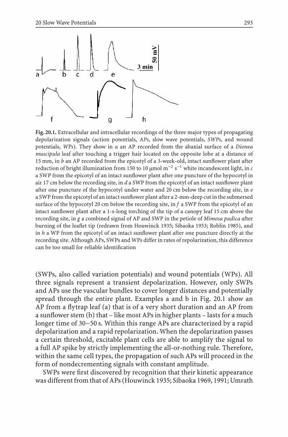
20 Slow Wave Potentials 293
Fig.20.1. Extracellular and intracellular recordings of the three major types of propagatingdepolarization signals (action potentials, APs, slow wave potentials, SWPs, and woundpotentials, WPs). They show in a an AP recorded from the abaxial surface of a Dioneamuscipula leaf after touching a trigger hair located on the opposite lobe at a distance of15 mm, in b an AP recorded from the epicotyl of a 3-week-old, intact sunflower plant afterreduction of bright illumination from 150 to 10 µmol m−2 s−1 white incandescent light, in ca SWP from the epicotyl of an intact sunflower plant after one puncture of the hypocotyl inair 17 cm below the recording site, in d a SWP from the epicotyl of an intact sunflower plantafter one puncture of the hypocotyl under water and 20 cm below the recording site, in ea SWP from the epicotyl of an intact sunflower plant after a 2-mm-deep cut in the submersedsurface of the hypocotyl 20 cm below the recording site, in f a SWP from the epicotyl of anintact sunflower plant after a 1-s-long torching of the tip of a canopy leaf 15 cm above therecording site, in g a combined signal of AP and SWP in the petiole of Mimosa pudica afterburning of the leaflet tip (redrawn from Houwinck 1935; Sibaoka 1953; Roblin 1985), andin h a WP from the epicotyl of an intact sunflower plant after one puncture directly at therecording site. Although APs, SWPs and WPs differ in rates of repolarization, this differencecan be too small for reliable identification
(SWPs, also called variation potentials) and wound potentials (WPs). Allthree signals represent a transient depolarization. However, only SWPsand APs use the vascular bundles to cover longer distances and potentiallyspread through the entire plant. Examples a and b in Fig. 20.1 show anAP from a flytrap leaf (a) that is of a very short duration and an AP froma sunflower stem (b) that – like most APs in higher plants – lasts for a muchlonger time of 30−50 s. Within this range APs are characterized by a rapiddepolarization and a rapid repolarization. When the depolarization passesa certain threshold, excitable plant cells are able to amplify the signal toa full AP spike by strictly implementing the all-or-nothing rule. Therefore,within the same cell types, the propagation of such APs will proceed in theform of nondecrementing signals with constant amplitude.
SWPs were first discovered by recognition that their kinetic appearancewas different from that of APs (Houwinck 1935; Sibaoka 1969, 1991; Umrath

294 R. Stahlberg, R.E. Cleland, E. Van Volkenburgh
1959). Electrical recordings in Mimosa showed an electrical excitation sig-nal in the form of a sequential combination of a short AP spike and a longerwavelike SWP (Fig. 20.1, trace g). Such a sequence was understood to implythat the AP moved faster through the Mimosa leaf and therefore appearedearlier at the recording site at the petiole base than the slow wave. The samesequence of signals was found in Vicia faba (Roblin 1985), but in sunflow-ers and cucumbers SWPs can be faster or slower than APs (Stankovic et al.1997; Stahlberg and Cosgrove 1997c).
Today the “slow” in SWP refers to the slow repolarization and the result-ing wavelike appearance rather than to inferior propagation rates (Fig. 20.1,traces e–g). To date a low rate is the most frequently applied criterion todifferentiate SWPs from APs (Stankovic et al. 1997; Dziubinska et al. 2001).However, even if measured in the same location (sunflower stem) SWPscan cover an astounding range in repolarization times and their resultingshapes closely approach that of either APs or WPs (Fig. 20.1, traces c–g).Repolarization times are extended after flame induction and root excision(Fig. 20.1, traces e and f), but short when initiated by a needle puncture(Fig. 20.1, traces c and d). SWP signals can be contaminated with actionspikes (Fig. 20.1, traces f and g). Such mixed signals have been foundamong many species, e.g., tomatoes, cucumbers and sunflowers (Roblin1985; Stahlberg and Cosgrove 1997c; Stankovic et al. 1998b). Moreover,uncontaminated SWPs occur in pea epicotyls (Stahlberg and Cosgrove1992, 1994, 1995, 1996, 1997c) and perhaps Tradescantia shoots (Tsaplevand Zatsepina 1980). In addition to the slower repolarization, uncontam-inated SWPs show slower depolarizations than APs and a round ratherthan pointed signal shape (see Fig. 20.1, traces e and g, plus SWPs frompea epicotyls in Stahlberg and Cosgrove 1992, 1996, 1997a). Unlike APs,SWP induction does not follow an all-or-nothing rule and SWP amplitudestherefore decrement during propagation (see later).
Finally, there are WPs as a direct depolarization response in the vicinityof injured cells (Fig. 20.1, trace h). WPs have very long repolarization timesand show a range from less than 1-mm to 40-mm distance (Shimmen2001; Stahlberg and Cosgrove 1994). The overlap in appearance of thedepolarization–repolarization events makes it difficult to distinguish thethree signals (Fig. 20.1); therefore, other distinguishing characteristics areneeded, e.g., stimuli causing SWPs to appear, ionic mechanisms mediatingdepolarization and repolarization, in rates, mechanisms and pathways ofSWP propagation. What then is a SWP and what are its characteristics?

20 Slow Wave Potentials 295
20.3SWPs are Hydraulically-Induced Depolarizations
The classic way to induce SWPs is to bring an open flame in contact witha leaf or another part of the plant (Houwinck 1935; Umrath 1959; Roblin1985; Wildon et al. 1992; Stankovic et al. 1997; Dziubinska et al. 2001).Flaming was considered as a model wound stimulation and an entire set ofindirect data suggested that the excitation in Mimosa and other plants wasmediated by the transpirational transport of wound substances emanatingfrom the burned site (Ricca 1916; Umrath 1959; Schildknecht 1984). How-ever, heat increases gas volume and pressure in the intercellular spaces (re-flectedasan increase in leaf thickness;Malone1992, 1996;Boari andMalone1993) and – more importantly – transiently increases volume and pressurein the narrow xylem conduits of the vascular bundles (Stahlberg and Cos-grove 1997c). Therefore, flaming acts as a strong hydraulic signal that ap-pears as a rapid increase in xylem pressure (Stahlberg and Cosgrove 1997c),turgor pressure (Malone and Stankovic 1991), growth rate (Stahlberg andCosgrove 1992, 1996), and leaf and stem thickness (Boari and Malone 1993).
The idea that hydraulic signals are accompanied by an electrical depolar-ization was clearly expressed by two independent studies in the early 1990s(Malone and Stankovic 1991; Stahlberg and Cosgrove 1992). A hydrauli-cally propagated signal had already been suggested to exist in flamed Mi-mosa leaflets (Haberlandt 1890) but experimental evidence for a hydraulicwave paralleling AP propagation did not materialize (Tinz-Fuchtmeyer andGradmann 1990). It was 100 years later that the exposure of the root of in-tact pea seedlings to modest pressure steps showed the appearance andpropagation of a well-resolved transient depolarization in the pea epicotyl(as in Fig. 20.6). This propagating electrical signal, however, was not an APbut had the typical shape and slow-repolarization characteristics of a SWP(Stahlberg and Cosgrove 1992, 1996, 1997a, 1997c).
The large, propagating depolarizations of a SWP are generated by theapplication of positive, not negative, steps in xylem pressure (Fig. 20.2b).Rapid axial propagation of the hydraulic signal is manifested by an almostimmediate water uptake into the apical growth zone in both pea and sun-flower shoots (Stahlberg and Cosgrove 1992, 1995, 1996; Stankovic et al.1997) and equally rapid changes in turgor and xylem pressure (Malone andStankovic 1991; Stahlberg and Cosgrove 1995). How does a pressure signalthat is almost instantly rising throughout the stem axis relate to an electricsignal that takes minutes to climb the stem? An analysis of this question inpea epicotyls found that the induced slow wave depolarizations increasedamplitude, rate and range in proportion to the size of the applied pres-sure steps while their lag phases were declining (Fig. 20.3; Stahlberg andCosgrove 1997a). Figure 20.3 explains an important point: depolarizations
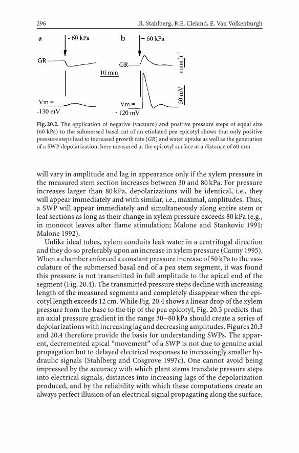
296 R. Stahlberg, R.E. Cleland, E. Van Volkenburgh
Fig.20.2. The application of negative (vacuum) and positive pressure steps of equal size(60 kPa) to the submersed basal cut of an etiolated pea epicotyl shows that only positivepressure steps lead to increased growth rate (GR) and water uptake as well as the generationof a SWP depolarization, here measured at the epicotyl surface at a distance of 60 mm
will vary in amplitude and lag in appearance only if the xylem pressure inthe measured stem section increases between 30 and 80 kPa. For pressureincreases larger than 80 kPa, depolarizations will be identical, i.e., theywill appear immediately and with similar, i.e., maximal, amplitudes. Thus,a SWP will appear immediately and simultaneously along entire stem orleaf sections as long as their change in xylem pressure exceeds 80 kPa (e.g.,in monocot leaves after flame stimulation; Malone and Stankovic 1991;Malone 1992).
Unlike ideal tubes, xylem conduits leak water in a centrifugal directionand they do so preferably upon an increase in xylem pressure (Canny 1995).When a chamber enforced a constant pressure increase of 50 kPa to the vas-culature of the submersed basal end of a pea stem segment, it was foundthis pressure is not transmitted in full amplitude to the apical end of thesegment (Fig. 20.4). The transmitted pressure steps decline with increasinglength of the measured segments and completely disappear when the epi-cotyl length exceeds 12 cm. While Fig. 20.4 shows a linear drop of the xylempressure from the base to the tip of the pea epicotyl, Fig. 20.3 predicts thatan axial pressure gradient in the range 30−80 kPa should create a series ofdepolarizationswith increasing lag anddecreasingamplitudes. Figures 20.3and 20.4 therefore provide the basis for understanding SWPs. The appar-ent, decremented apical “movement” of a SWP is not due to genuine axialpropagation but to delayed electrical responses to increasingly smaller hy-draulic signals (Stahlberg and Cosgrove 1997c). One cannot avoid beingimpressed by the accuracy with which plant stems translate pressure stepsinto electrical signals, distances into increasing lags of the depolarizationproduced, and by the reliability with which these computations create analways perfect illusion of an electrical signal propagating along the surface.

20 Slow Wave Potentials 297
Fig.20.3. A compilation of measurements shows that SWP characteristics such as lag phases(crosses) and amplitudes (circles) of SWP depolarizations depend on the size of the xylempressure steps (which are somewhat smaller than the actually shown pressure steps that wereapplied to the submersed cut end of an etiolated pea seedling at a distance of 60 mm from therecording site, see Fig. 20.4). Note that a propagating SWP appears only at lower pressuresteps. At pressures above 100 kPa the induced slow wave (SW) depolarizations becomeindistinguishable in time of appearance (lag phase→0 s) and amplitude. (Compiled fromunpublished and published data from Stahlberg and Cosgrove 1997a)
Fig.20.4.Regressionanalysis of pressurepropagation in epicotyl segmentsof various lengthsshows the linear dissipation of a constant pressure step of 50 kPa from the basal end ofapplication towards the apex. The basal ends of pea epicotyl segments 2−12 cm long weresubmersed and sealed into a pressure chamber while the apical end was sealed to a pressureprobe. A loss of about 4 kPacm−1 reflects the radial leakiness of the xylem. (Redrawn fromStahlberg and Cosgrove 1997a)

298 R. Stahlberg, R.E. Cleland, E. Van Volkenburgh
The SWP phenomenon suggests that the epidermal cells of many speciesandorgans showadepolarizationasa consequenceof a rapidly rising turgorpressure. The quantitative characterization of this relationship, e.g., withthe use of a combination probe measuring turgor pressure and membranepotential, would fill the remaining void about the hydraulic induction of theSWP. Pressure steps can become severely reduced on their centrifugal pathfrom the xylem to the epidermis (Westgate and Steudle 1985). Applicationof 100 kPa pressure steps to 3 cm-long epicotyl segments was insufficientto cause a measurable increase in epidermal turgor pressure (Stahlbergand Cosgrove 1992). Stankovic and Malone (1991) measured large turgorpressure changes in the epidermal cells of torched wheat leaves but notthe increase in xylem pressure. Together with the radial dissipation ofxylem pressure steps, a parallel study of the radial propagation of thedepolarization from the vascular bundles to the epidermis would be usefulto fully understand the conversion of pressure into electrical signals. So farsuch studies exist only for APs (Rhodes et al. 1996; Herde et al. 1998).
Induction of SWPs by small pressure steps applied without injury tointact plants also presents a powerful argument against participation ofchemical wound factors and for a purely hydraulic induction of SWPs(Stahlberg and Cosgrove 1995, 1996). In spite of this, it is still consid-ered that some plant species may use electrogenic substances to inducepropagating electrical signals. The idea draws support from the findingthat raw extracts from Mimosa, Biophytum and tomato plants were able toinduce propagating depolarizations (Ricca 1916; Umrath 1959; Van Sam-beek et al. 1976; Cheeseman and Pickard 1977; Sibaoka 1997). On the otherhand, feeding of wound sap to excised pea epicotyls showed clearly thatpeas do not use wound substances for SWP generation (Stahlberg andCosgrove 1992). Figure 20.5 shows a test of whether the xylem-mediatedtransport of strongly depolarizing agents like cyanide and azide is capableof generating a propagating depolarization in excised sunflower shoots.The induced signal moves slowly (less than 1 mm s−1) in comparison witha hydraulically induced SWP (5−10 mm s−1; e.g., Fig. 20.6) with depolariza-tions being sustained rather than transient. In order to produce SWP-likesignals, potential excitation substances must (1) be shown to accumulatein sufficient quantity to cause a large and rapid depolarization, (2) be ableeasily to access and exit the vascular bundles and (3) cause transient de-polarizations. None of the many SWPs recorded so far have been shown tofulfill these criteria.
While experimental methods of induction explore signal character andeffects, it is equally important to find natural circumstances under whichplants generate SWPs. Such situations include puncture wounds by sap-sucking insects (AlarconandMalone1994;VolkovandHaak1995;Fig. 20.6);embolisms (Stahlberg and Cosgrove 1996), soil hydration during rains and

20 Slow Wave Potentials 299
Fig.20.5. After the submersed excision of the lower hypocotyl of an intact sunflower plantand a waiting period of 60 min that allowed the ascending transpiration stream to returnto its normal rate, the basal end of the shoot was subjected to a 5 mM solution of sodiumazide (arrow), an electrogenic model substance known to cause a large depolarization inplant cells. The transport and release of this substance from the xylem causes a wave ofdepolarizations that ascends the stem from the hypocotyl (lower trace; the distance to thebasal cut was adjusted to be 10 cm) to the epicotyl (center trace; the distance to the basalcut was 20 cm) to a leaf blade (upper trace; the distance to the basal cut was 30 cm). Thetransport of this depolarizing chemical produces a signal that differs from a hydraulic SWPby a much lower propagation speed and the absence of repolarization and transience
floods (Stahlberg et al. 2005a), and perhaps also the reestablishment of pos-itive xylem pressure during the night in root-pressure-generating plants,and strong bending of plants under wind and other mechanical influ-ences.
For as long as they have been recognized as different entities, SWPsand APs have been believed to originate from different causes (Sibaoka1953; Umrath 1959). While the cooling of roots and the application of smallelectric currents in the tissue seem to induce exclusively APs, the induc-tion by heat (leaf flaming) has been reported to induce SWPs as well as APs(Roblin 1985; Wildon et al. 1992; Stankovic et al. 1997, 1998). Repeated flam-ing of sunflower leaves changed the shape of the resulting stem responsefrom a clear SWP to an AP-like signal (Davies et al. 1991). These data in-dicate the possibility of an interaction between SWPs and APs that has notbeen investigated. Both APs and SWPs are propagated within the vascular

300 R. Stahlberg, R.E. Cleland, E. Van Volkenburgh
Fig.20.6. SWPs in the stem of intact sunflower plants arise after a puncturing wound ineither a nonsubmersed (a) or a submersed (b) part of the hypocotyl with a 0.4-mm-wideneedle.Causedby thedifferencebetweenexternal atmosphericpressureanda transpiration-dependent tension inside the plant, a pressure wave ascends from the wound up the stemin the punctured vascular bundle(s) with high speed and is followed by the appearanceof a slower moving wave of depolarizations at the stem surface. In the case of a limitedwater supply at the puncture wound, the hydraulic signal exhausts itself and the range ofthe SWP ends between the first and the second electrode position at the stem (a). When thepuncture is made in the submersed hypocotyl, the SWP becomes systemic, i.e., it increasesits range to include all electrode positions along the stem (b). The data show that the rangeof wound-induced SWP depends on the availability of apoplastic water near the wound side
Table 20.1. Characteristics of slow wave potentials in comparison with action potentials andwound potentials. For details and references about the statements see the text
Characteristics of Slow wave potentials Action potentials Wound potentials
Induction Increase in turgor Depolarization Turgor lossand xylem pressure beyond of neighboring
certain threshold cells
Amplitude Graded; Amplitude fixed Graded; dependsamplitude function by all-or-nothing on degreeof stimulus size principle of injury
Induction methods Heat, wounding, Heat, cold, Woundingpressure chamber touch
Ionic mechanism P-type H+ pump, Ca2+, Cl2− P-type H+ pump,± ion channels? ion channels ±ion channels?
Repolarization Slow; > 1−30 min Quick; < 1 min Slow; 3−90 min
Propagation As pressure As electric Unknownsignal in vascular signal in vascularbundles/xylem bundles/phloem

20 Slow Wave Potentials 301
bundles (Table 20.1). Since the depolarization of a SWP lingers longer thanthat of an AP, SWPs may be more effective than APs in triggering theopening of excitable, i.e., voltage-gated Ca2+ and anion channels neededfor AP induction and propagation. SWPs in Vicia, cucumber and sunflowerplants are frequently accompanied by action spikes (Roblin 1985; Roblinand Bonnemain 1985; Stahlberg and Cosgrove 1994, 1997a; Stankovic andDavies 1998; Stankovic et al. 1997). Conversely, the fact that no depolariza-tion has ever been reported to cause a propagating SWP suggests that APsare unable to trigger SWPs.
20.4The Propagation of SWPs
According to previously introduced considerations (Figs. 20.3 and 20.4),SWPs only undergo an apparent propagation with an appearance and rangethat is determined by the gradual decline of the inducing pressure signal.Unlike flames and pressure chambers, wounding by puncturing, surfacecuts and organ excision generates a hydraulic signal that is based on exist-ing pressure gradients in the plants (Stahlberg and Cosgrove 1997c). Thisprovides useful information of how, e.g., species, age, light and other envi-ronmental conditions affect the ability of plants to generate SWPs as wellas their size and range (Alarcon and Malone 1994; Stahlberg and Cosgrove1994, 1995; Stahlberg et al. 2005). An example is given in Fig. 20.6.
Figure 20.6, example B shows the rapid ascent of a SWP from a punc-ture wound in the hypocotyl to the upper hypocotyl, epicotyl and leaf inan intact, illuminated sunflower plant. When the nonsubmersed part ofa hypocotyl was punctured, the limited water supply near the puncturedvascular bundle supported only a SWP with a short range limited to thehypocotyl (Fig. 20.6, example A). When punctured under water, the gener-ated SWP had an increased, systemic range. The increased range is a clearindication of the hydraulic nature of SWP propagation. Using a stimulusthat is close to the small scale of insect-inflicted wounds, we present herea short SWP that could be more representative than those published beforeas a consequence of larger injuries. It propagates with the same rate as thoseinduced by root excision and shows the same range extension to woundingunder water (Stahlberg et al. 2005b).
Although propagation rates of SWPs and APs overlap (Roblin 1985; Rob-lin and Bonnemain 1985; Stankovic et al. 1997), SWPs have four distinctivefeatures that clearly separate their movement from that of APs. First, SWPspropagate with a measurable decrement (loss of amplitude) along the plantsurface. Excision-induced SWPs in sunflower stems have a decrement of2.5% cm−1 (Stahlberg et al. 2005b). Second, SWPs propagate from the stem

302 R. Stahlberg, R.E. Cleland, E. Van Volkenburgh
into the leaves and vice versa, whereas APs (at least in sunflowers) cannotdo so (Dziubinska et al. 2001; Stahlberg et al. 2005b). A third defining par-ticularity of hydraulic propagation is that it enables SWPs – but not APs –to pass through stem sections where the living cells were killed or poisoned(Stahlberg and Cosgrove 1992). Fourth, SWPs depend on the pressure gra-dient between the atmosphere and an intact plant interior being undertension. Means to specifically suppress SWP propagation include workingwith excised sections or intact plants under water or an atmosphere of100% humidity (Mancuso 1998; Stahlberg et al. 2005b).
20.5The Ionic Mechanism of SWPs
A highly negative membrane potential in plant cells characterizes an activestate that involves the participation of the P-type H+ pump and generateslarge potential gradients for ions such H+, K+, Ca2+ and Cl− across theplasma membrane. Three major ion fluxes can generate large depolariza-tions in such cells: (1) an increased inward flow of Ca2+ ions from the cellwalls into the cytoplasm, (2) an increased outward flow of Cl− and otheranions and (3) a rapid stop in the outward pumping of H+ ions by theP-type ATPase.
While plant APs have been shown to involve the opening of ion channels(Sibaoka 1969, 1991; Fromm and Bauer 1994); SWPs are thought to reflecta transient shutdown of the P-type proton pump at the plasma membrane(Table 20.1). Evidence for this mechanism is that (1) SWPs depolarize cellsby a maximal amount of 90–100 mV, leaving the membrane potential ata negative voltage near the Nernst potential for K+ ions, (2) amplitudes ofthe SWP depolarization change continuously with the applied pressure size,(3) SW depolarizations are reduced or suppressed by the use of metabolicinhibitors (Julien et al. 1991; Stahlberg and Cosgrove 1992), (4) no mea-surable change in the cell-input resistance accompanied the large SWPdepolarization of pea epicotyl cells (Stahlberg and Cosgrove 1992, 1996,1997c), (5) SWP and the pH increase in the apoplast showed matching ki-netics when measured with the fluorescent indicator DM-NERF (Stahlbergand Cosgrove 1996), and that the growth rate of apical stems drops withthe arrival of the SWP signal (Stahlberg and Cosgrove 1997c; Stankovic etal. 1998). Being the fastest effect on this mechanism known (see Palmgren1998) the hydraulic or stretch-activated inhibition of the P-type H+ pumpdeserves more investigation. One cannot yet exclude that the ionic mecha-nism of SWPs could be more complex and in some species involve the par-ticipation of turgor-controlled or stretch-activated/inactivated ion chan-nels. Unlike pea epicotyls, epidermal cells of cucumber hypocotyls show

20 Slow Wave Potentials 303
transient increases in the cell input resistance during pressure-inducedSWPs (Stahlberg and Cosgrove 1997c).
Why is the repolarization of SWPs slower than in APs? Repolarization ofplant APs is believed to involve the combined action of voltage-dependentclosures of the depolarizing ion channels, the voltage-dependent activationof repolarizing K+ currents and an increased activity of P-type H+ pumps.Never going far beyond the Nernst potential for K+ ions, SWP depolar-izations are not likely to be compensated by large outward K+ currents.Another cause for the delayed repolarization of SWPs is the eliminationof the role P-type H+ pumps play in the repolarization efforts of plantcells. If the second stage of AP repolarization is mediated by a P-type H+
pumping ATPase (as suggested by Orpitov et al. 2002 for cucurbit cells)a turgor-inhibited pump would explain the slower repolarization of SWPs.
SWPs share one important feature with APs and WPs; a refractory periodduring which the plant cells are unable to repeat the voltage signal whensubjected to the same stimulus (Zawadzki et al. 1991). When a sequence ofpressure steps was applied with 10 min intervals in between, all steps causeda transient increase in growth rate but only the first pressure applicationgenerated a SWP (Stahlberg and Cosgrove 1996). Systematic studies ofrefractory periods of SWPs in green plants are completely wanting.
20.6The Effects of SWPs: Targeted Organs
SWPs trail hydraulic signals and very few studies differentiate whether aneffect is caused by either the hydraulic or the electric component. One at-tempt was the comparison of the growth behavior of cucumber and peaseedlings before, during and after the passage of the electrical SWP signalinto the growth zone (Stahlberg and Cosgrove 1997c). Application of small,sustained pressure steps to the stem base rapidly and transiently increasedthe growth rate due to a hydraulically mediated increase in apoplastic (andturgor) pressure. The delayed appearance of the electrical signal in theapical growth zone coincided with an unexpected, drastic drop in growthrate. The sustained slow wave depolarization in cucumbers paralleled a sus-tained growth inhibition of their hypocotyls, while a transient slow wavedepolarization in peas had a transient effect on the epicotyl growth. Relatedresults from sunflowers show a sustained shrinking of the upper stem afterthe passage of a flame-stimulated SWP (Stankovic et al. 1998).
Mobility of SWPs in both directions of the plant axis suggests two po-tential targets: the growing shoots with young canopy leaves and the root.In addition to stem growth, dramatic responses have been reported forleaves known to undergo particularly large, amplified SW depolarizations

304 R. Stahlberg, R.E. Cleland, E. Van Volkenburgh
(Stahlberg et al. 2005b). Leaf effects range from shutdown of stomata(Wildon et al. 1993; Pena-Cortes et al. 1995), to shutdown of photosyn-thesis (Koziolek et al. 2003), increased production of jasmonic acid andup-regulated transcription of proteinase inhibitor II and calmodulin intomato plants (Herde et al. 1996; Stankovic and Davies 1998). Less is knownabout root responses to SWPs. Sunflower and cucumber plants develop rootpressure that is metabolically supported and sensitive to pressure signalsthat can eliminate it in a rapid and drastic all-or-nothing manner (Stahlbergand Cosgrove 1997b). Leaf flaming generates basipetal electrical and pres-sure signals that could switch the root pump off and could cause in thisway the observed flame-induced shrinking of sunflower stems (Stankovicet al. 1997). This has not been tested yet. Although there is almost no in-formation on SWPs in roots, early work in Vicia indicates that the proteinmetabolism in roots is as sensitive to hydraulic signals as in shoots (Theiletet al. 1982).
20.7WPs and SWPs
Although both APs and SWPs have been called wound signals, there isan electrical signal more deserving of this name. WPs occur not only inhigher plants, but also in excised plant organs, nonvascular plants and algae(Shimmen 2001). A tandem pair of Chara internodal cells is a simple sys-tem to study cellular interactions. When one cell is damaged or killed, theneighboring cell undergoes a WP in the form of a large transient depolar-ization, sometimes with and sometimes without the occurrence of spikes(Shimmen 2001). Although cellular networks are more complex, cucumberhypocotyls showed identical responses (StahlbergandCosgrove1994).WPsin cucumber hypocotyls extend for a distance of 40 mm (a length corre-sponding to about 200 epidermal cells), 10 mm in pea epicotyls (Stahlbergand Cosgrove 1994), 5 mm in corn roots (Chastain and Hanson 1982),and 1 mm in barley roots (Mertz and Higinbotham 1976). WPs appear asuniversal signals with specific extensions for different species and organs.Touch causes a similar response; a slowly repolarizing local depolarizationwith amplitudes that depend on the strength of the mechanical stimulus(Okamoto 1955; Zerrenthin and Stahlberg 1982).
Evidence exists from Chara tandem cells, sugar beet roots and cucumberhypocotyls that the size of the turgor pressure of the victim cell(s) beforeinjury affects the amplitude of the generated WPs (Kinraide and Wyse1986; Shimmen 2001; Stahlberg and Cosgrove 1997a). WPs in higher plantsseem to be caused by a rapid inhibition of P-type H+ pumps in the effectedcells (Chastain and Hanson 1982; Gronewald and Hanson 1980; Kinraide

20 Slow Wave Potentials 305
and Wyse 1986). Accordingly, WPs (1) are accompanied by a strong re-duction in the growth rate of the cucumber hypocotyl in a 40-mm rangefrom the wound site and (2) proceed without a change in cell input re-sistance (Stahlberg and Cosgrove 1994, 1997a). Although WPs and SWPsseem to share a similar ionic mechanism, WPs lack an important definingcharacteristic of SWPs: distant propagation (Table 20.1). SWPs may haveevolved as a type of propagating WP.
Haberlandt (1890) suggested the existence of hydraulically propagatedelectric potentials at a time when the only known electrical signals wereAPs. It took time to find such signals and to understand that they coincidewith SWPs rather than APs. We slowly begin to realize that SWPs are notsimply ubiquitous but characteristic, defining signals for higher plants thatare missing in lower plants or animals.
Acknowledgements. We thank Daniel J. Cosgrove (Pennsylvania State Uni-versity) for supporting early work on SWPs and William E. Bradley (Uni-versity of Washington; deceased) for stimulating recent studies on thistopic.
References
Alarcon J-J, Malone M (1994) Substantial hydraulic signals are triggered by leaf-bitinginsects in tomato. J Exp Bot 45:953–957
Boari F, Malone M (1993) Rapid and systemic hydraulic signals are induced by localizedwounding in a wide range of species. J Exp Bot 44:741–746
Burdon-Sanderson J (1873) Note on the electrical phenomena which accompany irritationof the leaf of Dionea muscipula in the excited and unexcited states. Proc R Soc Lond21:491–496
Canny MJ (1995) Apoplastic water and solute movement: new rules for an old space. AnnuRev Plant Physiol Plant Molec Biol 46:215–236
Cheeseman JM, Pickard BG (1977) Depolarization of cell membranes in leaves of Lycoper-sicon by extract containing Ricca’s factor. Can J Bot 55:511–519
Davies E, Zawadzki T, Witters D (1991) Electrical activity and signal transmission inplants: how do plants know? In: Penelk C, Greppin H (eds) Plant signaling, plasmamembrane and change of state. University of Geneva, Switzerland, pp 119–137
Dziubinska H, Trebasz K, Zawadzki T (2001) Transmission route for action potentials andvariation potentials in Helianthus annuus L. J Plant Physiol 158:1167–1172
FrommJ,BauerT(1994)Actionpotentials inmaize sieve tubeschangephloemtranslocation.J Exp Bot 273:463–469
Fromm J, Eschrich W (1988) Transport processes in stimulated and non-stimulated leavesof Mimosa pudica. Trees 2:7–24
Gunar II, Sinykhin AM (1962) A spreading wave of excitation in higher plants. Proc AcadSci USSR (Botany) 142:214–215
Gunar II, Sinykhin AM (1963) Functional significance of action currents affecting the gasexchange of higher plants. Sov Plant Physiol 10:219–226

306 R. Stahlberg, R.E. Cleland, E. Van Volkenburgh
Green TR, Ryan CA (1972) Wound-induced proteinase inhibitor in plant leaves: a possibledefense mechanism against insects. Science 175:776–777
Gronewald JW, Hansen JB (1980) Sensitivity of proton and ion transport mechanisms ofcorn roots injury to injury. Plant Sci Lett 18:143–150
Haberlandt G (1890) Das reizleitende Gewebesystem der Sinnpflanze. Thieme, LeipzigHerde O, Fuss H, Pena-Cortes H, Fisahn J (1995) Proteinase inhibitor II gene expression
inducedbyelectrical stimulationandcontrol of photosynthetic activity in tomatoplants.Plant Cell Physiol 36:737–742
Herde O, Atzorn R, Fisahn J, Wasternak C, Willmitzer L, Pena-Cortes H (1996) Local-ized wounding by heat initiates the accumulation of proteinase inhibitor II in abscisicacid deficient tomato plants by triggering jasmonic acid biosynthesis. Plant Physiol112:853–860
Herde O, Fuss H, Pena-Cortes H, Willmitzer L, Fisahn J (1998) Remote stimulation byheat induces characteristic membrane-potential responses in the veins of wild-type andabscisic acid-deficient tomato plants. Planta 206:146–153
Houwinck AL (1935) The conduction of excitation in Mimosa pudica. Rec Trav Bot Neerl32:51–91
Julien JL, Desbiez MO, de Jaeger G, Frachisse JM (1991) Characteristics of the wave ofdepolarization induced by wounding in Bidens pilosa L. J Exp Bot 42:131–137
Kinraide TB, Wyse RE (1986) Electrical evidence for turgor inhibition of proton extrusionin sugar beet taproots. Plant Physiol 82:1148–1150
Koopowitz H, Dhyse R, Fosket DE (1975) Cell membrane potentials of higher plants: changesinduced by wounding. J Exp Bot 26:131–137
Koziolek C, Grams TE, Schreiber U, Matyssek R, Fromm J (2003) Transient knockout ofphotosynthesis mediated by electrical signals. New Phytol 161:715–722
Malone M (1992) Kinetics of wound-induced hydraulic signals and variation potentials inwheat seedlings. Planta 187:505–510
Malone M (1996) Rapid, long-distance signal transmission in higher plants. Adv Bot Res22:163–228
Malone M, Stankovic B (1991) Surface potentials and hydraulic signals in wheat leavesfollowing localized wounding by heat. Plant Cell Environ 14:431–436
Malone M, Palumbo L, Boari F, Monteleone M, Jones HG (1994) The relationship betweenwound-induced proteinase inhibitors and hydraulic signals in tomato seedlings. PlantCell Environ 17:81–87
Mancuso S (1999) Hydraulic and electrical transmission of wound-induced signals in Vitisvinifera. Aust J Plant Physiol 26:55–61
Mertz SM, Higinbotham N (1976) Transmembrane electropotentials in barley roots asrelated to cell type, cell location, and cutting and aging effects. Plant Physiol 57:123-128
Okamoto H (1955) On the distribution of electric potential in the seedling of Vignasesquipedalis and its change by light stimulation. Bot Mag (Tokyo) 68:1–15
Opritov VA (1978) Propagating excitation and assimilate transport in the phloem. Sov PlantPhysiol 25:1042–1048
Opritov VA, Pyatygin SS, Vodeneev VA (2002) Direct coupling of action potential generationin cells of a higher plant (Cucurbita pepo) with the operation of an electrogenic pump.Russ J Plant Physiol. 49:142–147
Palmgren MG (1998) Proton gradients and plant growth: role of the plasma membraneH+-ATPase. Adv Bot Res 28:1–70
Pena-Cortes H, Fisahn J, Wilmitzer L (1995) Signals involved in wound-induced proteinaseinhibitor II gene expression in tomato and potato plants. Proc Nat Acad Sci USA 92:4106–4113
Pickard BG (1973) Action potentials in higher plants. Bot Rev 39:172–201

20 Slow Wave Potentials 307
Rhodes JD, Thain JF, Wildon DC (1996) The pathway for systemic electrical signal conduc-tion in the wounded tomato plant. Planta 200:50–57
Ricca U (1916) Soluzione d’un problema di fisiologia: la propagazione di stimulo nellaMimosa. Nuovo G Bot Ital 23:51–170
Roblin G (1985) Analysis of the variation potential induced by wounding in plants. PlantCell Physiol 26:455–461
Roblin G, Bonnemain J-L (1985) Propagation in Vicia faba stem of a potential variation bywounding. Plant Cell Physiol 26:1273–1283
Schildknecht H (1984) Turgorins – new chemical messengers for plant behavior. Endeav NS8:113–117
Sibaoka T (1953) Some aspects of the slow conduction of stimuli in the leaf of Mimosapudica. Sci Rep Tohoku Univ 20:72–88
Sibaoka T (1969) Physiology of rapid movements in higher plants. Annu Rev Plant Physiol20:165–184
Sibaoka T (1991) Rapid plant movements triggered by action potentials. Bot Mag (Tokyo)104:73–95
Sibaoka T (1997) Application of leaf extract causes repetitive action potentials in Biophytumsensitivum. J Plant Res 110:485–487
Shimmen T (2001) Electrical perception of “death message” in Chara: involvement of turgorpressure. Plant Cell Physiol 42:366–373
Sinyukhin AM, Britikov EA (1967) Action potentials in the reproductive system of plants.Nature 215:1278–1280
Spanjers AW (1981) Biolelectric potential changes in the style of Lilium longiflorum Thunb.after self- and cross-pollination of the stigma. Planta 153:1–5
Stahlberg R, Cosgrove DJ (1992) Rapid alteration in growth rate and electric potentials uponstem excision in pea seedlings. Planta 187:523–531
Stahlberg R, Cosgrove DJ (1994) Comparison of electric and growth responses to excisionin in cucumber and pea seedlings. I. Short-distance effects are due to wounding. PlantCell Environ 18:33–41
Stahlberg R, Cosgrove DJ (1995) Comparison of electric and growth responses to excisionin cucumber and pea seedlings. II. Long-distance effects are due to hydraulic signals.Plant Cell Environ 18:33–41
Stahlberg R, Cosgrove DJ (1996) Induction and ionic basis of slow wave potentials inseedlings of Pisum sativum L. Planta 200:416–425
Stahlberg R, Cosgrove DJ (1997a) The propagation of slow wave potentials in pea epicotyls.Plant Physiol 113:209–217
Stahlberg R, Cosgrove DJ (1997b) Mannitol inhibits growth of intact cucumber but not peaseedlings by mechanically collapsing the root pressure. Plant Cell Environm 20:1135–1144
Stahlberg R, Cosgrove DJ (1997c) Slow wave potentials in cucumber differ in form andgrowth effect from those in pea seedlings. Physiol Plant 101:379–388
Stahlberg R, Cleland RE, Van Volkenburgh E (2005a) Propagating electrical signals can beinduced by environmental stimuli. Abstracts of the American Society of Plant Biologists
Stahlberg R, Cleland RE, Van Volkenburgh E (2005b). Decrement and amplification ofslow wave potentials during their propagation in Helianthus annuus L. shoots. Planta220:550–558
Stankovic B, Davies E (1996) Both action potentials and variation potentials induce pro-teinase inhibitor gene expression in tomato. FEBS Lett 390:275–279
Stankovic B, Davies E (1998) The wound response in tomato involves rapid growth andelectric responses, systemically up-regulated transcription of proteinase inhibitor andcalmodulin and down-regulated translation. Plant Cell Physiol 39:268–274

308 R. Stahlberg, R.E. Cleland, E. Van Volkenburgh
Stankovic B, Zawadzki T, Davies E (1997) Characterization of the variation potential insunflower. Plant Physiol 115:1083–1088
Stankovic B, Witters DL, Zawadzki T, Davies E (1998) Action and variation potentials insunflower: an analysis of their relationship and distinguishing characteristics. PhysiolPlant 103:51–58
Theilet C, Delpeyroux F, Fiszman M, Reigner P, Esnault R (1982) Influence of excisionshock on on the protein metabolism of Vicia faba L. meristematic root cells. Planta155:478–485
Tinz-Fuchtmeyer A, Gradmann D (1990) Laser-interferometric re-examination of rapidconductance of excitation in Mimosa pudica. J Exp Bot 41:15–19
Tsaplev YB, Zatsepina GN (1980) The electrical nature of the propagation of the variationpotential in Tradescantia. Biofizika 25:723–728
Umrath K (1959) Der Erregungsvorgang. In: Ruhland W (ed) Handbuch der Pflanzenphys-iologie, vol 17. Springer, Berlin Heidelberg New York, pp 24–110
Van Sambeek JW, Pickard BG (1976) Mediation of rapid electrical, metabolic, transpirationaland photosynthetic changes by factors released from wounds. Can J Bot 54:2662–2671
Volkov AG, Haak RA (1995) Bioelectrochemical signals in potato plants. Russ J Plant Physiol42:17–23
Westgate ME, Steudle E (1985) Water transport in the midrib of tissue of maize leaves. Directmeasurement of the propagation of changes in cell turgor across a plant tissue. PlantPhysiol 78:183–191
Wildon DC, Thain JF, Minchin PEH, Gubb IR Reilly AJ, Skipper YD, Doherty HM, O’Don-nell PJ, BowlesDJ (1992)Electrical signalingand systemicproteinase inhibitor inductionin the wounded plant. Nature 360:62–65
Zawadzki T, Davies E, Dziubinska H, Trebasz K (1991) Characteristics of action potentialsin Helianthus annuus. Physiol Plant 83:601–604
Zerrenthin U, Stahlberg R (1981) Die Nutzung der bioelektrischen Kurzzeitreaktion aufmechanische Reizung zur Ermittlung von Kennwerten und Sortenunterschioeden beiGetreidekeimpflanzen. Arch Acker- Pflanzenbau und Boden 25:197–203

21 Electrical Signals, the Cytoskeleton,and Gene Expression: a Hypothesison the Coherence of the CellularResponses to Environmental InsultEric Davies, Bratislav Stankovic
Abstract When plant tissue is abiotically injured by crushing, cutting, heat-wounding,electrical stimulation, or by several other means, the injured (perceiving) tissue generateselectrical signals (action potentials and variation potentials) and transmits them to distant(responding) tissue. Here they evoke apparently disparate responses, such as callose forma-tion, closing of plasmodesmata, stoppage of cytoplasmic streaming, inhibition of ribosomemovement along messenger RNA (mRNA), and ultrarapid but transient accumulation ofover 100 transcripts, which are degraded without being translated. These apparently dis-parate responses can be reconciled by one fundamental hypothesis that assumes that “theplant does not know what hit it” and thus “expecting the worst” mounts a holistic defenseresponse against its most potent nemesis, a putative viral invasion. We postulate that the ba-sis for this response is calcium influx into the cytoplasm via voltage-gated channels (actionpotential) associated with the microtubules, or via mechano-sensitive channels (varia-tion potential) associated with microfilaments. The calcium interacts with calcium and/orcalmodulin-dependent cytoskeleton-associated protein kinases. This causes the phospho-rylation of myosin, which stops cytoplasmic streaming, and of elongation factor 2F, whichslows elongation and termination and causes ribosomes to pile up on polyribosomes. Thisdecreases protein synthesis, but protects preexisting “host” transcripts from degradation.The phosphorylation signal then passes into the nucleus, where it phosphorylates RNApolymerase II, which goes into overdrive (i.e., does not stop at accuracy checkpoints), thuscausing the synthesis of large amounts of mismade mRNA. The mRNA is transported intothe cytoplasm, where it is scanned (checked for accuracy) by ribosomes, and found to be in-correct. This surveillance mechanism stimulates ribonuclease activity, which degrades thefree (non-polysome-associated), mismade RNA, but leaves the original, “host” transcriptsunscathed since they are protected by ribosomes. The ribonuclease also (and here is thecrux of the matter) attacks other free mRNAs, including viral mRNAs, so these are disposedof before they can be translated. Within minutes this reaction is over, cytoplasmic steamingresumes, translation continues, ribosomes are released and so can be used to translate new(correctly made) transcripts.
21.1Introduction to the Hypothesis
There are several different kinds of electrical activities in plants, includingaction potentials (APs), variation potentials (VPs), voltage spikes or volt-age transients, and rhythmic electrical activities but their role is far fromunderstood (Davies 1987b, 2004; Davies et al. 1991). They all involve ionfluxes across membranes and bring about changes in membrane potential.
Communication in PlantsF. Baluška, S. Mancuso, D. Volkmann (Eds.)© Springer-Verlag Berlin Heidelberg 2006

310 E. Davies, B. Stankovic
It is assumed that they are a means of communication (i.e., intercellularsignals) that can be generated and transmitted much more rapidly thancan conventional chemical (hormonal) signals.
The two major types of putative signal are APs and VPs (Fig. 21.1). APsare defined as all-or-nothing, self-perpetuating signals, which are trans-mitted with essentially constant velocity and magnitude and are driven byvoltage-gated channels (Zawadzki et al. 1991). Plant APs are very unlikeneuronal APs, but are very similar to APs in other animal tissues (heart,epithelium). The neuronal AP can be thought of as the biological equivalentof a telephone, being designed for maximum rate of information transmis-sion (velocity) and minimum amount of information leakage (security).This is done by having an ion-impermeable sheath over much of the neu-
Fig.21.1. Typical action potentials (APs) and variation potentials (VPs) measured in sun-flower. a The plant was stimulated electrically (5 V for 1 s) at a point about 5 cm below thelowest petiole (±) or heat-wounded with a gentle flame applied to the tip of a leaf (W).Measuring electrodes (inserted silver wires) were placed along the stem, and a referenceelectrode was placed in the pot. The electrical responses to electrical stimulus are actionpotentials that are shown in b. The electrical responses to the heat-wounding stimulus arevariation potentials that are shown in c. Note that electrical stimulation evoked a pure AP,while flame-wounding evoked a combined AP/VP, with the former “traveling” faster thanthe latter (Reproduced from Davies et al. 1991, with permission)

21 Wounding, electrical signals, the cytoskeleton, and gene expression 311
ron’s length. This prevents the tissue through which it is passing being“informed”, and also speeds up the rate of transmission by having thesignal (ion flux, change in membrane potential) jump from “leak-point”to “leak-point.” In contrast, APs in plants and in nonneuronal tissues canbe thought of as megaphones, where signal velocity is reduced in order tomaximize information spread, i.e., all the cells on the pathway (especiallythe phloem) are informed, and presumably modified, by the passing signal(Davies 1987a, 1987b; Zawadzki et al. 1991).
The VP differs considerably from the AP; it is not all-or-nothing, nor isit self-perpetuating (Stankovic et al. 1997c), rather it is a hydraulic surge,or possibly a transported chemical (Malone 1996) in the xylem evokinglocal changes in membrane potential in adjacent living cells. The rapid lossof tension in (dead) xylem elements is transduced into local changes inion flux through mechano-sensory ion channels in the adjacent living cells(Stankovic et al. 1997b,c, 1998) or perhaps via ligand-activated channels ifthere is a chemical transported (Malone 1996). Thus, continuing with theinformation transmission analogy, the VP could be likened unto a radiosignal being broadcast throughout the plant and “heard” on all the cells inthe vicinity.
Regardless of the exact mechanism of transmission of APs and VPs,the responses they evoke must depend on either the ions traversing themembrane or the change in membrane potential, or both. Evidence sug-gests that both signals might involve calcium influx followed by chlorideand potassium efflux. However, even if the same ions are involved, thedownstream events might not be the same, since the flux of any ion willdepend on the kind, location, number, connections, and other proper-ties of the channel through which it passes. For instance, voltage-gatedcalcium channels involved in APs might open only transiently, be few innumber, be located primarily in the phloem along the longitudinal axisand be connected to the microtubules (Thuleau et al. 1998), where theycan interact with microtubule-associated Ca-binding proteins. In contrast,mechano-sensitive calcium channels involved in VPs might be open forlonger periods, be abundant, be located primarily in living cells adjacentto the xylem around the entire cell and be connected to the microfilamentswhere they can interact with microfilament-associated Ca-binding proteins(Davies 1993; Wang et al. 2004). Finally, voltage-gated, mechano-sensitive,and especially ligand-activated calcium channels are likely to release somecalcium into the soluble phase in the vicinity of the plasma membrane,where they can activate enzymes such as phospholipase C which will re-lease another second messenger, IP3, from membranes, which, in turn,causes the release of more calcium from internal stores (Heilmann et al.2001).

312 E. Davies, B. Stankovic
21.2Evidence for Our Hypothesis
Since this paper is a hypothesis based on limited evidence, we will focuson those aspects of research conducted in our laboratory, supplementedwhere necessary by work from others.
21.2.1Electrical Signals and Translation
We began to suspect a role for electrical signals and their involvement inwound-modulated gene expression in plants (aged etiolated pea epicotyls)when we found that polysome formation occurred very rapidly both aboveand below the site of wounding, implying that a rapidly generated bidirec-tionally transmitted signal, presumably an electrical signal, was involved(Davies and Schuster 1981). Since there was only a slight increase in thepoly(A)RNA content of the polysome fraction, but a huge increase in the ca-pacity to make protein both in vivo and in vitro, we assumed that woundingwas increasing translation, presumably by increasing ribosome initiation(Davies and Schuster 1981).
This interpretation received support from subsequent experiments,which showed that the reinitiating ability in vitro of ribosomes isolatedfrom wounded peas was markedly increased (Ramaiah and Davies 1985).However, it was subsequently disproved when the experiments were redoneso that protein synthesis in vivo was conducted using totally intact (un-wounded) tissue (Davies et al. 1986), rather than in tissue slices (wounded)as had been done earlier (Davies and Schuster 1981; Schuster and Davies1983).Using the totally intact systemweshowedthat, even thoughpolysomeformation was occurring, protein synthesis in vivo declined markedly(Fig. 21.2). This effect of wounding (formation of polysomes, inhibitionof protein synthesis) could be mimicked in unwounded tissue by cyclohex-imide (Davies at al. 1986), a known inhibitor of elongation/termination.It became apparent that wounding was blocking ribosome movementalong messenger RNA (mRNA), and we now suspect that this results fromphosphorylation of the translation factor EF2, presumably via a calcium-dependent protein kinase.
21.2.2Calcium, the Cytoskeleton, and Translation
We reasoned that if influxes of calcium occurred as part of the woundsignal (AP), then cytoplasmic streaming should be inhibited, but to check

21 Wounding, electrical signals, the cytoskeleton, and gene expression 313
Fig.21.2.Polysome formation and protein synthesis. Intact tissue – 2-cm-long aged epicotylswhich had been excised 2 days earlier (to provide a cut surface for amino acid uptake andto permit recovery from the wound) were left undisturbed (unwounded), while others werenicked at the apex (wounded) for various time periods and then placed with their basesin labeled methionine for 30 min and then the basal 2 mm was extracted and assayed forprotein synthesis. Similar samples were frozen in liquid nitrogen immediately after the endof the wound treatment and used for polysome isolation. Excised tissue – similar epicotylswere used, but the 2-mm basal piece was excised before the period of incubation andassay for polysomes. Note that similar polysome formation occurred in both tissues afterwounding, but intact tissue exhibited a massive initial decrease in protein synthesis, whileexcised tissue exhibited an ongoing increase (Reproduced from Davies et al. 1986, withpermission)
this we needed a system that would allow continuous monitoring of cyto-plasmic streaming as well as ready uptake of exogenous compounds. Forthese studies we chose the aquatic plants, Elodea and Vallisneria (Davies1990). We showed that wounding did, indeed, inhibit both protein synthe-sis (Fig. 21.3a) and cytoplasmic streaming (Fig. 21.3b) in these plants, asdid treatment of unwounded plants with calcium plus ionophore, whileremoval of calcium relieved the inhibition.
Taken together these data suggest that wounding causes an increase incytosolic calcium, which, in turn, inhibits both cytoplasmic streaming andprotein synthesis. The question then became “Does cytoplasmic streamingdepend on protein synthesis, does protein synthesis depend on cytoplasmicstreaming, or are both processes co-dependent on some other event?” Wefound no evidence for either interdependence, but did find evidence fora possible co-dependence – both processes might be governed by proteinphosphorylation.
It has been known for some time that phosphorylation of myosin inhibitscytoplasmic streaming in plants, while phosphorylation of EF2 inhibits

314 E. Davies, B. Stankovic
0
5
10
15
20
25
30
35
40
Intact Wounded Cytochalasin B
Incubation conditions
35S
-Met
upta
ke
(%)
a
0
10
20
30
40
50
60
70
80
90
100
110
0-10 10-20 20-30 30-40 40-50Time after treatment (min)
Nu
mb
erof
cell
sst
ream
ing
(%)
Intact cells
Wounded
Cytochalasin B
b
Fig.21.3. Protein synthesis and cytoplasmic streaming in Elodea. a Leaves of the aquaticplant Elodea were either submerged intact in artificial pond water (APW), 2-cm piecesexcised (wounded), or submerged intact into APW containing cytochalasin B and incubatedfor 1 h with labeled methionine to assay protein synthesis. Wounding and cytochalasin Bcaused similar inhibition (approximately 50%) of protein synthesis. b Similar samples wereincubated in theabsenceofmethionineandviewedcontinuouslyunder themicroscopeat10-min intervals to evaluate cytoplasmic streaming (Pfeifer and Davies, unpublished results).Note that wounding caused a consistent 45–50% inhibition of cytoplasmic streaming overthe 50-min time period, whereas cytochalasin B caused an increasing (85–98%) inhibitionover time, but removal of cytochalasin B relieved the inhibition (data not shown)
protein synthesis in animals by preventing ribosome movement along themRNA (Ryazanov et al. 1988; Shestakova et al. 1991). But this still did notadequately explain the similar kinetics of inhibition and recovery of bothprocesses. They were so tightly linked that they might be associated withthe same structure – and if so, that structure was most likely the cytoskele-ton. Already in the animal literature there was evidence for “detergent-resistant” polysomes that remained with the cell after all membranes hadbeen removed, and so we set out to determine if such “cytoskeleton-bound

21 Wounding, electrical signals, the cytoskeleton, and gene expression 315
polysomes” exist in plants. They do. They are present in pea stems, pearoots, corn endosperm, and every other tissue we have examined (Davieset al. 1996), and they seem to be more translationally active than freepolysomes (Davies et al. 1998). Indeed, the cytoskeleton seems to playa major role in the targeting, tethering, transport, and translation of mRNA(Davies et al. 2001).
21.2.3Calcium Channels, the Cytoskeleton, and Transcription
Our work initially had been on translation (protein synthesis); research inthis area seems to be less interesting (i.e., less funding is available) thanresearch on transcription. This need for funding motivated us to changetracks and work on the model system for wound-evoked changes in tran-scription– the tomatoplant.With this systemweshowed thatbothelectricalstimulation and heat-wounding evoked the accumulation of proteinase in-hibitor (PIN) transcripts, but only heat stimulus could evoke accumulationof calmodulin mRNA (Stankovic and Davies 1996, 1997a, 1997b).
While the bulk of workers looking at transcriptional responses to elec-trical signals wait several hours to make their first measurements, we con-jectured that if plants generate ultrarapid signals, then they presumablyexist in order to evoke ultrarapid responses. In our earlier experiments weisolated RNA within 15 min of wounding – and found some transcriptsshowed maximum accumulation at that time, before declining to basal lev-els and then rising again (Fig. 21.4). Interestingly, when we analyzed mRNAlevels in polysomes (the currently translated mRNA), we found that mostof the newly synthesized transcripts were not recruited into polysomes(i.e., never translated), but were degraded. These same transcripts usuallyincreased again at a later time and these later-synthesized transcripts wererecruited into polysomes for translation. Indeed, many transcripts showedthis four-phase pattern: (1) a period of rapid accumulation, followed by (2)a period of equally rapid decline, followed by (3) a period of slow accumu-lation along with (4) their recruitment into polysomes (Fig. 21.4). When weshortened the wounding period to 2 min and used Northern blots (Davieset al. 1997), quantitative PCR (Coker et al. 2005) and DNA microarrays (un-published data), we found over 20 transcripts reached a peak at that timepoint – some increasing as much as tenfold in 2 min. How could transcriptaccumulation occur so rapidly?
The simplest explanation for ultrarapid accumulation of transcripts isthrough enhancing transcription by activating the major enzyme respon-sible, RNA polymerase II (pol2). Evidence from the animal literature showsthat pol2 normally adds 100 or more nucleotides to the nascent transcript,

316 E. Davies, B. Stankovic
0
20
40
60
80
100
120
140
160
0 5 10 15 20 25
Time after wounding (min)
Rel
ativ
em
RN
Aac
cum
ula
tion
(%)
Total mRNA
Polysomal mRNA
I II
IIIIV
Fig.21.4. Wound-induced systemic accumulation of total and polysomal messenger RNA(mRNA). Plants were wounded at zero time. Total and polysomal mRNA were isolated andthe general pattern for 12 different mRNAs (including calmodulin, Rubisco SS, chloroplastmRNA-binding protein, bZIP DNA-binding protein) is shown. The four phases of transcriptaccumulation are indicated with Roman numerals (Davies et al., unpublished results).Note that the rapidly synthesized transcripts (phase 1) are degraded (phase 2) and notused in protein synthesis. The slowly accumulating transcripts (phase 3) are recruited intopolysomes (phase 4) for protein synthesis
but then pauses to check for accuracy. However, when it is phosphory-lated, it “goes into overdrive” (Landick 1999) and keeps adding nucleotideswithout checking for errors; thus, mRNA made by phosphorylated pol2is virtually certain to be error-ridden. An alternative explanation for theultrarapid increase in synthesis of mRNA is that pol2 copies one strand,then, rather than detaching and finding the appropriate start site on thesense strand, reveres direction and copies the nonsense strand (Cornel-lissen 1989). We have used primers for the antisense mRNA, but have notfound any; thus, the most likely explanation for ultrarapid transcript ac-cumulation is through speeding up (activation, phosphorylation) of pol2.This might take place via the same microfilament system used to phos-phorylate myosin and EF2, since it has been shown that actin filamentspenetrate directly into the nucleus and are associated with both pol2 andactive transcription sites (Hofmann et al. 2004).
What would be the consequences of the cell making error-ridden mRNA?First, the ribosome would scan it for accuracy, find it in error and preventother ribosomes from attaching to it. Activated ribonucleases (Abler andGreen 1996; Gutierrez et al. 2002; LeBrasseur et al. 2002) would then de-grade it in a wonderfully orchestrated surveillance mechanism (Kozak

21 Wounding, electrical signals, the cytoskeleton, and gene expression 317
Table 21.1. The first 10 min: early events following heat-wounding a leaf of a tomato plant
Duration (s) Response Effect Reference
< 1 Loss of xylem tension Systemic awareness Davies et al. (1991)throughout the plant of perturbation
1–3 Mechano-sensing Local awareness Davies (1993),calcium channels open of perturbation Fisahn et al. (2004)
3–10 Calcium influx Binds to Roberts andinto the cytoplasm calcium-binding Harmon (1992),
proteins on MF Davies et al. (1996)5–15 Activation “Current” Davies (1993),
of microfilament- of phosphorylation Wang et al. (2004)linked CDPK along MF
5–15 Phosphorylation Prevents Davies et al. (1996)(inactivation) cytoplasmicof myosin streaming
5–15 Phosphorylation Stops translation, Ryazanov et al. (1988),(inactivation) ribosomes protect Shestakova et al. (1991)of EF2 host mRNA
5–15 Phosphorylation Makes error-ridden Landick (2004),(activation) mRNA, goes Hofmann et al. (2004)of pol2 into cytoplasm
30–120 Mismade RNA Competes with viral Landick (2004)accumulates RNA for ribosomes
120–500 Ca activation Degrades Larkins andof RNase unprotected viral Davies (1973)
and mismade RNA120–500 Disappearance Newly made mRNA Fig. 21.4
of newly-made RNA never translated3–10 Calcium influx Binds Roberts and
into the cytoplasm to calcium-binding Harmon (1992)proteins near PM
5–15 Activation of Releases IP3 Heilmann et al. (2001)phospholipase C from membranes
20–60 IP3 passes from cell Calcium released from Heilmann et al. (2001)to cell internal stores
30–120 IP3/Ca stimulates Mismade Salinas-MondragónRNA synthesis transcripts et al. (2001),
accumulate York et al. (1999)120–600 Activation Degrades Larkins and
of RNase unprotected viral Davies (1973)and mismade RNA
120–600 Disappearance Newly made Fig. 21.4of newly mRNA nevermade RNA translated
MF microfilament, CDPK calcium-dependent protein kinase, pol2 RNA polymerase II,mRNA messenger RNA, PM plasma membrane
318 E. Davies, B. Stankovic
1992; Beelman and Parker 1995; Hentzke and Kolozik 1999). Thus, in ourheat-wounded tomato system, these rapidly synthesized transcripts arenot recruited into polysomes; instead they are degraded and never used forprotein synthesis. Thus heat-wounding of a mature leaf leads to a complexchain of events occurring systemically in the youngest leaf (summarized inTable 21.1).
21.3Conclusions and Perspectives:The “Help! It’s a Virus” HypothesisWhy would a cell make a copious amount of mRNA, but degrade it withoutusing it for protein synthesis. One possibility is that it has no function –but in this case one would expect it to be such an economic disadvantagethat it would have been weeded out by evolutionary pressure. Anotherpossibility is that the RNA, including viral RNA, is used for intercellular(interorgan) signaling. This does not seem to be the case, however, sincevirtually identical kinetics of transcript accumulation and disappearancetake place in other parts of the plant, so there is nowhere else for preexistingmRNA to originate.
Our preferred hypothesis is that when the plant’s integrity is compro-mised, the plant does not know what hit it, but mounts a defense againstits most potent nemesis, a virus. Thus, essentially the instant its integrity iscompromised, the wounded leaf informs the entire plant via the hydraulicsurge and calcium channels open in all the cells capable of detecting suchchanges in turgor. Subsequent inhibition of cytoplasmic streaming willslow down virus movement within the cell; plugging of plasmodesmatawill compartmentalize the virus and prevent its spread; slowing of ribo-some movement along preexisting polysomes will cause ribosome “pile-up” on preexisting transcripts, thus protecting them from degradation byRNase; increasing the rate of transcription will cause an increase in errorrate; flooding the cytoplasm with mismade RNA will lessen the availabilityof ribosomes to translate viral RNA as well as to stimulate breakdown ofunprotected RNA. Within minutes, the plant has repelled its enemy, these(automatic?) defensive reactions cease, and now, having identified the spe-cific nature of the recent insult, the plant generates an appropriate responseand concludes “All is well with the world.”
ReferencesAbler ML, Green PJ (1996) Control of mRNA stability in higher plants. Plant Mol Biol
32:63–71

21 Wounding, electrical signals, the cytoskeleton, and gene expression 319
Beelman CA, Parker R (1995) Degradation of mRNA in eukaryotes. Cell 81:179–183Coker JS, Vian A, Davies E (2005) Identification, accumulation, and functional prediction of
novel tomato transcripts systemically up-regulated after flame-wounding. Physiol Plant124:311–322
Cole CN (2000) mRNA export: the long and winding road. Nat Cell Biol 2:E55-E58Cornelissen M (1989) Nuclear and cytoplasmic sites for anti-sense control. Nucl Acids Res
17:7203–7209Cullen BR (2000) Connections between the processing and nuclear export of mRNA: evi-
dence for an export license. Proc Natl Acad Sci USA 97:46–54Davies E (1987a) Wound responses in plants. Biochem Plants 12:243–264Davies E (1987b) Action potentials as multi-functional signals in plants: A hypothesis at-
tempting to unify apparently disparate wound responses. Plant Cell Environ 10:623–631Davies E (1990) Plant wound signals and translation. In: Proceedings of the 13th interna-
tional congress on plant growth substances, pp 519–530Davies E (1993) Intercellular and intracellular signals in plants and their transduction via
the membrane-cytoskeleton interface. Semin Cell Biol 4:139–147Davies E (2004) Commentary: new functions for electrical signals in plants. New Phytol
161:607–610Davies E, Larkins BA (1974) Polyribosome degradation as a sensitive assay for endolytic
messenger ribonuclease activity. Anal Biochem 61:155–164Davies E, Schuster A (981) Intercellular communication in plants: evidence for a rapidly-
generated, bidirectionally-transmitted wound signal. Proc Natl Acad Sci USA 78:2422–2426
Davies E, Ramaiah KVA, Abe S (1986) Wounding inhibits protein synthesis yet stimulatespolysome formation in aged, excised pea epicotyls. Plant Cell Physiol 27:1377–1386
Davies E, Zawadzki T, Witters D (1991 Electrical activity and signal transmission in plants:how do plants know? In: Penel C, Greppin H (eds) Plant signaling, plasma membraneand change of state. University of Geneva, Switzerland, pp 119–137
Davies E, Fillingham BD, Abe S (1996) The plant cytoskeleton. In: Hesketh JE, Pryme LF(eds) The cytoskeleton, vol 3. JAI, Greenwich, CT, pp 405–449
Davies E, Vian A, Vian C, Stankovic B (1997) Rapid systemic up-regulation of genes afterheat-wounding and electrical stimulation. Acta Physiol Plant 19:571–576
Davies E, Abe S, Larkins BA, Clore AM, Quatrano RS, Weidner S (1998) The role of thecytoskeleton in plant protein synthesis. In: Bailey-Serres J, Gallie DR (eds) A look be-yond transcription: mechanisms determining mRNA stability and translation in plants.American Society of Plant Physiologists, Rockville, MD, pp 115–124
Davies E, Shimps B, Brown K, Stankovic B (1999) Gravity, stress, calcium and gene expres-sion. J Gravitat Physiol 6:21–22
Davies E, Stankovic B, Azama S, Shibata K, Abe S (2001) Novel components of the plantcytoskeleton: a beginning to plant “cytomics”. Plant Sci 160:185–196
Fisahn J, Herde O, Willmitzer L, Pena-Cortez H (2004) Analysis of the transient increasein cytosolic Ca2+ during the action potential of higher plants with high temporal reso-lution: requirement of Ca2+ transients for induction of jasmonic acid biosynthesis andPINII gene expression. Plant Cell Physiol 45:456–459
Gutierrez RA, Ewing RM, Cherry JM, Green PJ (2002) Identification of unstable transcriptsin Arabidopsis by cDNA microarray analysis: rapid decay is associated with a group oftouch- and specific clock-controlled genes. Proc Natl Acad Sci USA 99:11513–11518
Heilmann I, Perera IY, Stevenson JM, Boss WF (2001) Sense and sensibility: inositol phos-pholipids as mediators of abiotic stress responses. In: Cherry JH et al. (eds) Plant toler-ance to abiotic stresses in agriculture: role of genetic engineering. Kluwer, Dordrecht,pp 285–296

320 E. Davies, B. Stankovic
HentzeMW,KulozikAE(1999)Aperfectmessage:RNAsurveillanceandnonsense-mediateddecay. Cell 96:307–310
Hofmann WA et al. (2004) Actin is part of the pre-initiation complexes and is necessary fortranscription by RNA polymerase II. Nat Cell Biol 6:1094–1101
Kozak M (1992) Regulation of translation in eukaryotic systems. Annu Rev Cell Biol8:197-225
Landick R (1999) Shifting RNA polymerase into overdrive. Science 284:598–599Larkins BA, Davies E (1973) Polyribosomes from peas III: stimulation of polysome degra-
dation by exogenous and endogenous calcium. Plant Physiol 52:655–659LeBrasseur ND, MacIntosh GC, Perez-Amador MA, Saitch M, Green PJ (2002) Local and
systemic wound-induction of RNase and nuclease activities in Arabidopsis: RNS1 asa marker for a JA-independent systemic signaling pathway. Plant J 29:393–403
Malone M (1996) Rapid, long-distance signal transmission in higher plants. In: Callow JA(ed) Advances in botanical research. Academic, San Diego, pp 163–228
Ramaiah KVA, Davies E (1985) Wounding of aged pea epicotyls enhances the reinitiatingability of isolated ribosomes. Plant Cell Physiol 26:1223–1231
Ryazanov AG, Shestakova EA, Natapov PG (1988) Phosphorylation of elongation factor 2 byEF-2 kinase affects rate of translation. Nature 334:170–173
Salinas-Mondragón R, Atkinson CA, Davies E (2001) Ultra-rapid wound responses inplants: changes in inositol 1,4,5-trisphosphate (IP3) and gene expression. Poster pre-sented at Plant Biology 2001, 21–25 July 2001, Providence, RI, USA. Abstract 686,http://abstracts.aspb.org/pb2001/public/P45/0230.html
Schuster A, Davies E (1983) Protein and RNA metabolism in pea epicotyls. II. The responseto wounding. Plant Physiol 73:817–821
Shestakova EA, Motoz LP, Minin AA, Gelfand VI, Gavrilova LP (1991) Some of the eukaryoticelongation factor 2 is co-localized with actin microfilament bundles in mouse embryofibroblasts. Cell Biol Int Rep 15:75–84
Stankovic B, Davies E (1996) Both action potentials and variation potentials induce pro-teinase inhibitor gene expression in tomato. FEBS Lett 390:275–279
Stankovic B, Davies E (1997a) Intercellular communication in plants: electrical stimulationof proteinase inhibitor gene expression in tomato. Planta 202:402–406
Stankovic B, Davies E (1997b) Wounding evokes rapid changes in tissue deformation, elec-trical potential, transcription, and translation in tomato. Plant Cell Physiol 39:268–274
Stankovic B, Zawadzki T, Davies E (1997) Characterization of the variation potential insunflower. Plant Physiol 115:1083–1088
Stankovic B, Witters DL, Zawadzki T, Davies E (1998) Action potentials and variation poten-tials in sunflower: an analysis of their relationships and distinguishing characteristics.Physiol Plant 105:51–58
Stankovic B, Vian A, Henry-Vian C, Davies E (2000) Molecular cloning and characterizationof a tomato cDNA encoding a systemically wound-inducible bZIP DNA-binding protein.Planta 212:60–66
Thuleau P, Schroder JL, Ranjeva R (1998) Recent advances in the regulation of plant calciumchannels: evidence for regulation of G-proteins, the cytoskeleton and second messen-gers. Curr Opin Cell Biol 1:424–427
Wang YF, Fan L-M, Zhang W-Z, Zhang W, Wu W-H (2004) Ca2+-permeable channels in theplasma membrane of Arabidopsis pollen are regulated by actin microfilaments. PlantPhysiol 136:3892–3904
York JD, Odom AR, Murphy R, Ives EB, Wente SR (1999) A phospholipase C-dependentinositol polyphosphate kinase pathway required for efficient mRNA export. Science285:96–100
Zawadzki T, Davies E, Dziubinska H, Trebacz K (1991) Characteristics of action potentialsin Helianthus annuus L. Physiol Plant 83:601–604

22 Characteristics and Functionsof Phloem-Transmitted ElectricalSignals in Higher PlantsJörg Fromm, Silke Lautner
Abstract Electrical signalling along the phloem has been studied in a number of specieslike maize, willow and Mimosa. It appears that sieve tubes with their large sieve pores areused to transmit information over long distances, while plasmodesmata serve as a means forthe propagation of electrical signals over short distances between cells. By using the aphidtechnique the phloem pathway has been shown to transmit action potentials with a velocityup to 10 cm s−1. With regard to the ion fluxes which create the conditions necessary forthe generation of an action potential, we found that calcium influx as well as potassiumand chloride efflux are involved. Some of their corresponding ion channels were identified.AKT2/3-like channels, expressed in the phloem and capable of mediating both uptake andreleaseofK+ in response to changes inmembranepotential,were identified in several speciessuch as Arabidopsis, maize and broad bean. Concerning physiological functions of electricalsignalling, evidence was found for a link between the signals and photosynthetic response inMimosa, apart from the regulation of rapid leaf movements. In addition, electrical signalsin maize play a role in the regulation of phloem transport as well as in root-to-shootcommunication of entire plants.
22.1IntroductionAlmost the entire chemistry of the neuromotoric system in animals is alsoavailable to plants. They do not possess nerves but neurotransmitters suchas acetylcholine, cellular messengers like calmodulin, cellular motors, e.g.actin and myosin, voltage-gated ion channels and sensors for touch, light,gravity and temperature. This exciting cellular chemistry raises the ques-tion: Why do plants need cellular equipment similar to nerves? Althoughthe degree of development and complexity of plant cells is not comparableto that of the nervous system in animals, plants seem to have developeda simple neural network within the phloem which serves the communi-cation between plant organs over long distances. They have presumablydeveloped such a system in order to cope in the best possible way withenvironmental stress factors. Having sensed environmental stimuli, thesensor region needs to transmit a signal to the responding region. The na-ture of this signal may be chemical (e.g. hormonal), hydraulic (e.g. pressurechanges) or electrical (e.g. ionic). The electrical signals, in particular, havethe capacity to rapidly transmit information over long distances. However,the conduction rate of most of the plant action potentials studied so far is
Communication in PlantsF. Baluška, S. Mancuso, D. Volkmann (Eds.)© Springer-Verlag Berlin Heidelberg 2006

322 J. Fromm, S. Lautner
in the range 0.01−0.2 m s−1, i.e. much slower than the conduction velocityof action potentials in nerves, which is between 0.4 and 42 m s−1. Simi-lar to the velocities of action potentials in nerves are signals induced bypentachlorophenol in soybean, reaching a speed of 30 m s−1 (Volkov et al.2000).
22.2Signal Perception and Short-Distance Electrical Signalling
Environmental stimuli such as changes in light, temperature, touch orwounding can induce electrical signals at any site of the symplasm, andare transmitted via plasmodesmata to all the other symplasmic cells (vanBel and Ehlers 2005). For instance, mechanical stimulation of Chara cellsgenerates the depolarization of the plasma membrane known as a receptorpotential (Kishimoto 1968). Usually, the receptor potential lasts as long asthe stimulus is present, being an electrical replica of the initial stimulus.If the stimulus is sufficiently great to cause the membrane potential todepolarize below a certain threshold, this will cause an action potentialto be generated. It shows a large transient depolarization which is self-perpetuating and therefore allows the rapid transmission of informationover long distances. To demonstrate intercellular coupling Spanswick andCosterton (1967) injected a current into a Nitella cell and managed to detectit using a microelectrode several cells away from the injected cell. Electri-cal coupling was also shown in a variety of tissues such as Elodea leavesand Avena coleoptiles (Spanswick 1972) as well as Lupinus stem phloem(van Bel and van Rijen 1994). These studies suggest that plasmodesmataare relays in a signalling network at the local level. If the informationhas to be transmitted to other parts of the plant further away, electricalcommunication via the phloem appears to be used (Fig. 22.1). Sieve tubesmay be considered low-resistance pathways for the propagation of electri-cal signals over long distances. The few existing plasmodesmata betweensieve elements/companion cells and phloem parenchyma cells (Kemperset al. 1998) may open up, making way for the electrical signals to movelaterally from neighbouring cells into the sieve elements/companion cells.Therefore, the signalling cascade within the plant depends on the electri-cal conductance of plasmodesmata in a lateral direction and on the highdegree of electrical coupling via the sieve pores in a longitudinal direction(van Bel and van Rijen 1994).

22 Electrical signaling via the phloem 323
Fig.22.1. Intercellular electrical communication in plants: Short-distance electrical sig-nalling via plasmodesmata (below) and long-distance signalling along the sieve tube path-way (above). A stimulus like cold-shock or touch (star) induces calcium influx into a livingcell, e.g. a mesophyll cell (MC). After the membrane potential is depolarized below a cer-tain threshold level, an action potential is elicited by chloride and potassium efflux. Thesignal is propagated over short distances through plasmodesmal (P) networks and, after ithas passed the few plasmodesmata between sieve element/companion cells (SE/CCs) andphloem parenchyma cells (PA), it will enter the SE/CC complex to be transmitted overlong distances. Sieve pores (SP) with their large functional diameters present low-resistancecorridors for the rapid propagation of electrical signals along the SE plasma membrane.Such signals can leave the phloem pathway at any site via plasmodesmata to induce certainphysiological responses in the neighbouring tissue
22.3Long-Distance Signalling via the Phloem
Apart from assimilate transport, long-distance signalling between variousorgans by physical and chemical signals travelling along the sieve tubesis a well-known process. Concerning electrical signals the transmissionof action potentials along the plasma membranes of phloem cells is alsoan established phenomenon in plants which exhibit rapid leaf movementssuch as Mimosa (Samejima and Sibaoka 1983; Fromm and Eschrich 1988b).Previous studies using dye-filled microelectrodes reported that in Mimosapetioles the excitable phloem cells are small parenchyma or companion

324 J. Fromm, S. Lautner
cells (Samejima and Sibaoka 1983). Further studies showed that the sievetubes also play a role in conducting action potentials. This assumptionwas confirmed in studies which used the aphid stylet technique, as firstdescribed by Wright and Fisher (1981) for Salix exigua and found to besuitable for studies on other species such as Mimosa (Fromm and Eschrich1988b). Cooling the apical end of a Mimosa petiole generates a rapidlymoving action potential which propagates basipetally through the sievetubes. As for animal cells, an action potential is defined as a propagat-ing, transient change in voltage with an all-or-none response, dependenton voltage-gated ion channels and capable of travelling through any liv-ing cells sharing common membranes. In contrast to action potentials,wounding, e.g. by flame-stimulation, causes the appearance of an irregular,so-called variation potential (Sibaoka 1966, 1969; Malone 1996). There iswidespread agreement in the literature that the variation potential is nota self-propagating signal, but a local electrical response to the passage ofchemical substances released from the wound site and propagated throughthe xylem by hydraulic dispersal. In contrast to action potentials, variationpotentials are able to pass through a zone of killed plant tissue. In thiscontext, the idea of a hydraulic conductance of excitation was re-examinedin Mimosa, using a combination of electric and interferometric recordings(Tinz-Füchtmeier and Gradmann 1990). Since no significant correlationwas detected between flame-stimulated electrical excitation and turgorchanges, the results render a hydraulic conductance of excitation unlikely;but rather confirm the primary role of electrical events in rapid conduc-tance of excitation in plants.
In the last 2 decades strong evidence has been accumulated that electricaltransmission in sieve tubes also takes place in species that do not displayreadily visible reactions. In maize leaves both electrical stimulus and cold-shock trigger action potentials with amplitudes exceeding 50 mV which aretransmitted without diminution in sieve tubes at velocities of 3−5 cm s−1
(Fromm and Bauer 1994). Sieve tubes also serve as a pathway for electricalsignalling in root-to-shoot communication of water-stressed maize plants(Fromm and Fei 1998). Watering the root system after a drought period of4 days induced a rapidly transmitted action potential. By contrast, water-stressing of roots through the addition of osmolyte to the root mediumcaused a different electrical signal to be measured in sieve tubes, indicatingthat the form of the generated signals depends on the type of stimulation.Not only in maize, but in the wounded tomato plant too, the pathway forsystemic electrical signal transduction may be associated with the phloem(Rhodes et al. 1996).
22 Electrical signaling via the phloem 325
22.4Characteristics of Phloem-Transmitted Action Potentials
Almost all living plant cells have a resting potential with a net negativecharge localized on the inner side of the plasma membrane, caused by un-equal distribution of positive and negative ions via the membrane. Withinthe phloem, the membrane potential of companion cells is slightly morenegative than that of sieve elements (van der Schoot and van Bel 1989).When a cell is stimulated, its electrical characteristics change significantly.The conformation of ion channels in the plasma membrane change inresponse to heat, touch, light or other environmental cues, leading to an in-crease or a decrease of the pore of the channels. This in turn controls the rateat which ions can migrate across the membrane and the size of the electricalpotential difference. The latter changes from a negative to a more positivevalue (depolarization) during an action potential. Subsequent to depolar-ization the original resting potential is re-established (repolarization).
The ion transport processes which create the conditions necessary for thegeneration of an action potential were investigated intensively in membersof the green algal family Characaea (Tazawa et al. 1987), indicating thatcalcium influx as well as chloride and potassium efflux are involved (Beilbyand Coster 1979; Lunevsky et al. 1983; Spyropoulos et al. 1961; Oda 1976).The previously described ion displacements during an action potentialwere confirmed in trees by a method involving inhibitors of ionic channelsas well as energy-dispersive X-ray microanalysis (Fromm and Spanswick1993). Results from these studies lead to the conclusion that calcium influxas well as potassium and chloride efflux are involved in the propagation ofaction potentials (Fig. 22.1). In willow roots, an apparent efflux of anionsand cations of 200−700 pmol cm−2 per action potential was calculated, us-ing a vibrating probe in combination with the microelectrode technique(Fromm et al. 1997). Electrical stimulation of willow trees showed that thestimulus required to trigger an action potential depends on both its inten-sity and its duration (Fromm and Spanswick 1993). Increasing stimulusstrength changes neither the amplitude nor the shape of the action poten-tial, indicating that it conforms to the all-or-none principle. Stimulus sensi-tivity seems to depend on the individual plant because the excitability var-ied considerably although the trees were grown under identical conditions.Since thedegreeof excitabilitymayalso change in thecourseof adayand theseason, it is likely that excitability depends on diurnal and seasonal rhythms(Zawadzki et al. 1991). As regards refractory periods, they were found tobe much longer in plants than in animals, in the range between 2 min and5 h (Zawadzki et al. 1991; Fromm and Spanswick 1993), while transmissionvelocities are between 2 and 10 cm s−1 (Fromm 1991; Fromm and Bauer1994; Mancuso 1999), and in soybean up to 30 ms−1 (Volkov et al. 2000).

326 J. Fromm, S. Lautner
22.5Ion Channels of the Phloem
The propagation of electrical signals along sieve tubes is achieved by open-ing and closing movements of ion channels in their plasma membranes.Since calcium, chloride and potassium fluxes are involved in the genera-tion of action potentials in plants (Fig. 22.1), some of their correspondingchannels were identified. Most of the work focuses on K+ channels. Themembrane potential of the sieve tubes, measured by means of the aphidtechnique, was shown to be dominated by K+ conductance (Ache et al.2001). Corresponding AKT2/3-like channels expressed in the phloem wereidentified in several species such as Arabidopsis, maize and broad bean(Marten et al. 1999; Deeken et al. 2000; Bauer et al. 2000; Lacombe et al.2000). AKT2/3 is capable of mediating both uptake and release of K+ in re-sponse to changes in membrane potential in a calcium- and pH-dependentfashion. Since AKT2/3 loss-of-function mutants (akt2/3–1) from Arabidop-sis thaliana, lacking the phloem channels of the AKT2/3 type, possessedonly half the sucrose content of the wild type, the authors assumed that thechannel is involved in the loading of sugar into the phloem (Deeken et al.2002). Furthermore, the akt2/3–1 mutant exhibited a reduced K+ depen-dence of the phloem potential. Most likely, the channel is also involved inthe generation of electrical signals, making it the subject of further studies.Concerning calcium, dihydropyridine-type Ca2+ channels were localizedin the phloem of leaf veins from Nicotiana tabacum and Pistia stratiotesby immunolabeling techniques at the light and electron microscopic level(Volk and Franceschi 2000). The results indicate that sieve elements maybe enriched with Ca2+ channels which may be involved in long-distanceelectrical signalling.
22.6Functions of Electrical Signals in Higher Plants
There are numerous functions of short-distance electrical signalling viaplasmodesmata in plants. For instance, insectivorous plants that live innitrogen-depleted areas use electrical signals to capture insects in orderto secure their nitrogen supply. When the outer Drosera leaf tentacles aretouched by insects, the plasma membrane of the cells of the sensitive tipis depolarized (Williams and Pickard 1972a). Once depolarization exceedsa certain threshold, a series of action potentials is generated and propagatedat a rate of 5 mm s−1 along the tentacle stalk. As soon as the base of thetentacle is reached, it is induced to wrap itself around the insect. Actionpotentials are also propagated to neighbouring tentacles and cause them

22 Electrical signaling via the phloem 327
to wrap themselves around the insect too, so as to make a really securetrap (Williams and Pickard 1972a,b). Subsequently, secretory cells exudeenzymes to digest the prey. Furthermore, the lobes of the trap leaf fromDionaea close when an insect strikes two hairs or one hair twice (Williamsand Benett 1982; Hodick and Sievers 1989) to ensure that the trap onlycloses around live prey. The catching process starts with a release of calciuminto the cytosol of the sensor cells (Hodick and Sievers 1988), induced bymechanical pressure. Subsequently, a depolarization signal is propagatedat a speed of approximately 20 cm s−1, causing the snapping of the trap(Sibaoka 1966). The fast closure of the trap is based on a snap-bucklinginstability, the onset of which is controlled actively by the plant (Forterreet al. 2005).
With regard to pollination, bioelectric potential changes were observedin the style of flowers, presumably transmitted via plasmodesmata downthe style (Sinyukhin and Britikov 1967). Stimulation of the Hibiscus stigmaby pollen, heat or cold-shock evokes electrical potential changes in the stylewhich propagate towards the ovary at the rate of 1.3−1.5 cm s−1, affectingits metabolism (Fromm et al. 1995). Self-pollination and cross-pollinationinduce signals which cause a transient increase in the ovarian respirationrate, indicating that the ovary metabolism was prepared for fertilization.
Long-distance electrical signalling via the phloem pathway is best knownin plants performing rapid leaf movements. After touching a leaf of Mimosaan action potential is evoked and transmitted along the sieve tubes (Frommand Eschrich 1988b; Fromm 1991) to cause shifts in ions and water betweenextensor and flexor cells in the pulvinus. As a result, the leaflets fold to-gether and the petiole bends downwards making the leaf look dead andunappealing to a would-be herbivore. In the Mimosa petiole the vascularbundles are surrounded by a sclerenchyma sheath in order to restrict elec-trical signalling to the phloem. When a signal reached a pulvinus, it can betransmitted laterally via numerous plasmodesmata to the cells of the motorcortex. The latter possess voltage-gated ion channels which respond to elec-trical signals, causing ion influxes and effluxes associated with water fluxesthat lead to leaf movements (Fromm and Eschrich 1988a–c). In addition,considerable amounts of photoassimilates are accumulated in the pulvini.Apart from the role of action potentials in the regulation of leaf movements,evidence was found for a link between electrical signalling and photosyn-thetic response inMimosa (Kozioleket al. 2004). Followingflame-woundingof a leaf pinna electrical signals travel rapidly into the neighbouring pinnaof the leaf to eliminate the net-CO2 uptake rate. At the same time the photo-system II quantum yield of electron transport is reduced. Two-dimensionalimaging analysis of the chlorophyll fluorescence signal revealed that thisyield reduction spreads acropetally through the pinna and via the veinsthrough the leaflets (Koziolek et al. 2004). Further photosynthetic research

328 J. Fromm, S. Lautner
has to be done on the responsiveness of the various types of molecules thatare involved in electron transport during electrical signalling.
Apart from Mimosa, consequences of phloem-transmitted electrical sig-nals were also reported for tomato plants. They respond to wounding by theinduction of proteinase inhibitor (PI) activity in parts of the shoot distantfrom the wound (Ryan 1990; Bowles 1990). The signal linking the woundsite with the induction of PI activity was suggested to be either chemi-cal, hydraulic or electrical. Wildon et al. (1992) provided evidence that thesystemic wound signal is a propagated electrical signal. Their hypothesisis that the electrical signal passes from the wound site to the site of PIinduction through electrically excitable cells that are coupled via plasmod-esmata. Rhodes et al. (1996) studied the pathway of the electrical signalin tomato and were able to show that the phloem is involved. In addition,Stankovic and Davies (1997) found a definite relationship to exist betweenaction potentials that were stimulated electrically and large, rapid increasesin pin transcript.
Since long-distance transmission of electrical signals is associated withthe phloem pathway it is obvious that action potentials have an effect onassimilate transport in the phloem. In maize leaves cold-shock as wellas electric stimulation evoke action potentials which propagate via thesieve tubes away from the site of stimulation (Fromm and Bauer 1994).Simultaneously, phloem transport in distant leaf parts is sharply reduced,made visible by autoradiography at a distance of over 15 cm from thestimulation site. Evidence of a link between electrical signalling and theinterruption of phloem translocation was found through a decrease insymplasmic K+ and Cl− concentrations during action potentials. Accordingto Shiina and Tazawa (1986), who studied the effects of action potentialson the elongation growth in the stem of Luffa cylindrica, K+ and Cl− effluxfrom stimulated cells into the apoplast may reduce cell turgor and causegrowth retardation. In maize, ion efflux during action potentials may alsoreduce the turgor of sieve tubes and trigger water efflux. Since water isthe transport medium for assimilates, a reduction in water content willdecrease phloem translocation (Fromm and Bauer 1994). However, theinterruption of phloem translocation may also be attributed to reducedphloem loading because this process depends on the membrane potentialand the K+ concentration in sieve tubes. Therefore, repeated irrigations ofthe membrane potential during electrical signalling may have an effect onapoplastic phloem loading.
Further studies on the consequences of phloem-transmitted electricalsignalling showed that action potentials are involved in the regulation ofgas exchange in maize (Fromm and Fei 1998). In these plants, the CO2
uptake and transpiration rate decreased strongly in drying soil. Subse-quently, plants were watered and increases in CO2 and H2O exchange were

22 Electrical signaling via the phloem 329
demonstrated to follow the arrival of an action potential in the leaves. Theseresults therefore strongly support theview thatphloem-transmitted electri-cal signalling plays an important role in the root-to-shoot communicationof entire plants.
22.7Conclusions and Future Perspectives
Although we cannot as yet fully image the genetic and metabolic com-plexity of plants we have managed to gain first insights into their mul-tidimensional electrical communication system. Obviously, the signallingnetwork responds to a variety of environmental factors which may be bioticor abiotic. Stimuli perceived in one part of the symplasm can be rapidlytransmitted via electrical signals to other cells, tissues and plant organs. Atthe short-distance level, electrical coupling via plasmodesmata is a well-established phenomenon (van Bel and Ehlers 2005) and was demonstratedin a variety of tissues and species by conventional electrophysiology. Af-ter moving laterally through plasmodesmata, electrical signals are capableof entering the phloem network to effect fast communication over longdistances (Fig. 22.1). The signals prefer to make their way through thelow-resistance, longitudinally arranged sieve tubes.
Numerous examples exist with regard to the physiological or ecologicalfunctions of electrical signalling. Mimosa leaves look dead and unappealingto herbivores once the plant is touched. Insectivorous plants obtain theirnitrogen by capturing and digesting insects. In ordinary plants that do notpossess motor activity evidence was provided that action potentials mayregulate a wide variety of physiological responses, including elongationgrowth (Shiina and Tazawa 1986), respiration (Dziubinska et al. 1989),water uptake (Davies et al. 1991), activation of PI genes (Wildon et al.1992; Stankovic and Davies 1997), phloem transport (Fromm and Bauer1994), gas exchange (Fromm and Fei 1998) and photosynthesis (Kozioleket al. 2004). For a better understanding of the complexity of electricalcommunication in plants, further studies involving novel methods withimproved resolution will have to be conducted in the future.
References
Ache P, Becker D, Deeken R, Dreyer I, Weber H, Fromm J, Hedrich R (2001) VFK1, a Viciafaba K+ channel involved in phloem unloading. Plant J 27:571–580
Bauer CS, Hoth S, Haga K, Philippar K, Aoki K, Hedrich R (2000) Differential expression andregulation of K+ channels in the maize coleoptile: molecular and biophysical analysisof cells isolated from cortex and vasculature. Plant J 24:139–145

330 J. Fromm, S. Lautner
Beilby MJ, Coster HGL (1979) The action potential in Chara corallina. II. Two activation-inactivation transients in voltage clamps of plasmalemma. Aust J Plant Phys 6:329–335
Bowles DJ (1990) Defence-related proteins in higher plants. Annu Rev Biochem 59:873–907Davies E, Zawadzki T, Witters D (1991) Electrical activity and signal transmission in plants:
how do plants know? In: University of Geneva, Geneva, Switzerland, pp 119–137Deeken R, Geiger D, Fromm J, Koroleva O, Ache P, Langenfeld-Heyser R, Sauer N, May ST,
Hedrich R (2002) Loss of the AKT2/3 potassium channel affects sugar loading into thephloem of Arabidopsis. Planta 216:334–344
Deeken R, Sanders D, Ache P, Hedrich R (2000) Development and light-dependent regulationof a phloem-localised K+ channel of Arabidopsis thaliana. Plant J 23:285–290
Dziubinska H, Trebacz K, Zawadzki T (1989) The effect of excitation on the rate of respirationin the liverwort Conocephalum conicum. Physiol Plant 75:417–423
Forterre Y, Skotheim JM, Dumais J, Mahadevan L (2005) How the venus flytrap snaps.Nature 433:421–425
Fromm J (1991) Control of phloem unloading by action potentials in Mimosa. Physiol Plant83:529–533
FrommJ,BauerT(1994)Actionpotentials inmaize sieve tubeschangephloemtranslocation.J Exp Bot 45:463–469
Fromm J, Eschrich W (1988a) Transport processes in stimulated and non-stimulated leavesof Mimosa pudica. I. The movement of 14C-labelled photoassimilates. Trees 2:7–17
Fromm J, Eschrich W (1988b) Transport processes in stimulated and non-stimulated leavesof Mimosa pudica. II. Energesis and transmission of seismic stimulations. Trees 2:18–24
Fromm J, Eschrich W (1988c) Transport processes in stimulated and non-stimulated leavesof Mimosa pudica. III. Displacement of ions during seismonastic leaf movements. Trees2:65–72
Fromm J, Fei H (1998) Electrical signaling and gas exchange in maize plants of drying soil.Plant Sci 132:203–213
Fromm J, Spanswick R (1993) Characteristics of action potentials in willow (Salix viminalisL.). J Exp Bot 44:1119–1125
Fromm J, Hajirezaei M, Wilke I (1995) The biochemical response of electrical signaling inthe reproductive system of Hibiscus plants. Plant Physiol 109:375–384
FrommJ,MeyerAJ,WeisenseelMH(1997)Growth,membranepotential andendogenous ioncurrents of willow (Salix viminalis) roots are all affected by abscisic acid and spermine.Physiol Plant 99:529–537
Hodick D, Sievers A (1988) The action potential of Dionaea muscipula Ellis. Planta 174:8–18Hodick D, Sievers A (1989) On the mechanism of trap closure of Venus flytrap (Dionaea
muscipula Ellis). Planta 179:32–42Kempers R, Ammerlaan A, van Bel AJE (1998) Symplasmic constriction and ultrastruc-
tural features of the sieve element/companion cell complex in the transport phloem ofapoplasmically and symplasmically phloem-loading species. Plant Physiol 116:271–278
Kishimoto U (1968) Response of Chara internodes to mechanical stimulation. Annu RepBiol Works Fac Sci Osaka Univ 16:61–66
Koziolek C, Grams TEE, Schreiber U, Matyssek R, Fromm J (2004) Transient knockout ofphotosynthesis mediated by electrical signals. New Phytol 161:715–722
Lacombe B, Pilot G, Michard E, Gaymard F, Sentenac H, Thibaud JB (2000) A shaker-likeK+ channel with weak rectification is expressed in both source and sink phloem tissuesof Arabidopsis. Plant Cell 12:837–851
Lunevsky VZ, Zherelova OM, Vostrikov IY, Berestovsky GN (1983) Excitation of Characeaecell membranes as a result of activation of calcium and chloride channels. J MembraneBiol 72:43–58

22 Electrical signaling via the phloem 331
Malone M (1996) Rapid, long-distance signal transmission in higher plants. Adv Bot Res22:163–228
Mancuso S (1999) Hydraulic and electrical transmission of wound-induced signals in Vitisvinifera. Aust J Plant Physiol 26:55–61
Marten I, Hoth S, Deeken R, Ketchum KA, Hoshi T, Hedrich R (1999) AKT3, a phloem-localised K+ channel is blocked by protons. Proc Natl Acad Sci USA 96:7581–7586
Oda K (1976) Simultaneous recording of potassium and chloride effluxes during an actionpotential in Chara corallina. Plant Cell Physiol 17:1085–1088
Rhodes J,Thain JF,WildonDC(1996)Thepathway for systemicelectrical signal transductionin the wounded tomato plant. Planta 200:50–57
Ryan CA (1990) Protease inhibitors in plants: Genes for improving defences against insectsand pathogens. Annu Rev Phytopathol 28:425–449
Samejima M, Sibaoka T (1983) Identification of the excitable cells in the petiole of Mimosapudica by intracellular injection of procion yellow. Plant Cell Physiol 24:33–39
Shiina T, Tazawa M (1986) Action potential in Luffa cylindrica and its effects on elongationgrowth. Plant Cell Physiol 27:1081–1089
Sibaoka T (1966) Action potentials in plant organs. Symp Soc Exp Biol 20:49–73Sibaoka T (1969) Physiology of rapid movements in higher plants. Annu Rev Plant Physiol
20:165–184Sinyukhin AM, Britikov EA (1967) Action potentials in the reproductive system of plants.
Nature 215:1278–1280Spanswick RM (1972) Electrical coupling between cells of higher plants: a direct demon-
stration of intercellular communication. Planta 102:215–227Spanswick RM, Costerton JWF (1967) Plasmodesmata in Nitella translucens: structure and
electrical resistance. J Cell Sci 2:451–464Spyropoulos CS, Tasaki I, Hayward G (1961) Fractination of tracer effluxes during action
potential. Science 133:2064–2065Stankovic B, Davies E (1997) Intercellular communication in plants: electrical stimulation
of proteinase inhibitor gene expression in tomato. Planta 202:402–406Tazawa M, Shimmen T, Mimura T (1987) Membrane control in the Characeae. Annu Rev
Plant Physiol 38:95–117Tinz-Füchtmeier A, Gradmann D (1990) Laser-interferometric reexamination of rapid con-
ductance of excitation in Mimosa pudica. J Exp Bot 41:15–19Van Bel AJE, Ehlers K (2005) Electrical signalling via plasmodesmata. In: Oparka KJ (ed)
Plasmodesmata. Blackwell, Oxford, pp 263–278Van Bel AJE, Van Rijen HVM (1994) Microelectrode-recorded development of symplasmic
autonomy of the sieve element/companion cell complex in the stem phloem of Lupinusluteus. Planta 192:165–175
Van der Schoot C, Van Bel AJE (1989) Glass microelectrode measurements of sieve tubemembrane potentials in internode discs and petiole strips of tomato (Solanum lycoper-sicum L.). Protoplasma 149:144–154
Volk G, Franceschi VR (2000) Localization of a calcium-channel-like protein in the sieveelement plasma membrane. Aust J Plant Physiol 27:779–786
Volkov AG, Collins DJ, Mwesigwa J (2000) Plant electrophysiology: pentachlorophenolinduces fast action potentials in soybean. Plant Sci 153:185–190
Wildon DC, Thain JF, Minchin PEH, Gubb IR, Reilly AJ, Skipper YD, Doherty HM, O’Don-nell PJ, Bowles DJ (1992) Electrical signaling and systemic proteinase-inhibitor induc-tion in the wounded plant. Nature 360:62–65
Williams SE, Benett AB (1982) Leaf closure in the Venus flytrap: an acid growth response.Science 218:1120–1122

332 J. Fromm, S. Lautner
Williams SE, Pickard BG (1972a) Properties of action potentials in Drosera tentacles. Planta103:193–221
Williams SE, Pickard BG (1972b) Receptor potentials and action potentials in Droseratentacles. Planta 103:222–240
Wright JP, Fisher DB (1981) Measurement of the sieve tube membrane potential. PlantPhysiol 67:845–848
Zawadzki T, Davies E, Dziubinska H, Trebacz K (1991) Characteristics of action potentialsin Helianthus annuus. Physiol Plant 83:601–604
23 Long-Distance SignalTransmission in TreesStefano Mancuso, Sergio Mugnai
Abstract Long-distance transmission of signals is a critical event in the life of trees.Many physiological studies have deduced that hormone-like substances together with hy-draulic/electrical signals mediate this important function of the plant life. However thenature of the complex network of signalling in trees has remained essentially unexplored.Recent molecular and genetic studies offer new approaches to understanding the mecha-nisms underlying the transmission of signals.
23.1IntroductionThe ability to sense and respond to environmental parameters is one com-mon feature of living being. This talent is particularly significant in landplants because of their necessity to manage all the environmental informa-tion and stimuli without escaping. Among plants, trees, for their necessityto respond in a short time to environmental stimuli despite the often bigdimensions, are the most obvious and interesting subjects of study for thelong-distance signal transmission.
Trees live in a continuously changing environment and although not allparts of the tree are exposed to the same stimuli at the same time, differentparts of it respond in a coordinated fashion, demonstrating the existence ofcommunication between various regions of the tree. For years, researchershave concentrated their efforts on the study of chemical (hormonal) signalsin trees, neglecting the fact that plants also use and rely on electrical andhydraulic signals. In addition, it is especially in big plants such as treesthat the need for signals other than hormones becomes more important.Considering the large leaf area of a tree, enormous amounts of chemicalswould need to be synthesized and transported in order to respond toa signal coming from the roots. In contrast, both electrical and hydraulicsignals consume no chemicals in their propagation.
Here, “long distance” will refer to signals running for distances that can-not be covered in reasonable times (a few hours) by diffusion in the aqueousphase, and that often have to cover several metres as in the case of root-to-shoot communication. For a long time, plant physiologists have presumedthat signalling systems in plants must involve transport of “something”through the vascular system, effectively ignoring any other kind of signal
Communication in PlantsF. Baluška, S. Mancuso, D. Volkmann (Eds.)© Springer-Verlag Berlin Heidelberg 2006

334 S. Mancuso, S. Mugnai
like the electrical and hydraulic ones, whose importance is today increas-ingly confirmed by experimental results as is widely illustrated in manychapters of this book.
Trees may also transmit a multiplicity of signals to neighbouring or-ganisms. These comprise the myriad of fatty acid derivatives, benzenoids,terpenoids and other scented substances emitted from flowers mainly toattract pollinators (Knudsen et al. 1993), but also the emission of isoprene(Loreto and Velikova 2001) or other volatiles from foliage or other vegeta-tive parts of the trees (Paré and Tumlinson 1999). In the following pages,five different potential mechanisms for long-distance signalling in plantswill be discussed:
1. Transmission of chemicals
2. Hydraulic signals
3. Electrical signals
4. Airborne flow of volatile messengers
5. Colour signals
23.2Transmission of Chemicals
The roots of higher plants comprise a metabolically active and largelyunexplored biological frontier. Their prime features include the ability tosynthesize a remarkably diverse group of metabolites, and to adjust theirmetabolic activities in response to different abiotic and biotic stresses.Various experiments have shown that stomatal responses are often moreclosely linked to soil moisture content than to leaf water status, indicatinga likely role of root-sourced chemical signals (e.g. abscisic acid, ABA) inthe regulation of stomatal conductance in response to soil drying. Much isknown about the role of ABA in closing stomata, as well as its productionin dehydrating roots and its circulation in herbaceous species (Chaves et al.2002). It is unclear, however, whether this is also true in mature trees, wherelong-distance transport of chemical signals from the roots to the shootswould be required (Jackson et al. 1995). In many instances the dynamics ofABA in trees is linked to changes in stomatal conductance (Blake and Ferrell1977; Khalil and Grace 1993; Liang et al. 1997; Loewenstein and Pallardy1998a,b; Maurel et al. 2004), but these results derive mostly from workswith potted plants and/or in controlled conditions, and are species-specific(Wartinger et al. 1990; Tenhunen et al. 1994; Correia et al. 1995; Triboulotet al. 1996; Sturm et al. 1998; Niinemets et al. 1999). Particularly important

23 Long-Distance Signal Transmission in Trees 335
in much of the root-signal research in trees has been the use of split-rootculture, where individual plants are grown with the root system dividedamong two or more separated soil volumes with independent moisturecontent. This situation shows that having a portion of the roots in dry soilcan trigger strong stomatal closure even when shoot water potential doesnot decline, strongly supporting the hypothesis of a chemical signal derivedfrom roots triggering stomatal closure (Gowing et al. 1990; Stoll et al. 2000;Augè and Moore 2002; Maurel et al. 2004).
23.2.1From Where Does ABA Come?
It has been suggested that increased ABA delivery by the fraction of rootsgrowing in the drying soil, rather than an increase in shoot xylem sapABA concentration, is the signal for stomatal closure. Many studies haveshown that, with increasing water stress, ABA is released from root tips intothe transpiration stream and transported to the leaves, where it triggersstomatal closure (Khalil and Grace 1993; Triboulot et al. 1996; Fort et al.1998). However, Fort et al. (1997) reported no increase in ABA delivery tothe shoots of Quercus seedlings subjected to soil drying even though rootxylem sap ABA concentration was increased and stomatal conductance wasdecreased. The importance of root-originated ABA in stomatal control, aswell as the apparent sensitivity of stomatal conductance to root-originatedABA, may vary with genotype, within genotypes as a function of pheno-typic plasticity, or with short-term changes in environmental parameters.Furthermore, apparent sensitivity of stomatal conductance to xylem sapABA concentration decreases with time in water-stressed plants (Correiaet al. 1995).
23.2.2How Much ABA Is Involved in the Response of Trees to Drought?
According to field studies, a threshold response of stomata to xylem sapABA concentration may exist, above which there are disproportionate in-creases in stomatal closure with xylem sap ABA concentration. This xylemsap ABA concentration was 500 nM in Pinus (Sturm et al. 1998) and Cean-othus (Tenhunen et al. 1994) but 200 nM in Prunus (Wartinger et al. 1990),indicating the existence of species differences in the stomatal control byABA. Maximum ABA concentrations were only on the order of 100−150 nMin the study of Triboulot et al. (1996) and reached values of 300−350 nMin Tilia and 200–220 nM in Populus (Niinemets et al. 1999). Augé et al.(2000) found different values in xylem sap ABA concentration, from 600 to

336 S. Mancuso, S. Mugnai
2,000 nM during the season. The peak xylem sap ABA concentration foundby Perks et al. (2002) during a period of severe drought (600 µmol m−3) wasof the same order of magnitude as previously reported in mature trees ofother conifer species (Wartinger et al. 1990; Triboulot et al. 1996; Sturm etal. 1998), but no significant increase in ABA flux was recorded. Fluxes ofABA remained between 10 and 100 pmol m−2 s−1 in the work of Augé et al.(2000). Sap flow velocities recorded on a 41 year-old Pinus by Perks et al.(2002) are similar to those reported for mature conifer trees of other species(130 cm day−1 at 1 m and 240 cm day−1 at 9 m in water-stressed plants): datafrom Milburn (1979) for Pinus equate to a rate of 86 cm day−1 and data fromKöstner et al. (1996) for P. sylvestris equate to a rate of 204 cm day−1. Duringthe period of severe drought, Perks et al. (2002) estimated that the timetaken for a signal to travel from the roots to the crown increased to morethan 6 weeks. These data support the conclusion of Schulze (1991) that"in conifers a root signal transported in the xylem may be too slow to beeffective".
23.2.3ABA and Xylem Sap pH
Someauthorshave suggested that g inherbaceousplantsmightbe regulatedby xylem sap pH (Thompson et al. 1997; Wilkinson and Davies 1997) orthat stomatal sensitivity to ABA concentration might be modified by xylemsap pH (Jia and Zhang 1997; Zhang et al. 1997): pH gradients in the leafcontrol ABA distribution in the leaf and ABA concentration at guard cells,and thereby influence stomatal aperture. Results in trees cannot supportthis hypothesis, as stronger correlations of g with ABA concentration thanwith pH of xylem sap have been observed by Loewenstein and Pallardy(1998a,b). Augé et al. (2000) also found no significant association of gvalues with xylem sap pH.
23.3Hydraulic Signals
Hydraulic pressure signals are propagating changes in water pressure in-side plant tissues (Malone 1996). Plant tissues have plenty of hydraulicconnections mainly composed of xylem, and they also provide a pathwayfor long-distance transmission of hydraulic signals (for an extensive reviewon hydraulic architecture of trees see Cruiziat et al. 2002). Pressure wavescan be relatively quick and fast, as they can diffuse through the plant at thespeed of sound (1,500 m s−1 in water), but, to be physiologically important,

23 Long-Distance Signal Transmission in Trees 337
a hydraulic signal must cause a significant change in turgor pressure in-side a cell. As plant cells can be elastic, their turgor will change only whena strong influx (or efflux) of water occurs: the flux is strictly linked with thehydraulic capacitance of the cell. Thus, hydraulic signals must involve sig-nificant mass flow of water; for example, to increase the turgor pressure inleaf cells by 1 bar, a net water influx equivalent to 1–5% of the total volumeof a leaf must occur (Malone 1996). Clearly, the kinetics of pressure changeinside plant tissues; they are correlated to and depend on the magnitudeand distribution of hydraulic resistances along the pathway. Leaf hydraulicresistances have been measured for a large number of tree species. Mostof the resistance in the aboveground part of a tree is located within theleaf blade. For example, the leaf resistance expressed as a percentage of thetotal resistance between trunk and leaves is 80–90% for Quercus (Tyree etal. 1993a), around 80% for Juglans (Tyree et al. 1993b) and less than 50%for Acer (Yang and Tyree 1994). Measurements of leaf resistance in youngapical and old basal branches of a Fraxinus tree have yielded contrastingresults (Cochard et al. 1997). Most of the resistance was indeed located inthe leaf blade in the apical shoots, but for older shoots, the resistance wasmainly in the axis.
In addition, an important factor in hydraulic signal transmission is thehydraulic capacitanceof the receiving tissue.There is considerable evidencethat treesundergo seasonal anddiurnal fluctuations inwater content. Thesefluctuations can be viewed as water going into and out of storage. Water-storage capacity can be defined in different ways (Cruiziat et al. 2002). Therelationship between water content and water potential is known as thehydraulic capacitance (Cw) of plant tissue and means the mass of water(∆Mw) that can be extracted per unit change in water potential (∆Ψ) ofthe tissue: Cw=∆Mw/∆Ψ. In general, hydraulic capacitance is difficult tomeasure, especially because it is not constant, but varies with the waterpotential.
The mass flow associated with hydraulic signals can be divided intotwo components (Malone 1996). The major one, characterized by a longaxial pathway (xylem), has a volumetric flow rate approximated by theHagen–Poiseuille law:
Jv ≈ πr4∆P8ηl
, (23.1)
where Jv is the volumetric flow rate, r is the tube radius, ∆P is the pressuregradient, η is the kinematic viscosity of the fluid and l is the tube length.The viscosity of water inside xylem vessels varies from negligible valueswith dilute solutes to considerably higher values when concentrated solutes,like sugars, are present. It is important to note that Jv is proportional to

338 S. Mancuso, S. Mugnai
the fourth power of the capillary (xylem vessel) diameter. This meansthat a slight increase in vessel diameter causes a considerable increase inconductivity, and thus in hydraulic signal entity, when the other parametershave constant values. The smaller component is defined by a short radialpathway through cells at each end of the flow, which present more hydraulicresistance than the axial pathway.
In a transpiring plant, the water status of all tissues will firstly approacha dynamic equilibrium with their local xylem, and then with the entireplant. In fact, a change in the flux of water at any site can be transmittedthroughout the xylem to any other site of the plant, thus affecting theturgor pressure of living cells. Examples of local changes are, for example,microvariations in the soil matric potential at root level or in light at leaflevel (a cloud or wind movements).
The mechanisms by which stomata could sense changes in xylem pres-sure to adjust g remain largely hypothetical. It seems improbable that xylempressure itself is the triggering parameter, so a variable correlated to xylempressure during water stress must be identified. Cavitation is the abruptchange from liquid water under tension to water vapour (Cochard et al.2002). As water is withdrawn from the cavitated conduit, vapour expandsto fill the entire lumen. In a short moment of time (hours or less), airdiffuses in and the pressure rises. The vessel then becomes “embolized”(air-blocked). The replacement of water vapour by air is the key point thatmakes embolism serious since air cannot be dissolved spontaneously in wa-ter as can water vapour. It is now clear that drought can induce cavitationand xylem embolism. Xylem cavitation is generally seen as a potentiallycatastrophic dysfunction of the axial water-conducting system (Salleo et al.2000), but it may also act as a rapid hydraulic signal initiating the stom-atal response, provided that it can be reversed without any major damageto water conduction. Because the onset of cavitation events in leaf bladeswas correlated with the onset of stomatal closure in Laurus, it has beensuggested that stomata were responding to hydraulic signals generatedby cavitation (Salleo et al. 2000; Nardini et al. 2001). However, in Juglans(Cochard et al. 2002), stomatal closure occurred before the onset of cavita-tion in leaf blades and midribs and only after 70% loss of conductance inthe trunk.
23.4Integration of Chemical and Hydraulic Signals
While it is difficult to reject the strong evidence for positive, root-sourcedsignals in trees, and the likely presence of ABA as the commonest signalsfor detecting water stress at soil-root level, it has also been hard not to

23 Long-Distance Signal Transmission in Trees 339
consider the leaf water potential (Ψl) to explain the full range of responsesin stomatal conductance. Recent studies (Salleo et al. 2000) suggest that thestomatal response to drought is the result of the integration of hydraulic andchemical root-generated signals. For example, to link the dosage-responsecharacteristics of the guard cells to ABA with observed xylem imports overa drought sequence, it is often necessary to assume increased sensitivity toABA at low Ψl, an inherent merging of chemical versus hydraulic signallingconcepts (Comstock 2002). Numerous reports detail a wide range of dif-ferent stress treatments in various experiments which are associated withremarkably constant values of Ψl when comparing stressed and controlplants. This constancy of Ψl is often cited as evidence against a hydraulicsignal. Such a conclusion, however, implies that the diverse treatments are(1) as a side effect, causing large alterations in hydraulic conductance, (2)that ABA is having an entirely independent effect on stomatal conduc-tance and (3) that these independent effects on hydraulic and stomatalconductance just happen to be so consistently well balanced and produceno measurable perturbation in Ψl. This is possible in some cases, but itseems an improbable explanation of such general behaviour. More likelyhypotheses would be either that both chemical and hydraulic signals areoperative and are integrated at the level of stomatal regulation, or even thathydraulic conductance itself is somehow being actively regulated. As hasbeen pointed out previously (Tardieu and Davies 1993) it is often preciselyin those species in which Ψl shows the least variation that a component ofhydraulic signalling may be most clearly present. Nevertheless, statementsthat observed homeostatic conservation of Ψl during various treatmentsrules out hydraulic signals are still common in the literature. In describingthe method by which leaf hydration controls g in woody plants, Saliendraet al. (1995) noted that both hydroactive and hydropassive processes arelikely to be important and would involve both hormonal and hydraulicmechanisms. Correia et al. (1995), Thomas and Eamus (1999) and Augé etal. (2000) also noted the likely interaction of both hormonal and hydraulicinfluences in modulating g.
23.5Electrical Signals
The wounding of a leaf or a part of the shoot is known to cause variationsin the extracellular electrical potential measured with surface contact elec-trodes (van Sambeek and Pickard 1976; Shiina and Tazawa 1986; Wildon etal. 1989) or with platinum or silver wires inserted directly into the tissues(Roblin 1985; Zawadzki et al. 1995). Wound- or stimulus-induced electri-cal phenomena in plants consist of a so-called variation potential (VP) or
340 S. Mancuso, S. Mugnai
“slow-wave”, which appears as a wave of negativity with a variable length,shape, amplitude and propagation velocity, capable of passing throughdead tissues (Roblin and Bonnemain 1985; Malone 1996; Mancuso 1999)and linked with xylem tension (Mancuso 1999), and briefer and faster sig-nals, called action potentials (AP), considered to be real self-propagatedelectrical signals (Pickard 1973; Malone and Stankovic 1991; Stankovic etal. 1997). It is also suggested that APs play a major role in intercellular andintracellular communication and for regulation of physiological processesat the molecular and the organism level (Davies 1987). Mancuso (1999)focused on differences between VPs and APs in their mechanisms of prop-agation. In fact, an AP cannot pass through a dead region of a tissue, is stillpresent in plants at saturating humidity and the amplitude and propagationvelocities of APs are fairly constant through the shoots.
Therefore, APs are propagating electrical signals and not merely a lo-cal response to a hydraulic dispersal. Though the pathway of APs is notcompletely clarified, intracellular recordings tend to locate the activity inthe phloem parenchyma (Samejima and Sibaoka 1983; Fromm and Span-swick 1993) or in the phloem sieve tubes (Wildon et al. 1992; Fromm andEschrich 1988a). Numerous papers have been published on the study ofVPs and APs. For example, Davies (2004) reviewed the subject, answeringto the question "What properties do electrical signals have that chemicalsignals do not have?" with four terms: rapidity, ubiquity, information, andtransience.
Researchers have rarely focused on woody plants (Tilia and Prunus,Boari and Malone 1993; Salix, Fromm and Spanswick 1993; Vitis, Mancuso1999) although it is in such plants that the need for rapid and efficientsignals other than chemicals becomes more obvious. Instead, they havebeen mainly limited to studying the electrical signals in herbaceous orsensitive plants like Mimosa pudica and related species because of theirvisible response to the stimuli (Ricca 1916; Houwink 1935; Weintraub 1952;Sibaoka 1969; Fromm and Spanswick 1993; Malone 1994b; Koziolek et al.2004).
Since electrical signals have been shown to be widespread in the plantkingdom(Pickard1973), it is important to study thephysiological processesthat might be under their control. Recently, Koziolek et al. (2004) describedM. pudica responses in light and dark reactions of photosynthesis that in-dicate electrical signals play an important role in triggering photosyntheticresponse across long distances within the plant, giving evidence for a linkbetween electrical signalling and photosynthetic response of plants. Elec-trical signals also regulate assimilate partitioning in M. pudica (Frommand Eschrich 1988b; Fromm 1991), showing that APs trigger sucrose un-loading from the pulvinar phloem and cause the turgor-dependent leafmovements.

23 Long-Distance Signal Transmission in Trees 341
A strong interaction between APs and hormones has been shown inwillow roots by Fromm and Eschrich (1993), with a physiological role inthe gas exchange of leaves. So, APs may be evoked by plant hormones. Theapplication of indole acetic acid (IAA) or isopentenyladenine (IPA) in theroot medium triggered a propagating AP with an amplitude of more than80 mV (IAA) or 50 mV (IPA): 3 min later the CO2 uptake increased andthe transpiration rate first slightly decreased and then increased (IAA) ordefinitely decreased (IPA). In contrast, ABA treatment resulted in a hyper-polarization on the membrane potential, explained by Fromm et al. (1997)assuming that K+ leaves the cortex cells. Consequently, CO2 uptake andtranspiration decreased sharply after 3 min following stimulation. Fromthese results, ABA-induced stomatal closure seems not to be based onthe hormone itself, but on the ABA-induced electrical signal, which bymembrane processes causes the stomata to close. Accordingly, Fromm andEschrich (1993) proposed that information on the soil water content waselectrically transmitted to the leaves. With the use of inhibitors of ion chan-nels and energy-dispersive X-ray microanalysis it was demonstrated thatinflux of Ca2+ and efflux of Cl− and K+ are responsible for the currentflowing during APs in willow roots (Fromm and Spanswick 1993). Effluxof negative ions would reduce the endogenous outward current at the basalelongation zone and enhance the endogenous inward current at the apicalelongation zone.
Grapevine (Vitis vinifera) plants exhibit different forms of rapid commu-nication after a stimulus. Following perception of environmental stimuli,hydraulic and electrical signals, travelling for long distances in the plant,are early events in the coordination of the whole plant or some of its organs.The velocity of propagation of the front of the main negative-going signal(VP) was 2.7 mm s−1, while an AP propagated along the shoot with a veloc-ity of about 100 mm s−1 (Mancuso 1999). Koziolek et al. (2004) showed thatwound-induced electrical signals propagate with a velocity of 4−8 mm s−1
within different pinnae of a M. pudica leaf. Another type of signal that couldbe involved in the regulation of photosynthesis after wounding is a chem-ical signal spreading from the stimulation site through the phloem. Thetransport velocities in the phloem typically proved to be 50−100 cm h−1
(Canny 1975), which is much too slow to account for observed modifica-tions in gas exchange. Also, the possibility of a chemical transport in thexylem can be ruled out because the stimulus was applied upstream withinthe leaf. Transport through the symplasm might be another pathway fora chemical signal, but the speed of this process (up to 15 µM m s−1 inhigher plants; Tyree 1970) is even slower than the transport velocity in thephloem.

342 S. Mancuso, S. Mugnai
23.6Airborne Flow of Volatile Messengers
Gaseous signal transmission in plants, from ethylene to nitric oxide, hasa long and established history. The gaseous plant hormone ethylene wasdescribed in 1934, but since antiquity the fact that plants emit numerousvolatile compounds from flowers, fruits and vegetative parts that exert ac-tivity on other organisms has been realized. For example, floral volatilesserve as attractants for species-specific pollinators, whereas the volatilesemitted from vegetative parts, especially those released after herbivorefeeding, appear to protect plants by deterring herbivores and by attractingthe enemies of herbivores (Kessler and Baldwin 2001; Pichersky and Ger-shenzon 2002). Leaves normally release small quantities of volatile chem-icals, but when a plant is damaged by herbivorous insects, this quantityrapidly increases. An undamaged plant maintains small levels of volatilesas a constitutive chemical reserve, which includes monoterpens, sesquiter-pens and aromatics (Markovic et al. 1996, in Fraxinus). In contrast, fol-lowing insect damage, plants release a variety of newly formed volatilesfrom the damaged site: the composition of the volatile profile changes,because of a de novo synthesis. However, these compounds are not storedin the plant (Paré and Tumlinson 1999), but are quickly released in thesurroundings. The metabolic cost of these phytochemical emissions canalso be high. In particular, terpenoids are more expensive to manufactureper gram than most other primary and secondary metabolites owing tothe need for extensive chemical reduction (Gershenzon 1994). It appearsthat volatiles need to be judiciously synthesized and safely stored, as in-creased synthesis can be costly and potentially toxic to the plant. However,decreases in terpene accumulation may make an individual plant morevulnerable to insect pest attacks or temperature stress. In addition to therelease of volatiles at the site of herbivore feeding, analysis of volatile emis-sions from unharmed leaves of insect-damaged plants has established thatthere is a systemic response. Chemical labelling experiments showed thesystemic volatiles are synthesized at the site of release, suggesting thata mobile chemical messenger is transported from the damage location todistal, undamaged leaves to trigger synthesis and volatile release, movingboth acropetally (Jones et al. 1993) and basipetally (Davis et al. 1991). Theobserved signal transduction from sink to source leaves leads to the ques-tion of the nature of the systemic signal. Different signal types have beenwidely proposed, from electrical (Stankovic and Davies 1996), to chemi-cal (Malone et al. 1994; Malone and Alarcon 1995; Rhodes et al. 1999) tohydraulic signals (Alarcon and Malone 1994). Recently, for the first timeelectrophysiological recordings were performed by Pophof et al. (2005) onsingle olfactory sensilla of Cactoblastis cactorum. Eight volatile organic

23 Long-Distance Signal Transmission in Trees 343
compounds emitted by Opuntia stricta, a host plant of C. cactorum, wereidentified using gas chromatography–mass spectrometry, β-caryophyllenebeing the major compound. Five compounds identified by gas chromatog-raphy in the headspace of O. stricta elicited responses in olfactory receptorcells of C. cactorum, nonanal being the most active compound and thereforea candidate attractant of C. cactorum.
23.7Colour Signals
Colour change and colour pattern are powerful tools in plant–animal com-munication. The functional and evolutionary importance of colour sig-nalling in animals has received great consideration in zoology, resulting innumerous theories and wide experimentation (Majerus 1998). In contrast,with the exclusion of studies on the colour importance for the attractionof pollinators to flowers (Chittka et al. 1999) and frugivors to fruit (Ridley1930), the biological relevance of colour has been extensively underesti-mated in plant sciences. Yet, visual signals sent to animals are usually moreefficient than olfactory signals on long-distance signalling, owing to thegreat influence of the environment on the diffusion of volatiles (Dobson1994; Anderson and Dobson 2003).
One the most exciting colour signals produced by plants is the bright au-tumn coloration displayed by many deciduous trees. Why some tree speciesmake this spectacular exhibition of colour is one of the most puzzlingquestions in tree biology. The usual explanation is that autumn colours aresimply a secondary and mere side effect of leaf senescence. In autumn thedegeneration of chloroplasts and the degradation of chlorophyll pigmentsin colourless low molecular products allows the red and yellow pigments(carotenoids and flavonoids) to appear from the background (Sanger 1971;Goodwin and Mercer 1983). This point of view, however, overlooks twoimportant facts: many trees do not show any bright colouration in autumnand, more important, there are numerous pieces of evidence that colourchange is also due to the synthesis of new pigments (Chang et al. 1989;Matile et al. 1992).
Two recent papers have challenged this interpretation by suggesting thatthese red and yellow leaf colours are an honest signal of tree’s ability todefend itself against potential insect pests (Archetti 2000; Hamilton andBrown 2001). Hamilton and Brown’s theory explains that the bright colourof autumn foliage is not just a side effect of chlorophyll reabsorption butacts as a signal, for aphids that are looking for places to lay their eggs, toindicate that the tree has invested heavily in chemical defence, and it is,therefore, not suitable for aphids. Hamilton and Brown (2001) predicted

344 S. Mancuso, S. Mugnai
that tree species which suffer greater insect damage invest more in autumncolour than less troubled species. Maples, for example, which exhibit one ofthe most impressive autumn displays, are some of the most heavily aphidinfested species (Blackman and Eastop 1994).
Hagen et al. (2003) explored experimentally Hamilton and Brown’s au-tumn signalling hypothesis in Betula. As predicted by the theory, earlyautumn colour change (i.e. more colourful trees in autumn) results in lessinsect damage the following spring. In addition, from an index of treeconditions (fluctuating asymmetry), they found a positive relationship be-tween tree condition and colour signal intensity. Recently, Archetti andLeather (2005) published the first direct observation of a key assumptionof the theory, that parasites avoid bright colours. By monitoring the col-onization of the aphid Rhopalosiphum padi on individual tress of Prunuspadus in autumn they were able to observe a strong preference of aphidsfor trees with green leaves and to demonstrate that aphids colonizing treeswith green leaves develop better in spring than aphids colonizing trees withbright autumn colours, which is consistent with the main assumption ofthe theory.
23.8Conclusions and Future Prospects
In the past year several exciting reports have suggested new models for theproduction and transmission of long-distance signals in physiological anddevelopmental controls of trees. Nevertheless, we still have a long way togo to fully understand the complex roles of the different mechanisms. Forexample, the findings described in this review mark only the beginningof an interesting challenge to elucidate the complex regulatory networkof chemical, hydraulic and electrical signalling and responses. Continuingadvances in genomics, especially in the availability of mutants and genomesequences, along with developments in chip microarray technology, shouldcause rapid progress in this field. In addition, speaking about trees, we can-not forget to mention that grafting has been an essential technique in thediscovery of long-distance signal pathways (e.g. root-to-shoot communi-cation). We think that a refined grafting technique, also today, could bethe key for allowing identification of alterations in gene expression whichwould give insights into long-distance signal transduction and gene func-tions.

23 Long-Distance Signal Transmission in Trees 345
ReferencesAnderson S, Dobson HEM (2003) Behavioural foraging responses by the butterfly Heliconius
melpamene to Lantana camara floral scent. J Chem Ecol 29:2303–2318Alarcon JJ, Malone M (1994) Substantial hydraulic signals are triggered by leaf-biting insects
in tomato. J Exp Bot 45:953–957Archetti M (2000) The origin of autumn colour by coevolution. J Theor Biol 205:625–630Archetti M, Leather SR (2005) A test of the coevolution theory of autumn colours: colour
preference of Rhopalosiphum padi on Prunus padus. Oikos 110:339–343Augé RM, Moore JL (2002) Stomatal response to nonhydraulic root-to-shoot communica-
tion of partial soil drying in relation to foliar dehydration tolerance. Environ Exp Bot47:217–229
Augé RM, Green CD, Stodola A, Saxton A, Olinick JB, Evans RM (2000) Correlations ofstomatal conductance with hydraulic and chemical factors in several deciduous treespecies in a natural habitat. New Phytol 145:483–500
Blackman RL, Eastop VF (1994) Aphids on the world’s trees. CABI, Wallingford, UKBlake J, Ferrell WK (1977) The association between soil and xylem water potential, leaf
resistance and abscisic acid content in droughted seedlings of Douglas-fir (Pseudotsugamenziesii). Physiol Plant 39:106–109
Boari F, Malone M (1993) Wound-induced hydraulic signals: survey of occurrence in a rangeof species. J Exp Bot 44:741–746
Canny MJP (1975) Mass transfer. In: Zimmermann HM, Milburn JA (eds) Encyclopedia ofplant physiology. Springer, Berlin Heidelberg, New York, pp 139–153
Chang KG, Fechner GH, Schroeder HA (1989) Anthocyanins in autumn leaves of quakingaspen in Colorado (USA). For Sci 35:229–236
Chaves MM, Pereira JS, Maroco J, Rodrigues ML, Ricardo CPP, Osòrio ML, Carvalho I,Faria T, Pinheiro C (2002) How plants cope with water stress in the field. Photosynthesisand growth. Ann Bot 89:907–916
Chittka L, Thomson JD, Waser NM (1999) Flower constancy, insect psychology, and plantevolution. Naturwissenschaften 86:361–377
Cochard H, Peiffer M, Le Gall K, Granier A (1997) Developmental control of xylem hydraulicresistances and vulnerability to embolism in Fraxinus excelsior L.: impacts on waterrelations. J Exp Bot 48:655–663
Cochard H, Coll L, Le Roux X, Améglio T (2002) Unraveling the effects of plant hydraulicson stomatal closure during water stress in walnut. Plant Physiol 128:282–290
Comstock JP (2002) Hydraulic and chemical signalling in the control of stomatal conduc-tance and transpiration. J Exp Bot 53:195–200
Correia MJ, Pereira JS, Chaves MM, Rodrigues ML, Pacheco CA (1995) ABA xylem con-centrations determine maximum daily leaf conductance of field-grown Vitis viniferaplants. Plant Cell Environ 18:511–521
Cruiziat P, Cochard H, Améglio T (2002) Hydraulic architecture of trees: main concepts andresults. Ann For Sci 59:723–752
Davies E (1987) Action potentials as multifunctional signals in plants: a unifying hypothesisto explain apparently disparate wound response. Plant Cell Environ 10:623–631
Davies E (2004) New functions for electrical signals in plants. New Phytol 161:607–610Davis JM, Gordon MP, Smit BA (1991) Assimilate movement dictates remote sites of
wound-induced gene expression in poplar leaves. Proc Natl Acad Sci USA 88:2393–2396
Dobson H (1994) Floral volatiles in insect biology. In: Bernays EA (ed) Insect–plant inter-actions. CRC, Boca Raton, pp 47–81

346 S. Mancuso, S. Mugnai
Fort C, Fauveau ML, Muller F, Label P, Granier A, Dreyer E (1997) Stomatal conductance,growth and root signalling in young oak seedlings subjected to partial soil drying. TreePhysiol 17:281–289
Fort C, Muller F, Label P, Granier A, Dreyer E (1998) Stomatal conductance, growth androot signalling in Betula pendula seedlings subjected to partial soil drying. Tree Physiol18:769–776
Fromm J (1991) Control of phloem unloading by action potentials in Mimosa. Physiol Plant83:529–533
Fromm J, Eschrich W (1988a) Transport processes in stimulated and non-stimulated leavesof Mimosa pudica. Trees 2:18–24
Fromm J, Eschrich W (1988b) Transport processes in stimulated and non-stimulated leavesof Mimosa pudica. I: The movement of 14C-labelled photoassimilates. Trees 2:2–17
Fromm J, Eschrich W (1993) Electric signals released from roots of willow (Salix viminalisL.) change transpiration and photosynthesis. J Plant Physiol 141:673–680
Fromm J, Spanswick R (1993) Characteristics of action potentials in willow (Salix viminalisL.). J Exp Bot 44:1119–1125
Fromm J, Meyer AJ, Weisenseel MH (1997) Growth, membrane potential and endogenousion currents of willow (Salix viminalis L.) roots are all affected by abscisic acid andspermine. Physiol Plant 99:529–537
Gershenzon J (1994) Metabolic costs of terpenoid accumulation in higher plants. J ChemEcol 20:1281–1328
Goodwin TW, Mercer EI (1983) Introduction to plant biochemistry, 2nd edn. Pergamon,New York
Gowing DJG, Jones HG, Davies WJ (1993) Xylem-transported abscisic acid: the relativeimportance of its mass and its concentration in the control of stomatal aperture. PlantCell Environ 16:453–459
Hagen SB, Folstad I, Jakobsen SW (2003) Autumn colouration and herbivore resistance inmountain birch (Betula pubescens). Ecol Lett 6:807–811
Hamilton WD, Brown SP (2001) Autumn tree colours as a handicap signal. Proc R Soc Lond268:1489–1493
Houwink AL (1935) The conduction of excitation in Mimosa pudica L. Rec Tav Bot Neerl32:51–91
Jackson GE, Irvine J, Grace J, Khalil AM (1995) Abscisic acid concentrations and fluxes indroughted conifer saplings. Plant Cell Environ 18:13–22
Jia W, Zhang J (1997) Comparison of exportation and metabolism of xylem-deliveredABA in maize leaves at different water status and xylem sap pH. Plant Growth Regul21:43–49
Jones CG, Hopper RF, Coleman JS, Krischik VA (1993) Control of systematically inducedherbivore resistance by plant vascular architecture. Oecologia 93:452–456
Kessler A, Baldwin IT (2001) Defensive function of herbivore-induced plant volatile emis-sions in nature. Science 291:2141–2144
Khalil AAM, Grace J (1993) Does xylem sap ABA control the stomatal behaviour of water-stressed sycamore (Acer pseudoplatanus L.) seedlings. J Exp Bot 44:1127–1134
Knudsen JT, Tollsten L, Bergstrom LG (1993) Floral scents, a checklist of volatile compoundsisolated by head-space techniques. Phytochemistry 33:253–280
Köstner B, Biron P, Siegwolf R, Granier A (1996) Estimates of water vapour flux and canopyconductance of Scots pine at the tree level utilizing different xylem sap flow methods.Theor App Clim 53:105–113
Koziolek C, Grams TEE, Schreiber U, Matyssek R, Fromm J (2004) Transient knockout ofphotosynthesis mediated by electrical signals. New Phytol 161:715–722

23 Long-Distance Signal Transmission in Trees 347
Liang J, Zhang J, Wong MH (1997) How do roots control xylem sap ABA concentration inresponse to soil drying? Plant Cell Physiol 38:10–16
Loewenstein NJ, Pallardy SG (1998a) Drought tolerance, xylem sap abscisic acid and stom-atal conductance during soil drying: a comparison of young plants of four temperatedeciduous angiosperms. Tree Physiol 18:421–430
Loewenstein NJ, Pallardy SG (1998b) Drought tolerance, xylem sap abscisic acid and stom-atal conductance during soil drying: a comparison of canopy trees of three temperatedeciduous angiosperms. Tree Physiol 18:431–439
Loreto F, Velikova V (2001) Isoprene produced by leaves protects the photosynthetic appa-ratus against ozone damage, quenches ozone products, and reduces lipid peroxidationof cellular membranes. Plant Physiol 127:1781–1787
Majerus MEN (1998) Melanism. Evolution in action. Oxford University Press, OxfordMalone M (1994a) Rapid inhibition of leaf growth by root cooling in wheat: kinetics and
mechanisms. J Exp Bot 44:1663–1669Malone M (1994b) Wound-induced hydraulic signals and stimulus transmission in Mimosa
pudica L. New Phytol 128:49–56Malone M (1996) Rapid, long-distance signal transmission in higher plants. Adv Bot Res
22:163–217Malone M, Alarcon JJ (1995) Only xylem-borne factors can account for systemic wound
signalling in the tomato plant. Planta 196:740–746Malone M, Alarcon JJ, Palumbo L (1994) An hydraulic interpretation of rapid, long-distance
wound signalling in the tomato. Planta 193:181–185Malone M, Stankovic B (1991) Surface potentials and hydraulic signals in wheat leaves
following localised wounding by heat. Plant Cell Environ 14:431–436Mancuso S (1999) Hydraulic and electrical transmission of wound-induced signals in Vitis
vinifera. Aust J Plant Physiol 26:55–61Markovic I, Norris DM, Phillips JK, Webster FX (1996) Volatiles involved in the nonhost
rejection of Fraxinus pennsylvanica by Lymantria dispar larvae. J Agric Food Chem44:929–935
Matile P, Flach BMP, Eller BM (1992) Autumn leaves of Ginkgo biloba: optical properties,pigments and optical brightners. Bot Acta 105:13–17
Maurel M, Robin C, Simonneau T, Loustau D, Dreyer E, Desprez-Loustau ML(2004) Stomatal conductance and root-to-shoot signalling in chestnut saplingsexposed to Phytophtora cinnamomi or partial soil drying. Funct Plant Biol31:41–51
Milburn JA (1979) Water flow in plants. Longman, London, pp 81–103Nardini A, Tyree MT, Salleo S (2001) Xylem cavitation in the leaf of Prunus laurocerasus and
its impact on leaf hydraulics. Plant Physiol 125:1700–1709Niinemets Ü, Söber A, Kull O, Hartung W, Tenhunen JD (1999) Apparent controls of leaf
conductance by soil water availability and via light-acclimation of foliage structuraland physiological properties in a mixed deciduous, temperate forest. Int J Plant Sci160:707–721
Paré PW, Tumlinson JH (1999) Plant volatiles as a defense against insect herbivores. PlantPhysiol 121:325–331
Perks MP, Irvine J, Grace J (2002) Canopy stomatal conductance and xylem sap abscisicacid (ABA) in mature Scots pine during a gradually imposed drought. Tree Physiol22:877–883
PicherskyE,Gershenzon J (2002)The formationand the functionofplantvolatiles: perfumesfor pollinator attraction and defense. Curr Opin Plant Biol 5:237–243
Pickard B (1973) Action potentials in higher plants. Bot Rev 39:172–201

348 S. Mancuso, S. Mugnai
Pophof B, StangeG, Abrell L (2005) Volatile organic compounds as signals in a plant–herbivore system: electrophysiological responses in olfactory sensilla of the moth Cac-toblastis cactorum. Chem Senses 30:51–68
Rhodes JD, Thain JF, Wildon DC (1999) Evidence for physically distinct systemic signallingpathways in the wounded tomato plant. Ann Bot 84:109–116
Ricca U (1916) Soluzione di un problema di fisiologia: la propagazione di stimolo nella‘Mimosa’. Nuovo G Bot Ital 23:51–51
Ridley HN (1930) The dispersal of plants throughout the world. Reeve, UKRoblin G (1985) Analysis of the variation potentials induced by wounding in plants. Plant
Cell Physiol 26:455–461RoblinG,Bonnemain JL (1985)Propagation in Vicia faba stemofpotential variation induced
by wounding. Plant Cell Physiol 26:1273–1282Saliendra NZ, Sperry JS, Comstock JP (1995) Influence of leaf water status on stomatal
response to humidity, hydraulic conductance and soil drought in Betula occidentalis.Planta 196:357–366
Salleo S, Nardini A, Pitt F, and Lo Gullo MA (2000) Xylem cavitation and hydraulic controlof stomatal conductance in laurel (Laurus nobilis L.). Plant Cell Environ 23:71–79
van Sambeek JW, Pickard BG (1976) Mediation of a rapid electrical, metabolic, tran-spirational and photosynthetic changes by factors released from wounds. I. Varia-tion potentials and putative action potentials in intact plants. Can J Bot 54:2642–2650
Samejima M, Sibaoka T (1983) Identification of the excitable cells in the petiole of Mimosapudica by intracellular injection of procion yellow. Plant Cell Physiol 24:33–39
Sanger JE (1971) Quantitative investigations of leaf pigments from their inception in budsthrough autumn coloration to decomposition in falling leaves. Ecology 52:1075–1089
Schulze ED (1991) Water and nutrient interactions with plant water stress. In: Mooney HA,Winner WE, Pell EJ (eds) Response of plants to multiple stresses. Academic, New York,p 89
Shiina T, Tazawa M (1986) Action potentials in Luffa cylindrical and its effects on elongationgrowth. Plant Cell Physiol 27:1081–1089
Sibaoka T (1969) Physiology of rapid movements in higher plants. Annu Rev Plant Physiol20:165–184
Stoll M, Loveys B, Dry P (2000) Hormonal changes induced by partial rootzone drying ofirrigated grapevine. J Exp Bot 51:1627–1634
Stankovic B, Davies E (1996) Both action potentials and variation potentials induce pro-teinase inhibitor gene expression in tomato. FEBS Lett 390:275–279
Stankovic B, Zawadzki T, Davies E (1997) Characterization of the variation potential insunflower. Plant Physiol 115:1083–1088
Sturm N, Köstner B, Hartung W, Tenhunen JD (1998) Environmental and endogenouscontrols on leaf- and stand-level water conductance in a Scots pine plantation. Ann SciFor 55:237–253
Tardieu F, Davies WJ (1993) Integration of hydraulic and chemical signalling in the con-trol of stomatal conductance and water status of droughted plants. Plant Cell Environ16:341–349
Tenhunen JD, Hanano R, Abril M, Weiler EW, Hartung W (1994) Above- and below-groundenvironmental influences on leaf conductance of Ceanothus thyrsiflorus growing ina chaparral environment: drought response and the role of abscisic acid. Oecologia99:306–314
Thomas DS, Eamus D (1999) The influence of predawn leaf water potential on stomatal re-sponses to atmospheric water content at constant Ci and on stem hydraulic conductanceand foliar ABA concentrations. J Exp Bot 50:243–251

23 Long-Distance Signal Transmission in Trees 349
Thompson DS, Wilkinson S, Bacon MA, Davies WJ (1997) Multiple signals and mechanismsthat regulate leaf growth and stomatal behaviour during water deficit. Physiol Plant100:303–313
Triboulot MB, Fauveau ML, Brèda N, Label P, Dreyer E (1996) Stomatal conductance andxylem sap abscisic acid (ABA) in adult oak trees during a gradually imposed drought.Ann Sci For 53:207–220
Tyree MT (1970) Symplast concept: a general theory of symplastic transport according tothermodynamics of irreversible processes. J Theor Biol 26:181–214
Tyree MT, Sinclair B, Lu P, Granier A (1993a) Whole shoot hydraulic resistance in Quercusspecies measured with a new high-pressure flowmeter. Ann Sci For 50:417–423
Tyree MT, Cochard H, Cruiziat P, Sinclair B, Ameglio T (1993b) Drought-induced leafshedding in walnut. Evidence for vulnerability segmentation. Plant Cell Environ 16:879–882
Yang S, Tyree MY (1994) Hydraulic architecture of Acer saccharum and Acer rubrum:comparison of branches to whole trees and the contribution of leaves to hydraulicresistance. J Exp Bot 45:179–186
Wartinger A, Heilmeier H, Hartung W, Schulze ED (1990) Daily and seasonal courses of leafconductance and abscisic acid in the xylem sap of almond trees (Prunus dulcis) underdesert conditions. New Phytol 78:821–825
Weintraub M (1952) Leaf movements in Mimosa pudica L. New Phytol 50:357–382Wildon DC, Doherty HM, Eagles G, Bowles DJ, Thain GF (1989) Systemic responses arising
from localised heat stimuli in tomato plants. Ann Bot 64:691–695Wildon DC, Thain JF, Minchin PEH, Gubb IR, Reilly AJ, Skipper YD, Doherty HM, O’Don-
nell PJ, Bowles DJ (1992) Electrical signalling and systemic proteinase inhibitor induc-tion in the wounded plant. Nature 360:62–65
Wilkinson S, Davies WJ (1997) Xylem sap pH increase: a drought signal received at theapoplastic face of the guard cell that involves the suppression of saturable abscisic aciduptake by the epidermal symplast. Plant Physiol 113:559–573
Zawadzki T, Dziubinska H, Davies E (1995) Characteristics of action potentials generatedspontaneously in Helianthus annuus. Physiol Plant 93:291–297
Zhang J, Jia W, Zhang DP (1997) Effect of leaf water status and xylem pH on metabolism ofxylem-transported abscisic acid. Plant Growth Regul 21:51–58
24 Electrophysiology and PhototropismAlexander G. Volkov
Abstract Plants continually gather informationabout their environment.Theconductionofbioelectrochemical excitation is a fundamental property of living organisms. Cells, tissues,and organs transmit electrochemical signals over short and long distances. The sensitivemembranes in phloem cells facilitate the passage of electrical excitations in the form ofaction potentials. We have created a unique electrophysiological workstation that can ef-fectively register this electrical activity in real time. It allows basic properties of electricalcommunication in green plants to be established. Our workstation has very high inputimpedance and a resolution of 0.01 ms. Excitation waves in higher plants are possible mech-anisms for intercellular and intracellular communication in the presence of environmentalchanges. Ionic channels, as natural nanodevices, control the plasma membrane potentialand the movement of ions across membranes regulating various biological functions. Somevoltage-gated ion channels work as plasma membrane nanopotentiostats. Blockers of ionicchannels, such as tetraethylammonium chloride and ZnCl2, stop the propagation of actionpotentials in soybean induced by blue light and inhibit phototropism in soybean plants.Voltage-gated ionic channels control the plasma membrane potential and the movement ofions across membranes regulating various biological functions. These biological nanode-vices play vital roles in signal transduction in higher plants. Tetraethylammonium chlorideand ZnCl2 block K+ and Ca2+ ionic channels. These blockers inhibit the propagation ofaction potentials induced by blue light, and inhibit phototropism in soybean plants. Theirradiation of soybean plants at 450 ± 50 nm induces action potentials with duration timesof about 1 ms and amplitudes around 60 mV. The role of the electrified interface of theplasma membrane in signal transduction is discussed.
24.1IntroductionPlants continually gather information about their environment (Ksenzhekand Volkov 1998). This conglomerate of information supports the main-tenance of homeostasis. Environmental changes elicit various biologicalresponses (Volkov 2000; Volkov and Mwesigwa 2001a,b). Plants synchro-nize their normal biological functions with their responses to the environ-ment. The synchronization of internal functions, based on external events,is linked with the phenomenon of excitability in plant cells. The cells, tis-sues, and organs of plants possess the ability to become excited underthe influence of environmental factors, referred to as irritants. The extremesensitivity of the protoplasm to chemical stimuli is the basis for excitability;these signals can be monitored.
Communication in PlantsF. Baluška, S. Mancuso, D. Volkmann (Eds.)© Springer-Verlag Berlin Heidelberg 2006

352 A.G. Volkov
Nerve cells in animals and phloem cells in plants share one fundamentalproperty: they possess excitable membranes through which electrical ex-citations, in the form of action potentials, can propagate. Plants generatebioelectrochemical signals that resemble nerve impulses, and are present inplants at all evolutionary levels (Goldsworthy 1983). Prior to the morpho-logical differentiation of nervous tissues, the inducement of nonexcitabilityafter excitation and the summation of subthreshold irritations were devel-oped in the vegetative and animal kingdoms in protoplasmatic structures.
The cells, tissues, and organs ofplants transmit electrochemical impulsesover short and long distances via the plasma membrane (Bose 1925; fromand Bauer 1994; Volkov and Jovanov 2002). It is conceivable that actionpotentials are the mechanisms for intercellular and intracellular commu-nication in response to environmental irritants (Bertholon 1783; Labady etal. 2002; Mwesigwa et al. 2000). Initially, plants respond to irritants at thesite of stimulation; however, excitation waves can be distributed across themembranes throughout the entire plant. Bioelectrical impulses travel fromthe root to the stem and vice versa. Chemical treatment, the intensity of theirritation, mechanical wounding, previous excitations, temperature, andother irritants influence the speed of propagation (Shvetsova et al. 2001,2002; Sinyukhin and Britikov 1967; Volkov et al. 2000, 2001, 2002, 2004,2005).
The phloem is a sophisticated tissue in the vascular system of higherplants. Representing a continuum of plasma membranes, the phloem isa potential pathway for transmission of electrical signals. It consists of twotypes of conducting cells: the characteristic sieve-tube elements, and thecompanion cells. Sieve-tube elements are elongated cells that have end wallsperforated by numerous minute pores through which dissolved materialscan pass. Sieve-tube elements are connected in a vertical series known assieve tubes. Sieve-tube elements are alive at maturity; however, before theelement begins its conductive function, their nuclei dissipate. The smallercompanion cells have nuclei at maturity and are living. They are adjacentto the sieve-tube elements. It is hypothesized that they control the processof conduction in the sieve tubes. Thus, when the phloem is stimulated atany point, the action potential is propagated over the entire length of thecell membrane and along the phloem with a constant voltage.
Electrical potentials have been measured at the tissue and whole plantlevel (Davies 1987). At the cellular level, electrical potentials exist acrossmembranes, and thus between cellular and specific compartments. Elec-trolytic species such as K+, Ca2+, H+, and Cl− are actively involved in theestablishment and modulation of electrical potentials. The highly selectiveion channels serve as natural electrochemical nanodevices. Voltage-gatedion channels, as nanopotentiostats, regulate the flow of electrolytic species,and determine the membrane potential (Brown et al. 2005).

24 Electrophysiology and Phototropism 353
The huge amount of experimental material testifies that the main lawsof excitability such as the inducement of nonexcitability after excitationand the summation of subthreshold irritations were developed in the veg-etative and animal kingdoms in protoplasmatic structures earlier than themorphological differentiation of nervous tissues (Volkov 2000). These pro-toplasmatic excitable structures consolidated into the organs of a nervoussystem and adjusted the interaction of the organism with the environ-ment.
Volkov and Haack (1995) studied the role of electrical signals induced byinsects in long-distance communication in plants and confirmed the mech-anism by which electrical signals can directly influence both biophysicaland biochemical processes in remote tissues.
Bose (1925) has established the availability of a reflex arch in plantssuch as Mimosa pudica. When plants are excited sensory cells generateimpulses which terminate at motor cells. The character of their distri-bution depends upon the physiological condition of plants. The signalfrom a beam of the sun is transmitted to the tissues of a stem at anextremely high speed and consequently the stem will curve toward thesource of light. After excitation, the illuminated top of a stem causes animpulse to be distributed among the tissues (Volkov et al. 2005). Whenthe impulse reaches motor cells, the stem bends. Thus, after electrochem-ical signals have reached the cell, deep cytophysiological reactions oc-cur.
24.2Phototropism and Photosensors
Light is an essential source of energy on which many of the biological func-tions of plants depend. The sun’s radiant energy optimizes germination,photosynthesis, flowering, and other processes needed to maintain home-ostasis. The first experiment on phototropism is probably lost in antiquity.Most likely someone noticed an indoor potted plant bending toward a win-dow and rotated the pot 180◦ and then noticed later that the plant againbent back toward the window. The first scientists to really make progressin explaining phototropism were Charles and Francis Darwin (1888). Theygrew canary grass seedling and placed little caps of tinfoil or this glasspainted black on the tips of the coleoptiles and determined what portionof the coleoptile had to receive light in order for phototropism to occur.Darwin (1888) found the tip was the light-sensitive part, but the bend-ing also occurred well below the tip. Darwin (1888) hypothesized that an“influence” was translocated from the illuminated tip to the part wherebending occurred.

354 A.G. Volkov
Plants contain specific photoreceptors that perceive light ranging fromUV to far-red light. Natural radiation concurrently excites multiple pho-toreceptors in higher plants. Specific receptors initiate distinct signalingpathways, leading to wavelength-specific light responses. Photoreceptorsphototropins, cryptochromes, and phytochromes have been identified atthe molecular level (Ahmad and Cashmore 1993; Ahmad et al. 1998; Casal2000; Lasceve et al. 1999; Lin et al. 1996; Quail 1997; Reymond et al. 1992;Swartz et al. 2001).
Phototropin is a blue light (360−500 nm) flavoprotein photoreceptor re-sponsible for phototropism, rapid inhibition of hypocotyls growth, stomataopening, chloroplast movements, and leaf expansion. The phototropins,such as phot1 and phot2, are a family of flavoproteins that function asthe primary photoreceptors in plant phototropism and in intracellularchloroplast movements. Phot1, which is a plasma-membrane-associatedmember of this family, has two 12-kD flavin mononucleotide (FMN) bind-ing domains LOV1 (light, oxygen, and voltage) and LOV2 within its N-terminal region and a C-terminal serine/threonine protein kinase domain.Phototropin, when activated by light, undergoes a conformational change.Phot1andphot2bindFMNandundergo light-dependentautophosphoryla-tion. Phot2 is localized in the plasma membrane. It regulates phototropismand intracellular chloroplast movements. Phot1 and phot2 bind FMN andundergo light-dependent autophosphorylation.
Phytochrome is a bluish protein photoreceptor which regulates many as-pects of plant development (Quail 1997; Smith 2000). Phytochrome A andphytochrome B perceive light as an environmental signal for the adap-tation to fluctuating circumstances by essentially different manners interms of effective wavelengths, required fluence, and photoreversibility.Phytochrome at its N-terminus contains a chromoprotein that can adopttwo spectroscopically distinct, but interconvertible forms Pr and Pfr. Phy-tochrome is synthesized in the dark as Pr, the inactive form of the pigment.Pr absorbs red light (600−700 nm) and it is converted to Pfr, the active formof phytochrome. In turn, active Pfr absorbs far-red light (700−750 nm) andis converted to Pr. In vivo, Pr is synthesized at night and converted to Pfr,the active form, during the day. Then, the following night the reverse canoccur and Pfr can be converted back to Pr. Hence, phytochrome enablesplants to measure and adapt to temporal changes in light conditions. Italso functions as a time-keeping mechanism. There are five forms of phy-tochromes which have been identified: phy A–phy E. Plant phytochromesare also light-modulated protein kinases that process dual ATP-dependentautophosphorylation and protein phosphotransferase activities.
Cryptochromes (cry1 and cry2) are flavoproteins of the family of pho-toreceptors responsible for photomorphogenesis (Cashmore et al. 1999).They perceive (UV-A) light as well as blue light (360−500 nm). Although

24 Electrophysiology and Phototropism 355
cryptochromes and phototropin share many similarities they have differ-ent transduction pathways. Cry1 plays a significant role in the synthesis ofanthocyanin and the entrainment of circadian rhythms. Cry2 plays a partin the photoperiodic flowering and cotyledon expansion. Cryptochromeswere found to be predominantly in the nucleus.
Blue light and UV irradiation influence the opening of the stomata (Foltaet al. 2001). It is suggested that zeaxanthin is a blue/green light photore-ceptor (Eisinger et al. 2000).
Phototropism is one of the best-known plant tropic responses. A positivephototropic response is characterized by a bending or turning toward thesource of light. When plants bend or turn away from the source of light,the phototropic response is considered negative. A phototropic response isa sequence of the four following processes: reception of the directional lightsignal, signal transduction, transformation of the signal to a physiologicalresponse, and the production of directional growth response.
24.3Electrochemical Circuits
The ends of a correctly constructed electrochemical circuit measuringthe electrical potential difference must always have metals or conductorswith identical chemical composition. This is usually achieved by simpleconnection of two metals by copper wires. The inclusion between twometal conductors of a third metal conductor according to Volta’s law doesnot change the potential difference at the output of a circuit (Volkov et al.1998). The potential difference in an electrochemical circuit at equilibriumis caused by the change of Gibbs free energy ∆G during the appropriateelectrochemical reaction:
E = −∆G/nF , (24.1)
where F =96,500 C is the Faraday constant and n is the number of electrons.The electrical potential difference of an electrochemical circuit at equilib-rium (E) is the electromotive force. The value of nFE characterizes themaximum electrical work that can be received through an electrochemicalcircuit. Equation 24.1 is the basis for the calculation of the Gibbs free energyfor different electrochemical reactions. The electrode potential at a giventemperature and pressure is determined by the magnitude of the standardelectrode potential and the activities of the substances taking part in theelectrode reaction. The standard potential is a constant, which is specific foreach electrode, while the activities of the reacting species may be different,depending on the concentration of the reaction medium.

356 A.G. Volkov
The classification of electrodes is based upon the chemical nature of thesubstances participating in the electrochemical process. Electrodes of thefirst type are systems in which the reduced forms are metals of electrodesand oxidized forms are ions of the same metal. Electrodes of the secondtype are systems in which the metal is covered by a layer of low-solubilitysalts (or oxide), and the solution contains anions of these salts (for oxide –OH− ions ). The Nernst equation for electrodes of the second type can bewritten as
E = E0 −RTnFln aAn− , (24.2)
where E0 is the standard potential of an electrode of the second kind. Asfollows from Eq. 24.2 that electrodes of the second kind are anions re-versible. The potential of an electrode of the second kind depends on theanionic activity of the sparingly soluble compound of the electrode mate-rial. The values of the potentials of electrodes of the second kind are readilyreproducible and stable. These electrodes are often employed as standardhalf-cell or reference electrodes with respect to which the potentials ofother electrodes are measured (Volkov et al. 1998). Of greatest interest inpractice are reversible Ag/AgCl electrodes. Ag/AgCl electrodes consist ofa piece of silver wire covered with a layer of silver chloride and immersedin an electrolyte solution containing chloride ions. Ag/AgCl electrodes areinitially unstable; however, stabilization can be accomplished by placingtwo electrodes for 24 h in 0.05 M KCl solution and connecting a short cir-cuit between them. Ag/AgCl electrodes should be stored in the dark andprotected from light while in use. Ag/AgCl electrodes are sensitive to tem-perature and should be maintained at constant temperature (Ksenzhek andVolkov 1998).
24.4Measuring of Action, Graded,and Variation Potentials in Plants
Two distinct classes of cell bioelectrochemical potential measurementsexist. The intracellular action potential is measured with one electrodeplaced inside a cell while the reference electrode is situated in the con-ducting medium surrounding the cell. The extracellular action potential ismeasured with both electrodes in contact with the conducting tissue em-bodying larger groups of cells. In the latter case the signal observed is dueto the depolarization–repolarization process in a group of cells. Measure-ments of these two types have been made in animal electrophysiology. The

24 Electrophysiology and Phototropism 357
FARADAY CAGE
SOIL
LIGHT
NI SC-2040SimultaneousSample andHold
PCNi 6052 DAQ CardLabViewSigma Plot
Ag/AgCl Electrode
ShieldedCable
Ag/AgCl Electrode
Ag/AgCl Electrode
Ag/AgCl Electrode
LIGHT
a
b
Fig.24.1. Experimental setup for measuring electrical signals in green plants
well-known methods such as electromyograms (EMG), electrocardiograms(EKG) and electroencephalograms (EEG) are based upon measurements ofthe summed bioelectrical activity in large groups of cells.
Figure 24.1 shows the experimental setup for bioelectrochemical mea-surements inside a Faraday cage mounted on a vibration-stabilized table.An IBM-compatible microcomputer with a NI 6052E DAQ multi I/O plug-in data acquisition board (National Instruments) was interfaced througha NI SC-2040 simultaneous sample and hold (National Instruments). Themultifunction NI 6052E data acquisition board provides a high resolutionand a wide gain range and supports continuous, high-speed data acquisi-tion. Single channels can be sampled at any gain up to 333,000 samples persecond. Measuring signals were recorded using LabView (National Instru-ments) software.
A fundamental rule of sampled data systems is that the input signal mustbe sampled at a rate greater than twice the highest-frequency componentin the signal. This is known as the Shannon sampling theorem, and thecritical sampling rate is called the Nyquist rate. Stated as a formula, it saysthat fs/2 > fa, where fs is the sampling frequency and fa is the maximumfrequency of the signal being sampled. Violation of the Nyquist criterion is

358 A.G. Volkov
called undersampling and results in aliasing. The maximum sample rate isspecified in samples per second, and not in hertz.
Ag/AgCl electrodes were prepared from Teflon-coated silver wire (A-MSystems) according to the method of Ksenzhek and Volkov (1998). TheAg/AgCl electrodes were identical; one electrode is referred to as the ref-erence electrode, and the other as the working electrode. The referenceelectrode (–) was inserted in the stem or root of a soybean, whereas theworking electrode (+) was inserted in the stem or a leaf of a soybean. Thepotential difference between the working electrode and the reference elec-trode is referred to as the variation potential in the plant. The mechanismof generation of a variation potential is not clear. Variation potentials mea-sured in green plants, trees, and fruits typically have amplitudes from 20to 140 mV.
24.5Light-Induced Electrophysiological Signaling in Plants
The soybean was irradiated in direction A shown in Fig. 24.1 with whitelight in a Faraday cage for 2 dayswith a 12 h : 12 h light-to-dark photoperiodprior to conducting the experiments. Action potentials are not generatedwhen the lights are turned off and on (Fig. 24.2).
After 1−2 min of irradiation, a change in the direction of irradiationfrom direction A to direction B generates action potentials in soybean
Fig.24.2. Potential difference between two Ag/AgCl electrodes in the stem of soybean underirradiation (on) and in the dark (off ). The distance between the electrodes was 5 cm. The soilwas preliminarily treated with water every day. The volume of soil was 0.5 L. The frequencyof scanning was 50,000 samples/s

24 Electrophysiology and Phototropism 359
Fig.24.3. Action potentials in soybean induced by irradiation at 450 nm in direction B asshown in Fig. 24.1. The irradiance was 8.5 µE/m2s. The distance between the electrodes was5 cm. The soil was preliminarily treated with water every day. The volume of soil was 0.5 L.The frequency of scanning was 50,000 samples/s
(Figs. 24.3–24.5) depending on the wavelength of light irradiation. Irradi-ation at wavelengths 400−500 nm induces fast action potentials in soybeanwith a duration time of about 0.3 ms; conversely, the irradiation of soybeanin direction B at wavelengths between 500 and 600 nm fails to generateaction potentials. The action spectrum of light-induced action potentialsis illustrated in Fig. 24.6. Irradiation between 500 and 700 nm does notinduce phototropism. Irradiation of soybean by blue light induces positivephototropism (Fig. 24.7).
The sensitive membranes in phloem cells facilitate the passage of elec-trical excitations in the form of action potentials. The action potential hasa stereotypical form and an essentially fixed amplitude – an “all-or-none”response to a stimulus. Each impulse is followed by the absolute refractory

360 A.G. Volkov
Fig.24.4. Action potentials in soybean induced by irradiation at 470 nm in direction B asshown in Fig. 24.1. The irradiance was 8.5 µE/m2s. The distance between electrodes was5 cm. The soil was preliminarily treated with water every day. The volume of soil was 0.5 L.The frequency of scanning was 50,000 samples/s.
period. The fiber cannot transmit a second impulse during the refractoryperiod. The integral organism of a plant can be maintained and developedin a continuously varying environment only if all cells, tissues, and organsfunction in concordance (Volkov et al. 2002).
These propagating excitations are theoretically modeled as travelingwave solutions of certain parameter-dependant nonlinear reaction-diffu-sion equations coupled with some nonlinear ordinary differential equa-tions. These traveling wave solutions can be classified as single and multi-ple loop pulses, front and back waves, or periodic waves of different wavespeed. This classification is matched by the classification of the electro-chemical responses observed in plants. The experimental observations alsoshow that under the influence of various pathogens, the shapes and speeds

24 Electrophysiology and Phototropism 361
Fig.24.5. Action potentials in a soybean plant induced by changing the direction of whitelight irradiation from direction A to direction B as shown in Fig. 24.1. The irradiance was10 mE/m2s. The distance between electrodes was 5 cm. The soil was preliminarily treatedwith water every day. The volume of soil was 0.5 L. The frequency of scanning was 50,000samples/s
of the electrochemical responses undergo changes. From the theoreticalperspective, the changes in the shapes and wave speeds of the travelingwaves can be accounted for by appropriate changes in the parameters inthe corresponding nonlinear differential equations (Volkov and Mwesigva2001a).
Hodgkin and Huxley’s membrane model accounts for K+, Na+, and ion-leakage channels in squid giant axons (Fig. 24.8a). The membrane restingpotential for each ion species is treated like a battery and a variable resistormodels the degree to which the channel is open.
Fromm and Spanswick (1993) discovered that the electrical stimula-tion of the plant is followed by ion shifts, which are most striking in

362 A.G. Volkov
Fig.24.6. Action spectrum: dependence of action potential amplitude on wavelength ofirradiation
the phloem cells. The amount of cytoplasmic calcium increased slightly,while the content of K+ and Cl− was diminished after stimulation. Suchevidence leads to the conclusion that Ca2+ influx as well as K+ and Cl−
efflux are involved in the propagation of action potentials. In an axon thereis K+ and Na+ transmembrane transport; conversely, in phloem cells theK+, Ca+, and more than likely H+ channels are involved in this process(Fig. 24.8b).
Babourina et al. (2002) found that blue light induces significant changesin activity of H+ and Ca2+ transporters within the first 10 min of exposureto blue light, peaking between 3 and 5 min. Blue light induced the openingof potassium and anion channels in plants and plant cells.
Some voltage-gated ion channels work as plasma membrane nanopoten-tiostats. Blockers of ionic channels such as tetraethylammonium chloride(TEACl) and ZnCl2 stopped the propagation of action potentials in soy-bean induced by blue light and inhibited phototropism in soybean plants.Voltage-gated ionic channels control the plasma membrane potential andthe movement of ions across membranes; thereby, regulating various bi-ological functions. These biological nanodevices play vital roles in signaltransduction in higher plants.
TEACl blocks voltage-gated potassium channels (Hille 2001). ZnCl2 in-hibits the function of calcium channels and possibly proton channels (Hille2001). The propagation of action potentials with a constant amplitude andspeed depends on the work of the ion channels. These blockers inhibit thepropagation of action potentials induced by blue light (Volkov et al. 2005),and inhibit phototropism in soybean plants (Fig. 24.9).

24 Electrophysiology and Phototropism 363
a b c
d e f
g h i
Fig.24.7.Positive phototropism in a soybean plant at 450 nm(a–c light intensity 8.5 µE/m2s),at 470 nm (d–f light intensity 9.8 µE/m2s), and under 10 mW/cm2 white light irradiation(g–h). Soybean was irradiated from the left side (g,h) or from the right side (i). Photographswere taken 10 s (a,d,g), 10 min (b,e,h) and 40 min (c,f,i) after beginning the irradiation

364 A.G. Volkov
Fig.24.8. The Hodgkin–Huxley (HH) equivalent circuit for an axon (a) and the modifiedHH circuit for sieve tubes in phloem (b)
a b c d
e f g h
Fig.24.9. Soybean plant after irradiation at 450 nm in direction B. The light intensity was8.5 µE/m2s. Photographs were taken 10 s (a), 10 min (b), 20 min (c), 30 min (d), 40 min (e),50 min (f), 60 min (g), and 70 min (h) after beginning of the irradiation. Soil around the soy-beanwas treatedwith tetraethylammoniumchloride 24 hbefore thephotographswere taken
The duration of the action potential is not influenced by the locationof the working electrode in the stem or leaves of the plant, or on thedistance between the working and reference electrodes. Solitary waves, dueto impulses generated by changes in environmental conditions, function ascarriers of information in soybeans.

24 Electrophysiology and Phototropism 365
Fig.24.10. Mechanism of biosignaling in green plants
All processes of life have been found to generate electrical fields in everyorganism that has been examined with suitable and sufficiently sensitivemeasuring techniques. The conduction of electrochemical excitation mustbe regarded as one of the most universal properties of living organisms.It arose in connection with the need for the transmission of a signal toan external influence from one part of a biological system to another. Thestudy of the nature of regulatory relations of the plant organism with theenvironment is a basic bioelectrochemical problem, one that has a directbearing on tasks of controlling the growth and development of plants.Figure 24.10 illustrates the mechanism of biosignaling in green plants.
Voltage-gated Ca2+ and K+ ionic channels of green plants can functionas biological nanopotentiostats. The study of their electrical activity hastremendous medical and biological applications. Green plants are a uniquecanvas for studying signal transduction. It is the foundation to discover-ing and improving biosensors for monitoring the environment, detectingeffects of pollutants, pesticides, and defoliants, predicting and monitor-ing climate changes, agriculture, and directing and quickly controlling theconditions influencing the harvest.
References
Ahmad M, Cashmore AR (1993) HY4 gene of A. thaliana encodes a protein with character-istics of a blue-light photoreceptor. Nature 366:162–166
Ahmad M, Jarillo JA, Smirnova O, Cashmore AR (1998) Cryptochrome blue-light photore-ceptors implicated in phototropism. Nature 392:720–723

366 A.G. Volkov
Babourina O, Newman I, Shabala S (2002) Blue light-induced kinetics of H+ and Ca2+ fluxesin etiolated wild-type and phototropin-mutant Arabidopsis seedlings. Proc Natl AcadSci USA 99:2433–2438
Bertholon M (1783) De l’electricite des vegetaux: ouvrage dans lequel on traite de l’electricitede l’atmosphere sur les plantes, de ses effets sur l’economie des vegetaux, de leurs vertusmedicaux. Didotjeune, Paris
Bose JC (1925) Transmission of stimuli in plants. Nature 115:457–457Brown CL, Mbyirurukira G, Osei AJ, Volkov AG (2005) Effects of ion channel blockers on
signal transduction in green plants. Biophys J 88:430a-430aCasal JJ (2000) Phytochromes, cryptochromes, phototropin: photoreceptor interactions in
plants. Photochem Photobiol 71:1–11Cashmore AR, Jarillo JA, Wu YJ, Liu D (1999) Cryptochromes: blue light receptors for plants
and animals. Science 284:760–765Darwin C (1888) The power of movement in plants. Appleton, New YorkDavies E (1983) Action potentials as multifunctional signals in plants: a unifying hypothesis
to explain apparently disparate wound responses. Plant Cell Environ 10:623–631Eisinger W, Swartz TE, Bogomolni RA, Taiz L (2000) The ultraviolet action spectrum for
stomatal opening in broad bean. Plant Physiol 122:99–105Folta KM, Spalding EP (2001) Unexpected roles for cryptochrome 2 and phototropin re-
vealed by high-resolution analysis of blue light-mediated hypocotyls growth inhibition.Plant J 26:471–478
Frechilla S, Talbott LD, Bogomolni RA, Zeiger E (2000) Reversal of blue light-stimulatedstomatal opening by green light. Plant Physiol 122:99–106
FrommJ,BauerT(1994)Actionpotentials inmaize sieve tubeschangephloemtranslocation.J Exp Bot 45:463–469
Fromm J, Spanswick R (1993) Characteristics of action potentials in willow (Salix viminalisL.). J Exp Bot 44: 1119–1125
Goldsworthy A (1983) The evolution of plant action potentials. J Theor Biol 103:645–648Hille B (2001) Ion channels of excitable membranes. Sinauer, Sunderland, MAKsenzhek OS, Volkov AG (1998) Plant energetics. Academic, San DiegoLabady A, Thomas D’J, Shvetsova T, Volkov AG (2002) Plant electrophysiology: excitation
waves and effects of CCCP on electrical signaling in soybean. Bioelectrochem 57:47–53Lasceve G, Leymarie J, Olney MA, Liscum E, Christie M, Vavasseur A, Briggs WR (1999)
Arabidopsis contains at least four independent blue-light-activated signal transductionpathways. Plant Physiol 120:605–614
Lin C, Ahmad M, Cashmore AR (1996) Arabidopsis cryptochrome is a soluble proteinmediating blue light-dependent regulation of plant growth and development. Plant J10:893–902
Mwesigwa J, Collins DJ, Volkov AG (2000) Electrochemical signaling in green plants: ef-fects of 2,4-dinitrophenol on resting and action potentials in soybean. Bioelectrochem51:201–205
Quail PH (1997) An emerging molecular map of the phytochromes. Plant Cell Environ20:657–665
Reymond P, Short TW, Briggs WR (1992) Blue light activates a specific protein kinase inhigher plants. Plant Physiol 100:655–661
Short TW, Briggs WR (1994) The transduction of blue light signals in higher plants. AnnuRev Plant Physiol Plant Mol Biol 45:143–171
Shvetsova T, Mwesigwa J, Volkov AG (2001) Plant electrophysiology: FCCP induces fastelectrical signaling in soybean. Plant Sci 161:901–909
Shvetsova T, Mwesigwa J, Labady A, Kelly S, Thomas D’J, Lewis K, Volkov AG (2002) Soybeanelectrophysiology: effects of acid rain. Plant Sci 162:723–731

24 Electrophysiology and Phototropism 367
Sinukhin AM, Britikov EA (1967) Action potentials in the reproductive system of plant.Nature 215:1278–1280
Smith H (2000) Phytochromes and light signal perception by plants – an emerging synthesis.Nature 407:585–591
Swartz TE, Corchnoy SB, Christie JM, Lewis JW, Szundi I, Briggs WR, Bogomolni RA (2001)The photocycle of a flavin-binding domain of the blue light photoreceptor phototropin.J Biol Chem 276:36493–36500
Volkov AG (2000) Green plants: electrochemical interfaces. J Electroanal Chem 483: 150–156Volkov AG (2002) Biocatalysis: Electrochemical mechanisms of respiration and photosyn-
thesis. In: Volkov AG (ed) Interfacial catalysis. Dekker, New York, pp 1–22Volkov AG, Collins DJ, Mwesigwa J (2000) Plant electrophysiology: pentachlorophenol
induces fast action potentials in soybean. Plant Sci 153:185–190Volkov AG, Deamer DW, Tanelian DL, Markin VS (1998) Liquid interfaces in chemistry and
biology. Wiley, New YorkVolkov AG, Dunkley TC, Labady AJ, Ruff D, Morgan SA (2004) Electrochemical signaling in
green plants induced by photosensory systems: molecular recognition of the directionof light. In: Bruckner-Lea C, Hunter G, Miura K, Vanysek P, Egashira M, Mizutani F(eds) Chemical sensors VI: chemical and biological sensors and analytical methods.The Electrochemical Society, Pennington, pp 344–353
Volkov AG, Dunkley TC, Labady AJ, Brown C (2005) Phototropism and electrified interfacesin green plants. Electrochim Acta 50:4241–4247
VolkovAG,DunkleyTC,MorganSA,RuffDII,BoyceY,LabadyAJ (2004)Bioelectrochemicalsignaling in green plants induced by photosensory systems. Bioelectrochemisty 63:91–94
Volkov AG, Haack RA (1995) Insect induced bioelectrochemical signals in potato plants.Bioelectrochem Bioenerg 35:55–60
Volkov AG, Jovanov E (2002) Electrical signaling in green plants: action potentials. In: Jan J,Kozumplik J, Provaznik J (eds) Analysis of biomedical signals and images. Vutum, Brno,pp 36–38
Volkov AG, Labady A, Thomas D’J, Shvetsova T (2001) Green plants as environmentalbiosensors: electrochemical effects of carbonyl cyanide 3-chlorophenylhydrazone onsoybean. Anal Sci 17 Suppl:i359–i362
Volkov AG, Mwesigwa J (2001a) Interfacial electrical phenomena in green plants: actionpotentials. In: Volkov AG (ed.) Liquid interfaces in chemical, biological, and pharma-ceutical applications. Dekker, New York, pp 649–681
Volkov AG, Mwesigwa J (2001b) Electrochemistry of soybean: effects of uncouplers, pollu-tants, and pesticides. J Electroanal Chem 496:153–157
Volkov AG, Mwesigwa J, Jovanov E, Labady A, Thomas DJ, Lewis K, Shvetsova T (2002)Acid rain induces action potentials in green plants. In: Cerutti S, Akay M, MainardiLT, Sato S, Zywietz C (eds) Proceedings of the IV international workshop on biosignalinterpretation BSI2002. Polytechnic University Press, Milan, pp 513–517
Volkov AG, Mwesigwa J, Labady A, Kelly S, Lewis K, Shvetsova T (2002) Soybean electro-physiology: Effects of acid rain. Plant Sci 162:723–731
Volkov AG, Mwesigwa J, Shvetsova T (2001) Soybean as an environmental biosensor: Actionpotentials and excitation waves. In: Butler M, Vanysek P, Yamazoe N (eds) Chemical andbiological sensors and analytical methods II. The Electrochemical Society, Pennington,pp 229–238
Volkov AG, Shvetsova T, Markin VS (2002) Waves of excitation and action potentials ingreen plants. Biophys J 82:218a

25 Hydro-Electrochemical Integrationof the Higher Plant – Basisfor Electrogenic Flower InductionEdgar Wagner, Lars Lehner, Johannes Normann,Justyna Veit, Jolana Albrechtová
Abstract The integration of activity of Chenopodium plants on a hydraulic–electrochemicallevel is expressed by a diurnal rhythm in the resting membrane potential measured withcontact electrodes. The membrane state could be gated by the energy state of cells. Fromearlier studies we compiled evidence in favour of a circadian rhythm in overall energy trans-duction producing a circadian rhythm in energy charge and redox state (NADPH2/NADP).The ratio of metabolic coupling nucleotides would be relatively temperature independentand thus could fulfil the requirements for precise temperature-compensated time-keeping.The phytochrome photoreceptors, involved in photoperiodic control of development, couldvia changes in pyridine nucleotide pool sizes and changes in nucleotide ratios regulatetranscription-translational loops by redox and phosphorylation controlled transcriptionfactors. Spontaneous action potentials (APs) have been shown to correlate with turgor-controlled growth movements. The accumulation of spontaneous APs at specific timesduring daily light–dark spans were recorded, giving specific electrophysiograms, represen-tative for flower-inducing and vegetative conditions. It is anticipated that hydraulic changesat the apex leading to flower initiation are mediated by a specific hydro-electrochemicalcommunication between leaves, the shoot apex and the root system. These results have beenused to substitute a flower-inducing photoperiod by specific timing of electric stimulationvia surface electrodes.
25.1State of the Art in Photoperiodic Controlof Flowering in Short- and Long-Day PlantsPhotoperiodic control of flowering involves perception of the critical pho-toperiod by the leaves, the production of the flower-inducing stimulus inthe leaves, and its transduction to the shoot apex, where flower inductionand evocation takes place. There is substantial evidence that the communi-cation between leaves and the shoot apex involves frequency-coded signalsin both organs and the change in their phase relationship upon flowerinduction.
To develop a coherent view on photoperiodic control in long- and short-day plants, the observations in Table 25.1 have to be considered, not leastfor the development of a hypothesis on electrogenic flower induction inshort- and long-day plants.
Tostudy interorgancommunicationbetween the signal-perceivingorgan(leaf) and the target tissue (stem/apex) implied in the control of flowering,
Communication in PlantsF. Baluška, S. Mancuso, D. Volkmann (Eds.)© Springer-Verlag Berlin Heidelberg 2006

370 E. Wagner et al.
Table 25.1. Flowering of short-day plants (SDP) and long-day plants (LDP): essentials of thephotoperiodic reaction
SDP and LDP show opposite reactions to a given photoperiod
Reactions result from coincidence or non-coincidence of light and dark phasesof the photoperiod with corresponding phases of an endogeneous circadian rhythm
The main photoreceptors are the plant sensory pigment systems phytochromeand cryptochrome
Circadian rhythm and phytochrome have the same properties in SDP and LDP
Critical photoperiodical induction produces irreversible changes in the leavesof SDP and LDP leading to a common state both in SDP and in LDP(proven by grafting experiments)
There is no difference between SDP and LDP in their response towards a commoninductor from a grafted leaf from an induced SDP or LDP
rhythmic kinetics of the stem extension rate (SER) and leaf movement (LM)were investigated in the short-day plant Chenopodium rubrum and in thelong-day plant Chenopodium murale.
An undampened circadian rhythm in the SER was observed in continu-ous light. Total stem elongation depends on the precise cooperation of stemelongation of single internodes. In a specific experiment the first internodecompleted growth while the second and the third internodes both con-tributed to the total SER. The fraction of stem elongation due to the secondinternode declined, while the growth rate of the third internode increased.The two internodes thus displayed individual rhythms in the SER with thesame phasing and a reciprocal change in amplitude, precisely controlled(Lecharny and Wagner 1984). As the stem grows, the internodes sustainingthe undamped circadian rhythm in the SER move up.
To observe whole plant behaviour time-lapse photography was usedshowing rhythmic integration of the main shoot axis and side branches inrhythmic growth as well as in LMs. The SER was continuously monitoredusing an auxanometric system, while simultaneously analysing LM viaa video system. Changes in organ surface potential were investigated usingbipolar recordings with surface platinum electrodes (Fig. 25.1).
Cytoplasmic pH and Ca2+ concentration at the apical meristem wereanalysed using confocal laser-scanning microscopy and fluorescent dyes.Rhythmic LMs in Chenopodium spp. do not depend on differential turgorchanges of flexor and tensor cells, as there are no pulvini at the basis ofthe petioles, but they are due to the timing of differential growth at theupper and lower surface of petioles and leaf basis. Detailed observationof time lapse movies clearly shows that the rhythmic folding up of leavesstarts at the uppermost leaves surrounding the apical bud, progressing

25 Hydro-Electrochemical Integration of the Higher Plant 371
Fig.25.1. Experimental setup. Measuring device for long-term recording of changes inelectric surface membrane potential, leaf movements (LM) and stem elongation rate (SER).Video imaging at 950 nm for continuous monitoring of LMs in light–dark cycles. Linearvoltage differential transformers (LVDTs) hooked to the plant stem with a spring-loadedconstant pull of 1.5 g. Platinum electrodes are used together with a commercial contactgel for measuring and stimulation. Additional sensors monitor electromagnetic noise,temperature, light intensity and humidity
down the stem axis to the lowest still growing pair of leaves. The rhythmicLMs reflect rhythmic changes in hydraulics. Such changes in hydraulics arealso obvious at the basal end of the plant from a circadian rhythm in rootexudation (Fig. 25.2).
Control of cell volume and water relations at the plasma membrane mostlikely involves stretch-activated ion channels (Kloda and Martinac 2002;Lang and Waldegger 1997). Securing the integrity of the plasma membranetherefore seems imperative. Maintaining integrity of the plasma membranemight be the basis of hydro-electrochemical activity reflected in actionpotentials as discussed by Goldsworthy (1983).
Membrane potentials, being ubiquitous in all living cells, with the insideof the cell about 100 mV negative compared with the outside, are main-tained by the activity of electrogenic ion pumps providing the energy forthe active transport of many substances across the membrane. Depolarisa-tion of cells leads to action potentials. Goldsworthy (1983) proposed thataction potentials might have evolved as a mechanism for rapidly switching

372 E. Wagner et al.
0.0
0.2
0.4
0.6
0.8
1.0
1.2
1.4
1.6
1.8
2.0
2.2
2.4
0 12 24 36 48 60 72 84 96
Time [h]
Exu
dati
on
[µl/
10
min
]
Fig.25.2. Circadian rhythm of root exudation. Isolated root stock of a 5-week-oldChenopodium rubrum (ecotyp 184) under constant conditions (25 ◦C; 40% Hoagland’ssolution, dim white light) (Wagner et al. 1997)
off the membrane potential of cells to allow repair of a damaged cell mem-brane without losing too many ions from a localised injury. The generationof action potentials in plant cells would depend on the sensitivity of cellsso that permeability of ions is increased by turgor-mediated mechanicaldeformation. Such changes depend on the metabolic activity of the cellsleading to a change in turgor with subsequent changes in the activity ofmechano-transductive ion channels (Kloda and Martinac 2002; Lang andWaldegger 1997).
Observed volume changes at the apex upon flower induction (Albrech-tová et al. 2004) and the rhythmic changes in root exudation (Fig. 25.2)might be the basis for changes in the electrical activity at the root andshoot apical meristems. The observed diurnal rhythm in the resting mem-brane potential with significant changes for light-to-dark and dark-to-lighttransitions possibly reflects a change between a photosynthesis-driven(Fridlyand and Scheibe 1999; Karpinski et al. 1999) and a respiration-driven energy metabolism. Such a rhythmic change in energy metabolismas displayed in C. rubrum might be the essence of a circadian oscillator notonly in higher plants but also in cyanobacteria (Ivleva et al. 2005).
This working hypothesis will be developed in more detail (Sect. 25.4).Redox and phosphorylation states are considered to be gating parametersof energy metabolism to adapt the development of living systems in dailylight–dark cycles to avoid, for example, oxidative damage during lightconditions.

25 Hydro-Electrochemical Integration of the Higher Plant 373
These considerations support the view that action potentials and the cir-cadian rhythmic organisation of energy metabolism are very early achieve-ments in the adaptation of living systems to their natural environment.
25.2Rhythms in SER as Markers of PhotoperiodicControl and Interorgan Communicationin a Long- and a Short-Day Plant
Both species (C. murale and C. rubrum) exhibit a circadian rhythm in theSER with period lengths of 24.3±0.5 h (C. rubrum) and 27.5±0.5 h (C. mu-rale). Flowering plants show a significantly shorter period length in theSER of 23.44±0.75 h (C. rubrum) and 26.6±0.66 h (C. murale). The periodlengths of LM mirror the kinetics in the SER, displaying similar increasesof frequency in the flower-induced state. While in vegetative plants thekinetics of the SER and LM are 180◦ out of phase, this phase relationshipis shifted after flower induction. Both parameters display clear movementand growth patterns with photoperiod-specific reactions to “light-on” and“light-off” signals. Flower induction correlates to a threshold value of stemgrowth of 0.6 (C. rubrum) and 4.0 (C. murale) for the ratio of the integralgrowth during the dark span over the integral growth in the light span.Two hours after the beginning of the critical dark period the pattern of cy-toplasmic pH and Ca2+ concentration at the apical meristem has changed,possibly indicating the arrival of the inductive signal (Albrechtová et al.2001; Walczysko et al. 2000).
25.3Early Changes at the Shoot Apical MeristemDuring Flower Induction
The observations just discussed marked the beginning of a detailed kineticsanalysis of early changes at the shoot apical meristem after the beginning ofthe inductive darkness. The cell physiological studies, paralleled by molec-ular studies, are well suited to closing the gap in recent investigations onthe genetic control of flowering via a network of transcription factors,which led to the discovery of distinct signalling pathways predominantlyin the model plant Arabidopsis (Blázquez and Weigel 2000; Reeves andCoupland 2000). In fact very little is known about the physiological basisof the morphological changes during flower induction and early flower de-velopment. However, recent studies demonstrated changes in carbohydrate
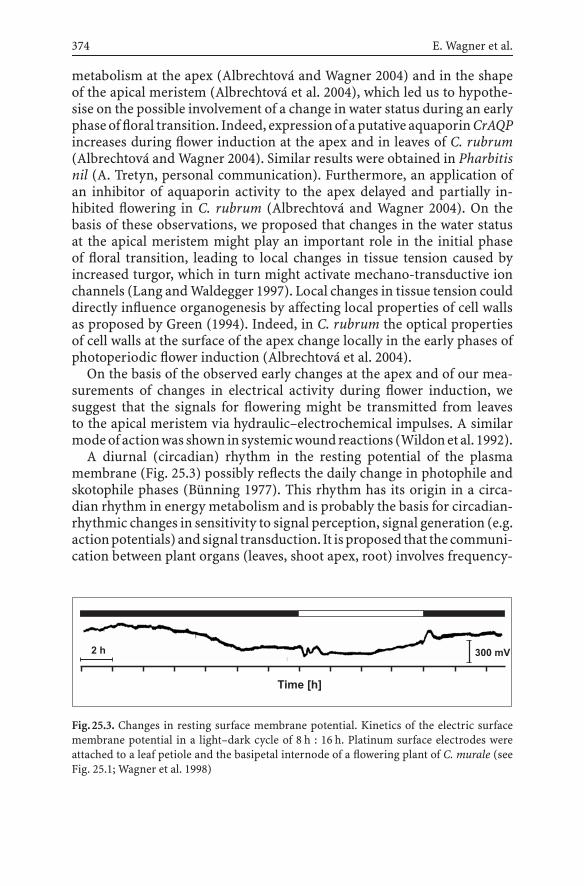
374 E. Wagner et al.
metabolism at the apex (Albrechtová and Wagner 2004) and in the shapeof the apical meristem (Albrechtová et al. 2004), which led us to hypothe-sise on the possible involvement of a change in water status during an earlyphase of floral transition. Indeed, expression of a putative aquaporin CrAQPincreases during flower induction at the apex and in leaves of C. rubrum(Albrechtová and Wagner 2004). Similar results were obtained in Pharbitisnil (A. Tretyn, personal communication). Furthermore, an application ofan inhibitor of aquaporin activity to the apex delayed and partially in-hibited flowering in C. rubrum (Albrechtová and Wagner 2004). On thebasis of these observations, we proposed that changes in the water statusat the apical meristem might play an important role in the initial phaseof floral transition, leading to local changes in tissue tension caused byincreased turgor, which in turn might activate mechano-transductive ionchannels (Lang and Waldegger 1997). Local changes in tissue tension coulddirectly influence organogenesis by affecting local properties of cell wallsas proposed by Green (1994). Indeed, in C. rubrum the optical propertiesof cell walls at the surface of the apex change locally in the early phases ofphotoperiodic flower induction (Albrechtová et al. 2004).
On the basis of the observed early changes at the apex and of our mea-surements of changes in electrical activity during flower induction, wesuggest that the signals for flowering might be transmitted from leavesto the apical meristem via hydraulic–electrochemical impulses. A similarmodeof actionwas shown in systemicwoundreactions (Wildonet al. 1992).
A diurnal (circadian) rhythm in the resting potential of the plasmamembrane (Fig. 25.3) possibly reflects the daily change in photophile andskotophile phases (Bünning 1977). This rhythm has its origin in a circa-dian rhythm in energy metabolism and is probably the basis for circadian-rhythmic changes in sensitivity to signal perception, signal generation (e.g.actionpotentials) andsignal transduction. It isproposed that the communi-cation between plant organs (leaves, shoot apex, root) involves frequency-
2 h
Time [h]
300 mV
Fig.25.3. Changes in resting surface membrane potential. Kinetics of the electric surfacemembrane potential in a light–dark cycle of 8 h : 16 h. Platinum surface electrodes wereattached to a leaf petiole and the basipetal internode of a flowering plant of C. murale (seeFig. 25.1; Wagner et al. 1998)

25 Hydro-Electrochemical Integration of the Higher Plant 375
coded (electric) signals (Wagner et al. 1998). Rhythmic integration over thewhole plant possibly involves modulation of turgor pressure via stretch-activated ion channels and aquaporins, with concomitant changes in mem-brane potential (Fig. 25.3).
The perception of a flower-inducing dark period might lead to a changein electrochemical signalling between leaves and the stem and thus couldrepresent “florigen”. The involvement of action and variation potentialsfor integration of the whole plant was anticipated (Wagner et al. 1998).Signal arrival at the apex might trigger cytoplasmic changes in pH andCa2+ concentration as secondary messengers in photoperiodic control ofdevelopment (Love et al. 2004). Finally, the switch from the vegetative tothe flowering state is a threshold response, systemic in nature and involvingnot only the apical meristem but also the axillary buds.
25.4Evolution of Circadian Frequencies –Timing of Metabolic ControlsConsidering metabolic control of timing in photoperiodism (Wagner andCumming 1970; Wagner et al. 1975), it has to be kept in mind that evolutionfrom prokaryotic to eukaryotic organisms was paralleled by a correspond-ing evolution in energy metabolism. From primary fermentation, energyconservation progressed to anaerobic photosynthesis and then to carbondioxide fixation with acceptance of electrons by water and evolution ofoxygen (Bekker et al. 2004). In a progressively oxygenic biosphere respi-ration developed with oxygen as the terminal electron acceptor. Evolvinglife was paralleled by the corresponding evolution of tropospheric O2/CO2
composition and feedback of oxygen on life processes via reactive oxy-gen and reactive nitrogen species, which as signalling molecules becamecrucial for control of development of prokaryotic and eukaryotic livingsystems. Adaptation to the seasonal variation in day length resulted inphotoperiodic control of development with a circadian rhythm in energyconservation and transformation to optimise energy-harvesting by pho-tosynthesis (Foyer and Noctor 2003; Wagner et al. 1975). Photosynthesis,on the other hand, acts as a metabolic regulator via redox signals (Oh andKaplan 2000; Pfannschmidt 2003; Pfannschmidt et al. 2001; Sherameti etal. 2002; Zeilstra-Ryalls et al. 1998) in addition to specific photoreceptorsystems like phytochromes and cryptochromes. Finally, redox control inte-grates rhythmic gene expression in prokaryotes (Ditty et al. 2003; Dvornyket al. 2003; Rutter et al. 2001; Tomita et al. 2004), as well as in chloroplasts,mitochondria and the nucleus of eukaryotes (Forsberg et al. 2001; Tron etal. 2002).

376 E. Wagner et al.
The circadian rhythmic cell (cyanobacterial and eukaryotic) is consid-ered to be a hydro-electrochemical oscillator (Wagner et al. 1997) synchro-nised by the daily light–dark cycles, with temporal compartmentation ofmetabolismand a network ofmetabolic sequences to compensate for oxida-tive stress in adapting to the light environment, e.g. by separating nitrogenfixation from photosynthetic oxygen production (Sherameti et al. 2002).
25.5Circadian Rhythmic Organisation of EnergyMetabolism in C. rubrum and the Gatingof Photoreceptor (Phytochrome) Action
In C. rubrum a circadian rhythm in overall energy transduction has beenobserved. The rhythm results from an oscillatory network between glycoly-sis and oxidative phosphorylation, coupled to photophosphorylation. Thisnetwork produces a circadian rhythm in adenylate energy charge and re-dox state (NADP/NADPH2). The nucleotide ratios themselves could act asrate effectors in compartmental feedback and thus fulfil the requirementsof precise temperature-compensated time-keeping (Wagner et al. 1975,
Table 25.2. Rhythmic organisation of metabolism in Chenopodium rubrum (the periodlengths of the subpeaks are in parentheses) (Complemented after Bünning 1977)
Phenomenon Period length (h)
Photoperiodic light sensitivity 30Betacyanine accumulation 24–30 (15)Betacyanine turnover 24–30Adenylate kinase activity 30 (15)Energy charge (ATP + 1/2 ADP/ATP + ADP + AMP) 21–24 (11–13)NADPH2/NADP ratio 21–24Dark respiration 21–24Chlorophyll accumulation 15Net photosynthesis 15Triose phosphate dehydrogenase activity (NADH2; NADPH2) 15Malate dehydrogenase activity 12–15Glutamate dehydrogenase activity 12–15Glucose-6-phosphate dehydrogenase activity 12–15Gluconate-6-phosphate dehydrogenase activity 12–15Pyridine nucleotide, pool size [NAD(H2); NADP(H2)] 12–15 (6)
Turgor-controlled growth phenomenaStem extension rate 23–27Leaf movement 23–27

25 Hydro-Electrochemical Integration of the Higher Plant 377
12 24 36 480
0.56
4
0.65
5
6
7
Darkness [h]
20°C 20°C
4.5 days 24 h various hours (0 - 72)
3
2
1
0
60 72
[ATP] + 0.5 [ADP]
[ATP] + [ADP] + [AMP]
[NADPH2 / NADP]
32.5°C / 10°C
En
erg
yC
ha
rge
[N
AD
PH
2/N
AD
P]
Fig.25.4. Circadian rhythm of NADPH2-to-NADP ratio and of “energy charge” of C. rubrumseedlings. The seedlings were germinated in constant light and alternating conditions oftemperature for 4.5 days, thereafter transferred to constant 20 ◦C for 24 h followed by a darkperiod of varied duration (3-h increments) (Wagner et al. 1975)
1983, 1998). In C. rubrum there are semicircadian oscillations with specificphasing in enzyme activities involved in glycolysis, photosynthesis andrespiration resulting in a circadian rhythm of adenylate energy charge andredox state (Table 25.2, Fig. 25.4).
There is an inverse phase relationship between glyceraldehyde-3-phos-phate dehydrogenase (GPD) linked to photosynthesis (NADP-GPD) and theenzyme linked to glycolysis (NAD-GDP) (Fig. 25.5). Adenylate kinase, a keyenzyme in energy dissipation displays circadian/semicircadian oscillationsin activity. The enzyme exists as isoenzymes in chloroplasts, mitochondria,the cytoplasm and the nucleus. The enzyme activities are modulated bylight (phytochrome, cryptochrome), glucose feeding and photoperiod andthermoperiod (Figs. 25.6–25.8).
The rhythm in enzyme activities may be amplitude-modulated by phy-tochrome. The phytochrome action is gated by the phasing of the endoge-nous rhythm reflecting a sequence of photophile and skotophile phases asconceived by Bünning (1977) for the functioning of the physiological clock(Figs. 25.9 and 25.10).

378 E. Wagner et al.
Fig.25.5. Time course of adenylate kinase (AK), NAD- and NADP-glyceraldehyde-3-phosphate dehydrogenase (GPD) activity during darkness. C. rubrum (ecotype 184/68)seedlings germinated in alternating light intensity and temperature for 4.5 days. Thereafterconstant 20 ◦C, 6,000 lx fluorescent white light for 24 h followed by a dark period of variedduration (3-h increments). The enzyme activities were measured at the end of each respec-tive dark period. The dark period medium was either Hoagland’s solution or 0.4 M glucosein Hoagland’s solution under sterile conditions. There are semicircadian oscillations with aninverse phase relationship between photosynthetic NADP-GPD and glycolytic NAD-GPD.Glucose feeding in darkness has no effect on NADP-GPD but stimulates the other enzymeactivities (Frosch et al. 1973)
25.6Hydraulic–Electrochemical Integration of the Whole Plant
The integration of metabolic activity of C. rubrum plants on a hydraulic–electrochemical level is represented by a diurnal rhythm in compoundsurface-membrane potential (Fig. 25.2). Spontaneous surface-membraneaction potentials could be shown to correlate with turgor-controlled hy-draulic growth movements of leaves and stem extension (Fig. 25.11), whichare controlled by the photoperiod. Recorded electrophysiograms can be

25 Hydro-Electrochemical Integration of the Higher Plant 379
Fig.25.6. Pattern of AK isozymes from C. rubrum (ecotype 374) after chromatography on(diethylamino)ethyl (DEAE) cellulose. Top Seedlings grown on Hoagland’s solution with0.5% agar at 6,500 lx continuous white fluorescent light for 24 days. Bottom The sameconditions with one inductive dark period of 12 h, 5.5 days after sowing. The abundanceof the different isozymes in the cellular compartments changes after flower induction;in particular the relation between chloroplasts (AK V) and mitochondria (AK I). IsozymeAK IV (nucleus) was not detected (Wagner et al. 1983). AK I mitochondria, AK II cytoplasm,AK III chloroplasts, AK IV nucleus, AK V chloroplasts, P phosphatase
used for the control of development, i.e. via electrogenic induction of flow-ering by DC pulses (Wagner et al. 2004).
At the apex, photoperiodic conditions inducing flowering have beenstudied at the cellular level in C. rubrum (Sect. 25.3). There are very earlychanges in calcium and pH patterning (Albrechtová et al. 2003) and carbo-hydrate metabolism, which could lead to an increase in osmotic pressurein the cells of the apical meristem, and thus build a driving force for watertransport (Albrechtová and Wagner 2004). It is well known that aquaporinsare involved in regulating water relations, and studies in C. rubrum revealedthe expression of a novel aquaporin (CrAQP) with a transient increase inthe expression a few hours before the maximal increase in the size of themeristem (Albrechtová and Wagner 2004; Albrechtová et al. 2004). It was

380 E. Wagner et al.
Fig.25.7. Pattern of AK isozymes from C. rubrum (ecotype 374) after chromatographyon DEAE-cellulose. Seedlings grown for 4.5 days at 12 h : 12 h 32.5 ◦C:10 ◦C and 6,500 lxcontinuous white light on filter paper with H2O; thereafter on Hoagland’s solution at 20 ◦Cfor 1 day followed by 5.5 days on 0.2 M glucose in Hoagland’s solution. The abundanceof the different isozymes in the cellular compartments changes after glucose applicationas compared with that in untreated plants (c.f. vegetative pattern as a control, Fig. 25.6).There is an increase of AK II and AK III abundance in the cytoplasm and the chloroplasts,respectively. AK IV (nucleus) was not detected (Wagner et al. 1983)
concluded that the change of aquaporin expression at the apical meristemduring floral transition could be responsible for increased water movementinto the meristem provoking its expansion. Further studies should reveal ifintracellular pH and calcium concentration can influence water transportby regulating CrAQP activity (Lopez et al. 2003).
25.7Electrophysiological Integration of Activityof the Whole Plant – Monitoring of Surface Sum Potentials
Automatic measurements of up to 4 weeks’ duration could be performedusing a measuring system with surface electrodes for the recording ofsurface sum potentials. The following measuring procedures were used(Wagner et al. 2004).

25 Hydro-Electrochemical Integration of the Higher Plant 381
Fig.25.8. Pattern of AK isozymes after chromatography on DEAE-cellulose. Growing condi-tions as in Fig. 25.7, however, with 0.3 M glucose application. There is a dramatic increase ofAK I abundance in mitochondria as compared with that in untreated plants (c.f. vegetativepattern, control, Fig. 25.6). The abundance of AK IV in the nucleus increased over thedetection limit (Wagner et al. 1983)
The electrodes essentially cover, i.e. surround, the stem axis or the peti-oles of leaves completely. This geometry of contact between surface elec-trodes and the plant tissue assures, in contrast to intracellular penetratingelectrodes, that the potential changes to be recorded are not due to singlecells but that they represent the electrochemical activity of the whole area ofcontact of the electrode with the plant tissue in question. For this reason thepotentials recorded with surface electrodes are named “surface sum poten-tial” or “surface potential”. To reduce the resistance between the plant andthe electrode surface, the contact surfaces between electrode and plant canbe covered with a thin contact gel like one used in medicine, e.g. for record-ing electroencephalograms. The analysis or evaluation of the recordedchanges inmembranepotential areperformedon thebasisof the frequency,the temporal distribution and/or the direction of propagation of action po-tentials. In long-term experiments, using bipolar surface electrodes specificchanges in the direction of propagation of action potentials in response todifferent flower-inducing and non-flower-inducing photoperiods could beobserved. The recordings have shown that the propagation of action poten-tials along the stem axis of the model plants under investigation, C. rubrum

382 E. Wagner et al.
Fig.25.9. Time course of NADP-GPD activity during darkness. C. rubrum seedlings germi-nating in alternating conditions of light intensity and temperature for 4.5 days. Thereafterconstant 20 ◦C, 6,000 lx fluorescent white light for A 12 h, B 24 h and C 36 h followed bya dark period of varied duration (3-h increments). At the beginning of the dark period theseedlings received 5 min of red (solid line), 5 min of far-red (dashed line), or 5 min of far-redplus 5 min of red (dotted line) light (corresponding to a dusk signal in daily light–darkcycles), in order to modulate the level of active phytochrome. The enzyme activities weremeasured at the end of each respective dark period. The rhythmic germination conditionssynchronise the endogenous circadian rhythm to exactly 24 h with an alternation of sko-tophile and photophile phases. The imposition of darkness after 12 and 36 h of constantconditions begins at the same phase, while the beginning of darkness after 24 h of constantconditions is 12 h out of phase with respect to the germination rhythm and to the othertwo treatment as is obvious from the semicircadian oscillations of enzyme activity in thethree parts of the figure. The results demonstrate the different sensitivity to phytochromeamplitude modulation of enzyme activity oscillations depending on the phase of the under-lying cycle of photophile and skotophile phases. Clearly the endogenous rhythm is gatingthe window for phytochrome action (Frosch and Wagner 1973)

25 Hydro-Electrochemical Integration of the Higher Plant 383
Fig.25.10.Time course of AK activity during darkness. Experimental protocol as for Fig. 25.8(Frosch and Wagner 1973)
and C. murale, depends on the photoperiod inductive or non-inductive forflowering. The statistic evaluation of the propagation direction of the actionpotentials could be used as a marker for the induction of flowering inshort- and long-day plants. On the basis of these data, the state of flow-ering or flower induction of the plant can be determined long before thefirst morphological changes at the apical meristem become visible. Ourresults furthermore indicate that the action potentials under the influenceof flower-inducing and non-flower-inducing photoperiods are temporar-ily not uniformly distributed in the dark and light phases of the differentphotoperiods, but that they follow a characteristic distribution pattern. Toanalyse the correlation between the photoperiodic induction of floweringas a specific state of the plant material, and the temporal distribution ofaction potentials and their direction of propagation, the observed actionpotentials had to be classified. Of particular relevance are signals which can

384 E. Wagner et al.
Fig.25.11. a Kinetics of LM (green line), SER (blue line) (millimetres per hour) and timeof appearance of action potentials (APs, red vertical lines) in C. murale (6-week-old plants)during exposure to non-inductive photoperiod with a light-to-dark ratio of 4 h : 20 h.b Schematic drawing of LM of Chenopodium showing the diurnal change of leaf position
Fig.25.12. Specific changes in the phasing of SER and LM kinetics appear upon flowerinduction. Both parameters depend on turgor-mediated growth and their interactionsdemonstrate rhythmic interorgan communication. LMs and SER are quantified by com-puter controlled time lapse photography (see Fig. 25.1)

25 Hydro-Electrochemical Integration of the Higher Plant 385
be detected shortly after a light–dark or a dark–light transition (Wagner etal 1998). Measurements under the non-inductive light conditions for flow-ering did show that in the long-day plant C. murale mainly light-on signalswere observed, while in the short-day plant C. rubrum an accumulation ofthe signals was observed after the light–dark transition. Under the influ-ence of the flower-inducing photoperiods, both groups of plants showed aninverse pattern of action potential accumulation. In long-term measure-ments, the correlation between flower induction and the classification ofaction potentials either as light-on or as light-off signals could clearly bedemonstrated. Therefore, this criterion is an important indicator for thestate of flowering or for the differentiation between long- and short-dayplant material. Thus, it has been shown that short-day plants and long-dayplants each display characteristic distributions of action potentials duringdark and light phases, which are characteristic for flower-inducing andnon-flower-inducing conditions. It follows that the temporal distributionof action potentials over and within dark and light spans can also be reliablyused as a marker for the flowering state of a given plant material (Wagneret al. 2004).
25.8Substitution of Photoperiodic Flower Inductionby Electrogenic Flower Induction
Specific electrophysiograms characteristic for photoperiodic flower induc-tion were generated providing information concerning the electrical signalpatterns of flower-induced plants. Experiments on electrostimulation usingspecific impulse patterns of DC current were aimed at inducing floweringunder non-inductive light conditions. The results obtained showed thatDC pulses during a fixed period of time for 1.5 h on seven consecutive daysclearly induced seven of the 12 plants under investigation. With inversepolarity of the stimulating DC current, the apices of the stimulated plantsdid not show any difference compared to the apices of non-stimulatedcontrol plants (Wagner et al. 2004). These results clearly show that the ad-equate polarity of the stimulating electrodes is of decisive importance forflower induction. The temporal patterns of the stimulating currents shouldbe selected depending on the type of plant (short-day or long-day) andthe temporal pattern of the transition between endogenous oscillation inphotophile and skotophile phases. The results obtained prove that flowerinduction in the plant can be induced by electrophysiological stimulationunder non-flower-inducing environmental conditions.

386 E. Wagner et al.
25.9Conclusions and Future Perspectives
The hydro-electrochemical communication of the higher plant is intrin-sically linked with and depends on the membrane potential of cells andorganelles, which can be modulated by the light environment (Trebacz andSievers 1998). High-frequency (e.g. 24 min) temperature-compensated os-cillations like in NADH oxidase activity of plasma membranes (Morré andMorré 1998) might be a high-frequency time scale for redox-control ona circadian scale. Populations of synchronised chloroplasts and mitochon-dria might be involved in the generation of proton-conducting structureslike the mitochondrial network in animal cells (Giorgi et al. 2000). Thehydro-electrochemical integration of the whole plant is the control net forintegrating plant and environment in daily and seasonal adaptations usingaction potentials in a frequency-coded communication. The most impor-tant questions concern the mechanism of oscillator functioning in thevarious plant organs and the mode of coupling of oscillators, not to forgetoscillator generation (Fig. 25.12). The following questions need immediateattention:
1. Where exactly are the action potentials for the systemic signalling gen-erated?
2. What is the structural basis for stem polarity in the propagation of actionpotentials?
3. How does an induced leaf define propagation of action potentials in thestem?
4. What is the induced state of a leaf on the physiological level?
5. How does the photoperiod, e.g. via phytochrome (cryptochrome), createthe induced state of a leaf?
6. What is common in an induced leaf from a short-day and a long-dayplant?
7. How do circadian rhythmicity and hydro-electrochemical activity inte-grate the plant as a whole?
Acknowledgements. We gratefully acknowledge the generous support ofCarl Lücking at the Neurocenter Freiburg.

25 Hydro-Electrochemical Integration of the Higher Plant 387
References
Albrechtová JTP, Wagner E (2004) Mechanisms of changing organogenesis at the apexof Chenopodium rubrum during photoperiodic flower induction. Flower Newslett38:27–33
Albrechtová JTP, Metzger C, Wagner E (2001) pH-patterning at the shoot apical meris-tem as related to time of day during different light treatments. Plant Physiol Biochem39:115–120
Albrechtová JTP, Heilscher S, Leske L, Walczysko P, Wagner E (2003) Calcium and pH pat-terning at the apical meristem are specifically altered by photoperiodic flower inductionin Chenopodium spp. Plant Cell Environ 26:1985–1994
Albrechtová JTP, Dueggelin M, Durrenberger M, Wagner E (2004) Changes in geometry ofthe apical meristem and concomitant changes in cell wall properties during photoperi-odic induction of flowering in Chenopodium rubrum. New Phytol 163:263–269
Bekker A, Holland HD, Wang PL, Rumble D III, Stein HJ, Hannah JL, Coetzee LL, Beukes NJ(2004) Dating the rise of atmospheric oxygen. Nature 427:117–120
Blázquez MA, Weigel D (2000) Integration of floral inductive signals in Arabidopsis. Nature404:889–892
Bünning E (1977) Die physiologische Uhr. Springer, Berlin Heidelberg New YorkDitty JL, Williams SB, Golden SS (2003) A cyanobacterial circadian timing mechanism.
Annu Rev Genet 37:513–543Dvornyk V, Vinogradova O, Nevo E (2003) Origin and evolution of circadian clock genes in
prokaryotes. Proc Natl Acad Sci USA 100:2495–2500Forsberg J, Rosenquist M, Fraysse L, Allen JF (2001) Redox signalling in chloroplasts and mi-
tochondria: genomic and biochemical evidence for two-component regulatory systemsin bioenergetic organelles. Biochem Soc Trans 29:403–407
Foyer CH, Noctor G (2003) Redox sensing and signalling associated with reactive oxygen inchloroplasts, peroxisomes and mitochondria. Physiol Plant 119:355–364
Fridlyand LE, Scheibe R (1999) Regulation of the Calvin cycle for CO2 fixation as an examplefor general control mechanisms in metabolic cycles. BioSystems 51:79–93
Frosch S, Wagner E (1973) Endogeneous rhythmicity and energy transduction. II. Phy-tochrome action and the conditioning of rhythmicity of adenylate kinase, NAD- andNADP-linked glyceraldehyde-3-phosphate dehydrogenase in Chenopodium rubrum bytemperature and light intensity cycles during germination. Can J Bot 51:1521–1528
Frosch S, Wagner E, Cumming BG (1973) Endogeneous rhythmicity and energy transduc-tion. I. Rhythmicity in adenylate kinase, NAD- and NADP-linked glyceraldehyde-3-phosphate dehydrogenase in Chenopodium rubrum. Can J Bot 51:1355–1367
Giorgi De F, Lartigue L, Ichas F (2000) Electrical coupling and plasticity of the mitochondrialnetwork. Cell Calcium 28:365–370
Goldsworthy A (1983) The evolution of plant action potentials. J Theor Biol 103:645–648Green PB (1994) Connecting gene and hormone action to form, pattern and organogenesis:
biophysical transductions. J Exp Bot 45:1775–1788Ivleva NB, Bramlett MR, Lindahl PA, Golden SS (2005) LdpA: a component of the circadian
clock senses redox state of the cell. EMBO J 24:1202–1210Karpinski S, Reynolds H, Karpinska B, Wingsle G, Creissen G, Mullineaux P (1999) Systemic
signalling and acclimation in response to excitation energy in Arabidopsis. Science284:654–657
KlodaA,MartinacB (2002)Commonevolutionaryoriginsofmechanosensitive ionchannelsin Archaea, Bacteria and cell-walled Eukarya. Archaea 1:35–44
Lang F, Waldegger S (1997) Regulating cell volume. Am Sci 85:456–463
388 E. Wagner et al.
Lecharny A, Wagner E (1984) Stem extension rate in light-grown plants. Evidence for anendogenous circadian rhythm in Chenopodium rubrum. Physiol Plant 60:437–443
Lopez F, Bousser A, Sissoeff I, Gaspar M, Lachaise B, Hoarau J, Mahé A (2003) Diurnal reg-ulation of water transport and aquaporin gene expression in maize roots: contributionof PIP2 proteins. Plant Cell Physiol 44:1384–1395
Love J, Dodd AN, Webb AAR (2004) Circadian and diurnal calcium oscillations encodephotoperiodic information in Arabidopsis. Plant Cell 16:956–966
Morré DJ, Morré DM(1998) NADHoxidase activity of soybean plasma membranes oscillateswith a temperature compensated period of 24 min. Plant J 16:277–284
Oh JI, Kaplan S (2000) Redox signaling: globalization of gene expression. EMBO J 19:4237–4247
Pfannschmidt T (2003) Chloroplast redox signals: how photosynthesis controls its owngenes. Trends Plant Sci 8:33–41
Pfannschmidt T, Allen JF, Oelmüller R (2001) Principles of redox control in photosynthesisgene expression. Physiol Plant 112:1–9
Reeves PH, Coupland G (2000) Response of plant development to environment: Control offlowering by daylength and temperature. Curr Opin Plant Biol 3:37–42
Rutter J, Reick M, Wu LC, McKnight SL (2001) Regulation of clock and NPAS2 DANN bindingby the redox state of NAD cofactors. Science 293:510–514
Sherameti I, Sopory SK, Trebicka A, Pfannschmidt T, Oelmüller R (2002) Photosyntheticelectron transport determines nitrate reductase gene expression and activity in higherplants. J Biol Chem 277:46594–46600
Tomita J, Nakajima M, Kondo T, Iwasaki H (2004) No transcription-translation feedback incircadian rhythm of KaiC phosporylation. Science 307:251–254
Trebacz K, Sievers A (1998) Action potentials evoked by light in traps of Dionea muscipulaEllis. Plant Cell Physiol 39:369–372
Tron AE, Bertoncini CW, Chan RL, Gonzalez DH (2002) Redox regulation of planthemeodomai transcription factors. J Biol Chem 38:34800–34807
Wagner E, Cumming BG (1970) Betacyanin accumulation, chlorophyll content, and flowerinitiation in Chenopodium rubrum as related to endogenous rhythmicity and phy-tochrome action. Can J Bot 48:1–18
Wagner E, Deitzer GF, Fischer S, Frosch S, Kempf O, Stroebele L (1975) Endogenous os-cillations in pathways of energy transduction as related to circadian rhythmicity andphotoperiodic control. BioSystems 7:68–76
Wagner E, Haertlé U, Kossmann I, Frosch S (1983) Metabolic and developmental adaptationof eukaryotic cells as related to endogenous and exogenous control of translocatorsbetween subcellular compartments. In: Schenk HEA, Schwemmler W (eds) Endocyto-biology, vol II. de Gruyter, Berlin, pp 341–351
Wagner E, Normann J, Albrechtová JTP, Bonzon M, Greppin H (1997) Photoperiodic controlof flowering: electrochemical-hydraulic communication between plant organs – ‘flori-gen’ a frequency-coded electric signal? In: Greppin H, Penel C, Simon P (eds) Travellingshot on plant development. University of Geneva, Geneva, pp 19–34
Wagner E, Normann J, Albrechtová JTP, Walczysko P, Bonzon M, Greppin H (1998)Electrochemical-hydraulic signalling in photoperiodic control of flowering: Is “flori-gen” a frequency-coded electric signal? Flower Newslett 26:62–74
Wagner E, Lehner L, Normann J, Albrechtová JTP (2004) Electrogenic flower initiation –perspectives for whole plant physiology and for applications in horticulture, agricultureand silviculture. Flower Newslett 38:3–9
Walczysko P, Wagner E, Albrechtová JTP (2000) Use of co-loaded Fluo-3 and Fura Redfluorescent indicators for studying the cytosolic Ca2+ concentrations distribution inliving plant tissue. Cell Calcium 28:23–32

25 Hydro-Electrochemical Integration of the Higher Plant 389
Wildon DC, Thain JF, Minchin PEH, Gubb IR, Reilly AJ, Skipper YD, Doherty HM, O’Don-nell PJ, Bowles DJ (1992) Electrical signalling and systemic proteinase inhibitor induc-tion in the wounded plant. Nature 360:62–65
Zeilstra-Ryalls J, Gomelsky M, Eraso JM, Yeliseev A, O’Gara J, Kaplan S (1998) Control ofphotosystem formation in Rhodobacter sphaeroides. J Bacteriol 180:2801–2809

26 Signals and Signalling Pathwaysin Plant Wound ResponsesJeremy D. Rhodes, John F. Thain, David C. Wildon
Abstract The focus is on the most widely studied wound response: the systemic synthesisin tomato of the defence proteins known as proteinase inhibitors (PIs). In wounded tomatoseedlings, the severity of the wound is a major determinant of the type of wound signalling.Minor wounds produce outcomes that are consistent with the transport in the phloem ofa chemical elicitor of PI synthesis. Severe wounds produce outcomes that are consistent withthe distribution of elicitors of PI synthesis and of electrical activity by hydraulic dispersal inthe xylem. The main electrical events associated with severe wounds are action-potential-like depolarisations in the sieve-tube-element/companion cell complexes; these events areconsidered in relation to the use of the terms “action potential” and “variation potential”.
26.1Introduction
Plants respond to wounding both locally at the wound site and at sitesdistant from the wound (systemic responses). The most widely studiedresponse is the systemic synthesis of defence proteins, known as proteinaseinhibitors (PIs), in tomato seedlings following mechanical wounding, firstdiscovered by Green and Ryan (1972). Mechanical wounding has beenwidely used to simulate insect damage. Our interest has been to elucidatethe nature of the signals and signalling pathways underlying this systemicresponse. We believe that we can distinguish two situations: rapid signallingfollowing a severe wound, and signalling in the phloem following a minorwound. The distinction between the effects of severe and minor wounds, inour view, enables a synthesis of the published results from several differentresearch groups.
Several candidateshavebeenproposedasasignal linking the localwoundto the systemic response, including both chemical and physical signals,and a combination of the chemical and the physical: hydraulic dispersal.Given the complex nature of a wound, Bowles (1998) has highlighted theprobability that such an event would give rise to multiple signals.
Possible chemical signals include oligosaccharides, jasmonic acid, andthe peptide systemin; all have been shown to be elicitors of PI synthesis(Bowles 1998). Current evidence, based on mutations affecting PI synthesis,favours jasmonic acid as being a systemic signal, with systemin acting lo-cally at or near the wound site – GA Howe’s group, using minor mechanical
Communication in PlantsF. Baluška, S. Mancuso, D. Volkmann (Eds.)© Springer-Verlag Berlin Heidelberg 2006

392 J.D. Rhodes, J.F. Thain, D.C. Wildon
wounding; see references in the review by Stratmann (2003). FollowingBaydoun and Fry (1985), it has generally been assumed that oligosaccha-rides act only locally, but the later work of Rigby et al. (1994) showedtranslocation in the phloem of an oligogalacturonide that induced PI syn-thesis. Thus, oligosaccharides remain as possible candidates for a systemicsignal. Both oligosaccharides (Thain et al. 1995) and systemin (Moyen andJohannes 1996) produce transient membrane depolarisation, which couldbe a factor in the electrical activity associated with severe wounds.
Possible physical signals include a hydraulic pressure wave that ariseswhen a wound releases the tension in the xylem of a transpiring plant. Sucha pressure wave propagates through the plant from the wound site at highspeeds, 100 mm s−1 or more; see the review by Malone (1996). A pressurewave could bring about its effects either by the activation of mechano-sensitive (stretch-activated) channels (Stankovic and Davies 1997) or bythe effect of signal molecules. Malone and co-workers showed, in tomato,that pressure pulses could be induced without significant wounding bysubmerged excision of a single leaflet through its submerged petiole; sucha pressure pulse did not induce PI synthesis in tomato leaves. Instead,release of tension in the xylem sets up a reverse flow of xylem sap froma wound site into the rest of the plant, a phenomenon which they termedhydraulic dispersal; when this flow carried sap from wounded leaf tissue(e.g. a heat wound), systemic PI synthesis ensued, presumably owing to thepresence of chemical elicitors in the sap.
Wounding also gives rise to electrical events that have usually been de-tected with surface-contact electrodes, and that appear to move through theplant from the wound site. These observations have led to the suggestion,e.g. Wildon et al. (1992), that the systemic signal for induction of PI syn-thesis is an electrical one, i.e. a self-propagating action potential similar tothose in nervous conduction or in animal epithelial conduction: cell-to-cellconduction of action potentials (Mackie 1965). If that were true it should bepossible to initiate the signal by depolarisation of the membrane potentialof cells in the signalling pathway, preferably by injection of current viaintracellular microelectrodes. It would also be expected that the electricalsignals would show other characteristics typical of action potentials suchas the all-or-nothing response, a strength–duration relationship for theinitiating current, and a refractory period.
Evidence for the occurrence of action potentials in plants has been re-viewed by Thain and Wildon (1996). There is clear evidence for actionpotentials in the charophyte algae, and in a number of higher plant species,which suggests that the ability to generate action potentials is widespreadin the plant kingdom. Electrical stimulation has been used to induce PIgene expression (Herde et al. 1995; Peña-Cortès et al. 1995; Stankovic andDavies 1996, 1997), although intracellular electrodes were not used either

26 Signals and Signalling Pathways in Plant Wound Responses 393
for the stimulus or for the recording, and the effect of heat generated at theplant–electrode interface cannot be discounted.
The wound-induced electrical events recorded with surface-contact elec-trodes are generally divided into two kinds (Mancuso 1999): those of ir-regular shape and of long duration (tens of seconds or more), which aregenerally called variation potentials; and those of simple shape (e.g. a sin-gle spike) and of shorter duration (a few seconds), which have generallybeen called action potentials. Clear examples of these two kinds can beseen in Fig. 1 of the paper by Stankovic and Davies (1996) but intermediateforms also occur. It must be emphasised that, for electrical events recordedwith surface-contact electrodes, these terms are based purely on the shapesand durations of the electrical events; surface-contact electrodes record thesum, biased by the resistances of the various pathways between the cellsand the surface, of all the membrane currents flowing in the underlying tis-sues. Unfortunately the terms variation potential and action potential areoften taken to imply a mechanism. For example, it is widely assumed thatvariation potentials are the local electrical effects of chemicals flowing inthe xylem stream. This could be true in many cases (Malone 1996), but it isalso possible that an irregularly shaped event recorded with surface-contactelectrodes could be the summation of a number of action potentials travel-ling along different pathways at slightly different times. Similarly the use ofthe term action potential to imply a mechanism is insecure in the absence ofevidence from intracellular microelectrodes used to electrically stimulateand record from identifiable cells in the signalling pathway. At least for theelectrical events following wounding, it may be best to avoid the variationpotential/action potential terminology until a better understanding of thecellular nature of these events is obtained.
Obvious pathways for systemic chemical signals are the xylem and thephloem. A third would be the symplasm, but rates of transport, 15 µm s−1,are too low for rapid systemic signalling. Rates of movement in both thexylem and the phloem can be high, with maximum reported speeds of0.6−4.0 mm s−1 in the phloem (Baker and Milburn 1989), and 250 mm s−1
in the xylem (Passioura 1972). As already indicated, speeds for possiblephysical signals and hydraulic dispersal, are also high; thus, it may not bepossible to use speed as a criterion to identify either the pathway or themechanism. Several approaches can be used to distinguish between thevarious signalling possibilities: steaming of a short length of the pathway,followed by a recovery period, to disrupt the phloem but not the xylem;chilling to transiently block phloem translocation in some species, includ-ing tomato; the use of appropriate fluorescent dyes to visualise flows in thexylem or the phloem.
With regard to electrical signals, the phloem sieve-tube-element/com-panion cell (STE/CC) complex provides a suitable pathway with good

394 J.D. Rhodes, J.F. Thain, D.C. Wildon
longitudinal connectivity, a functional plasma membrane, and low lateralconnectivity with surrounding tissues (van Bel 2003). However the phe-nomenon of epithelial conduction in animals shows that action potentialscan propagate through tissue consisting of relatively unspecialised cells,so it is not impossible that action potentials could propagate along othertissues, as long as they have adequate electrical connectivity via plasmod-esmata.
We now consider the possibilities for signals and signalling pathwaysmainly in the light of results from our laboratory.
26.2Patterns of Proteinase Inhibitor Activityand Electrical Activity Following a Varietyof Wounding Protocols Applied to Tomato Seedlings
In our work (Rhodes et al. 1999) we used five different wounding protocols:(1) a razor cut through a cotyledon in air; (2) a razor cut simultaneouslythrough a cotyledon and a droplet of Lucifer Yellow CH (LY) solutionplaced on its surface (i.e. cut under water); (3) a small (area about 60 mm2)mechanical wound; (4) a large (area about 150 mm2) mechanical wound;(5) a heat wound using a hot spatula held under, but not touching, thelamina. Wound-type 3 is referred to as a minor wound; wound-types 4and 5 are referred to as severe wounds. For all the wounding protocols, weused surface electrodes on leaves 1 and 2 (Fig. 26.1) to check for electricalactivity. Significant results are summarised in Table 26.1.
A razor cut through the lamina of a cotyledon in air (wound-type 1)produced neither electrical activity at leaf 1, nor systemic induction ofPI activity (Table 26.1), as would be expected for a wound type in whichneither a hydraulic pressure wave, nor hydraulic dispersal occurs.
A razor cut through a droplet of water on the lamina of a cotyledon(wound-type 2) induced systemic PI activity, but did not result in anyelectrical activity (Table 26.1), showing that electrical activity is not causedby reversed xylem flow alone. The pattern of induction of PI activity wasstriking: when cotyledon 1 (see Fig. 26.1, legend) was cut, more PI activitywas seen in the right side of leaf 1 and the left side of leaf 2 (Fig. 26.1: lowvalues indicate high PI activity). The pattern was reversed when cotyledon 2was cut. To trace the pattern of flow in the xylem, the razor cut was madethrough a droplet of LY solution on the surface of the cotyledon. The flowof dye was observed in real time, and travelled with an average speed of5 mm s−1; petiole sections confirmed that the flow was in the xylem. Thedistribution of LY in leaves 1 and 2 depended on which cotyledon was cut:

26 Signals and Signalling Pathways in Plant Wound Responses 395
Fig.26.1. The results of experiments with wound-type 2. Numbers show the pattern of theactivity of chymotrypsin added to leaf extracts of leaf 1, the oldest true leaf, and leaf 2,and are expressed as a percentage of unwounded control plant samples; low values indicatehigh proteinase inhibitor activity. The arrows show the pattern of movement of the dyeLucifer Yellow CH (LY) from a droplet placed on the surface of the lamina of cotyledon 1(the cotyledon facing the observer when the plant was positioned with leaf 1 pointing tothe right), through which a clean cut was made using a razor blade. (Reproduced fromRhodes et al. 1999, with the permission of Oxford University Press, copyright of the Annalsof Botany Company)
when cotyledon 1 was cut the LY entered the right side of leaf 1 and the leftside of leaf 2 (Fig. 26.1); the pattern was reversed when cotyledon 2 wascut. The patterns of dye movement are directly linked to the architectureof the xylem. Thus, there was an observable reversal of flow in the xylemout of the wounded leaf, which could carry chemical elicitors to the rest ofthe plant. The distribution pattern of this hydraulic dispersal in the xylemcoincided with the pattern of PI induction.
To track the movement of water in the xylem following other types ofwounding, LY was injected into the lamina of a cotyledon, a procedurethat did not cause systemic PI activity. Both a large mechanical wound(wound-type 4) and a heat wound (wound-type 5) when applied to injectedcotyledon 1 caused uptake of LY by the xylem; flow of LY into leaves 1 and2 followed the pattern already described for a cut across a droplet of LYon the lamina of cotyledon 1 (Fig. 26.1). The pattern was reversed whencotyledon 2 was injected and severely wounded. These severe wounds wereaccompanied by electrical activity, whose pattern coincided with the pat-tern of flow of LY. Severe wounds to cotyledons induced high levels of PI

396 J.D. Rhodes, J.F. Thain, D.C. Wildon
Table 26.1. Summary of significant results. (Reproduced from Rhodes et al. 1999, with thepermission of Oxford University Press, copyright of the Annals of Botany Company)
Wound type Electricaleventrecordedwithsurfaceelectrodes
Xylemreversal
Systemicproteinaseinhibitor –no steamingof petioleof woundedleaf orcotyledon
Systemicproteinaseinhibitor –after steamingof petioleof woundedleaf 1 – wound-types 3–5
Razor cut through cotyledonin air (1)
– – –
Razor cut through cotyledon,under water (2)
– + +
Small mechanical woundto terminal leaflet of leaf 1 (3)
– – + –
Large mechanical woundto terminal leaflet of leaf 1 (4)
+ + + +
Heat woundto terminal leaflet of leaf 1 (5)
+ + + +
activity in all parts of the plant; the pattern of PI activity was less obviousthan for wound-type 2, although where differences were found they alwaysfollowed the pattern described before. Steam girdling the petiole of leaf 1did not prevent the systemic induction of PI activity by a severe wound(wound-types 4 and 5) to the terminal leaflet of that leaf (Table 26.1); sim-ilar results were obtained by Malone and co-workers using heat-girdling.Following severe wounds, the patterns of PI activity, electrical activity, andthe distribution of LY are consistent with the distribution of elicitors of PIsynthesis and of electrical activity by hydraulic dispersal in the xylem; forsuch wounds, some involvement of the phloem is also possible (Rhodes etal. 1999).
Following a small mechanical wound (wound-type 3), in most cases, noLY flowed out of a cotyledon. LY was seen to flow out of leaf 1 followinga large mechanical wound to its terminal leaflet, but not following a smallmechanical wound to the same structure. Small mechanical wounds re-sulted in much lower levels of PI activity and a different pattern of distri-bution than those recorded for severe wounds or a razor cut under water:significant PI activity was found in the wounded terminal leaflet of leaf 1,and in leaf 3 and in the apex – organs which were identified, using car-boxyfluorescein, as being active importers via the phloem, but very littlePI activity was found in either of the cotyledons or leaf 2, which corre-

26 Signals and Signalling Pathways in Plant Wound Responses 397
spondingly were not active importers via the phloem. These small woundswere not accompanied by electrical activity (Table 26.1). Steam girdlingthe petiole of leaf 1 prevented the systemic induction of PI activity bya small mechanical wound to that leaf (Table 26.1). These results are con-sistent with the report (Nelson et al. 1983) from Ryan’s group that systemicsignalling was prevented by prior hot air (80 ◦C) treatment of the petioleof the leaf that was to be wounded; the wounding protocol used by thatgroup is, in our view, similar to wounding method 3, i.e. a small mechan-ical wound. We conclude that for small mechanical wounds the systemicsignal is a chemical elicitor that is exported from the wounded leaf in thephloem.
Electrical events are associated only with severe wounds. Malone (1996)has provided evidence, on several grounds, that such electrical events arelikely to be associated with hydraulic dispersal. On this basis, and fromour evidence presented earlier for the similarities of the patterns of hy-draulic dispersal (Fig. 26.1) and electrical activity, we conclude that theelectrical events could be responses to chemicals transported in the xylemby hydraulic dispersal from the wound site, rather than action potentialspropagated from that site. This is contrary to the conclusion in an earlierpaper reporting work from our laboratory in which we used only severewounds (Wildon et al. 1992); see also Davies (2004).
The conclusions here raise interesting questions about the results re-ported in our previous paper (Rhodes et al. 1996), which was the mostdetailed study to date of electrical events at the cell level in the woundedplant. In that work we used intracellular recording from all the cell types inthe petiole of unwounded leaf 1 to further characterise electrical events fol-lowing a severe wound (heat, wound-type 5) to cotyledon 1. The recordingsetup is shown in Fig. 26.2.
Fig.26.2. Schematic diagram of the arrangement of electrodes on the petiole of leaf 1 ofa tomato seedling, used to record wound-induced electrical events. Each plant was wounded,using heat produced by passing an electric current for 30 s through a small wire elementheld over, but not touching, cotyledon 1 (Reproduced from Rhodes et al. 1996, with thepermission of Springer-Verlag)

398 J.D. Rhodes, J.F. Thain, D.C. Wildon
We obtained intracellular electrical recordings from the epidermal cells,cortical cells, phloem parenchyma cells, vascular parenchyma cells, and theSTE/CC complex. The identification of the various cell types was achievedby iontophoretic injection of LY at the end of electrical recording. Fig-ure 26.3 shows three of the cell types, and electrical recordings from thesecell types: an epidermal cell (Fig. 26.3a,b), a cortical cell (Fig. 26.3c,d) anda STE/CC (Fig. 26.3e,f). Results from the other two cell types (phloemparenchyma cells, vascular parenchyma cells) were similar to those ob-tained from the cortical cells.
Only the phloem STE/CC complexes showed large electrical responses towounding. These were large ‘spike’ depolarisations, which were very simi-lar in shape and duration to known plant action potentials. The other fourcell types showed only small depolarisations of longer duration, usually(except in the case of epidermal cells) with a small initial spike as is seen inthe cortical cell record of Fig. 26.3d. It is possible that the small initial spikesseen with these other cell types are due to current from the large STE/CCspikes dissipating through surrounding cells via limited plasmodesmatalconnections; visual comparison with the surface electrode and bath elec-trode traces suggests that the small spikes approximately coincide in timewith the large STE/CC ones.
The large spike depolarisations seen in the STE/CC complexes are themain electrical events in the petiole following severe wounding. The originof these spikes is not known. From their shape and duration they could beaction potentials propagating along the STE/CC pathway. If they are actionpotentials, then presumably they have a function, but it would appear not tobe the systemic signal for PI synthesis (Table 26.1). Alternatively they couldbe local responses to chemicals, as yet unidentified, but possibly oligosac-charides or systemin, carried in the xylem by hydraulic dispersal from thewound site. This latter possibility would lead to the following conclusions:firstly that, of all the cell types in the petiole, only the STE/CC complexesare sensitive to these chemicals; secondly that the STE/CC complexes havea mechanism for rapidly restoring their membrane potential to its normalvalue, presumably to protect them from the damaging effects of the largedepolarisation. Without more detailed knowledge of the membrane eventsthat cause these spikes, it is not possible to decide whether they are trueaction potentials.
While each experiment using intracellular electrodes allowed us torecord the electrical events in only one STE/CC complex, these large depo-larisations must have been happening in many of the STE/CC complexes inthe petiole, either simultaneously or at slightly different times. The recordsfrom the surface and bath electrodes represent the summation of all themembrane currents flowing in the underlying tissues but, as noted before,these are primarily in the phloem STE/CC complexes. The shapes and du-
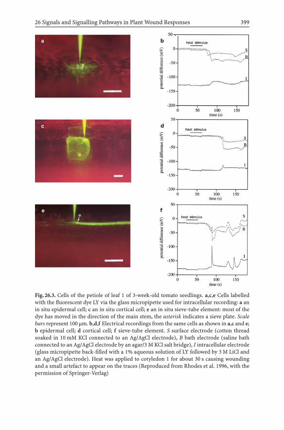
26 Signals and Signalling Pathways in Plant Wound Responses 399
Fig.26.3. Cells of the petiole of leaf 1 of 3-week-old tomato seedlings. a,c,e Cells labelledwith the fluorescent dye LY via the glass micropipette used for intracellular recording: a anin situ epidermal cell; c an in situ cortical cell; e an in situ sieve-tube element: most of thedye has moved in the direction of the main stem, the asterisk indicates a sieve plate. Scalebars represent 100 µm. b,d,f Electrical recordings from the same cells as shown in a,c and e;b epidermal cell; d cortical cell; f sieve-tube element. S surface electrode (cotton threadsoaked in 10 mM KCl connected to an Ag/AgCl electrode), B bath electrode (saline bathconnected to an Ag/AgCl electrode by an agar/3 M KCl salt bridge), I intracellular electrode(glass micropipette back-filled with a 1% aqueous solution of LY followed by 3 M LiCl andan Ag/AgCl electrode). Heat was applied to cotyledon 1 for about 30 s causing woundingand a small artefact to appear on the traces (Reproduced from Rhodes et al. 1996, with thepermission of Springer-Verlag)

400 J.D. Rhodes, J.F. Thain, D.C. Wildon
rations of the electrical events recorded by the surface and bath electrodeswould normally lead to their classification as variation potentials with theimplication that they reflect depolarisations in the underlying tissues dueto chemicals that have travelled in the xylem from the wound site. How-ever, given our current ignorance of detailed mechanisms at the cell level,they could reflect the summation of action potentials travelling at slightlydifferent times in the different STE/CC complexes of the petiole.
26.3Conclusions and Future Prospects
For severe wounding, the results are consistent with the distribution ofelicitors of PI synthesis and electrical activity by hydraulic dispersal in thexylem. We conclude that the electrical events are likely to be responses tochemicals transported in the xylem by hydraulic dispersal from the woundsite, rather than action potentials propagated from the wound site.
Following a small crushing wound, the pattern of systemic PI synthesiswas consistent with the transport of a chemical elicitor in the phloem fromthe wound site.
The lack of an electrical event following a small wound indicates thatelectrical signals are not an essential part of the systemic signalling systemthat induces PI synthesis.
The fact that a severe wound leads to large rapid action-potential-likedepolarisations in the cells of the STE/CC complexes in the petiole, but notin other cell types, is intriguing. Do these depolarisations have a function?
In the absence of detailed studies at the cell level, the description ofwound-induced electrical events recorded with surface-contact electrodesas either variation potentials or action potentials could carry misleadingimplications about their cellular mechanisms.
A significant step forward in plant signalling would be the identificationof the cellular mechanism of wound-induced electrical events; this could beachieved using a similar approach to that described here and in Rhodes etal. (1996). However, since the mapping of its genome, Arabidopsis may bethe preferred model organism; Arabidopsis does show electrical events fol-lowing severe wounds (our unpublished observations). The identificationof the ion channels responsible for the electrical events could be studied bythe use of a range of agonists and inhibitors which could be introduced viathe xylem stream and their electrical consequences in the phloem could bemonitored using microelectrodes.
Acknowledgements. The work from our laboratory was funded by theBiotechnology and Biological Sciences Research Council, UK.

26 Signals and Signalling Pathways in Plant Wound Responses 401
ReferencesBaker DA, Milburn JA (1989) Appendix Table X: speed of assimilate transport in the phloem
of plant species analysed by differing techniques. In: Baker DA, Milburn JA (eds) Trans-port of photoassimilates. Longman, Harlow, pp 354–355
Baydoun EA-H, Fry SC (1985) The immobility of pectic substances in injured tomato leavesand its bearing on the identity of the wound hormone. Planta 165:269–276
Bowles D (1998) Signal transduction in the wound response of tomato plants. Philos TransR Soc Lond Ser B 353:1495–1510
Davies E (2004) New functions for electrical signals in plants. New Phytol 161:607–610Green TR, Ryan CA (1972) Wound-induced proteinase inhibitor in plant leaves: a possible
defense mechanism against insects. Science 175:776–777Herde O, Fuss H, Peña-Cortès H, Fisahn J (1995) Proteinase inhibitor II gene expression
induced by electrical stimulation and control of photosynthetic activity in tomato leaves.Plant Cell Physiol 36:737–742
Mackie GO (1965) Conduction in nerve-free epithelia of siphonophores. Am Zool 5:439–453Malone M (1996) Rapid, long-distance signal transmission in higher plants. Adv Bot Res
22:163–228Mancuso S (1999) Hydraulic and electrical transmission of wound-induced signals in Vitis
vinifera. Aust J Plant Physiol 26:55–61Moyen C, Johannes E (1996) Systemin transiently depolarizes the tomato mesophyll cell
membrane and antagonizes fusicoccin-induced extracellular acidification of mesophylltissue. Plant Cell Environ 19:464–470
Nelson CE, Walker-Simmons M, Makus D, Zuroske G, Graham J, Ryan CA (1983) Regulationof synthesis and accumulation of proteinase inhibitors in leaves of wounded tomatoplants. In: Hedin PA (ed) Plant resistance to insects. ACS symposium series no 208.American Chemical Society, Washington, DC, pp 103–122
Passioura JB (1972) The effect of root geometry on the yield of wheat growing on storedwater. Aust J Agric Res 23:745–752
Peña-Cortès H, Fisahn J, Willmitzer L (1995) Signals involved in wound-induced proteinaseinhibitor II gene expression in tomato and potato plants. Proc Natl Acad Sci USA92:4106–4113
Rhodes JD, Thain JF, Wildon DC (1996) The pathway for systemic electrical signal conduc-tion in the wounded tomato plant. Planta 200:50–57
Rhodes JD, Thain JF, Wildon DC (1999) Evidence for physically distinct systemic signallingpathways in the wounded tomato plant. Ann Bot 84:109–116
Rigby NM, MacDougall AJ, Needs PW, Selvendran RR (1994) Phloem translocation ofa reduced oligogalacturonide in Ricinus communis L. Planta 193:536–541
Stankovic B, Davies E (1996) Both action potentials and variation potentials induce pro-teinase inhibitor gene expression in tomato. FEBS Lett 390:275–279
Stankovic B, Davies E (1997) Intercellular communication in plants: electrical stimulationof proteinase inhibitor gene expression in tomato. Planta 202:402–406
Stratmann JW (2003) Long distance run in the wound response – jasmonic acid is pullingahead. Trends Plant Sci 8:247–250
Thain JF, Wildon DC (1996) Electrical signalling in plants. In: Smallwood M, Knox JP,Bowles DJ (eds) Membranes: specialized functions in plants. Bios, Oxford, pp 301–317
Thain JF, Gubb IR, Wildon DC (1995) Depolarization of tomato leaf cells by oligogalactur-onide elicitors. Plant Cell Environ 18:211–214
van Bel AJE (2003) The phloem, a miracle of ingenuity. Plant Cell Environ 26:125–149Wildon DC, Thain JF, Minchin PEH, Gubb IR, Reilly AJ, Skipper YD, Doherty HM, O’Don-
nel PJ, BowlesDJ (1992)Electrical signalling and systemicproteinase inhibitor inductionin the wounded plant. Nature 360:62–65

27 Root Exudation and RhizosphereBiology: Multiple Functionsof a Plant Secondary MetaboliteLaura G. Perry, Tiffany L. Weir, Balakrishnan Prithiviraj,Mark W. Paschke, Jorge M. Vivanco
Abstract Plant root exudates have important effects on plant and soil communities, me-diating positive and antagonistic plant–plant and plant–microbe interactions, and alteringsoil processes. Here, we discuss the potential for a single plant secondary metabolite to servemultiple functions in the rhizosphere. Secondary metabolites that serve multiple functionsmay provide plants with multiple benefits at relatively low metabolic cost, with conse-quences for competitive ability, disease resistance, and resource availability. Specifically, wedescribe recent research on (±)-catechin, a secondary metabolite exuded from the roots ofthe invasive weed Centaurea maculosa. Depending on concentration and the species thatare present, (±)-catechin has the potential to act as an allelochemical, autoinhibitor, andantimicrobial agent, and to increase soil nutrient availability.
27.1Introduction
Plants secrete a wide array of organic compounds into the soil surroundingtheir roots (i.e, the rhizosphere), including amino acids, carbohydrates,mucilage, and secondary metabolites (Bertin et al. 2003; Walker et al. 2003).The quantities of carbon released as root exudates are surprisingly large.Young plants (seedlings) may secrete over 30% of their photosynthates asroot exudates (Sauerbeck et al. 1981), while older plants release between 5and 21% of their photosynthates into the soil (Marschner 1995). Secondarymetabolites, which are low molecular weight compounds produced duringsecondary metabolism, constitute the most diverse group of root exudates,and include an array of organic acids, flavonoids, tannins, terpenoids,alkaloids, polyacteylenes, and simple phenolics (Flores et al. 1999; Bertinet al. 2003). To date, more than 100,000 plant secondary metabolites havebeen identified (Dixon 2001).
Plant root exudates have important and diverse effects on soil chemistry,biology, and ecology. In particular, root exudates appear to mediate pos-itive and antagonistic belowground communication between plants andtheir competitors, mutualists, and enemies. Secondary metabolites in rootexudates are involved in the formation of symbiotic associations betweenplants and mycorrhizal fungi and bacteria (Dakora and Phillips 2002; Marx2004). Other secondary metabolites in root exudates have been shown topossess phytotoxic, antimicrobial, antibiotic, insecticidal, and hormonal
Communication in PlantsF. Baluška, S. Mancuso, D. Volkmann (Eds.)© Springer-Verlag Berlin Heidelberg 2006

404 L.G. Perry et al.
properties (Sudha and Ravishankar 2002; Bais et al. 2004, Bais et al. 2005),leading to inhibition of competitors, pathogens, herbivores, and parasites.Still other root exudates elicit herbivore defense responses in neighboringplants (Dicke and Dijkman 2001). In addition, root exudates can alter nu-trient cycling, facilitate root movement, increase nutrient acquisition, andreduce metal toxicity (Dakora and Phillips 2002; Hawes et al. 2003).
The role of phytotoxic secondary metabolites in plant–plant interactions(i.e., allelopathy) has been the subject of considerable research and debate(Nilsen 2002; Bertin et al. 2003; Weir et al. 2004). Plants that produce and ac-cumulate phytotoxins in the soil are thought to limit establishment, growth,and survival of neighboring plants, thus reducing local resource competi-tion and increasing their own success. Potent phytotoxins have been foundin plant leaf and root tissue, leaf leachates, leaf volatiles, and root exudates.However, because effects of phytotoxins on plant interactions are difficultto separate from effects of resource competition, many ecologists are notconvinced that allelopathy plays an important role in plant communities(Fitter 2003).
In addition, the evolution of phytotoxic secondary metabolites in plantsis not well understood, in contrast to the evolution of plant secondarymetabolites that repel or attract insects and microbes (Ehrlich and Raven1964; Whittaker and Feeny 1971; Scribner 2002). The selection pressuresthat could account for the development of phytotoxins in plants are rela-tively easy to imagine (e.g., reducedcompetition fromneighbors).However,the selection pressures that might maintain the production of phytotoxicsecondary metabolites over time are unclear (Fitter 2003). The argumentthat natural selection should operate against continued phytotoxin pro-duction is as follows. Allelopathic plants must be exposed to relatively highconcentrations of their own phytotoxin. Consequently, to benefit frombeing allelopathic, a plant must be at least partly resistant to its own al-lelochemical. Further, the metabolic cost of resistance together with themetabolic cost of production must be low relative to the resource benefit ofcompetitor inhibition. If an allelopathic plant is able to develop relativelycheap resistance to its own allelochemical, then other plants should also beable to evolve resistance at relatively little cost. Once other plants developresistance, the competitive benefits of producing the phytotoxin shoulddisappear. The cost of producing the phytotoxin once its benefits are lostshould result in evolution of reduced production.
Examples of allelopathic plants that have become invasive when trans-ported to new continents suggest that allelopathy may be particularly effec-tive in novel habitats where native species have not had the opportunity toevolve resistance to the invaders’ allelochemicals (Rabotnov 1982; Callawayand Aschehoug 2000; Bais et al. 2003; Vivanco et al. 2004). These studies em-phasize the potential importance of allelopathy in novel plant interactions,

27 Root exudation and rhizosphere biology 405
and the potential for the benefits of allelopathy to disappear over time asthe competitors develop resistance. However, the selection pressures thatlead to continued production of phytotoxins by allelopathic species in theirnative range, where their competitors have developed resistance, remainunclear. Plant–plant chemical interactions may be more complex than tra-ditionally thought. For example, indirect effects of allelochemicals on soilcommunities may sometimes be more important to plant community com-position than direct effects on plant growth or survival (Wardle et al. 1990;Inderjit and Weiner 2001). Such indirect modes of chemical interactionbetween plants have rarely been examined explicitly.
Recently, our research on phytotoxic plant root exudates has high-lighted how single plant secondary metabolites can perform many func-tions within plant and soil communities, depending on concentration, thespecies that are present, and perhaps environmental conditions. The po-tential for single secondary metabolites to have numerous effects in therhizosphere provides insights into the complex nature of the selectionpressures that may act on secondary metabolite production and secretion.A secondary metabolite that can serve multiple functions may be producedinitially for one purpose, and later for a different purpose. Further, themetabolic costs, and associated selection pressures, of producing a sec-ondary metabolite may be substantially reduced relative to the benefits ifthe metabolite serves multiple functions. In this chapter, we will focus onone secondary metabolite, catechin, which is exuded as a racemic mixtureof (+)-catechin and (–)-catechin from the roots of Centaurea maculosa(Lam.) (spotted knapweed), an exotic invasive weed in North Americangrasslands. Recent research indicates that (±)-catechin has the potential toact as an allelochemical, autoinhibitor, and antimicrobial and nematicidalagent, and to increase soil nutrient availability in C. maculosa communities(Fig. 27.1).
27.2C. maculosa Invasion Ecology
C. maculosa is a tap-rooted, short-lived perennial, native to grasslandsteppes of central and eastern Europe. C. maculosa was accidentally in-troduced to northwestern North America in the late 1800s (Sheley et al.1998). To date, C. maculosa has infested over 2.8 million hectares of NorthAmerican grassland, mainly in the midwestern and western regions ofNorth America (Müller-Scharer and Schroeder 1993). While C. maculosais not particularly abundant in European grasslands, it is often the domi-nant plant in invaded North American grasslands (Ridenour and Callaway2001). In North American infestations, C. maculosa densities can exceed

406 L.G. Perry et al.
Fig.27.1. A schematic depiction of the potential roles of (±)-catechin in Centaurea mac-ulosa root exudates. (±)-Catechin is a potent phytotoxin with negative effects on survivaland growth of interspecific competitors of C. maculosa (Bais et al. 2002, 2003). Reducedcompetitor growth owing to (±)-catechin exudation increases C. maculosa growth (Ride-nour and Callaway 2001). Higher catechin concentrations lead to inhibition of C. maculosaseedling establishment (i.e., autoinhibition), perhaps serving as a mechanism for regulationof population density (Perry et al. 2005b). (+)-Catechin is antibacterial and antifungal, while(–)-catechin is nematocidal, perhaps leading to changes in pathogen and mutualist popu-lations (Bais et al. 2002; Veluri et al. 2004b). Finally, (±)-catechin is a strong metal chelatorwith the potential to increase phosphorus and micronutrient availability (Callaway andRidenour 2004). (±) Effects of catechin on nematode populations and perhaps mycorrhizalfungi may also alter nutrient cycling and other soil processes

27 Root exudation and rhizosphere biology 407
400 individuals per square meter (Jacobs and Sheley 1998), and seed setcan exceed 10,000 seeds per square meter (Schirman 1981). Monodominantstands of C. maculosa decrease plant biodiversity (Tyser and Key 1988; Ri-denour and Callaway 2001), increase water runoff and soil erosion (Laceyet al. 1989), and reduce forage quality for livestock and wildlife (Watsonand Reddy 1974; Thompson 1996).
Recent work on C. maculosa root exudates indicates that C. maculosainvasions in North America may be partly mediated by root exudationof a potent phytotoxin, (±)-catechin (Bais et al. 2003). C. maculosa inva-siveness in North America probably results from a combination of factors,including allelopathy, high reproductive capacity (Schirman 1981), affinityfor disturbance (Marcus et al. 1998), and competitive ability for limitingresources. C. maculosa does not appear to be a better competitor thanNorth American species for nitrogen or water (Blicker et al. 2003; Olsenand Blicker 2003), but may be a better competitor for phosphorus in thepresence of arbuscular mycorrhizal (AM) fungi (Zabinski et al. 2002). Inaddition, AM fungi may mediate carbon transfer from some North Ameri-can grasses to C. maculosa (Carey et al. 2004). In the following, we describethe current knowledge of effects of (±)-catechin exudation on C. maculosaecology and invasion.
27.3(±)-Catechin, Allelopathy, and Cell Death
Evidence forallelopathy inC.maculosawasfirst reported in1963byFletcherand Renny, who observed high concentrations of a phytotoxin, cnicin, inC. maculosa leaf tissue. However, cnicin concentrations in C. maculosasoils were found to be insufficient to inhibit establishment and growthof neighboring plants (Locken and Kelsey 1987), indicating that C. mac-ulosa cnicin production was unlikely to be important for inhibiting plantcompetitors. More recently, Ridenour and Callaway (2001) tested whetherC. maculosa root exudates might contain phytotoxins that inhibit growthof North American grassland species. They used activated carbon, whichadsorbs organic compounds but has little effect on inorganic compounds(Mahall and Callaway 1992), to adsorb C. maculosa root exudates in the soil.Growing C. maculosa in soil with activated carbon reduced the inhibitoryeffect of C. maculosa on a native grass, Festuca idahoensis (Idaho fescue),suggesting that organic compounds in the soil around C. maculosa rootssuppressed F. idahoensis growth. Further, the competitive effect of F. ida-hoensis on C. maculosa growth was 30% greater when activated carbon wasadded to the soil, suggesting that adsorption of C. maculosa allelochemicalsshifted the balance of competition in favor of F. idahoensis. However, the

408 L.G. Perry et al.
phytotoxins in C. maculosa root exudates needed to be identified to evalu-ate the role of allelopathy in C. maculosa dominance over North Americangrassland species.
27.3.1Identification of the Allelochemical
To overcome the difficulty of isolating root-secreted allelochemicals fromsoil, Bais et al. (2002) grew C. maculosa in vitro and collected the rootexudates in sterile media. Addition of crude C. maculosa exudates to liquidmedia where Linaria dalmatica (Dalmatian toadflax), Verbascum thap-sus (common mullein), Bromus tectorum (downy brome), Kochia scoparia(kochia), C. diffusa (diffuse knapweed), and Arabidopsis thaliana weregrowing reduced root growth and induced plant mortality by 14 daysafter treatment, indicating that a component of the root exudates hadbroad-spectrum phytotoxic activity (Bais et al. 2002). The active fraction ofthe exudates was identified as (±)-catechin using high-performance liquidchromatography separations and identification by mass spectrometry and1H and 13C NMR techniques. By testing commercially purchased isomers ofpure catechin, Bais et al. (2002) initially attributed the phytotoxic activityto (–)-catechin. However, it was recently determined that (+)-catechin isalso phytotoxic (Iqbal et al. 2003), although less potent than (–)-catechin(Veluri et al. 2004a).
27.3.2Catechin Induces Reactive Oxygen Speciesand Ca2+-Mediated Cell Death
As is the case for many allelochemicals, the exact cellular target of (±)-catechin is still unknown. However, many of the cell signaling and indirectmolecular events that precede catechin-mediated cell death have been iden-tified. Using real-time video imaging and fluorescent viability dyes, withA. thaliana and C. diffusa as target species, Bais et al. (2003) observedthat cell death began about 15 min after catechin treatment, following con-densation of the cytoplasm. Cell death occurred first in the meristematiczone of the root tip and moved upward to the central elongation zone, sug-gesting that catechin or catechin-induced signals are transported throughthe vascular system. To more fully understand the molecular basis forcatechin-mediated cell death, Bais et al. (2003) monitored the kinetics ofseveral cellular signals, including generation of reactive oxygen species(ROS), fluctuations in cytoplasmic calcium concentrations ([Ca2+]cyt), and

27 Root exudation and rhizosphere biology 409
pH changes, to determine which signaling events occurred upon catechinexposure. ROS have been implicated in signal transduction events lead-ing to downstream responses, such as modulation of gene expression, thataffect several cellular and developmental processes (Huckelhoven and Ko-gel 2003; Finkel 1998). ROS are also known to accumulate in plant cellsin response to compatible pathogen infections and may damage cellularstructures and nucleic acids, resulting in cell death (Huckelhoven and Ko-gel 2003). Fluctuations in [Ca2+]cyt are also known to occur in responseto stress, including high salinity, osmoticum, heavy metals, and oxida-tive damage (Fasano et al. 2001; Jones et al. 1998). Finally, cytoplasmicpH changes are associated with signal transduction and regulation of rootgrowth (Fasano et al.2001; Scott and Allen 1999), and cell viability is closelytied to homeostasis of cytoplasmic pH.
Bais et al. (2003) showed that all three factorsplayan important role in theinitial events leading to catechin phytotoxicity. Using the fluorescent dye 6-carboxy-2′, 7′-dichlorodihydrofluorescein diacetate-di(acetoxymethly es-ter), they determined that within 10 sec, catechin treatment induced a 12-fold increase in ROS generation in C. diffusa and A. thaliana seedlings.Like the pattern of cell death induction, the increase in ROS originated inthe meristematic region of the root tip and moved into the central elon-gation zone before progressing back through the main axis of the root ina wavelike fashion. Catechin treatment also resulted in rapid, transient el-evations in root-tip-localized [Ca2+]cyt levels. These increases in [Ca2+]cyt
occurred about 30 sec after catechin treatment, following the ROS signal,but preceding cell death. Approximately 15−20 min after catechin adminis-tration, consistent with the initial appearance of cell death, the cytosolic pHdropped from approximately 7.2 to 5.6. These changes most likely reflectthe loss of ion homeostasis associated with cell mortality. Bais et al. (2003)also demonstrated that exogenous application of an antioxidant, ascorbicacid, prevented the generation of the initial ROS signal, the subsequentspike in meristematic [Ca2+]cyt levels, and cell death, suggesting that thephytotoxicity of catechin is directly or indirectly due to oxidative stress.
27.3.3Catechin Exposure Leads to Genome-Wide Changes in Arabidopsis
Because the model plant A. thaliana is susceptible to catechin, Bais et al.(2003) were able to monitor changes in global gene expression to deter-mine potential transcriptional events associated with catechin phytotoxic-ity. Using a 12,000 gene oligoarray, they monitored changes in transcription10 min, 1 h, and 12 h after Arabidopsis roots had been subjected to phyto-toxic levels of catechin. Within 1 h of treatment a number of genes relatedto oxidative stress were upregulated, including glutathione transferase,

410 L.G. Perry et al.
monooxygenase, lipid transfer protein, heat shock protein, DNA-J protein,and blue copper-binding protein (Bais et al. 2003). Phenylpropanoid andterpenoid phytoalexin pathway genes, a number of which produce enzymesthat can act as antioxidants (Sticher et al. 1997), were also induced in theroots 1 h after catechin treatment. However, the most interesting changein gene expression that they observed was the upregulation of ten genesin the first 10 min after catechin treatment. These included genes associ-ated with calcium signaling and oxidative stress, as well as four unknowngenes lacking homology with genes from other organisms. Identificationof the function of these unknown genes could provide insights into thefactors that cause plants to be susceptible to catechin, and into potentialmechanisms of resistance.
27.3.4(±)-Catechin Is Present at PhytotoxicConcentrations in C. maculosa Soils
While laboratory experiments demonstrated that (±)-catechin is a potentphytotoxin with strong effects on plant biochemistry and gene expression,field observations were required to gauge the importance of (±)-catechinin C. maculosa competitive interactions under natural conditions. In par-ticular, for (±)-catechin to influence plant interactions, it must be presentat phytotoxic concentrations in C. maculosa field soils. Several studieshave reported exceptionally high soil (±)-catechin concentrations in NorthAmerican C. maculosa soils (Bais et al. 2002, 2003; Perry et al. 2005b), in-dicating that soil (±)-catechin is present in sufficient quantities in C. mac-ulosa soil to inhibit plant neighbors. Bais et al. (2003) reported a mean soil(±)-catechin concentration of 2.24±0.20 mg g−1 and Perry et al. (2005b)reported a mean soil (±)-catechin concentration of 1. 55 ± 1.27 mg g−1 drysoil. In addition, tests of the soil extracts from one site confirmed that the(±)-catechin in C. maculosa soils has similar phytotoxicity to (±)-catechinfrom commercial sources (Perry et al. 2005b). Bais et al. (2002) found that(±)-catechin concentrations declined with distance from the C. maculosataproot, and with increasing soil depth. However, Perry et al. (2005b) foundthat soil (±)-catechin concentrations did not change with distance from theC. maculosa taproot but remained high as far as 25 cm from the taproot,indicating that high soil (±)-catechin concentrations may be ubiquitous inat least some well-established C. maculosa populations. The differences insoil (±)-catechin concentrations among the studies arenot surprising, sincethe studies were conducted in different locations and at different times. Theeffects of soil characteristics, climate, and season on (±)-catechin secretion,stability, and soil absorption are not yet understood.

27 Root exudation and rhizosphere biology 411
27.3.5The Role of (±)-Catechin in C. maculosa Invasion
For (±)-catechin to facilitate C. maculosa invasion, it must inhibit the NorthAmerican grassland species with which C. maculosa competes. Effects of(±)-catechin on seedling growth and survival have been examined for morethan 25 North American grassland species that are native to the types ofplant communities invaded by C. maculosa (Bais et al. 2003; Weir et al. 2003;Perry et al. 2005a). (±)-Catechin concentrations as low as 50 µg ml−1 reducethe growth of sensitive North American grassland species, such as F. ida-hoensis (Idaho fescue) and Koeleria cristata (prairie junegrass), grown invitro in liquid media (Weir et al. 2003). Higher (±)-catechin concentra-tions (125−500 µg ml−1) are required to significantly reduce the growth ofsensitive North American species germinated on filter paper (Perry et al.2005a), but these concentrations are still well below reported C. maculosasoil (±)-catechin concentrations. North American grassland species varyconsiderably in sensitivity to (±)-catechin (Perry et al. 2005a), indicat-ing that (±)-catechin exudation probably gives C. maculosa an advantageover some species and not others. In particular, seedlings of larger-seededspecies tend to be more resistant to (±)-catechin than seedlings of smaller-seeded species (Perry et al. 2005a). Some of the North American speciesthat are relatively resistant to (±)-catechin appear to persist in C. macu-losa populations, while others may be displaced by C. maculosa throughresource competition rather than allelopathy.
One hypothesis to explain C. maculosa invasiveness in North Amer-ica is that North American grassland species may be on average moresensitive to (±)-catechin than the European species with which C. macu-losa naturally coexists, because the North American species have not hadan opportunity to evolve resistance to the allelochemical (i.e., the “novelweapons” hypothesis; Callaway and Aschehoug 2000). To explore whetherNorth American grassland species are more sensitive to (±)-catechin thanEuropean species, Bais et al. (2003) compared the effects of (±)-catechin onthree North American grasses that are commonly displaced by C. maculosaand three European congeners that naturally coexist with C. maculosa. TheNorth American species were significantly more inhibited by (±)-catechintreatment than the European species, suggesting that (±)-catechin maygive C. maculosa a greater advantage over its competitors in North Amer-ica than in Europe, probably contributing to C. maculosa invasiveness inNorth American grasslands.

412 L.G. Perry et al.
27.4(±)-Catechin and C. maculosa Autoinhibition
In addition to reducing the success of C. maculosa’s interspecific com-petitors, (±)-catechin appears to reduce C. maculosa seedling recruitmentin well-established populations. In C. maculosa stands in North America,adult plants are sometimes widely spaced, with the interspaces betweenplants unoccupied, occupied by C. maculosa seedlings that fail to establish,or occupied by other species. Perry et al. (2005b) examined whether thispattern might be accounted for by (±)-catechin inhibition of C. maculosaseedling establishment (i.e., autoinhibition).
To test whether organic compounds in the soil in C. maculosa standslimited C. maculosa seedling establishment, Perry et al. (2005b) added ac-tivated carbon, which adsorbs organic compounds (Mahall and Callaway1992), to the soil around adult C. maculosa plants in a well-establishedpopulation in the field. Activated carbon addition increased C. maculosaseedling densities by more than 30%, indicating a strong negative effect oforganic compounds in the soil around C. maculosa adults on C. maculosaseedling establishment. Seedling densities were still visibly greater in thesoil with activated carbon than in the soil without activated carbon in someplots 1 year after the start of the experiment (L. Perry, personal observa-tion), indicating that the effects of organic compounds in the soil aroundC. maculosa adults on C. maculosa seedling establishment are persistent.
Measurements of soil (±)-catechin around adult C. maculosa at the fieldsite indicated that (±)-catechin concentrations in the site were extremelyhigh (mean 1.55 mg g−1 dry soil). C. maculosa seedling densities at the sitewere very low (mean 28 seedlings per square meter). To test whether thehigh soil (±)-catechin concentrations could account in part for the lowC. maculosa seedling establishment, Perry et al. (2005b) examined the re-sponse of C. maculosa seedlings to (±)-catechin in laboratory experiments.Treatment with 1.0 mg ml−1 of (±)-catechin reduced C. maculosa seedlingroot lengths by 50%, demonstrating that C. maculosa soil (±)-catechinconcentrations are sufficient to substantially reduce C. maculosa seedlinggrowth. Higher (±)-catechin concentrations (2 and 4 mg ml−1) reducedC. maculosa root elongation by as much as 75%. Reduced root elongationdid not result in seedling mortality under laboratory conditions, but wouldbe expected to reduce survival under field conditions with limited water. Inaddition, when (±)-catechin was maintained in solution in 10% methanolfor 2 days after treatment, treatment with 1.0 mg ml−1 of (±)-catechin re-duced C. maculosa germination by more than 70%. Tetrazolium analyses ofseed viability indicated that (±)-catechin treatment did not reduce seed sur-vival, suggesting that (±)-catechin dissolved in methanol may have delayedgermination of C. maculosa seeds.

27 Root exudation and rhizosphere biology 413
Plant autoinhibition mediated by secondary metabolites has been thesubject of much research (Singh et al. 1999). However, (±)-catechin is oneof the first secondary metabolites determined to act as both an allelochem-ical and an autoinhibitor. Autoinhibition of seedling establishment mayinfluence plant populations in several ways. First, autoinhibition may bea mechanism through which adults avoid establishment of intraspecificcompetitors in dense populations (Schenk et al. 1999). Second, seedlingsthat produce autoinhibitors may reduce intraspecific competition by pre-venting the establishment of their siblings and unrelated seedlings (Dyer2004). Third, autoinhibitors that delay germination, rather than killingseeds or seedlings, may postpone germination in areas where intraspe-cific competition from adults would prevent seedling survival (Picmanand Picman 1984). Determining the specific effects of autoinhibition onC. maculosa populations will require further research to determine whether(±)-catechin reduces C. maculosa seedling root elongation or delays ger-mination under field conditions.
Autoinhibition by C. maculosa is probably most important in well-established stands, where soil (±)-catechin is likely to be concentratedand widespread. (±)-Catechin concentrations lower than 0.5 mg ml−1 havelittle effect on C. maculosa root elongation, and no effect on germination(Perry et al. 2005b). However, much lower (±)-catechin concentrations havelarge effects on other, more (±)-catechin-sensitive species (Bais et al. 2003;Weir et al. 2003; Perry et al. 2005a). Thus, C. maculosa seedlings shouldhave an advantage over more (±)-catechin-sensitive species in soils withmoderate (±)-catechin concentrations, while very high (±)-catechin con-centrations may prevent establishment of C. maculosa seedlings as wellas other species. In well-established C. maculosa populations with highsoil (±)-catechin concentrations, autoinhibition, water stress (Jacobs andSheley 1998), and seed predation (Muller-Schärer and Schroeder 1993)probably operate together to limit C. maculosa seedling establishment.
27.5(±)-Catechin Effects on Soil Communities
In addition to the inhibitory effects of (±)-catechin on plants, (+)-catechinand (–)-catechin both reduce survival of some soil organisms. However,the two enantiomers appear to affect different organisms. Bais et al. (2002)found that (+)-catechin, but not (–)-catechin, inhibited growth of severalsoil bacteria. Six bacterial strains were exposed to each of the enantiomersor the racemic mixture, applied to filter discs. Xanthomonas campestrisssp. vesicatoria, Erwinia amylovora, Pseudomonas fluorescens, and E. caro-tovora were each susceptible to 100 µg ml−1 (+)-catechin or to 200 µg ml−1

414 L.G. Perry et al.
(±)-catechin. In contrast, Agrobacterium radiobacter was inhibited only byhigher concentrations (300 µg ml−1 (+)-catechin or more), and Agrobac-terium rhizogenes (15834) was not affected even by high (+)-catechinconcentrations (Bais et al. 2002). None of the bacteria were affected by(–)-catechin treatment. Similarly, Veluri et al. (2004b) found that low con-centrations of (+)-catechin (12.5−25 µg ml−1) inhibited growth of severalroot-colonizing fungi, including Tricodermareesi, T. viridis, Fusariumoxys-porum, Aspergillus niger, and Penicillium sp. Veluri et al. (2004b) com-pared the antimicrobial activity of several catechin derivatives and foundthat (+)-catechin was the most active against E. carotovora, E. amylovora,X. campestris, and A. radiobacter, while the (+)-catechin derivatives (+)-pentaacetylcatechin, (+)-tetramethoxycatechin, and (+)-isopropylidylcat-echin showed less antimicrobial activity. (+)-Pentaacetylcatechin and (+)-tetramethoxycatechin also inhibited some of the root-colonizing fungi.Notably, (+)-catechin antibacterial activity may be limited to gram nega-tive species (Veluri et al. 2004b). (+)-Catechin has no effect or only a veryweak effect on a number of gram positive human pathogens (Palma et al.1999; Puupponen-Pimiš et al. 2001).
Although(–)-catechindoesnotappear toaffect soilbacteria, (–)-catechindoes appear to inhibit other soil organisms. Specifically, recent studies con-ducted in our laboratory have shown that (–)-catechin is nematicidal (B.Prithiviraj and J. Vivanco, unpublished data). Addition of low concentra-tions (1−10 g ml−1) of (–)-catechin to the growth medium resulted in 100%mortality of the bacterial-feeding, saprophytic nematode Caenorhbditiselegans in 3−4 days. No previous studies have reported (±)-catechin in-hibition of plant pathogenic or saprophytic nematodes. Such nematidicalactivity could have large effects on soil processes (discussed later) and onsoil microbial populations. Free-living nematodes in the soil are often bac-terial or fungal feeders, and thus could influence population dynamics ofplant pathogens or mutualists.
In addition, soil fungi, and perhaps other soil microbes, may influence(±)-catechin secretion by C. maculosa. Fungal cell walls isolated from thesoil-borne pathogen Phytophthora cinnamommi elicited increased root ex-udation of (±)-catechin by C. maculosa (Bais et al. 2002). C. maculosaplants grown together in vitro produced approximately 83.2 µg ml−1 of(±)-catechin in their pooled root exudates. When P. cinnamoni cell wallisolates were applied to C. maculosa plants in vitro, the (±)-catechin con-centration in the root exudates increased to 185.04 µg ml−1. The responseof C. maculosa to fungal cell wall elicitors suggests that (±)-catechin may bean inducible plant chemical defense, similar to phytoalexins, that C. mac-ulosa secretes in larger quantities when under attack by pathogenic mi-crobes in the rhizosphere. Both constitutive and induced (±)-catechin ex-udation have the potential to affect the population dynamics of a wide

27 Root exudation and rhizosphere biology 415
variety of rhizosphere microbiota under natural conditions. The influenceof (±)-catechin on soil communities and the consequences for pathogenand mutualist abundances in C. maculosa soils warrant further investiga-tion.
27.6(±)-Catechin, Soil Processes, and Nutrient Availability
(±)-Catechin may also influence soil nutrient availability and nutrient cy-cling in the C. maculosa rhizosphere, through both direct chemical interac-tions with soil nutrients and indirect effects on soil communities (Callawayand Ridenour 2004). In laboratory experiments, catechin has been shownto be a relatively strong metal chelator, able to form stable complexes withiron, aluminum, and copper ions (Mhatre et al. 1993; Mira et al. 2002;Khokhar and Apenten 2003). Metal chelators in root exudates are thoughtto increase availability of soil micronutrients, including iron, manganese,copper, andzinc, by formingcomplexeswith themetals and increasing theirsolubility and mobility (Dakora and Phillips 2002). Evidence that chelatorsin plant root exudates increase soil micronutrient availability is particularlystrong with regard to graminoid secretion of phytosiderophores (Treeby etal. 1989; Cesco et al. 2002; Jones et al. 2004), but many phenolics, includingcatechin, produced by dicots also have the potential to form complexeswith insoluble micronutrients and may have similar effects (Olsen et al.1981; Dakora and Phillips 2002). In addition, metal chelators in root exu-dates are thought to increase availability of soil phosphorus, a frequentlylimiting macronutrient in terrestrial ecosystems. A large portion of soilphosphorus is often unavailable to plants because it is bound in insolu-ble ferric, aluminum, and calcium phosphates (Mengel and Kirkby 1987).Metal chelators, by binding to iron and aluminum in ferric and aluminumphosphates, release plant-available phosphates at the same time that theyincrease metal solubility (Masaoka et al. 1993; Dakora and Phillips 2002).Thus, (±)-catechin exudation may increase phosphorus and micronutrientavailability in the C. maculosa rhizosphere, although effects of (±)-catechinchelation on nutrient availability have not yet been examined.
(±)-Catechin may also affect soil nutrient availability by altering soilcommunities. As described earlier, (±)-catechin is known to be toxic tosome soil-borne pathogens and nematodes. The nematicidal effects of (±)-catechin could have profound effects on nutrient cycling. Laboratory exper-iments and field studies have demonstrated that nematodes play a criticalrole in influencing turnover of soil microbial biomass and nutrient avail-ability (Bardgett et al. 1999; Bongers and Ferris 1999; Yeates 2003). Insome ecosystems, nematode activity accounts for up to 40% of nutrient

416 L.G. Perry et al.
mineralization as a result of nematode excretion and nematode effects onbacterial populations (De Ruiter et al. 1993). In addition, (±)-catechin inthe rhizosphere may affect soil mycorrhizal fungi. Catechin concentrationsin the roots of several tree species have been shown to decline in associationwith mycorrhizal infection (Münzenberger et al. 1990, 1995; Beyeler andHeyser 1997), suggesting either that tree roots reduce catechin concentra-tions to allow mycorrhizal infection to occur, or that mycorrhizal fungi usethe catechin in infected roots as a carbon source (Beyeler and Heyser 1997).Several fungi have been shown to degrade catechin (Galiotou-Panayotouet al. 1988; Vasudevan and Mahadevan 1990). In either case, high soil (±)-catechin concentrations would be expected to influence the abundance ofsoilmycorrhizal fungi. (±)-Catechinexudationmay increase theabundanceof soil microbes that can survive high soil (±)-catechin concentrations, orthat can use (±)-catechin as a carbon source, and reduce the abundanceof those microbes that cannot. Such changes have the potential to alternutrient mineralization, immobilization, and transformation rates, as wellas mutualistic associations that affect resource availability (Laakso et al.2000; Hamel 2004). Research on the effects of (±)-catechin on soil com-munities, particularly mycorrhizal communities, and on nutrient cyclingis needed to determine the effects of C. maculosa (±)-catechin exudationon soil processes.
27.7Conclusions and Future Prospects
In summary, (±)-catechin, a secondary metabolite exuded from the roots ofC. maculosa, has been shown to mediate positive and antagonistic plant–plant and plant–microbe communication, with the potential for strongeffects on (1) survival and growth of interspecific competitors, (2) conspe-cific seedling establishment, (3) survival of pathogenic and nonpathogenicsoil organisms, and (4) soil processes. Given the numerous functions that(±)-catechin may have in plant communities, it will be difficult to deter-mine the selection pressures that resulted in the evolution of C. maculosa(±)-catechin production and that continue to select for its production inits native and exotic ranges. It is likely that some or perhaps many of thepotential effects of C. maculosa (±)-catechin production are unintendedconsequences of a compound produced for a particular function or com-bination of functions. More research will be needed to determine the fieldconditions under which (±)-catechin operates as an allelochemical, au-toinhibitor, antimicrobial agent, or soil nutrition enhancer, and whetherand when these functions might act in concert. Regardless, the myriad ofroles that (±)-catechin may play in communities occupied by C. maculosa
27 Root exudation and rhizosphere biology 417
highlights the importance of considering multiple functions in attemptingto understand the effects and evolution of secondary metabolite produc-tion.
Acknowledgements. U.S. Dept. of Defense SERDP (SI-1388) provided fund-ing for this chapter.
ReferencesBais HP, Prithiviraj B, Jha AK, Ausubel FM, Vivanco JM (2005) Mediation of pathogen
resistance by exudation of antimicrobials from roots. Nature 434:217–221Bais HP, Park S, Weir TL, Callaway RM, Vivanco JM (2004) How plants communicate using
the underground information superhighway. Trends Plant Sci 9:26–32Bais HP, Vepachedu R, Gilroy S, Callaway RM, Vivanco JM (2003) Allelopathy and exotic
plant invasion: from molecules and genes to species interactions. Science 301:1377–1380Bais HP, Walker TS, Stermitz FR, Hufbauer RA, Vivanco JM (2002) Enantiomeric-dependent
phytotoxic and antimicrobial activity of (±)-catechin. A rhizosecreted racemic mixturefrom spotted knapweed. Plant Physiol 128:1173–1179
Bardgett RD, Cook R, Yeates GW, Denton CS (1999) The influence of nematodes on below-ground processes in grassland ecosystems. Plant Soil 212:23–33
Bertin C, Yang X, Weston LA (2003) The role of root exudates and allelochemicals in therhizosphere. Plant Soil 256:67–83
Beyeler M, Heyser W (1997) The influence of mycorrhizal colonization on growth in thegreenhouse and on catechin, epicatechin and procyanidin in roots of Fagus sylvatica L.Mycorrhiza 7:171–177
Blicker PS, Olsen BE, Wraith JM (2003) Water use and water-use efficiency of the invasiveCentaurea maculosa and three native grasses. Plant Soil 254:371–381
Bongers T, Ferris H (1999) Nematode community structure as a bioindicator in environ-mental monitoring. Trends Ecol Evol 14:224–228
Callaway RM, Aschehoug ET (2000) Invasive plants versus their new and old neighbors:a mechanism for exotic invasion. Science 290:521–523
Callaway RM, Ridenour WM (2004) Novel weapons: invasive success and the evolution ofincreased competitive ability. Front Ecol Environ 2:436–443
Callaway RM, Newingham B, Zabinski CA, Mahall BE (2001) Compensatory growth andcompetitive ability of an invasive weed are enhanced by soil fungi and native neighbours.Ecol Lett 5:429–433
Cary EV, Marler MJ, Callaway, RM (2004) Mycorrizae transfer carbon from a native grass toan invasive weed: evidence from stable isotopes and physiology. Plant Ecol 172:133–141
Cesco S, Nikolic M, Romheld V, Varanini Z, Pinton R (2002) Uptake of 59Fe from soluble59Fe-humate complexes by cucumber and barley plants. Plant Soil 241:121–128
Dakora FD, Phillips DA (2002) Root exudates as mediators of mineral acquisition in low-nutrient environments. Plant Soil 245:35–47
De Ruiter PC, Moore JC, Zwart KB, Bouwman LA, Hassink J, Bloem J, De Vos JA, MarinissenJCY, Didden WAM, Lebbink G, Brussaard L (1993) Simulation of nitrogen mineralizationin the below-ground food webs of two winter wheat fields. J Appl Ecol 30:95–106
Dicke M, Dijkman H (2001) Within-plant circulation of systemic elicitor of induced defenceand release from roots of elicitor that affects neighboring plants. Biochem Syst Ecol29:1075–1087
Dixon RA (2001) Natural products and plant disease resistance. Nature 411:843–847

418 L.G. Perry et al.
Dyer AR (2004) Maternal and sibling factors induce dormancy in dimorphic seed pairs ofAegilops triuncialis. Plant Ecol 172:211–218
Ehrlich PR, Raven PH (1964) Butterflies and plants: a study in coevolution. Evolution18:586–608
Fasano JM, Swanson SJ, Blancoflor EB, Dowd PE, Kao TH, Gilroy S (2001) Changes inroot cap pH are required for the gravity response of the Arabidopsis root. Plant Cell13:907–921
Finkel T (1998) Oxygen radicals and signaling. Curr Opin Cell Biol 10:248–253Fitter A (2003) Making allelopathy respectable. Science 301:1337–1338Fletcher RA, Renny AJ (1963) A growth inhibitor found in Centaurea spp. Can J Plant Sci
43:475–481Flores HE, Vivanco JM, Loyola-Vargas VM (1999) “Radicle” biochemistry: the biology of
root-specific metabolism. Trends Plant Sci 4:220–226Galiotou-Panayotou M, Rodis P, Macris BJ, Stathakos D (1988) Purification of a novel en-
zyme involved in catechin degradation by Clavatia gigantea. Appl Microbiol Biotechnol28:543–545
Hamel C (2004) Impact of arbuscular mycorrhizal fungi on N and P cycling in the root zone.Can J Plant Sci 84:383–395
Hawes MC, Bengough G, Cassab G, Ponce G (2003) Root caps and rhizosphere. J PlantGrowth Regul 21:352–367
Huckelhoven R, Kogel KH (2003) Reactive oxygen intermediates in plant-microbe interac-tions:Who is who in powdery mildew resistance? Planta 216:891–902
Iqbal Z, Hiradate S, Noda A, Isojima SI, Fujii Y (2003) Allelopathic activity of buckwheat:isolation and characterization of phenolics. Weed Sci 51:657–662
Inderjit, Weiner J (2001) Plant allelochemical interference or soil chemical ecology? PerspectPlant Ecol 4:3–12
Jacobs JS, Sheley RL (1998) Observation: life history of spotted knapweed. J Range Manage51:665–673
Jones DL, Hodge A, Kuzyakov Y (2004) Plant and mycorrhizal regulation of rhizodeposition.New Phytol 163:459–480
Jones DL, Gilroy S, Larsen PB, Howell SH, Kochian LV (1998) Effect of aluminumon cytoplasmic Ca2+ homeostasis in root hairs of Arabidopsis thaliana (L.). Planta206:378–387
Khokhar S, Apenten RKO (2003) Iron binding characteristics of phenolic compounds: sometentative structure-activity relations. Food Chem 81:133–140
Laakso J, Setälä H, Palojärvi A (2000) Influence of decomposer food web structure andnitrogen availability on plant growth. Plant Soil 225:153–165
Lacey JR, Marlow CB, Lane JR (1989) Influence of spotted knapweed (Centaurea maculosa)on surface runoff and sediment yield. Weed Technol 3:627–631
Locken LJ, Kelsey RG (1987) Cnicin concentrations in Centaurea maculosa (spotted knap-weed). Biochem Syst Ecol 15:313–320
Mahall BE, Callaway RM (1992) Root communication mechanisms and intracommunitydistributions of two mojave desert shrubs. Ecology 73:2145–2151
Marschner H (1995) Mineral nutrition of higher plants, second edition. Academic, Lon-don, UK
Marcus WA, Milner G, Maxwell B (1998) Spotted knapweed distribution in stock camps andtrails of the Selway-Bitterroot Wilderness. Great Basin Nat 58:156–166
Marx J (2004) The roots of plant-microbe collaborations. Science 304:234–236Masaoka Y, Kojima M, Sugihara S, Yoshihara T, Koshino M, Ichihara A (1993) Dissolu-
tion of ferric phosphate by alfalfa (Medicago sativa L.) root exudates. Plant Soil 155/156:75–78

27 Root exudation and rhizosphere biology 419
Mengel K, Kirkby EA (1987) Principles of plant nutrition, fourth edition. InternationalPotash Institute, Worblaufen-Bern, Switzerland
Mhatre SN, Iyer RK, Moorthy PN (1993) Characterization of aluminum complexes in teaextract. 1. Al-27 NMR studies. Magn Reson Chem 31:169–175
Mira L, Fernandez MT, Santos M, Rocha R, Florencio MH, Jennings KR (2002) Interactionsof flavonoids with iron and copper ions: a mechanism for their antioxidant activity. FreeRadical Res 36:1199–1208
Muller-Schärer H, Schroeder D (1993) The biological control of Centaurea spp. in NorthAmerica: do insects solve the problem? Pestic Sci 37:343–353
Münzenberger B, Heilemann J, Strack D, Kottke I, Oberwinkler F (1990) Phenolics ofmycorrhizas and non-mycorrhizal roots of Norway spruce. Planta 182:142–148
Münzenberger B, Kottke I, Oberwinkler F (1995) Reduction of phenolics in mycorrhizas ofLarix decidua Mill. Tree Physiol 15:191–196
NilsenET(2002)Ecological relevanceof allelopathy: someconsiderations related toMediter-ranean subtropical, temperate, and boreal forest shrubs. In: Inderjit, Mallik AU (eds)Chemical ecologyofplants: allelopathy inaquaticandterrestrial ecosystems.Birkhäuser,Basel, Switzerland, pp 109–129
Olsen BE, Blicker PS (2003) Response of the invasive Centaurea maculosa and two nativegrasses to N-pulses. Plant Soil 254:457–467
Olsen RA, Bennett JH, Blume D, Brown JC (1981) Chemical aspects of Fe stress responsemechanism in tomatoes. J Plant Nutr 3:905–921
Palma M, Taylor LT, Varela RM, Cutler SJ, Cutler HG (1999) Fractional extraction of com-pounds from grape seed by supercritical fluid extraction and analysis for antimicrobialand agrochemical activities. J Agric Food Chem 47:5044–5048
Perry LG, Johnson C, Alford ÉR, Vivanco JM, Paschke MW (2005a) Screening of grasslandplants for restoration after spotted knapweed invasion. Restor Ecol (in press)
Perry LG, Thelen GC, Ridenour WM, Weir TL, Callaway RM, Paschke MW, Vivanco JM(2005b) Dual role for an allelochemical: (±)-catechin from Centaurea maculosa rootexudates regulates conspecific seedling establishment. J Ecol (in press)
Puupponen-Pimiš, Nohynek L, Meier C, Kšhkšnene M, Hopia A, Oksman-Caldentey K-M(2001) Antimicrobial properties of phenolic compounds from berries. J Appl Microbiol90:494–507
Rabotnov TA (1982) Importance of the evolutionary approach to the study of allelopathy.Sov J Ecol 12:127–130
Ridenour WM, Callaway RM (2001) The relative importance of allelopathy in interference:the effects of an invasive weed on a native bunchgrass. Oecologia 126:444–450
Sauerbeck D, Nonnen S, Allard JL (1981) Consumption and turnover of photosynthesisin the rhizosphere depending on plant species and growth conditions. LandwirtschForschung Sonderh 37:207–216
Schenck J, Mahall BE, Callaway RM (1999) Spatial segregation of roots. Adv Ecol Res pp145–180
Schirman R (1981) Seed production and spring seedlings establishment of diffuse andspotted knapweed. J Range Manage 34:45–47
Scribner JM (2002) Evolution of insect-plant relationships: chemical constraints, coadapta-tion, and concordance of insect/plant traits. Entomol Exp Appl 104:217–235
Scott, AC, Allen NS (1999) Changes in cytosolic pH within Arabidopsis root columellacells play a key role in the early signaling pathway for root gravitropism. Plant Physiol121:1291–1298
Sheley RL, Jacobs JS, Carpinelli MF (1998) Distribution, biology, and management of diffuseknapweed (Centaurea diffusa) and spotted knapweed (Centaurea maculosa). WeedTechnol 12:353–362

420 L.G. Perry et al.
Singh HP, Batish DR, Kohli RK (1999) Autotoxicity: concept, organisms, and ecologicalsignificance. Crit Rev Plant Sci 18:757–772
Sticher L, Mauch-Mani B, Metraux JP (1997) Systemic acquired resistance. Annu Rev Phy-topathol 35:235–270
Sudha G, Ravishankar GA (2002) Involvement and interaction of various signaling com-pounds on the plant metabolic events during defense response, resistance to stressfactors, formation of secondary metabolites and their molecular aspects. Plant Cell TissOrg 71:181–212
Thompson MJ (1996) Winter foraging responses to elk to spotted knapweed removal.Northwest Sci 70:10–19
Treeby M, Marschner H, Romheld V (1989) Mobilization of iron and other micronutrientcations from calcareous soil by plant-borne, microbial, and synthetic metal chelators.Plant Soil 114:217–226
Tyser RW, Key CH (1988) Spotted knapweed in natural area fescue grasslands: an ecologicalassessment. Northwest Sci 62:981–987
Vasudevan N, Mahadevan A (1990) Utilization of catechin by microorganisms. Curr SciIndia 59:1323–1325
Veluri R, Weir TL, Bais HP, Stermitz FR, Vivanco JM (2004a) Phytotoxic and antimicrobialactivities of catechin derivatives. J Agric Food Chem 52:7746–7746
Veluri R, Weir TL, Bais HP, Stermitz FR, Vivanco JM (2004b) Phytotoxic and antimicrobialactivities of catechin derivatives. J Agric Food Chem 52:1077–1082
Vivanco JM,BaisHP, StermitzFR,ThelenGC,CallawayRM(2004)Biogeographical variationin community response to root allelochemistry: novel weapons and exotic invasion. EcolLett 7:285–292
Walker TS, Bais HP, Halligan KM, Stermitz FR, Vivanco JM (2003) Metabolic profiling ofroot exudates of Arabidopsis thaliana. J Agric Food Chem 51:2548–2554
Wardle DA, Nilsson M, Gallet C, Zackrisson O (1990) An ecosystem-level perspective ofallelopathy. Biol Rev 73:305–319
Watson AK, Renney AJ (1974) The biology of Canadian weeds. 6. Centaurea diffusa andCentaurea maculosa. Can J Plant Sci 54:687–701
Weir TL, Park S, Vivanco JM (2004) Biochemical and physiological mechanisms mediatedby allelochemicals. Curr Opin Plant Biol 7:472–479
Weir TL, Bais HP, Vivanco JM (2003) Intraspecific and interspecific interactions mediatedby a phytotoxin, (–)-catechin, secreted by the roots of Centaurea maculosa (spottedknapweed). J Chem Ecol 29:2379–2393
Whittaker RH, Feeny PP (1971) Allelochemics: chemical interactions between species. Sci-ence 171:757–770
Yeates GW (2003) Nematodes as soil indicators: functional and biodiversity aspects. BiolFert Soils 37:199–210
ZabinskiCA,QuinnL,CallawayRM(2002)Phosphorusuptake,not carbon transfer, explainsarbuscular mycorrhizal enhancement of Centaurea maculosa in the presence of nativegrassland species. Funct Ecol 16:758–765

28 Communication BetweenUndamaged Plants by Volatiles:the Role of AllelobiosisVelemir Ninkovic, Robert Glinwood, Jan Pettersson
Abstract Plant–plant signalling is discussed from a tritrophic perspective, with specialreference to results from experiments with a model system consisting of barley, aphidsand ladybirds. Experimental support for the following statements is discussed: (1) barleyplants communicate via volatile substances and, in certain combinations of genotypes, thiscommunication leads to changes in biomass allocation, (2) communication between barleyplants of certain genotypes changes the pattern of host plant acceptance by the bird cherry-oat aphid, Rhopalosiphum padi, (3) odour stimuli from barley and common weeds affect thesearching behaviour of the seven-spotted ladybird, Coccinella septempunctata. The resultsindicate that an active response of the barley plant to exposure to weed Cirsium arvensevolatiles may be involved. The tritrophic effects of plant–plant communication in barleyadd a new dimension to the term allelopathy. Thus, we use the term allelobiosis to denoteinteractions in which exchange of plant chemicals has an informative value for the receivingplant, and the response of the receiving plant affects its growth strategy, and relations withherbivores and their natural enemies.
28.1Introduction
Coexistence with other plants is the commonest type of biotic challengethat an individual plant faces. With very few exceptions, plants have to staywhere thepropagationunit, suchasa seed,happens to start itsdevelopment.Thus, the survival of a plant depends on its capacity to meet challenges inthegrowingenvironment, includingcompetitionwithneighbouringplants.One strategy for plants to deal with this may be to detect cues from theirneighbours, and to respond in a way that reduces the negative effects ofcompetition.
One type of chemical interaction between individual plants can take theform of ‘chemical warfare’, in which compounds that escape from one plantinto the environment may affect the growth and development of neighbour-ing plants and their organs (Rice 1984). This was first described by Molisch(1937) who named it ‘allelopathy’ after the Greek allelon ‘of each other’,and pathos, ‘to suffer’. The active compounds are usually non-nutritionalchemicals whose effects on other plants depend upon their concentrationand the environmental conditions. Allelopathy has long been an importantissue in agricultural science, and has been shown to affect many aspects of
Communication in PlantsF. Baluška, S. Mancuso, D. Volkmann (Eds.)© Springer-Verlag Berlin Heidelberg 2006

422 V. Ninkovic, R. Glinwood, J. Pettersson
Fig.28.1. Allelobiosis: the process of chemical interaction between undamaged pants andits effects on other trophic levels, e.g. insect herbivores and their natural enemies
plant coexistence and competition. However, from an ecological perspec-tive, its role is still only partly understood and its general evolutionaryimportance is still a matter of speculation. For example, interactions of thistype may affect not only the plant, but also herbivores and their naturalenemies that are associated with the plant.
Wehave introducedanew term,allelobiosis, todescribe thewider trophiceffects of plant interaction via chemicals (Pettersson et al. 2003) (Fig. 28.1).The three key aspects of our definition of allelobiosis are (1) the chemicalinteraction occurs between undamaged plants, (2) the interaction may bebeneficial for the receiving plant and (3) the responses of the receiving plantaffect organisms at other trophic levels. Aspect 1 separates allelobiosis froma large body of research on interplant signalling, which focuses on signalsreleased by infected/infested plants, while aspect 3 separates allelobiosisfrom the plant-focussed approach of allelopathy.
In theory, chemicals released by one plant may have an informative valuefor a neighbouring plant, and represent a stimulus that promotes changesin the growth strategy of the ‘listening plant’. Potential effects on growthare changes in biomass allocation that, in the longer term, increase a plant’scapacity to exploit resources such as light, water and soil nutrients. Thealtered growth strategy may also affect the physiological status of the plant,with implications for other organisms such as herbivores and their naturalenemies.
In this review we explore the current knowledge of allelobiosis, and itseffects across three trophic levels. Although allelobiosis can take place viaseveral routes, including root exudates, we will focus here on plant volatiles.Special reference will be made to experiments on a model system consistingof barley, an aphid herbivore and a natural enemy, a ladybird.

28 Communication Between Undamaged Plants by Volatiles 423
28.1.1Plant–Plant Communication via Volatiles – a Complex Language
Well-known examples of interactions mediated by volatiles from undam-aged plants are the dominance achieved by shrub species (e.g. Salvia leuco-phylla, S. apiana, S. mellifera and Artemisia californica) through emissionof chemicals such as α-pinene, β-pinene, camphene, champhor, cienoleand dipentene that inhibit germination and root growth in seeds of herba-ceous species (Muller et al. 1964). Methyl jasmonate produced by A. tri-dentata plants induced resistance to herbivores in leaves of neighbour-ing tomato plants by initiating the accumulation of proteinase inhibitors(Farmer 2001). Karban et al. (2000) showed that mechanically damagedA. tridentata plants increase production of methyl jasmonate and inducedefensive responses to herbivores in wild tobacco, Nicotiana attenuata,although a recent study suggests that methyl jasmonate is not the activesignal in this interaction (Preston et al. 2004).
There has been an explosion of interest in plant signalling in recentyears, with a strong emphasis on volatiles emitted by plants in response toattack by herbivores or infection with pathogens. Such signals can inducea defence response in neighbouring, non-attacked plants, making them lessattractive to herbivores. Studies have also shown that volatiles producedby plants induced in this way can promote searching behaviour of naturalenemies of the herbivores. It is not our intention to review the entirespectrum of volatile plant–plant communication; however, a number ofexcellent reviews are available, including those by Bruin and Dicke (2001)and Farmer (2001).
28.1.2Experimental Considerations in Plant–Plant Communication
Distinguishing the effects of plant interaction via chemicals from theeffects of competition for resources has been the major hindrance tostudies of allelopathy/allelobiosis in both laboratory and natural condi-tions. The first reports of communication between infested and uninfestedplants (Rhoades 1983; Baldwin and Schultz 1983) met with some crit-icism focusing on the experimental design, particularly problems withpseudoreplication (Fowler and Lawton 1985). Pettersson et al. (1999) andNinkovic et al. (2002) introduced a new method for plant exposure thatseparates allelobiosis from other interference mechanisms. The methodis based on a large number of two-chamber cages, each of which consti-tutes an experimental replicate (Fig. 28.2). Pots containing the plants areplaced in separate chambers, preventing competition for nutrients, light

424 V. Ninkovic, R. Glinwood, J. Pettersson
Fig.28.2. Twin-chamber cages for the exposure of barley plants to volatile chemicals fromneighbouring plants. The chambers permit only interaction via plant volatiles, while elim-inating root contact and competition for water and nutrients
and space. To prevent communication between roots, the pots are placed inPetri dishes (Fig. 28.2), or are grown in separate plastic bags placed underthe bench (Ninkovic 2003). Air is drawn into the first chamber, where it col-lects volatiles from the inducing plant, it then passes through a hole in thewall to the second chamber containing the responding plant and is drawnout from the top of the second cage to a fan-equipped vacuum tank be-fore being vented out of the room. A computer-controlled watering systemsimultaneously delivers precise amounts to the plants in all the cages.
28.2Allelobiosis in Barley
28.2.1Barley Plant Responses to Plant Volatiles
Studies on plant–plant communication in barley have mostly focussed onbarley, Hordeum vulgare, as a responder to, rather than an emitter of,volatile cues. For instance, the growth of barley seedlings and the respira-tion rate of germinating seeds were inhibited when they were exposed tovolatiles released from leaves of A. tridentata var. vaseyana (Weaver andKlovich 1977) and sasa, Sasa cernua (Li et al. 1992). These, and further,negative effects on barley plants, e.g. lowered content of water and chloro-phyll, were observed in experiments with crude volatile oils and the pureterpenes from leaves of Eucalyptus globulus and E. citriodara (Kohli andSingh 1991).

28 Communication Between Undamaged Plants by Volatiles 425
Intraspecific allelobiotic interactions between plants have rarely beenstudied. In the case of interaction between barley plants, this phenomenonhas been addressed in very few studies and usually from the viewpoint ofinduced resistance. Fujiwara et al. (1987) reported that volatile compoundsreleased after pruning of barley leaves induced systemic resistance againstpowdery mildew fungus in exposed, intact barley seedlings. This resistancewasmoreprominent in theprimary leaf than in the secondary leaf. Fromtheperspective of plant resistance to aphid, Pettersson et al. (1996) tested aphidacceptance of plants at the two-leaf stage that were exposed to volatilesfrom aphid-attacked plants or to powdery mildew-infested plants. In bothcases aphid acceptance of exposed plants was significantly decreased incomparison with that of plants treated with clean air. The results of thisstudy support a link between induced resistance to herbivores and diseasein barley.
28.2.2Allelobiosis and Plant Responses
In productive habitats such as fertilized arable land, similarities in resourcerequirements between individual plants of the same species intensify thestruggle for capture of available resources. In the early stages of plantgrowth (seedling phase), shoots and leaves scarcely interfere with eachother and competitive interactions, where they occur at all, are most likelyto be limited to those operating within the soil. Plants can modify theirgrowth in response to environmental conditions, allocating biomass to ei-ther aboveground or belowground organs in a way that maximizes growth.This trade-off between allocation to shoots and roots may be one of theplant’s primary responses to competition with other plants (Grime 2001).If this is so, then it should benefit plants to detect competition not onlythrough the depletion of resources, but by responding to the actual pres-ence of potentially competing plant individuals. In this way plants couldrespond promptly to the presence of competitors, e.g. during seed germi-nation or the early seedling phase, allowing them to minimize the negativeeffects of competition.
In a particular combination of barley cultivars, in which interactionbetween roots was prevented, seedlings responded to volatiles from neigh-bouringplantswithchanges in theirpatternofbiomassallocation(Ninkovic2003). Plants exposed to volatiles from a different cultivar allocated morebiomass to roots than did plants exposed to volatiles from the same cul-tivar, or to air alone. However, the total dry weight did not differ betweentreatments, and thus the principal effect was on the allocation of biomass,not on the total biomass (Fig. 28.3).

426 V. Ninkovic, R. Glinwood, J. Pettersson
Fig.28.3. Change in pattern of biomass allocation in barley after exposure of a plant of onecultivar to volatiles from a plant of a different cultivar. The exposed plant allocated greaterbiomass to roots compared with the unexposed plant, but total biomass was unchanged
Even though plants exposed to volatiles from a different cultivar allo-cated more biomass to roots than to shoots and leaves, their relative growthrate (RGR, increase in biomass per unit biomass per unit time) and unitleaf rate (ULR, increase in biomass per unit time and leaf area, a physio-logical component of the RGR) were not significantly different from thatof either type of control plant. However, specific leaf area (SLA, leaf areaper leaf dry weight), one of the two morphological components of RGR,was significantly increased in plants exposed to volatiles from the othercultivar. This is in line with previous studies showing that reduced biomassallocation to leaves can be compensated for by higher SLA (Aerst et al.1991). It has been speculated that fast-growing plants benefit from a highSLA only if leaves increase their photosynthetic activity to the maximum(Van der Werf 1996).
Gersani et al. (2001) showed that individual plants sharing rhizospherespace with another plant produce more root mass than when one plant‘owns’ that space. However, it seems that root growth can be stimulatedmerely by volatiles from a neighbouring plant, even though the plants donot share rhizosphere space (Ninkovic 2003). This indicates that allelobioticresponses affect the whole plant, not only specific parts such as roots orleaves.

28 Communication Between Undamaged Plants by Volatiles 427
In laboratory experiments, barley responses to allelobiotic interactionsvia volatiles from a different barley cultivar were measured in terms ofchanges in leaf temperature using an infrared camera (Pettersson et al.1999). All possible pairwise combinations of four barley cultivars weretested. When certain cultivars were exposed to volatiles from certain othercultivars, significant reductions in leaf temperature were found.
Changes in leaf temperature result from active regulation of stomatalaperture transpiration, which depends on the water status of the plantmodified by prevailing environmental conditions. Stomatal conductance,in turn, is a measure of transpiration. It has been reported that infraredmeasurements correlate well with data obtained using a diffusion porome-ter andwithgas-exchangemeasurements, andstudieshavealso shown localleaf temperature increases in regions where stomatal closure was inducedby initial virus infection, before disease symptoms were visible (Chaerleand van der Streaten 2000).
Reduction in barley leaf temperature following allelobiosis indicatesincreased transpiration rates in exposed plants. It is probable that anincreased need for water necessitates increased allocation of availablebiomass to roots in these plants. Higher stomatal conductance enhancesthe influx of CO2, which is required to maintain a higher photosyntheticactivity. The ULR is a physiological component of the RGR that is gener-ally strongly correlated with the rate of photosynthesis (Poorter and Nagel2000). To maintain the same level of the ULR, it seems that the photosyn-thetic activity of allelobiosis-exposed plants increased.
28.3Allelobiosis and Insect Responses
Studies on the effects of plant–plant communication on insects have fo-cused almost exclusively on interactions in which the responding plant isexposed to volatiles from herbivore- or pathogen-attacked plants. Volatilesproduced by plants attacked in this way can induce responses in neighbour-ing undamaged plants, making them less attractive to herbivores (Bruinand Dicke 2001) and more attractive to the herbivores’ natural enemies(Dicke and Van Loon 2000). Recent studies at the biochemical and ge-netic level have started to clarify the set of changes induced in respondingplants by exposure to volatiles from herbivore- or pathogen-attacked plants(Arimura et al. 2000; Farmer 2001; Pickett et al. 2001). However, volatilecommunication between undamaged plants, and its implications for highertrophic levels, i.e. insect herbivores and their natural enemies, has been lessstudied.

428 V. Ninkovic, R. Glinwood, J. Pettersson
28.3.1Allelobiosis and Aphid Response
Aphids belong to widely distributed group of insects, of which numerousspecies are serious pests that damage plants mainly by sucking phloemsap, but also by transmitting plant viruses. Aphids make considerable use ofchemical information in host plant location and selection, and are sensitiveto changes in the quality and physiological status of their host plant (Pickettet al. 1992). Aphids feed by inserting a long flexible mouthpart, the stylet,directly into the phloem. Together, these factors make aphids an excellentmodel herbivore to detect changes in plants following allelobiosis.
28.3.1.1Interspecific Allelobiosis and Aphid Host Plant AcceptanceWhen barley plants of certain cultivars were exposed to allelobiotic chem-icals from common weeds (using the exposure system described earlier),they became significantly less acceptable to the aphid Rhopalosiphum padi.This occurred when barley was exposed to root exudates from the couchgrass Elytrigia repens (Glinwood et al. 2003) and to volatiles from the this-tles Cirsium arvense and C. vulgare (Glinwood et al. 2004). R. padi showedno behavioural response to volatiles from Cirsium spp. directly, and a rangeof compounds previously identified in E. repens root exudates did not neg-atively affect R. padi feeding on an artificial medium. Thus, exposure toallelobiosis from both weeds induced changes in barley plants that madethem less acceptable for aphid feeding.
28.3.1.2Intraspecific Allelobiosis and Aphid Host Plant AcceptanceWhen barley plants of one cultivar were exposed to volatiles from plants ofa different cultivar (using the exposure system described earlier), they be-came significantly less acceptable to R. padi. This occurred only in certainpairwise combinations of four barley cultivars. This reduction of accept-ability to the aphid also occurred when certain cultivars were exposed tovolatiles from the same cultivar, i.e. self-exposure (Pettersson et al. 1999).
When the pairwise combinations were planted together in alternate rowsin the field, certain combinations again led to reduced aphid acceptance ofparticular cultivars, compared with pure stands of that cultivar (Ninkovicet al. 2002) (Fig. 28.4). Whereas in the laboratory system plants interact onlyvia volatiles, planting in the field allows for interaction via both volatilesand root exudates, as well as the effects of competition and environmentalfactors.

28 Communication Between Undamaged Plants by Volatiles 429
0 0.2 0.4 0.6 0.8 1
Kara exposed to
Kara
Kara exposed to
Hulda
Kara exposed to
Frida
Kara exposed to
Alva *a
0 0.2 0.4 0.6 0.8 1
Only Kara plants
Kara mixed with
Hulda
Kara mixed with
Frida
Kara mixed with
Alva
*
*b
Fig.28.4. Change in aphid plant acceptance of barley cultivar (Kara) a after exposure tovolatiles from certain other barley cultivars in laboratory studies and b growing togetherwith certain other barley cultivars in a field experiment (∗P < 0. 05) (Ninkovic et al. 2002)
To our knowledge, effects on herbivores following aerial allelobiosis be-tween undamaged plants of the same species have not previously beenshown. The results indicate that the plant response is a genotype-regulatedphenomenon that is dependent upon the characteristics of both the induc-ingandrespondingcultivars.Genetic variations inbarley cultivar responsesto weed-produced allelopathic compounds have been demonstrated (Rayand Hastings 1992).

430 V. Ninkovic, R. Glinwood, J. Pettersson
28.3.1.3Intraspecific Allelobiosis and Aphid Olfactory ResponsePlant odour plays an important role for aphids in host plant location andselection (Pickett et al. 1992), and changes in volatile profiles can indicateplant physiological status. In olfactometer experiments, R. padi was signifi-cantly less attracted to the combined odour of two different barley cultivarsthat were allowed to interact via volatiles than to the combined odour fromthe same two cultivars that were isolated from each other (Pettersson etal. 2003). Odour from a plant of a single cultivar that had been previouslyexposed to volatiles from a plant of the other was also less attractive thanodour from an unexposed plant.
This indicates that exposure to volatiles from a different cultivar mayinduce a systemic change in the plant that can lead to a modified volatileprofile. This may be one factor responsible for reduced aphid plant accep-tance of exposed plants.
28.3.2Allelobiosis and Ladybird Searching Behaviour
The seven-spotted ladybird, Coccinella septempunctata, is an importantaphid predator, but its impact as a control agent is variable. The searchingbehaviour of the seven-spotted ladybird has been studied from many per-spectives (Dixon 2000), and food-searching behaviour of adults is influ-enced by volatiles emitted by barley plants infested with aphids (Ninkovicet al. 2001).
It seems to be a general phenomenon that increased botanical diversityreduces the incidence of pests, and enhances the impact of their naturalenemies. So far the effects of aerial allelobiosis between undamaged plantssharing an environment have not been considered as a factor that cancontribute to this phenomenon. In a field study, the abundance of adultC. septempunctata was greater in barley plots containing high naturallyoccurring densities of the common weeds C. arvense and E. repens than inother plots containing only barley (Ninkovic and Pettersson 2003).
In a subsequent laboratory study, adult C. septempunctata showed sig-nificantly stronger attraction to mixed odours of barley and each of thetwo weeds, than to barley alone. Ladybirds responded differently to barleyplants that had been previously exposed to volatiles from the two weeds.The barley plants that had been exposed to E. repens lost their attractionwhen E. repens was removed, whereas barley plants that had been exposedto C. arvense remained attractive even after C. arvense was removed. Thisindicates that volatiles from these weeds can induce effects in barley plantsthat affect habitat-searching behaviour by ladybirds.

28 Communication Between Undamaged Plants by Volatiles 431
28.4Conclusions and Future Prospects
The work reviewed here shows that chemical communication takes placebetween undamaged plants, supporting aspect 1 in our definition of al-lelobiosis (see “Introduction”). It appears that this communication affectsnot only the plant itself, but also organisms at higher trophic levels, namelyinsect herbivores and their natural enemies (aspect 3). It also raises the ideathat a barley individual may benefit from the communication (aspect 2) bydetecting neighbouring plants via the airborne volatiles they produce. It ismost relevant to discuss this latter aspect in the context of the results onplant biomass allocation.
In experiments with two barley cultivars, Ninkovic (2003) showed thatwhen plants of a particular barley cultivar were exposed to volatiles froma different cultivar the exposed plant changed its pattern of biomass al-location, allocating more biomass to roots and less to leaves (Fig. 28.3).This pattern did not arise when plants were exposed to volatiles from thesame cultivar (self-induction). This selectivity may be due to the presenceor absence of specific substances in the volatile blends, but this is not nec-essarily the case. It is important not to overlook the possibility that theamounts of trivial plant compounds or the specific ratios between some ofthese might constitute the active signal. This presents a challenge to thechemical identification of the signal mechanism itself, but clearly this hashigh priority for study since increased knowledge in this area will lead bothto greater understanding of the ecological role of allelobiosis as well as topossible applications in crop management.
Even though plants of the exposed cultivar modified their pattern ofbiomass allocation, both the total biomass and the RGR were not signif-icantly different from those of unexposed plants. Further, exposed plantsdid not undergo any alteration of physiological activity compared withunexposed plants. Here it is interesting to speculate whether, in the bar-ley model system, volatiles from a different cultivar represent a stimulusthat mobilises a morphological plasticity in the exposed cultivar that al-lows it to respond to potential competition from a neighbouring plant. Inthis scenario, allelobiosis would represent a source of information for theresponding plant.
Allelobiosis causes changes in exposed barley plants that make them lesssuitable for aphid settling than unexposed plants. From the perspective ofthe responding plant, allelobiosis can be viewed as a route for obtaininginformation on plant competitors, which should give an advantage. Alter-natively, it can be considered as a chemical disturbance that is detrimental.For the herbivore, however, it should be advantageous to detect the changesin plant status associated with either situation, if these make the plant less

432 V. Ninkovic, R. Glinwood, J. Pettersson
suitable as a host. Aphids are well known for their capacity to detect anduse chemical indicators of plant condition, and can detect barley plantsthat are engaged in allelobiosis with neighbouring plants, via volatile cuesbefore contacting the exposed plant itself. Owing to their sophisticated hostselection behaviour and mechanism of feeding, aphids represent an excel-lent indicator of allelobiosis effects in plants, and future work will focus onidentifying the changes that occur in exposed plants, and their importancefor the herbivore as well as the plant itself. Ladybirds are polyphagouspredators, representing the third trophic level in the barley crop system.The experimental results indicate that they respond positively to volatilesreleased by barley exposed to allelobiosis from other plant species. Thissupports the idea that the habitat-searching behaviour of adult ladybirdsis influenced by indicators of plant status, and contributes to understand-ing of the mechanisms behind the effects of habitat diversity on naturalenemies of pests in managed systems.
Ecological and evolutionary understanding of allelobiosis is still at a veryearly stage, and furtherknowledgeof thepotential benefitsandcosts tobothemitting and responding plants is necessary to understand the importanceof plant interactions of this type in natural and managed habitats. Deeperunderstanding of how plants receive and translate volatile signals will lenda fascinating perspective to research on the signalling systems involved.The most exciting question will be to what extent allelobiosis representsa source of information to the listening plant.
References
Aerts R, Boot RGA, van der Aart PJM (1991) The relation between above- and belowgroundbiomass allocation patterns and competitive ability. Oecologia 87:551–559
Arimura GI, Ozawa R, Shimoda T, Nishioka T, Boland W, Takabayashi J (2000) Herbivory-induced volatiles elicit defence genes in lima beans. Nature 406:512–514
Baldwin IT, Shultz JC (1983) Rapid changes in tree leaf chemistry induced by damage:evidence for communication between plants. Science 221:277–279
Bruin J, Dicke M (2001) Chemical information transfer between wounded and unwoundedplants: backing up the future. Biochem Syst Ecol 29:1103–1113
Chaerle L, Van der Streaten (2000) Imaging techniques and the early detection of plantstress. Trends Plant Sci 5:495–501
Dicke M, van Loon JA (2000) Multitrophic effects of herbivore-induced plant volatiles in anevolutionary context. Entomol Exp Appl 97:237–249
Dixon AFG (2000) Insect predator-prey dynamics. Cambridge University Press, CambridgeFarmer EE (2001) Surface-to-air signals. Nature 411:854–856Fowler SV, Lawton JH (1985) Rapidly induced defenses and talking trees: the devil’s advocate
position. Am Nat 126:181–195Fujiwara M, Oku H, Shiraishi T (1987) Involvement of volatile substances in systemic
resistance of barley against Erysiphe graminis f. sp. hordei induced by pruning of leaves.J Phytopathol 120:81–84

28 Communication Between Undamaged Plants by Volatiles 433
Gersani M, Brown JS, O’Brien EE, Maina GM, Abramsky Z (2001) Tragedy of the commonsas a result of root competition. J Ecol 89:660–669
Glinwood R, Ninkovic V, Pettersson J, Ahmed E (2004) Barley exposed to aerial allelopathyfrom thistles (Cirsium spp.) became less acceptable to aphids. Ecol Entomol 29:188–195
Glinwood R, Pettersson J, Ahmed E, Ninkovic V, Birkett M, Pickett J (2003) Change inacceptance of barley plants to aphids after exposure to allelochemicals from couch-grass (Elytrigia repens). J Chem Ecol 29:259–272
Grime JP (2001) Plant strategies, vegetation processes, and ecosystem properties. Wiley andSons, Chichester
Karban R, Baldwin IT, Baxter KJ, Laue G, Felton GW (2000) Communication between plants:induced resistance in wild tobacco plants following clipping of neighboring sagebrush.Oecologia 125:66–71
Kohli RK, Singh D (1991) Allelopathic impact of volatile components from Eucalyptus oncrop plants. Biol Plant 33:475–483
Li HH, Nishimura H, Hasegawa K, Mizutani J (1992) Allelopathy of Sasa cemua. J ChemEcol 18:1785–1796
Molisch H (1937) Der Einfluss einer Pflanze auf die andere Allelopathie. Fischer, JenaMuller CH, Muller WH, Haines BL (1964) Volatile growth inhibitors produced by shrubs.
Science 143:471–473Ninkovic V (2003) Volatile communication between barley plants affects biomass allocation.
J Exp Bot 54:1931–1939Ninkovic V, Al Abassi S, Pettersson J (2001) The influence of aphid-induced plant volatiles
on ladybird beetle searching behaviour. Biol Control 21:191–195Ninkovic V, Olsson U, Pettersson J (2002) Mixing barley cultivars affects aphid host plant
acceptance in field experiments. Entomol Exp Appl 102:177–182Ninkovic V, Pettersson J (2003) Searching behaviour of sevenspotted ladybird, Coccinella
septempunctata – effects of plant-plant odour interaction. Oikos 100:65–70Pettersson J, Ninkovic V, Ahmed E (1999) Volatiles from different barley cultivars affect
aphid acceptance of neighbouring plants. Acta Agric Scand B 49:152–157Pettersson J, Ninkovic V, Glinwood R (2003) In: Plant activation of barley by intercropped
conspecifics and weeds: allelobiosis, vol 2. BCPC Crop Science and Technology, pp 1135–1144
Pettersson J, Quiroz A, Fahad AE (1996) Aphid antixenosis mediated by volatiles in cereals.Acta Agric Scand B 46:135–140
Pickett JA, Rasmussen HB, Woodcock CM, Matthes M, Napier JA (2001) Plant stress sig-nalling: understanding and exploiting plant-plant interactions. Biochem Soc Trans31:123–127
Pickett J, Wadhams LJ, Woodcock CM, Hardy J (1992) The chemical ecology of aphids. AnnuRev Entomol 37:67–90
Poorter H, Nagel O (2000) The role of biomass allocation in the growth response of plantsdifferent levels of light, CO2 nutrients and water: a quantitative review. Aust J PlantPhysiol 27:595–607
Preston CA, Laue G, Baldwin IT (2004) Plant-plant signalling: application of trans or cismethyl jasmonate equivalent to sagebrush releases does not elicit direct defenses innative tobacco. J Chem Ecol 30:2193–2213
Ray H, Hastings PJ (1992) Variation within flax (Linum usitatissimum) and barley (Hordeumvulgare) in response to allelopathic chemicals. Theor Appl Genet 84:460–465
Rhoades DF (1983) Responses of alder and willow to attack by tent caterpillars and web-worms: evidence for pheromonal sensitivity of willows. In: Hedin PA (ed) Plant resis-tance to insects, vol 4. American Chemical Society, Washington, DC, pp 55–68
Rice EL (1984) Allelopathy. Academic, New York

434 V. Ninkovic, R. Glinwood, J. Pettersson
Van der Werf A (1996) Growth analysis and photoassimilate partitioning, vol 1. In: Zam-ski E, Schaffer AA (eds) Photoassimilate distribution implants and crops: source-sinkrelationships. Dekker, New York, pp 1–20
Weaver TW, Klovich D (1977) Allelopathic effects of volatile substances from Artemisiatridentata Nutt. Am Midl Nat 97:508–512

Subject Index
abscisic acid (ABA) 78, 187, 215,243, 334
acetylcholine 137, 138, 144, 177,236, 321
actin 24, 27, 75–83, 87–89actin binding proteins (ABPs) 75, 76, 88action potential (AP) 19, 25, 67, 192, 277,
278, 283, 291, 293, 300, 309, 310, 321–329,340, 351, 352, 356, 358–364, 369, 371–373,375, 378, 381, 383–386, 391–400
algae 30, 68, 69, 97, 277, 283, 304, 392allelobiosis 421–425, 427–432allelopathy 405, 421–423amino acid transport 153–170anandamide 205–216aphids 343, 344, 421, 425, 428, 430, 432apical meristem 27, 28, 124, 130, 132, 370,
373–375, 379, 380, 383apyrases 223, 227, 230, 231aquaporin 374, 375, 379, 380Arabidopsis 99–103, 164–166, 174–177,
187–200, 209–211, 223–231, 238–243Aristotle 20ATP 27, 179, 221–232, 239, 243, 262, 279,
354, 376, 377ATPγs 229, 230ATPase 160, 282, 291, 302, 303autoinhibition 406, 412, 413auxin 19, 22–25, 27, 28, 40–44, 46, 47, 53,
60, 61, 123–133, 140–142, 148, 229, 251
barley 97, 304, 421– 432behaviour 1–12, 28–31, 199, 242, 261,
267–272, 370, 421, 423, 428, 430, 432beta-aminobutyric acid (BABA) 190beta-methylaminoalanine (BMAA)
145–147, 195, 196beta-oxalylaminoalanine (BOAA) 145biomass allocation 421, 422, 425, 426, 431
blue light 351, 354, 355, 362brain 2, 5, 6, 8
calcium 21, 23, 76, 81, 83, 87, 95, 113, 190,199, 207, 221, 223–227, 238, 242, 253, 261,278–282, 287, 309–318, 321, 323,325–327, 362, 379, 380, 408, 410, 415
calmodulin (CaM) 139, 171, 172, 221,227, 254, 279, 304, 309, 315, 316, 321
caspase 75, 84–88cationic amino acid transporters (CATs)
159, 160cell cycle 126, 128, 131channels 113, 116, 117, 143, 148, 154, 159,
173, 194, 199, 207, 221, 235–244,277–287, 291–305, 309–318, 321–329,341, 351, 352, 362, 365, 371–375, 392, 401
circumnutation 21, 265cofilin 78, 81communication 1, 7, 8, 19, 21, 31, 66, 72,
89, 148, 174, 236, 240, 244, 270, 277, 310,321–324, 329, 333, 340, 341–344, 351–353,369, 373, 374, 385, 386, 403, 416, 421–432
competition 3, 8–12, 404, 407, 411, 413,421–425, 428, 431
competition between organs 53, 54,58–61
cryptochrome 354, 355, 370, 375, 377, 386cyclic GMP (cGMP) 113, 116, 117,
129–132cyclins 128, 129cytochrome c 84–88cytokinins 70, 140, 141, 142, 148cytoplasmic streaming 78, 79, 222, 223,
309, 312, 313, 314, 318cytoskeleton 27, 46, 75–89, 215, 284,
309–318
decision 1, 6, 7, 8–12, 19–21, 37, 41, 53,66, 89

436 Subject Index
defense 67, 95–105, 111–118depolarization 173, 177, 223, 277, 280,
291–305, 322, 325–327, 356deterministic chaos 261, 270, 271developmental plasticity 62developmental selection 53, 61, 62Dionaea 249, 250, 277, 282–285
ectoapyrase 228, 231, 232, 243elongation factor 97, 309electric potentials 291, 305electrical signalling via the phloem
321–329electrical signals 43, 47, 67, 70, 277–287,
291–305, 309–318, 333, 334, 339–341,352, 353, 357, 392, 394, 400
electrical stimulation 325, 352, 361,385, 392
electrodes 392, 393, 397, 400electrophysiograms 378, 385elicitation 213, 216environmental heterogeneity 56environmental information 265, 268, 333environmental insults 31, 309–318epithelial conduction 392, 394etiolation 56, 189, 192evolution 7, 41, 47, 54, 61, 65, 68, 95–100,
104, 187, 267–271, 280, 318, 343, 352, 375,404, 416, 417, 422, 432,
fitness 1–12flowering 369, 373, 374, 375, 379, 383–385fungi 22–24, 29–31, 97, 156, 277, 403, 406,
407, 414, 416
gamma–amino butyric acid (GABA) 76,137, 155–157, 165, 171–181, 189–191,195, 199
gamma-hydroxybutyrate (GHB) 171–181gelsolin (PrABP80) 75, 78, 81, 82gene expression 102, 103, 116, 117, 174,
197, 214, 222, 225, 228, 236, 242, 239–257,277, 286, 309–318, 344, 375, 392, 409, 410
germination 76, 80, 195, 196, 199, 212,215, 230, 231, 243, 254, 262, 272, 353,382, 412
glutamate 21, 137, 154, 155, 157, 160, 162,178, 179, 187–200, 238–244
glutamate receptors 174, 177, 178, 193,195, 197, 242, 243
glutamate decarboxylase (GAD) 171, 172glycine 157, 187, 189–195, 198, 199,
239, 242
herbivory 56, 58, 137, 146, 226hormonal signals 58, 60hydathodes 166hydraulic dispersal 392, 396, 400hydraulic signals 19, 26, 70, 295, 296, 300,
301–304, 333, 334–339, 342hydro-electrochemical integration
369, 378hyperforin 137, 143, 144hypocotyl 304
indole acetic acid (IAA) 41, 129–132, 341innate immunity 95integration of information 60, 61invasion ecology 405, 407, 411
jasmonic acid (JA) 255, 304, 391
ladybird 430, 432leaf expansion 354leaf movement (LM) 263, 370, 327lipopolysaccharides (LPS) 96living system theory (LST) 37luciferin-luciferase 226, 232
mechanical deformation 372mechanoreceptors 292mechano-transducing ion channels 372melatonin 137–142membrane potential 223, 311memory 5, 6, 8, 43, 45, 46, 47, 284microarray technology 253, 315Mimosa 249, 291, 282, 323, 327, 340, 353mitogen-activated protein kinase
(MAPKinase) 102, 225motor cells 353myosin 309, 313, 316, 317, 321
NADH oxidase 385nematode (Cenorhabditis) 414neural Darwinism 61neural network 321neurotoxins 137, 144, 145, 147

Subject Index 437
neurotransmitter transport 153–170neurotransmitters 137–148, 155, 166, 181,
236, 242, 321nitric oxide (NO) 28, 102, 111–118,
123–133, 342non-selective cation channels (NSCCs)
235–244nutation 264
oligogalacturidonides (OGA) 226oligosaccharides 391, 392, 398organogenesis 374oscillators 261, 262, 385oxidative damage 372oxylipin 209, 211, 212
pathogen attack 84, 102, 112, 114, 190,214, 226, 227, 427
pathogen-associated molecular pattern(PAMP) 95
pathogens 19, 20, 23, 30, 78, 96, 98–101,112, 114, 115, 133, 212, 214, 231, 242, 243,255, 360, 404, 414, 415, 423
pattern formation 61pattern recognition receptor 98pH cytoplasmic 370, 373, 380, 408phloem 165, 311, 328, 352, 359, 393, 394,
397, 400phosphodiesterase (PDE) 130, 132,
209, 210phospholipase C (PLC) 281, 311, 317,phospholipase D (PLD) 208, 209phosphorylation 4, 6, 77, 85, 88, 103, 112,
207, 267, 309, 312–317, 354, 369, 372, 376photoperiodic control 369, 373, 385, 386photoreceptors 353, 354, 369, 370photoreversibility 354phototropin 354phototropism 351, 353, 355, 362phytoalexins 112, 414phytochrome 354, 375, 377, 386plant cultivar-specific resistance 96plant integrity 318plant mineral nutrition 237plant-plant communication 421, 423,
424, 427plant species-specific resistance 96plasmodesmata 31, 46, 68, 69, 72, 283,
309, 318, 321–323, 326–329, 394, 398
pollen 230, 231pollen tube 75–88pollen-pistil 75–88pollination 285, 327polysomes 312, 315, 318positional information 270positive feedback 58, 60profilin 75, 78, 81–83, 88programmed cell death (PCD) 75–77,
83–89prolin transporter (ProT) 164protein kinases 5, 77, 156, 253, 254,
309, 354proteinase inhibitors 328, 391, 394–397purinceptors 221, 222, 229, 232
quiescent centre 25, 28, 41–44
reactive oxygen species (ROS) 102, 111,236, 408
receptor potential 322receptors 5, 9, 10, 95,98–101, 104, 111,
112, 142, 148, 155, 173–181, 188–200,205–207, 214–216, 221–231, 236, 243,254, 354
resistance genes 100, 104, 255resonance 271respiration 286rhizoids 68, 69, 280rhythm circadian 371, 373, 374, 376, 386rhythm diurnal 372, 378rhythmic electrical activity 309root-colonizing fungi 414root elongation 408, 412root exudates 372, 403, 407root hairs 78, 125, 223, 244root primordia 24, 126, 129, 132
second messenger 5, 76–78, 86, 88, 117,125, 131, 207, 236, 254, 311
secondary metabolites 403, 405seedling establishment 412selection 404self/non-self 2, 9, 21, 31, 39, 95, 98, 104self-awareness 21,22self incompatibility (SI) 75–89sensory cells 353shedding of branches 53, 57, 61sieve-tube elements 352

438 Subject Index
sieve-tube-element/companion cellcomplex (STE/CC) 398
slow wave potentials (SWPs) 291, 300soil communities 410 413soil microbes 413soil mycorrhizal fungi 415soil nutrients 415solute carriers (SLCs) 154–158stem extension rate (SER) 370stomatal patchiness 264stretch activated/inactivated channels
302, 371, 392superoxide 227–229surface electrodes 381susceptibility 413sustained wound potential (WP) 291synapses 19, 21, 22, 24, 27, 28, 31, 42–44,
46, 144, 155synchronisation 270, 351synchronized chloroplasts 385systems theory 269
tendrils 11, 251, 292thigmomorphogenesis 252thigmotropism 251tissue tension 374touch 225, 226, 227touch genes (TCH) 249, 251
transcription 315–318transcription factor 373transition zone 22, 26–28, 39,
40–43, 65transmission 25, 26, 37, 45, 67–70, 137,
153, 173, 222, 283, 284, 287, 310, 311,322–325, 328, 333–337, 344, 352, 365
trees 333turgor 8, 263–267, 284, 285, 291, 295,
298–304, 318, 324, 328, 337–340,369–378, 386
two shoot peas 54, 55, 56, 57
variation potential 309, 324, 393vascular bundles 298, 300, 301vascular differentiation 58, 60vascular system 333, 352Venus’ flytrap 249, 250virus 318volatile 421–433
wound/wounding 223, 226, 228, 298, 310,312, 315, 374
xylem pressure 295, 296
yeast 83, 87, 97,103, 118, 153, 156, 159,160, 166, 175–177, 180, 199