of the chlamydomonas &nhadtii i-cmi mobile intron homing
TRANSCRIPT
Copyright 8 1997 by the Genetics Society of America
Genetic Analysis of the Chlamydomonas &nhadtii I-CmI Mobile Intron Homing System in Escherichia coli
Lenny M. Seligman,*’t Kathryn M. Stephens,* Jeremiah H. Savaget and Raymond J. Monnat, Jr.* *Department of Pathology, University of Washington, Seattle, Washington 98195-7705 and
?Department of Biology, Pomona College, Claremont, California 91 71 1
Manuscript received May 22, 1997 Accepted for publication September 17, 1997
ABSTRACT We have developed and used a genetic selection system in Eschm’chia coli to study functional require-
ments for homing site recognition and cleavage by a representative eukaryotic mobile intron endonucle- ase. The homing endonuclease, I-CreI, was originally isolated from the chloroplast of the unicellular green alga Chlamydomonas reinhardtii. I-CreI homing site mutants contained base pair substitutions or single base deletions that altered the rate of homing site cleavage and/or product release. I-CreI endonu- clease mutants fell into six phenotypic classes that differed in in vivo activity, toxicity or genetic domi- nance. Inactivating mutations clustered in the N-terminal 60% of the I-CreI amino acid sequence, and two frameshift mutations were isolated that resulted in premature translation termination though re- tained partial activity. These mutations indicate that the N-terminal two-thirds of the I-CreI endonuclease is sufficient for homing site recognition and cleavage. Substitution mutations altered in four potential active site residues were examined DZON, Q47H or R70A substitutions inactivated endonuclease activity, whereas S22A did not. The genetic approach we have taken complements phylogenetic and structural studies of mobile intron endonucleases and has provided new information on the mechanistic basis of I-CreI homing site recognition and cleavage.
M OBILE intron endonucleases are a diverse family of proteins encoded by introns that can self-
splice at the RNA level or, as protein introns (“in- teins”) , at the protein level. The endonucleolytic activ- ity of these proteins can promote lateral transfer (or “homing”) of their encoding introns to specific sites in complex genomes. The intron homing cycle (Figure 1A) is initiated and targeted by a site-specific DNA dou- ble strand break made by the intron-encoded endonu- clease in a homing site allele that lacks the intron. A copy of the intron containing the endonuclease open reading frame is transferred to the cleaved homing site by DNA double strand break repair or gene conversion (LAMBOWITZ and BELFORT 1993; MUELLER et al. 1993; BELFORT and PERLMAN 1995; BELFORT and ROBERTS 1997). The inserted intron is replicated with host DNA and expresses the intronencoded endonuclease, thus retaining the potential for homing to other endonucle- ase-sensitive, intron-minus alleles.
Mobile introns encoding endonucleases have been identified in organelles, and in a few instances the nu- clei, of many different unicellular eucaryotes. They have also been identified in eubacteria, in Archea and in bac- teriophage genomes. Homing of these encoding in- trons occurs in the context of mating or transformation
Cmresponding author: Raymond J. Monnat, Jr., Department of Pa- thology, University of Washington, Box 357705, Seattle, WA 98195 7705. E-mail: [email protected]
Genetics 147: 1653-1664 (December, 1997)
between intron-plus and intron-minus strains or, in the case of bacteriophage mobile introns, in the context of infection. Mobile intron endonuclease homing sites differ markedly from the recognition and cleavage sites of other site-specific endonucleases. For example, the type I 1 restriction endonucleases recognize and cleave short (4-8 bp) DNA sequences that are often perfectly twofold sequence-symmetric or palindromic (ROBERTS and HALFORD 1993). Mobile intron homing sites, in contrast, are considerably longer (15-40 bp), and often display little or no twofold sequence symmetry (LA” BOWITZ and BELFORT lP93; MUELLER et al. 1993; BEL FORT and ROBERTS 1997).
The length and specificity of cleavage of many hom- ing sites, their location in physiologically important host genes and the ability of many mobile introns to self- splice at the RNA or protein level suggest that intron homing may have arisen as a form of “smart” DNA parasitism: once present within an essential gene, a mo- bile intron can self-splice at the RNA or protein level to minimize deleterious effects on the host and thus insure intron propagation with the potential for lateral transfer (reviewed in LAMBOWITZ and BELFORT 1993; MUELLER et al. 1993; BELFORT et al. 1995; BELFORT and ROBERTS 1997).
We have developed and used a genetic system in Esch- erichia coli to study functional requirements for homing site recognition and cleavage by 1-CreI, a eucaryotic mo- bile intron endonuclease. The 888-bp I-CreI mobile in-
16.34
A intron+ allele
I.. M. Seligman et 01.
B
homing site resolution Ir'l "
I dsb repair
/
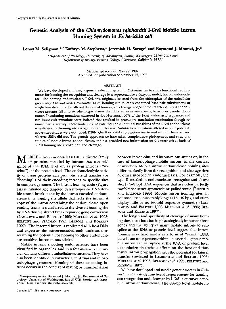
FIGL'KE l."I-CreI mobile intron homing cyclc and genetic assav for I-Crel homing site recognition and cleavage in E coli. (A) Homing of the I-Crel mobile intron is initiated hy a site-specific DNA double strand break macle by I-Crd endonuclease encoded hy the mobile intron open reading frame (filled box of intron' allele, at top center). The I-Crd homing site is located in the C. rei17/tnrd/ii chloroplast 23s rRNA gene (open hox, top left). The cleaved homing site is a substrate for double strand break repair off the intron-plus allele. Repair transfers a copy of the intron to the cleaved recipient allele (dsh repair, hottom center). Resolution (resolution, top right) generates two alleles containing the mobile intron, each flanked hv a disrupted I-CreI homing site. (€5) A genetic assay for 1-Crel endonuclease function was estahlished in E. coli by placing the I-Crel endonuclease open reading frame on a plasmid, pBE, under the control of arahinose operon regulatory sequences (the arn promoter, r, and araC regulator) and an I-Crel homing site on an FI28 F' Factor acljacent to a kanamycin resistance gene (F'S). Expression of active I-Crel endonuclease leads to the loss of the F' kanamscin marker, presumably due to homing site cleavage. Mutations that inactivate either endonuclease function or that render the homing site resistant to cleavage generate cells that retain kanamycin resistance. Homing with intron transfer does not occur in this assay due to lack of the 888-hp mohile intron (see A).
tron was originally identified in the chloroplast genome of the unicellular green alga Clzlnmyiomonm rhllnrdtii (ROCHAIX and MAI.NOE 1978; ROCHXIX e/ nl. 198.5; LE- MIEUX et nl. 1989). This intron can self-splice at the RNA level and can home to copies of the 23s rRNA gene that lack the mobile intron (HERRIS et nl. 1990; DPRREN- RERGER and ROCHAIX 1991; THOMI'SOS and HERRIS 1991; DCRRENRERGER et nl. 1996).
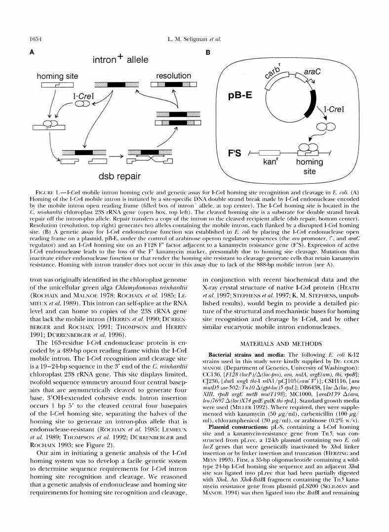
The 163-residue I-CreI endonuclease protein is en- coded by a 489-bp open reading frame within the I-Crd mobile intron. The I-Crd recognition and cleavage site is a 19-24-bp sequence in the 3' end of the C. rpinhnrdtii chloroplast 23s rRNA gene. This site displays limited, twofold sequence symmetry around four central basep airs that are asymmetrically cleaved to generate four base, 3'0Hextended cohesive ends. Intron insertion occurs 1 bp .5' to the cleaved central four basepairs of the I-CrpI homing site, separating the halves of the homing site to generate an intron-plus allele that is endonuclease-resistant (ROCHAIX et nl. 1985; LEMIELIX pt nl. 1989; THOMPSOS d nl. 1992; D~RRESRERGER and ROCHAIX 1993; see Figure 2).
Our aim in initiating a genetic analysis of the I-Crd homing system was to develop a facile genetic system to determine sequence requirements for I-CrrI intron homing site recognition and cleavage. We reasoned that a genetic analysis of endonuclease and homing site requirements for homing site recognition and cleavage,
in conjunction with recent biochemical data and the X-ray crystal structure of native I-CreI protein (HEATH ~t ol. 1997; STEPHENS et nl. 1997; K. M. STEPHENS, unpub lished results), would begin to provide a detailed pic- ture of the structural and mechanistic bases for homing site recognition and cleavage by I-Crd, and by other similar eucaryotic mobile intron endonucleases.
MATERIALS AND METHODS
Bacterial strains and media: The following E. coli K-12 strains used in this studv were kindly supplied by Dr. COLIN MASOII. (Department of Genetics, University of Washington): CCl36, [ F12X ( lac~) /A( lac+ro) , am, naM, nrgE(am), thi, rpoB]; CJ236, [dull zing1 /hi-l reAl/pCJl05(cnm'F')]; CSH116, [am lnlllL)5 zn~-502::7'n10 A(g)c,/-lac)5 rpsI.]; DB6438, [lac A(lac, pro) Xlll, 1poB argE mptB m1tt7198J; MC1000, [arnD139 A ( n m , /~u)7697 A(Inr)X74 gnKgalK /hi rpsl,]. Standard growth media were used (MILLER 1992). Where required, they were supple- mented with kanamycin (50 pg/ml), carhenicillin (100 pg/ mi), chloramphenicol (30 pg/ml), or arahinose (0.2% w/v).
Plasmid constructions: pL-S, containing a I-Crd homing site and a kanamvcin-resistance gene from Tn5, was con- structed from pLrec, a 12-kh plasmid containing two E. coli Car% genes that were genetically inactivated by XhoI linker insertion or hy linker insertion and truncation (HERZING and MM' 1993). First, a 35-hp oligonucleotide containing a wild- type 24-hp I-CreI homing site sequence and an adjacent XOaI site was ligated into pLrec that had been partially digested with XhoI. An XI~OI-BSIBI fragment containing the Tn5 kana- mycin resistance gene from plasmid pLS200 (SEI.IGMAN and M.ASOII. 1994) was then ligated into the BstBI and remaining
I-CreI Homing System Mutants 1655
&I sites of pLrecCre2 to generate pLs. The orientations of the oligonucleotide and restriction fragment were verified by restriction digestion and DNA sequencing. pLac+ was iso- lated after p L s transformation into E. coli, followed by screen- ing for the generation of Lac+ colonies on indicator plates. pLac+ appears to have arisen as the result of homologous recombination in the -6OO-bp region of homology between the genetically inactive pLS lacZ direct repeats to generate an intact lac2 open reading frame.
PEE and PA-E are compatible plasmids that encode I-CreI endonuclease under control of the promoter and regulatory gene (aruC) of the ur&AD (arabinose) operon of E. coli. p E E was constructed by ligating an XbuI-EcolN fragment from PI-CreI, a PET1 1-based expression vector containing the I-Crd open reading frame (THOMPSON et al. 1992; kindly supplied by Dr. DAVID HERRIN, University of Texas-Austin) into Xbd- and HindIII-cleaved pBAD18 DNA (GUZMAN et al. 1995). Be- fore ligation, the Hind111 site of pBAD18 was treated with E. coli DNA polymerase I Klenow fragment to render it blunt- ended (SAMBROOK et aL 1989). PA-E was constructed by trans- femng a CM-XhoI fragment containing the aruBAD regula- tory sequences and the I-Cd open reading frame from PEE to ClaI and SalI-cleaved pACYC184 DNA (CHANG and COHEN 1978; New England Biolabs, Beverly, MA).
The I-Cd open reading frame we used differed at seven nucleotide positions from the published sequence (ROCHAIX et ul. 1985). The revised I-CreI open reading frame sequence, determined by sequencing two independently cloned C. rein- hurdtii chloroplast 23s rRNA restriction fragments, has se- quence changes at codons 42 (ACG, not GCG) and codons 110-111 (GAACAA, not TGGCGG). The remaining A --t G base substitution in codon 56 was found only in PI-CreI. This D56G I-CreI variant, hereafter referred to as wild-type I-Cd endonuclease, was also used for the X-ray crystal structure determination of I-CreI (HEATH et ul. 1997; STEPHENS et al. 1997). A sitedirected G56D revertant was generated by over- lap extension PCR (VILEJO et al. 1995) for comparison assays (see Table 1 and RESULTS).
Three other PEE derivatives were constructed to study po- tential active site residues. These I-CreI variants, with D20N, S22A or R70A substitutions, were constructed by oligonucleo- tidedirected mutagenesis. Briefly, the I-CreI open reading frame was transferred to pBluescript I1 KS(+) (Stratagene, La Jolla, C A ) as an &I-XbuI restriction fragment to generate pBSCre. Uracil-containing single-stranded phagemid DNA, isolated by transforming pBSCre into E. coli strain CJ236, was then used for oligonucleotidedirected mutagenesis as pre- viously described (KUNKEL et al. 1987). The resulting muta- tions were transferred to PEE on &I-xbuI restriction frag- ments and then sequence-verified.
pKS155, a pBluescriptI1 =(+)derived plasmid that con- tains a wild-type I-Crd homing site was constructed by ligating a 35-bp oligonucleotide containing I-Cd and XbuI sites into Sulkleaved pBluescriptI1 KS( +) plasmid DNA. Plasmids con- taining mutant wild-type (pKS162) or mutant (pKS163-169 and pKS172) I-CmI homing sites were constructed by transfer- ring pLs BstBI-EcoRV restriction fragments containing hom- ing sites to ClaI- and EcoRV-cleaved pBluescriptI1 KS( +) DNA. Two mutations contained in pKS166 were separated by cleav- ing with Asp700 and I-Crd endonuclease, followed by the re- cloning of homing site fragments into Asp700- and I-Cd- cleaved pKS162 plasmid DNA to generate pKS170 and pKS171. Standard recombinant DNA procedures were used for plasmid constructions and for the isolation and restriction analysis of plasmid DNAs (HOLMES and QUIGLEY 1981; SAM- BROOK et cd. 1989).
Bacterial manipulations: Reciprocal recombination was used to transfer the kanamycin resistance gene and adjacent
I-CreI homing site of p L s to the F' factor F128 (HOLLOWAY and%@W 1996) of E. coli strain CC136. The resulting F'S recombinants were selected by isolating kanamycin-resistant colonies after mating, followed by screening for carbenicillin sensitivity to eliminate cointegrants (MILLER 1992). Mutant homing sites were transferred by reciprocal recombination from F'S onto pLac+ by transforming MClOOOFS with pLac+. Plasmid DNA from transformants was isolated and trans formed into MClOOO to isolate kanamycin-resistant colonies.
Mutant isolation: Homing site mutants were obtained by transforming strain CSH116F'S with PEE plasmid DNA. Car- benicillin- and kanamycin-resistant colonies were purified by restreaking onto antibiotic agar, then cured of plasmid by growth in liquid media containing kanamycin and arabinose (0.2% w/v). Under these growth conditions, wild-type I-Crd- encoding plasmid is efficiently lost. Carbenicillin-sensitive col- onies containing putative homing site mutants were then tested for the ability to transfer kanamycin resistance upon mating to MClOOO containing PEE.
I-Cd endonuclease mutants were generated by passaging p E E plasmid DNA through mutator strains CSHl16 (mutD5) or DB6438 (mu&'). Mutagenized plasmid DNA, isolated from over- night liquid cultures grown from independent colonies, was transformed into MC1000F'S to isolate carbenicillin- and kana- mycin-resistant colonies. PEE plasmid DNA was prepared from colonies purified by restreaking on carbenicillin- and kanamy. cin-containing agar, then transformed into MC1000F'S. Plasmid DNAs giving comparable transformation efficiencies on carbeni- cillin- and on carbenicillin- and kanamycincontaining agar were characterized as putative endonuclease mutants.
Mutant charactexbation: The in vitro cleavage properties of the mutant I-CreI homing sites were determined as follows. Two hundred nanograms of an Ahd-linearized pKS163-172 plasmid DNA was mixed with 200 ng of AM-linearized pKS155 DNA (a 3-kb plasmid containing a wild-type I-CreI homing site) in 25 or 50 pl of 20 mM Tris pH 9.0, 10 mM MgC12. Digestions were performed with a 2.9-fold molar ex- cess of purified I-CreI for 6 hr at 37", or with limiting (equimo- lar) amounts of I-Cd for 20 min at 37". Cleavage reactions were stopped by placing digestions on ice, followed by the addition of an equal volume of stop solution containing brom- phenol blue, 20 mM EDTA and, unless otherwise noted, SDS (to 0.5% w/v) before electrophoresis through 1.2% agarose gels run in 1 X TBE buffer (SAMBROOK et ul. 1989). The gel buffer did not contain SDS. The DNA sequence of mutant I- CreI homing sites was determined by chain terminator se- quencing of the homing site region of independent pLac+ recombinants, using an end-labeled 20-base primer as pre- viously described ( MONNAT et al. 1992).
The overexpression toxicity and/or activity of I-Cd endonu- clease mutants was determined by retransforming MC1000F'S with individual PEE mutant plasmids, followed by selection on agar containing carbenicillin and 0.2% (w/v) arabinose. PEE plasmids that contain a wild-type I-Crd open readiig fiame do not generate colonies after transformation under these growth conditions. To determine whether toxicity reflected the reten- tion of partial endonuclease activity, endonuclease mutants were transformed and grown in the presence of 0.2% arabinose then assayed for retention of the F'S kanamycin resistance marker. Thii was done by restreaking carbenicillin-resistant colonies onto agar containing carbenicillin and kanamycin.
The genetic dominance of I-Cd endonuclease mutants was assayed by determining the ability of mutants to interfere with wild-type I-Cd elimination of the F'Slinked kanamycin resistance marker. MC1000F'S containing a PEE mutant was transformed with PA-E, a compatible plasmid that expresses wild-type I-CreI endonuclease. Transformants were then se- lected on agar containing chloramphenicol and carbenicillin
16.56 I,. M. Seligman rt N/.
in the presence or absence of kanamycin. Endonuclease mu- tants with chloramphenicol and kanamycin-resistatit t o chlor- ampI~enicoI-resistat~t ( i . r . , total) colony ratios of 5 IO-” , the hackground in this system, were scored as recessive.
The DSX sequence of both strands of the K”r1 open reacl- ing frame of pRE mutants was dctet-mined by flrlorcscinated chain terminator sequencing using a set of four, 16-2O-hase primers that hybridized in or acljaccnt t o the I-CrrI open reatl- ing frame. These analyses \cerr performed by the Murdock Center (University of Montana, Missoula), by the Departmrnt of Biochemistry Sequencing Facility at thc Univcrsity o f \,!’ash- ington (Seattle), or by the DNA Sequencing Facility at R;lncho Santa Ana Botanic Garden (Claremont, ( A ) .
RESULTS
Strategy for mutant isolation: We have developed and used a genetic assay fbr I-Crd activity in k,‘. coli to determine the sequence requirements for I-Crd hom- ing site recognition and cleavage. This assay system con- sists of a host strain containing the I-Crd homing endo- nuclease and homing site, though lacking a copy of the 888-bp I-Crd mobile intron (Figure 1B). Plasmid-borne I-CrpI endonuclease is expressed under control of an arabinose-inducible promoter (pB-E; see MATEKIXLS
ASD METHODS), while the I -Cd homing site is adjacent to a kanamycin-resistance gene on an F’ factor (F‘S). In this system constitutive, low level expression of I-Crd endonuclease in the absence of arabinose induction (GVZMAS PI nl. 1995) leads to the efficient loss of the F‘S kanamycin-resistance marker.
I-CreI homing site mutants: I-Crd-resistant homing site mutants were isolated by transforming rnu/1)5 host cells harboring F’S with the I-Crd plasmid pB-E. Kana- mycin- and carbenicillin-resistant colonies containing putative mutant homing sites arose at a frequency of -4 X lo-:’. To demonstrate that cells carried mutations linked to the F’, mutants were cured of pRE plasmid, then assayed for the ability to transfer the F‘ kanamycin- resistance marker into host cells carrying wild-type pR E plasmid. Twenty-one mutant I-Crd homing sites iden- tified in this manner were transferred onto pLac+ for further analysis.
DNA sequence analysis of the mutant homing sites revealed at least one mutation in each 24-bp homing site: 20 mutants differed at a single basepair from the wild-type I-Crd homing site sequence, while the re- mainingmutant contained two base substitutions within the I-CrpI homing site. Mutations included base substitu- tions at positions .5, 8, 14, 1.5 and 17; and single base deletions at positions .?, 6 and 8. Multiple, independent mutations of a given type were isolated at positions .i, 8 and 1.5, the same three positions where different mo- lecular types of mutations (base pair deletions and/or base substitutions) were identified (see Figure 2). Two mutations contained in one mutant were separated by recloning and then analyzed indidually.
The in vifro cleavage sensitivity of mutant I -Cd hom-
1 5 10 15 ?o 3 1
A 1 A JhI G A A A C ” T C G * T C G A G C A 9 2 1 3 1 1 2 2 1
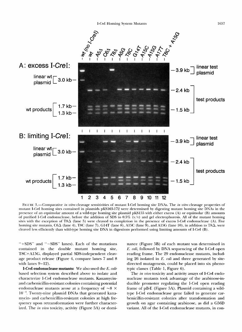
ing sites was determined by digesting mutant homing site plasmids w i t h excess or limiting amounts of purified I-CroI endonuclease. An equimolar amount of wild-type I-Crd homing site DNA was included in each assay to provide an internal standard for assessing the relative cleavage competence of mutant homing sites under dif- ferent digest conditions (see bli\-rEKIa,\L.s ASL) METIIODS). Site-specific cleavage of the 1-G-d homing site of the 3.9-kb pBlueScriptII-derived “test” plasmids containing a mutant homing site yielded fragments of 2.4 and l..5 kb (Figure 3, “test products”). Cleavage of the I-CwI homing site of the 3-kb control plasmid yielded frag- ments of 1.7 and 1.3 kb (Figure 3, “wt product$”). All of the control plasmid and, surprisingly, nine of the 10 homing site mutants were cleaved by excess I-CrpI (Fig- ure SA). The remaining homing site mutant, T8A, was resistant LO I-CrpI cleavage (Figure 3A, lane .5). Diges- tions performed using just enough I-Crd endonuclease to cleave a majority of wild-type plasmid identified five additional mutant homing sites in addition to T8A were cleaved less efficiently than wild-type homing site DNA (Figure 3B; mutant$ C6A, T8C, G14T, Al.5C, and Al5G in lanes 4, 3, 7-10). The remaining four homing site mutants were cleaved as efficiently as wild-type homing site DNA (Figure 30; A.?A, A5G, A17T and the double mutant in lanes 3, 6 , 1 1 and 12). Gel shift assays using I-C~PI endonuclease have shown that these mutant hom- ing sites are bound less efficiently than is wild-type hom- ing site DNA (K. M. STIPIIESS, unpublished results).
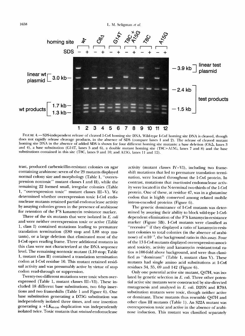
During the course of these experiments we found that I-Crd endonuclease requires the addition of SDS to release cleaved homing site DNA. We thus compared the ability of I-Crd endonuclease to cleave and release mutant, as opposed to wild type, homing site DNAs in the nBscncp of SDS (Figure 4). Three mutant homing sites displayed SDSindependent cleavage product re- lease (Figure 4; C6A, G14T and T8C+A1.5G, compare
1-Q.d Homing System Mwants 16.37
9 e!? 8 2. p"
@ v Y 7 Y 7 @ 0 W ( 9 ( 9 4 0 * * p C , k p . h @ p p p h < r , b Q ) b 0 ' \ ' \ \ ' \ 0
k 0 e k %
' 1 1 1 1 1 1 1 1 1 1 1
A: excess I-CreI: linear test - 3'9 kbl plasmid
linear wt [ 3.0 kb- plasmid
[
-2.4 kb 1 test products
wt products 1.7 kb- 1.3 kb- - 1.5 kb
I I I I I I I I I I I I
9: limiting I-CreI: " "
- 3'9 kbl plasmid linear test
linear wt [ 3.0 kb- plasmid
-2.4 kb
[ 1 test products
wt products 1.7 kb- 1.3 kb-
- 1.5 kb
1 2 3 4 5 6 7 8 9 1 0 1 1 1 2 FI(;L'RE %-Comparative in 7dro cleavage sensitivities of mutant I-G-c1 homing site DNAs. The in vitro cleavage properties of
mutant I-Crd homing sites contained in plasmids pKSl63-172 were determined by digesting mutant homing site DNAs i n the presence of an equimolar amount ofa wild-type homing site plasmid pKs1.5.3 with eihcr excess (A) or equimolar ( R ) amount5 of purified I-Crcl endonuclease, before the addition of SDS to 0.5% (v/v) and gel electrophoresis. All of the mutant homing sites with the exception of T8A (lane 5) were cleaved to completion i n the presence of excess I-Crcl endonuclease (A). Five homing site mutants, C6A (lane 4), TXC (lane 7), G14T (lane X), 4 1 . X (lane < I ) , and A15G (lane lo), in addition to TXA, were cleaved less efficiently than wild-type homing site DNA i n digestions performed using limiting amounts of I-Crpl (R) .
"+SDS" and "-SDS" lanes). Each of the mutations contained in the double mutant homing site, TFIC+Al%, displayed partial SDSindependent cleav- age product release (Figure 4, compare lanes 7 and 8 with lanes 9-12).
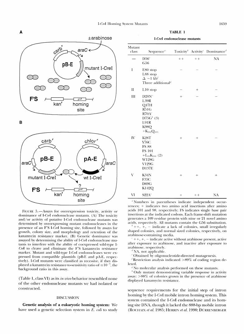
I-CreI endonuclease mutants: M'e also used the I;. c o b based selection system described above to isolate and characterize I-Crd endonuclease mutants. Kanamycin- and carbenicillin-resistant colonies containing potential endonuclease mutants arose at a frequency of "8 X lo-". Twenty-nine plasmid DNAs that generated kana- mycin- and carbenicillin-resistant colonies at high fre- quency upon retransformation were further character- ized. The in vivo toxicity, activity (Figure 5A) or domi-
nance (Figure 5B) of each mutant was determined in I:: coli, followed by DNA sequencing of the I-Crd open reading frame. The 29 endonuclease mutants, includ- ing 26 isolated in E. coli and three generated by site- directed mutagenesis, could be placed into six pheno- typic classes (Table l , Figure 6).
The in vivo toxicity and activity assays of I-CrpI endo- nuclease mutants took advantage of the arabinose-in- ducible promoter regulating the I-Crd open reading frame of pB-E (Figure 5A). Plasmid containing a wild- type I-Crd endonuclease gene failed to generate car- benicillin-resistant colonies after transformation and growth on agar containing arabinose, as did a G56D variant. All of the I-CreI endonuclease mutants, in con-
1658 L. M. Seligman r/ nl.
A so 0I OL? O P
homing site * 8 $ @ v @ p“ nnnnnn
S D S - + - + - + - + - + - +
- 3’9 kbl plasmid linear test
linear wt [I 3.0 kb - plasmid
-2.4 kb
r 1 test products 1.7 kb- 1.3 kb-
wt products - 1.5 kb
1 2 3 4 5 6 7 8 9 1 0 1 1 1 2 FIGURE 4.-SDSindependent release of cleaved I-Crd homing site DNA. Wild-type I-CwI homing site DNA is cleaved. though
does not rapidly release cleavage products, in the absence of SDS (compare lanes 1 and 2). The release of cleaved mutant homing site DNA in the absence of added SDS is shown for four different homing site mutants: a base deletion (CsA, lanes 3 and 4). a base substitution (G14T, lanes 5 and 6 ) , a double mutant homing site (TAC+Al.X;, lanes 7 and 8) and the base substitutions contained in this site (T8C, lanes 9 and 10; and A l X , lanes 1 1 and 12).
trast, produced carbenicillin-resistant colonies on agar containing arabinose: seven of the 29 mutants displayed normal colony size and morphology (Table 1 , “overex- pression nontoxic” mutant classes I and II ) , while the remaining 22 formed small, irregular colonies (Table 1, “overexpression toxic” mutant classes III-V). We determined whether overexpression toxic I-CreI endo- nuclease mutants retained partial endonuclease activity by assaying colonies grown in the presence of arabinose for retention of the F‘S kanamycin resistance marker.
Three of the six mutants that were isolated in E. coli and were neither overexpression toxic or active (Table 1 , class I ) contained mutations leading to premature translation termination (E80 stop and L88 stop mu- tants), or a large deletion that eliminated most of the I-CreI open reading frame. Three additional mutants in this class were not characterized at the DNA sequence level. The remaining nontoxic mutant (L10 stop; Table 1, mutant class 11) contained a translation termination codon at I-CreI residue 10. This mutant retained resid- ual activity and was presumably active by virtue of stop codon read-through or suppression.
Twenty-two different mutations were toxic when over- expressed (Table 1, mutant classes III-VI) . These in- cluded 18 different base substitutions, two 6 b p inser- tions and two frameshifts (Table 1 and Figure 6). One base substitution generating a D75G substitution was independently isolated three times, and one insertion generating a +LK.n.loo duplication was independently isolated twice. Toxic mutants that re/ninPd endonuclease
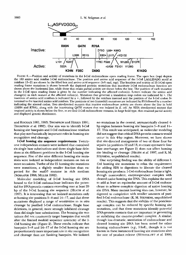
activity (mutant classes IV-VI), including two frame- shift mutations that led to premature translation termi- nation, were located throughout the I-CreI protein. In contrast, mutations that innclivated endonuclease activ- ity were located in the N-terminal two-thirds of the I-CreI protein. One of these, at residue 47, was in a glutamine codon that is highly conserved among related mobile intron-encoded proteins (Figure 6).
The genetic dominance of I-Crd mutants was deter- mined by assaying their ability to block wild-type I-CreI- dependent elimination of the F’S kanamycin-resistance marker (Figure 5B). I-CrpI mutantq were classified as “recessive” if they displayed a ratio of kanamycin-resis- tant colonies to total colonies (in the absence of arabi- nose) of 5 IO-“, the background ratio in this assay. Four of the 13 I-Crd mutants displayed overexpression-associ- ated toxicity, activity and kanamycin resistant:total ra- tios 2 100-fold above background, and were thus classi- fied as “dominant” (Table 1, mutant class V). These mutants had single amino acid substitutions at I-CreI residues 34, 35, 69 and 142 (Figure 6).
Only one potential active site mutant, Q47H, was iso- lated by genetic selection in E. coli. Three other poten- tial active site mutants were constructed by sitedirected mutagenesis and analyzed in E. coli. D20N and R70A substitution mutants were toxic, though neither active or dominant. These mutants thus resemble Q47H and other class 111 mutants (Table 1). An S22A mutant was overexpression toxic and active in the absence of arabi- nose induction. This mutant was classified separately
16.39
A *arabinose
F's - \ kan' homing /
site
B
/
mutant I-CreI"
3 F's 1 kan' homing
site
F I G ~ I W .i.-Assays for overexpression toxicity, activity or dominance of I-CrcI endonuclease mutants. (A) The toxicity and/or activity of putative I-CrpI endonuclcasc lnutants was determined by overexpressing mutant entlonucleasrs i n the presence of an F'S I - C w I homing site, followed by assays for growth, colony size, and morphology antl retention of the kanamycin resistance marker. (R) Gcnetic dominance was assayed by determining the ability of I-G.rl cntlonrlclease mu- tants to interfere with the ability o f coexpressetl wild-type I- Crd to cleave and eliminate the F'S kanamycin rrsistancr marker. Mutant antl wild-type I-C'rd cndonuclcascs were cx- pressed from compatible plasmids (pRE and p;\-E, respcc- tivelv). I - C w I mutants wcre classified ;IS recessive, if they dis- played a kanamycin I-esistatlce-ttrscnsitivity ratio o f 5 IO-", the background ratio i n this assay.
(Table 1, class VI) as its in vivo behavior resembled none of the other endonuclease mutants we had isolated or constructed.
DISCUSSION
Genetic analysis of a eukaryotic homing system: M'e have used a genetic selection system i n L. roli to study
TABLE 1
I-CreI endonuclease mutants
II : I
- + + -
+ +
+ + +
I' Slumbers in pawntheses indicate indepenclenr occur- rrnces; + indic;Itrs t w o amino acid insertions after amino acitls 1 0 1 and 98, rcspcctively: FS indicates single base pair insrrtions a t the indicated cotlons. Each fi-ame-shift mutation generatcs a 109 1-csidue protein w i t h nine or 21 novel amino acids, respectively. All tnlttants contain the G X substitution.
" ++. +, - indicate a Iack of colonies, small irregularly sh;qxd colonies, ;md nortnal sized colonies, respectively, on al-;~hinosecontaining media.
' ++, +. - indicate active without arabinose present, active after esposu~-e t o ;tr;thinose, and inactive after exposure to arabinose, rcspcctivdy.
" NA, not ;IppIicahIc. "Ol~taincd by oligonltcleotide~irected mutagenesis. 'Rrstriction analysis indicated >89% of coding region de-
E So molccular ;Inalysis performed on these mutants. " Only mutant tlen~onstrating variable response i n activity
assay: >88%, of colonies grown in the presence o f arabinose displayed kanamycin rcsistancc.
leted.
sequence requirements for the initial step of intron homing by the I-&I mobile intron homing system. This system contained the I-Crd endonuclease and its hom- ing site DNA, though it lacked the 888-bp mobile intron (Roc:tq;\rs I>/ N I . 198.5; HEKKIS PI ( I / . 1990; D~:KRESRERGER
1660 L. M. Seligman et al.
3 20 40 60 80 100 120 140 163
D20N Q47H R70A Inactive: D75G L88* K98Q
L39R R51G
Active: +LK99 FSlOl V129G K34N F35C D69G K142Q
FIGURE 6.-Position and activity of mutations in the I-CreI endonuclease open reading frame. The open box (top) depicts the 163 amino acid residue I-CreI endonuclease. The position and amino acid sequence of the I-CreI LAGLIDADG motif at residues 13-21 are shown by the filled box and amino acid sequence (left end, top). The location and activity of 25 I-CreI open reading frame mutations is shown beneath the depicted protein: mutations that inactivate I-CreI endonuclease function are shown above the horizontal line, while those that retain partial activity are shown below the line. The position of each mutation in the I-CreI open reading frame is given by the number indicating the affected codon(s). Letters indicate the amino acid change(s) in each mutant at the affected codon(s). Mutations that generate a translation stop codon are indicated by *. The insertion of amino acid residues is indicated by a +, followed by the residues inserted and the position of the I-CreI codon N- terminal to the inserted amino acid residues. The positions of two frameshift mutations are indicated by FS followed by a number indicating the altered codon. Two sitedirected mutants that inactive endonuclease activity are shown above the line in bold (D20N and R70A), along with the inactivating Q47H mutant that was isolated in E. coli. An S22A sitedirected mutant that retained activity is shown below the line, as are four I-CreI endonuclease mutants, in large bold type, that retained partial activity and displayed genetic dominance.
and ROCHAIX 1991,1993; THOMPSON and HERRIN 1991; THOMPSON et al. 1992). Our aim was to identify I-CreI homing site basepairs and I-CreI endonuclease residues that play mechanistically important roles in homing site recognition and cleavage.
I-CreI homing site sequence requirements Twenty- one independent mutants were isolated that contained six single base substitutions and three single base dele- tions at six different positions in the I-CreI homing site sequence. Five of the nine different homing site muta- tions were isolated as independent mutants on two or more occasions. Twelve of the 21 homing site mutations were transitions, a slightly smaller fraction than ex- pected for the mutD5 mutator in rich medium (SCHAAPER 1988; MILLER 1992).
Molecular modeling of I-CreI homing site DNA bound to the I-CreI endonuclease indicates the poten- tial for DNA-protein contacts extending over at least 20 bp of the I-CreI homing site sequence (HEATH et al. 1997). It is interesting that we isolated mutations only at homing site positions 5-8 and 14-17, and that these mutations displayed a range of sensitivities to in vitro cleavage by purified I-CreI endonuclease. Single base deletions, in general, more strongly inhibited cleavage than did single base substitutions. The homing site mu- tations did not consistently target basepairs that would alter the limited twofold sequence symmetry of the I- CreI homing site (Figure 2). These results indicate that basepairs 5-8 and 14-17 of the I-CreI homing site are proportionately more important role in site recognition and cleavage than are flanking basepairs. We isolated
no mutations in the central, asymmetrically cleaved 4 bp region between homing site basepairs 5-8 and 14- 17. This result was anticipated, as molecular modeling did not suggest that critical DNA-protein contacts would occur in this 4 b p region. Moreover, we have shown that site-directed mutagenesis of two of these four ba- sepairs (at positions 12 and 13, to create symmetric four base overhangs; see Figure 2) does not affect homing site binding or cleavage (HEATH et al. 1997, and K. M. STEPHENS, unpublished results).
One surprising finding was the ability of different I- CreI homing site mutations to relax the requirement for adding SDS to digestions to liberate the cleaved homing site products. I-CreI endonuclease forms a tight, though noncovalent, enzyme-product complex with cleaved native homing site DNA. This explains the need to add at least an equimolar amount of I-CreI endonu- clease to achieve complete digestion of native homing site DNA. Many mutant homing sites can, however, be digested to completion with smaller amounts of puri- fied I-CreI endonuclease (K. M. STEPHENS, unpublished results). This suggests that the stability of the postcleav- age complex can be reduced by specific homing site mutations, and that these mutations identify potential DNA-protein contacts that are important in generating or stabilizing the enzyme-product complex. A similar, though less dramatic, association of endonuclease and cleaved homing site DNA has been noted for other homing endonucleases (e.g., I-SceI), though it is not known in these instances if homing site mutations alter the rate of product release (MONTEILHET et ul. 1990;
I-CreI Homing System Mutants 1661
PLESSIS et al. 1992; PERRIN et al. 1993). One postulated role for these endonuclease-product complexes is fo facilitate intron insertion by protecting cleaved homing site DNA ends from exonucleolytic degradation. It is not known, however, whether I-CreI or I-SceI form stable, noncovalent complexes with their cleaved homing site DNAs in vivo.
Our mutant homing site data indicate that selection in E. coli provides a useful way to isolate homing site mutations that reduce or abolish homing site cleavage. This selection system also allowed us to fortuitously re- cover homing site mutants that altered the rate of cleav- age product release. The partial sensitivity of many of our homing site mutants to I-CreI digestion in vitro sug- gests that our selection system is able to reveal small decrements in the ability of I-CreI to cleave F’S I-CreI homing site DNA. Comparable mutant homing sites for a second mobile intron endonuclease, I-DmoI, have been selected in vitro on the basis of resistance to cleav- age with limiting amounts of endonuclease (hem et al. 1997). Further biochemical analyses of mutant 1- CreI homing sites, in conjunction with a high-resolution cocrystal structure of I-CreI bound to homing site DNA, should allow us to identify the specific DNA-protein contacts involved in homing site recognition, in cleav- age and in the generation of a tight, though noncova- lent, enzyme-product complex.
Functional organization of the 1-CreI endonucle- ase: We used a modification of the genetic selection system described above to isolate and characterize I- CreI endonuclease mutants. A combination of toxicity, activity and genetic dominance assays in E. coli were used to further characterize and identify the most po- tentially informative I-CreI endonuclease mutants (cf : PAKULA and SAUER 1989). Six different mutant classes could be identified among 29 endonuclease mutants. The mutants isolated in E. coli contained 26 unique mutations in the I-CreI open reading frame (Table 1 and Figure 6) that were close to the expected spectra for the mutator strains that we employed: 11 of 14 muta- tions isolated by passaging through mut T were A + C transversions, whereas half (five of 10) of the mutD derived mutants consisted of A + G transitions ( SCHAAPER 1988; MILLER 1992).
Mutations that inactivated endonuclease function in- cluded six single amino acid substitutions that were largely nonconservative, a duplication of the lysyl-gluta- mine dipeptide at positions 102-103, and two chain termination codons at amino acid residues 80 and 88. These mutations are clustered in the N-terminal two- thirds of the I-CreI sequence, the portion of the open reading frame that is retained in two independent, en- zymatically active frameshift mutants. These results indi- cate that homing site recognition and cleavage are me- diated by the N-terminal two-thirds of I-CreI, as sug- gested by the X-ray crystal structure of native I-CreI (Figure 7; see also below). Many of the amino acid
substitutions that inactivate I-CreI endonuclease func- tion Htain toxicity upon overexpression in E. coli. This suggests that toxicity and endonuclease activity are not tightly linked. I-CreI endonuclease has a strong net basic charge, with a PI of -10.5. Thus enzymatically inactive I-CreI might retain the potential to act as a DNA binding protein that could alter chromosome structure or gene expression in E. coli. A complementary set of results have been obtained for mutants of I-CeuI, where mu- tants originally selected on the basis of reduced toxicity displayed a range of enzyme activities (TURMEL et al. 1997).
A particularly interesting subset of I-CreI mutants dis- played overexpression activity and toxicity, and were able to interfere with the ability of wild-type I-CreI to cleave homing site DNA in E. coli. Each of these domi- nant mutants had nonconservative amino acid substitu- tions in the I-CreI open reading frame. The “overex- pression active” phenotype of these four mutants sug- gests that each has a lower affinity for homing site DNA than wild-type I-CreI, and that dominance results from the ability of mutants to sequester wild-type I-CreI endo- nuclease in heterodimers that can no longer efficiently recognize or cleave homing site DNA. The positions of mutations in three of the four dominant mutants sup- port this argument. The K34N and F35C substitutions occur in a loop between sheets pl and 02 that has the potential to contact specific base pairs at the ends of the I-CreI homing site. The D69G substitution, in contrast, is positioned near the substrate binding surface and potential active site residue R70. The remaining domi- nant mutant, K142Q is enigmatic: it is located in the Gterminus of I-CreI where no additional mutations have been found, and where the native I-CreI crystal structure is disordered (Figure 7 and HEATH et al. 1997). Mutant forms of I-CreI that retained the ability to form dimers with wild-type I-CreI may be useful for analyzing the role of subunit communication in homing site binding, cleavage and product release (see, e.g., STAHL et al. 1996).
We were surprised that we isolated no inactivating mutations in the single copy of the LAGLIDADG motif at I-CreI residues 13-21. This motif has been found in many other mobile intron endonucleases and matur- ases, where it has been postulated to play a role in substrate binding and/or cleavage (MICHEL et al. 1982; HENSCENS et al. 1983; WARING et a,!. 1983; HODCES et al. 1992; MUELLER et al. 1993; PIETROKOVSKI 1994; BELFORT and PERLMAN 1995; GIMBLE and STEPHENS 1995; HENKE et al. 1995; PIETROKOVSKI 1997). The X-ray crystal struc- ture of native I-CreI indicates that the LAGLIDADG mo- tif of I-CreI plays two roles: it forms the dimer interface and contributes amino acid residues, most notably D20, to a putative endonuclease active site in each monomer. A comparable dual role that we predicted for mobile intron endonucleases containing two copies of the LAGLIDADG motif (HEATH et al. 1997) has been re-
1662 L. M. Seligman et al.
A
B
FIGURE 7.-Model of native I-CreI endonuclease docked to I-CreI homing site DNA and active site mutants. (A) The active form of I-CreI is an endonucle9se homodimer2 in which the four antiparallel p strands of each monomer form a DNA binding surface or saddle that is -20 A wide and 70 A long. The LAGLIDADG motifs of the two monomers (al/al’) form a novel a- helical dimer interface, and contribute aspartic acid residues D20 and D20’ to a predicted bipartite active site at the center of the homodimer. The I-CreI DNA binding surface makes contact with at least 20 bp of the I-CreI homing site. The most distant potential contacts are by loops between strands D l and p 2 that can contact homing site bp 2 and 3 and 19-21 via the major groove. The positions of three dominant mutants (K34N, F35C and D69G) are indicated in the ribbon diagram by arrows. (B) Stereo diagram of the postulated I-CreI endonuclease active site residues in an I-CreI monomer. The green strand indicates the position of the phosphodiester backbone of one strand of the homing site DNA. Potential active site residues are indicated in red (D20, S22, Q47 and R70). The phenotypes of D20, Q47 and R70 mutants indicate that these three residues are likely to be active site residues, whereas S22 is not (see text). The phosphodiester bond cleaved is located at the center of the triangle formed by D20, S22 and Q47.
cently confirmed with publication of the structure of PI-SceI, an intein-encoded mobile intron endonuclease ( D u m et al. 1997).
The X-ray crystal structure of I-CreI and phylogenetic data on related mobile intron-encoded proteins indi- cate that four different amino acid residues may con- tribute to the I-CreI active site: D20 in the LAGLIDADG motif, and residues S22, Q47 and R70 (HEATH et al.
1997; TURMEL et al. 1997). We isolated one I-CreI endo- nuclease mutant in E. coli, a Q47H substitution that was enzymatically inactive (Table l ) , and thus similar to an inactivating substitution found at the comparable residue of I-CeuI (Q93R TURMEL et al. 1997). To deter- mine the functional importance of the other three po- tential active site residues, we constructed sitedirected mutations to generate D20N, S22A and R70A amino
I-CwI Homing System Mutants 1663
acid substitutions. The phenotypes of the D20N and R70A substitutions were identical to those of Q47H: toxic and inactive, though not dominant when ex- pressed in E. coli. These two mutants thus resembled mutants (I-CeuI E66K and Kll6Q respectively) that have been shown to inactive I-CeuI endonuclease activity (TURMEL et al. 1997). The S29A I-CreI mutant, in con- trast, was active in the absence of arabinose induction. However, unlike either wild-type or G56D I-CreI, S22A could generate colonies on agar containing arabinose. These results suggest that S22 is unlikely to play a func- tionally important role in the I-CreI active site.
The I-CreI active site model we favor has two symme- tq-related metal binding sites, formed by D20, Q47 and R70 in each monomer, positioned -1OA apart at the center of the I-CreI DNA binding surface. These active sites are appropriately positioned to simultaneously cleave homing site DNA bound in the presence of Mg+ across the minor groove to liberate four base, 3’0H- extended cohesive ends (ANDERSON 1993; AGCARWAL 1995; HEATH et al. 1997). The postulated I-CreI active site would thus resemble the active sites of several well characterized Type I1 restriction endonucleases in that it is bipartite, with each metal-binding site formed by acidic (D20), basic (R70) and polar (Q47) residues that are widely distributed in the protein primary sequence (AGGARWAL 1995; HEATH et al. 1997). In contrast, the recently determined structure of the PI-SceI mobile in- tron endonuclease appears to contain a single active site consisting of two aspartic acid residues and a lysine. The single PI-SceI active site may act sequentially to cleave both strands of the PI-SceI homing site to gener- ate four base, 3’0Hextended cohesive ends (DUAN et al. 1997).
netic selection system in E. coli to explore the sequence requirements for homing site recognition and cleavage by the I-CreI eukaryotic mobile intron endonuclease. This genetic approach should be applicable to the anal- ysis of other eukaryotic mobile intron homing systems (see, e.g., TURMEL et al. 1997) and may have practical applications. For example, genetic selection could be used to isolate mutant forms of I-CreI, or of other mobile intron endonucleases, that are able to recognize and cleave variant or novel homing sites. Such mutant pro- teins could be used to target or recruit endonucleolytic or other specific biochemical functions to genes con- taining these target sites in cells. The feasibility of this approach is being explored in plant and mammalian cells, where it is known that at least four mobile intron endonucleases, I-CreI, I-SceI, PI-SceI and I-PpoI, can find and cleave their cognate homing sites with high speci- ficity (reviewed in JASIN 1996; R. J. MONNAT, JR., F. M. H. IIACKMANN, and M. A. CANTRELL, unpublished results).
We thank GINGER ARMBRUST, FRANz D~RRENBURGER, ELIZABETH HARRIS, PAT HEATH, DAVID HERRIN, JEAN-DAVID ROCHAIX and BARRY
S , . We have developed and used a simple ge-
STO~DABD for helpful discussions and for communicating results be- fore publication. ELIZABETH HARRIS, DAVID HERRIN, COtIN WOIL, STEVE M m , JEAN-DAVID ROCHA~X and BETH TRAXLER kindly provided materials or strains. PATRICK COLLTNS performed E. coli phenotyping assays on several mutants. ALDEN I-h- generated Figures 1-5 and PAT HEATH generated Figure 7. BAEK KIM, COLIN MANOIL and ANDREW TAYLOR provided useful comments on a draft of this manu- script. This work was supported by grants from the National Institutes on Aging, the National Cancer Institute and the University of Wash- ington Royalty Research Fund to R.J.M., Jr. L.M.S. and K.M.S. were supported by National Institutes of Health Training Grants T32 CA09437 and T32 ES07032, respectively. J.H.S. was supported by Na- tional Science Foundation REU grant BIR-9531713.
LITERATURE CITED
AAGAARD, C., M. J. AWAYEZ and R A. CARREn, 1997 Profile of the DNA recognition site of the archeal homing endonuclease I- DmoI. Nucleic Acids Res. 2 5 1523-1530.
AGGARWAL, A. K, 1995 Structure and function of restriction end* nucleases. Curr. Opin. Struct. Biol. 5: 11-19.
ANDERSON, J. E., 1993 Restriction endonucleases and modification methylases. Cum. Opin. Struct. Biol. 3: 24-30.
BELFORT, M., and P. S. PERLMAN, 1995 Mechanisms of intron mobil- ity. J. Biol. Chem. 270 30237-30240.
BELFORT, M., and R ROBERTS, 1997 Homing endonucleases-keep ing the house in order. Nucleic Acids Res. 2 5 3379-3388.
BELFORT, M., M. E. REABAN, T. COETZEE and J. Z. DALCAARD, 1995 Prokaryotic introns and inteins: a panoply of form and function. J. Bacteriol. 177: 3897-3903.
CHANG, A. C. Y., and S . N. COHEN, 1978 Construction and character- ization of amplifiable multicopy DNA cloning vehicles derived from the P15A cryptic miniplasmid. J. Bacteriol. 154: 1141- 1156.
DUAN, X., F. S . GIMBLE and F. A. QUIOCHO, 1997 Crystal structure of PI-Scd, a homing endonuclease with protein splicing activity.
D~RRENBERGER, F., and J.-D. ROCHAM, 1991 Chloroplast ribosomal intron of Chlamydmwnus reinhardtii in vitro self-splicing, DNA endonuclease activity and in vivo mobility. EMBO J. 10: 3495- 3501.
D~JRRENBERGER, F., and J.D. ROCHAIX, 1993 Characterization of the cleavage site and the recognition sequence of the I -Cd DNA endonuclease encoded by the chloroplast ribosomal intron of Chlumydomoncls reinhadii. Mol. Gen. Genet. 236: 409-414.
D~RRENBERGER, F., A. J. THOMPSON, D. L. HEWN and J.D. ROCHAM, 1996 Double strand break-induced recombination in Chlamyob monas winhardtii chloroplasts. Nucleic Acids Res. 2 4 3323-3331.
GIMBLE, F. S., and B.W. STEPHENS, 1995 Substitutions in conserved dodecapeptide motifs that uncouple the DNA binding and DNA cleavage activities of PI-Sd endonuclease. J. Biol. Chem. 270
GUZMAN, L., D. BELIN, M. J. CARSON and J. BECKWITH, 1995 Tight regulation, modulation, and high-level expression by vectors con- taining the arabinose PBAD promoter. J. Bacteriol. 177: 4121- 4131.
HEATH, P. J., K M. STEPHENS, R J. MONNAT, JR. and B. L. STODDARD, 1997 The structure of I-Crd: a Group I intron-encoded homing endonuclease. Nature Struct. Biol. 4: 468-476.
HENKE, R M., R. A. BUTOW and P. S . P E W , 1995 Maturase and endonuclease functions depend on separate conserved domains of the bifunctional protein encoded by the group I intron aI4a of yeast mitochondrial DNA. EMBO J. 14: 5094-5099.
HENSGENS, L. A. M., L. BONEN, M. DE W, G. VAN DER HORST and L. A. GRIVELL, 1983 Two intron sequences in yeast mitochon- drial COX1 gene: homology among UW-containing introns and suaindependent variation in flanking exons. Cell 34: 379-389.
HERRIN, D. L., Y. CHEN and G. W. SCHMIDT, 1990 RNA splicing in Chlamydomonas chloroplasts: self-splicing of 23s preRNA. J. Biol. Chem. 265 21134-21140.
HERZING, L. B. K, and M. S . MEW, 1993 Novel LacZbased recombi- nation vectors for mammalian cells. Gene 137: 163-169.
HODGES, R., F. B. PERLER, C. J. NOREN and W. E. JACK, 1992 Protein splicing removes intervening sequences in an archaea DNA poly- merase. Nucleic Acids Res. 20: 6153-6157.
Cell 8 9 555-564.
5849-5856.
1664 L. M. Seligman et al.
HOLLOWAY, B., and K. B. LOW, 1996 F-prime and R-prime factors, pp. 2413-2420 in Escherichia coli and Salmonella: Cellular and Molec- ular Biorbgv, Ed. 2, edited by F. C. NEIDHARDT, R. I . CURTIS, J. L. INGRAHAM, E. C. C. LIN, K. B. LOW, JR., et al. ASM Press, Washing- ton, D.C.
HOLMES, D. S., and M. QUIGLEY, 1981 A rapid boiling method for the preparation of bacterial plasmids. Anal. Biochem. 114: 193- 197.
JASIN, M., 1996 Genetic manipulation of genomes with rarecutting endonucleases. Trends Genet 12: 224-228.
KUNKEL, T. A., J. D. ROBERTS and R. A. ZAKOUR, 1987 Rapid and efficient site-specific mutagenesis without phenotypic selection. Methods Enzymol. 154: 367-382.
LAMBOWITZ, A. M., and M. BELFORT, 1993 Introns as mobile genetic elements. Annu. Rev. Biochem. 6 2 587-622.
LEMIEUX, C., J. BOULANGER, C. OTIS and M. TURMEL, 1989 Nucleo- tide sequence of the chloroplast large subunit rRNA gene from Chlamydomonas reinhardtii. Nucleic Acids Res. 17: 7997.
MICHEL, F., A. JACQUIER and B. DUJON, 1982 Comparison of fungal mitochondrial introns reveals extensive homologies in RNA sec- ondary structure. Biochimie 64: 867-881.
MILLER, J. H., 1992 A Short Course in Bactm’al Genetics: A Laboratq Manual and Hanahoh for Escherichia coli and Related Bacteria, Ed. 1. Cold Spring Harbor Laboratory Press, Cold Spring Harbor, NY.
MONNAT, R. J., JR., A. F. M. HACKMANN and T. A. CHIAVEROTTI, 1992 Nucleotide sequence analysis of human hypoxanthine phospho- ribosylvansferase gene deletions. Genomics 13 777-787.
MONTEILHET, C., A. PERRIN, A. THIERRY, L. COLLEAUX and B. DUJON, 1990 Purification and characterization of the in vitro activity of I-SceI, a novel and highly specific endonuclease encoded by a group I intron. Nucleic Acids Res. 18: 1407-1413.
MUELLER, J. E., M. BRYK, N. LOIZOS and M. BEWORT, 1993 Homing endonucleases, pp. 111-143 in Nucleasa, Ed. 2, edited by S. M. LINN, R. S. LLOYD and R J. ROBERTS. Cold Spring Harbor Labora- tory Press, Cold Spring Harbor, NY.
PAKUM, A. A., and R. T. SAUER, 1989 Genetic analysis of protein stability and function. Annu. Rev. Genet. 23: 289-310.
PERRIN, A, M. BUCKLE and B. DUJON, 1993 Asymmetrical recogni- tion and activity of the I-SceI endonuclease on its site and on intron-exon junctions. EMBO J. 12: 2939-2947.
PIETROKOVSKI, S., 1994 Conserved sequence features of inteins (pro- tein introns) and their use in identifying new inteins and related proteins. Protein Sci. 3 2340-2350.
PIETROKOVSKI, S., 1997 Modular organization of inteins and C-ter- mind autocatalytic domains. Protein Sci. (in press).
PLESSIS, A., A. PERRIN, J. E. HABER and B. DUJON, 1992 Site-specific recombination determined by I-Scel, a mitochondrial group I
intronencoded endonuclease expressed in the yeast nucleus. Genetics 130 451-460.
ROBERTS, R., and S. WORD, 1993 Type I 1 restriction endonucle- ases, pp.35-88 in N u c l e w , Ed.2, edited by S. LINN, R. LLOYD and R J. ROBERTS. Cold Spring Harbor Laboratory Press, Cold Spring Harbor, NY.
ROCHAIX, J. D., and P. MALNOE, 1978 Anatomy of the chloroplast ribosomal DNA of Chlamydomonas rknhurdtii. Cell 1 5 661-670.
ROCH~IX, J. D., M. WIRE and F. MICHEL, 1985 The chloroplast ribosomal intron of Chlamydomonas reinhardtii codes for a poly- peptide related to mitochondrial maturases. Nucleic Acids Res.
SAMBROOK, J., E. F. FRITSCH and T. MANIATIS, 1989 Molecular Clun- ing; A Laboratq Manual, Ed. 2. Cold Spring Harbor Laboratory Press, Cold Spring Harbor, NY.
SCHAAPER, R. M., 1988 Mechanisms of mutagenesis in the Eschachia coli mutator muD5: role of mismatch repair. Proc. Natl. Acad. Sci. USA 85: 8126-8130.
SELIGMAN, L.. and C. MANOIL, 1994 An amphipathic sequence de- terminant of membrane protein topology. J. Biol. Chem. 269
STAHL, F., W. WENDE, A. JELTSCH and A. PINGOUD, 1996 Introduc- tion of asymmetry in the naturally symmetric restriction endonu- clease EcoRV to investigate intersubunit communication in the homodimeric protein. Proc. Natl. Acad. Sci. USA 93: 6175-6180.
STEPHENS, K. M., R. J. MONNAT, JR., P. J. HEATH and B. L. STODDARD, 1997 Crystallization and preliminary X-ray studies of I-CreI: a group I intron-encoded endonuclease from C. reinhardtii. Pro- teins: SFG 2 8 137-139.
THOMPSON, A. J., and D. L. HERRIN, 1991 In vitro self-splicing reac- tions of the chloroplast group I intron Cr.LSU from Chlamydom
Nucleic Acids Res. 19: 6611-6618. nas reinhardtii and in vivo manipulation via gene replacement.
THOMPSON,A. J.,X.YUAN,W.KUDLICKI~~~D. L. HERRIN, 1992 Cleav- age and recognition pattern of a double-strand-specific endonu- clease (I-CreI) encoded by the chloroplast 23s rRNA intron of Chlamydomonas reinhardtii. Gene 119: 247-251.
TURMEL, M., C. Ons, V. C 6 ~ and C. LEMIEUX, 1997 Evolutionarily conserved and functionally important residues in the I-CeuI hom- ing endonuclease. Nucleic Acids Res. 2 5 2610-2619.
VILEJO, A. N., R. J. POGUIJS and L.R. PEASE, 1995 Mutagenesis and synthesis of novel recombinant genes using PCR, pp. 603-612 in PCR primer: A Laboratq Manual, Ed. 1, edited by C. W. DIEF- FENBACH and G. S. DVEKSLER. Cold Spring Harbor Laboratoly
WARING, R. B., R. W. DAVIES, C. SWZOCCHIO and T. A. BROWN, 1982 Press, Cold Spring Harbor, NY.
Internal structure of a mitochondrial intron of AspergiUus nidu- lans. Proc. Natl. Acad. Sci. USA 7 9 6332-6336.
13: 975-984.
19888-19896.
Communicating editor: R. MAURER